Pneumosinus dilatans - Rhinology Internation Journal
Pneumosinus dilatans - Rhinology Internation Journal
Pneumosinus dilatans - Rhinology Internation Journal

You also want an ePaper? Increase the reach of your titles
YUMPU automatically turns print PDFs into web optimized ePapers that Google loves.
CASE REPORT<br />
<strong>Rhinology</strong>, 36, 40–42, 1998<br />
<strong>Pneumosinus</strong> <strong>dilatans</strong>: A discussion of four cases<br />
and the possible aetiology*†<br />
W.M. Adams 1 , R.I. Jones 1 , S.I. Chavda 1 , A.L. Pahor 2 , K.T. Taifa 2<br />
1<br />
2<br />
Department of Radiology, City Hospital, Birmingham, United Kingdom<br />
Department of Otorhinolaryngology, City Hospital, Birmingham, United Kingdom<br />
SUMMARY<br />
<strong>Pneumosinus</strong> <strong>dilatans</strong> is an abnormal dilatation of the paranasal sinuses, which contains<br />
only air and is lined by normal mucosa. It is a rare condition, the aetiology of which is unclear.<br />
We describe four patients who presented to our department with pneumosinus <strong>dilatans</strong>. The<br />
aetiology was either developmental hydrocephalus (n=1), post-traumatic (n=1) or idiopathic<br />
(n=2). Two patients underwent surgery, and follow-up is at least 12 months to date. The radiological<br />
aspects of this rare condition and the possible aetiologies are discussed.<br />
Key words: paranasal sinuses, pneumosinus <strong>dilatans</strong>, paranasal anatomy, sinus dilatation<br />
INTRODUCTION<br />
<strong>Pneumosinus</strong> <strong>dilatans</strong> is an abnormal dilatation of the paranasal<br />
sinuses, which contains only air and is lined by normal mucosa<br />
(Smith et al., 1987). It is a rare condition, the aetiology of which<br />
is unclear. We describe four patients who presented to our<br />
department with pneumosinus <strong>dilatans</strong>.<br />
CASE REPORTS<br />
Patient 1<br />
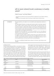
This patient was a 21-year-old man with a long and complex history<br />
who initially presented as a child with hydrocephalus. He<br />
was born with a large por-encephalic cyst with subdural fluid<br />
collections and required repeated insertion of ventriculo-peritoneal<br />
shunts for hydrocephalus. He had a long-standing history<br />
of walking difficulties and epilepsy. The finding of pneumosinus<br />
<strong>dilatans</strong> affecting the frontal and sphenoid sinus was an<br />
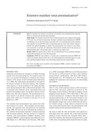
incidental finding and asymptomatic (Figure 1).<br />
Patient 2<br />
A 29-year-old man was referred with intermittent swelling of<br />
the right frontal region occurring over eight years, which started<br />
after fracturing his nose. The swelling could increase in size<br />
overnight and then subside over a similar period. It was associated<br />
with blurred vision and headaches but no diplopia. He was<br />
employed as a metal polisher. High-resolution coronal CTscans<br />
through the paranasal sinuses showed a hypoplastic and<br />
acellular left frontal sinus and a relatively large right frontal<br />
sinus. There was no definite evidence of fracture involving the<br />
Figure 1. Axial T 2 -weighted MR. There is pneumosinus <strong>dilatans</strong> of<br />
the frontal sinuses with encephalomalacia of the frontal lobes.<br />
* Received for publication March 10, 1997; accepted September 8, 1997<br />
† Presented at the 16th Congress of the European Rhinologic Society in Ghent, September 7-13, 1996
<strong>Pneumosinus</strong> <strong>dilatans</strong> 41<br />
origin of the right nasal frontal duct. The nasal septum was<br />
straight. There was evidence of minor chronic sinusitis involving<br />
the ethmoid air cells and the maxillary antra.<br />
He underwent fibre-optic nasendoscopy. There was evidence of<br />
metal dust in the nose; both sides of the nasal septum (in particular<br />
Little’s area) demonstrated signs of generalised congestion.<br />
The nasal septum deviated to the right with a bulge on the right<br />
side extending to the anterior part of area III. A submucosal resection<br />
was performed to try to open up the fronto-nasal duct without<br />
relief of symptoms, but he declined more definitive surgery.<br />
Patient 3<br />
A 15-year-old girl was first seen regarding sinus problems on<br />
16th May 1995 with a history of post-nasal discharge (especially<br />
during colds), yellowish in colour. She complained of daily<br />
headaches and pain above and behind the left eye. This was first<br />
diagnosed as migraine and she received anti-migraine treatment.<br />
The pain had occasionally made her feel as if the left eye<br />
was about to “pop out.” CT scan of the sinuses showed pneumosinus<br />
<strong>dilatans</strong> of the left sphenoid sinus.<br />
On 29th August 1995, the left sphenoid sinus was explored<br />
through a posterior septal approach. The septum between the<br />
left and right sinuses was divided. Post-operatively, the pain<br />
related to the left eye had disappeared completely. She was last<br />
reviewed on 10th October 1995. There were no further pains<br />
related to her left eye, but she still complained of tension headaches.<br />
Pre-operatively, she used to have puffiness over the eyes,<br />
but this disappeared following the operation.<br />
Patient 4<br />
A 54-year-old lady was referred with a 6-month history of nasal<br />
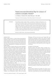
obstruction and headaches (Figure 2). Clinical examination<br />
showed evidence of frontal bossing. High-resolution coronal<br />
CT scans showed pneumosinus <strong>dilatans</strong> affecting the frontal<br />
sinuses. Via FESS and an external approach (through an Eagle<br />
incision of the frontal sinuses) the fronto-nasal duct was enlarged<br />
bilaterally. Some of the fronto-ethmoidal cells were removed,<br />
the septum between the two frontal sinuses was divided,<br />
and a muscle plug obtained from the thigh was inserted. This<br />
achieved resolution of her symptoms.<br />
DISCUSSION<br />
The term “pneumosinus <strong>dilatans</strong>” was first coined by Benjamins<br />
in 1918, who described a rare case of abnormal dilatation of the<br />
frontal sinus containing only air. Lombardi in 1967 reported 51<br />
cases and described its predilection for certain sites: It occurs<br />
most frequently in the frontal sinuses followed by the sphenoid,<br />
maxillary and ethmoidal sinuses, respectively. Som et al. (1987)<br />
proposed the following classification: Hypersinus: A frontal sinus<br />
that has developed beyond the upper limits of a normal frontal<br />
sinus. The sinus is aerated and its walls are normal. It does not<br />
extend beyond the normal boundaries of the frontal bone.<br />
<strong>Pneumosinus</strong> <strong>dilatans</strong>: An aerated sinus that is abnormally<br />
expanded. The sinus walls, although intact and of normal thickness,<br />
have been displaced outwardly to cause frontal bossing,<br />
intracranial extension or ethmoid, nasal or orbital encroachment.<br />
Pneumocoele: An aerated sinus with either focal or generalised<br />
thinning of the bony sinus walls. However, Reicher et al.<br />
(1986) prefer to use the term pneumosinus <strong>dilatans</strong> for all cases<br />
of dilated, air-filled sinuses of uncertain origin with outwardly<br />
bulging walls, because differentiating pneumosinus <strong>dilatans</strong><br />
from pneumocoele radiographically is impossible, and both present<br />
with identical clinical symptoms.<br />
In normal subjects, there is a wide variation in the degree of<br />
pneumatisation of the sinuses. Overgrowth may occur in acromegalic<br />
subjects or in cases of agenesis of a cerebral hemisphere.<br />
The aetiology of pneumosinus <strong>dilatans</strong> is poorly understood as<br />
may be evident from the many mechanisms that have been proposed<br />
by various authors. Most authors attribute it to a ballvalve<br />
action secondary to either redundant mucosa or a minor<br />
inflammatory process with consequent rise in pressure within<br />
the sinus (Dhillon and Williams, 1987). Benedikt et al. (1991)<br />
have suggested that spontaneous drainage of a mucocoele is the<br />
causative agent in a case of air-filled expansion of the ethmoid<br />
sinus, whereas others think that hormonal influence of osteoblastic<br />
activity allows ingrowth and expansion of the sinus<br />
(Smith et al., 1987). Still other authors think it is due to a congenital<br />
abnormality leading to unchecked development and<br />
growth of the sinus cavity (Som et al., 1987). It has been associated<br />
– particularly in the sphenoid sinus – with pathological<br />
factors, such as optic meningioma and fibro-osseous disease<br />
(Lloyd, 1985), which are thought to produce a stimulating effect<br />
on osteoblastic activity.<br />
The fact that some of our patients responded to surgery would<br />
suggest some form of ball-valve mechanism, which is relieved<br />
by widening of the ostium to allow adequate aeration. Surgery<br />
should be considered for cosmetic reasons and is mandatory if<br />
ocular problems supervene. The association with meningioma<br />
and fibro-osseous disease means that other forms of imaging<br />
such as CT or MR may be required.<br />
Figure 2. Plain X-ray of the skull showing bilateral pneumosinus <strong>dilatans</strong><br />
of the frontal sinuses.<br />
REFERENCES<br />
1. Benedikt RA, Brown DC, Roth MK, Geyer CA, Ghaed VN (1991)<br />
Amer J Neuroradiol 12: 729-731.<br />
2. Benjamins CE (1918) <strong>Pneumosinus</strong> frontalis <strong>dilatans</strong>. Acta<br />
Otolaryngol (Stockh) 1: 412-422.<br />
3. Dhillon RS, Williams DC (1987) <strong>Pneumosinus</strong> <strong>dilatans</strong>. J Laryngol<br />
Otol 101: 828-832.
42 Adams et al.<br />
4. Lloyd GAS (1985) Orbital pneumosinus <strong>dilatans</strong>. Clin Radiol 36:<br />
381-386.<br />
5. Lombardi (1967) ??<br />
6. Reicher MA, Bentson JR, Halbach W (1986) <strong>Pneumosinus</strong> <strong>dilatans</strong><br />
of the sphenoid sinus. Amer J Neuroradiol 7: 865-868.<br />
7. Smith IM, Maran AGD, Von Haacke NP (1987) <strong>Pneumosinus</strong> <strong>dilatans</strong>.<br />
Ann Otol Rhinol Laryngol 96: 210-212.<br />
8. Som PS, Edelstein D, Urken ML, Lawson W, Weber AL, Biller HF<br />
(1987) Abnormally large frontal sinus. II. Nomenclature, pathology<br />
and symptoms. Laryngoscope 97: 606-611.<br />
A.L. Pahor, F.R.C.S.<br />
Department of Otorhinolaryngology<br />
City Hospital<br />
Dudley Road<br />
Birmingham B18 7QH<br />
United Kingdom<br />
ANNOUNCEMENT