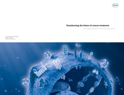
Transforming the future of cancer treatment Oncology ... - Roche
Transforming the future of cancer treatment Oncology ... - Roche
Transforming the future of cancer treatment Oncology ... - Roche

You also want an ePaper? Increase the reach of your titles
YUMPU automatically turns print PDFs into web optimized ePapers that Google loves.
© 2010 <strong>Roche</strong>. All rights reserved.<br />
LH5002-10425000<br />
Printed on recycled paper.<br />
<strong>Transforming</strong> <strong>the</strong> <strong>future</strong> <strong>of</strong> <strong>cancer</strong> <strong>treatment</strong><br />
<strong>Oncology</strong> Research and Development
Table <strong>of</strong> contents<br />
Introduction . . 3<br />
Our new organizational structure 4<br />
Areas <strong>of</strong> focus<br />
Antibody-drug conjugates (ADCs). . 6<br />
Angiogenic signaling. 8<br />
Anti-EGFL7 MAb (MEGF0444A, RG7414) 9<br />
Anti-NRP1 MAb (MNRP1685, RG7347). 9<br />
Anti-PlGF MAb (TB-403, RG7334). 9<br />
Apoptosis. 10<br />
Navitoclax (Bcl-2/Bcl-X L<br />
inhibitor, ABT-263, RG7433) 11<br />
MDM2 antagonist (RG7112). . 11<br />
Dulanermin (rhApo2L/TRAIL, RG3639) 11<br />
B-cell surface proteins . . 12<br />
GA101 (anti-CD20 MAb, RO5072759, RG7159) . 13<br />
Hedgehog signaling. 14<br />
Hedgehog pathway inhibitor (GDC-0449, RG3616). . 15<br />
HER signaling 16<br />
T-DM1 (HER2-targeted antibody-drug conjugate, HER2 ADC, RG3502) 17<br />
Pertuzumab (HER2 dimerization inhibitor MAb, RG1273) 17<br />
Anti-EGFR huMAb (GA201, RG7160). . 17<br />
MAPK signaling. . 18<br />
BRAF inhibitor (PLX4032, RG7204) 19<br />
MEK inhibitor (GDC-0973, RG7420) 19<br />
MEK inhibitor, CIF (RG7167). . 19<br />
MEK inhibitor, CKI27 (RG7304) 19<br />
MET signaling . . 20<br />
MetMAb (RG3638) 21<br />
PI3K signaling. . 22<br />
PI3 kinase inhibitor (GDC-0941, RG7321) 23<br />
PI3 kinase/mTOR inhibitor (GDC-0980, RG7422) 23<br />
Notch signaling 24<br />
Gamma secretase inhibitor (RG4733). . 24<br />
References 24<br />
Contact information<br />
Corporate Web sites<br />
www.roche.com<br />
www.gene.com<br />
Clinical trials<br />
• For inquiries about clinical trials in <strong>the</strong> US, please call <strong>the</strong><br />
Trial Information Support Line at (888) 662-6728<br />
or email genentechclinicaltrials@druginfo.com<br />
• <strong>Roche</strong> Clinical Trial Protocol Registry: www.roche-trials.com<br />
• For clinical trials outside <strong>the</strong> US, contact your local affiliate<br />
through <strong>the</strong> “<strong>Roche</strong> Worldwide” link at www.roche.com<br />
• National Institutes <strong>of</strong> Health Web site: www.clinicaltrials.gov<br />
Early development programs<br />
• Genentech Early Development (gRED): earlydevelopment@gene.com<br />
• Pharma Early Development (pRED): global.pred@roche.com<br />
Potential collaborators, please contact<br />
gPartnering<br />
www.gene.com/partnering<br />
busdev@gene.com<br />
<strong>Roche</strong> Partnering<br />
www.roche.com/partnering<br />
pharma.partnering@roche.com<br />
NMEs in oncology 28<br />
Contact information . . 29
Introduction<br />
Headquartered in Basel, Switzerland, <strong>Roche</strong> is a leader in<br />
research-focused healthcare with combined strengths in<br />
pharmaceuticals and diagnostics. <strong>Roche</strong> is <strong>the</strong> world’s largest<br />
biotechnology company, with truly differentiated medicines in<br />
oncology, virology, inflammation, metabolism, and CNS. <strong>Roche</strong> is<br />
also <strong>the</strong> world leader in in vitro diagnostics, tissue-based <strong>cancer</strong><br />
diagnostics, and a pioneer in diabetes management. <strong>Roche</strong>’s<br />
personalized healthcare strategy aims to provide medicines and<br />
diagnostic tools that enable tangible improvements in <strong>the</strong> health,<br />
quality <strong>of</strong> life, and survival <strong>of</strong> patients.<br />
In March 2009, Genentech became a wholly owned member <strong>of</strong> <strong>the</strong><br />
<strong>Roche</strong> Group. The new organization leverages <strong>the</strong> combined strength<br />
<strong>of</strong> both companies while maintaining <strong>the</strong> diversity <strong>of</strong> approaches<br />
essential for successful innovation.<br />
Our oncology pipeline has 23 new molecular entities (NMEs)<br />
under investigation, including 5 NMEs in late-stage development.<br />
With <strong>the</strong> potential to develop into first- and best-in-class medicines,<br />
our industry-leading pipeline is poised to deliver a new generation<br />
<strong>of</strong> targeted <strong>the</strong>rapeutics for <strong>cancer</strong> patients.<br />
For more information about <strong>Roche</strong> and Genentech, visit<br />
www.roche.com and www.gene.com. Additional contact<br />
information can be found on page 29.<br />
3
Our new organizational structure<br />
The <strong>Roche</strong> Group ensures continued research innovation by<br />
maintaining a diversity <strong>of</strong> approaches and combining <strong>the</strong><br />
cutting-edge research <strong>of</strong> <strong>Roche</strong> and Genentech, as well as<br />
Chugai Pharmaceuticals (Japan).<br />
As part <strong>of</strong> <strong>the</strong> merger agreement, Genentech’s Research and<br />
Early Development (Phase I and Phase II) organizations were<br />
reformed as a single independent center for discovery research<br />
and early clinical development. Genentech Research and Early<br />
Development ( gRED ) is focused on continuing <strong>the</strong> pioneering<br />
biotechnology work and tradition established more than 30 years<br />
ago and includes <strong>the</strong>rapeutic antibodies, antibody-drug conjugates,<br />
and small molecules.<br />
In parallel, Pharma Research and Early Development ( pRED )<br />
combines <strong>the</strong> global early development and research organizations<br />
that existed within <strong>Roche</strong>, with a focus on creating value from a<br />
deep understanding <strong>of</strong> life sciences. pRED is dedicated to <strong>the</strong><br />
translation and understanding <strong>of</strong> disease biology in <strong>the</strong> clinical<br />
setting, and providing novel <strong>the</strong>rapeutics to <strong>the</strong> specific patient<br />
populations in 5 disease areas.<br />
Maintaining <strong>the</strong>se distinct and independent centers for research and<br />
early development provides <strong>the</strong> diversity <strong>of</strong> approaches essential for<br />
successful innovation. Drug candidates from both gRED and pRED<br />
that have progressed to Lifecycle (pivotal studies and beyond) are<br />
managed by <strong>the</strong> late-stage development group, Pharma Medicines<br />
( pMED ). The <strong>Roche</strong> Group has a vast array <strong>of</strong> novel approaches for<br />
treating <strong>cancer</strong> and is at <strong>the</strong> forefront <strong>of</strong> <strong>cancer</strong> research.<br />
Genentech Research and Early Development ( gRED )<br />
Genentech Research and Early Development (gRED) comprises <strong>the</strong> combined Research and Early Clinical<br />
Development components <strong>of</strong> Genentech, all located, as before in South San Francisco. Operating as an<br />
independent entity, gRED aspires to make fundamental scientific discoveries and to develop <strong>the</strong>se<br />
discoveries globally into first- and best-in-class <strong>the</strong>rapeutics that provide unique benefits to patients.<br />
Interfacing with <strong>the</strong> o<strong>the</strong>r key members <strong>of</strong> <strong>the</strong> <strong>Roche</strong> family, gRED is responsible for <strong>the</strong> development <strong>of</strong><br />
drug candidates and predictive markers that result from Genentech Research, specifically from early<br />
development “Go” to “pivotal-trial ready.” gPartnering, comprised <strong>of</strong> Business Development and Alliance<br />
Management, focuses on establishing strategic alliances to support gRED’s mission, working closely with<br />
Genentech’s Research and Early Development groups. By leveraging a unique understanding <strong>of</strong> disease<br />
mechanisms, a diversity <strong>of</strong> approaches to <strong>the</strong> <strong>treatment</strong> <strong>of</strong> <strong>cancer</strong>, and collaborative partnering, gRED<br />
remains focused on bringing promising drug candidates to patients, and <strong>the</strong> means to identify <strong>the</strong> patients<br />
who may benefit <strong>the</strong> most. In addition to activities in oncology, gRED focuses on Immunology, Infectious<br />
Diseases, and Neuroscience.<br />
Pharma Research and Early Development ( pRED )<br />
Pharma Research and Early Development (pRED) combines <strong>the</strong> former <strong>Roche</strong> Research and Early<br />
Development groups as well as several support functions. Uniting more than 3500 colleagues globally,<br />
pRED is committed to driving and delivering world-class science through discovery groups that operate<br />
across a diversity <strong>of</strong> <strong>the</strong>rapeutic modalities and translational and diagnostic capabilities. pRED is focused<br />
on bringing new, medically significant <strong>the</strong>rapies to patients in need through <strong>the</strong> implementation <strong>of</strong><br />
<strong>Roche</strong>’s Personalized Healthcare strategy. In collaboration with <strong>Roche</strong> Partnering, pRED is exploring<br />
external innovation and emerging technologies to create sustainable, value-adding industry partnerships.<br />
pRED relies on <strong>the</strong> global footprint <strong>of</strong> its numerous sites as well as on a number <strong>of</strong> external Translational<br />
Medicine Hubs with <strong>the</strong> academic community to expand its access to innovation.<br />
<strong>Roche</strong> Molecular Diagnostics<br />
<strong>Roche</strong>’s Diagnostics Division is a leading supplier <strong>of</strong> in vitro diagnostics (IVDs)—products used to test<br />
body fluids and tissue samples to obtain information to diagnose, treat, and manage disease. In oncology,<br />
an integrated approach combines specific anti<strong>cancer</strong> drugs with molecular diagnostic tests, improving<br />
<strong>treatment</strong> outcome by selecting suitable patients and monitoring <strong>the</strong> progress <strong>of</strong> <strong>the</strong>rapy. Once <strong>the</strong><br />
relationship between a biomarker and a drug <strong>treatment</strong> has been established, a standardized molecular<br />
diagnostic test can be developed during clinical registration trials and prepared for regulatory approval<br />
alongside <strong>the</strong> drug. By combining drug and diagnostic test development expertise under <strong>the</strong> <strong>Roche</strong> global<br />
biomarker program umbrella, we are uniquely positioned to deliver on individual patient needs through <strong>the</strong><br />
implementation <strong>of</strong> targeted <strong>treatment</strong>s.<br />
Contact information for gRED, gPartnering, pRED, and <strong>Roche</strong> Partnering can be found on page 29 <strong>of</strong><br />
this brochure.<br />
4 5
Antibody-drug conjugates (ADCs)<br />
An ADC is a unique combination <strong>of</strong> a targeted monoclonal antibody (MAb), a stable linker,<br />
and a potent cytotoxic and is designed to selectively kill <strong>cancer</strong> cells while minimizing<br />
effects on normal tissue. 1-4<br />
ADCs<br />
gRED<br />
ADC technology is an intensive area <strong>of</strong> focus at gRED. The concept <strong>of</strong> conjugating potent cytotoxics to<br />
MAbs in order to specifically target tumor cells has been investigated for over 20 years, yet until recently,<br />
success had been limited. 1,5,6 Inappropriate choices <strong>of</strong> target antigens, insufficient linker stabilities, and<br />
inadequate cytotoxic activity resulted in <strong>the</strong> suboptimal performance <strong>of</strong> early ADCs. 1<br />
Recent advances in antibody technologies, target selection, and cytotoxic potency, coupled with intensive<br />
investigation <strong>of</strong> linkers have resulted in <strong>the</strong> capacity to create a new generation <strong>of</strong> ADCs. 6 We are dedicated<br />
to aggressively researching ADCs because we believe <strong>the</strong>se agents may have <strong>the</strong> potential to treat <strong>cancer</strong><br />
and improve patients’ lives. 2<br />
7
Angiogenic signaling<br />
Angiogenesis is a vital process that facilitates tumor growth and survival. 1-3 Tumor angiogenesis refers<br />
to <strong>the</strong> ability <strong>of</strong> a tumor to stimulate new blood vessel formation. 1,2 This critical step in development<br />
enables tumor expansion, local invasion, and dissemination through <strong>the</strong> following 1-3 :<br />
• Delivery <strong>of</strong> oxygen, nutrients, and survival factors<br />
• Production <strong>of</strong> growth factors that benefit tumor cells<br />
• Formation <strong>of</strong> a route for tumor cell egress<br />
Anti-EGFL7 MAb (MEGF0444A, RG7414)<br />
gRED<br />
New vessels and <strong>the</strong>ir surrounding cells lay down extracellular matrix with important growth-promoting and<br />
survival factors, including EGF-like domain-containing protein 7 (EGFL7). 4-8 The anti-EGFL7 MAb RG7414<br />
is designed to block endo<strong>the</strong>lial cell adhesion to <strong>the</strong> EGFL7 protein. 9 This antibody may disrupt vascular<br />
tube formation 7 and increase stress-induced endo<strong>the</strong>lial cell apoptosis, 8,9 <strong>the</strong>reby inhibiting angiogenesis.<br />
In multiple preclinical tumor models, RG7414 was shown to enhance <strong>the</strong> inhibition <strong>of</strong> tumor vascularization<br />
by vascular endo<strong>the</strong>lial growth factor (VEGF) blockage. 9<br />
Anti-NRP1 MAb (MNRP1685, RG7347)<br />
gRED<br />
Neuropilin-1 (NRP1) is a growth factor receptor that is important for promoting vascular growth and<br />
Tumor<br />
maturation. 10-13 The anti-NRP1 MAb RG7347 is designed to target NRP1 and inhibit angiogenesis and<br />
<strong>the</strong> subsequent vascular maturation that is necessary to form <strong>the</strong> functional vasculature that supports<br />
tumor growth. RG7347 has been shown to reduce tumor growth in preclinical models, both alone and in<br />
combination with VEGF inhibition. Fur<strong>the</strong>rmore, RG7347 may complement and potentiate <strong>the</strong> antitumor<br />
effects <strong>of</strong> VEGF inhibition. 12,14<br />
VEGF<br />
EGFL7<br />
Sprouting vessel<br />
Anti-PlGF MAb (TB-403, RG7334)<br />
pRED<br />
Placental growth factor (PlGF), VEGF-A, and VEGF-B are ligands for VEGF receptor-1 (VEGFR-1),<br />
which is expressed on endo<strong>the</strong>lial cells as well as monocytes/macrophages, and some tumor cells. 3,15<br />
PIGF<br />
VEGFR2<br />
The humanized anti-PlGF MAb RG7334 is directed against PlGF. 16 Antitumor activity has been demonstrated<br />
VEGFR1 VEGFRI<br />
with RG7334 in human tumor xenograft models <strong>of</strong> renal cell carcinoma and hepatocellular carcinoma. 16<br />
The mechanism <strong>of</strong> action <strong>of</strong> RG7334 is currently under investigation; preclinical studies suggest its effects<br />
Pericyte<br />
Pericyte<br />
may include blocking tumor angiogenesis and primary tumor growth as well as inhibition <strong>of</strong> metastasis. 15,16<br />
In addition, PIGF inhibition may complement and potentiate <strong>the</strong> antitumor effects <strong>of</strong> VEGF inhibition, in part<br />
NRP1<br />
by inhibiting macrophage recruitment. 15-17<br />
Vessel maturation<br />
Angiogenesis<br />
9
Apoptosis<br />
Dysregulation <strong>of</strong> apoptosis (programmed cell death) is a key process in <strong>cancer</strong> development<br />
and progression. 1,2 The ability <strong>of</strong> <strong>cancer</strong> cells to avoid apoptosis and continue to proliferate<br />
is one <strong>of</strong> <strong>the</strong> fundamental hallmarks <strong>of</strong> <strong>cancer</strong> and is a major focus <strong>of</strong> <strong>cancer</strong> <strong>the</strong>rapy development. 2<br />
Developing novel molecules that promote apoptosis by targeting both <strong>the</strong> intrinsic and extrinsic<br />
apoptotic pathways advances our understanding <strong>of</strong> <strong>the</strong> mechanisms behind tumor cell proliferation,<br />
which may also lead to <strong>the</strong> development <strong>of</strong> effective <strong>cancer</strong> <strong>the</strong>rapies. Apoptosis is triggered through<br />
2 main signaling pathways 1,3,4 :<br />
• The extrinsic pathway may be activated in response to multiple external pro-apoptotic signals,<br />
including endogenous Apo2L/TRAIL and o<strong>the</strong>r pro-apoptotic receptor agonists (PARAs)<br />
• The intrinsic pathway is initiated by cellular developmental cues or as a result <strong>of</strong> severe<br />
cellular stress (eg, DNA damage)<br />
Endogenous ligand<br />
Apo2L/TRAIL<br />
DR4<br />
DR5<br />
Navitoclax (Bcl-2/Bcl-X L inhibitor, ABT-263, RG7433)<br />
gRED<br />
Overexpression <strong>of</strong> <strong>the</strong> prosurvival Bcl-2 family members (eg, Bcl-2, Bcl-X L<br />
) is associated with tumor<br />
progression. The Bcl-2 family members inhibit <strong>the</strong> intrinsic apoptotic pathway by sequestering and<br />
neutralizing pro-apoptotic proteins. Navitoclax is a small-molecule inhibitor that targets anti-apoptotic Bcl-2<br />
family proteins such as Bcl-2 and Bcl-X L<br />
, and thus may cause cells to undergo programmed cell death. 5<br />
MDM2 antagonist (RG7112)<br />
pRED<br />
The p53 tumor suppressor is a potent growth suppressive and pro-apoptotic molecule that is frequently<br />
inhibited in <strong>cancer</strong> cells by overproduction <strong>of</strong> its negative regulator, MDM2. 6-8 Inhibitors <strong>of</strong> <strong>the</strong> p53-MDM2<br />
interaction can release p53 from negative control and restore its antitumor activity. 6-8 RG7112 is a<br />
first-in-class, potent and selective MDM2 antagonist that activates <strong>the</strong> p53 pathway in <strong>cancer</strong> cells where<br />
p53 is not mutated, leading to cell cycle arrest and apoptosis. 8 RG7112 has demonstrated preclinical<br />
antitumor activity against a variety <strong>of</strong> tumor types expressing wild-type p53 both in vitro and in vivo. 8<br />
FLIP<br />
Procaspase 8<br />
t-Bid<br />
Mitochondria<br />
Bcl-2<br />
Bak<br />
Cytochrome c<br />
Bax<br />
Bcl-2/Bcl-X L<br />
/MCL-1<br />
NOXA<br />
MDM2<br />
p53<br />
PUMA<br />
FADD<br />
FLIP<br />
Procaspase 8<br />
Caspase 8<br />
Bid<br />
Caspase 8<br />
Cytochrome c Apaf-1<br />
SMAC/DIABLO<br />
Apoptosome<br />
IAP<br />
Caspase 9<br />
Caspase 3, 6 & 7<br />
Dulanermin (rhApo2L/TRAIL, RG3639)<br />
gRED<br />
Dulanermin is a pro-apoptotic soluble protein based on naturally occurring Apo2L/TRAIL. Dulanermin<br />
is <strong>the</strong> first dual PARA that directly activates <strong>the</strong> extrinsic apoptotic pathway via <strong>the</strong> pro-apoptotic death<br />
receptors DR4 and DR5. 1,9-11 Acting independently <strong>of</strong> p53, a tumor-suppressor protein that is inactivated<br />
in many <strong>cancer</strong>s, dulanermin has <strong>the</strong> potential to induce <strong>the</strong> death <strong>of</strong> tumor cells that may o<strong>the</strong>rwise be<br />
resistant to standard chemo<strong>the</strong>rapy agents. 11<br />
Dulanermin may also activate <strong>the</strong> intrinsic apoptotic pathway through <strong>the</strong> mitochondrial amplification loop. 11<br />
Binding <strong>of</strong> dulanermin selectively induces apoptosis in <strong>cancer</strong> cells while sparing normal cells. 9<br />
DNA damage<br />
Apoptosis<br />
11
B-cell surface proteins<br />
B cells are a fundamental component <strong>of</strong> <strong>the</strong> body’s immune system. However, like most cells in<br />
<strong>the</strong> body, B cells can become <strong>cancer</strong>ous—leading to diseases such as non-Hodgkin’s lymphoma (NHL)<br />
and chronic lymphocytic leukemia (CLL). Targeting B-cell surface antigens that are highly<br />
expressed in B-cell malignancies with monoclonal antibodies may lead to 1 or more <strong>of</strong> <strong>the</strong><br />
following mechanisms 1-3 :<br />
• Apoptosis or direct cell death <strong>of</strong> <strong>cancer</strong> cells<br />
• Identification and destruction <strong>of</strong> <strong>the</strong> <strong>cancer</strong> cells by immune effector cells in a process called<br />
antibody-dependent cellular cytotoxicity (ADCC)<br />
• Facilitation <strong>of</strong> <strong>the</strong> binding <strong>of</strong> <strong>the</strong> complement system—proteins that destroy <strong>the</strong> cell membrane<br />
integrity—in a process called complement-dependent cytotoxicity (CDC)<br />
GA101 (anti-CD20 MAb, RO5072759, RG7159)<br />
pMED<br />
CD20 is a B-cell surface protein that is expressed on malignancies <strong>of</strong> B-cell precursors and mature B cells,<br />
including NHL and CLL. 2,4,5 GA101 is a type II, glycoengineered, humanized anti-CD20 MAb designed to<br />
enhance direct cell death and ADCC mechanisms when binding to CD20-positive malignant B cells. 6<br />
By virtue <strong>of</strong> its type II antibody characteristics, GA101 causes higher direct cell death induction <strong>of</strong> <strong>cancer</strong><br />
cells and lower complement recruitment compared with type I anti-CD20 antibodies. Afucosylation <strong>of</strong> <strong>the</strong><br />
Fc region <strong>of</strong> GA101 (GlycoMAb TM technology) has resulted in stronger FcgRIIIa binding, which translates to<br />
enhanced ADCC. Preclinical studies demonstrate that GA101 induces greater B-cell depletion in peripheral<br />
blood as well as in lymphoid tissue compared with o<strong>the</strong>r available monoclonal antibodies. It also mediates<br />
high antitumor activity in NHL xenograft models. 6<br />
CD20<br />
Growth<br />
Survival<br />
13
Hedgehog signaling<br />
The Hedgehog (Hh) signaling pathway plays a crucial role in human embryogenesis, but is largely<br />
inactive in adult tissues under normal conditions. 1,2 The Hh signal is relayed by a number <strong>of</strong> key<br />
proteins, including Patched (PTCH) and Smoo<strong>the</strong>ned (SMO), at <strong>the</strong> cell surface. In <strong>the</strong> absence <strong>of</strong><br />
Hh ligand, PTCH acts as a suppressor <strong>of</strong> SMO. 1,3 Upon ligand binding to PTCH, SMO is translocated<br />
to <strong>the</strong> primary cilium, where <strong>the</strong> transcription factor GLI is activated, translocates to <strong>the</strong> nucleus, and<br />
leads to <strong>the</strong> expression <strong>of</strong> Hh target genes. 1,3 GLI mediates <strong>the</strong> expression <strong>of</strong> genes involved in cell<br />
growth and differentiation. 1<br />
Hedgehog pathway inhibitor (GDC-0449, RG3616)<br />
pMED<br />
Overactive Hh signaling may occur in <strong>cancer</strong> by 2 distinct mechanisms: ligand-dependent and<br />
mutation-driven (ligand-independent) signaling. 3,4 Aberrant activation <strong>of</strong> <strong>the</strong> Hh signaling pathway<br />
has been implicated in <strong>the</strong> development <strong>of</strong> many types <strong>of</strong> <strong>cancer</strong>. 1,5<br />
Emerging preclinical evidence suggests that tumor growth occurring in mutation-driven models and in<br />
ligand overexpression models can be inhibited by blocking key components <strong>of</strong> <strong>the</strong> pathway, such as<br />
SMO. 4,6-8 RG3616 is a small-molecule inhibitor designed to specifically inhibit SMO. 1,6,8 In preclinical models,<br />
RG3616 inhibits overactive SMO signaling and tumor growth driven by mutations or by elevated levels <strong>of</strong> Hh<br />
ligands, <strong>of</strong>fering <strong>the</strong> potential for broad application. 1,6,7 Inhibition <strong>of</strong> SMO signaling renders <strong>the</strong> transcription<br />
factor GLI inactive, preventing <strong>the</strong> expression <strong>of</strong> genes that mediate <strong>the</strong> role <strong>of</strong> Hh on tumor growth. 1<br />
Tumor cell<br />
Signaling<br />
molecules<br />
SMO<br />
GLI<br />
GLI<br />
Cilium<br />
Transcription<br />
SMO<br />
Ligand<br />
Cilium<br />
PTCH<br />
Stromal cell<br />
Signaling<br />
molecules<br />
GLI<br />
Transcription<br />
15
HER signaling<br />
Human epidermal growth factor receptor (HER) pathways play a critical role in <strong>cancer</strong> biology.<br />
Dysregulation <strong>of</strong> HER-mediated signaling pathways results in <strong>the</strong> growth and spread <strong>of</strong> <strong>cancer</strong> cells. 1<br />
The HER family consists <strong>of</strong> 4 structurally related receptors: HER1 (EGFR), HER2, HER3, and HER4. 1,2<br />
Inappropriate signaling may occur as a result <strong>of</strong> receptor overexpression or dysregulation <strong>of</strong> receptor<br />
activation, which may lead to <strong>the</strong> following 3-5 :<br />
• Increased/uncontrolled cell proliferation<br />
• Resistance to apoptosis (programmed cell death)<br />
• Enhanced <strong>cancer</strong> cell motility<br />
• Angiogenesis<br />
T-DM1 (HER2-targeted antibody-drug conjugate,<br />
HER2 ADC, RG3502)<br />
pMED<br />
Trastuzumab-DM1 (T-DM1) is an antibody-drug conjugate (ADC) composed <strong>of</strong> trastuzumab, a unique<br />
stable linker, and <strong>the</strong> potent cytotoxic agent DM1. It is designed to bind to <strong>the</strong> HER2 receptor on <strong>the</strong><br />
surface <strong>of</strong> <strong>cancer</strong> cells and selectively kill <strong>cancer</strong> cells while minimizing cytotoxic effects on normal tissue. 6<br />
T-DM1 has multiple mechanisms <strong>of</strong> action. Preclinical studies show that <strong>the</strong> potent monoclonal<br />
antibody–mediated antitumor effects include antibody-dependent cellular cytotoxicity (ADCC), inhibition<br />
<strong>of</strong> cell signaling, and induction <strong>of</strong> cell cycle arrest. 6 After internalization, <strong>the</strong> potent cytotoxic agent DM1<br />
causes cell death by inhibiting <strong>the</strong> polymerization <strong>of</strong> microtubules. 7 In preclinical animal models <strong>of</strong><br />
HER2-overexpressing <strong>cancer</strong>, T-DM1 demonstrated activity against both trastuzumab-sensitive and<br />
trastuzumab-insensitive tumors. 6,8<br />
HER3<br />
Ligand<br />
HER2<br />
HER2<br />
Pertuzumab (HER2 dimerization inhibitor MAb, RG1273)<br />
HER2<br />
EGFR<br />
pMED<br />
Pertuzumab is <strong>the</strong> first in a new class <strong>of</strong> targeted anti<strong>cancer</strong> <strong>the</strong>rapeutic agents known as HER2<br />
dimerization inhibitors (HDIs) and is designed to block <strong>the</strong> dimerization <strong>of</strong> HER2 with o<strong>the</strong>r members <strong>of</strong><br />
HER2<br />
PTEN<br />
Bad<br />
PI3K<br />
AKT<br />
GRB2<br />
SOS<br />
RAF<br />
RAS<br />
<strong>the</strong> HER family. 9,10 Inhibition <strong>of</strong> HER2 dimerization may prevent abnormal activation <strong>of</strong> HER-mediated<br />
intracellular signaling cascades in <strong>cancer</strong> cells. 9 Preclinical studies have identified HER dimerization as a<br />
key step in <strong>the</strong> ability <strong>of</strong> cells to independently proliferate, regardless <strong>of</strong> whe<strong>the</strong>r HER2 is overexpressed. 9<br />
These preclinical studies suggest that pertuzumab may be an effective anti<strong>cancer</strong> strategy in tumors<br />
Apoptosis<br />
GSK<br />
Survival<br />
MEK<br />
HER3<br />
with ei<strong>the</strong>r normal or elevated expression <strong>of</strong> HER2. 9<br />
Endocytosis<br />
ERK<br />
HER2<br />
Anti-EGFR huMAb (GA201, RG7160)<br />
pRED<br />
Cell division<br />
The anti-EGFR MAb GA201 is a recombinant, humanized, and glycoengineered MAb <strong>of</strong> <strong>the</strong> immunoglobulin<br />
G1 (IgG1) subclass directed against EGFR/HER1. 11 GA201 has demonstrated high-affinity binding to EGFR<br />
Lysosomal<br />
degradation<br />
with inhibition <strong>of</strong> EGF ligand binding, EGFR/HER2 heterodimerization, EGFR downstream signaling and cell<br />
proliferation, and induction <strong>of</strong> cell death. 11 In addition, GA201 was engineered for high ADCC potency. 11<br />
In preclinical models, GA201 has demonstrated greater activity than o<strong>the</strong>r anti-EGFR antibodies. 11<br />
17
MAPK signaling<br />
The MAPK signaling pathway plays a key role in <strong>the</strong> regulation <strong>of</strong> gene expression, cellular growth, and<br />
survival. 1,2 MAPK signaling is initiated by receptor tyrosine kinases upon <strong>the</strong>ir activation by growth<br />
factors in <strong>the</strong> extracellular space. 3 Adaptor molecules that interact directly with <strong>the</strong> intracellular<br />
portion <strong>of</strong> <strong>the</strong> receptor mediate <strong>the</strong> recruitment and activation <strong>of</strong> signaling molecules <strong>of</strong> this pathway,<br />
which include Ras, Raf, MEK, and ERK, also known as MAPK. 1,2<br />
Dysregulated MAPK signaling is implicated in a wide range <strong>of</strong> <strong>cancer</strong>s and occurs via multiple<br />
mechanisms, including abnormal expression or activating mutations in receptors and activating<br />
K-Ras and B-Raf mutations. 1-3 Abnormal MAPK signaling may lead to <strong>the</strong> following 1,2 :<br />
• Increased/uncontrolled cell proliferation<br />
• Resistance to apoptosis (programmed cell death)<br />
• Resistance to chemo<strong>the</strong>rapy, radio<strong>the</strong>rapy, and targeted <strong>the</strong>rapies<br />
BRAF inhibitor (PLX4032, RG7204)<br />
pMED<br />
The V600E mutation in <strong>the</strong> BRAF gene results in constitutively active oncogenic BRAF V600E protein and leads<br />
to excessive cell proliferation and survival, regardless <strong>of</strong> <strong>the</strong> presence <strong>of</strong> growth factors. 1 The BRAF inhibitor<br />
RG7204 is a potential first-in-class <strong>the</strong>rapeutic small molecule designed to selectively inhibit mutated V600<br />
BRAF, a key driver <strong>of</strong> cellular transformation. In preclinical models, RG7204 was shown to bind oncogenic<br />
BRAF and inhibit its activity as reflected by <strong>the</strong> inhibition <strong>of</strong> downstream molecules MEK and ERK. The<br />
inhibition <strong>of</strong> oncogenic BRAF leads to a block in tumor cell proliferation and <strong>the</strong> induction <strong>of</strong> apoptosis.<br />
In preclinical studies, RG7204 has been shown to cause significant growth delay or regression<br />
in tumors harboring this mutation. 4,5<br />
MEK inhibitor (GDC-0973, RG7420)<br />
Receptors<br />
gRED<br />
The inhibition <strong>of</strong> MAPK signaling with agents targeted toward critical proteins in <strong>the</strong> pathway, such as MEK,<br />
GRB2<br />
SOS<br />
RAF<br />
MEK<br />
RAS<br />
has <strong>the</strong> potential to inhibit growth in a variety <strong>of</strong> tumor types. 6 The MEK inhibitor GDC-0973 is a potent,<br />
selective, orally bioavailable small-molecule inhibitor <strong>of</strong> MEK that is designed to bind to MEK in a site<br />
adjacent to <strong>the</strong> ATP-binding site, resulting in high specificity. 6,7 Inhibiting MEK may overcome activating<br />
mutations that occur upstream in <strong>the</strong> MAPK cascade. In multiple preclinical studies, GDC-0973 has been<br />
shown to inhibit cell growth and induce tumor regression. 7<br />
MEK inhibitor, CIF (RG7167)<br />
pRED<br />
ERK<br />
RG7167 is a potent, orally bioavailable, highly selective MEK inhibitor. It potently inhibits <strong>the</strong> MAPK signaling<br />
pathway activation and tumor cell growth. Single-agent oral administration resulted in complete tumor<br />
Survival<br />
regression in xenograft models. 8<br />
Proliferation<br />
Transcription<br />
factor<br />
ERK<br />
MEK inhibitor, CKI27 (RG7304)<br />
pRED<br />
RG7304 is a selective dual Raf and MEK inhibitor under investigation for <strong>cancer</strong>s with hyperactive MAPK<br />
signaling. It is designed to selectively inhibit Raf1 (C-Raf), B-Raf, mutant B-Raf (V600E), and MEK1. The<br />
inhibition <strong>of</strong> MAPK signaling with agents targeting critical proteins in <strong>the</strong> pathway has <strong>the</strong> potential to inhibit<br />
growth in a variety <strong>of</strong> <strong>cancer</strong>s. 6<br />
19
MET signaling<br />
Met activates signaling pathways leading to invasive <strong>cancer</strong> growth. Met expression has been<br />
correlated with a poorer prognosis in a variety <strong>of</strong> tumor types. 1,2 Met activation occurs following<br />
binding <strong>of</strong> its ligand, hepatocyte growth factor (HGF), also known as scatter factor (SF). 3 Once<br />
activated, <strong>the</strong> intracellular domains <strong>of</strong> Met recruit a variety <strong>of</strong> signaling proteins, such as PI3K, a<br />
kinase that drives tumor cell survival signals, and <strong>the</strong> signaling adaptors Gab1 and Grb2 that fur<strong>the</strong>r<br />
amplify and transmit signals leading to cellular proliferation and migration in preclinical models. 4,5<br />
Met signaling in tumor cells may lead to <strong>the</strong> following 6-8 :<br />
• Increased/uncontrolled cell proliferation<br />
• Angiogenesis<br />
• Metastasis<br />
• Resistance to<br />
— Apoptosis (programmed cell death)<br />
— Chemo<strong>the</strong>rapy, radio<strong>the</strong>rapy, and targeted <strong>the</strong>rapies<br />
MetMAb (RG3638)<br />
gRED<br />
Enhanced Met signaling in tumors can result from increased levels <strong>of</strong> HGF provided by <strong>the</strong> tumor cells<br />
and/or supporting cells, activating mutations within Met, and/or receptor overexpression with or without<br />
gene amplification. 6,7 Activation <strong>of</strong> Met via its ligand, HGF, is <strong>the</strong> predominant mechanism by which Met<br />
becomes activated. Inhibition <strong>of</strong> ligand binding by targeting <strong>the</strong> extracellular region <strong>of</strong> Met can block<br />
Met-induced tumor cell growth and survival, as well as metastasis <strong>of</strong> ligand-dependent tumor cells. 4,5,9,10<br />
MetMAb is a monovalent anti-Met MAb consisting <strong>of</strong> a single Fab region. Its monovalent design enables<br />
MetMAb to bind Met and effectively inhibit HGF binding and receptor activation without inducing Met<br />
dimerization. 3,9,10 In preclinical studies, MetMAb demonstrated antiproliferative, anti-angiogenic, and<br />
pro-apoptotic effects. 9,10<br />
HGF<br />
c-Met<br />
PDK<br />
RAS<br />
SOS<br />
Gab1<br />
ATK<br />
PI3K<br />
CRK<br />
SHP2<br />
PLCg<br />
MAPK<br />
Survival<br />
Metastasis<br />
Proliferation<br />
21
PI3K signaling<br />
The phosphatidylinositol 3-kinase (PI3K) pathway is critical for cell survival and cell growth, and can<br />
be activated by growth factors binding to cell surface receptors. It is an intricate signaling cascade that<br />
is among <strong>the</strong> most frequently activated pathways in <strong>cancer</strong>. PI3K is composed <strong>of</strong> a catalytic subunit<br />
that confers enzyme activity, and a regulatory subunit that binds to both cell surface receptors and to<br />
<strong>the</strong> Ras protein. The p110 alpha catalytic subunit <strong>of</strong> PI3K as well as <strong>the</strong> Phosphatase and Tensin<br />
homolog tumor suppressor, PTEN, a negative regulator <strong>of</strong> this pathway, are commonly mutated in a<br />
wide range <strong>of</strong> human tumors. Additionally, many cell surface receptors that activate PI3K are subject<br />
to undergoing mutation or amplification in tumors. Because PI3K signaling can affect many cellular<br />
processes, dysregulation <strong>of</strong> <strong>the</strong> PI3K pathway is a key step in tumorigenesis. 1 Abnormal PI3K signaling<br />
may lead to <strong>the</strong> following 1 :<br />
• Abnormal cell growth<br />
• Increased/uncontrolled proliferation<br />
• Increased cell survival<br />
• Enhanced <strong>cancer</strong> cell motility<br />
Receptors<br />
PI3 kinase inhibitor (GDC-0941, RG7321)<br />
gRED<br />
Components <strong>of</strong> <strong>the</strong> PI3K pathway are frequently mutated or amplified in a broad range <strong>of</strong> <strong>cancer</strong>s.<br />
Developing novel molecules that effectively and specifically block <strong>the</strong> PI3K pathway may inhibit <strong>the</strong><br />
proliferation and growth <strong>of</strong> tumor cells and sensitize <strong>the</strong>m to apoptosis. 2<br />
In preclinical studies, <strong>the</strong> PI3K inhibitor GDC-0941 exhibited selectivity for PI3K over o<strong>the</strong>r kinases and<br />
inhibited activation <strong>of</strong> downstream signaling components in <strong>the</strong> PI3K pathway. GDC-0941 leads to cell cycle<br />
arrest and apoptosis in certain human tumor cell lines. In preclinical studies, when administered as a single<br />
agent or in combination <strong>the</strong>rapy with o<strong>the</strong>r anti<strong>cancer</strong> agents, GDC-0941 has demonstrated significant<br />
antitumor activity. 2<br />
PI3 kinase/mTOR inhibitor (GDC-0980, RG7422)<br />
gRED<br />
Inhibition <strong>of</strong> both PI3 kinase and mTOR may provide a more efficient blockade <strong>of</strong> <strong>the</strong> PI3K pathway<br />
and may prevent development <strong>of</strong> resistance as well. 3,4 In preclinical studies, <strong>the</strong> PI3K inhibitor GDC-0980<br />
exhibits selectivity for PI3K and mTOR over o<strong>the</strong>r kinases and inhibits activation <strong>of</strong> downstream signaling<br />
components <strong>of</strong> <strong>the</strong> pathway. GDC-0980 leads to cell cycle arrest and apoptosis in certain human tumor<br />
Tyrosine<br />
kinase<br />
domain<br />
p85<br />
p110<br />
PI3K<br />
PIP 2<br />
PIP 3<br />
PTEN<br />
cell lines. 5<br />
Akt<br />
mTOR<br />
Caspase<br />
Cell growth<br />
Apoptosis<br />
23
Notch signaling<br />
pRED<br />
Gamma secretase is a key enzyme in <strong>the</strong> intramembrane proteolytic processing <strong>of</strong> several signaling<br />
receptors, including Notch, a cell surface receptor critical in cell fate signaling. 1 The gamma secretase<br />
processing <strong>of</strong> Notch produces its active form, called intracellular Notch (ICN). 1,2 This protein translocates<br />
to <strong>the</strong> nucleus and forms part <strong>of</strong> a large transcription complex that directly alters <strong>the</strong> expression <strong>of</strong> key<br />
proliferation- and differentiation-specific genes. 1 Notch’s role in cell fate determination points to<br />
3 potential drivers <strong>of</strong> efficacies: (1) promotion <strong>of</strong> cell differentiation, (2) reduction <strong>of</strong> <strong>the</strong> <strong>cancer</strong> stem cell<br />
tumor subpopulation, (3) inhibition <strong>of</strong> angiogenesis by targeting Notch signaling in tumor endo<strong>the</strong>lial cells. 1<br />
Gamma secretase inhibitor (RG4733)<br />
The gamma secretase inhibitor RG4733 is a potent and selective inhibitor <strong>of</strong> gamma secretase, inhibiting<br />
Notch signaling in tumor cells. 1 Blocking <strong>of</strong> Notch signaling produces a slower-growing, less-transformed<br />
phenotype in preclinical <strong>cancer</strong> models, resulting in significant inhibition <strong>of</strong> tumor growth in vivo. 1,2<br />
References<br />
ADCs<br />
1. Jaracz S, Chen J, Kuznetsova LV, Ojima I. Recent advances in tumor-targeting anti<strong>cancer</strong> drug conjugates.<br />
Bioorg Med Chem. 2005;13:5043-5054.<br />
2. Wu AM, Senter PD. Arming antibodies: prospects and challenges for immunoconjugates. Nat Biotechnol. 2005;23:1137-1146.<br />
3. Ricart AD, Tolcher AW. Technology insight: cytotoxic drug immunoconjugates for <strong>cancer</strong> <strong>the</strong>rapy. Nat Clin Pract Oncol.<br />
2007;4:245-255.<br />
4. Junutula JR, Raab H, Clark S, et al. Site-specific conjugation <strong>of</strong> a cytotoxic drug to an antibody improves <strong>the</strong> <strong>the</strong>rapeutic index.<br />
Nat Biotechnol. 2008;26:925-932.<br />
5. Ghose TT, Blair AH. Antibody-linked cytotoxic agents in <strong>the</strong> <strong>treatment</strong> <strong>of</strong> <strong>cancer</strong>: current status and <strong>future</strong> prospects.<br />
J Natl Cancer Inst. 1978;61:657-676.<br />
6. Carter PJ, Senter PD. Antibody-drug conjugates for <strong>cancer</strong> <strong>the</strong>rapy. Cancer J. 2008;14:154-169.<br />
Angiogenic signaling<br />
1. Hanahan D, Weinberg RA. The hallmarks <strong>of</strong> <strong>cancer</strong>. Cell. 2000;100:57-70.<br />
2. Bergers G, Benjamin LE. Tumorigenesis and <strong>the</strong> angiogenic switch. Nat Rev Cancer. 2003;3:401-410.<br />
3. Hicklin DJ, Ellis LM. Role <strong>of</strong> <strong>the</strong> vascular endo<strong>the</strong>lial growth factor pathway in tumor growth and angiogenesis.<br />
J Clin Oncol. 2005;23:1011-1027.<br />
4. Schmidt M, Paes K, De Mazière A, et al. EGFL7 regulates <strong>the</strong> collective migration <strong>of</strong> endo<strong>the</strong>lial cells by restricting <strong>the</strong>ir spatial<br />
distribution. Development. 2007;134:2913-2923.<br />
5. Campagnolo L, Leahy A, Chitnis S, et al. EGFL7 is a chemoattractant for endo<strong>the</strong>lial cells and is up-regulated in angiogenesis<br />
and arterial injury. Am J Pathol. 2005;167:275-284.<br />
6. Fitch MJ, Campagnolo L, Kuhnert F, Stuhlmann H. Egfl7, a novel epidermal growth factor domain gene expressed in endo<strong>the</strong>lial<br />
cells. Dev Dyn. 2004;230:316-324.<br />
7. Parker LH, Schmidt M, Jin S-W, et al. The endo<strong>the</strong>lial-cell-derived secreted factor Egfl7 regulates vascular tube formation.<br />
Nature. 2004;428:754-758.<br />
8. Xu D, Perez RE, Ekekezie II, Navarro A, Truog WE. Epidermal growth factor-like domain 7 protects endo<strong>the</strong>lial cells from<br />
hyperoxia-induced cell death. Am J Physiol Lung Cell Mol Physiol. 2008;294:L17-L23.<br />
9. Yeung S, Smyczek T, Cheng J, et al. Inhibiting vascular morphogenesis in tumors: EGFL7 as a novel <strong>the</strong>rapeutic target.<br />
Presented at: 11th International Symposium on Anti-Angiogenic Agents; February 5-7, 2009; San Diego, CA.<br />
10. Otrock ZK, Makarem JA, Shamseddine AI. Vascular endo<strong>the</strong>lial growth factor family <strong>of</strong> ligands and receptors: review.<br />
Blood Cells Mol Dis. 2007;38:258-268.<br />
11. Whitaker GB, Limberg BJ, Rosenbaum SJ. Vascular endo<strong>the</strong>lial growth factor receptor-2 and neuropilin-1 form a receptor<br />
complex that is responsible for <strong>the</strong> differential signaling potency <strong>of</strong> VEGF 165<br />
and VEGF 121<br />
. J Biol Chem. 2001;276:25520-25531.<br />
12. Bagri A, Tessier-Lavigne M, Watts RJ. Neuropilins in tumor biology. Clin Cancer Res. 2009;15:1860-1864.<br />
13. Appleton BA, Wu P, Maloney J, et al. Structural studies <strong>of</strong> neuropilin/antibody complexes provide insights into semaphorin<br />
and VEGF binding. EMBO J. 2007;26:4902-4912.<br />
14. Pan Q, Chan<strong>the</strong>ry Y, Liang W-C, et al. Blocking neuropilin-1 function has an additive effect with anti-VEGF to inhibit tumor<br />
growth. Cancer Cell. 2007;11:53-67.<br />
15. Fischer C, Jonckx B, Mazzone M, et al. Anti-PlGF inhibits growth <strong>of</strong> VEGF(R)-inhibitor-resistant tumors without affecting<br />
healthy vessels. Cell. 2007;131:463-475.<br />
16. Rizzo C, Yin XY, Packman K, et al. RO5323441, a humanized monoclonal antibody against <strong>the</strong> placenta growth factor, blocks<br />
PlGF-induced VEGFR-1 phosphorylation in vitro and tumor growth in vivo. Presented at: 101st American Association for Cancer<br />
Research Annual Meeting; April 17-21, 2010; Washington, DC. Abstract 1370.<br />
17. Van de Veire S, Stalmans I, Heindryckx F, et al. Fur<strong>the</strong>r pharmacological and genetic evidence for <strong>the</strong> efficacy <strong>of</strong> PlGF inhibition<br />
in <strong>cancer</strong> and eye disease. Cell. 2010;141:178-190.<br />
Apoptosis<br />
1. Ashkenazi A. Targeting death and decoy receptors <strong>of</strong> <strong>the</strong> tumour-necrosis factor superfamily. Nat Rev Cancer. 2002;2:420-430.<br />
2. Hanahan D, Weinberg RA. The hallmarks <strong>of</strong> <strong>cancer</strong>. Cell. 2000;100:57-70.<br />
3. Jin Z, El-Deiry WS. Overview <strong>of</strong> cell death signaling pathways. Cancer Biol Ther. 2005;4:139-163.<br />
4. Ghobrial IM, Witzig TE, Adjei AA. Targeting apoptosis pathways in <strong>cancer</strong> <strong>the</strong>rapy. CA Cancer J Clin. 2005;55:178-194.<br />
5. Tse C, Shoemaker AR, Adickes J, et al. ABT-263: a potent and orally bioavailable Bcl-2 family inhibitor. Cancer Res.<br />
2008;68:3421-3428.<br />
6. Vassilev LT. MDM2 inhibitors for <strong>cancer</strong> <strong>the</strong>rapy. Trends Mol Med. 2007;13:23-31.<br />
7. Vassilev LT, Vu BT, Graves B, et al. In vivo activation <strong>of</strong> <strong>the</strong> p53 pathway by small-molecule antagonists <strong>of</strong> MDM2. Science.<br />
2004;303:844-848.<br />
8. Tovar C, Rosinski J, Filipovic Z, et al. Small-molecule MDM2 antagonists reveal aberrant p53 signaling in <strong>cancer</strong>:<br />
implications for <strong>the</strong>rapy. Proc Natl Acad Sci U S A. 2006;103:1888-1893.<br />
9. Ashkenazi A, Pai RC, Fong S, et al. Safety and antitumor activity <strong>of</strong> recombinant soluble Apo2 ligand. J Clin Invest.<br />
1999;104:155-162.<br />
10. Kischkel RC, Lawrence DA, Chuntharapai A, Schow P, Kim KJ, Ashkenazi A. Apo2L/TRAIL-dependent recruitment <strong>of</strong><br />
endogenous FADD and caspase-8 to death receptors 4 and 5. Immunity. 2000;12:611-620.<br />
11. Ashkenazi A, Holland P, Eckhardt SG. Ligand-based targeting <strong>of</strong> apoptosis in <strong>cancer</strong>: <strong>the</strong> potential <strong>of</strong> recombinant human<br />
apoptosis ligand 2/tumor necrosis factor-related apoptosis-inducing ligand (rhApo2L/TRAIL). J Clin Oncol.<br />
2008;26:3621-3630.<br />
B-cell surface proteins<br />
1. US Department <strong>of</strong> Health and Human Services. What you need to know about TM Non-Hodgkin’s Lymphoma. NIH Publication<br />
No. 05-1567.<br />
2. Riley JK, Sliwkowski MX. CD20: a gene in search <strong>of</strong> a function. Semin Oncol. 2000;27(6 suppl 12):17-24.<br />
3. Imai K, Takaoka A. Comparing antibody and small-molecule <strong>the</strong>rapies for <strong>cancer</strong>. Nat Rev Cancer. 2006;6:714-727.<br />
4. Reff ME, Carner K, Chambers KS, et al. Depletion <strong>of</strong> B cells in vivo by a chimeric mouse human monoclonal antibody to CD20.<br />
Blood. 1994;83:435-445.<br />
5. Wojciechowski W, Li H, Marshall S, et al. Enhanced expression <strong>of</strong> CD20 in human tumor B cells is controlled through<br />
ERK-dependent mechanisms. J Immunol. 2005;174:7859-7868.<br />
6. Mössner E, Brünker P, Moser S, et al. Increasing <strong>the</strong> efficacy <strong>of</strong> CD20 antibody <strong>the</strong>rapy through <strong>the</strong> engineering <strong>of</strong> a new type<br />
II anti-CD20 antibody with enhanced direct and immune effector cell-mediated B-cell cytotoxicity [published online ahead <strong>of</strong><br />
print March 1, 2010]. Blood. doi:10.1182/blood-2009-06-225979.<br />
24<br />
25
Hedgehog signaling<br />
1. Rubin LL, de Sauvage FJ. Targeting <strong>the</strong> Hedgehog pathway in <strong>cancer</strong>. Nat Rev Drug Discov. 2006;5:1026-1033.<br />
2. Ingham PW, McMahon AP. Hedgehog signaling in animal development: paradigms and principles. Genes Dev.<br />
2001;15:3059-3087.<br />
3. Evangelista M, Tian H, de Sauvage FJ. The Hedgehog signaling pathway in <strong>cancer</strong>. Clin Cancer Res. 2006;12:5924-5928.<br />
4. Yauch RL, Gould SE, Scales SJ, et al. A paracrine requirement for hedgehog signalling in <strong>cancer</strong>. Nature. 2008;455:406-410.<br />
5. Scales SJ, de Sauvage FJ. Mechanisms <strong>of</strong> Hedgehog pathway activation in <strong>cancer</strong> and implications for <strong>the</strong>rapy.<br />
Trends Pharmacol Sci. 2009;30:303-312.<br />
6. Williams JA, Guicherit OM, Zaharian BI, et al. Identification <strong>of</strong> a small molecule inhibitor <strong>of</strong> <strong>the</strong> hedgehog signaling pathway:<br />
effects on basal cell carcinoma-like lesions. Proc Natl Acad Sci U S A. 2003;100:4616-4621.<br />
7. Romer JT, Kimura H, Magdaleno S, et al. Suppression <strong>of</strong> <strong>the</strong> Shh pathway using a small molecule inhibitor eliminates<br />
medulloblastoma in Ptc1 +/- p53 -/- mice. Cancer Cell. 2004;6:229-240.<br />
8. Robarge KD, Brunton SA, Castanedo GM, et al. GDC-0449—a potent inhibitor <strong>of</strong> <strong>the</strong> hedgehog pathway.<br />
Bioorg Med Chem Lett. 2009;19:5576-5581.<br />
HER signaling<br />
1. Ménard S, Tagliabue E, Campiglio M, Pupa SM. Role <strong>of</strong> HER2 gene overexpression in breast carcinoma. J Cell Physiol.<br />
2000;182:150-162.<br />
2. Sliwkowski MX. In: Harris JR, Lippman ME, Morrow M, Osborne CK, eds. Diseases <strong>of</strong> <strong>the</strong> Breast. 3rd ed. Philadelphia, PA:<br />
Lippincott Williams & Wilkins; 2004:415-426.<br />
3. Prenzel N, Fischer OM, Streit S, Hart S, Ullrich A. The epidermal growth factor receptor family as a central element for cellular<br />
signal transduction and diversification. Endocr Relat Cancer. 2001;8:11-31.<br />
4. Hynes NE, Stern DF. The biology <strong>of</strong> erbB-2/neu/HER-2 and its role in <strong>cancer</strong>. Biochim Biophys Acta. 1994;1198:165-184.<br />
5. Linggi B, Carpenter G. ErbB receptors: new insights on mechanisms and biology. Trends Cell Biol. 2006;16:649-656.<br />
6. Lewis Phillips GD, Li G, Dugger DL, et al. Targeting HER2-positive breast <strong>cancer</strong> with trastuzumab-DM1, an antibody-cytotoxic<br />
drug conjugate. Cancer Res. 2008;68:9280-9288.<br />
7. Cassady JM, Chan KK, Floss HG, Leistner E. Recent developments in <strong>the</strong> maytansinoid antitumor agents.<br />
Chem Pharm Bull (Tokyo). 2004;52:1-26.<br />
8. Parsons K, Crocker L, Leipold D, et al. Trastuzumab directed cytotoxic <strong>the</strong>rapy: efficacy against HER2-positive trastuzumabinsensitive<br />
breast <strong>cancer</strong> models and enhanced response in trastuzumab-sensitive models. Presented at: 98th American<br />
Association for Cancer Research Annual Meeting; April 14-18, 2007; Los Angeles, CA. Abstract 649.<br />
9. Adams CW, Allison DE, Flagella K, et al. Humanization <strong>of</strong> a recombinant monoclonal antibody to produce a <strong>the</strong>rapeutic HER<br />
dimerization inhibitor, pertuzumab. Cancer Immunol Immuno<strong>the</strong>r. 2006;55:717-727.<br />
10. Agus DB, Akita RW, Fox WD, et al. Targeting ligand-activated ErbB2 signaling inhibits breast and prostate tumor growth.<br />
Cancer Cell. 2002;2:127-137.<br />
11. Gerdes C, Patre M, Nicolini V, et al. GA201, a novel humanized, glycoengineered EGFR antibody with enhanced ADCC and<br />
superior in vivo efficacy in xenograft models. Presented at: 99th American Association for Cancer Research Annual<br />
Meeting; April 12-16, 2008; San Diego, CA. Abstract 3973.<br />
MAPK signaling<br />
1. McCubrey JA, Steelman LS, Chappell WH, et al. Roles <strong>of</strong> <strong>the</strong> Raf/MEK/ERK pathway in cell growth, malignant transformation<br />
and drug resistance. Biochim Biophys Acta. 2007;1773:1263-1284.<br />
2. Sebolt-Leopold JS, Herrera R. Targeting <strong>the</strong> mitogen-activated protein kinase cascade to treat <strong>cancer</strong>. Nat Rev Cancer.<br />
2004;4:937-947.<br />
3. McCubrey JA, Steelman LS, Abrams SL, et al. Roles <strong>of</strong> <strong>the</strong> RAF/MEK/ERK and PI3K/PTEN/AKT pathways in malignant<br />
transformation and drug resistance. Advan Enzyme Regul. 2006;46:249-279.<br />
4. Sala E, Mologni L, Truffa S, Gaetano C, Bollag GE, Gambacorti-Passerini C. BRAF silencing by short hairpin RNA or chemical<br />
blockade by PLX4032 leads to different responses in melanoma and thyroid carcinoma cells. Mol Cancer Res. 2008;6:751-759.<br />
5. Tsai J, Lee JT, Wang W, et al. Discovery <strong>of</strong> a selective inhibitor <strong>of</strong> oncogenic B-Raf kinase with potent antimelanoma activity.<br />
Proc Natl Acad Sci U S A. 2008;105:3041-3046.<br />
6. Wong K-K. Recent developments in anti-<strong>cancer</strong> agents targeting <strong>the</strong> Ras/Raf/MEK/ERK pathway.<br />
Recent Pat Anti<strong>cancer</strong> Drug Discov. 2009;4:28-35.<br />
7. Johnston S. XL518, a potent, selective, orally bioavailable MEK1 inhibitor, downregulates <strong>the</strong> Ras/Raf/MEK/ERK pathway<br />
in vivo, resulting in tumor growth inhibition and regression in preclinical models. Presented at: 19th AACR-NCI-EORTC<br />
Symposium on Molecular Targets and Cancer Therapeutics; October 22, 2007; San Francisco, CA. Abstract C209.<br />
8. Lee L, Niu H, Rueger R, et al. The safety, tolerability, pharmacokinetics, and pharmacodynamics <strong>of</strong> single oral doses <strong>of</strong><br />
CH4987655 in healthy volunteers: target suppression using a biomarker. Clin Cancer Res. 2009;15:7368-7374.<br />
MET signaling<br />
1. Lai AZ, Abella JV, Park M. Crosstalk in Met receptor oncogenesis. Trends Cell Biol. 2009;19:542-551.<br />
2. Van Andel Institute Web site. Hepatocyte growth factor/scatter factor, Met and <strong>cancer</strong> references. http://www.vai.org/met/.<br />
Accessed April 7, 2010.<br />
3. Kong-Beltran M, Stamos J, Wickramasinghe D. The Sema domain <strong>of</strong> Met is necessary for receptor dimerization and activation.<br />
Cancer Cell. 2004;6:75-84.<br />
4. Peruzzi B, Bottaro DP. Targeting <strong>the</strong> c-Met signaling pathway in <strong>cancer</strong>. Clin Cancer Res. 2006;12:3657-3660.<br />
5. Ma PC, Maulik G, Christensen J, Salgia R. c-Met: structure, functions and potential for <strong>the</strong>rapeutic inhibition.<br />
Cancer Metastasis Rev. 2003;22:309-325.<br />
6. Birchmeier C, Birchmeier W, Gherardi E, Vande Woude GF. Met, metastasis, motility and more. Nat Rev Mol Cell Biol.<br />
2003;4:915-925.<br />
7. Engelman JA, Zejnullahu K, Mitsudomi T, et al. MET amplification leads to gefitinib resistance in lung <strong>cancer</strong> by activating<br />
ERBB3 signaling. Science. 2007;316:1039-1043.<br />
8. Fan S, Meng Q, Laterra JJ, Rosen EM. Role <strong>of</strong> Src signal transduction pathways in scatter factor-mediated cellular protection.<br />
J Biol Chem. 2009;284:7561-7577.<br />
9. Martens T, Schmidt N-O, Echerich C, et al. A novel one-armed anti-c-Met antibody inhibits glioblastoma growth in vivo.<br />
Clin Cancer Res. 2006;12:6144-6152.<br />
10. Jin H, Yang R, Zheng Z, et al. MetMAb, <strong>the</strong> one-armed 5D5 anti-c-Met antibody, inhibits orthotopic pancreatic tumor growth<br />
and improves survival. Cancer Res. 2008;68:4360-4368.<br />
PI3K signaling<br />
1. Knight ZA, Shokat KM. Chemically targeting <strong>the</strong> PI3K family. Biochem Soc Trans. 2007;35:245-249.<br />
2. Folkes AJ, Ahmadi K, Alderton WK, et al. The identification <strong>of</strong> 2-(1H-Indazol-4-yl)-6-(4-methanesulfonyl-piperazin-1-ylmethyl)-<br />
4-morpholin-4-yl thieno[3,2-d]pyrimidine (GDC-0941) as a potent, selective, orally bioavailable inhibitor <strong>of</strong> class I PI3 kinase for<br />
<strong>the</strong> <strong>treatment</strong> <strong>of</strong> <strong>cancer</strong>. J Med Chem. 2008;51:5522-5532.<br />
3. Carracedo A, Pandolfi PP. The PTEN–PI3K pathway: <strong>of</strong> feedbacks and cross-talks. Oncogene. 2008;27:5527-5541.<br />
4. Yuan TL, Cantley LC. PI3K pathway alterations in <strong>cancer</strong>: variations on a <strong>the</strong>me. Oncogene. 2008;27:5497-5510.<br />
5. Friedman L, Belvin M, Berry L, et al. A potent and selective novel inhibitor <strong>of</strong> PI3K/mTOR, GDC-0980, currently in phase I clinical<br />
trials. Presented at: AACR-NCI-EORTC Molecular Targets and Cancer Therapeutics; November 15-19, 2009; Boston, MA.<br />
Abstract C201.<br />
Notch signaling<br />
1. Luistro L, He W, Smith M, et al. Preclinical pr<strong>of</strong>ile <strong>of</strong> a potent γ-secretase inhibitor targeting notch signaling with in vivo efficacy<br />
and pharmacodynamic properties. Cancer Res. 2009;69:7672-7680.<br />
2. Tien A-C, Rajan A, Bellen HJ. A notch updated. J Cell Biol. 2009;184:621-629.<br />
26<br />
27
NMEs in oncology*<br />
Phase I<br />
MDM2 antagonist<br />
(RG7112)<br />
Solid tumors & hematologic malignancies<br />
Anti-EGFR huMAb<br />
(GA201, RG7160)<br />
Head and neck squamous cell carcinoma,<br />
non-small cell lung <strong>cancer</strong>, solid tumors<br />
MEK inhibitor, CKI27<br />
(RG7304) a<br />
Solid tumors<br />
MEK inhibitor, CIF<br />
(RG7167) b<br />
Solid tumors<br />
Gamma secretase inhibitor<br />
(RG4733)<br />
Solid tumors<br />
Anti-PlGF MAb<br />
(TB-403, RG7334) c<br />
Solid tumors<br />
PI3 kinase inhibitor<br />
(GDC-0941, RG7321)<br />
Metastatic breast <strong>cancer</strong>, metastatic non-small cell<br />
lung <strong>cancer</strong><br />
PI3 kinase/mTOR inhibitor<br />
(GDC-0980, RG7422)<br />
Solid tumors, non-Hodgkin’s lymphoma<br />
MEK inhibitor<br />
(GDC-0973, RG7420) d<br />
Solid tumors<br />
Anti-NRP1 MAb<br />
(MNRP1685, RG7347)<br />
Solid tumors<br />
Anti-EGFL7 MAb<br />
(MEGF0444A, RG7414)<br />
Solid tumors<br />
Dulanermin<br />
(rhApo2L/TRAIL, RG3639) e<br />
Colorectal <strong>cancer</strong><br />
Antibody-drug conjugate (ADC)<br />
(DCDT2980S) f<br />
Hematologic malignancies<br />
Phase II<br />
Pertuzumab<br />
(HER2 dimerization inhibitor MAb, RG1273) g<br />
Neoadjuvant HER2+ breast <strong>cancer</strong>, second-line<br />
HER2+ metastatic breast <strong>cancer</strong><br />
Phase III<br />
Pertuzumab<br />
(HER2 dimerization inhibitor MAb, RG1273) g<br />
First-line HER2+ metastatic breast <strong>cancer</strong><br />
Safety:10.5"<br />
Trim:11"<br />
Bleed:11.25"<br />
T-DM1<br />
(HER2-targeted antibody-drug conjugate,<br />
HER2 ADC, RG3502) h<br />
First- & third-line HER2+ metastatic breast <strong>cancer</strong><br />
T-DM1<br />
(HER2-targeted antibody-drug conjugate,<br />
HER2 ADC, RG3502) h<br />
Second-line HER2+ metastatic breast <strong>cancer</strong><br />
GA101<br />
(anti-CD20 MAb, RO5072759, RG7159) i<br />
Chronic lymphocytic leukemia,<br />
non-Hodgkin’s lymphoma<br />
GA101<br />
(anti-CD20 MAb, RO5072759, RG7159) i<br />
First-line chronic lymphocytic leukemia<br />
BRAF inhibitor<br />
(PLX4032, RG7204) j<br />
Second- & third-line metastatic melanoma<br />
BRAF inhibitor<br />
(PLX4032, RG7204) j<br />
First-line metastatic melanoma<br />
Hedgehog pathway inhibitor<br />
(GDC-0449, RG3616) k<br />
Advanced basal cell carcinoma, first-line metastatic<br />
colorectal <strong>cancer</strong>, ovarian <strong>cancer</strong> in second or third<br />
complete remission<br />
MetMAb<br />
(RG3638)<br />
Second- & third-line metastatic non-small cell<br />
lung <strong>cancer</strong><br />
Navitoclax<br />
(Bcl-2/Bcl-X L<br />
inhibitor, ABT-263, RG7433) l<br />
Relapsed chronic lymphocytic leukemia, relapsed<br />
lymphoid malignancies, small-cell lung <strong>cancer</strong><br />
NMEs developed by pRED (Pharma Research and Early Development)<br />
NMEs developed by gRED (Genentech Research and Early Development)<br />
NMEs developed by pMED (Pharma Medicines, late-stage development)<br />
28<br />
*Products under investigational study have not been approved for <strong>the</strong> use<br />
under investigation. This information is presented only for purposes <strong>of</strong><br />
providing an overview <strong>of</strong> clinical trials and should not be construed as a<br />
recommendation for use <strong>of</strong> any product for unapproved uses.<br />
a<br />
RG7304 (CKI27): developed in collaboration with Chugai.<br />
b<br />
RG7167 (CIF): developed in collaboration with Chugai.<br />
c<br />
RG7334 <br />
(anti-PlGF): developed in collaboration with<br />
ThromboGenics/BioInvent.<br />
d<br />
RG7420 (GDC-0973): developed in collaboration with Exelixis.<br />
e<br />
RG3639 (dulanermin): developed in collaboration with Amgen.<br />
f<br />
DCDT2980S (ADC): developed in collaboration with Seattle Genetics.<br />
g<br />
RG1273 (pertuzumab): developed in collaboration with Chugai.<br />
h<br />
RG3502 (T-DM1): developed in collaboration with ImmunoGen.<br />
i<br />
RG7159 (GA101): developed in collaboration with Biogen Idec.<br />
j<br />
RG7204 <br />
is an oral agent being developed by <strong>Roche</strong> and Genentech, in<br />
partnership with Plexxikon Inc.<br />
k<br />
GDC-0449 <br />
was discovered by Genentech and was jointly validated with<br />
Curis, Inc. through a series <strong>of</strong> preclinical studies. Genentech and <strong>Roche</strong><br />
are responsible for <strong>the</strong> clinical development and commercialization <strong>of</strong><br />
GDC-0449.<br />
l<br />
RG7433 (navitoclax): developed in collaboration with Abbott.<br />
Information current as <strong>of</strong> April 2010.