Corneoscleral graft in Mooren's ulcer: a case report
Corneoscleral graft in Mooren's ulcer: a case report
Corneoscleral graft in Mooren's ulcer: a case report

Create successful ePaper yourself
Turn your PDF publications into a flip-book with our unique Google optimized e-Paper software.
Cases Journal<br />
Case Report<br />
<strong>Corneoscleral</strong> <strong>graft</strong> <strong>in</strong> <strong>Mooren's</strong> <strong>ulcer</strong>: a <strong>case</strong> <strong>report</strong><br />
Mauro Cell<strong>in</strong>i*, Michela Fres<strong>in</strong>a, Ernesto Strobbe, Corrado Gizzi and<br />
Emilio C Campos<br />
BioMed Central<br />
Open Access<br />
Address: Department of Surgery Science and Anesthesiology University of Bologna, Ophthalmology Service, Via Palagi 9 - 40138 Bologna, Italy<br />
Email: Mauro Cell<strong>in</strong>i* - mauro.cell<strong>in</strong>i@unibo.it; Michela Fres<strong>in</strong>a - michela.fres<strong>in</strong>a2@unibo.it; Ernesto Strobbe - strobern@libero.it;<br />
Corrado Gizzi - corrado.gizzi@gmail.com; Emilio C Campos - emilio.campos@unibo.it<br />
* Correspond<strong>in</strong>g author<br />
Published: 2 November 2009<br />
Cases Journal 2009, 2:180 doi:10.1186/1757-1626-2-180<br />
This article is available from: http://www.<strong>case</strong>sjournal.com/content/2/1/180<br />
Abstract<br />
Introduction: <strong>Mooren's</strong> <strong>ulcer</strong> is a rare disorder of unknown etiology that is refractory to<br />
treatment. It can affect not just the cornea but also the scleral tissue and can <strong>in</strong>volve both eyes.<br />
Case presentation: We <strong>report</strong> a <strong>case</strong> of a 74-year-old man with a history of bilateral and<br />
malignant <strong>Mooren's</strong> <strong>ulcer</strong>. The patient had undergone an exenteratio bulbi of the left eye because<br />
of the perforation of a <strong>Mooren's</strong> corneal <strong>ulcer</strong>. The perforated <strong>Mooren's</strong> corneal <strong>ulcer</strong> also<br />
presented <strong>in</strong> the right eye and <strong>in</strong>volved the adjacent scleral tissue. It was decided to perform a<br />
corneal-scleral <strong>graft</strong> to preserve the anatomical <strong>in</strong>tegrity of the eye.<br />
Conclusion: This <strong>report</strong> highlights how a corneal-scleral <strong>graft</strong> followed by systemic and local<br />
immunosuppressive treatment should be considered <strong>in</strong> monocular patients with malignant<br />
<strong>Mooren's</strong> <strong>ulcer</strong> where there is serious damage to the corneal and scleral tissue.<br />
Introduction<br />
<strong>Mooren's</strong> <strong>ulcer</strong> is a rare disorder <strong>in</strong>volv<strong>in</strong>g the chronic and<br />
pa<strong>in</strong>ful <strong>ulcer</strong>ation of the cornea. The lesion with overhang<strong>in</strong>g<br />
edges generally starts on the periphery and tends<br />
to spread progressively to the entire circumference or<br />
towards the centre of the cornea. As well as the cornea, the<br />
sclera can also be <strong>in</strong>volved with an <strong>in</strong>cidence of 13.5% of<br />
eye perforation and loss of vision [1]. When not only the<br />
cornea but also the sclera is <strong>in</strong>volved <strong>in</strong> eye perforation it<br />
can be necessary to perform a large corneoscleral <strong>graft</strong> to<br />
save the eye. We describe a <strong>case</strong> of anterior segment reconstruction<br />
us<strong>in</strong>g corneoscleral <strong>graft</strong>s of 14 mm <strong>in</strong> diameter<br />
<strong>in</strong> a patient with <strong>Mooren's</strong> <strong>ulcer</strong> and eye perforation.<br />
Case presentation<br />
A 74-year-old male presented at our cl<strong>in</strong>ic with a 10-day<br />
history of mild pa<strong>in</strong> and loss of vision <strong>in</strong> his right eye. In<br />
Received: 20 October 2009<br />
Accepted: 2 November 2009<br />
© 2009 Cell<strong>in</strong>i et al; licensee BioMed Central Ltd.<br />
This is an Open Access article distributed under the terms of the Creative Commons Attribution License (http://creativecommons.org/licenses/by/2.0),<br />
which permits unrestricted use, distribution, and reproduction <strong>in</strong> any medium, provided the orig<strong>in</strong>al work is properly cited.<br />
2003, he had undergone an exenteratio bulbi with the<br />
<strong>in</strong>sertion of a prosthesis <strong>in</strong> the left eye follow<strong>in</strong>g the corneal<br />
perforation of a <strong>Mooren's</strong> <strong>ulcer</strong>. Medical exam<strong>in</strong>ation<br />
revealed acute conjunctival hyperaemia and a large perforated<br />
<strong>Mooren's</strong> <strong>ulcer</strong> <strong>in</strong> the right eye. The anterior chamber<br />
was absent and a seclusio pupillae with cataract were<br />
also present (Figure 1). Visual acuity was hand movements.<br />
Under general anaesthesia, we performed a 14 mm<br />
corneoscleral transplantation <strong>in</strong> association with the extracapsular<br />
extraction of the cataract, <strong>in</strong>traocular lens<br />
implantation and superior basal iridectomy.<br />
The surgical technique <strong>in</strong>cluded a total limbal peritomy <strong>in</strong><br />
attempt to save limbal stem cells. A 14 mm treph<strong>in</strong>e was<br />
used to mark and partially cut the scleral surface. After<br />
careful haemostasis, the anterior chamber was entered<br />
obliquely with a diamond knife and the sclera was cut<br />
Page 1 of 3<br />
(page number not for citation purposes)
Cases Journal 2009, 2:180 http://www.<strong>case</strong>sjournal.com/content/2/1/180<br />
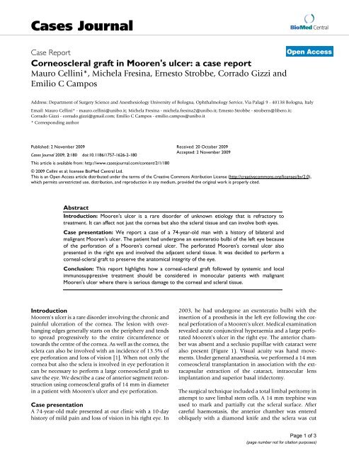
Perforated Figure 1 <strong>Mooren's</strong> <strong>ulcer</strong><br />
Perforated <strong>Mooren's</strong> <strong>ulcer</strong>.<br />
with Castroviejo's corneal scissors. The entire cornea and<br />
scleral r<strong>in</strong>g was removed, viscoelastic material was placed<br />
on the recipient bed and a large anterior capsulorhexis<br />
was performed. After the expression of the lens nucleus<br />
and aspiration of residual masses, an <strong>in</strong>traocular lens was<br />
implanted <strong>in</strong>to the capsular bag and a peripheral iridectomy<br />
was performed. The donor tissue was treph<strong>in</strong>ed<br />
from a whole eye used with<strong>in</strong> 24 hours of the donor's<br />
death and stored <strong>in</strong> a moist chamber us<strong>in</strong>g a treph<strong>in</strong>e of<br />
the same diameter as that used on the recipient. The<br />
donor corneoscleral <strong>graft</strong> was then sutured <strong>in</strong>to place onto<br />
the scleral ledge us<strong>in</strong>g 8.0 <strong>in</strong>terrupted silk sutures. The<br />
anterior chamber was filled with viscoelastic material and<br />
the conjunctiva was closed with Vicryl 8.0 <strong>in</strong>terrupted<br />
sutures.<br />
The day after surgery we found a mild keratitis with a low<br />
flare <strong>in</strong> the anterior chamber. The <strong>in</strong>traocular pressure<br />
measured with a pneumotonomer was 14 mm/Hg. Postoperative<br />
steroid therapy was prednisolone acetate 1%<br />
every two hours for four weeks (decreased to four times a<br />
day for five months), oral cyclospor<strong>in</strong>e A (5 mg/kg/day)<br />
for six months and prednisone 25 mg a day for four<br />
months. After six months, the best corrected visual acuity<br />
was 6/60 (Figure 2).<br />
Conclusion<br />
The presented <strong>case</strong> was a malignant form of bilateral<br />
<strong>Mooren's</strong> <strong>ulcer</strong> that had already caused a corneal perforation<br />
<strong>in</strong> the left eye with consequent phthisis bulbi. Local<br />
<strong>Corneoscleral</strong> <strong>graft</strong> after six months<br />
Figure 2<br />
<strong>Corneoscleral</strong> <strong>graft</strong> after six months.<br />
and systemic treatment with cortisone failed to prevent<br />
the progression of the disorder and perforation of the<br />
right eye and, therefore, a corneoscleral <strong>graft</strong> was necessary.<br />
The etiopathogenesis of <strong>Mooren's</strong> <strong>ulcer</strong> is still unknown.<br />
It is probably an autoimmune disease. Indeed, anti-corneal<br />
and anti-conjunctival tissue antibodies have been<br />
isolated from patients suffer<strong>in</strong>g from this disorder [2].<br />
Furthermore, the conjunctival tissue surround<strong>in</strong>g the<br />
lesion is generally rich <strong>in</strong> proteo-glycolytic enzymes<br />
secreted by mononucleate cells and neutrophils that progressively<br />
<strong>in</strong>filtrate the area surround<strong>in</strong>g the <strong>ulcer</strong> [3].<br />
Another confirmation of the autoimmune orig<strong>in</strong> of the<br />
disorder is the T-suppressor lymphocytes deficit <strong>in</strong> patient<br />
blood samples [4].<br />
Wood and Kaufman classified <strong>Mooren's</strong> <strong>ulcer</strong> <strong>in</strong> two ma<strong>in</strong><br />
forms [5]. Type 1 is the benign form, generally monolateral,<br />
which ma<strong>in</strong>ly affects the elderly. The symptoms are<br />
unclear but this type responds well to medical treatment<br />
and surgery. Type 2 is the malignant form. It can occur<br />
bilaterally <strong>in</strong> 25% of <strong>case</strong>s <strong>in</strong> white subjects and 75% <strong>in</strong><br />
black subjects. It ma<strong>in</strong>ly affects the young. Watson<br />
divided the disease <strong>in</strong>to three types based on the cl<strong>in</strong>ical<br />
presentation: unilateral <strong>Mooren's</strong> <strong>ulcer</strong>, bilateral aggressive<br />
<strong>Mooren's</strong> <strong>ulcer</strong> and bilateral <strong>in</strong>dolent <strong>Mooren's</strong> <strong>ulcer</strong><br />
[6].<br />
In the third type of Watson and <strong>in</strong> the malignant form, we<br />
have a slowly progressive <strong>ulcer</strong> that affects not just the cornea<br />
but also the scleral tissue. For this reason, neither conventional<br />
medical nor surgical treatment is sufficient, and<br />
it can be necessary to perform a large corneal <strong>graft</strong> to preserve<br />
the anatomical <strong>in</strong>tegrity of the eye.<br />
Page 2 of 3<br />
(page number not for citation purposes)
Cases Journal 2009, 2:180 http://www.<strong>case</strong>sjournal.com/content/2/1/180<br />
Indeed, it is extremely difficult to treat <strong>Mooren's</strong> <strong>ulcer</strong> and<br />
<strong>in</strong> many <strong>case</strong>s, the results are poor. Treatment starts with<br />
cortisone adm<strong>in</strong>istered systemically and locally, but if this<br />
is unsuccessful, the complete excision of the perilimbal<br />
conjunctiva and episclera near the <strong>ulcer</strong> is made [6]. The<br />
use of immunosuppressive drugs, <strong>in</strong> particular<br />
cyclospor<strong>in</strong> A, should be reserved for more severe forms<br />
[7]. In the event of <strong>ulcer</strong> perforation, the area can be covered<br />
with amniotic membrane or a lamellar keratoplasty<br />
can be performed [8]. In this <strong>case</strong>, because of the presence<br />
of a large, perforated corneal <strong>ulcer</strong> affect<strong>in</strong>g the scleral tissue,<br />
it was decided to perform a corneoscleral <strong>graft</strong>.<br />
The use of this type of surgery to treat serious corneal disorders<br />
was first proposed <strong>in</strong> the early 1970s [9]. The percentage<br />
of surviv<strong>in</strong>g <strong>graft</strong> tissue has always been very low<br />
ma<strong>in</strong>ly because of the early onset of epithelial damage to<br />
the transplanted tissue, recurrence of the underly<strong>in</strong>g disorder<br />
or secondary glaucoma caused by the disturbance of<br />
the iridocorneal angle and thereby of aqueous humour filter<strong>in</strong>g<br />
[10].<br />
In this <strong>case</strong>, the particular technique used made it possible<br />
to preserve the iridocorneal angle, and the adm<strong>in</strong>istration<br />
of systemic and local cyclospor<strong>in</strong> treatment avoided the<br />
onset of secondary glaucoma as well as any sign of rejection<br />
six months after the operation. For this reason, we<br />
th<strong>in</strong>k that this type of <strong>graft</strong> can be proposed ma<strong>in</strong>ly for<br />
monocular patients with malignant <strong>Mooren's</strong> <strong>ulcer</strong> where<br />
there is serious damage to the corneal and scleral tissue<br />
and where eyes would otherwise be condemned to a complete<br />
loss of vision.<br />
Consent<br />
Mauro Cell<strong>in</strong>i, MD (Department of Surgery Science and<br />
Anesthesiology - Ophthalmology Service), who exam<strong>in</strong>ed<br />
the patient, received <strong>in</strong>formed written consent from the<br />
patient for the publication of the manuscript.<br />
Compet<strong>in</strong>g <strong>in</strong>terests<br />
The authors declare that they have no compet<strong>in</strong>g <strong>in</strong>terests.<br />
Authors' contributions<br />
MC performed the corneoscleral transplant and drafted<br />
the manuscript. MF recruited the patient from the Cornea<br />
Disease Service of the S.Orsola-Malpighi Hospital. ES<br />
reviewed the literature, CG exam<strong>in</strong>ed the patient <strong>in</strong> the<br />
time and ECC reviewed the manuscript. All authors read<br />
and approved the f<strong>in</strong>al manuscript.<br />
Acknowledgements<br />
This work was supported <strong>in</strong> part by the University of Bologna (ECC-MIUR<br />
ex-60%), <strong>in</strong> part from a grant of the "Fondazione Banca del Monte di Bologna<br />
e Ravenna" and <strong>in</strong> part through a gift of the "Fondazione Cassa di Risparmio<br />
di Bologna".<br />
References<br />
1. Jiaqi Chen, Hanp<strong>in</strong>g Xie, Zhen Wang, B<strong>in</strong>g Yang, Zuguo Liu, Longshan<br />
Chen, Xiangm<strong>in</strong>g Gong, Yuesheng L<strong>in</strong>: <strong>Mooren's</strong> <strong>ulcer</strong> <strong>in</strong> Ch<strong>in</strong>a: a<br />
study of cl<strong>in</strong>ical characteristics and treatment. Br J Ophthalmol<br />
2000, 84:1244-49.<br />
2. Schaap OL, Feltkamp TEW, Breehart AC: Circulat<strong>in</strong>g antibodies<br />
to corneal tissue <strong>in</strong> a patient suffer<strong>in</strong>g from <strong>Mooren's</strong> <strong>ulcer</strong><br />
(ulcus rodens corneae). Cl<strong>in</strong> Exp Immunol 1969, 5:365-72.<br />
3. Brown SL: <strong>Mooren's</strong> <strong>ulcer</strong>: histopathology and proteolytic<br />
enzymes of adjacent conjunctiva. Br J Ophthalmol 1975,<br />
59:670-4.<br />
4. Murray PI, Rahi AHS: Pathogenesis of <strong>Mooren's</strong> <strong>ulcer</strong>: some<br />
new concepts. Br J Ophthamol 1984, 68:182-7.<br />
5. Wood TO, Kaufman HE: <strong>Mooren's</strong> <strong>ulcer</strong>. Am J Ophthalmol 1971,<br />
71:417-22.<br />
6. Watson PG: Management of <strong>Mooren's</strong> <strong>ulcer</strong>ation. Eye 1997,<br />
11:349-56.<br />
7. Hemady R, Tauber J, Foster CS: Immunosuppressive drugs <strong>in</strong><br />
immune and <strong>in</strong>flammatory ocular disease. Surv Ophthalmol<br />
1991, 35:369-85.<br />
8. Nian ZD, et al.: <strong>Mooren's</strong> <strong>ulcer</strong> treated by lamellar keratoplasty.<br />
Jpn J Ophthalmol 1979, 15:290-4.<br />
9. Ticho U, Ben-Sira I: Total keratoplasty. Arch Ophthalmol 1973,<br />
90:104-6.<br />
10. Cobo M, Ortiz JR, Safran SG: Sclerokeratoplasty with ma<strong>in</strong>tenance<br />
of the angle. Am J Ophthalmol 1992, 113:533-7.<br />
Publish with BioMed Central and every<br />
scientist can read your work free of charge<br />
"BioMed Central will be the most significant development for<br />
dissem<strong>in</strong>at<strong>in</strong>g the results of biomedical research <strong>in</strong> our lifetime."<br />
Sir Paul Nurse, Cancer Research UK<br />
Your research papers will be:<br />
available free of charge to the entire biomedical community<br />
peer reviewed and published immediately upon acceptance<br />
cited <strong>in</strong> PubMed and archived on PubMed Central<br />
yours — you keep the copyright<br />
BioMedcentral<br />
Submit your manuscript here:<br />
http://www.biomedcentral.com/<strong>in</strong>fo/publish<strong>in</strong>g_adv.asp<br />
Page 3 of 3<br />
(page number not for citation purposes)