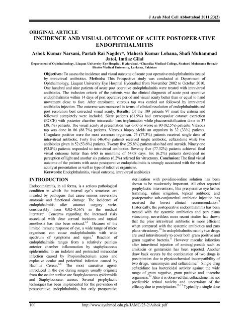
incidence and visual outcome of acute postoperative endophthalmitis
incidence and visual outcome of acute postoperative endophthalmitis
incidence and visual outcome of acute postoperative endophthalmitis

Create successful ePaper yourself
Turn your PDF publications into a flip-book with our unique Google optimized e-Paper software.
J Ayub Med Coll Abbottabad 2011;23(2)<br />
ORIGINAL ARTICLE<br />
INCIDENCE AND VISUAL OUTCOME OF ACUTE POSTOPERATIVE<br />
ENDOPHTHALMITIS<br />
Ashok Kumar Narsani, Partab Rai Nagdev*, Mahesh Kumar Lohana, Shafi Muhammad<br />
Jatoi, Imtiaz Gilal<br />
Department <strong>of</strong> Ophthalmology, Liaquat University Eye Hospital, Hyderabad, *Ch<strong>and</strong>ka Medical College, Shaheed Mohtrama Benazir<br />
Bhutto Medical University, Larkana, Pakistan<br />
Objectives: To assess the <strong>incidence</strong> <strong>and</strong> <strong>visual</strong> <strong>outcome</strong> <strong>of</strong> <strong>acute</strong> post operative <strong>endophthalmitis</strong> treated<br />
by intravitreal antibiotics. Methods: This Prospective study was conducted at Department <strong>of</strong><br />
Ophthalmology, Liaquat University Eye Hospital Hyderabad from November 2002 to October 2010.<br />
One hundred <strong>and</strong> nine patients <strong>of</strong> <strong>acute</strong> post operative <strong>endophthalmitis</strong> were treated with intravitreal<br />
antibiotics. The inclusion criteria <strong>of</strong> the patients was the clinical diagnosis <strong>of</strong> <strong>acute</strong> post operative<br />
<strong>endophthalmitis</strong> within 14 days <strong>of</strong> post operative period <strong>and</strong> <strong>visual</strong> acuity better than or equal to h<strong>and</strong><br />
movement close to face. After enrolment, vitreous tap was carried out followed by intravitreal<br />
antibiotics injection. The <strong>outcome</strong> was measured in terms <strong>of</strong> clinical resolution <strong>of</strong> <strong>endophthalmitis</strong> <strong>and</strong><br />
post resolution best corrected <strong>visual</strong> acuity. Results: Of the 109 patients 97 meet the criteria <strong>and</strong><br />
followed completely were included. Sixty patients (61.9%) had extracapsular cataract extraction<br />
(ECCE) with posterior chamber intraocular lens implantation while phacoemulsification done in 37<br />
(38.1%) patients. The <strong>visual</strong> acuity at presentation was 6/60 or worse in 80 (82.5%) patients. Vitreous<br />
tap was done in 86 (88.7%) patients. Vitreous biopsy yields an organism in 32 (33%) patients.<br />
Coagulase positive were the most common organism. 75 (77.3%) patients received single dose <strong>of</strong><br />
intravitreal antibiotic. Forty five (46.4%) patients received single antibiotic, ceftazidime while two<br />
antibiotics given in 52 (53.6%) patients. Twenty five (25.8%) patients also had oral steroids. Ninety one<br />
(93.8%) patients responded to intravitreal antibiotics. Seventy five (77.32%) patients achieved final<br />
<strong>visual</strong> <strong>outcome</strong> better than 6/60 in meantime <strong>of</strong> 54.08 days. Six (6.2%) patients developed no<br />
perception <strong>of</strong> light <strong>and</strong> another six patients (6.2%) referred for vitrectomy. Conclusion: The final <strong>visual</strong><br />
<strong>outcome</strong> <strong>of</strong> the patients with <strong>acute</strong> <strong>postoperative</strong> <strong>endophthalmitis</strong> is strongly associated with the <strong>visual</strong><br />
acuity at presentation as well as type <strong>of</strong> infective organisms.<br />
Keywords: Endophthalmitis, <strong>visual</strong> <strong>outcome</strong>, intravitreal antibiotics<br />
INTRODUCTION<br />
Endophthalmitis, in all forms, is a serious pathological<br />
condition in which the internal eye’s structures are<br />
invaded by pathogens that cause serious irreversible<br />
anatomic <strong>and</strong> functional damage. The <strong>incidence</strong> <strong>of</strong><br />
<strong>endophthalmitis</strong> after cataract surgery varies<br />
considerably from 0.02–0.56% in the medical<br />
literature 2 . Concerns regarding the increased risks<br />
associated with clear corneal incisions <strong>and</strong> topical<br />
anesthesia has also been noticed. 3,4 Because <strong>of</strong> the<br />
limited immune response <strong>of</strong> eye, a wide range <strong>of</strong> micro<br />
organisms can cause <strong>endophthalmitis</strong> with wide<br />
spectrum <strong>of</strong> symptoms <strong>and</strong> signs. 5 Reaction <strong>of</strong><br />
<strong>endophthalmitis</strong> ranges from a relatively painless<br />
anterior chamber inflammation by staphylococcus<br />
epidermidis, to an indolent <strong>and</strong> protracted intraocular<br />
infection caused by Propionibacterium acnes <strong>and</strong><br />
explosive ocular <strong>and</strong> periorbital infection caused by<br />
Bacillus Cereus. 6-8 The most causative agents<br />
introduced in the eye during surgery usually originate<br />
from the ocular surface are Staphylococcus epidermidis<br />
<strong>and</strong> Staphylococcus aureus. Several prophylactic<br />
techniques has been implemented for the prevention <strong>of</strong><br />
<strong>postoperative</strong> <strong>endophthalmitis</strong>, but only preoperative<br />
sterilization with povidine-iodine solution has been<br />
shown to be moderately important. All other reported<br />
prophylactic interventions, like preoperative eye lashes<br />
trimming, saline irrigation, topical antibiotic <strong>and</strong><br />
<strong>postoperative</strong> sub-conjunctival antibiotic injection has<br />
received the lowest clinical recommendation. 9<br />
Historically, the <strong>postoperative</strong> <strong>endophthalmitis</strong> has been<br />
treated with the systemic antibiotics <strong>and</strong> pars plana<br />
vitrectomy, neverthless more recent studies has shown<br />
that the prior intravitreal antibiotic is more efficient<br />
when compared with the systemic antibiotics <strong>and</strong> pars<br />
plana vitrectomy. 10 In <strong>endophthalmitis</strong> mainly two drugs<br />
are used intravitreously to cover both gram positive <strong>and</strong><br />
gram negative bacteria. 11 However macular infarction<br />
after intravitreal injection <strong>of</strong> aminoglycoside such as<br />
amikacin or gentamicin has been reported. Another<br />
draw back occurs by the combination <strong>of</strong> two drugs is<br />
precipitation due to physicochemical incompatibility <strong>of</strong><br />
two drugs, vancomycin <strong>and</strong> ceftazidime. 11 Single drug<br />
ceftazidime has bactericidal activity against the wide<br />
range <strong>of</strong> gram negative, gram positive <strong>and</strong> anaerobe<br />
organisms. 12 Also it is observed that ceftazidime has not<br />
predictable retinal toxicity <strong>and</strong> uncertainty <strong>of</strong> the<br />
efficacy due to precipitation. 13–15 Typically a single dose<br />
100<br />
http://www.ayubmed.edu.pk/JAMC/23-2/Ashok.pdf
J Ayub Med Coll Abbottabad 2011;23(2)<br />
is sufficient, though some cases require repeated dose<br />
for virulent or slow replicating organisms. 16<br />
MATERIAL AND METHODS<br />
This, eight years prospective hospital based study was<br />
conducted from November 2002 to October 2010 at the<br />
Department <strong>of</strong> Ophthalmology Liaquat University <strong>of</strong><br />
Medical <strong>and</strong> Health Sciences, Eye Hospital Hyderabad.<br />
The patients were enrolled with data including name,<br />
age, sex, associated systemic disease, interval between<br />
surgery <strong>and</strong> onset <strong>of</strong> symptoms <strong>and</strong> signs. Ocular<br />
evaluation included the best corrected <strong>visual</strong> acuity, slit<br />
lamp biomicroscopic examination, indirect<br />
ophthalmoscopy <strong>and</strong> ultrasonography for posterior<br />
segment evaluation. The inclusion criteria was best<br />
corrected <strong>visual</strong> acuity <strong>of</strong> better than or equal to h<strong>and</strong><br />
movement close to face (HMCTF), reaction in the<br />
anterior chamber, hypopyon, reduced / absent fundal<br />
glow <strong>and</strong> exudates in the vitreous with presentation<br />
within 14 days <strong>of</strong> cataract surgery. The exclusion<br />
criteria was patients with chronic post-operative<br />
<strong>endophthalmitis</strong>, post traumatic <strong>endophthalmitis</strong>,<br />
previously treated <strong>endophthalmitis</strong> following intraocular<br />
surgery other than cataract <strong>and</strong> <strong>acute</strong> post-operative<br />
<strong>endophthalmitis</strong> with <strong>visual</strong> acuity <strong>of</strong> light perception or<br />
worse, retinal detachment, one eyed. All patients were<br />
operated in the same department by different surgeons<br />
with same local anesthesia (peribulbar technique)<br />
through clear corneal incision.<br />
The intravitreal injection was given under<br />
topical anesthesia in the operation theatre under<br />
operating microscope after completing aseptic<br />
measures. The tap site was measured by caliper 3.5mm<br />
behind the limbus at 10 'O clock position. A 25 gauge<br />
needle on 1cc disposable syringe was used to aspirate<br />
0.1 ml to 0.2 ml <strong>of</strong> the vitreous. Keeping the needle in<br />
place the syringe was replaced by another 1cc syringe<br />
containing prepared intravitreal antibiotic (ceftazidime<br />
2mg/0.1ml or vancomycin 2mg/0.1ml <strong>and</strong> amikacin<br />
0.4mg/0.1ml) injected into the vitreous cavity with the<br />
bevel <strong>of</strong> the needle facing away from the macula. If the<br />
eye showed no improvement or worsening <strong>of</strong> the signs<br />
<strong>and</strong> symptoms after an interval <strong>of</strong> 48 hours, the<br />
intravitreal injection was repeated. In addition all<br />
patients received fortified topical antibiotic therapy<br />
(ceftazidime 50mg/ml, vancomycin 50mg/ml,) at 1 hour<br />
interval along with cycloplegics (atropine 1% three<br />
times a day). Patient having less severe features <strong>and</strong><br />
negative culture also treated with low dose oral steroids<br />
prednesolone. A decrease in symptoms <strong>and</strong> reaction in<br />
the anterior chamber <strong>and</strong> vitreous cavity <strong>and</strong> increase in<br />
best corrected <strong>visual</strong> acuity was the criteria taken as a<br />
response to intravitreal antibiotic injections. The patients<br />
whose condition further deteriorates in spite <strong>of</strong> maximal<br />
tolerated medical therapy were referred for pars plana<br />
vitrectomy. The patients were evaluated daily until<br />
discharged, once per week for one month <strong>and</strong> then every<br />
month until the end <strong>of</strong> third month. The data was<br />
analysed on SPSS-16.<br />
RESULTS<br />
In our study the overall <strong>incidence</strong> <strong>of</strong> <strong>acute</strong> <strong>postoperative</strong><br />
<strong>endophthalmitis</strong> following cataract extraction was 0.65%<br />
in the last eight years. Of the total 109 patients, 97<br />
(89.9%) had followed completely, the remaining 12<br />
(10.1%) has lost the follow up <strong>and</strong> they were excluded<br />
from the study. There were 63 (65%) males <strong>and</strong> 34(35%)<br />
females with a mean age <strong>of</strong> 43.5 years (Table-1).<br />
Table-1: Age <strong>of</strong> patients<br />
Age (years) Frequency Percentage<br />
10–20 2 2.1<br />
21–30 4 4.1<br />
31–40 17 17.5<br />
41–50 15 15.5<br />
51–60 28 28.9<br />
61–70 23 23.7<br />
71–80 6 6.2<br />
>80 2 2.1<br />
TOTAL 97 100.0<br />
In 60 (61.9%) patients conventional<br />
extracapsular cataract extraction (ECCE) with posterior<br />
chamber intraocular lens implantation (PCIOL) was<br />
done, while (PCIOL) with phacoemulsification was<br />
done in 37 (38.1%) patients. The uneventful surgery<br />
was done successfully in 93 (95.9%) patients while rent<br />
in posterior capsule was happened in 4 (4.1%) patients.<br />
At the end <strong>of</strong> surgery all patients’ received subconjunctival<br />
injection gentamycin 20mg/0.5ml <strong>and</strong><br />
dexamethasone 2mg/0.5ml. Post operatively all the<br />
patients were treated by combined tobramycin 0.3% <strong>and</strong><br />
dexamethasone 0.1% eye drops. The mean time from<br />
surgery to presentation with <strong>endophthalmitis</strong> was 3.2<br />
days (ranged 1–14 days). The <strong>visual</strong> acuity <strong>of</strong> patients at<br />
presentation was 6/60 or worse in 80 (82.5%) patients,<br />
6/36 -6/12 in 14 (14.4%) patients <strong>and</strong> better than 6/12 in<br />
3 (3.1%) patients (Table-2).<br />
Table-2: Best corrected vision present with<br />
<strong>endophthalmitis</strong> (BCVpwE)<br />
Visual Acuity Frequency Percentage<br />
HM-3/60 57 58.8<br />
4/60-6/60 23 23.7<br />
6/36-6/12 14 14.4<br />
6/9-6/6 03 3.1<br />
TOTAL 97 100.0<br />
The vitreous was tapped out easily in 86<br />
(88.7%) patients <strong>and</strong> in 11 (11.3%) patients it was not<br />
tapped out due to thick organized vitreous. The vitreous<br />
biopsy showed micro organisms in 32 (33%) patients as<br />
follows: staphylococcus aureus in 10 (10.3%) patients,<br />
staphylococcus epidermidis 9 (9.3%) patients,<br />
streptococcus pneumonia 6 (6.2%), pseudomonas 5<br />
(5.2%) <strong>and</strong> E-coli 2 (2.06%) patients. In 54 (55.7%)<br />
http://www.ayubmed.edu.pk/JAMC/23-2/Ashok.pdf 101
J Ayub Med Coll Abbottabad 2011;23(2)<br />
patients vitreous biopsy showed no growth <strong>of</strong> micro<br />
organisms.<br />
Of the 97 patients, 75 (77.3%) received<br />
intravitreal antibiotic for single time while 22 (22.7%)<br />
patients received same intravitreal antibiotics for second<br />
time. Among the 97 patients, 45 (46.4%) patients<br />
received single intravitreal antibiotic (ceftazidime),<br />
while two antibiotics (vancomycin <strong>and</strong> amikacin) given<br />
in 52 (53.6%) patients. Also all patients treated by<br />
intensively topical fortified antibiotics <strong>and</strong> steroids,<br />
cycloplaegics. Oral antibiotic cipr<strong>of</strong>loxacin 500 mg two<br />
times a day for one week <strong>and</strong> oral analgesic,<br />
flurbipr<strong>of</strong>en 100 mg two times until patient pain free.<br />
Twenty five (25.8%) patients also had oral steroids. In<br />
the result 91 (93.8%) patients responded clinically well<br />
to intravitreal antibiotics while 6 (6.2%) patients not<br />
responded <strong>and</strong> were referred for vitrectomy. The best<br />
corrected <strong>visual</strong> acuity (BCVA) achieved to 6/6-6/9 in<br />
13 (13.4%) patients, 6/12-6/36 in 22 (22.7%) patients,<br />
6/24-6/60 in 40 (41.23%) patients, less than 6/60 in 16<br />
(16.5%) patients <strong>and</strong> no perception <strong>of</strong> light in 6 (6.2%)<br />
patients (Table-3).<br />
Table-3: Best corrected vision Final (BCV Final)<br />
Visual Acuity Frequency Percentage<br />
HM–3/60 16 16.5<br />
4/60–6/60 40 41.2<br />
6/36–6/12 22 22.7<br />
6/9–6/6 13 13.4<br />
NPL 6 6.2<br />
TOTAL 97 100.0<br />
The mean time <strong>of</strong> achieving final <strong>visual</strong> <strong>outcome</strong> was<br />
54.08 days. There was statistically significant (p=0.00)<br />
relationship between poor vision at presentation <strong>and</strong><br />
good <strong>visual</strong> out come. The mean <strong>visual</strong> acuity before<br />
treatment was 1.62 <strong>and</strong> the mean <strong>visual</strong> acuity after<br />
treatment was increased to 2.52.<br />
Similarly type <strong>of</strong> organisms (p=0.002) <strong>and</strong><br />
intravitreal dose (p=0.002) had statistically significant<br />
association with better final <strong>visual</strong> <strong>outcome</strong>.<br />
Complicated surgery (p=0.178), systemic disease<br />
(p=0.500), oral steroid (p=0.099), intravitreal regime<br />
(p=0.307), time <strong>of</strong> presentation (p=0.551), positive<br />
vitreous biopsy (p=0.34) not found to be statistically<br />
significant<br />
DISCUSSION<br />
Postoperative <strong>endophthalmitis</strong> is the most devasting<br />
complication following cataract surgery. In our study,<br />
<strong>incidence</strong> <strong>of</strong> post operative <strong>endophthalmitis</strong> is 0.65%<br />
which is higher than Kamala rajah S 14 <strong>and</strong> Desai P<br />
studies 17 . A number <strong>of</strong> studies have raised concern<br />
about the higher <strong>incidence</strong> <strong>of</strong> <strong>endophthalmitis</strong> in the<br />
clear corneal incisions than scleral tunnel incisions 18 .<br />
Infective <strong>endophthalmitis</strong> is usually appeared with rapid<br />
loss <strong>of</strong> vision <strong>and</strong> intense intraocular inflammation<br />
particularly in the early post operative period. In our<br />
study 17(17.53%) patients showed <strong>acute</strong> low grade<br />
<strong>endophthalmitis</strong> with negative microbial growth in<br />
vitreous tap, which reflect the difficulty in<br />
distinguishing between toxic anterior segment<br />
syndromes with infective <strong>endophthalmitis</strong>. The<br />
microbiological culture growth pr<strong>of</strong>ile has regional<br />
variation. In our study vitreous tap was done in 86<br />
(88.7%) patients, <strong>of</strong> which 32 (37.21%) showed positive<br />
microbial growth, while Carrim et al 19 in their study<br />
reported 55% <strong>of</strong> positive results. Gram positive are the<br />
78.13% <strong>of</strong> total positive microbial growth having 50%<br />
Coagulase negative type. These findings are consistent<br />
with studies <strong>of</strong> Mayer et al 20 he has reported gram<br />
positive organisms in 76% patients. 14 <strong>and</strong> <strong>of</strong> Kamala<br />
rajah S et al 14 he has reported 49% <strong>of</strong> gram positive <strong>and</strong><br />
coagulase negative microorganisms. In our study<br />
75(77.3%) patients responded well to single dose <strong>of</strong><br />
intravitreal antibiotic while 22 (22.7%) patients required<br />
second time injection, while Kamala rajah S et al 14<br />
reported, repeated second time intravitreal injections in<br />
20% <strong>of</strong> patients. In our study the <strong>visual</strong> acuity at<br />
presentation was 6/60 or worse in 80 (82.50%) <strong>of</strong><br />
patients which was consistent to the survey <strong>of</strong> British<br />
Ophthalmological Surveillance Unit (BOSU) survey<br />
seen in 85% <strong>of</strong> patients. The <strong>visual</strong> <strong>outcome</strong> seen in this<br />
study is strongly proportional to the <strong>visual</strong> acuity before<br />
starting the treatment. Like Endophthalmitis Vitrectomy<br />
Study Group (EVSG) 21 , <strong>visual</strong> <strong>outcome</strong> in this study has<br />
also strongly associated with the type <strong>of</strong> infective<br />
microorganism. Patients with gram negative <strong>and</strong><br />
coagulase positive organisms showed poor <strong>outcome</strong><br />
than coagulase negative. This was seen in vitreous tap<br />
<strong>of</strong> 15 patients which showing pseudomonas in 5 patients<br />
<strong>and</strong> staphylococcus aureus in 10 patients. Out <strong>of</strong> 6<br />
patients with <strong>visual</strong> acuity (VA) <strong>of</strong> no perception <strong>of</strong><br />
light (NPL) 4 (4.1%) patients vitreous tap showed gram<br />
negative organisms, nearly same organisms on culture<br />
are seen by Desai P 22 in 5% patients. One more thing<br />
observed in this study was the strong association<br />
between the numbers <strong>of</strong> doses <strong>of</strong> intravitreal antibiotics.<br />
A patient with single dose has fewer <strong>outcome</strong>s<br />
irrespective to the symptoms, than those who received<br />
two doses. 23<br />
CONCLUSION<br />
The final <strong>visual</strong> <strong>outcome</strong> <strong>of</strong> the patients with <strong>acute</strong><br />
<strong>postoperative</strong> <strong>endophthalmitis</strong> is strongly associated<br />
with the <strong>visual</strong> acuity at presentation as well as type <strong>of</strong><br />
infective organisms.<br />
REFERENCES<br />
1. Chisari G. Endophthalmitis:Gram Positive Etiological agents <strong>and</strong><br />
susceptiblity to Glycolipids. Current clinical pharmacology.<br />
2008;3:153–5.<br />
2. Lloyd JC, Braga-Mele R. Incidence <strong>of</strong> <strong>postoperative</strong><br />
<strong>endophthalmitis</strong> in a high-volume cataract surgicentre in Canada.<br />
Canadian journal <strong>of</strong> ophthalmology. 2009;44:288–92.<br />
102<br />
http://www.ayubmed.edu.pk/JAMC/23-2/Ashok.pdf
J Ayub Med Coll Abbottabad 2011;23(2)<br />
3. Bohigian GM. A retrospective study <strong>of</strong> the <strong>incidence</strong> <strong>of</strong> culture<br />
positive <strong>endophthalmitis</strong> after cataract surgery <strong>and</strong> the use <strong>of</strong><br />
preoperative antibiotics. Ophthalmic Surg Lasers Imaging<br />
2007;38:103–6.<br />
4. Taban M, Behrens A, Newcomb RL, Nobe MY, Saedi G,<br />
Sweet PM, et al. Acute <strong>endophthalmitis</strong> following cataract<br />
surgery: a systemic review <strong>of</strong> the literature, Arch Ophthalmol<br />
2005;123:613–20.<br />
5 Callegan MC, Engelbert M, Parke DW 2nd, Jett BD, Gilmore<br />
MS. Bacterial <strong>endophthalmitis</strong>: epidemiology, therapeutics, <strong>and</strong><br />
bacterium-host interactions. Clin Microbiol Rev 2002;15:111–24.<br />
6 Aaberg TM Jr, Flynn HW Jr, Schiffman J, Newton J.<br />
Nosocomial <strong>acute</strong>-onset <strong>postoperative</strong> <strong>endophthalmitis</strong> survey. A<br />
10-year review <strong>of</strong> <strong>incidence</strong> <strong>and</strong> <strong>outcome</strong>s. Ophthalmology<br />
1998;105:1004–10.<br />
7 Clark WL, Kaiser PK, Flynn HW Jr, Belfort A, Miller D, Meisler<br />
DM. Treatment strategies <strong>and</strong> <strong>visual</strong> acuity <strong>outcome</strong>s in chronic<br />
<strong>postoperative</strong> Propionibacterium acnes <strong>endophthalmitis</strong>.<br />
Ophthalmology 1999;106:1665–70.<br />
8 Schemmer GR, Driebe WT. Post-traumatic Bacillus cereus<br />
<strong>endophthalmitis</strong>. Arch. Ophthalmol 1987;105:342–4.<br />
9 Costello P, Bakri SJ, Beer PM, Singh RJ, Falk NS, Peter GB, et<br />
al, Vitreous penetration <strong>of</strong> topical moxifloxcin <strong>and</strong> gatifloxcin in<br />
humans. Retina 2005;25:1.<br />
10 Hanscom TA. Postoperative <strong>endophthalmitis</strong>. Clin Infect Dis<br />
2004;38:542–6.<br />
11 Kwok AK, Hui M, Pang CP, Chan RC, Cheung SW, Yip CM, et<br />
al. An In Vitro study <strong>of</strong> Ceftazidime <strong>and</strong> Vancomycin<br />
Concentrations in Various Fluid Media: Implications for Use in<br />
Treating Endophthalmitis. Invest Ophthalmol Vis Sci<br />
2002;43:1182–8.<br />
12 Rains CP, Bryson HM, Peters DH. Ceftazidime. An update <strong>of</strong> its<br />
antibacterial activity, pharmacokinetic properties <strong>and</strong> therapeutic<br />
efficacy. Drugs 1995;49:577–617.<br />
13 Elder MJ, Morlet N; EPSWA. Endophthalmitis Population<br />
Study <strong>of</strong> Western Australia. Endophthalmitis. Clin Experiment<br />
Ophthalmol 2002;30:394–8.<br />
14 Kamala Rajah S, Silvestri G, Sharma N, Khan A, Foot B, Ling R,<br />
et al. Surveillance <strong>of</strong> <strong>endophthalmitis</strong> following cataract surgery<br />
in the UK. Eye 2004;18:580–7.<br />
15 Campochiaro PA, Conway BP. Aminoglycoside toxicity--a<br />
survey <strong>of</strong> retinal specialists. Implications for ocular use. Arch<br />
Ophthalmol 1991;109:946–50.<br />
16 Abelson M, Forbes M. The Engima <strong>of</strong> Endophthalmitis. Rev<br />
Ophthalmol 2002;30:394–8.<br />
17 Desai P, Minassian DC, Reidly A. National cataract surgery<br />
survey 1997–8. A report <strong>of</strong> the result <strong>of</strong> the cilinical <strong>outcome</strong>s. Br<br />
J Ophthalmol 1999;83:893–6<br />
18 Nichamin LD, Chang DF, Johonson SH, Mamalis N, Masket S,<br />
Packard RB, et al. ASCRS white paper: what is the association<br />
between clear corneal incision <strong>and</strong> post operative<br />
<strong>endophthalmitis</strong>? J Catract Refrct Surg 2006;32:1556–9.<br />
19 Carrim ZI, Richardson J, Wykes WN. Incidence <strong>and</strong> <strong>visual</strong> out<br />
come <strong>of</strong> <strong>acute</strong> <strong>endophthalmitis</strong> after cataract surgery-Experience<br />
<strong>of</strong> a single eye department in Scotl<strong>and</strong>. Br J Ophthalmol<br />
2009;93:721–5.<br />
20 Mayer E, Cadman D, Ewings P, , Twomey JM, Gray RH,<br />
Claridge KG, et al. A 10 years retrospective survey <strong>of</strong> cataract<br />
surgery <strong>and</strong> <strong>endophthalmitis</strong> in a single eye unit: injectable lenses<br />
lower the <strong>incidence</strong> <strong>of</strong> <strong>endophthalmitis</strong>. Br J Ophthalmol<br />
2003;87:867–9.<br />
21 Endophthalmitis Vitrectomy Study Group. Microbiologic factors<br />
<strong>and</strong> <strong>visual</strong> <strong>outcome</strong> in the Endophthalmitis Vitrectomy Study.<br />
Am J Ophthalmol 1996;122:830–46.<br />
22 Desai P. The National cataract surgery survey: Clinical<br />
<strong>outcome</strong>s. Eye 1993;7:489–94.<br />
23 Mollan SP, Gao A, Lockwood A, Durrani OM, Butler L.<br />
Postcataract <strong>endophthalmitis</strong>: <strong>incidence</strong> <strong>and</strong> microbial isolates in<br />
a united kingdom region from 1996 through 2004. J Ctaract<br />
Refract Surg 2007;33:265–8.<br />
Address for Correspondence:<br />
Dr. Ashok Kumar Narsani, Associate Pr<strong>of</strong>essor, Department <strong>of</strong> Ophthalmology, Liaquat University Eye Hospital,<br />
Hyderabad, Pakistan. Cell: +92-302-3929326<br />
Email: amnarsani@hotmail.com<br />
http://www.ayubmed.edu.pk/JAMC/23-2/Ashok.pdf 103