Evaluation of the Antimutagenic Effect of Vitamin C against DNA ...
Evaluation of the Antimutagenic Effect of Vitamin C against DNA ...
Evaluation of the Antimutagenic Effect of Vitamin C against DNA ...

Create successful ePaper yourself
Turn your PDF publications into a flip-book with our unique Google optimized e-Paper software.
Nature and Science 2009; 7(12)<br />
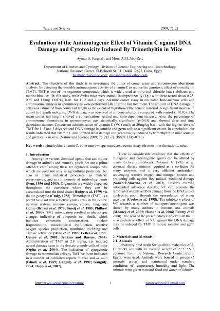
<strong>Evaluation</strong> <strong>of</strong> <strong>the</strong> <strong>Antimutagenic</strong> <strong>Effect</strong> <strong>of</strong> <strong>Vitamin</strong> C <strong>against</strong> <strong>DNA</strong><br />
Damage and Cytotoxicity Induced By Trimethyltin in Mice<br />
Ayman A. Farghaly and Mona A.M. Abo-Zeid<br />
Department <strong>of</strong> Genetics and Cytology, Division <strong>of</strong> Genetic Engineering and Biotechnology,<br />
National Research Center, El-Behooth St. 31, Dokki 12622, Cairo, Egypt.<br />
farghaly_5@yahoo.com, monaabozeid@yahoo.com<br />
Abstract: The objective <strong>of</strong> this study is to investigate <strong>the</strong> utility <strong>of</strong> comet assay and chromosome aberrations<br />
analysis for detecting <strong>the</strong> possible antimutagenic activity <strong>of</strong> vitamin C to reduce <strong>the</strong> genotoxic effect <strong>of</strong> trimethyltin<br />
(TMT). TMT is one <strong>of</strong> <strong>the</strong> organotin compounds which is widely used as polyvinyl chloride heat stabilizers and<br />
marine biocides. In this study, male Swiss mice were treated interapretoneally (i.p.) with three tested doses 0.25,<br />
0.50 and 1.0mg TMT/kg b.wt. for 1, 2 and 3 days. Alkaline comet assay in nucleated bone-marrow cells and<br />
chromosome analysis in spermatocytes were performed 24h after <strong>the</strong> last treatment. The amount <strong>of</strong> <strong>DNA</strong> damage in<br />
cells was estimated from comet tail length as <strong>the</strong> extent <strong>of</strong> migration <strong>of</strong> <strong>the</strong> genetic material. A significant increase in<br />
comet tail length indicating <strong>DNA</strong> damage was observed at all concentrations compared with control (p
Nature and Science 2009; 7(12)<br />
2.2. Chemicals<br />
TMT was purchased from BDH Chemicals Poole,<br />
England. VC was purchased from Sigma, USA. All<br />
o<strong>the</strong>r chemicals used were <strong>of</strong> analytical grade.<br />
2.3. Doses and experimental design<br />
The experimental design involved: 1- Mice were<br />
treated i.p. with <strong>the</strong> doses 0.25, 0.5 and 1.0mg TMT/kg<br />
b.wt. for 1, 2 and 3 days. 2- Concurrent administration<br />
<strong>of</strong> VC at 20mg/kg b.wt. and 1mg TMT/kg b.wt. for 1,2<br />
and 3 days. The samples were taken 24h after <strong>the</strong> last<br />
treatment from <strong>the</strong> bone-marrow cells for <strong>DNA</strong> damage<br />
detection by comet assay and spermatocyte cells for<br />
chromosome aberrations detection. In all experiments,<br />
<strong>the</strong> animals groups were treated with vehicle as a<br />
negative control, 20mg VC/kg b.wt. and 1mg<br />
mitomycin C/kg b.wt. as a positive control. All samples<br />
were collected after 24h.<br />
2.4. Cell viability<br />
To measure cytotoxicity, 15µl <strong>of</strong> each original cell<br />
suspension was mixed with15µl <strong>of</strong> 0.4% solution <strong>of</strong><br />
trypan blue vital dye. Cells were analyzed with a light<br />
microscope and <strong>the</strong> percentage <strong>of</strong> viable cells was<br />
determined from 200 cells for each experimental group<br />
(Zamorano-Ponce et al, 2004).<br />
2.5. Comet assay<br />
The comet assay was performed as described by<br />
Tice et al (2000). Mice femurs were dissected out and<br />
bone marrow was aspirated from each femur into media<br />
solution. The cell suspension (25 µl) was mixed 1:10<br />
with 250 µl molten low melting point (LMP) agarose,<br />
and samples <strong>of</strong> 75 µl <strong>of</strong> <strong>the</strong> mixture were rapidly spread<br />
on CometSlides. After gelling for 20 min at 4ºC in <strong>the</strong><br />
dark, slides were put in a tank filled with lysis solution<br />
(2.5M NaCl, 0.1M EDTA, 10mM Tris base, 1% sodium<br />
lauryl sarcosinate and 1% Triton X-100) for 1h at 4ºC in<br />
<strong>the</strong> dark. Slides were <strong>the</strong>n washed three times with<br />
neutralization buffer (0.4M Tris, pH 7.5) for 5 min and<br />
incubated in fresh alkaline buffer (0.3M NaOH and<br />
1mM EDTA, pH>13) for 30min at room temperature to<br />
allow unwinding <strong>of</strong> <strong>DNA</strong>. Electrophoresis was <strong>the</strong>n<br />
carried out at room temperature in fresh ice-cold<br />
alkaline electrophoresis buffer for 30 min (1V/cm;<br />
300mA). After electrophoresis, slides were gently<br />
washed three times for 5 min in fresh neutralization<br />
buffer and exposed to 70% ethanol for 5 min. After<br />
drying at room temperature, slides were stained with 25<br />
µl <strong>of</strong> ethidium bromide solution (20µg/ml) and covered<br />
with cover slip. Comets were examined at 200X<br />
magnification using a fluorescence microscope.<br />
Figure 1: Diagram represents (a) Comet tail length (µm ±<br />
SE) (b) Chromosome aberration (% ± SE) induced by<br />
different doses <strong>of</strong> TMT for three times intervals<br />
compared with MMC as a positive control.<br />
Table 1: The percentage <strong>of</strong> cell viability and comet<br />
tail length (µm) in mice nucleated bone marrow<br />
cells after treatment with TMT alone and in<br />
combination with vitamin C.<br />
Treatments<br />
I. Control<br />
(vehicle)<br />
II.VC<br />
20mg/kg b.wt.<br />
III. MMC<br />
(1mg/kg b. wt.)<br />
positive control<br />
IV. TMT<br />
0.25mg/kg b. wt.<br />
0.5mg/kg b. wt.<br />
1.0mg/kg b. wt.<br />
V.TMT+V.C.<br />
1.0mg/kg b. wt.<br />
+ 20mg/kg b.wt.<br />
Time <strong>of</strong><br />
Treatment<br />
(Day)<br />
(%) <strong>of</strong><br />
Viable<br />
Cells<br />
Comet tail<br />
length<br />
Mean ±SE<br />
- 93 1.14±0.23<br />
- 94 1.17±0.28<br />
- 72 11.03±1.12 a<br />
1<br />
2<br />
3<br />
1<br />
2<br />
3<br />
1<br />
2<br />
3<br />
1<br />
2<br />
3<br />
91<br />
87<br />
82<br />
85<br />
80<br />
78<br />
82<br />
77<br />
70<br />
84<br />
81<br />
76<br />
3.72±0.46 a<br />
3.92±0.58 a<br />
4.48±0.91 a<br />
3.90±0.48 a<br />
5.31±0.52 a<br />
8.37±0.97 a<br />
4.31±0.97 a<br />
6.70±1.20 a<br />
9.01±1.61 a<br />
3.42±0.55 a<br />
4.71±1.19 ab<br />
5.86±0.76 ab<br />
a) Significant compared to vehicle control (p
Nature and Science 2009; 7(12)<br />
Table 2: Number and mean percentage <strong>of</strong> diakinase metaphase I cells with chromosome aberrations in mice<br />
spermatocytes after treatment with TMT alone and in combination with vitamin C.<br />
Treatments<br />
Time <strong>of</strong> XY un. Auto. un. XY+ Auto. Chain (IV) Total Aberrations<br />
Treatment<br />
un.<br />
(Day)<br />
No. Mean (%)±SE<br />
I. Control (vehicle).<br />
-<br />
7<br />
5<br />
-<br />
-<br />
12<br />
2.4±0.65<br />
II.VC (20mg/kg b. wt.)<br />
-<br />
5<br />
4<br />
-<br />
-<br />
9<br />
1.8±0.52<br />
III. MMC (1mg/kg b. wt.)<br />
positive control<br />
- 35 20 4 2 61 12.2±1.2 a<br />
IV. TMT<br />
0.25 mg/kg b. wt.<br />
1<br />
2<br />
3<br />
14<br />
15<br />
18<br />
6<br />
7<br />
8<br />
-<br />
-<br />
-<br />
-<br />
-<br />
-<br />
20<br />
22<br />
26<br />
4.0±0.54<br />
4.4±0.40<br />
5.2±0.60 a<br />
0.5 mg/kg b. wt.<br />
1<br />
2<br />
3<br />
16<br />
15<br />
18<br />
7<br />
12<br />
13<br />
-<br />
-<br />
1<br />
-<br />
-<br />
-<br />
23<br />
27<br />
32<br />
4.6±0.42<br />
5.4±0.54 a<br />
6.4±0.48 a<br />
1.0 mg/kg b. wt.<br />
1<br />
2<br />
3<br />
19<br />
22<br />
28<br />
13<br />
15<br />
17<br />
-<br />
1<br />
-<br />
-<br />
1<br />
2<br />
32<br />
39<br />
47<br />
6.4±0.42 a<br />
7.8±0.56 a<br />
9.4±0.58 a<br />
V. TMT+V.C.<br />
1.0mg/kg b. wt.<br />
+ 20mg/kg b.wt.<br />
1<br />
2<br />
3<br />
20<br />
18<br />
21<br />
The total number <strong>of</strong> scored cells is 500 (5 animals/ group), XY un.: XY univalent; Auto. un.: Autosomal univalent<br />
a) Significant compared to vehicle control (p
Nature and Science 2009; 7(12)<br />
3.2. Comet assay:<br />
The amount <strong>of</strong> <strong>DNA</strong> damage in <strong>the</strong> cell was<br />
estimated from tail length as <strong>the</strong> extent <strong>of</strong> <strong>the</strong> migration<br />
<strong>of</strong> <strong>the</strong> genetic material in <strong>the</strong> direction <strong>of</strong> <strong>the</strong> anode.<br />
During electrophoresis, cell <strong>DNA</strong> was seen to more<br />
rapidly migrate towards <strong>the</strong> anode at <strong>the</strong> highest<br />
concentration than at <strong>the</strong> lowest concentration. Even <strong>the</strong><br />
comet tail tended to increase when exposure was<br />
prolonged from 1 to 3 days. Mean tail length (µm) <strong>of</strong><br />
comets obtained by TMT treatment is given in table (1).<br />
The trend <strong>of</strong> increase in comet tail length with increase<br />
in concentration and duration is depicted in figure (2).<br />
All <strong>the</strong> concentrations and <strong>the</strong>ir respective duration<br />
evoked significant <strong>DNA</strong> damage (p
Nature and Science 2009; 7(12)<br />
progression may result (L<strong>of</strong>t and Poulsen, 1996;<br />
Pryor, 1997). 2- This <strong>DNA</strong> damage could be also<br />
originated from apoptotic cells. Several laboratories<br />
have reported that <strong>the</strong> onset <strong>of</strong> apoptosis can give comet<br />
images with cell aspect and tail parameter values <strong>of</strong> <strong>the</strong><br />
same orders as those <strong>of</strong> cells with moderate <strong>DNA</strong><br />
damages (Florent et al, 1999; Choucroun et al, 2001).<br />
Jenkins and Barone (2004) reported that TMT had <strong>the</strong><br />
ability to induce apoptosis in PC12 cells by initiating<br />
apoptotic pathway requiring oxidative stress, caspase<br />
activation and P38 protein kinase activity leading to cell<br />
death. Also, Kawada et al (2008) observed that i.p.<br />
injection <strong>of</strong> TMT at <strong>the</strong> dose 2.8mg/kg b.wt. led to a<br />
dramatic increase in <strong>the</strong> number <strong>of</strong> degenerating cells in<br />
<strong>the</strong> granule cell layer <strong>of</strong> <strong>the</strong> OB and AON <strong>of</strong> <strong>the</strong> mouse<br />
brain cells.<br />
Interest in <strong>the</strong> chemopreventive functions <strong>of</strong><br />
antioxidants has grown considerably in recent years.<br />
Evidence accumulated over <strong>the</strong> years shows that people<br />
with high dietary intakes <strong>of</strong> fruits and vegetables are<br />
less likely to develop cancer than people who have low<br />
dietary intake <strong>of</strong> <strong>the</strong>se foods. While many<br />
chemopreventives in fruits and vegetables may have<br />
anticancer properties, much interest has focused on<br />
vitamin C (Mayne, 2003). This study represents one <strong>of</strong><br />
<strong>the</strong> premiere studies carried out to diminish <strong>the</strong> toxicity<br />
and <strong>the</strong> genotoxicity <strong>of</strong> <strong>the</strong> oxidative compound TMT<br />
by using <strong>the</strong> natural antioxidant compound VC. <strong>Vitamin</strong><br />
C is a highly effective antioxidant. It acts as a reducing<br />
agent that can terminate free radical driven oxidation by<br />
being converted to a resonance-stabilized free radical.<br />
In this respect VC can protect indispensable molecules<br />
in <strong>the</strong> body, such as protein, lipids, carbohydrates and<br />
nucleic acids (<strong>DNA</strong> and RNA). VC also regenerates<br />
o<strong>the</strong>r antioxidants such as vitamin E (Schneider et al,<br />
2001). Our results showed that concurrent<br />
administration <strong>of</strong> VC inhibited <strong>the</strong> <strong>DNA</strong> damage and<br />
chromosome aberrations induced by TMT in all tested<br />
doses. This ameliorative effect induced by VC may be<br />
resulted from enhancement <strong>of</strong> detoxification pathways<br />
that convert this reactive compound to less toxic and<br />
more easily excreted products (Vijayalaxmi and Venu,<br />
1999) and/or through its action as <strong>the</strong> free radical<br />
scavenging efficiency (Chaudiere and Ferrari-Iliou,<br />
1999). In addition, numerous in vitro and in vivo studies<br />
have evaluated <strong>the</strong> protective effects <strong>of</strong> VC <strong>against</strong><br />
several radical generating chemicals (Blasiak and<br />
Kawalik, 2001; Blasiak et al, 2004; Robichova et al,<br />
2004; Arranz et al, 2007).<br />
In conclusion, our results demonstrated that VC<br />
could be a suitable agent for preventing TMT- induced<br />
<strong>DNA</strong> and chromosome damage in an in vivo<br />
mammalian system.<br />
Correspondence to:<br />
Ayman A. Farghaly and Mona A.M. Abo-Zeid Department <strong>of</strong><br />
Genetics and Cytology,<br />
Division <strong>of</strong> Genetic Engineering and Biotechnology,<br />
National Research Center,<br />
El-Behooth St. 31, Dokki 12622, Cairo, Egypt.<br />
Emails: farghaly_5@yahoo.com,<br />
monaabozeid@yahoo.com<br />
References:<br />
[1] Arranz N, Haza AI, Garcia A, Rafter J, Morales P.<br />
Protective effect <strong>of</strong> vitamin C towards N-<br />
nitrosamine- induced <strong>DNA</strong> damage in <strong>the</strong> singlecell<br />
gel electrophoresis (SCGE)/HepG2 assay.<br />
Toxicol In Vitro 2007; 21: 1311-1317.<br />
[2] Blasiak J, Kowalik J. Protective action <strong>of</strong> vitamin C<br />
<strong>against</strong> <strong>DNA</strong> damage induced by selenium-cisplatin<br />
conjugate. Acta Biochim Pol 2001; 48(1): 233- 240.<br />
[3] Blasiak J, Gloc E, Wozniak K, Czechowska A.<br />
Genotoxicity <strong>of</strong> acrylamide in human lymphocytes.<br />
Chem Biol Interact 2004; 149(2-3): 137- 149.<br />
[4] Brown AW, Aldridge WN, Street BW, Verschoyle<br />
RD. The behavioral and neuropathologic sequelae<br />
<strong>of</strong> intoxication by trimethyltin compounds in <strong>the</strong> rat.<br />
Am J Pathol 1979; 97:59–82.<br />
[5] Cattanach BM, Pollard CE, Jssason JH. Ethyl<br />
methane-sulfonate induced chromosome breakage<br />
in <strong>the</strong> mouse. Mutat Res 1968; 6: 296-307.<br />
[6] Chaudiere J, Ferrari Iliou R. Intracellular<br />
antioxidants: from chemical to biochemical<br />
mechanisms. Food and Chemical Tox 1999; 37:<br />
949-962.<br />
[7] Choucroun P, Gillet D, Dorange G, Sawicki B,<br />
Dewitte JD. Comet assay and early apoptosis. Mutat<br />
Res 2001 ; 478: 89-96.<br />
[8] Cooke MS, Evans MD, Podmore ID, Podmore KE,<br />
Herbert KE, Mistry N, Mistry P, Hickenbotham PT,<br />
Hussieni A, Griffiths HR, Lunec J . Novel repair<br />
action <strong>of</strong> vitamin C upon in vivo oxidative <strong>DNA</strong><br />
damage. FEBS Lett 1998; 363: 363- 367.<br />
[9] Craig P. Biochemical cycles. National volatilization<br />
<strong>of</strong> tin. Nature 1998; 332: 309.<br />
[10] Dopp E, Hartmann LM, von Recklinghausen U,<br />
Florea AM, Rabieh S, Shokouhi B, Hirner AV, Obe<br />
G, Rettenmeier AW. The cyto- and genotoxicity <strong>of</strong><br />
organotin compounds is dependent on <strong>the</strong> cellular<br />
uptake capability. Toxicology 2007;232(3):226-234.<br />
[11] Evans EP, Breckon G, Ford CE. An air-drying<br />
method for meiotic preparations for mammalian<br />
testes. Cytogenetics 1964; 3: 289- 294.<br />
[12] Fahmy MA, Hassan NHA, Farghaly AA, Hassan<br />
EES. Studies on <strong>the</strong> genotoxic effect <strong>of</strong> beryllium<br />
chloride and <strong>the</strong> possible protective role <strong>of</strong><br />
selenium/vitamins A, C and E. Muta Res<br />
2008;652: 103-111.<br />
http://www.sciencepub.net/nature<br />
5<br />
naturesciencej@gmail.com
Nature and Science 2009; 7(12)<br />
[13] Fent K. Ecotoxicology <strong>of</strong> organotin compunds.<br />
Crit Rev Toxicol 1996; 26 (1): 1-117.<br />
[14] Fent K. Ecotoxicological problems associated with<br />
contaminated sites. Toxicol Lett 2003; 353-365.<br />
[15] Florent M, Godard T, Ballet JJ, Sola B. Detection<br />
by <strong>the</strong> comet assay <strong>of</strong> apoptosis induced by<br />
lymphoid cell lines after growth factor deprivation.<br />
Cell Biol Toxicol 1999; 15: 185-192.<br />
[16] Ganguly BB. Bone marrow clastogenicity <strong>of</strong><br />
trimethyltin. Mutat Res 1994; 312(1):9-15.<br />
[17] Ganguly BB, Talukdar G, Sharma A . Cytotoxicity<br />
<strong>of</strong> tin on human peripheral lymphocytes in vitro.<br />
Mutat Res 1992; 282(2): 61-67.<br />
[18] Geloso MC, Vercelli A, Corvino V, Repici M,<br />
Boca M, Haglid K, Zelano G, Michetti F.<br />
Cyclooxygenase-2 and caspase 3 expression in<br />
trimethyltin induced apoptosis in <strong>the</strong> mouse<br />
hippocampus. Exp Neurol 2002; 175:152–160.<br />
[19] Ghosh BB, Talukder G, Sharma A. Cytotoxic<br />
effects <strong>of</strong> trimethyltin chloride on human peripheral<br />
blood lymphocytes in vitro. Hum Toxicol 1989; 8:<br />
349-353.<br />
[20] Hassan NHA, Fahmy MA, Farghaly AA, Hassan<br />
EES. <strong>Antimutagenic</strong> effect <strong>of</strong> selenium and vitamins<br />
<strong>against</strong> <strong>the</strong> genotoxicity induced by cobalt chloride<br />
in mice. Cytologia 2006; 71: 213-222.<br />
[21] Hodge VF, Seidel SL, Goldberg ED. Determination<br />
<strong>of</strong> tin (IV) and organotin compounds in natural<br />
waters, coastal sediments and macro-algae by<br />
atomic absorption spectrometry. Anal Chem 1979;<br />
51: 1256-1259.<br />
[22] Jenkins SM, Barone S. The neurotoxicant<br />
trimethyltin induces apoptosis via caspase<br />
activation, p38 protein kinase and oxidative stress in<br />
PC12 cells. Toxicol Lett 2004; 147:63–72.<br />
[23] Jensen KG, Andersen O, Ronne M. Organotin<br />
compounds induce aneuploidy in human peripheral<br />
lymphocytes in vitro. Mutat Res 1991; 246(1): 109-<br />
112.<br />
[24] Kawada K, Yoneyama M, Nagashima R, Ogita K .<br />
In vivo acute treatment with trimethyltin chloride<br />
causes neuronal degeneration in <strong>the</strong> murine<br />
olfactory bulb and anterior olfactory nucleus by<br />
different cascades in each region. J Neurosci Res<br />
2008; 86(7):1635-1646.<br />
[25] LeBel CP, Ali SF, McKee M, Bondy SC.<br />
Organometal-induced increases in oxygen reactive<br />
species: <strong>the</strong> potential <strong>of</strong> 2_, 7_-dichlor<strong>of</strong>luorescin<br />
diacetate as an index <strong>of</strong> neurotoxic damage. Toxico<br />
Appl Pharmacol 1990; 104:17–24.<br />
[26] L<strong>of</strong>t S, Poulsen HE. Cancer risk and oxidative<br />
<strong>DNA</strong> damage in man. J Mol Med 1996; 74: 297-<br />
312. (Published erratum appears in J Mol Med 75:<br />
67-68).<br />
[27] Manas F, Paralta L, Raviolo J, Garcia Ovando H,<br />
Weyers A, Ugnia L, Gonzalez Cid M, Larripa I,<br />
Gorla N . Genotoxicity <strong>of</strong> AMPA, <strong>the</strong> environmental<br />
metabolite <strong>of</strong> glyphosate, assessed by <strong>the</strong> comet<br />
assay and cytogenetic tests. Ecotoxical Env Safety<br />
2009; 72: 834-837.<br />
[28] Mayne S. Antioxidants nutrients and chronic<br />
disease: use <strong>of</strong> biomarkers <strong>of</strong> exposure and<br />
oxidative stress status in epidemiologic research. J<br />
<strong>of</strong> Nutrit 2003; 133: 933-940.<br />
[29] Mooney LA, Madsen AM, Tang D, Orjuela MA,<br />
Tsai WY, Garduno ER, Perera FP. Antioxidant<br />
vitamin supplementation reduces benzo(a)pyrene-<br />
<strong>DNA</strong> adducts and potential cancer risk in female<br />
smokers. Cancer Epidemiol Biomarkers Prev 2005;<br />
14: 237-242<br />
[30] Ogita K, Nitta Y, Watanabe M, Nakatani Y,<br />
Nishiyama N, Sugiyama C, Yoneda Y. In vivo<br />
activation <strong>of</strong> c-Jun N-terminal kinase signaling<br />
cascade prior to granule cell death induced by<br />
trimethyltin in <strong>the</strong> dentate gyrus <strong>of</strong> mice.<br />
Neuropharmacology 2004; 47(4):619-30.<br />
[31] Philbert MA, Billingsley ML, Reuhl KR.<br />
Mechanisms <strong>of</strong> injury in <strong>the</strong> central nervous system.<br />
Toxicol Pathol 2000; 28:43–53.<br />
[32] Pryor WA. Cigarette smoke radicals and <strong>the</strong> role <strong>of</strong><br />
free radicals in chemical carcinogenicity. Environ.<br />
Health Perspect 1997; 105 (suppl): 875-882.<br />
[33] Rapp M, Therman E, Deniston C. Non-pairing <strong>of</strong><br />
<strong>the</strong> X and Y chromosome in <strong>the</strong> spermatocytes <strong>of</strong><br />
BDF1 mice. Cytogenet Cell Genet 1977; 19: 85-93.<br />
[34] Robichova S, Slamenova D, Chalupa I, Sebova L.<br />
<strong>DNA</strong> lesions and cytogenetic changes induced by<br />
N-nitrosomorpholine in HepG2, V79 and VH10<br />
cells: <strong>the</strong> protective effects <strong>of</strong> vitamins A, C and E.<br />
Mutat Res 2004; 560: 91- 99.<br />
[35] Sanchez-Moreno C, Paniague M, Madrid A, Martin<br />
A. Protective effect <strong>of</strong> vitamin C <strong>against</strong> <strong>the</strong> ethanol<br />
mediated toxic effects on human brain glial cells. J<br />
Nut Bioch 2003; 14: 606-613.<br />
[36] Schneider M, Diemer K, Engelhart K, Zankl H,<br />
Trommer WE, Biesalski HK. Protective effects <strong>of</strong><br />
vitamin C and E on <strong>the</strong> number <strong>of</strong> micronuclei in<br />
lymphocytes in smokers and <strong>the</strong>ir role in ascorbate<br />
free radical formation in plasma. Free Radic Res<br />
2001; 34: 209-219.<br />
[37] Sergent O, Morel I, Cillard J. Involvement <strong>of</strong> metal<br />
ions in lipid peroxidation: biological implications.<br />
In: Sigel A, Sigel H editors. Metal Ions in<br />
Biological Systems. Vol. 36. New York, NY: Marcel<br />
Dekker, 1999. p. 251.<br />
[38] Snoeij NJ, van Iersel AA, Penninks AH, Seinen W.<br />
Toxicity <strong>of</strong> triorganotin compounds: comparative in<br />
vivo studies with a series <strong>of</strong> trialkyltin compounds<br />
and triphenyltin chloride in male rats. Toxicol Appl<br />
Pharmacol 1985; 81:274–286.<br />
http://www.sciencepub.net/nature<br />
6<br />
naturesciencej@gmail.com
Nature and Science 2009; 7(12)<br />
[39] Stine KE, Reiter LW, Lemasters JJ . Alkyltin<br />
inhibition <strong>of</strong> ATPase activities in tissue<br />
homogenates and subcellular fractions from adult<br />
and neonatal rats. Toxicol Appl Pharmacol 1988;<br />
94:394–406.<br />
[40] Tice RR, Agurell E, Anderson D, Burlinson B,<br />
Hartmann A, Kobayashi H et al. Single cell<br />
gel/comet assay: guidelines for in vitro and in vivo<br />
genetic toxicology testing. Environ Mol Mutagen<br />
2000; 35: 206-221.<br />
[41] Vijayalaxmi KK, Venu R. In vivo anticlastogenic<br />
effects <strong>of</strong> L- ascorbic acid in mice. Mutat Res 1999;<br />
438: 47- 51.<br />
[42] William WA, Hsu TC. The genotoxic effects <strong>of</strong><br />
adriamycin in somatic and germinal cells <strong>of</strong> mouse.<br />
Mutat Res 1980; 79: 351-361.<br />
[43] Zamorano-Ponce E, Fernandez J, Vargas G, Rivera P,<br />
Carballo MA. Protective activity <strong>of</strong> cedron (Aloysia<br />
triphylla) infusion over genetic damage induced by<br />
cisplatin evaluated by <strong>the</strong> comet assay technique.<br />
Tox Lett 2004; 152: 85-90.<br />
5/6/2009<br />
http://www.sciencepub.net/nature<br />
7<br />
naturesciencej@gmail.com