Long term prognosis of symptomatic occipital lobe epilepsy ... - sepeap
Long term prognosis of symptomatic occipital lobe epilepsy ... - sepeap
Long term prognosis of symptomatic occipital lobe epilepsy ... - sepeap

You also want an ePaper? Increase the reach of your titles
YUMPU automatically turns print PDFs into web optimized ePapers that Google loves.
Epilepsy Research (2010) 88, 93—99<br />
journal homepage: www.elsevier.com/locate/<strong>epilepsy</strong>res<br />
REVIEW<br />
<strong>Long</strong> <strong>term</strong> <strong>prognosis</strong> <strong>of</strong> <strong>symptomatic</strong> <strong>occipital</strong> <strong>lobe</strong><br />
<strong>epilepsy</strong> secondary to neonatal hypoglycemia<br />
Hesham Montassir a,∗ , Yoshihiro Maegaki a , Kousaku Ohno a , Kaeko Ogura b<br />
a Division <strong>of</strong> Child Neurology, Institute <strong>of</strong> Neurological Sciences, Faculty <strong>of</strong> Medicine, Tottori University, 36-1 Nishi-Cho, Yonago,<br />
683-8504, Japan<br />
b Information Center for Persons with Developmental Disabilities, Research Institute, National Rehabilitation Center for Persons<br />
with Disabilities, 4-1 Namiki, Tokorozawa, Saitama, 359-8555, Japan<br />
Received 13 March 2009; received in revised form 13 July 2009; accepted 4 October 2009<br />
Available online 14 November 2009<br />
KEYWORDS<br />
Occipital <strong>lobe</strong><br />
<strong>epilepsy</strong>;<br />
Neonatal<br />
hypoglycemia;<br />
Status epilepticus;<br />
MRI;<br />
Prognosis<br />
Summary<br />
Purpose: To report on long-<strong>term</strong> clinical course in patients with <strong>symptomatic</strong> <strong>occipital</strong> <strong>lobe</strong><br />
<strong>epilepsy</strong> secondary to neonatal hypoglycemia.<br />
Methods: Six patients with neonatal hypoglycemia and <strong>symptomatic</strong> <strong>occipital</strong> <strong>lobe</strong> <strong>epilepsy</strong><br />
were studied in our hospital through reviewing their medical records retrospectively.<br />
Results: The median onset age <strong>of</strong> <strong>epilepsy</strong> was 2 years 8 months and median follow-up period<br />
was 12 years and 4 months. Initial seizure types were generalized convulsions in 4 patients,<br />
hemiconvulsion in 1, and infantile spasms in 1. Ictal manifestations <strong>of</strong> main seizures were<br />
identical to <strong>occipital</strong> <strong>lobe</strong> seizures, such as eye deviation, eye blinking, ictal vomiting, and<br />
visual hallucination. Seizure frequency was maximum during infancy and early childhood and<br />
decreased thereafter with no seizure in 2 patients, a few seizures a year in 3, and once a month<br />
in 1. All patients had status epilepticus in the early course <strong>of</strong> <strong>epilepsy</strong>. EEGs showed parieto<strong>occipital</strong><br />
spikes in all patients. MRI revealed cortical atrophy and T2 prolongation parieto<strong>occipital</strong>ly<br />
in 4 patients, hippocampal atrophy in 1, and unremarkable in 1.<br />
Conclusion: This study indicates that <strong>epilepsy</strong> secondary to neonatal hypoglycemia is intractable<br />
during infancy and early childhood with frequent status epilepticus but tends to decrease in<br />
older age.<br />
© 2009 Elsevier B.V. All rights reserved.<br />
Contents<br />
Introduction............................................................................................................... 94<br />
Subjects and methods ..................................................................................................... 94<br />
Results .................................................................................................................... 94<br />
∗ Corresponding author. Tel.: +81 859 38 6777; fax: +81 859 38 6779.<br />
E-mail address: hishammontassir@gmail.com (H. Montassir).<br />
0920-1211/$ — see front matter © 2009 Elsevier B.V. All rights reserved.<br />
doi:10.1016/j.eplepsyres.2009.10.001
94 H. Montassir et al.<br />
Perinatal history and neurological sequelae (Table 2)................................................................. 94<br />
Characteristics <strong>of</strong> epileptic seizure and clinical course <strong>of</strong> <strong>epilepsy</strong> (Table 3 and Figs. 1 and 2)........................ 95<br />
EEG and MRI findings (Table 4 and Figs. 3—5) ......................................................................... 97<br />
Discussion ............................................................................................................... 97<br />
References .............................................................................................................. 99<br />
Introduction<br />
Glucose is essentially important for brain metabolism. It<br />
is well known that severe neonatal hypoglycemia results<br />
in posterior cerebral injury. The neurological sequelae<br />
<strong>of</strong> severe neonatal hypoglycemic encephalopathy include<br />
developmental delay, learning or behavioral problems,<br />
<strong>epilepsy</strong>, and visual impairment. Although the prevalence<br />
<strong>of</strong> <strong>epilepsy</strong> in neonatal hypoglycemic encephalopathy was<br />
quite high, studies about <strong>epilepsy</strong> secondary to neonatal<br />
hypoglycemia are sparse (Norden, 2001). Caraballo et<br />
al. (2004) reported that <strong>prognosis</strong> <strong>of</strong> <strong>symptomatic</strong> <strong>occipital</strong><br />
<strong>lobe</strong> <strong>epilepsy</strong> following neonatal hypoglycemia was<br />
mostly good. In our own experience, <strong>symptomatic</strong> <strong>occipital</strong><br />
<strong>lobe</strong> <strong>epilepsy</strong> secondary to neonatal hypoglycemia is<br />
intractable to treatment with high prevalence <strong>of</strong> status<br />
epilepticus in infancy but remits in late childhood. <strong>Long</strong>itudinal<br />
clinical course <strong>of</strong> <strong>epilepsy</strong> secondary to neonatal<br />
hypoglycemia are not fully described. In this study, we<br />
sought to examine the long-<strong>term</strong> <strong>prognosis</strong> and longitudinal<br />
clinical course <strong>of</strong> <strong>symptomatic</strong> <strong>occipital</strong> <strong>lobe</strong> <strong>epilepsy</strong><br />
secondary to neonatal hypoglycemia.<br />
Subjects and methods<br />
We retrospectively extracted subjects from the database <strong>of</strong> outpatients<br />
who were referred to our child neurology clinic at Tottori<br />
University Hospital from 1980 to 2006 (12,463 patients). We<br />
extracted the data <strong>of</strong> 2028 epileptic patients from our database.<br />
Then, 81 patients with <strong>occipital</strong> <strong>lobe</strong> <strong>epilepsy</strong> were extracted.<br />
Patients with <strong>symptomatic</strong> <strong>occipital</strong> <strong>lobe</strong> <strong>epilepsy</strong> who had neonatal<br />
hypoglycemia were the subjects <strong>of</strong> this study. Occipital <strong>lobe</strong><br />
<strong>epilepsy</strong> was defined as having ictal semiology identical to <strong>occipital</strong><br />
<strong>lobe</strong> seizures, which include eye deviation or eye blinking with<br />
staring, ictal vomiting, or visual hallucination (Williamson et al.,<br />
1992), and interictal paroxysmal waves in the posterior derivation.<br />
Patients with <strong>occipital</strong> <strong>lobe</strong> <strong>epilepsy</strong> who had <strong>occipital</strong> <strong>lobe</strong><br />
injuries other than neonatal hypoglycemia were excluded. Severe<br />
hypoxic-ischemic brain injury is a high risk for the occurrence <strong>of</strong><br />
<strong>epilepsy</strong> regardless <strong>of</strong> hypoglycemia, while the mild to moderate<br />
type is not (Zafeiriou et al., 1999; Pisani et al., 2009). Therefore,<br />
patients with severe cerebral palsy who are not able to walk independently<br />
were excluded, while those with mild cerebral palsy were<br />
included. Epilepsy patients with pre<strong>term</strong> birth (less than 35 weeks<br />
gestation) or those with inborn errors <strong>of</strong> metabolism, congenital<br />
anomalies, brain malformation, or chromosomal abnormality were<br />
also excluded, even if they had histories <strong>of</strong> neonatal hypoglycemia<br />
(Table 1).<br />
Neonatal hypoglycemia was defined as whole-blood glucose concentration<br />
below 35 mg/dL, 40 mg/dL, and 45 mg/dL during 0—3 h,<br />
3—24 h, and after 24 h after birth, respectively (Srinivasan et al.,<br />
1986).<br />
Onsets <strong>of</strong> <strong>epilepsy</strong>, major seizure types, and clinical course <strong>of</strong><br />
<strong>epilepsy</strong>, EEG and neuroimaging findings were studied from their<br />
medical records. Status epilepticus was defined as clinical seizure<br />
lasting more than 30 min. MRI (1.5 T or 3.0 T recently) was performed<br />
repeatedly and included T1-, T2-, and FLAIR imaging. Mental<br />
or intellectual status was evaluated using the Wechsler intelligence<br />
scale for children or the Enjoji development scale. ‘‘Normal’’ was<br />
defined when IQ or DQ was more than 70, ‘‘mild’’ mental retardation<br />
(MR) or developmental delay was defined when it was between<br />
50 and 70, ‘‘moderate’’ when it was between 20 and 50, and<br />
‘‘severe’’ when it was below 20. We also reviewed clinical and<br />
laboratory data during perinatal periods.<br />
This study was approved by the ethical committee in Tottori<br />
University.<br />
Results<br />
The most common epileptic syndrome <strong>of</strong> <strong>occipital</strong> <strong>lobe</strong><br />
<strong>epilepsy</strong> was idiopathic childhood <strong>occipital</strong> <strong>epilepsy</strong><br />
(Panayiotopoulos syndrome and Gastaut type) and this was<br />
found in 54 <strong>of</strong> the patients. Twenty-one patients with <strong>occipital</strong><br />
<strong>lobe</strong> <strong>epilepsy</strong> were excluded and finally 6 patients (5<br />
males and 1 female) were the subjects <strong>of</strong> the study (Table 2).<br />
Perinatal history and neurological sequelae<br />
(Table 2)<br />
Perinatal histories and neurological sequelae are shown in<br />
Table 2. Patients 1—4 were taken care <strong>of</strong> in our neonatal<br />
intensive care unit and Patients 5 and 6 were treated in other<br />
hospitals. Perinatal histories closely correlated with neonatal<br />
hypoglycemia were highly present: low birth weight for<br />
gestational age in 5, low Apgar score after 1 min in 3, and<br />
neonatal seizure in 5. Severe neonatal hypoglycemia with a<br />
serum glucose level <strong>of</strong> less than 15 mg/dL was reported in<br />
3 patients and hypoglycemia was prolonged more than 10 h<br />
Table 1<br />
Inclusion and exclusion criteria.<br />
Inclusion criteria (no. <strong>of</strong> patients)<br />
Exclusion criteria<br />
Outpatients (12,463)<br />
OLE with <strong>occipital</strong><br />
injuries other than<br />
neonatal<br />
hypoglycemia<br />
Epilepsy patients (2028)<br />
Severe CP with<br />
inability to walk<br />
Occipital <strong>lobe</strong> <strong>epilepsy</strong> (81) LBW (less than 35<br />
weeks <strong>of</strong> gestation)<br />
Neonatal hypoglycemia (6) Inborn errors <strong>of</strong><br />
metabolism<br />
Congenital anomalies<br />
Brain malformations<br />
Chromosomal<br />
abnormalities<br />
CP: cerebral palsy; no.: number; OLE: <strong>occipital</strong> <strong>lobe</strong> <strong>epilepsy</strong>;<br />
LBW: low birth weight.
<strong>Long</strong>itudinal clinical course 95<br />
Table 2<br />
Perinatal histories and neurological sequelae.<br />
Patient no., sex,<br />
current age<br />
Gestational<br />
age<br />
(weeks)<br />
Birth weight<br />
(g)<br />
APGAR<br />
(1 min)<br />
Neonatal<br />
seizure<br />
Neonatal hypoglycemia Neurological<br />
sequelae<br />
Lowest level Duration (h) (IQ/DQ,<br />
examined age)<br />
1, m, 13 years 39 2100 9 + 35 24 Normal<br />
2, f, 17 years 42 2790 3 +
96 H. Montassir et al.<br />
Table 3<br />
Clinical characteristics <strong>of</strong> <strong>epilepsy</strong>.<br />
Patient Onset <strong>of</strong> <strong>epilepsy</strong> Seizure type Seizure frequency Number <strong>of</strong><br />
status<br />
epilepticus<br />
First<br />
seizure<br />
type<br />
Major<br />
seizure type<br />
Maximum<br />
(age)<br />
Present<br />
(age)<br />
Antiepileptic<br />
drugs<br />
1 2 years 9 months Generalized<br />
tonic-clonic<br />
convulsion<br />
2 2 years 1 month Generalized<br />
tonic-clonic<br />
convulsion<br />
3 2 years 7 months Generalized<br />
clonic<br />
convulsion<br />
4 2 years 9 months Generalized<br />
clonic<br />
convulsion<br />
5 10 months Infantile<br />
spasms<br />
6 3 years Left clonic<br />
hemiconvulsion<br />
Eye<br />
deviation,<br />
vomiting,<br />
cyanosis,<br />
visual hallucination<br />
Eye blinking<br />
and eye<br />
deviation,<br />
evolving to<br />
hemiconvulsion<br />
Eye<br />
deviation<br />
and<br />
vomiting<br />
Eye<br />
deviation<br />
10<br />
times/year<br />
(3—9<br />
years)<br />
8<br />
times/year<br />
(9—10<br />
years)<br />
2—4<br />
times/year<br />
(3—8<br />
years)<br />
4<br />
times/year<br />
(13 years)<br />
Seizure free<br />
since 13<br />
years (17<br />
years)<br />
Seizure free<br />
since 9<br />
years (23<br />
years)<br />
1—5 Twice/year<br />
times/month (14 years)<br />
(3—5<br />
years)<br />
Staring 2—3 Once/month<br />
times/month (4 years)<br />
(2—3<br />
years)<br />
Eye<br />
deviation,<br />
vomiting,<br />
and cyanosis<br />
20<br />
times/year<br />
(7—10<br />
years)<br />
Once/year<br />
(16 years)<br />
8 times VPA, CBZ, ZNS,<br />
NZP, CLB, ST<br />
9 times PB, VPA, CBZ<br />
16 times PB, CBZ, VPA,<br />
ZNS, PHT(#),<br />
AZA, CZP(#)<br />
Twice<br />
Once<br />
VPA, CBZ,<br />
ZNS(#), CZP(#)<br />
VPA, ZNS, CBZ,<br />
CLB<br />
20 times PB, CBZ(#),<br />
ZNS, CLB(#)<br />
AZA: azathioprine; CBZ: carbamazepine; CLB: clobazam; CZP: clonazepam; NZP: nitazepam; PB: phenobarbital; PHT: phenytoin; VPA:<br />
valproate; ZNS: zonisamide; #: effective.<br />
clobazam was used in 3 patients and was effective (seizure<br />
reduction >50%) in 1 (Patient 6); clonazepam was used and<br />
was effective in 2 patients (Patients 3 and 4); nitazepam<br />
was used but proved non-effective in 1 patient; phenobarbital<br />
was used and was non-effective in 3 patients; phenytoin<br />
was used and was partially effective in 1 patient (Patient<br />
3); valproate was used and non-effective in 5 patients;<br />
and zonisamide was used in 5 patients and was effective<br />
in 1 (Patient 4). Antiepileptic drugs were effective in 3<br />
patients (Patients 3, 4, and 6), while seizures decreased<br />
spontaneously without any change <strong>of</strong> drugs in 2 patients<br />
(Patients 1 and 2).<br />
Table 4<br />
EEG and MRI findings.<br />
Patient Major EEG findings and evolution MRI findings<br />
1 rt O spikes Cortical atrophy and white matter<br />
T2 prolongation (bilateral O)<br />
2 lt O spikes, rt and lt F spikes → Cz small spikes Cortical atrophy and white matter<br />
T2 prolongation (bilateral O, left F)<br />
3 rt O spikes → rt and lt F spikes → normalized Cortical atrophy and white matter<br />
T2 prolongation (bilateral O, left F)<br />
4 bilateral P—O spikes → rt F spikes Unremarkable<br />
5 Hypsarrhythmia → lt O spikes Cortical atrophy and white matter<br />
T2 prolongation (bilateral O and F)<br />
6 lt and rt O spikes Hippocampal atrophy on both sides<br />
F: frontal; O: <strong>occipital</strong>; P: parital.
<strong>Long</strong>itudinal clinical course 97<br />
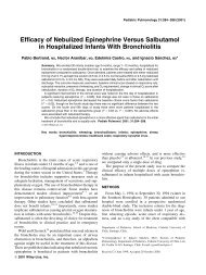
Figure 3 Sleep EEG recorded at the age <strong>of</strong> 5 years showing<br />
frequent sharp waves in the right <strong>occipital</strong> region.<br />
Figure 4 Sleep EEG recorded at the age <strong>of</strong> 5 years showing<br />
frequent sharp waves in the right <strong>occipital</strong> region.<br />
EEG and MRI findings (Table 4 and Figs. 3—5)<br />
Interictal EEGs showed <strong>occipital</strong> or parieto-<strong>occipital</strong> spikes<br />
in all patients (Figs. 3 and 4) during infancy and early childhood.<br />
Frequency and amplitude <strong>of</strong> spikes were gradually<br />
decreased and disappeared in 1 patient (Patient 3). Frontal<br />
spikes as well as <strong>occipital</strong> spikes were also seen in 1 patient<br />
(Patient 2). Frontal or central spikes were seen in late childhood<br />
or adolescence in 3 patients (Patients 2—4), while their<br />
clinical seizure symptoms went unchanged.<br />
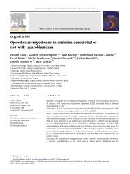
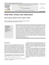
MRI revealed cortical atrophy and T2 prolongation in the<br />
parieto-<strong>occipital</strong> region in 4 patients, <strong>of</strong> whom additional<br />
frontal lesions were present in 3 patients (Figs. 2, 3 and 5).<br />
One patient (Patient 6) showed hippocampal atrophy on both<br />
sides without temporal <strong>lobe</strong> seizures. MRI was unremarkable<br />
in 1 patient (Patient 4).<br />
Discussion<br />
It is well known that hypoglycemic brain injury predominantly<br />
involves the <strong>occipital</strong> <strong>lobe</strong> (Murakmi et al., 1999;<br />
Caraballo et al., 2004; Filan et al., 2006; Yalnizoglu and<br />
Halioglu, 2007). We reported on 6 patients who had a history<br />
<strong>of</strong> neonatal hypoglycemia and <strong>occipital</strong> <strong>lobe</strong> <strong>epilepsy</strong><br />
later on. Our patients had ictal symptoms compatible with<br />
<strong>occipital</strong> <strong>lobe</strong> <strong>epilepsy</strong>, such as eye deviation, eye blinking,<br />
staring, visual hallucinations, and ictal vomiting (Williamson<br />
et al., 1992). Interictal EEGs showed spike waves in the<br />
posterior region during the active periods <strong>of</strong> <strong>epilepsy</strong> in all<br />
patients. MRI also revealed posterior cerebral damage in 4<br />
patients. These findings indicate that epileptic foci <strong>of</strong> the<br />
patients could mainly be caused by neonatal hypoglycemia.<br />
Although 2 patients (Patients 4 and 6) could not be identified<br />
with posterior cerebral lesion on MRI, they showed<br />
typical clinical symptoms and EEG findings for <strong>occipital</strong><br />
<strong>lobe</strong> <strong>epilepsy</strong>. They could not be diagnosed as having idiopathic<br />
childhood <strong>occipital</strong> <strong>epilepsy</strong> coincidentally, because<br />
the onset and clinical course <strong>of</strong> seizures were not compatible<br />
with those <strong>of</strong> Panayiotopoulos syndrome or Gastaut type.<br />
They may have had very mild <strong>occipital</strong> lesions which could<br />
not be recognized on MRI.<br />
Perinatal hypoxic-ischemic encephalopathy in <strong>term</strong><br />
infants also causes parieto-<strong>occipital</strong> <strong>lobe</strong> <strong>epilepsy</strong> (Gil-Nagel<br />
et al., 2005; Oguni et al., 2008). It is sometimes difficult to<br />
distinguish between perinatal hypoxic-ischemic brain injury<br />
and neonatal hypoglycemic brain injury in clinical symptoms<br />
and neuroimaging findings. The perinatal hypoxic-ischemic<br />
brain injury in <strong>term</strong> infants usually involves the deeper sulcal<br />
portion <strong>of</strong> the convolutions and spares the crowns, so<br />
called ‘‘mushroom gyri’’ or ‘‘ulegyria’’. The brain lesions<br />
are located in parasagittal watershed vascular territories,<br />
such as frontal and parieto-<strong>occipital</strong> <strong>lobe</strong>s. It is usually more<br />
marked in the posterior regions at the watershed zone <strong>of</strong><br />
the 3 major cerebral arteries. Perinatal hypoxic-ischemic<br />
encephalopathy in <strong>term</strong> infants usually causes moderate to<br />
severe neurological deficit, including mental or developmental<br />
delay, motor palsy, visual impairment, and <strong>epilepsy</strong>,<br />
while milder cases have been reported (Oguni et al., 2008).<br />
Although EEG abnormalities and epileptic foci may be multifocal<br />
or widespread, parieto-<strong>occipital</strong> <strong>lobe</strong> <strong>epilepsy</strong> may be<br />
common. These two conditions sometimes coexist simultaneously<br />
and would enhance each other, creating more brain<br />
injury. In the series <strong>of</strong> Oguni et al. (2008), 3 <strong>of</strong> the 10<br />
patients with parieto-<strong>occipital</strong> <strong>lobe</strong> <strong>epilepsy</strong> as sequela <strong>of</strong><br />
perinatal asphyxia were reported to have hypoglycemia in<br />
the neonatal period. In the reports <strong>of</strong> neonatal hypoglycemic<br />
encephalopathy, other associated factors including perinatal<br />
asphyxia and fetal distress were highly present (Murakmi<br />
et al., 1999; Yalnizoglu and Halioglu, 2007; Montassir et<br />
al., 2009). In the present study, additional perinatal factors<br />
which were possibly associated with brain injury other than<br />
hypoglycemia were highly present; low birth weight for gestational<br />
age in 5, low Apgar score in 3, neonatal seizure in 5.<br />
MRI demonstrated abnormalities in the frontal <strong>lobe</strong> as well<br />
as the <strong>occipital</strong> <strong>lobe</strong> in 3 patients (Patients 2, 3, and 5). MRI<br />
<strong>of</strong> Patients 2 and 5 showed ‘‘mushroom gyri’’ or ‘‘ulegyria’’<br />
in the posterior regions. These findings indicate that not<br />
only hypoglycemia but also perinatal hypoxia or ischemia<br />
played an important role in the genesis <strong>of</strong> brain injury. The<br />
vulnerability <strong>of</strong> the brain to hypoglycemia is enhanced by<br />
concomitant other perinatal factors. In <strong>term</strong> infants, therefore,<br />
hypoglycemic encephalopathy and hypoxic-ischemic<br />
encephalopathy <strong>of</strong>ten overlap and sometimes cannot be distinguishable<br />
in practice. There may be some difference in<br />
response to treatment; epileptic patients with perinatal
98 H. Montassir et al.<br />
Figure 5 A: patient 1, B: patient 2, C: patient 3, and D: patient 5. MRI images show parieto-<strong>occipital</strong> atrophies in both sides and<br />
white matter T2 prolongation bilatrally. CBZ, carbamazepine.<br />
hypoxia-ischemia <strong>of</strong>ten proved refractory to treatment but<br />
those with neonatal hypoglycemia showed relatively good<br />
prognoses (Villani et al., 2003; Caraballo et al., 2004; Gil-<br />
Nagel et al., 2005; Oguni et al., 2008). The present study<br />
agreed with this finding.<br />
Although the prevalence <strong>of</strong> <strong>epilepsy</strong> in neonatal hypoglycemic<br />
encephalopathy was greater than 50% (Murakmi<br />
et al., 1999; Alkalay et al., 2005; Yalnizoglu and Halioglu,<br />
2007), the characteristics and long-<strong>term</strong> <strong>prognosis</strong> were<br />
not well known. Norden (2001) described 4 patients with<br />
an epileptic syndrome secondary to neonatal hypoglycemia<br />
characterized by complex focal seizures originating from<br />
the <strong>occipital</strong> <strong>lobe</strong>s, with magnetic resonance imaging<br />
evidence <strong>of</strong> atrophic changes in the <strong>occipital</strong> cortices.<br />
Seizures were refractory to antiepileptic drugs, and individuals<br />
experienced pervasive delays in development. In<br />
the series <strong>of</strong> Caraballo et al. (2004), on the other hand,<br />
most patients with <strong>occipital</strong> <strong>lobe</strong> <strong>epilepsy</strong> had good prognoses.<br />
In the present study we found that <strong>symptomatic</strong><br />
<strong>occipital</strong> <strong>lobe</strong> <strong>epilepsy</strong> secondary to neonatal hypoglycemia<br />
was intractable and possessed quite a high risk <strong>of</strong> status<br />
epilepticus in infancy and early childhood but remitted in<br />
late childhood and adolescence.<br />
It is unclear if <strong>epilepsy</strong> following neonatal hypoglycemia<br />
has pharmaco-resistance and develops status epilepticus in<br />
the early course <strong>of</strong> the disease. Panayiotopoulos syndrome,<br />
early onset benign childhood <strong>occipital</strong> <strong>epilepsy</strong>, shows similar<br />
ictal symptoms and interictal EEG findings to the present<br />
patients, such as nocturnal seizures, ictal symptoms <strong>of</strong><br />
eye deviation and vomiting, liability to status epilepticus,<br />
<strong>occipital</strong> spikes, and high remission in late childhood. In<br />
the etiological study <strong>of</strong> status epilepticus in childhood,<br />
Panayiotopoulos syndrome and <strong>symptomatic</strong> <strong>occipital</strong> <strong>lobe</strong><br />
<strong>epilepsy</strong> secondary to neonatal hypoglycemia were the<br />
major cause and highest recurrence risk for status epilepticus<br />
(Okanishi et al., 2008). Therefore, epileptogenecity<br />
in the <strong>occipital</strong> <strong>lobe</strong> may be important for prolongation <strong>of</strong><br />
seizure activity in infancy.
<strong>Long</strong>itudinal clinical course 99<br />
The posterior cerebral predilection <strong>of</strong> neonatal hypoglycemic<br />
encephalopathy has been demonstrated by<br />
computed tomography and magnetic resonance imaging<br />
(Chiu, 1998; Traill et al., 1998; Kinnala et al., 1999). The<br />
possible reasons for the pattern <strong>of</strong> damage may relate to the<br />
high degree <strong>of</strong> axonal migration and synaptogenesis occurring<br />
within the <strong>occipital</strong> <strong>lobe</strong> during the neonatal period and<br />
the development <strong>of</strong> receptors for excitatory amino acids<br />
which can be stimulated by elevated levels <strong>of</strong> aspartate.<br />
This last mechanism could result in the selective death <strong>of</strong><br />
the postsynaptic neurons. It has also been hypothesized that<br />
high metabolic turnover during early life predisposes this<br />
area to injury as occurs in mitochondrial diseases (Dvorkin<br />
et al., 1987).<br />
A few limitations <strong>of</strong> this study can now be examined.<br />
First, the small number <strong>of</strong> patients may affect the accuracy<br />
<strong>of</strong> the results. Further studies with larger numbers<br />
are needed. Second, <strong>occipital</strong> <strong>lobe</strong> <strong>epilepsy</strong> was based on<br />
seizure semiology and interictal EEG. The possibility <strong>of</strong><br />
frontal or temporal <strong>lobe</strong> <strong>epilepsy</strong> cannot be completely<br />
denied in patients with extra-<strong>occipital</strong> <strong>lobe</strong> abnormality.<br />
Third, <strong>occipital</strong> <strong>lobe</strong> injuries could not be identified on MRI<br />
in 2 patients. Brain injuries could be subtle, while nevertheless<br />
we cannot deny the possibility <strong>of</strong> minor focal cortical<br />
dysplasia.<br />
In <strong>occipital</strong> <strong>lobe</strong> <strong>epilepsy</strong> in infancy and early childhood,<br />
neonatal hypoglycemia should be considered as an important<br />
possible etiology. Seizures are liable to be pharmacoresistant<br />
and develop status epileptics in infancy and early<br />
childhood, while they mostly decrease and are shortened in<br />
late childhood and adolescence.<br />
References<br />
Alkalay, A.l., Flares-Sarnat, L., Sarnat, H.B., Moser, F.G., Simmons,<br />
C.F., 2005. Brain imaging findincs in neonatal hypoglycemia: case<br />
report and review <strong>of</strong> 23 cases. Clin. Pediatr. (Phila) 44, 783—<br />
790.<br />
Caraballo, R.H., Sakr, D., Mozzi, M., Guerrero, A., Adi, J.N., Cersósimo,<br />
R.O., Fejerman, N., 2004. Symptomatic <strong>occipital</strong> <strong>lobe</strong><br />
<strong>epilepsy</strong> following neonatal hypoglycemia. Pediatr. Neurol. 31,<br />
24—29.<br />
Chiu, N.T., 1998. Technetium-99m-HMPAO brain SPECT in<br />
neonates with hypoglycemic encephalopathy. J. Nucl. Med. 39,<br />
1711—1713.<br />
Dvorkin, G.S., Andermann, F., Carpenter, S., Melanson, S., Verret,<br />
S., Jacob, C., Sherwin, A., Bekhor, S., Lugaresi, E., Sackellares,<br />
C., Willoughby, J., MacGregor, D., 1987. Classical migraine,<br />
intractable <strong>epilepsy</strong> and multiple strokes: a syndrome related to<br />
mitochondrial encephalomyopathy. In: Andermann, F., Lugaresi,<br />
E. (Eds.), Migraine and Epilepsy. Butterworths, London, pp.<br />
203—232.<br />
Filan, P.M., Inder, T.E., Cameron, F.J., Kean, M.J., Hunt, R.W., 2006.<br />
Neonatal hypoglycemia and <strong>occipital</strong> cerebral injury. J. Pediatr.<br />
148, 552—555.<br />
Gil-Nagel, A., García Morales, I., Jiménez Huete, A., Alvarez Linera,<br />
J., del Barrio, A., Ruiz Ocaña, C., Muñoz, D.G., 2005. Occipital<br />
<strong>lobe</strong> <strong>epilepsy</strong> secondary to ulegyria. J. Neurol. 252, 1178—1185.<br />
Kinnala, A., Rikalainen, H., Lapinleimu, H., Parkkola, R., Kormano,<br />
M., Kero, P., 1999. Cerebral magnetic resonance imaging<br />
and ultrasonography findings after neonatal hypoglycemia. Pediatrics<br />
103, 724—729.<br />
Montassir, H., Maegaki, Y., Ogura, K., Kurozawa, Y., Nagata, I.,<br />
Kanzaki, S., Ohno, K., 2009. Associated factors in neonatal hypoglycemic<br />
brain injury. Brain Dev. 31, 649—656.<br />
Murakmi, Y., Yamashita, Y., Matsuishi, T., Utsunomiya, T., Okudera,<br />
T., Hashimoto, T., 1999. Cranial MRI <strong>of</strong> neurologically impaired<br />
children suffering from neonatal hypoglycemia. Pediatr. Radiol.<br />
29, 23—27.<br />
Norden, A.D., 2001. A novel <strong>epilepsy</strong> syndrome secondary to neonatal<br />
hypoglycemia. Epilepsia 42 (Suppl. 7), S50.<br />
Oguni, H., Sugama, M., Osawa, M., 2008. Symptomatic parieto<strong>occipital</strong><br />
<strong>epilepsy</strong> as sequel <strong>of</strong> perinatal asphyxia. Pediatr.<br />
Neurol. 38, 345—352.<br />
Okanishi, T., Maegaki, Y., Ohno, K., Togari, H., 2008. Underlying<br />
neurologic disorders and recurrence rates <strong>of</strong> status epilepticus<br />
in childhood. Brain Dev. 30, 624—628.<br />
Pisani, F., Orsini, M., Braibanti, S., Copioli, C., Sisti, L., Turco,<br />
E.C., 2009. Development <strong>of</strong> <strong>epilepsy</strong> in newborn with moderate<br />
hypoxic-ischemic encephalopathy and neonatal seizures. Brain<br />
Dev. 31, 64—68.<br />
Srinivasan, G., Pildes, R.S., Cattamanchi, G., Voora, S., Lilien, L.D.,<br />
1986. Plasma glucose values in normal neonates: a new look. J.<br />
Pediatr. 109, 114—117.<br />
Traill, Z., Squier, M., Anslow, P., 1998. Brain imaging in neonatal<br />
hypoglycemia. Arch. Dis. Child. Fetal Neonatal Ed. 79,<br />
F145—F147.<br />
Villani, F., D’Incerti, L., Granata, T., Battaglia, G., Vitali, P., Chiapparini,<br />
L., Avanzini, G., 2003. Epileptic and imaging findings<br />
in perinatal hypoxic-ischemic encephalopathy with ulegyria.<br />
Epilepsy Res. 55, 235—243.<br />
Williamson, P.D., Thadani, V.M., Darcey, T.M., Spencer, D.D.,<br />
Spencer, S.S., Mattson, R.H., 1992. Occipital <strong>lobe</strong> <strong>epilepsy</strong>:<br />
clinical characteristics, seizure spread patterns, and results <strong>of</strong><br />
surgery. Ann. Neurol. 31, 3—13.<br />
Yalnizoglu, D., Halioglu, G., 2007. Neurological outcome in patients<br />
with MRI pattern <strong>of</strong> damage typical for neonatl hypoglycemia.<br />
Brain Dev. 29, 285—292.<br />
Zafeiriou, D.I., Kontopoulos, E.E., Tsikoulas, I., 1999. Characteristics<br />
and <strong>prognosis</strong> <strong>of</strong> <strong>epilepsy</strong> in children with cerebral palsy. J.<br />
Child Neurol. 14, 289—294.