UK retinopathy of prematurity guideline - sepeap
UK retinopathy of prematurity guideline - sepeap
UK retinopathy of prematurity guideline - sepeap

Create successful ePaper yourself
Turn your PDF publications into a flip-book with our unique Google optimized e-Paper software.
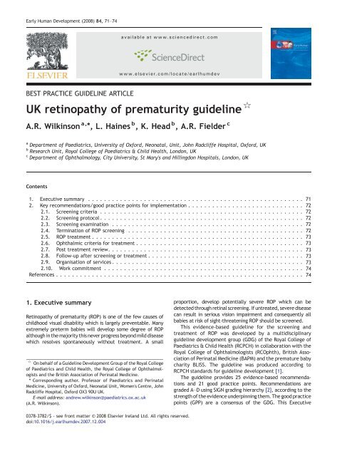
Early Human Development (2008) 84, 71–74<br />
available at www.sciencedirect.com<br />
www.elsevier.com/locate/earlhumdev<br />
BEST PRACTICE GUIDELINE ARTICLE<br />
<strong>UK</strong> <strong>retinopathy</strong> <strong>of</strong> <strong>prematurity</strong> <strong>guideline</strong> ☆<br />
A.R. Wilkinson a, ⁎, L. Haines b , K. Head b , A.R. Fielder c<br />
a Department <strong>of</strong> Paediatrics, University <strong>of</strong> Oxford, Neonatal, Unit, John Radcliffe Hospital, Oxford, <strong>UK</strong><br />
b Research Unit, Royal College <strong>of</strong> Paediatrics & Child Health, London, <strong>UK</strong><br />
c Department <strong>of</strong> Ophthalmology, City University, St Mary's and Hillingdon Hospitals, London, <strong>UK</strong><br />
Contents<br />
1. Executive summary ...................................................... 71<br />
2. Key recommendations/good practice points for implementation . . . .......................... 72<br />
2.1. Screening criteria ................................................... 72<br />
2.2. Screening protocol ................................................... 72<br />
2.3. Screening examination ................................................ 72<br />
2.4. Termination <strong>of</strong> ROP screening ............................................ 73 72<br />
2.5. ROP treatment ..................................................... 73<br />
2.6. Ophthalmic criteria for treatment .......................................... 73<br />
2.7. Post treatment review. ................................................ 74 73<br />
2.8. Follow-up after screening or treatment ....................................... 74 73<br />
2.9. Organisation <strong>of</strong> services ................................................. 73<br />
2.10. Work commitment ................................................... 74<br />
References .............................................................. 74<br />
1. Executive summary<br />
Retinopathy <strong>of</strong> <strong>prematurity</strong> (ROP) is one <strong>of</strong> the few causes <strong>of</strong><br />
childhood visual disability which is largely preventable. Many<br />
extremely preterm babies will develop some degree <strong>of</strong> ROP<br />
although in the majority this never progress beyond mild disease<br />
which resolves spontaneously without treatment. A small<br />
☆ On behalf <strong>of</strong> a Guideline Development Group <strong>of</strong> the Royal College<br />
<strong>of</strong> Paediatrics and Child Health, the Royal College <strong>of</strong> Ophthalmologists<br />
and the British Association <strong>of</strong> Perinatal Medicine.<br />
⁎ Corresponding author. Pr<strong>of</strong>essor <strong>of</strong> Paediatrics and Perinatal<br />
Medicine, University <strong>of</strong> Oxford, Neonatal Unit, Women's Centre, John<br />
Radcliffe Hospital, Oxford OX3 9DU <strong>UK</strong>.<br />
E-mail address: andrew.wilkinson@paediatrics.ox.ac.uk<br />
(A.R. Wilkinson).<br />
proportion, develop potentially severe ROP which can be<br />
detected through retinal screening. If untreated, severe disease<br />
can result in serious vision impairment and consequently all<br />
babies at risk <strong>of</strong> sight-threatening ROP should be screened.<br />
This evidence-based <strong>guideline</strong> for the screening and<br />
treatment <strong>of</strong> ROP was developed by a multidisciplinary<br />
<strong>guideline</strong> development group (GDG) <strong>of</strong> the Royal College <strong>of</strong><br />
Paediatrics & Child Health (RCPCH) in collaboration with the<br />
Royal College <strong>of</strong> Ophthalmologists (RCOphth), British Association<br />
<strong>of</strong> Perinatal Medicine (BAPM) and the premature baby<br />
charity BLISS. The <strong>guideline</strong> was produced according to<br />
RCPCH standards for <strong>guideline</strong> development [1].<br />
The <strong>guideline</strong> provides 25 evidence-based recommendations<br />
and 21 good practice points. Recommendations are<br />
graded A–D using SIGN grading hierarchy [2], according to the<br />
strength <strong>of</strong> the evidence underpinning them. The good practice<br />
points (GPP) are a consensus <strong>of</strong> the GDG. This Executive<br />
0378-3782/$ - see front matter © 2008 Elsevier Ireland Ltd. All rights reserved.<br />
doi:10.1016/j.earlhumdev.2007.12.004
72 A.R. Wilkinson et al.<br />
Summary highlights those recommendations and good<br />
practice points considered by the GDG to be priorities for<br />
implementation.<br />
This <strong>guideline</strong> has been produced specifically for use within<br />
the <strong>UK</strong> and supersedes the previous <strong>guideline</strong> [3].Itwillnotbe<br />
applicable in countries where more mature babies are at risk <strong>of</strong><br />
sight threatening ROP [4].<br />
Not all the recommendations are included in this<br />
Summary. The full Guideline should be consulted which also<br />
contains complete details <strong>of</strong> the Guideline methodology.<br />
Appendices A, B, C and D give a standardised sheet for<br />
recording screening results, an algorithm for ophthalmic<br />
criteria for screening and treatment, the International<br />
Classification <strong>of</strong> ROP Revisited [5], and parent information<br />
leaflets respectively. All the documents are available on the<br />
websites <strong>of</strong> the Royal College <strong>of</strong> Ophthalmologists www.<br />
rcophth.ac.uk, the Royal College <strong>of</strong> Paediatrics and Child<br />
Health www.rcpch.ac.uk or the British Association <strong>of</strong> Perinatal<br />
Medicine www.bapm.org.<br />
2. Key recommendations/good practice points<br />
for implementation<br />
2.1. Screening criteria<br />
■ All babies less than 32 weeks gestational age<br />
(up to 31 weeks and 6 days) or less than 1501 g<br />
birthweight should be screened for ROP. One<br />
criterion to be met for inclusion.<br />
■ All babies less than 31 weeks gestational age<br />
(up to 30 weeks and 6 days) or less than 1251 g<br />
birthweight must be screened for ROP. One<br />
criterion to be met for inclusion.<br />
2.2. Screening protocol<br />
GPP<br />
■ Babies born before 27 weeks gestational age<br />
B<br />
(i.e. up to 26 weeks and 6 days) — the first ROP<br />
screening examination should be undertaken at<br />
30 to 31 weeks postmenstrual age.<br />
■ Babies born between 27 and 32 weeks gestational B<br />
age (i.e. up to 31 weeks and 6 days) — the first<br />
ROP screening examination should be undertaken<br />
between 4 to 5 weeks (i.e. 28–35 days) postnatal age.<br />
■ Babies N32 weeks gestational age but with birthweight B<br />
b1501 grams — the first ROP screening examination<br />
should be undertaken between 4 to 5 weeks<br />
(i.e. 28–35 days) postnatal age.<br />
■ Minimum frequencies <strong>of</strong> screening should be weekly when: B<br />
■ the vessels end in zone I or posterior zone II; or<br />
■ there is any plus or pre-plus disease or<br />
■ there is any stage 3 disease in any zone<br />
■ Minimum frequencies <strong>of</strong> screening should be every 2 weeks: D<br />
■ In all other circumstances until the criteria for<br />
termination have been reached<br />
■ All babies b32 weeks gestational age or birthweight D<br />
b1501 g should have their first ROP screening<br />
examination prior to discharge.<br />
B<br />
Although screening for all babies at risk should follow the<br />
above protocol, it is acknowledged that there may be clinical<br />
or organisational circumstances which prevent this. In these<br />
circumstances the following is recommended as good<br />
practice to ensure that subsequent screening examinations<br />
are not missed.<br />
■ Where a decision is made not to screen a baby,<br />
the reasons for doing so should be clearly stated<br />
in the baby's medical record and the examination<br />
should be rescheduled within one week <strong>of</strong> the<br />
intended examination.<br />
2.3. Screening examination<br />
GPP<br />
The screening examination can be stressful for both babies and<br />
parents. The full <strong>guideline</strong> gives recommendations on preparation<br />
and care <strong>of</strong> the baby. The examination requires a<br />
well-dilated pupil so the peripheral retina can be fully<br />
visualised. The following are key recommendations and good<br />
practice points for this area.<br />
■ In addition to oral communication, parents should<br />
be given written information about the screening<br />
process prior to the first examination <strong>of</strong> their baby.<br />
■ It is important that the periphery <strong>of</strong> the retina can<br />
be seen and this may be facilitated by the use <strong>of</strong><br />
an eyelid speculum and scleral indentor suitable<br />
for neonatal use.<br />
■ Ophthalmological notes should be made after each<br />
ROP examination, detailing zone, stage, and extent<br />
in terms <strong>of</strong> clock hours <strong>of</strong> any ROP and the presence<br />
<strong>of</strong> any pre-plus or plus disease. These notes should<br />
include a recommendation for the timing <strong>of</strong> the<br />
next examination (if any) and be kept with the<br />
baby's medical record.<br />
■ Comfort care techniques (e.g. administering sucrose<br />
solution, nesting, swaddling and/or the use <strong>of</strong> a pacifier)<br />
during the screening examination may be considered.<br />
2.4. Termination <strong>of</strong> ROP screening<br />
GPP<br />
B<br />
GPP<br />
Screening can be stopped when a baby is no longer at risk <strong>of</strong><br />
sight-threatening ROP.<br />
In babies who never develop any ROP, the risk <strong>of</strong> sightthreatening<br />
ROP developing is minimal once the retinal<br />
vessels have entered zone III. That vessels are in zone III can<br />
be difficult to determine, but it is unlikely to occur before<br />
37 weeks postmenstrual age and a decision to stop screening<br />
before this must be carefully evaluated.<br />
■ In babies without ROP, there is minimal risk <strong>of</strong><br />
developing sight threatening ROP when vascularisation<br />
has extended into zone III and eye examinations may be<br />
stopped when this happens, usually after 36 completed<br />
weeks postmenstrual age.<br />
In babies developing ROP which does not meet the criteria<br />
for treatment, screening can be safely stopped when there<br />
B<br />
B
<strong>UK</strong> <strong>retinopathy</strong> <strong>of</strong> <strong>prematurity</strong> <strong>guideline</strong><br />
73<br />
are clear signs that the active progression <strong>of</strong> ROP has halted<br />
and regression has commenced.<br />
■ In the presence <strong>of</strong> ROP, screening for progressive active<br />
disease may be discontinued when any <strong>of</strong> the following<br />
characteristics <strong>of</strong> regression are seen on at least 2<br />
successive examinations:<br />
■ lack <strong>of</strong> increase in severity<br />
■ partial resolution progressing towards complete<br />
resolution<br />
■ change in colour in the ridge from salmon pink to white<br />
■ transgression <strong>of</strong> vessels through the demarcation line<br />
■ commencement <strong>of</strong> the process <strong>of</strong> replacement <strong>of</strong><br />
active ROP lesions by scar tissue<br />
2.5. ROP treatment<br />
Timely treatment for ROP is effective at preventing severe<br />
vision impairment. Previous guidance recommended treatment<br />
when the disease reached ‘Threshold’, as defined in<br />
section 7 <strong>of</strong> the main document [6]. Recent evidence shows<br />
benefit from earlier treatment.<br />
2.6. Ophthalmic criteria for treatment<br />
D<br />
Severe ROP requiring treatment is relatively infrequent<br />
and treatment is a specialised procedure. Although there is<br />
no research literature on treatment outcomes according to<br />
operator expertise, it is likely that those with the greatest<br />
experience will be the most skilled practitioners in the<br />
procedure.<br />
■ Babies with ROP should be treated by ophthalmologists GPP<br />
who have the appropriate competency.<br />
■ Each network should have identified individuals for GPP<br />
ROP treatment.<br />
2.7. Post treatment review<br />
Post operative review is important to monitor disease regression<br />
and to determine if retreatment is necessary. The GDG<br />
have agreed the following GPP in the absence <strong>of</strong> good quality<br />
evidence to inform these timings.<br />
■ The first examination post treatment should take<br />
place 5-7 days after treatment and should be<br />
continued at least weekly for signs <strong>of</strong> decreasing<br />
activity and regression.<br />
■ Re-treatment should be performed usually 10–14 days<br />
after initial treatment when there has been a failure<br />
<strong>of</strong> the ROP to regress.<br />
GPP<br />
GPP<br />
■ Treatment for ROP should be undertaken if any <strong>of</strong> the<br />
following indications are reached:<br />
■ Zone I, any ROP with plus disease,<br />
■ Zone I, stage 3 without plus disease,<br />
■ Zone II; stage 3 with plus disease.<br />
■ Treatment for ROP should be seriously considered if<br />
the following indication is reached:<br />
■ Zone II, stage 2 with plus disease<br />
B<br />
B<br />
2.8. Follow-up after screening or treatment<br />
■ After the acute phase, eyes that have reached<br />
stage 3 or have been treated should be monitored<br />
at a frequency dictated by the clinical condition<br />
to determine the risk <strong>of</strong> sequelae.<br />
GPP<br />
2.9. Organisation <strong>of</strong> services<br />
Although there is no specific evidence to inform the<br />
interval between reaching treatment criteria and treatment<br />
taking place, it is the view <strong>of</strong> the GDG that, given the<br />
encouraging results for early treatment obtained by treating<br />
within 48 h, this should be the target standard.<br />
■ Babies with aggressive ROP (as defined in ICROP<br />
revisited [5]) should be treated as soon as possible<br />
and within 48 h. ROP requiring treatment but<br />
which is not aggressive posterior ROP should<br />
normally be treated within 48–72 h.<br />
■ Transpupillary diode laser therapy is recommended<br />
as the first line treatment for ROP.<br />
■ Treatment with near-confluent (0.5–1 burn-width)<br />
laser burn spacing should be administered to the<br />
entire avascular retina.<br />
■ The unavailability <strong>of</strong> diode laser equipment or the<br />
inability to transfer to another centre should not<br />
prevent or delay the treatment <strong>of</strong> ROP. In these<br />
situations, treatment with cryotherapy or argon<br />
laser may be completed by an ophthalmologist<br />
experienced in these techniques.<br />
GPP<br />
B<br />
D<br />
GPP<br />
Effective services for ROP screening and treatment must be<br />
embedded in a robust organisational structure, with individual<br />
responsibilities identified. Particular efforts must be made to<br />
ensure that the service is delivered appropriately for all those<br />
at risk, as there is evidence that babies transferred or<br />
discharged home before screening is complete are at risk <strong>of</strong><br />
poor outcomes as a result <strong>of</strong> lack <strong>of</strong> follow-up.<br />
■ All units caring for babies at risk <strong>of</strong> ROP should<br />
have a written protocol in relation to the screening<br />
for, and treatment <strong>of</strong>, ROP. This should include<br />
responsibilities for follow-up <strong>of</strong> babies transferred<br />
or discharged from the unit before screening is<br />
complete, which should be the responsibility <strong>of</strong> the<br />
named consultant Neonatologist for each baby.<br />
■ If babies are transferred either before ROP screening<br />
is initiated or when it has been started but not<br />
completed, it is the responsibility <strong>of</strong> the consultant<br />
neonatologist to ensure that the neonatal team in<br />
the receiving unit is aware <strong>of</strong> the need to start or<br />
continue ROP screening.<br />
GPP<br />
GPP<br />
(continued on next page)
74 A.R. Wilkinson et al.<br />
■ There should be a record <strong>of</strong> all babies who require<br />
review and the arrangements for their follow-up.<br />
■ For babies who meet the ROP screening criteria,<br />
screening status and the need and arrangements<br />
for further screens must be recorded in all transfer<br />
letters so that screening may be continued.<br />
■ For babies discharged home before screening is<br />
complete the first follow-up out-patient appointment<br />
must be made before hospital discharge and the<br />
importance <strong>of</strong> attendance explained to the<br />
parents/carers.<br />
2.10. Work commitment<br />
■ Ophthalmologists regularly completing ROP screening<br />
and/or treatment should have sessional<br />
commitments allocated within their work plan.<br />
GPP<br />
D<br />
D<br />
GPP<br />
References<br />
[1] Standards for Development <strong>of</strong> Clinical Guidelines in Paediatrics<br />
and Child Health. RCPCH; June 2006.<br />
[2] Scottish Intercollegiate Guidelines Network. Sign 50: A Guideline<br />
Developers' Handbook; 2001.<br />
[3] The report <strong>of</strong> a Joint Working Party <strong>of</strong> The Royal College <strong>of</strong><br />
Ophthalmologists and the British Association <strong>of</strong> Perinatal<br />
Medicine. Retinopathy <strong>of</strong> <strong>prematurity</strong>: <strong>guideline</strong>s for screening<br />
and treatment. Early Hum Dev 1996;46(3):239–58.<br />
[4] Gilbert, Fielder A, Gordillo L, Quinn G, Semiglia R, Visintin P,<br />
et al. Characteristics <strong>of</strong> infants with severe <strong>retinopathy</strong> <strong>of</strong><br />
<strong>prematurity</strong> in countries with low, moderate and high levels <strong>of</strong><br />
development: implications for screening programs. Pediatrics<br />
2005;115(5):e518–25.<br />
[5] The International Classification <strong>of</strong> Retinopathy <strong>of</strong> Prematurity<br />
revisited. Arch Ophthalmol 2005;123(7):991–9.<br />
[6] Multicenter trial <strong>of</strong> cryotherapy for <strong>retinopathy</strong> <strong>of</strong> <strong>prematurity</strong>.<br />
Preliminary results. Arch Ophthalmol 1988;106(4):471–9.