Eldor Spinal Needle Versus Pencan Spinal Needle
Eldor Spinal Needle Versus Pencan Spinal Needle
Eldor Spinal Needle Versus Pencan Spinal Needle

Create successful ePaper yourself
Turn your PDF publications into a flip-book with our unique Google optimized e-Paper software.
210 Regional Anesthesia and Pain Medicine Vol. 25 No. 2 March-April 2000<br />
spinal fluid was observed at the needle hub in all. The<br />
injection of local anesthetics was followed by adequate<br />
spinal anesthesia. At 24-hour follow-up none of our<br />
patients complained about headache or back pain.<br />
Only a few studies are available pertaining to this new<br />
spinal needle. Yamazaki et aL l compared the penetration<br />
characteristics of the ball-pen needle with both a pencil point<br />
and a Quincke needle in vitro. They analyzed the pressure<br />
necessary to penetrate a standardized polyethylene membrane.<br />
The puncture with the Quincke needle required a<br />
maximum pressure of 35 g, which decreased after the penetration<br />
of the membrane to 20 g. The pencil-point needle<br />
required 60 g and 40 g, respectively, whereas the ball-pen<br />
needle required 80 g to penetrate followed by an immediate<br />
decrease to 25 g. Yamazaki et al.2 hypothesized that this<br />
sudden decrease of pressure after penetration in vitro would<br />
likely lead to an easier perception of dural puncture in vivo.<br />
This assumption is consistent with our clinical experiences<br />
with the ball-pen needle.<br />
In obese patients, the use of the ball-pen in 25- and<br />
27-gauges, respectively, is limited due to the need for a<br />
relatively long introducer. The company is addressing this<br />
by changing the length of the introducer.<br />
In condusion, the ball-pen needle seems to be an<br />
interesting and promising needle for spinal anesthesia.<br />
Further controlled studies are needed to demonstrate the<br />
potential benefit of this open-end pencil-point needle.<br />
t<br />
Peter Lierz, M.D.<br />
Department of Anaesthesiology and Intensive Care<br />
Marienkrankenhaus Soest<br />
Soest, Germany<br />
Burkhard Gustorff, M.D., D.E.A.A.<br />
Department of Anaesthesia and Intensive Care (B)<br />
University of Vienna<br />
Vienna, Austria<br />
Peter Felleiter, M.D.<br />
Department of Anesthesiology and Intensive Care<br />
Swiss Paraplegic Center<br />
Nottwil, Switzerland<br />
References<br />
1. Yamazaki O, Hukuyama A, Suzuki I', Kinehuchi Y, Itoh K,<br />
Takiguchi M. Study of penetration characteristics of pencil<br />
point type spinal anesthesia needles. JAnestheda 1999; 13:304.<br />
2. Yamazaki O, Rukuyama A, Kinehuc_hi Y, Suzuki 1", TaMguchi<br />
M. Comparative study on puncture resistance of spinal<br />
anesthesia needles. Jpn J Clin Mort 1999; 10:67.<br />
Accepted for publication September 1, 1999.<br />
<strong>Eldor</strong> <strong>Spinal</strong> <strong>Needle</strong><strong>Versus</strong> <strong>Pencan</strong><br />
<strong>Spinal</strong> <strong>Needle</strong><br />
To the Editor:<br />
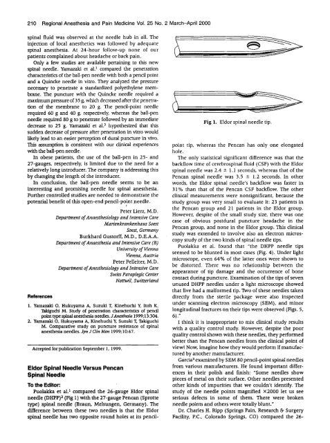
Puolakka et al.l compared the 26-gauge <strong>Eldor</strong> spinal<br />
needle (DHPP) 2 (Fig 1) with the 27-gauge <strong>Pencan</strong> (Sprotte<br />
type) spinal needle (Braun, Melsungen, Germany). The<br />
difference between these two needles is that the <strong>Eldor</strong><br />
spinal needle has two opposite round holes at its pencil-<br />
Fig 1. <strong>Eldor</strong> spinal needle tip.<br />
point tip, whereas the <strong>Pencan</strong> has only one elongated<br />
hole.<br />
The only statistical significant difference was that the<br />
backflow time of cerebrospinal fluid (CSF) with the <strong>Eldor</strong><br />
spinal needle was 2.4 _+ 1.1 seconds, whereas that of the<br />
<strong>Pencan</strong> spinal needle was 3.5 -+ 1.2 seconds. In other<br />
words, the <strong>Eldor</strong> spinal needle's backflow was faster in<br />
31% than that of the <strong>Pencan</strong> CSF backflow. The other<br />
clinical measurements were nonsignificant, because the<br />
study group was very small to evaluate it: 23 patients in<br />
the <strong>Pencan</strong> group and 21 patients in the <strong>Eldor</strong> group.<br />
However, despite of the small study size, there was one<br />
case of obvious postdural puncture headache in the<br />
<strong>Pencan</strong> group, and none in the <strong>Eldor</strong> group. This clinical<br />
study was extended to involve also an electron microscopy<br />
study of the two kinds of spinal needle tips.<br />
Puolakka et al. found that "the DHPP needle tips<br />
seemed to be blunted in most cases (Fig. 4). Under light<br />
microscope, even 64% of the latter ones were shown to<br />
be distorted. There was no relationship between the<br />
appearance of tip damage and the occurrence of bone<br />
contact during puncture. Examination of the tips of seven<br />
unused DHPP needles under a light microscope showed<br />
that five had a malformed tip. Two of these needles taken<br />
directly from the sterile package were also inspected<br />
under scanning electron microscopy (SEM), and minor<br />
longitudinal fractures on their tips were observed (Figs. 5,<br />
6)."<br />
I think it is inappropriate to mix clinical study results<br />
with a quality control study. However, despite the poor<br />
quality control shown with these needles, they performed<br />
better than the <strong>Pencan</strong> needles from the clinical point of<br />
viewl Now, imagine how they would perform if manufactured<br />
by another manufacturer.<br />
Garcia 3 examined by SEM 80 pencil-point spinal needles<br />
from various manufacturers. He found important differences<br />
in their polish and finish: "Some needles show<br />
pieces of metal on their surface. Other needles presented<br />
other kinds of impurities that we couldn't identify. The<br />
study of the needle points magnified x2000 let us see<br />
serious defects in some of them. There were broken<br />
needle points and others were totally blunt."<br />
Dr. Charles H. Ripp (Springs Pain, Research & Surgery<br />
Facility, P.C., Colorado Springs, CO) compared the 26-<br />
J
Letters to the Editor 211<br />
gauge <strong>Eldor</strong> spinal needle (DHPP spinal needle) with the<br />
25-gauge Whitacre needle. The two closed ended spinal<br />
injection needles were compared in regard to their ability<br />
to have injected solution spread immediately following<br />
injection. Each needle was placed in an ice cold water<br />
bath with the temperature measuring 32°1=. Subsequently,<br />
1 mL of 120°F water was injected through a 1-mL TB<br />
syringe rapidly. Baseline and 1 second postinjection infrared<br />
images were obtained on an Inframetric System<br />
(sensitivity to 0.1°F). The temperature of the water<br />
changed as the injected solution dispersed and was graphically<br />
depicted. The green temperature was warmer than<br />
the surrounding blue.<br />
The <strong>Eldor</strong> spinal needle showed a 5-fold increase in the<br />
immediate dispersal area compared with the Whitacre<br />
needle. The <strong>Eldor</strong> spinal needle showed greater immediate<br />
dispersal of injected solution, despite being a smaller<br />
gauge. This advantage can improve anesthetic spread,<br />
provide optimal anesthesia, and lessen the risk of local<br />
anesthetic toxicity.<br />
Gaynes Labs, Inc. (Bridgeview, It.) performed axial<br />
compression tests on the 26-gauge <strong>Eldor</strong> spinal needle<br />
and the 26-gauge Gertie Marx spinal needle. An individual<br />
needle (with its styler in place) was clamped in a<br />
holding fixture (two metal plates with an alignment<br />
groove). Three millimeters, as measured from the tip of<br />
the needle, was exposed outside of the clamp. The end<br />
opposite the tip was bent at a 90 ° angle over the end of the<br />
fixture so that the styler would remain in place. The<br />
needle and holding fixture was then mounted vertically<br />
in the jaws of the testing machine, with the tip of the<br />
needle pointing downward. The jaws were connected to a<br />
force measuring device, which was in turn connected to<br />
the movable ram of the testing machine. The needle<br />
advanced downward at a rate of 0.2 inches per minute,<br />
onto a hardened steel block, until the tip of the needle<br />
bent to a 90 ° angle. A data acquisition system recorded<br />
the compressive force (in pounds) that was being applied<br />
axially to the needle. The maximum compressive force<br />
that occurred during the test was recorded.<br />
The maximum force needed to bend the <strong>Eldor</strong> spinal<br />
needle was 9.65 lb. compared with the maximum force of<br />
9.I6 lb. needed to bend the Gertie Marx needle. Despite<br />
the fact that the <strong>Eldor</strong> needle has two holes at the tip<br />
compared with one hole of the Gertie Marx needle, the<br />
<strong>Eldor</strong> spinal needle is stronger than the Gertie Marx<br />
needle of the same gauge.<br />
Dr. Timo A.R. Palas from the department of Anesthesiology,<br />
Regionalspital Biel, 2502 Biel, Switzerland (1997<br />
Annual Meeting of the American Society of Regional<br />
Anesthesia, Atlanta Hilton Hotel, Atlanta, Georgia, April<br />
10-13, 1997) compared the 27-gauge <strong>Eldor</strong> spinal needle<br />
with the normal one-hole, pencil-point 27-gauge spinal<br />
needle (<strong>Pencan</strong>). Twenty patients were divided into two<br />
groups. The lumbar puncture was performed in the sitting<br />
position with the holes of the needles pointing upwards.<br />
Either plain 2 % prflocaine or plain 0.5 % bupivacaine was<br />
used. The mean age in both groups was 28 years (4 males<br />
and 6 females in each group). The backflow of the CSF<br />
could be seen in a mean time of 0.6 seconds in the <strong>Eldor</strong><br />
spinal needle compared with 2.1 seconds in the <strong>Pencan</strong><br />
needle. There were five cases of an anesthetic maldistribution<br />
in the <strong>Pencan</strong> group during the first 5 minutes after<br />
injection and none in the <strong>Eldor</strong> spinal needle group.<br />
Based on these studies and on the study by Puolakka et<br />
al., the conclusion reached by Puolakka et al. that "the<br />
present study has indicated that most of the DHPP needles<br />
are damaged in the manufacturing process thus making<br />
them less appealing for clinical use and probably more<br />
vulnerable to distortion during lumbar puncture. At the<br />
present, therefore, the SHPP needle seems preferable" is<br />
unfounded. We have already changed the <strong>Eldor</strong> spinal<br />
needle manufacturer. We didn't change our design.<br />
References<br />
Joseph <strong>Eldor</strong>, M.D.<br />
Department of Anesthesia,<br />
Misgav Ladach General Hospital<br />
Jerusalem, Israel<br />
1. Puolakka R, Haasio J, Rosenberg PH, Tuominen M. Comparison<br />
of double-hole and single-hole pencil-point needles<br />
for spinal anesthesia with hyperbaric bupivacaine. Reg<br />
Anesth Pain Med 1998;23:271-277.<br />
2. <strong>Eldor</strong> J. Double-hole pencil-point spinal needle. Reg Anesth<br />
1996;21:74-75.<br />
3. Garcia AL. A scanning electron microscopy and analysis of the<br />
latest spinal needle design. Highlights in regional anaesthesia<br />
and pain therapy. IV. The European Society of Regional<br />
Anaesthesia, Permanyer Publications, 1995; 115-118.<br />
Accepted for publication May 11, 1999.<br />
Acute Aphesia Following Tourniquet Release<br />
in Intravenous Regional Anesthesia With 0.75%<br />
Lidocaine<br />
To the Editor:<br />
I would like to report a case of acute aphesia following<br />
tourniquet release in intravenous regional anesthesia<br />
with 0.75% lidocaine.<br />
The patient was a 56-year-old man, weighing 68 kg,<br />
who was scheduled for removal of ganglion cyst over left<br />
wrist region under i.v. regional anesthesia (IVRA). The<br />
patient had no history of any systemic disease. Under the<br />
monitoring of electrocardiography, noninvasive blood<br />
pressure, and pulse oximetry, a standard procedure of<br />
IVRA with double-cuff tourniquet proceeded. Twenty<br />
milliliters of 0.75% lidocaine was administered. The<br />
operation time was 20 minutes, and no sedative drug was<br />
given. Acute aphesia was noted after the release of<br />
tourniquet. The vital signs were stable in the posttoumiquet<br />
release period. There were no symptoms or signs of<br />
circumoral numbness, visual or auditory disturbance, or<br />
loss of consciousness. He did complain.of lightheadness.<br />
His brain computed tomography scan revealed negative<br />
findings. Fortunately, the patient's speaking ability recovered<br />
20 hours later. No sequela was noted during the 3<br />
days of follow-up.<br />
This is a clinical report of acute aphesia following IVRA<br />
lidocaine administration. Aphesia refers to those motor or