Download - Society for General Microbiology
Download - Society for General Microbiology
Download - Society for General Microbiology

Create successful ePaper yourself
Turn your PDF publications into a flip-book with our unique Google optimized e-Paper software.
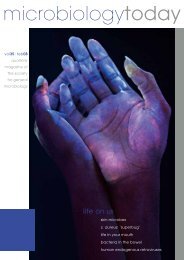
microbiologytoday<br />
vol35|nov08<br />
quarterly<br />
magazine of<br />
the society<br />
<strong>for</strong> general<br />
microbiology<br />
bugs on bugs<br />
microbial diseases of bees<br />
fungal farmers of the insect world<br />
wolbachia and gene transfer<br />
shedding light on photorhabdus<br />
an inside job – bdellovibrio<br />
nature’s experiment – bacteriophages
contents<br />
vol35(4)<br />
regular features<br />
159 News 202 Schoolzone 212 Hot off the Press<br />
166 Microshorts 206 Gradline 215 Going Public<br />
199 Conferences 209 Addresses 218 Reviews<br />
other items<br />
articles<br />
168 Microbial diseases of bees<br />
Travis Glare & Maureen O’Callaghan<br />
Bees play an essential role in the world’s ecosystems, but<br />
microbial diseases are posing a big threat to these vital<br />
insects.<br />
196 National Subject Profiles<br />
210 Obituary – Professor Chris Thurston<br />
184 An inside job: Bdellovibrio<br />
bacteriovorus<br />
Liz Sockett<br />
These predatory bacteria hunt down and eat their fellow<br />
organisms.<br />
172<br />
Ancient fungal farmers<br />
of the insect world<br />
Garret Suen & Cameron<br />
Currie<br />
Leaf-cutter ants not only grow fungi to eat. They<br />
weed their ‘gardens’ and apply pesticides too.<br />
176 Bacterial sequences in an<br />
invertebrate genome<br />
Julie Dunning Hotopp & Jason Rasgon<br />
Some arthropods and nematodes need their bacterial<br />
inhabitants to survive.<br />
180 Photorhabdus: shedding light<br />
on symbioses<br />
Susan Joyce and David Clarke<br />
Which amazing microbe can make nematodes glow in the<br />
dark and yet kill certain insects?<br />
188 Bacteriophages: nature’s most<br />
successful experiment<br />
Graham Hatfull<br />
Phages could well be the world’s biggest reservoir of<br />
unidentified genetic material.<br />
192<br />
1983: a vintage year <strong>for</strong><br />
pathogen discovery<br />
Robin Weiss<br />
Important findings about three major infectious<br />
diseases were made 25 years ago.<br />
220 Comment:<br />
Scotoma in contemporary<br />
microbiology<br />
Howard Gest<br />
Are some bacteria really ‘unculturable’? Probably not<br />
according to this writer.<br />
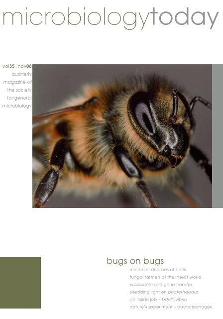
Cover image Macrophotograph of the head of a worker honey bee (Apis mellifera). Dr Jeremy Burgess / Science Photo Library<br />
Editor Dr Matt Hutchings––Editorial Board Dr Sue Assinder, Dr Paul Hoskisson, Professor Bert Rima––Managing Editor Janet Hurst––Assistant Editors Lucy Goodchild & Faye Stokes<br />
Editorial Assistant Yvonne Taylor––Design & Production Ian Atherton––Contributions are always welcome and should be addressed to the Editor c/o SGM HQ, Marlborough House,<br />
Basingstoke Road, Spencers Wood, Reading RG7 1AG–Tel. 0118 988 1809–Fax 0118 988 5656–email mtoday@sgm.ac.uk–web www.sgm.ac.uk<br />
Advertising David Lancaster, Ten Alps Publishing, London Office, 10 Savoy Street, London WC2E 7HR–t 0207 878 2316–f 0207 379 7118–e david.lancaster@tenalpspublishing.co.uk<br />
Regular feature images pp. 159 SGM; 203, 219 Comstock / Jupiter Images; 207, 213 Stockbyte; 209 Digital Vision / Getty<br />
© 2008 The <strong>Society</strong> <strong>for</strong> <strong>General</strong> <strong>Microbiology</strong>––ISSN 1464-0570––Printed by Latimer Trend & Company Ltd, Plymouth, UK<br />
The views expressed<br />
by contributors are not<br />
necessarily those of the<br />
<strong>Society</strong>; nor can the<br />
claims of advertisers<br />
be guaranteed.
Set your PhD on the right track with a<br />
New Student NEB Starter Pack<br />
news<br />
FREE<br />
to all new<br />
research<br />
students*<br />
REGISTER FOR YOUR STARTER PACK AT WWW.NEB.UK.COM<br />
Starter Pack Includes:<br />
Quick-Load 100bp & 1kb DNA Ladders<br />
Prestained Protein Marker<br />
Crimson Taq DNA Polymerase<br />
Phire Hot Start DNA Polymerase<br />
New England Biolabs (UK) Ltd, Knowl Piece, Wilbury Way, Hitchin, Herts SG4 0TY<br />
Call Free: 0800 318486 Call Free (Technical): 0800 6522890 Fax Free: 0800 435682 email: info@uk.neb.com<br />
*Starter Packs are available to all new research students who are not<br />
currently on the NEB UK mailing list.<br />
Please provide supervisor’s name and your name and departmental<br />
address when you place your request.<br />
A Cell Signaling Technology New Student Starter Pack is also available<br />
from NEB UK. Please visit www.neb.uk.com <strong>for</strong> details.<br />
Offer closes 12 th December 2008. Contents may vary from those shown above. This pack is<br />
only available in the UK <strong>for</strong> customers serviced by NEB (UK) Ltd (while stocks last).<br />
An NEB New Student Starter Pack is available to customers in Germany - please contact<br />
info@de.neb.com (pack contents may vary in Germany).<br />
Phire Hot Start DNA Polymerase is manufactured by Finnzymes, Oy.<br />
NEW ENGLAND<br />
®<br />
BioLabs<br />
the leader in enzyme technology<br />
New SGM Prize Medal<br />
Stanley B. Prusiner accepts award <strong>for</strong> 2009<br />
SGM is pleased to announce that Dr Stanley B. Prusiner,<br />
Director of the Institute <strong>for</strong> Neurodegenerative Diseases<br />
at University of Cali<strong>for</strong>nia, San Francisco, has accepted<br />
the invitation to be the first recipient of the <strong>Society</strong>’s new<br />
Prize Medal. Amongst many awards, Dr Prusiner won the<br />
Nobel Prize in Physiology or Medicine in 1997 <strong>for</strong> his work<br />
proposing an explanation <strong>for</strong> the cause of BSE (‘mad cow<br />
disease’) and its human equivalent, CJD. He coined the<br />
term prion, which comes from ‘proteinaceous infectious<br />
particle that lacks nucleic acid’ to refer to a previously<br />
undescribed <strong>for</strong>m of infection due to protein misfolding.<br />
Dr Prusiner will deliver his Prize Lecture entitled Prion<br />
Biology and Disease on 1 April 2009 at the SGM Spring<br />
Conference at Harrogate (www.sgmharrogate2009.org.uk).<br />
A special symposium on prion research will also take place<br />
on the same day.<br />
Back content<br />
of IJSEM goes<br />
online<br />
The whole back content of<br />
the journal, which started<br />
out as the International<br />
Bulletin of Bacteriological<br />
Nomenclature and Taxonomy<br />
in January 1951, becoming<br />
International Journal of<br />
Systematic Bacteriology in<br />
1966 be<strong>for</strong>e being retitled<br />
International Journal of<br />
Systematic and Evolutionary <strong>Microbiology</strong> in 2000, is now<br />
available on the HighWire site. This is a significant event in<br />
the history of microbial classification. It will greatly benefit<br />
the scientific community to have this archive freely available<br />
worldwide without a journal subscription (current access<br />
controls will remain <strong>for</strong> content that is less than 2 years<br />
old). Papers will be available in fully searchable PDF <strong>for</strong>mat.<br />
The archive will include hundreds of species descriptions<br />
and many seminal articles in prokaryotic systematics and<br />
taxonomy that have never been available online be<strong>for</strong>e in<br />
full text. IJSEM is the official journal of record <strong>for</strong> novel<br />
prokaryotic taxa and is published by the SGM on behalf of<br />
the International Committee on Systematics of Prokaryotes.<br />
The back content of Journal of <strong>General</strong> Virology is already<br />
online and that of <strong>Microbiology</strong> and Journal of Medical<br />
<strong>Microbiology</strong> will be available soon.<br />
Surveys on Open Access Journals<br />
I represent SGM on the Biosciences Federation (BSF) Journals<br />
Committee. We have recently carried out surveys of BSF<br />
learned societies with journal publishing interests, and of<br />
society members as authors and readers, to find out some of<br />
the wider implications of the open access (OA) movement, and<br />
the state of awareness about the issues. This stems from the<br />
desire of some research funders, such as the Wellcome Trust<br />
and Research Councils UK, that papers from work they have<br />
funded should be made freely available online at or shortly<br />
after the time of publication. The concern of publishers is that<br />
an increase in the proportion of articles with OA could lead to<br />
a loss of subscription income. Research funders have varied in<br />
their willingness to provide money <strong>for</strong> author-side payments <strong>for</strong><br />
OA, to replace this possible loss of subscription income. There<br />
appears also to be confusion about what mechanisms different<br />
funders have set up to provide OA funds.<br />
The first survey indicated that publishing societies earn<br />
surpluses on their journal sales, which they recycle to support<br />
student grants, educational activities, advocacy and public<br />
awareness, and subsidized conferences. Publishing scientific<br />
journals is of course a global business: on average, around 90%<br />
of our institutional sales are ‘exports’. However, to take a UK<br />
perspective, the 14 publishing societies in the survey received<br />
a total of £1,790k p.a. in subscription and other journal income<br />
from UK universities, but returned a total of £3,864k in direct<br />
support (grants, bursaries, conferences) and a further £2,299k<br />
in indirect support (educational and other charitable activities).<br />
If grossed up <strong>for</strong> all UK publishing learned societies, this clearly<br />
would amount to a very substantial amount of support <strong>for</strong> our<br />
education system and the students involved. It would be an<br />
un<strong>for</strong>tunate example of the Law of Unintended Consequences<br />
if this support was threatened by funders’ OA mandates which<br />
were not matched by appropriate funding.<br />
In the second survey, it was clear that authors and readers<br />
were much more in favour of OA through journals’ own online<br />
sites or established bodies such as PubMed, than they were<br />
of ‘self-archiving’ on personal or departmental web pages, or<br />
institutional repositories. However, it was also clear that many<br />
authors and readers were confused about what the different<br />
types of OA and self-archiving were, and about the difference<br />
between OA and online (subscription-controlled) journals.<br />
The BSF press release about the Committee’s report is available<br />
at www.bsf.ac.uk/journals/BSF_OA_press_release_Final.pdf<br />
and the text of the full report is at www.bsf.ac.uk/journals/<br />
BSF_survey_report_July_2008_FINAL.pdf. The Committee<br />
has also prepared a guide <strong>for</strong> authors about background issues<br />
and UK funders’ policies, which is at www.bsf.ac.uk/journals/<br />
journals_authors’guide.htm<br />
Ron Fraser, SGM Chief Executive<br />
microbiology today nov 08 159<br />
NEBStarterPackAdMBT08.indd 1<br />
30/9/08 11:01:36 am
Nobel Prize<br />
in Physiology<br />
or Medicine<br />
2008<br />
This year’s Nobel Prize<br />
rewards the discoveries<br />
of two viruses causing<br />
severe human diseases.<br />
One half will go to Harald<br />
zur Hausen (German<br />
Cancer Research Centre,<br />
Heidelberg, Germany)<br />
<strong>for</strong> his discovery of<br />
human papilloma viruses<br />
causing cervical cancer.<br />
The other half will be<br />
shared between Françoise<br />
Barré-Sinoussi (Institut<br />
Pasteur, Paris, France) and<br />
Luc Montagnier (World<br />
Foundation <strong>for</strong> AIDS<br />
Research and Prevention,<br />
Paris, France) <strong>for</strong> their<br />
discovery of human<br />
immunodeficiency virus.<br />
See the article by Robin<br />
Weiss on p. 192.<br />
2008<br />
Address Book<br />
A copy of the latest edition<br />
of the <strong>Society</strong>’s Address<br />
Book, giving contact details<br />
of members, should have<br />
been enclosed with your<br />
mailing of this magazine.<br />
If you did not receive one,<br />
please get in touch with<br />
the Membership Office<br />
(members@sgm.ac.uk).<br />
SGM Staff<br />
Council – new structure<br />
A Special Resolution to amend the <strong>Society</strong>’s Articles of Association was passed at the<br />
AGM on 9 September 2008. This will enable implementation of the changes to the SGM’s<br />
governing Council that were described on p. 106 of the August issue of <strong>Microbiology</strong><br />
Today. With effect from the AGM to be held in 2009, Council will consist of six Officers<br />
and the number of Ordinary Members will be reduced to six over the period from then<br />
to September 2011. Much of the business will be transacted by subcommittees. The new<br />
Articles are available on the website at www.sgm.ac.uk/about/articles.pdf<br />
Council – July meeting highlights<br />
The SGM Prize Medal<br />
Council devoted a significant amount of<br />
time to careful consideration of nominations<br />
<strong>for</strong> the new SGM Prize Medal to be<br />
awarded in 2009. It was agreed that the<br />
President should approach Dr Stanley<br />
Prusiner and he has been pleased to accept<br />
(see p. 159). A more detailed appreciation<br />
of Dr Prusiner’s work will be published in<br />
a future issue of <strong>Microbiology</strong> Today.<br />
Honorary Membership<br />
Council has bestowed Honorary<br />
Membership of the <strong>Society</strong> on Dr Volker<br />
ter Meulen, Professor Emeritus <strong>for</strong> Virology<br />
and Immunology, Universität Würzburg, and<br />
President of the ‘Leopoldina’, Gesellschaft<br />
für Natur<strong>for</strong>scher und Ärzte, Sachsen-<br />
Anhalt, in recognition of his outstanding<br />
contributions to the molecular biology of<br />
paramyxo- and coronaviruses and chronic<br />
virus–host relationships, as well as <strong>for</strong> his<br />
engagement in SGM activities, science<br />
management and international microbiology<br />
promotion.<br />
Professor Sir Howard Dalton<br />
Council was pleased to hear that SGM had<br />
been remembered in the will of <strong>for</strong>mer<br />
President, the late Professor Sir Howard<br />
Dalton FRS. The bequest of £2,000 will be<br />
used to promote microbiological projects<br />
in The Gambia where Sir Howard and his<br />
wife Kira carried out charitable work.<br />
SGM finances<br />
Council approved the membership fees<br />
and SGM journal subscription prices <strong>for</strong><br />
2009. These will increase by on average<br />
4%.<br />
Laboratory-based microbiology<br />
projects <strong>for</strong> medically qualified<br />
graduates<br />
The Treasurer announced that there will be<br />
a new grant scheme to support medically<br />
qualified graduates taking up a career in<br />
medical microbiology. The grants will fund<br />
the consumables part of short-term research<br />
projects in a ‘home’ hospital or another host<br />
laboratory. Applications to the scheme are<br />
invited <strong>for</strong> 2009; see www.sgm.ac.uk/<br />
grants<br />
Retiring members of Council<br />
The President thanked the retiring<br />
member of Council, Professor Bert<br />
Rima, Queen’s University, Belfast, <strong>for</strong> his<br />
highly appreciated input to the activities<br />
of Council. He also noted the significant<br />
contributions of Professors Iain Hagan<br />
and Rick Randall, who had resigned from<br />
Council earlier in the year, be<strong>for</strong>e the end<br />
of their terms of office.<br />
Ulrich Desselberger, <strong>General</strong> Secretary<br />
Congratulations to Stefan Sidorowicz and his wife Helen on the birth of a baby daughter Laura in July, and to Nicolas<br />
Fanget and his wife Amina on the birth of a baby son Bilal in October.<br />
Farewell to Gemma Sims who worked here <strong>for</strong> a year to develop a microbiology teaching resource to meet the<br />
requirements of the new A levels. This should be ready <strong>for</strong> distribution to UK schools early in 2009. We wish Gemma well<br />
in her new post as teacher of biology at Leighton Park School in Reading.<br />
New elected members of Council<br />
The following will serve on Council <strong>for</strong> 4 years from 9 September 2008:<br />
Professor Mark Harris<br />
I graduated with a first class honours degree in Biological Sciences from Plymouth Polytechnic in<br />
1983 and then undertook my PhD at the Institute of Virology in Glasgow, working with Ron Hay on<br />
adenovirus DNA replication. After a postdoc at the NERC Institute of Virology in Ox<strong>for</strong>d working on<br />
baculoviruses with Bob Possee, I moved back to Glasgow to the Department of Veterinary Pathology,<br />
switched from DNA to RNA viruses, and began working on the Nef protein of HIV-1 in the lab<br />
of Jim Neil. After 5 years as a postdoc I obtained an MRC Senior AIDS Research Fellowship and<br />
subsequently moved to Leeds in 1997, taking up a Lectureship post in what was then the Department<br />
of <strong>Microbiology</strong>. Whilst retaining an interest in HIV, my lab has moved over almost entirely to the<br />
study of hepatitis C virus. My research is focussed both on basic mechanisms of virus replication as<br />
well as virus–host protein interactions. Our funding comes from a variety of sources including research<br />
councils, the Wellcome Trust and industry. I have always been a strong supporter of the <strong>Society</strong> – I am<br />
currently an Editor of Journal of <strong>General</strong> Virology and serve on the Virus Division committee. I welcome<br />
the opportunity to make a further contribution to SGM activities as a member of Council.<br />
Dr Gary Rowley<br />
Gary Rowley is a lecturer of bacterial pathogenesis within the School of Biological Sciences, University<br />
of East Anglia. He did his PhD with Professor Mark Roberts, University of Glasgow, and then moved<br />
to the Institute of Food Research, as a postdoc in Professor Jay Hinton’s Laboratory. He moved to UEA<br />
in 2007 to take up a Faculty position. His research interests focus on the environmental regulation of<br />
bacterial virulence genes using the intracellular pathogen Salmonella Typhimurium as a model organism.<br />
Recent work has focused on investigating the role of the envelope stress response in pathogenesis and<br />
the mechanisms that Salmonella uses to detoxify nitric oxide.<br />
Congratulations to …<br />
SGM Education Officer Dr Sue Assinder on<br />
her appointment as Director of Education,<br />
Liverpool School of Tropical Medicine.<br />
Professor David Baulcombe (University of<br />
Cambridge) on winning the 2008 Albert<br />
Lasker Basic Medical Research Award. Along<br />
with Victor Ambros and Gary Ruvkun, the<br />
award honours the scientists who revealed<br />
an unanticipated world of tiny RNAs that<br />
regulate gene activity in plants and animals.<br />
Baulcombe made his discovery whilst<br />
probing how plants defend themselves<br />
against viruses.<br />
Professor Nigel L. Brown, <strong>for</strong>mer Director<br />
of Science and Technology at the BBSRC<br />
who has moved to the University of<br />
Edinburgh as Vice-Principal and Head of the<br />
College of Science and Engineering.<br />
Professor Iain Hunter (University of<br />
Strathclyde) on his new post as Dean of the<br />
University’s Faculty of Science. Professor<br />
Hunter has been Professor of Molecular<br />
<strong>Microbiology</strong> <strong>for</strong> the past 13 years.<br />
Professor Richard James (Head of the<br />
School of Molecular Medical Sciences,<br />
University of Nottingham) on being<br />
awarded the SfAM Communications Award<br />
2008 <strong>for</strong> raising the profile of his applied<br />
microbiology work to the public.<br />
Douglas Kell, Professor of Bioanalytical<br />
Science, University of Manchester, who<br />
is to be the new Chief Executive of the<br />
Biotechnology and Biological Sciences<br />
Research Council. He is a leading figure in<br />
the world of systems biology.<br />
Professor Hilary Lappin-Scott (University of<br />
Exeter and SGM Scientific Meetings Officer)<br />
who will be taking up a new post at Bangor<br />
University in January 2009 as Pro Vice<br />
Chancellor <strong>for</strong> Research.<br />
Deaths<br />
The <strong>Society</strong> notes<br />
with regret the deaths of:<br />
Professor Peter Gilbert<br />
(University of Manchester),<br />
a distinguished expert on<br />
biofilms and a member<br />
since 1974. Professor<br />
Gilbert was due to speak at<br />
the recent SGM meeting in<br />
Dublin and the programme<br />
was changed so that<br />
Professor Michael Brown<br />
could deliver a tribute. A<br />
reception was also held to<br />
honour Peter’s memory.<br />
Professor Christopher<br />
Thurston, a member since<br />
1972, died in August after a<br />
long illness. A full obituary<br />
appears on p. 210.<br />
160 microbiology today nov 08<br />
microbiology today nov 08 161
Grants<br />
NEW! – Medical Trainee Support Grants<br />
Funding <strong>for</strong> medical microbiology trainees (during<br />
foundation or specialist training) to carry out short labbased<br />
projects on a microbiological topic. The grant covers<br />
a contribution towards consumables costs only. Closing<br />
dates: 20 March and 25 September 2009.<br />
Student Schemes<br />
GRADSchool Grants<br />
Postgraduate Student Associate Members registered <strong>for</strong> a<br />
PhD in a UK university can apply <strong>for</strong> funding to support the<br />
full cost of course fees <strong>for</strong> a GRADschool. Students funded<br />
by Wellcome Trust, BBSRC, NERC, MRC or EPSRC are<br />
entitled to a free place on a GRADSchool course and should<br />
not apply to this scheme. Applications, on the appropriate<br />
<strong>for</strong>m, are considered throughout the year but must be<br />
made be<strong>for</strong>e booking a place on a course.<br />
Student Meetings Grants<br />
Grants contribute towards travel, registration and<br />
accommodation expenses <strong>for</strong> attendance at one SGM<br />
meeting each year. Applicants must be Postgraduate<br />
Student Associate Members resident and registered <strong>for</strong> a<br />
PhD in an EU country or Undergraduate Members based at<br />
a university in the UK or Ireland accepted to present work<br />
the meeting. Closing date <strong>for</strong> Edinburgh: 27 March 2009.<br />
Elective Grants<br />
Funding <strong>for</strong> medical/dental/veterinary students to work<br />
on microbiological projects in their elective periods. Closing<br />
dates: 20 March and 25 September 2009.<br />
Vacation Studentships<br />
The 2009 scheme is now open <strong>for</strong> applications. As<br />
described on p. 208, the scheme offers a great opportunity<br />
<strong>for</strong> undergraduates to work on microbiological research<br />
projects during the summer vacation be<strong>for</strong>e their final year.<br />
The awards, which are made by competition, aim to give<br />
students experience of research and to encourage them<br />
to consider a career in this area. The studentships provide<br />
support at a rate of £185 per week <strong>for</strong> a period of up to<br />
8 weeks. An additional sum of up to £400 <strong>for</strong> specific<br />
research costs may also awarded. Applications must be<br />
from SGM members on behalf of named students.<br />
The closing date <strong>for</strong> applications is 13 February 2009.<br />
Student <strong>Society</strong> Sponsored Lectures<br />
These cover the travel and other expenses of up to two<br />
speakers on microbiological topics per <strong>Society</strong> each year<br />
at student society meetings.<br />
Scientific Meetings Travel Grants<br />
This scheme is open to a range of early-career<br />
microbiologists resident within the EU, ranging from<br />
postgraduate students through to first postdocs and newly<br />
appointed lecturers. Funding is tiered according to the<br />
location of the meeting. The maximum grants are: UK (or<br />
country of residence) – £200; within Europe – £350; Rest<br />
of World – £500. These grants may also be used to support<br />
attendance on short courses.<br />
President’s Fund <strong>for</strong> Research Visits<br />
Grants are available to support short research visits<br />
(1–3 months) by early-career microbiologists resident<br />
within the EU, ranging from postgraduate students through<br />
to first postdocs and newly appointed lecturers. Funding is<br />
limited to a maximum of £3,000. Retrospective applications<br />
will not be accepted. Closing dates: 20 March and<br />
25 September 2009.<br />
Public Understanding of Science Awards<br />
Are you planning any projects to promote the public<br />
understanding of microbiology? Have you got a National<br />
Science Week event in mind? SGM can help. Grants of<br />
up to £1,000 are available to fund appropriate activities.<br />
Applications are considered on a first come, first served<br />
basis throughout the calendar year.<br />
SGM has a wide range of grant schemes to support<br />
microbiology. See www.sgm.ac.uk/grants <strong>for</strong> details<br />
and closing dates.<br />
Enquiries should be made to the Grants Office, SGM,<br />
Marlborough House, Basingstoke Road, Spencers Wood,<br />
Reading RG7 1AG (t 0118 988 1821; f 0118 988 5656;<br />
e grants@sgm.ac.uk).<br />
Lister Institute Research<br />
Prizes 2009<br />
Applications are now invited<br />
from young clinicians and<br />
biomedical scientists <strong>for</strong> the<br />
2009 Lister Research Prizes.<br />
The Prizes offer £200,000 to<br />
be spent on the recipient’s<br />
research in whatever way<br />
they choose, other than <strong>for</strong><br />
personal salary, and there<strong>for</strong>e<br />
provide unfettered research<br />
funding. Prizes will be<br />
allocated on the basis of the<br />
applicant’s research proposal<br />
and track record. Applications<br />
may be in any area of<br />
biomedical science or related<br />
areas. Further in<strong>for</strong>mation<br />
and <strong>for</strong>ms are available from<br />
the Lister’s website (www.<br />
lister-institute.org.uk) or<br />
directly from the Institute’s<br />
Administrator (secretary@<br />
lister-institute.org.uk). Closing<br />
date: 5 December 2008.<br />
microbiology today nov 08 163
KC_3717 AD:Layout 1 9/5/08 10:46 Page 1<br />
News<br />
Product<br />
catalogue<br />
Media<br />
manual<br />
FAQs<br />
Project1:Layout 1 2/10/08 11:09 Page 1<br />
Lab M online<br />
There’s a lot on<br />
it <strong>for</strong> you...<br />
• Culture media<br />
database and<br />
product guide<br />
• Articles and technical<br />
manual<br />
• <strong>Download</strong> quality<br />
certificates<br />
• Request a quote<br />
• Place an order<br />
• Sign up <strong>for</strong> regular<br />
eNews<br />
Centre <strong>for</strong> Bioscience – The Higher Education Academy<br />
National Teaching Fellows<br />
Individual awards <strong>for</strong> 2008 have been made to two SGM members, Dr Annette Cashmore (University of Leicester) and<br />
Dr Julian Park (University of Reading). The awards recognize and celebrate individuals who make a significant impact on the<br />
student learning experience.<br />
Report on first-year undergraduate practicals<br />
This report is the outcome of a workshop held in April 2008 by the Centre to discuss first-year undergraduate work in the<br />
biosciences. Participants shared experiences of delivering practical classes where problem-solving, research investigation,<br />
creativity and innovation are key features. Amongst several disciplines, the report describes five microbiology investigations<br />
which can be downloaded from the Centre website (www.bioscience.heacademy.ac.uk/events/themes/1styrpracticals.aspx).<br />
www.labm.com<br />
THE GATEWAY TO MICROBIOLOGY TM<br />
SGM membership subscriptions 2009<br />
The following rates were agreed at the AGM of the <strong>Society</strong> on 9 September 2008.<br />
Membership category Annual<br />
Additional subscriptions <strong>for</strong> publications (print only)<br />
subscription <strong>Microbiology</strong> JGV IJSEM JMM<br />
£ US$ £ US$ £ US$ £ US$ £ US$<br />
Ordinary 54 108 106 212 106 212 106 212 60 120<br />
Associate<br />
Postgraduate Student<br />
Retired<br />
Microbiologist with<br />
annual salary
microshorts<br />
Lucy Goodchild takes a look at some stories<br />
that have hit the headlines recently.<br />
m Cotton bollworm caterpillar. Nigel Cattlin /<br />
SPL<br />
c Biting midge (Culicoides sp.) feeding on<br />
human blood. Sinclair Stammers / SPL<br />
GM cotton<br />
reduces pest<br />
damage<br />
A 10-year study by scientists<br />
at the Chinese Academy of<br />
Agricultural Sciences in Beijing<br />
has revealed that the benefits<br />
of genetically modified cotton<br />
extend further than had<br />
previously been anticipated.<br />
Cotton modified with a gene<br />
from Bacillus thuringiensis<br />
to make its own insecticide<br />
was able to resist attack<br />
from its biggest pest, the<br />
cotton bollworm. The study,<br />
published in Science, showed<br />
that the GM crop resulted in<br />
a ‘dramatic long-term decline’<br />
in damage. The pest-resistant<br />
crop even reduced the<br />
cotton bollworm population<br />
in neighbouring fields, a<br />
surprising result<br />
<strong>for</strong> the researchers.<br />
www.timesonline.co.uk/tol/news/uk/<br />
science/article4783078.ece<br />
Vaccines <strong>for</strong> bacteria<br />
Bacteria used in industrial processes could be protected from<br />
virus infections using a kind of vaccine, according to research<br />
published in Science. German researchers at Wageningen University<br />
uncovered the mechanism some bacteria use to defend themselves<br />
in the long term against potentially lethal viruses: this has potential<br />
to protect good bacteria and target pathogenic species. Bacteria<br />
insert pieces of viral DNA into their own genome. The ‘adopted’<br />
segment is used like a snapshot to help the bacterium remember<br />
the virus and kill it during a subsequent infection. The researchers<br />
identified six bacterial proteins involved in the defence system; one<br />
cuts the ‘adopted’ segment out of the bacterial genome and helps<br />
the other five proteins to compare it to the DNA of the invading<br />
virus. This mechanism could be utilized to protect industrially<br />
important bacteria from being attacked by bacteriophages. It<br />
could also be targeted to combat antibiotic-resistant bacteria; by<br />
deactivating the system, bacteria would be left defenceless and<br />
susceptible to bacteriophage attack.<br />
www.alphagalileo.org/index.cfm?fuseaction=readrelease&releaseid=531517<br />
Scientists discover<br />
‘virophage’<br />
Scientists have discovered a virus that can be infected by another<br />
virus, according to research published in Nature. The giant virus,<br />
called mamavirus, infects amoebae. When researchers from the<br />
Université de la Méditerranée in Marseille, France, studied the<br />
virus using an electron microscope, they discovered an associated<br />
virus, which they called Sputnik. This smaller virus is incapable<br />
of infecting cells on its own. As it has only 21 genes, Sputnik<br />
hijacks mamavirus machinery in order to infect cells, so it has<br />
been dubbed a ‘virophage’. By hijacking it, Sputnik reduces<br />
the infectivity of mamavirus. Giant viruses are able to infect<br />
climatically important plankton, which produce dimethylsulfide.<br />
There<strong>for</strong>e, by reducing the infectivity of mamavirus, Sputnik virus<br />
could potentially affect climate change.<br />
www.nature.com/news/2008/080806/full/454677a.html<br />
Bar-coding midges to stop<br />
spread of bluetongue<br />
Scientists have developed a method of genetically ‘bar-coding’<br />
biting midges that could help prevent the spread of bluetongue<br />
disease. Researchers from the University of Aberdeen collected 1<br />
million midges in 37 light traps<br />
in Scotland between late 2007<br />
and early 2008. They used a<br />
pioneering DNA test to identify<br />
the midges Culicoides obsoletus,<br />
C. chiopterus, C. dewulfi and C.<br />
scoticus, and create a map of<br />
their geographical distribution<br />
in Scotland. In southern<br />
Europe, Culicoides imicola carries<br />
various strains of bluetongue<br />
virus and is responsible <strong>for</strong> its<br />
spread across the continent.<br />
However, the virus has been<br />
found in different midge species<br />
in the UK and scientists are<br />
tracking them to gauge the<br />
speed at which the virus might<br />
spread if it reaches Scotland.<br />
The study revealed that midge<br />
numbers were dependent<br />
on climatic and geographic<br />
conditions.<br />
www.alphagalileo.org/index.cfm?fuseact<br />
ion=readrelease&releaseid=531840<br />
m Two variants of the Harlequin ladybird. Sheila Terry / Science Photo Library<br />
Ecologists find invasive ladybird’s<br />
Achilles’ heel<br />
The Harlequin ladybird was introduced to the UK 4 years ago as a <strong>for</strong>m of biological<br />
control of aphids and it has since become an invasive species, posing a major threat to<br />
native ladybirds. The Harlequin is larger and more aggressive than native ladybirds and is<br />
also resistant to a deadly fungus, Beauveria bassiana, that threatens native species. Although<br />
Harlequin ladybirds do not succumb to infection with the fungus, the number of eggs they<br />
lay after exposure to the pathogen is dramatically reduced. However, because the fungus is<br />
so deadly to the already endangered native ladybirds, it is not a viable means of controlling<br />
the invaders. Speaking at the British Ecological <strong>Society</strong>’s conference at Imperial College<br />
London, scientists say they are now looking at semiochemicals, which the insects use <strong>for</strong><br />
communication, to control Harlequin ladybirds.<br />
www.alphagalileo.org/index.cfm?fuseaction=readrelease&releaseid=531792<br />
Mosquitoes lured by odourless<br />
chemical<br />
Catching mosquitoes is a key part of the<br />
surveillance of vector-borne diseases like<br />
West Nile virus, encephalitis and lymphatic<br />
filariasis. People who monitor the mosquito<br />
traps, and even those who live near them,<br />
have to suffer the highly offensive smell<br />
of the attractants currently in use. Now,<br />
scientists at the University of Cali<strong>for</strong>nia,<br />
Davis in the USA have developed a<br />
low-cost attractant that lures mosquitoes<br />
without making humans hold their<br />
noses; it is odourless to us, but enticing<br />
to mosquitoes. The synthetic mixture<br />
contains trimethylamine and nonanal in<br />
low doses and extensive field research in<br />
Brazil showed it is as effective as the lure<br />
currently used. Gravid female traps target<br />
mosquitoes that have fed on blood and<br />
are ready to lay eggs. Because mosquitoes<br />
lay hundreds of eggs at a time, catching<br />
females ready to lay can reduce the number<br />
of mosquitoes capable of spreading<br />
diseases dramatically. The research could<br />
play a key role in surveillance and control<br />
programmes <strong>for</strong> Culex mosquitoes.<br />
www.newspostonline.com/sci-tech/killingmosquitoes-without-raising-a-stink-just-became-areality-200808313789<br />
Historical research<br />
highlights<br />
The entire back-catalogue of IJSEM is now<br />
online – here’s a snippet from the content.<br />
2005: Scientists identify olive fly<br />
symbiont after 96-year search<br />
In 1909, Petri described an example of<br />
hereditary symbiosis in the olive fly after<br />
observing unidentified bacteria under a<br />
microscope. He suggested that the<br />
symbiont might be Bacterium (Pseudomonas)<br />
savastanoi, which causes olive knot disease,<br />
as it could be isolated from the larvae.<br />
Petri postulated that the bacterium might<br />
be unculturable, a speculation that has<br />
remained the case <strong>for</strong> almost a century. In<br />
1965, Buchner analysed the bacterium,<br />
followed closely by Hagen in 1966, but<br />
neither disputed Petri’s designation.<br />
In 2005 Capuzzo et al. from Università di<br />
Padova and Università di Udine in Italy<br />
proposed the novel species ‘Candidatus<br />
Erwinia dacicola’. By sequencing the entire<br />
16S rRNA gene, they were able to show<br />
marked similarity with enterobacterial<br />
lineages, with close matches to Erwinia<br />
persicina and Erwinia rhapontici.<br />
Adults of the olive fly Bactrocera oleae, the<br />
most important pest of olive trees, carry the<br />
bacteria in an organ called the oesophageal<br />
bulb. The bacteria replicate rapidly and<br />
<strong>for</strong>m masses that reach the midgut. The<br />
mother transmits the bacteria to her eggs<br />
during oviposition, then the bacteria<br />
multiply inside the larvae. By rearing<br />
the flies on artificial media, the scientists<br />
observed that the progressive loss of the<br />
symbiotic bacteria resulted in lower vitality<br />
and fertility of the flies. Furthermore, flies<br />
lacking the symbionts were more prone to<br />
infection by other microbial species.<br />
Although the evidence suggests a symbiotic<br />
relationship between the bacteria and<br />
the olive fly, the bacteria still could not<br />
be cultured. However, the availability of<br />
modern DNA-based methodologies allowed<br />
the researchers to succeed where Petri had<br />
not: to clarify the systematic placement of<br />
the microbes and to trace their connections<br />
to related species.<br />
IJSEM 55, 1641–1647 (doi: 10.1099/ijs.0.63653-0)<br />
166 microbiology today nov 08 microbiology today nov 08 167
Microbial<br />
diseases<br />
of bees<br />
Bees come under attack<br />
from a wide range of microbes.<br />
Travis R. Glare and Maureen<br />
O’Callaghan consider the role of bee<br />
diseases in the worldwide decline of<br />
these key ecosystem providers.<br />
The humble bee has a special<br />
place in our lives. Essential<br />
<strong>for</strong> pollination of many plants,<br />
including food crops, the<br />
provider of honey and royal<br />
jelly and many other products,<br />
bees are important to the economies<br />
of countries and, as ecosystem<br />
service providers, have few equals<br />
among insects. There is a quote, often<br />
attributed to Einstein, suggesting that<br />
if all the bees disappeared then<br />
humans would follow within 4 years.<br />
While this is perhaps an overstatement,<br />
a recent estimate of the<br />
contribution of insect pollination,<br />
mainly by bees, to agriculture was<br />
€153 bn.<br />
There are many threats to bee<br />
survival, including the risk of disease<br />
caused by micro-organisms. The vast<br />
majority of our knowledge of bee<br />
diseases focuses on the honey bee, Apis<br />
mellifera, although there are actually<br />
over 20,000 species, both stingless<br />
and stinging, from those with solitary<br />
lifestyles to complex societies such as<br />
honey bee hives.<br />
Viruses, fungi, protozoa and bacteria<br />
are all known to cause infections in<br />
bees, sometimes leading to collapse of<br />
colonies, and causing serious threats<br />
to the bee-keeping industry. Bees have<br />
two distinct life <strong>for</strong>ms, brood (egg, larva<br />
and pupal stages which develop within<br />
the hive) and adult. Most diseases are<br />
specific to just one of these life stages.<br />
While the list of diseases is quite long,<br />
only a few are of serious concern to<br />
apiculturists.<br />
Major disease of bees<br />
Various evocative names, based on the<br />
visual symptoms of diseased bees, are<br />
used to describe the most problematic<br />
diseases, <strong>for</strong> example foulbrood, sacbrood<br />
and chalkbrood.<br />
American foulbrood (AFB) is caused<br />
by the spore-<strong>for</strong>ming bacterium Paenibacillus<br />
larvae. The disease was first<br />
described in 1769. AFB is probably<br />
the most virulent disease of honey bee<br />
brood and is capable of causing the<br />
collapse of bee colonies. The name<br />
describes the symptoms of diseased<br />
brood; infected cells become discoloured,<br />
sunken and there is a characteristic<br />
smell. Adult bees are not<br />
affected. This disease has traditionally<br />
been treated with the antibiotic oxytetracycline,<br />
but some bacterial strains<br />
have developed resistance to this and<br />
the disease is now increasing in prevalence<br />
around the world. Although the<br />
disease was first described over 200<br />
years ago, much is still unknown about<br />
this infection. A German research team<br />
has only recently discovered how the<br />
bacteria kill larvae, by building up to<br />
very high numbers in the gut be<strong>for</strong>e<br />
bursting into the haemocoel, causing<br />
death.<br />
European foulbrood (EFB) is caused<br />
by the non-spore-<strong>for</strong>ming bacterium<br />
Melissococcus (=Streptococcus) plutonius.<br />
Unlike AFB, EFB usually affects<br />
unsealed brood, and the recently dead<br />
larvae present as watery and yellowish<br />
brown cadavers twisted inside the cell.<br />
Despite the importance of EFB, the<br />
disease is poorly understood, but like<br />
AFB, has increased in prevalence in<br />
recent years.<br />
Of the fungi known to infect bees,<br />
species of the fungus Ascosphaera are<br />
the most common. Ascosphaera apis is<br />
the causative agent of the well known<br />
chalkbrood disease in honey bees, so<br />
called because of the chalky appearance<br />
of infected brood. Chalkbrood is<br />
usually considered a minor disease of<br />
bees, as is stonebrood, caused by the<br />
fungus Aspergillus.<br />
Viruses can also cause devastation<br />
in bee colonies. At least 18 types of<br />
viruses have been found infecting<br />
honey bees alone. Going by some<br />
delightfully descriptive names (e.g.<br />
de<strong>for</strong>med wing virus, chronic paralysis<br />
virus, acute bee paralysis, sacbrood<br />
virus and black queen cell virus),<br />
these viruses range from non-lethal to<br />
causing significant mortality in nests.<br />
One of the more interesting aspects<br />
of viral disease is that many infections<br />
cause no obvious symptoms much of<br />
1<br />
2<br />
3<br />
m 1. Sunken brood capping with holes suggests<br />
American foulbrood (AFB). Zachary Huang, Michigan<br />
State University, USA<br />
2. A dead larva killed by AFB usually <strong>for</strong>ms a ‘false<br />
tougue’ pointing upward. M.V. Smith, University of<br />
Guelph, Canada<br />
3. Larvae showing typical European foulbrood (EFB)<br />
symptoms. These larvae show yellow streaks. M.V. Smith,<br />
University of Guelph, Canada<br />
b A honey bee (Apis mellifera) feeding. Dr John<br />
Brackenbury / Science Photo Library<br />
168 microbiology today nov 08 microbiology today nov 08 169
1 2<br />
3<br />
4<br />
m 1. Chalkbrood, whereby the larvae become mouldy with white<br />
hyphae, then hardened to be similar to pieces of white chalk. This<br />
disease is mostly considered a stress disease, only occuring in weak,<br />
or in otherwise stressed colonies. M.V. Smith, University of Guelph,<br />
Canada<br />
m 2. Close-up of the head of a larva killed by Sacbrood. M.V. Smith,<br />
University of Guelph, Canada<br />
c 3. A honey bee (Apis mellifera) with two Varroa jacobsoni mites on<br />
its thorax. Maryann Frazier / Science Photo Library<br />
c 4. Coloured SEM of a Varroa sp. honey bee mite. Steve Gschmeissner<br />
/ Science Photo Library<br />
the time. Kashmir bee virus can persist in bee populations<br />
causing no obvious symptoms, only to explode into lethal<br />
infections, possibly triggered by bee stress factors such as<br />
attack by the Varroa mite. Varroa mites are parasites on honey<br />
bees and have spread around most of the world, causing<br />
significant losses in hives as well acting as vectors <strong>for</strong> some<br />
viruses. Virus infections can be hard to detect and diagnose,<br />
as symptoms, if any, resemble other mortality causes.<br />
Emerging diseases<br />
Microbes are constantly evolving, leading to the emergence<br />
of new strains with novel pathogenic abilities. For example,<br />
some honey bee diseases appear to have widened their host<br />
range in recent years. Protozoa of the genus Nosema infect<br />
many invertebrates, and individual species are typically<br />
quite limited in their host range. Nosema apis has long been<br />
recognized as causing one of the most important diseases<br />
in adult honey bees, infecting the guts of adult bees. However,<br />
Nosema ceranae, thought to infect only the Asiatic or<br />
Eastern honey bee, Apis cerana, has recently been shown<br />
to infect the European honey bee, A. mellifera. Evidence<br />
is emerging of recent spread of N. ceranae in honey bee<br />
populations around the world since around 1998. There<br />
is ongoing risk that other highly virulent diseases of honey<br />
bees will emerge.<br />
Bees under stress<br />
There is still so much we don’t know about how combinations<br />
of microbial diseases, parasites, pollution and urbanization<br />
are affecting bees. Colony Collapse Disorder (CCD) is the<br />
name given to the recent widespread mortality of worker<br />
(adult) honey bees on several continents, especially North<br />
America. The sometimes startlingly high mortality rates have<br />
not been attributed to a particular cause. Several recent<br />
studies suggest that some colony collapse is caused by a<br />
combination of disease and the parasitic attentions of Varroa<br />
mites. Various studies have found that prevalence of viral<br />
and protozoan diseases is higher in Varroa -infected hives<br />
and Varroa is thought to be capable of acting as a vector<br />
<strong>for</strong> pathogenic microbes. In some cases, viral diseases that<br />
do not usually cause high mortality are rampant in hives<br />
with Varroa or have been associated with CCD. Israeli Acute<br />
Paralysis Virus (IAPV) was recently found to be the most<br />
consistent indicator of CCD, as well as Kashmir bee virus<br />
and Nosema spp. However, no causal link has been made<br />
between IAPV and CCD. As with all living things, stress<br />
increases the susceptibility of the host to a pathogen, and<br />
if bees are under stress, disease can be more debilitating.<br />
Whether CCD is only caused by the interaction between<br />
a specific stress such as Varroa and some diseases, or<br />
widespread interaction between a number of stresses is<br />
unclear. Combinations of stresses could include multiple<br />
diseases. Using molecular techniques, several studies have<br />
shown multiple diseases infecting single bees. So diseases,<br />
some of which may not normally cause death, could act<br />
together to kill. Additionally, nutritional stress can exacerbate<br />
the incidence of pathogens.<br />
How do diseases spread?<br />
How diseases spread between individuals is still largely<br />
unknown. Both horizontal transmission (where viruses are<br />
transmitted among individuals of the same generation), and<br />
vertical transmission (where the disease is passed from<br />
queens to their offspring) are known. Modern molecularbased<br />
techniques have contributed significantly to our<br />
understanding, allowing investigation of whether pathogens<br />
are present inside eggs, and by establishing the relatedness<br />
of occurrences of disease in different hives. It is obvious<br />
how some diseases spread; the presence of large numbers<br />
of spores, whether fungal, bacterial or protozoan, inside a<br />
hive will contaminate brood and/or workers that come in<br />
contact. However, some more unusual routes have also<br />
been demonstrated. DNA from viral pathogens has been<br />
detected in the semen of honey bee<br />
drones, suggesting that mating may<br />
spread some disease both horizontally<br />
and vertically. Pathogens can be<br />
transmitted in bees and sometimes<br />
in bee products, prompting many<br />
countries to closely regulate the<br />
importation of bees and honey.<br />
How do bees defend<br />
themselves from disease?<br />
The high density populations and<br />
conditions within the bee colony<br />
(enclosed, moist, dark, poorly ventilated)<br />
are ideal <strong>for</strong> the outbreak<br />
and spread of disease. Fortunately,<br />
because bees are constantly exposed<br />
to pathogenic micro-organisms, they<br />
have evolved strategies to resist infection.<br />
The cuticle of bees acts as a barrier<br />
to penetration, and immune systembased<br />
defence can prevent infection<br />
of many minor pathogens. However,<br />
the recent completion of the honey<br />
bee genome sequence has shown that<br />
they have only about a third of the<br />
number of known immunologically<br />
related genes when compared to flies<br />
or mosquitoes, suggesting that bees<br />
rely less on individual immunity than<br />
most insects.<br />
Bees, in common with a number of<br />
other social insects, have well developed<br />
behavioural responses to combat<br />
disease. These behavioural responses<br />
are collectively known as hygienic<br />
behaviour and include recognition and<br />
removal of diseased brood by worker<br />
bees. Bee species, and even different<br />
hives of the same species, differ in their<br />
ability to per<strong>for</strong>m hygienic behaviours,<br />
with some colonies far superior to<br />
others. Some strains of bees are capable<br />
of recognizing diseased brood well<br />
be<strong>for</strong>e it is a threat to the hive, and<br />
remove diseased individuals. In some<br />
cases, the task of disposing of diseased<br />
insects falls to specialist ‘undertaker<br />
bees’ that appear to be old workers.<br />
Bees are also assisted in resisting disease<br />
by propolis, present in the<br />
plant resins collected by honey bees<br />
and used as a sealant in the hives.<br />
Propolis is known <strong>for</strong> its antimicrobial<br />
properties.<br />
What hope is there <strong>for</strong><br />
the future?<br />
With increasing prevalence of disease,<br />
unexplained disappearance of bees on<br />
some continents and the emergence<br />
of new diseases, bee populations are<br />
under threat. Fortunately, increasing<br />
sophistication of research methods is<br />
allowing unprecedented understanding<br />
and insights into bee pathology<br />
by allowing detection of cryptic<br />
infections, generation of epidemiological<br />
data and detailed understanding<br />
of bee–pathogen interaction. With<br />
increasing understanding comes a<br />
better appreciation of the role of disease<br />
and methods <strong>for</strong> reducing impact.<br />
For example, the presence of Kashmir<br />
bee virus has been detected in the<br />
UK, despite never being identified as<br />
a cause of infection in UK bees based<br />
on visual symptoms. This suggests a<br />
potential non-lethal role <strong>for</strong> this virus.<br />
Detection of virus associated with<br />
CCD may also lead to a cure.<br />
Separating the various factors affecting<br />
bee colonies will allow the causal<br />
agents to be directly treated. Without<br />
understanding of the cause of CCD,<br />
no cure will be possible, but when<br />
the factors are known, many large and<br />
small mitigations can be used.<br />
The risk is that with increasing<br />
pressure from civilization, bees could<br />
suffer increasingly from threats and<br />
stress, including increasing prevalence<br />
of disease. A better understanding of<br />
bee dynamics and the development<br />
of mitigations is urgently required.<br />
Travis R. Glare & Maureen<br />
O’Callaghan<br />
AgResearch, Lincoln, Private Bag<br />
4749, Christchurch, New Zealand<br />
(t +64 3 321 8825; e travis.glare@<br />
agresearch.co.nz)<br />
Further reading<br />
Bailey, L. (1968). Honey bee pathology.<br />
Annu Rev Entomol 13, 191–212.<br />
Chen, Y.P. & Siede, R. (2007). Honey<br />
bee viruses. Adv Virus Res 70, 33–80.<br />
Cox-Foster, D.L. & others (2007). A<br />
metagenomic survey of microbes in<br />
honey bee colony collapse disorder.<br />
Science 318, 283–287.<br />
Evans, J.D. & others (2006). Immune<br />
pathways and defence mechanisms in<br />
honey bees Apis mellifera. Insect Mol Biol<br />
15, 645–656.<br />
Paxton, R.J. & others (2007). Nosema<br />
ceranae has infected Apis mellifera in<br />
Europe since at least 1998 and may<br />
be more virulent than Nosema apis.<br />
Apidologie 38, 558–565.<br />
Wilkins, S. & others (2007). The<br />
incidence of honey bee pests and<br />
diseases in England and Wales. Pest<br />
Manage Sci 63, 1062–1068.<br />
170 microbiology today nov 08 microbiology today nov 08 171
Ancient<br />
fungal<br />
farmers<br />
of the<br />
insect<br />
world<br />
Not only humans practise agriculture.<br />
As Garret Suen and Cameron R. Currie<br />
describe, ants have amazing systems of<br />
growing fungal crops in their ‘gardens’ too.<br />
b A worker of the leaf-cutting ant Acromyrmex octospinosus tends to her fungus garden. These<br />
ants grow bacteria on their body and use the antibiotics the bacteria produce to protect their<br />
gardens against infection from invading pathogens. Heidi Horn<br />
Take a stroll through a rain <strong>for</strong>est in South America<br />
and you might find yourself walking in a river, not<br />
of water, but of leaves. Leaf-cutter ants swarm in the<br />
underbrush, carrying their precious cargo back to<br />
their nest with an apparent single-minded determination.<br />
This conspicuous behaviour has made<br />
these ants one of the most dominant herbivores in the Neotropics,<br />
and one of the most successful social insects in nature.<br />
A closer look at the ants reveals that they are ancient farmers,<br />
having developed the secret of agriculture over 50 million<br />
years ago. Using their freshly-cut leaves, they incorporate<br />
them into gardens where they grow a specialized fungus<br />
that they consume <strong>for</strong> food. This relationship between ant<br />
and fungus has been described as a breakthrough in animal<br />
behaviour, and parallels the practice of sustainable agriculture<br />
in humans, arguably the most important development in<br />
human civilization that, in our opinion, resulted in the<br />
dominance of humans on planet Earth.<br />
Leaf-cutting ants are the most highly-derived group of ants<br />
that practice fungus growing. A total of four other fungusgrowing<br />
ant agricultural systems have been described,<br />
spanning over 200 different species of ants, each based on<br />
the type of fungus grown and the material incorporated into<br />
their gardens. The vast majority of fungus-growing ants do<br />
not cut leaves, but instead collect fruit,<br />
leaf litter and decomposing organic<br />
material, such as caterpillar dung, to<br />
grow their fungus.<br />
Fungus-growers also have diverse<br />
colony sizes, with some species containing<br />
only a few hundred workers,<br />
while many leaf-cutting ant species<br />
can contain upwards of 5 million. All<br />
species, however, follow the same life<br />
cycle. Organic material is brought into<br />
the colony by <strong>for</strong>agers and is then<br />
processed to <strong>for</strong>m a garden matrix<br />
where the fungus grows. New material<br />
is continuously incorporated into the<br />
gardens in order to propagate the<br />
fungus, and old material is removed<br />
by the ants and placed in special refuse<br />
dumps away from the colony. In many<br />
groups of fungus-growing ants, the<br />
fungus produces specialized packets<br />
of nutrients called gongylidia that the<br />
ants eat and feed to their developing<br />
brood. At the start of the rainy season,<br />
the colony produces male and female<br />
winged reproductives called aletes,<br />
which mate in a spectacular display<br />
of flying ants. The newly-mated queens<br />
then go on to found new colonies.<br />
Young queens transport a small piece<br />
of the fungus garden in a special organ<br />
known as an infrabuccal pocket when<br />
they leave the nest <strong>for</strong> their mating<br />
flights, and thus ensure that they can<br />
successfully start a fungus garden in<br />
the new colony.<br />
Garden microbiology<br />
Until about a decade and a half<br />
ago, research on fungus-growing ants<br />
focused primarily on the ants and their<br />
<strong>for</strong>aging behaviour. It wasn’t until the<br />
early 1990s that this focus shifted to<br />
the fungus gardens and their associated<br />
microbial communities. Since the<br />
ant gardens are maintained in soil<br />
chambers, they are routinely exposed<br />
to a number of potential pathogens<br />
172 microbiology today nov 08 microbiology today nov 08 173
Farming ants<br />
The fungus feeds on a supply of freshly<br />
cut leaves brought by the farming ants.<br />
Acromyrmex<br />
Lepiotaceae<br />
Fungus crop<br />
The fungus crop provides the<br />
ants with nutrient-rich food.<br />
b Fig. 1. The fungus-growing ant system.<br />
The ants grow a fungus crop <strong>for</strong> food<br />
in gardens, which often get attacked by<br />
invading crop pests. The ants deal with<br />
these attacks by growing bacteria on<br />
the surface of their bodies that produce<br />
antibiotics capable of stopping the pest.<br />
Cara Gibson & Angie Fox<br />
Bacteria<br />
Bacteria that grow on the surface of ants’ bodies<br />
produce an antibiotic that attacks the crop pest.<br />
They stop the pest from destroying the crop.<br />
Escovopsis<br />
Crop pest<br />
Like human farmers, the ants can’t<br />
keep their crops free of disease<br />
organisms. An invading pest called<br />
Escovopsis infects the fungus crop.<br />
. Fig. 2. A fungus garden from a 1-year-old<br />
colony of Acromyrmex echinatior. Note that<br />
many of the garden workers are covered<br />
with the antibiotic-producing bacteria.<br />
David R. Nash<br />
c Fig. 3. Rivers of leaves. Foragers of the leafcutter<br />
ant Atta cephalotes bring freshly-cut<br />
leaves back to their nest. Alexander Wild<br />
that could infect and overtake a garden.<br />
In fact, many of the ant colonies<br />
do become overgrown by fungal<br />
pathogens, often resulting in the death<br />
of the colony. Intensive sampling of<br />
the fungal communities within the gardens<br />
revealed that a specialized microfungal<br />
pathogen selectively attacks<br />
the gardens of the fungus-growing<br />
ants. These fungi, which belong to the<br />
genus Escovopsis, directly attack and<br />
kill the crop fungus, and can overrun<br />
the garden in a similar fashion to the<br />
way weeds and pests can ruin human<br />
gardens.<br />
A curious observation that researchers<br />
noted was that some workers had a<br />
white wax-like substance across<br />
their bodies. It was thought that this<br />
substance was a wax produced by the<br />
ants themselves, with an unknown<br />
function. However, when viewed under<br />
a microscope it was discovered that<br />
this covering was not a wax, but a<br />
bacterium! Isolation of these bacteria<br />
revealed that they belong to the genus<br />
Pseudonocardia, which are part of the<br />
actinobacteria, a group of prokaryotes<br />
that produces over 80% of the antibiotics<br />
used by humans. Further work<br />
on this ant-associated actinobacteria<br />
has shown that it produces antifungal<br />
compounds that inhibit the specialized<br />
microfungal pathogen that attacks the<br />
garden. As a result, it is now known<br />
that these ants employ these bacteria as<br />
a source of pesticides to control the invading<br />
pathogenic fungi. Interestingly,<br />
the spread of the actinobacteria on<br />
worker ant bodies is correlated to the<br />
incidence of infection. At the onset<br />
of invasion by Escovopsis, the actinobacteria<br />
will cover the workers’ bodies,<br />
presumably to increase the production<br />
of the pesticide. This discovery was<br />
the first demonstrated example of<br />
an animal, other than humans, that<br />
employ bacteria to produce antibiotics<br />
in order to deal with pathogens.<br />
As a second line of defence, the<br />
ants have also adopted the practice of<br />
weeding. Anyone who has ever weeded<br />
a garden can readily identify with this<br />
onerous task! When Escovopsis is<br />
detected by garden workers, there is<br />
an immediate flurry of activity as ants<br />
begin to comb through the garden<br />
matrix. Upon finding the pathogenic<br />
fungus, they weed them out and<br />
discard them into their refuse dumps<br />
away from the garden. By weeding<br />
and applying pesticides, the ants<br />
have developed a system to keep their<br />
gardens pest-free, an impressive feat<br />
given that they grow their fungal crop<br />
in monoculture, an ability which has<br />
evaded human agriculturalists.<br />
Mutualism happens<br />
The interaction between the ants and their fungus crop,<br />
and the ants and the bacteria is known as a mutualistic<br />
relationship. In general a mutualism is established when<br />
both members of the interaction derive a benefit from the<br />
association. In the ant–fungus mutualism, the ants obtain<br />
nutrients from the fungus, and use this to feed the entire<br />
colony. This mutualism is so tight, that the loss of fungus<br />
by the ants results in the death of the entire colony. In<br />
return, the fungus receives a continuous supply of growing<br />
material, protection from the environment, and the removal<br />
of disease-causing agents and competitors through the<br />
ants’ weeding behaviour and pesticide application.<br />
So what do the bacteria get out of producing pesticides<br />
<strong>for</strong> the ants? For starters, they get food. Many species of<br />
fungus-growing ants have evolved special crypts on their<br />
bodies where the bacteria live and grow. It is thought that<br />
the ants provide nutrients to the bacteria through glands<br />
connected to these crypts. Furthermore, the bacteria gain<br />
a protected environment in which to grow, away from the<br />
intense competition they would face if they lived in other<br />
environments such as the soil. Since the ants are invested in<br />
these bacteria as a producer of pesticides, they are carried<br />
by young queens that found new nests, and thus gain access<br />
to new resources that ensure their continued existence and<br />
survival.<br />
A chemical arms race<br />
Research in our laboratory has revealed a number of interesting<br />
properties between the bacteria and the pathogenic<br />
fungus. The bacteria appear to be specially suited to<br />
inhibiting the pathogenic fungi that infect the ants’ fungus<br />
garden. Even though these parasitic fungi belong to a single<br />
genus, they are differentiated into various species and strains<br />
that are each associated with particular groups of ants. We<br />
have found that the actinobacteria associated with any<br />
given species of fungus-growing ant is effective at inhibiting<br />
some strains of pathogenic fungi, but not all; they tend to be<br />
most effective against the pathogenic fungus that specifically<br />
infects the gardens of the ants they are associated with.<br />
Interestingly, the tight association between ant, bacteria and<br />
pathogen will sometimes result in the pathogen winning.<br />
This interplay has been described as a chemical ‘arms race’<br />
between the bacteria and fungus, with one side beating the<br />
other as new compounds are evolved. At the moment, we are<br />
beginning to understand the chemical warfare at the genetic<br />
level, and it is likely that these types of interactions are more<br />
prevalent in nature than previously thought.<br />
So how exactly does an ant go about <strong>for</strong>ming partnerships<br />
with a fungus and a bacterium? No one really knows. With<br />
new advances in molecular and genetic technologies, such<br />
as whole-genome sequencing, we will hopefully begin to<br />
understand how these associations were established, and<br />
gain further insight into how these interactions resulted in<br />
the remarkable fungus-growing ability of the ants.<br />
Garret Suen & Cameron R. Currie<br />
Department of Bacteriology, University of Wisconsin-<br />
Madison, Madison, Wisconsin 53706, USA<br />
(e gsuen@wisc.edu; currie@bact.wisc.edu)<br />
Further reading<br />
Currie, C.R. (2001). A community of ants, fungi, and bacteria:<br />
a multilateral approach to studying symbiosis. Annu Rev Microbiol<br />
55, 357–380.<br />
Currie, C.R., Scott, J.A., Summerbell, R.C. & Malloch, D.<br />
(1999). Fungus-growing ants use antibiotic producing bacteria<br />
to control garden parasites. Nature 398, 701–704.<br />
Currie, C.R., Poulsen, M., Mendenhall, J., Boomsma, J.J. &<br />
Billen, J. (2006). Coevolved crypts and exocrine glands support<br />
mutualistic bacteria in fungus-growing ants. Science 311, 81–83.<br />
Poulsen, M. & Boomsma, J.J. (2005). Mutualistic fungi control<br />
crop diversity in fungus-growing ants. Science 307, 741–744.<br />
Schultz, T.R. & Brady, S.G. (2008). Major evolutionary<br />
transitions in ant agriculture. Proc Natl Acad Sci U S A 14,<br />
5435–5440.<br />
174 microbiology today nov 08 microbiology today nov 08 175
Bacterial<br />
sequences in<br />
an invertebrate<br />
genome<br />
The genomes of<br />
many nematodes<br />
and arthropods<br />
contain bacterial<br />
sequences. How did<br />
they get there? Julie<br />
C. Dunning Hotopp<br />
and Jason Rasgon<br />
explain.<br />
m Wolbachia infect the most abundant animal<br />
phyla including nematodes and arthropods.<br />
This includes some bees and butterflies like<br />
those shown here. J.C. Dunning Hotopp<br />
Wolbachia pipientis is the most prolific<br />
intracellular endosymbiont on earth.<br />
These bacteria infect not only 70% of<br />
insects, but also the most abundant<br />
animal phyla, including nematodes and<br />
arthropods.<br />
The arthropod-infecting Wolbachia exert unusual effects on<br />
host reproduction, including: (1) parthenogenesis, whereby<br />
infected virgin females produce infected female offspring,<br />
(2) male killing, whereby infected male embryos fail to<br />
develop, (3) feminization, whereby genetic males develop<br />
into reproductively capable females, and (4) cytoplasmic<br />
incompatibility, the most common phenotype, whereby the<br />
offspring of uninfected females and infected males fail to<br />
develop. Wolbachia are maternally inherited, being transferred<br />
through the egg cytoplasm. There<strong>for</strong>e, these reproductive<br />
phenotypes favouring Wolbachia-infected females increase<br />
the proliferation of Wolbachia-infected arthropods. Wolbachia<br />
are parasitic endosymbionts, since the interaction benefits<br />
Wolbachia while exerting a negative effect on the host by<br />
limiting genetic exchange. However, a mutualistic role<br />
benefiting both organisms cannot be excluded.<br />
Unlike infections in arthropods,<br />
treatment of nematodes with antibiotics<br />
that are targeted at eliminating<br />
the Wolbachia infection also<br />
kills the host. This suggests that Wolbachia<br />
<strong>for</strong>m an obligate mutualistic<br />
symbiosis with filarial nematodes,<br />
since neither organism can survive<br />
without the other. The exact nature<br />
of the mutualistic interaction is not<br />
known, but it has been proposed<br />
that Wolbachia provide the host with<br />
necessary nucleotides, cofactors and<br />
vitamins.<br />
Despite maternal inheritance in<br />
arthropods, arthropod-borne Wolbachia<br />
do not evolve with the host. Instead, the<br />
bacteria are transmitted horizontally<br />
and infections are lost, although the<br />
mechanisms are not understood. In<br />
contrast, filarial nematodes and<br />
Wolbachia evolve together, reflecting<br />
the obligate symbioses between these<br />
bacteria and their hosts.<br />
Interdomain lateral gene<br />
transfer<br />
In 2001, Natsuko Kondo and<br />
colleagues described a variant of a<br />
bean beetle, Callosobruchus chinensis,<br />
where Wolbachia genes had moved<br />
into the insect chromosome. This<br />
movement of DNA from an organism<br />
176 microbiology today nov 08 microbiology today nov 08 177
to an unrelated one is called lateral gene transfer (LGT).<br />
In filarial nematodes, an LGT event is responsible <strong>for</strong> the<br />
presence of two degenerate Wolbachia gene fragments in<br />
the nematodes Onchocerca volvulus and O. ochengi. It has<br />
also been proposed that mosquitoes acquired a Wolbachia<br />
gene involved in the resistance of Anopheles mosquitoes to<br />
Wolbachia infection. However, this claim is supported only<br />
by homology searches, which are inadequate to assign the<br />
directionality of LGT events. In fact, these proteins have<br />
only been found in Wolbachia and mosquitoes, making it<br />
impossible to assign directionality.<br />
These are examples of interdomain LGT events, where<br />
DNA moves between two of the three domains of life<br />
(eukaryotes, eubacteria, archaea). Most described LGT events<br />
occur within a single domain of life. LGT is most common<br />
in eubacteria where it is responsible <strong>for</strong> movement of genes<br />
<strong>for</strong> antibiotic resistance, pathogenicity and bioremediation.<br />
A very striking example of interdomain LGT is in the<br />
hyperthermophilic eubacterial lineage Thermotoga maritima,<br />
which has 81 archaeal genes clustered in fifteen 4–20 kbp<br />
islands.<br />
Interdomain transfers in higher multicellular eukaryotes<br />
are thought to be uncommon and unimportant, but several<br />
important cases have been documented. Most notably, the<br />
plant pathogen Agrobacterium tumefaciens transfers 10–<br />
30 kbp of T-DNA from the Ti plasmid (200–800 kb) to<br />
plants. Upon transfer, the T-DNA is targeted to the nucleus,<br />
incorporated into the plant chromosome by illegitimate<br />
recombination, and the genes are transcribed from eukaryotic<br />
promoters encoded in the T-DNA. Although normally<br />
associated with crown gall disease and subsequently the<br />
plant’s death, this has also been used to introduce novel<br />
genes in plants (e.g. genetically modified crops).<br />
Lateral gene transfers between Wolbachia and<br />
host chromosomes<br />
In every genome sequencing project, sequences remain that<br />
do not end up in the final assembly and are often considered<br />
chaff or garbage. When combing through the chaff of the fruit<br />
fly Drosophila ananassae genome, we discovered numerous<br />
segments of DNA that belong to both Drosophila and<br />
Wolbachia genes. We verified the structure of some of these<br />
regions by PCR and demonstrated by in situ hybridization<br />
of polytene chromosomes that a large part of the Wolbachia<br />
genome has been integrated into the D. ananassae Hawaii 2L<br />
chromosome. Variably sized Wolbachia inserts were detected<br />
in four lines of D. ananassae from Asia and the Pacific,<br />
indicating that the insert may be widely distributed and<br />
degenerating. Lastly, we have found transcription of at least<br />
28 D. ananassae genes of Wolbachia origin.<br />
In the filarial nematode Brugia malayi, we characterized<br />
inserts in the whole-genome shotgun-sequencing project.<br />
As Wolbachia infection is required <strong>for</strong> the worm’s fertility<br />
and development, the genomes of both organisms were<br />
sequenced simultaneously, complicating assemblies and<br />
leading to the removal of many Wolbachia reads during<br />
genome assembly (>98% identity over 90% of the read<br />
length on the basis of the independent BAC-based genome<br />
sequence of wBm, the Wolbachia endosymbiont of B. malayi).<br />
Despite this, the genome of B. malayi contains 249 contigs<br />
with Wolbachia sequences; PCR and end-sequencing<br />
confirmed nine of these inserts. The transfer of at least one<br />
region containing a degenerate fragment of the Wolbachia<br />
aspartate aminotransferase gene (Wbm0002), predates the<br />
divergence of three species, B. malayi, B. timori and B. pahangi.<br />
While the D. ananassae nuclear insert appears to be in one<br />
large piece, the B. malayi genome seems to have multiple<br />
inserts scattered through the genome with some large inserts<br />
of numerous Wolbachia genes, and other smaller inserts of a<br />
single gene fragment.<br />
We have found numerous Wolbachia LGT inserts in the<br />
genomes of diverse invertebrate animals, including fruit<br />
flies, wasps and nematodes. Of 26 arthropod and nematode<br />
genomes in the trace repositories, 11 contain Wolbachia<br />
sequences. Of the Wolbachia-containing genomes, eight<br />
show evidence of having LGT between the endosymbiont<br />
genome and the host chromosomes. We have characterized<br />
host–endosymbiont LGT in five of these hosts. These results<br />
suggest that such inserts are common. Given that Wolbachia<br />
are among the most abundant endosymbionts, and their<br />
hosts represent the most abundant animal phyla, the view<br />
that interdomain transfers are uncommon and unimportant<br />
in multicellular organisms needs to be re-evaluated.<br />
Endosymbiont lateral gene transfer ratchet<br />
A lateral gene transfer ratchet has been proposed by W. Ford<br />
Doolittle to explain the accumulation of nuclear genes of<br />
organelle origin. The idea is that at some low frequency<br />
organelle genes will be inserted into the nuclear genome, and<br />
with some even lower frequency this will occur in positions<br />
that allow expression of the protein with an appropriate<br />
leader sequence <strong>for</strong> organelle targeting. Once this occurs,<br />
either the nuclear copy or the organelle copy will be lost. If<br />
the organelle copy is lost, the gene will become fixed in the<br />
nuclear genome, and as such becomes a successful transfer.<br />
Otherwise, the process can repeat itself, until all the genes<br />
that can insert in the nuclear genome have done so.<br />
It seems reasonable to extend this theory to endosymbionts<br />
and their hosts. In the case of endosymbionts, a<br />
second option is available upon transfer of the gene to its<br />
host. If the genes transferred now fulfil the host’s needs,<br />
the endosymbiont might become obsolete and can be lost.<br />
In the case of an obligate endosymbiont, this would result<br />
in the presence of endosymbiont-free lineages in clades of<br />
organisms with co-evolving obligate symbionts.<br />
Such a situation exists in filarial nematodes that <strong>for</strong>m an<br />
m Top. These cultured Anopheles gambiae Sua5B mosquito cells are<br />
infected with the Wolbachia strain wRi. The Wolbachia are red while<br />
the mosquito cell nuclei are stained blue. Jason Rasgon<br />
m Bottom. These Drosophila simulans ovaries are infected with the<br />
Wolbachia strain wRi. The Wolbachia are red while the Drosophila<br />
cell nuclei are stained blue. Jason Rasgon<br />
obligate mutualistic symbiosis with their Wolbachia strains.<br />
The origin of the symbiosis is thought to have arisen about<br />
50 million years ago and occurred in the ancestor of the<br />
Onchocercinae and the Dirofilariinae, the Wolbachia-infected<br />
clades. However, since that time there appears to have<br />
been at least six independent losses of Wolbachia infection.<br />
Although Wolbachia infections can successfully be transferred<br />
to Wolbachia-free lineages, the infection is lost on<br />
culturing in animals, suggesting that the association is no<br />
longer stable. The nature of the instability has not been<br />
established. But it is plausible that an LGT event from the<br />
endosymbiont to the filarial nematode may have conferred<br />
the nematode with the ability to live without its once-obligate<br />
symbiont.<br />
Julie C. Dunning Hotopp<br />
Institute <strong>for</strong> Genome Sciences, Department of<br />
<strong>Microbiology</strong> & Immunology, University of Maryland<br />
Baltimore, 20 Penn Street, Room S203B, Baltimore,<br />
MD 21201, USA (t +1 410 706 5673; f +1 410 706 1482;<br />
e jdhotopp@som.umaryland.edu)<br />
Jason L. Rasgon<br />
The W. Harry Feinstone Department of Molecular<br />
<strong>Microbiology</strong> and Immunology, The Johns Hopkins<br />
Malaria Research Institute, Bloomberg School of Public<br />
Health, Johns Hopkins University, 615 N Wolfe Street,<br />
Room E4626, Baltimore, MD 21205, USA (t +1 410 502<br />
2584; f +1 410 955 0105; e jrasgon@jhsph.edu)<br />
Further reading<br />
Doolittle, W.F. (1998). You are what you eat: a gene transfer<br />
ratchet could account <strong>for</strong> bacterial genes in eukaryotic nuclear<br />
genomes. Trends Genet 14, 307–311.<br />
Dunning Hotopp, J.C. & others (2007). Widespread lateral<br />
gene transfer from intracellular bacteria to multicellular<br />
eukaryotes. Science 317, 1753–1756.<br />
Keeling, P.J. & Palmer, J.D. (2008). Horizontal gene transfer in<br />
eukaryotic evolution. Nat Rev Genet 9, 605–618.<br />
Kondo, N., Nikoh, N., Ijichi, N., Shimada, M. & Fukatsu,<br />
T. (2002). Genome fragment of Wolbachia endosymbiont<br />
transferred to X chromosome of host insect. Proc Natl Acad Sci<br />
U S A 99, 14280–14285.<br />
Stouthamer, R., Breeuwer, J.A. & Hurst. G.D. (1999). Wolbachia<br />
pipientis: microbial manipulator of arthropod reproduction. Annu<br />
Rev Microbiol 53, 71–102.<br />
178 microbiology today nov 08 microbiology today nov 08 179
When living in a complex association with a<br />
nematode, the bioluminescent bacterium Photorhabdus<br />
can be deadly to insects, according to Susan A. Joyce<br />
and David J. Clarke.<br />
Photorhabdus:<br />
shedding light on<br />
symbioses<br />
Next time that you are on the beach, walk<br />
into the dunes and take a sample of the<br />
sandy soil within the area where the dune<br />
grass is growing. Place your soil sample<br />
in a flask, add a few insect larvae (readily<br />
available from your local bait or pet shop)<br />
and the chances are good that the insects will be dead<br />
within 2–3 days. Take these dead insects into the darkest<br />
room in your house and within 5–10 minutes you should<br />
see that some, if not all, of the insect cadavers will glow<br />
in the dark. This bioluminescence is due to the presence<br />
of the bacterium Photorhabdus, a highly virulent insect<br />
pathogen (entomopathogen) that you have isolated in a<br />
nematode vector from the soil. Together, Photorhabdus and<br />
the nematode vector (Heterorhabditis) <strong>for</strong>m a deadly complex<br />
that is naturally lethal to insect larvae.<br />
Photorhabdus are members of the family Enterobacteriaceae<br />
and are, there<strong>for</strong>e, quite closely related to familiar pathogens<br />
such as Escherichia coli, Salmonella, Yersinia and Erwinia spp.<br />
To date three species of Photorhabdus have been described;<br />
P. luminescens, P. temperata and P. asymbiotica. Although P.<br />
luminescens and P. temperata only appear to infect insects<br />
P. asymbiotica has been found, in a relatively small number<br />
m Insect larvae infected with Photorhabdus luminescens photographed<br />
in a dark room using the light produced by the bacteria. The<br />
insect hosts are final instar larvae of the greater waxmoth (Galleria<br />
mellonella). Susan Joyce (Sand dunes, Photos.com/Jupiter)<br />
of cases, to be associated with infections in humans. These<br />
infections, although not fatal, do result in rather serious<br />
wounds that are difficult to treat. Fortunately, these cases<br />
appear to be restricted to the southern states of the USA (in<br />
particular Texas) and the Gold Coast of Australia. However,<br />
it is rather intriguing that, under certain conditions Photorhabdus<br />
does appear capable of ‘jumping’ from insect to<br />
human. Interestingly, in the laboratory, most strains of P.<br />
luminescens and P. temperata do not grow at temperatures<br />
>32°C, whilst P. asymbiotica can grow at 37°C. There<strong>for</strong>e,<br />
perhaps the high ambient temperature of Texas and the Gold<br />
Coast has allowed P. asymbiotica to adapt to growth at the<br />
relatively high temperature of humans.<br />
Comparing the genomes of insect and human isolates of<br />
Photorhabdus may uncover some interesting insights into the<br />
evolution of mammalian pathogenicity in environmental<br />
micro-organisms. To this end, the genome sequence of P.<br />
luminescens TT01 has been available since 2003 (http://genolist.<br />
pasteur.fr/PhotoList) and the genome sequence of a strain of<br />
P. asymbiotica has just been completed<br />
at the Sanger Centre (www.sanger.ac.<br />
uk/Projects/P_asymbiotica/).<br />
Bioluminescence<br />
One of the most striking features<br />
of Photorhabdus bacteria is that they<br />
are bioluminescent and, indeed, it<br />
is this feature that gave the bacteria<br />
their name, i.e. photo (referring to the<br />
ability to produce light) and rhabdus<br />
(rod). Photorhabdus have a lux operon<br />
that contains all of the genes required<br />
<strong>for</strong> the production of light from fatty<br />
acids and molecular O 2<br />
, i.e. luxCDABE.<br />
The biochemistry of light production<br />
in bacteria is well described and it<br />
involves the conversion of a fatty acid<br />
into an aldehyde through the action<br />
of a fatty acid reductase (encoded by<br />
luxC and luxD). This reaction requires<br />
a supply of electrons that are delivered<br />
to the enzyme by the reduced electron<br />
carrier flavin mononucleotide (FMN).<br />
The aldehyde is then bound to the<br />
luciferase enzyme (encoded by luxA<br />
and luxB) which, together with O 2<br />
,<br />
<strong>for</strong>ms an unstable ternary complex<br />
that rapidly decomposes, releasing<br />
H 2<br />
O, fatty acid and energy in the<br />
<strong>for</strong>m of photons. The requirement <strong>for</strong><br />
electrons suggests that one function of<br />
light production may be to act as an<br />
electron shunt that allows the recycling<br />
of essential electron carriers without<br />
the production of ATP and/or biomass.<br />
Although bacterial bioluminescence is<br />
quite common in the marine environment,<br />
Photorhabdus is the only terrestrial<br />
bacterium that is known to produce<br />
light. In the sea, many light-producing<br />
bacteria are involved in symbioses<br />
with marine animals and the role of<br />
bacterial bioluminescence can vary<br />
from protection (counter-illumination<br />
in the Vibrio–squid symbiosis) to the<br />
attraction of a mate. Un<strong>for</strong>tunately the<br />
role of bioluminescence in Photorhabdus<br />
is not yet understood, although, as all<br />
isolated Photorhabdus strains produce<br />
light, it is likely that there is a strong<br />
selection <strong>for</strong> this activity.<br />
The life cycle<br />
Photorhabdus are normally found colonizing<br />
the guts of the infective juvenile<br />
(IJ) stage of the nematode Heterorhabditis.<br />
The IJ is a soil-dwelling, motile,<br />
non-feeding stage of the nematode<br />
whose role is to actively seek out and<br />
infect susceptible insect larvae and<br />
release the Photorhabdus into the insect<br />
bloodstream. Here the bacteria begin<br />
to divide whilst producing a wide<br />
range of toxins and hydrolytic enzymes<br />
that serve to kill the insect within 2–3<br />
days and facilitate the conversion of<br />
the internal organs and tissues of the<br />
insect into an environment where<br />
180 microbiology today nov 08 microbiology today nov 08 181
GFP-tagged Photorhabdus in the gut of a<br />
Heterorhabditis IJ. Bars, 50 µm (left) and<br />
20 µm (right). Catherine Easom<br />
. The Tc toxins are orally toxic to larvae<br />
of Manduca sexta (the tobacco hornworm<br />
moth). In a typical bioassay, 2nd instar<br />
insect larvae are allowed to feed on a<br />
wheatgerm-based diet that can be treated<br />
with preparations of the Tc toxin complex.<br />
The larvae do not feed on the food with<br />
the Tc complex added (right) and fail to<br />
develop compared with controls (left).<br />
Nick Waterfield<br />
the nematode can reproduce. During development, the<br />
nematodes feed on the Photorhabdus biomass within the<br />
insect cadaver and after 2–3 generations of reproduction,<br />
environmental conditions stimulate the development of IJs<br />
that are colonized by Photorhabdus be<strong>for</strong>e the IJ emerges<br />
from the insect cadaver into the soil. A single IJ infecting<br />
the insect will result in >100,000 IJs emerging from the<br />
insect cadaver 2–3 weeks later. The interaction between<br />
Photorhabdus and Heterorhabditis is, in effect, a highly effective<br />
symbiosis of pathogens and it is so effective that it is produced<br />
and marketed by several companies as a biocontrol agent<br />
<strong>for</strong> a variety of insect pests, in particular the weevil. There<strong>for</strong>e,<br />
this bacterium has the ability to act as a pathogen in one<br />
host (insect) and a mutualist in another host (nematode).<br />
Pathogenicity and toxin production<br />
Photorhabdus is extremely virulent and, in some insect hosts,<br />
injection of a single cell is sufficient to kill the host within<br />
2–3 days. Photorhabdus are capable of killing a wide range<br />
of insects from the Orders Coleoptera (beetles), Lepidoptera<br />
(moths, butterflies) and Diptera (flies). However, in the<br />
environment the host range appears to be limited by the<br />
ability of the IJ to penetrate the potential host. Photorhabdus<br />
grow exponentially in the insect, achieving high cell densities<br />
within the host [approx. 10 9 c.f.u. per Galleria mellonella<br />
(greater waxmoth) larva] and converting all of the internal<br />
organs and tissues of the insect into a biomass that is required<br />
<strong>for</strong> the growth and development of the nematode partner.<br />
There is a close correlation between the growth rate of<br />
Photorhabdus and the time taken to kill the insect, suggesting<br />
that pathogenicity is dependent on bacterial growth.<br />
Insects have a very sophisticated innate immune system<br />
that has striking parallels with the innate immune system in<br />
vertebrates. This system is highly effective at killing bacteria,<br />
m The molecular structure of 3’-5’-dihydroxy-4-isopropylstilbene (ST).<br />
David Clarke<br />
b A hand infected with P. asymbiotica. John Gerrard / Nick Waterfield<br />
and Photorhabdus produce toxins that target the cellular<br />
branch of innate immunity. Analysis of genome sequence data<br />
has revealed that Photorhabdus encodes an impressive array<br />
of toxins. Indeed studies in Photorhabdus have resulted in the<br />
identification of a new family of toxins that have since been<br />
shown to be found in other bacteria, including mammalian<br />
pathogens. The Tc toxins from this family are large protein<br />
complexes that can be poisonous when fed to insect larvae.<br />
When cloned into the model plant Arabidopsis, the Tc toxins<br />
provide protection against insect pests, suggesting a potential<br />
alternative to Bt toxin.<br />
Mutualism and signal production<br />
The Photorhabdus–Heterorhabditis association is highly<br />
specific, and nutritional interactions do make a contribution<br />
to this specificity. There<strong>for</strong>e, a particular isolate of<br />
Heterorhabditis cannot feed on all Photorhabdus bacteria.<br />
Nutritional interactions are common in many mutualistic<br />
associations, e.g. amino acid exchange between plant cells<br />
and Rhizobium bacteroides, but the details of these interactions<br />
are not yet known in the Photorhabdus–Heterorhabditis system.<br />
In addition, successful colonization of the IJ is essential <strong>for</strong><br />
both partners in the association, and it has been shown that<br />
IJs will only be colonized by the nematodes’ cognate bacterial<br />
partner (or in some cases a very close relative). Photorhabdus<br />
is transmitted to the IJ via the mother in a very complex<br />
process that results in her infection and death.<br />
During the stationary phase of growth, Photorhabdus<br />
produces several small bioactive molecules, including<br />
a redox-active, anthraquinone pigment and a stilbene<br />
molecule, 3′-5′-dihydroxy-4-isopropylstilbene (ST). The ST<br />
molecule is produced in large quantities when Photorhabdus<br />
are grown in either LB broth or insect larvae, and it was<br />
originally identified as an antibiotic. It was suggested that<br />
the role of ST was to protect the insect<br />
cadaver from saprophytic organisms in<br />
the soil. However, recent studies have<br />
shown that ST is also an inter-kingdom<br />
signal that is involved in controlling<br />
the development of Heterorhabditis into<br />
the IJ stage. The IJ is developmentally<br />
analogous to the dauer stage in the<br />
model nematode Caenorhabditis elegans.<br />
In C. elegans, dauer <strong>for</strong>mation is a response<br />
to the low availability of food and/<br />
or high nematode numbers. There<strong>for</strong>e,<br />
it is possible that ST is perceived by<br />
Heterorhabditis as an indication that<br />
food (i.e. bacteria) is plentiful. The<br />
ST molecule is multipotent and has<br />
also been shown to dampen the insect<br />
immune system so that the nematode<br />
and bacteria are invisible to the insect<br />
immune system post-infection.<br />
The future is bright (or should<br />
that be light-producing!)<br />
Photorhabdus is a Gram-negative bacterium<br />
that is equipped <strong>for</strong> life as a<br />
mutualist in one host and as a pathogen<br />
in another, including the occasional<br />
human. Its complex life cycle, relatedness<br />
to important mammalian pathogens<br />
and amenability to genetic<br />
manipulation have contributed to the<br />
development of this genus as an exciting<br />
model <strong>for</strong> studying bacteria–host<br />
interactions. However, Photorhabdus<br />
is also quickly emerging as a source of<br />
novel bioactive compounds. In total<br />
there are 22 genetic loci in the genome<br />
of P. luminescens TT01 that contain<br />
genes predicted to be involved in<br />
the production of small bioactives,<br />
including polyketide synthases (PKS),<br />
non-ribosmal peptide synthases (NRPS)<br />
or PKS-NRPS. These loci occupy 7% of<br />
the TT01 genome compared to 4% in<br />
Streptomyces, the organism responsible<br />
<strong>for</strong> the production of many important<br />
antibiotics. There<strong>for</strong>e, it is expected that<br />
screening libraries of Photorhabdus DNA<br />
will identify new bioactive compounds,<br />
including toxins and antibiotics.<br />
From a more fundamental perspective,<br />
we expect that the tripartite interaction<br />
between Photorhabdus, Heterohabditis<br />
and insect will also help us to answer<br />
a very important question: what is the<br />
real role of such bioactive molecules<br />
in nature?<br />
Susan A. Joyce & David J. Clarke<br />
Department of <strong>Microbiology</strong>,<br />
University College Cork, Ireland<br />
(t +353 21 490 3624,<br />
e david.clarke@ucc.ie)<br />
Further reading<br />
Ciche, T.A., Kim, K.S., Kaufmann-<br />
Daszczuk, B., Nguyen, K.C. &<br />
Hall, D.H. (2008). Cell invasion<br />
and matricide during Photorhabdus<br />
luminescens transmission by<br />
Heterorhabditis bacteriophora nematodes.<br />
Appl Environ Microbiol 74, 2275–2287.<br />
Clarke, D.J. (2008). Photorhabdus: a<br />
model <strong>for</strong> the analysis of pathogenicity<br />
and mutualism. Cell Microbiol doi<br />
10.1111/j.1462-5822.2008.01209.x<br />
Schmidt, E.W. (2008). Trading<br />
molecules and tracking targets in<br />
symbiotic interactions. Nat Chem Biol<br />
4, 466–473.<br />
Waterfield, N.R., Wren, B.W. &<br />
Ffrench-Constant, R.H. (2004).<br />
Invertebrates as a source of emerging<br />
human pathogens. Nat Rev Microbiol<br />
2, 833–841.<br />
Wilson, T. & Hastings, J.W. (1998).<br />
Bioluminescence. Annu Rev Cell Dev<br />
Biol 14, 197–230.<br />
182 microbiology today nov 08 microbiology today nov 08 183
An inside job:<br />
Bdellovibrio<br />
bacteriovorus<br />
Some predatory ‘bugs’ eat other ‘bugs’ from inside,<br />
as Liz Sockett and her research group have found.<br />
Bdellovibrio are small (0.25×<br />
1.0 μm), flagellate, motile,<br />
Gram-negative deltaproteobacteria<br />
which invade<br />
and kill other Gram-negative<br />
bacteria, entering the<br />
prey cell’s periplasm, replicating within<br />
it and using the contents of that<br />
bacterium as their nutrient source.<br />
Bdellovibrio bacteriovorus HD100 is the<br />
sequenced strain and has a 3.8 Mb genome,<br />
indicating that although Bdellovibrio<br />
have evolved to invade and<br />
‘eat’ other bacteria, they have not lost<br />
large numbers of genes, as is the case<br />
with truly parasitic bacteria which rely<br />
on their hosts. The interaction between<br />
Bdellovibrio and other bacteria can be<br />
thought of as predatory rather than<br />
parasitic as, in most cases, the Bdellovibrio<br />
kill the prey cell within 15 minutes<br />
of entry and do not establish a longlived<br />
parasitic relationship within it.<br />
In keeping with its large genome,<br />
B. bacteriovorus can exist in two different<br />
growth phases, as host-dependent<br />
(HD) cells that require prey <strong>for</strong> growth<br />
and division, and as host-independent<br />
(HI) cells, when growing in rich nutrient<br />
media as might be found in biofilms<br />
and sediments in nature. The<br />
balance between HD and HI growth<br />
in natural environments is not known,<br />
but HI growth is a useful tool <strong>for</strong> saving<br />
non-predatory mutants <strong>for</strong> study in the<br />
lab setting. Predatory growth seems to<br />
be a kind of evolutionary trade-off in<br />
Bdellovibrio where the acquisition of a large number of genes<br />
<strong>for</strong> hydrolytic enzymes, and <strong>for</strong> penetrating and resealing<br />
the prey cell, give access to an intracellular niche, bounded<br />
by the prey-cell outer membrane, where the Bdellovibrio can<br />
‘dine privately’ on the inner contents of the prey cell without<br />
competition <strong>for</strong> nutrients from other bacteria. This contrasts<br />
with the ‘eating habits’ of other deltaproteobacteria such<br />
as the myxobacteria which digest prey externally and take<br />
up digested prey nutrients partly in competition with other<br />
bacteria in their surroundings.<br />
m Bdellovibrio life cycle, showing predatory (HD) and (rarer)<br />
axenic (HI) growth modes. Liz Sockett<br />
b False-coloured, negatively stained electron micrograph of a<br />
B. bacteriovorus bdelloplast. David Milner and Laura Hobley<br />
Predatory growth<br />
When growing predatorily, Bdellovibrio exhibit a biphasic lifestyle<br />
with a free-swimming attack phase and a sessile intraperiplasmic<br />
growth phase, during which they reside inside the<br />
dead prey cell, degrading it by secreting enzymes across the<br />
prey cytoplasmic membrane to digest prey macromolecules,<br />
and taking up the products <strong>for</strong> Bdellovibrio growth. Bdellovibrio<br />
seem to be mostly ‘locked on’ to predatory growth in dilute<br />
environments and cannot productively switch to HI growth to<br />
survive, but depend on finding prey <strong>for</strong> replication. During the<br />
free-swimming attack phase, rapid prey location, attachment<br />
and recognition are vital to the successful replication of the<br />
Bdellovibrio, as they typically have a half-life of about 10<br />
hours during starvation in buffered environments. A large<br />
complement of genes <strong>for</strong> flagellar and chemotaxis systems<br />
is seen in the genome to aid movement towards prey-rich<br />
regions.<br />
184 microbiology today nov 08 microbiology today nov 08 185
Bdellovibrio attach to many kinds of surfaces reversibly,<br />
including inorganic particles and Gram-positive cells (which<br />
are not prey as they do not have a periplasm). How they<br />
tell prey from ‘junk’ is unknown. There seems to be a<br />
brief recognition period, during which the Bdellovibrio cell<br />
identifies its prey, attachment between Bdellovibrio and prey<br />
becomes irreversible, and the Bdellovibrio begins its invasion<br />
process. A small pore is produced in the outer membrane of<br />
the prey, through which the Bdellovibrio bacterium seems to<br />
squeeze. Once inside, the pore is resealed, and modification<br />
of the prey’s peptidoglycan causes the rounding of the prey<br />
cell and the <strong>for</strong>mation of a structure called a bdelloplast.<br />
This is followed by the intraperiplasmic hydrolysis of prey,<br />
as mentioned above. The Bdellovibrio cell elongates, and<br />
then septates at multiple fission sites. The multiple progeny<br />
cells become flagellate, be<strong>for</strong>e releasing a final wave of lytic<br />
enzymes to burst from the confines of the bdelloplast and<br />
seek out more prey.<br />
We and others are actively engaged in functional analyses<br />
of the genes within the B. bacteriovorus HD100 genome,<br />
studying expression patterns and defining roles <strong>for</strong> them<br />
in the Bdellovibrio life cycle. We have shown that, although<br />
Bdellovibrio swim actively using chemotaxis to find prey-rich<br />
regions, they do not use actively rotating flagella to ‘bore’<br />
into prey. We have found that preventing expression of the<br />
Bdellovibrio PilA fibre protein of Type IV pili abolishes prey<br />
entry, showing that pili are involved in productive prey<br />
attachment and or penetration.<br />
Where are Bdellovibrio found?<br />
Bdellovibrio are found throughout nature; wherever there<br />
are suitable prey to be ‘eaten’, Bdellovibrio are usually found<br />
eating them! Soil and fresh water samples are often found<br />
to contain Bdellovibrio, or its closely related cousins Bacteriolyticus<br />
and Peredibacter, whilst salt-water samples often yield<br />
Bacteriovorax spp., another closely related cousin. Bdellovibrio<br />
have also been isolated from sewage,<br />
and from the gut flora of humans,<br />
horses and chickens. Bdellovibrio 16S<br />
rDNA sequences have been found<br />
in a variety of metagenomic studies,<br />
including those from marine sediments<br />
and even human skin. Bdellovibrio are<br />
found strongly associated with natural<br />
biofilms, and recent studies have<br />
shown that effective predation occurs<br />
in these naturally occurring bacterial<br />
communities.<br />
Living antibiotics!<br />
In July 2008, the Health Protection<br />
Agency published further worrying<br />
statistics about the rise of antibioticresistant<br />
Gram-negative infections<br />
1 2 3<br />
in the UK, charting the levels of<br />
resistance particularly in pathogenic<br />
Escherichia coli species. Bdellovibrio<br />
naturally attack and kill a wide range<br />
of pathogenic Gram-negative bacteria,<br />
including Salmonella, E. coli, Proteus,<br />
Pseudomonas, Burkholderia, Serratia<br />
and others. If applied as ‘living<br />
antibiotics’, Bdellovibrio would avoid<br />
many of the shortcomings of phage<br />
therapy which utilizes bacteriophage<br />
as a treatment against bacterial<br />
infection. Bdellovibrio are not known<br />
to be prey-specific; they infect a variety<br />
of Gram-negative hosts, and have no<br />
known specific host recognition sites.<br />
In contrast, bacteriophage attach to<br />
specific molecular targets, hence are<br />
only effective against a narrow range<br />
of bacteria, which can in turn become<br />
resistant by simple point mutations in<br />
genes encoding these protein targets.<br />
In addition, some phage are unable<br />
to invade cells with capsules, whereas<br />
bacterial capsules have been shown to<br />
be an ineffective barrier to predation<br />
by Bdellovibrio. Bdellovibrio have been<br />
shown to be unable to enter and infect<br />
mammalian cells, which is of great<br />
importance when considering their<br />
potential as an antibiotic treatment.<br />
This means that they could infect and<br />
kill the pathogenic bacteria causing the<br />
infection whilst not causing harm to a<br />
patient.<br />
The potential applications of<br />
Bdellovibrio offer an exciting avenue<br />
<strong>for</strong> further research, and may one<br />
day <strong>for</strong>m part of a new generation<br />
of antimicrobial therapeutics. In the<br />
future you may visit a doctor and be<br />
prescribed ‘Bdellovibrio therapy’ to<br />
combat your infection!<br />
Liz Sockett, Laura Hobley, Andrew<br />
Fenton, Richard Woods, Carey<br />
Lambert, Chien-Yi Chang, Robert<br />
Atterbury, Rob Till, Marilyn<br />
Whitworth, Mike Capeness, Rowena<br />
Fung, Rashidah Ahmad, David<br />
Milner, Tom Lerner & Simon King<br />
Institute of Genetics, University<br />
of Nottingham, School of Biology,<br />
Medical School, QMC, Derby<br />
Road, Nottingham NG7 2UH,<br />
UK (t +44 115 8230325;<br />
e liz.sockett@nottingham.ac.uk)<br />
Further reading<br />
Evans, K.J., Lambert, C. & Sockett,<br />
R.E. (2007). Predation by Bdellovibrio<br />
bacteriovorus HD100 requires type IV<br />
pili. J Bacteriol 189, 4850–4859.<br />
Health Protection Agency Centre<br />
<strong>for</strong> Infections (2008). Antimicrobial<br />
Resistance and Prescribing in England,<br />
Wales and Northern Ireland, 2008. London<br />
Health Protection Agency.<br />
Lambert, C., Morehouse, K.A.,<br />
Chang, C.Y. & Sockett, R.E. (2006).<br />
Bdellovibrio: growth and development<br />
during the predatory cycle. Curr Opin<br />
Microbiol 9, 639–644.<br />
Sockett, R.E. & Lambert, C. (2004).<br />
Bdellovibrio as therapeutic agents: a<br />
predatory renaissance? Nat Rev Microbiol<br />
2, 669–675.<br />
Stolp, H. & Starr, M.P. (1963).<br />
Bdellovibrio bacteriovorus gen. et sp. n., a<br />
predatory, ectoparasitic, and bacteriolytic<br />
microorganism. Antonie Van Leeuwenhoek<br />
29, 217–248.<br />
Rendulic, S., Jagtap, P., Rosinus, A.<br />
& others (2004). A predator unmasked:<br />
life cycle of Bdellovibrio bacteriovorus<br />
from a genomic perspective. Science<br />
303, 689–692.<br />
m Negatively stained electron micrographs of Bdellovibrio bacteriovorus HD100.<br />
(1) Free, motile, attack-phase swimming cells hoping to encounter Gram-negative bacterial<br />
prey. The cells have long sheathed flagella with an unusual wave-<strong>for</strong>m caused by the presence<br />
of several different flagellin proteins making up the flagellum.<br />
(2) Attaching to Gram-negative bacterial prey <strong>for</strong> invasion through the prey outer membrane.<br />
(3) Living within bacterial prey <strong>for</strong>ming a bdelloplast where the prey bacterium is beginning<br />
to be consumed and the Bdellovibrio is starting to elongate and grow prior to replication.<br />
Andrew Fenton, David Milner and Laura Hobley<br />
186 microbiology today nov 08 microbiology today nov 08 187
Bacteriophages:<br />
Phages exist on our planet not only in huge numbers<br />
but infinite variety as Graham F. Hatfull describes.<br />
nature’s most<br />
successful<br />
experiment<br />
m Coloured electron micrographs of a<br />
variety of bacteriophages. From left to<br />
right: SEM of T phages attacking Escherichia<br />
coli (Eye of Science / SPL); TEM of lambda<br />
phages (CNRI / SPL); TEM of a cluster of<br />
P1 phages (Biozentrum / SPL); TEM of<br />
T phage particles attacking E. coli (Eye of<br />
Science / SPL).<br />
You may well be under the<br />
impression that the largest<br />
number of undiscovered<br />
species – and the greatest<br />
pool of unknown genes –<br />
lie within the considerable<br />
biodiversity of the tropical rain <strong>for</strong>ests.<br />
Not so. A compelling argument can be<br />
made that the biggest reservoir of unidentified<br />
genetic in<strong>for</strong>mation is all<br />
around us, in the global population of<br />
bacteriophages.<br />
How many phages are there?<br />
There are two main components to<br />
this conclusion: the amazing abundance<br />
of phages in the environment<br />
and the emerging picture of their<br />
genetic diversity. Over the past few<br />
years it has been calculated that the<br />
total number of phage particles in the<br />
biosphere is a stunning 10 31 . This is a remarkable<br />
number, because it suggests<br />
that phages are a numerical majority<br />
of all biological entities – i.e. there are<br />
more phage particles than all other<br />
biological <strong>for</strong>ms added together. If<br />
abundance can be equated with success,<br />
then phages represent the result of<br />
nature’s most successful experiment!<br />
It is helpful to understand how this<br />
abundance is calculated. Samples from<br />
the environment can be stained with<br />
dyes that cause viruses and bacteria to<br />
fluoresce, and the number of particles<br />
counted using fluorescent microscopy.<br />
There are two main observations: that<br />
viral particles are typically present at<br />
10 6 –10 7 per ml seawater, and that there<br />
are 5- to 10-fold more viruses than bacteria.<br />
The viral abundance is broadly<br />
similar whether seawater or terrestrial<br />
samples are analysed, and does not change significantly when<br />
comparing coastal, oceanic, surface or deep-water samples.<br />
A simple extrapolation to the total volume of seawater and<br />
inclusion of terrestrial counts leads to a total number of<br />
phage particles of 10 31 . An independent estimate of the total<br />
number of bacteria in the biosphere arrived at 10 30 , providing<br />
some com<strong>for</strong>t in the validity of these estimations.<br />
A dynamic population<br />
The abundance of phage particles is impressive, but would<br />
be rather less interesting if these were just a large number of<br />
a small number of types. However, there are clues suggesting<br />
that this is not the case. First, it has been estimated that there<br />
are approximately 10 24 viral infections of bacteria per second<br />
on a global scale, suggesting that the entire phage population<br />
turns over every few days – far from being static, the population<br />
is highly dynamic, with each cycle of infections having<br />
the potential to generate altered or mutant particles. Second,<br />
it seems likely that the evolutionary origin of phages is not<br />
too distant from that of their bacterial hosts, so this dynamic<br />
relationship has been going on <strong>for</strong> at least 2 billion years!<br />
What type of viral population has arisen, what do they<br />
look like, and how different are their genomes? Phages<br />
were among the first ‘invisible’ entities to be observed by<br />
electron microscopy, which showed them to be well-defined<br />
structures containing a protein head (capsid) surrounding<br />
the genetic material, attached to a tail. While these<br />
tailed phages containing double-stranded DNA (dsDNA) are<br />
perhaps the most common <strong>for</strong>ms, a large variety of different<br />
morphotypes have been described, with some spectacular<br />
shapes described recently <strong>for</strong> viruses of archaeal hosts. The<br />
genetic material in phages can be either DNA or RNA, either<br />
in single- or double-stranded <strong>for</strong>ms.<br />
Genomics<br />
Currently, just over 500 completely sequenced phage genomes<br />
are listed in the Genomes section at the National Center<br />
<strong>for</strong> Biotechnology In<strong>for</strong>mation (NCBI). The size of this collection<br />
has grown considerably over the past 5 years, but is<br />
still dwarfed by the number of sequenced bacterial genomes<br />
(currently ~750), even though these are 100 times larger.<br />
While these sequenced phage genomes represent only a<br />
188 microbiology today nov 08 microbiology today nov 08 189
tiny slice of the total phage population, their comparative<br />
genomic analysis has provided some fascinating insights<br />
into the diversity of the population and the evolutionary<br />
mechanisms generating this diversity.<br />
There are two general ways of thinking about the genetic<br />
relationships within the phage population. One is to consider<br />
the similarities between groups of phages that infect<br />
different bacterial hosts; the other is to compare genomes<br />
of phages that infect a common bacterial host. The 500<br />
sequenced phage genomes correspond to about 70 different<br />
bacterial hosts (a small subset of the ~470 bacterial species<br />
that have been sequenced), and because phages probably<br />
exist <strong>for</strong> the vast majority of bacterial species, this is likely<br />
to correspond to a small and highly unrepresentative sample.<br />
Nonetheless, comparisons of these genomes show that they<br />
are highly diverse, and phages of one bacterial host will<br />
often have little or no recognizable nucleic acid sequence<br />
similarity with phages of other hosts. Although amino acid<br />
sequence comparison of predicted gene products can detect<br />
more distant relationships, relatively few genes emerge as<br />
having common evolutionary origins. However, it appears<br />
as though the more closely related the bacterial hosts, the<br />
greater the chance that their phages may share common<br />
genes. For example, phage PA6 of Propionibacterium acnes<br />
contains a number of genes that are shared by phages of the<br />
mycobacteria. Overall, it seems reasonable to suppose that<br />
phages <strong>for</strong>m a continuum of relationships that will only be<br />
fully understood with a substantial increase in the number of<br />
sequenced genomes.<br />
Substantial groups of sequenced genomes have now been<br />
accumulated that share common hosts, with more than 20<br />
genomes <strong>for</strong> each of Burkholderia, enterobacteria, lactococci,<br />
mycobacteria, pseudomonads, staphylococci and streptococci.<br />
An emerging theme is that these groups are also<br />
quite divergent, although there are clusters of phages that<br />
are more closely related to each other than to others of that<br />
host. This probably reflects a similar, but perhaps more<br />
focussed view to that of the phage population as a whole,<br />
with the clusters probably representing unequal sampling<br />
Wildcat<br />
Porky<br />
Boomer<br />
of the population rather than specific<br />
and stable population structures. Viral<br />
metagenomic studies – in which DNA<br />
fragments from the total collection of<br />
viruses in a sample are sequenced –<br />
support this view of a highly diverse<br />
phage population.<br />
Mosaicism<br />
One of the clearest and most amazing<br />
aspects of phage genomes is their mosaic<br />
architecture. Comparative analysis<br />
reveals segments or modules that are<br />
shared by two or more phages, but<br />
which are flanked by different modules<br />
These modules can be groups of genes –<br />
especially those whose functions need<br />
to work together, such as the virion<br />
structural genes – or single genes. In<br />
the mycobacteriophages, this singlegene<br />
mosaicism within the non-structural<br />
genes is particularly prevalent with<br />
large groups of contiguous genes having<br />
distinct and different evolutionary<br />
histories. The diversity of phage genomes<br />
suggests that the number of different<br />
modules is very large and that<br />
only a very small subset have as yet been<br />
identified. Each individual phage genome<br />
can thus be viewed as a unique<br />
assembly of modules, and the combinatorial<br />
possibilities are enormous.<br />
Evolutionary mechanisms<br />
The evolutionary mechanisms that give<br />
rise to these mosaic genome structures<br />
is not clear, although both homologous<br />
and illegitimate recombination appear<br />
to be involved. One particular mystery<br />
is how the junctions between modules<br />
are generated, and although these could<br />
arise from homologous recombination<br />
between short conserved boundary<br />
sequences, these are not found<br />
in most phage genomes. An alternative<br />
explanation is that illegitimate<br />
recombination occurs at randomly<br />
chosen positions and with randomly<br />
chosen partners, which could be<br />
other phage genomes, plasmids or the<br />
bacterial chromosome. Most of these<br />
events will generate genomic trash,<br />
but with selection <strong>for</strong> appropriatelysized<br />
genomes that can be packaged<br />
into capsids, and <strong>for</strong> maintenance of a<br />
functional set of genes, viable progeny<br />
could arise. The overall process is not<br />
expected to be efficient or frequent,<br />
but with such a dynamic population<br />
evolving over such a long period of<br />
time, this would not seem to be a problem.<br />
Other events such as transposition<br />
and site-specific recombination<br />
events will also generate further<br />
rearrangements, and homologous<br />
recombinationl mediates exchanges at<br />
common gene sequences.<br />
Gene functions<br />
What do all these phage genes do?<br />
While the genes required <strong>for</strong> virion<br />
structure and assembly can often be<br />
recognized – especially since they are<br />
often arranged in common gene<br />
orders – along with some recombination<br />
and DNA replication activities,<br />
the functions of most other phage<br />
genes remains largely unknown. When<br />
they are compared against databases<br />
such as GenBank, matches revealing<br />
putative functions are relatively rare,<br />
and typically fewer than 30% of genes<br />
in newly sequenced phage genomes<br />
can be assigned putative functions.<br />
Genomics and bioin<strong>for</strong>matics are thus<br />
unlikely to provide a comprehensive<br />
understanding of phage gene functions,<br />
and experimental approaches are<br />
clearly needed.<br />
Phage genomics are clearly in their<br />
infancy. The current state of the field<br />
provides a tantalizingly tasty appetizer,<br />
but with such a vast landscape of<br />
unexplored phages yet to be studied,<br />
one cannot but feel that the main<br />
course is yet to come. In the next few<br />
years, the advent of ultra-high throughput<br />
DNA sequencing technologies will<br />
generate a multitude of new phage<br />
genome sequences, and functional and<br />
structural genomic approaches will<br />
help us to elucidate their biology. While<br />
we may never know <strong>for</strong> sure what the<br />
whole phage population looks like,<br />
perhaps we can at least learn enough<br />
to appreciate how much of it we really<br />
don’t understand.<br />
Graham F. Hatfull<br />
Department of Biological Sciences<br />
and Pittsburgh Bacteriophage<br />
Institute, University of Pittsburgh,<br />
4249 5th Avenue, Pittsburgh,<br />
PA 15260, USA (t +1 412 624<br />
4350; f +1 412 624 4870;<br />
e gfh@pitt.edu)<br />
m Virion morphology of mycobacteriophage Bxz1. Electron<br />
microscopy shows that Bxz1 contains an icosahedral capsid<br />
containing a dsDNA genome, and a contractile tail. Graham Hatfull<br />
c Mosaicism in mycobacteriophage genomes. Short segments<br />
of five mycobacteriophage genomes are shown with the genes<br />
represented as coloured boxes. Each gene has been assigned to<br />
a phamily (Pham) of related sequences and the Pham number<br />
presented above the box, along with the number of genes in<br />
that Pham in parentheses; genes of the same Pham are coloured<br />
accordingly. Genes without any relatives elsewhere in the<br />
collection of 50 completely sequenced mycobacteriophage<br />
genomes are shown as white boxes. Phages Porky, Boomer,<br />
Cjw1 and TM4 all contain at least one gene that is related to<br />
one in Wildcat in this region, and coloured stripes indicate these<br />
relationships. Careful inspection will reveal additional genes<br />
shared by some of these phages. Graham Hatfull<br />
Cjw1<br />
TM4<br />
190 microbiology today nov 08 microbiology today nov 08 191
This year marks the 25th anniversary of three<br />
independent discoveries concerning human infectious<br />
diseases. In 1983, Helicobacter pylori was found<br />
to be associated with stomach ulcers by Robin<br />
Warren and Barry Marshall, human papilloma virus<br />
(HPV) type 16 was identified as a candidate agent of<br />
cervical cancer by Harald zur Hausen’s group, and human<br />
immunodeficiency virus (HIV) was first isolated from a<br />
patient with symptoms suggestive of the early stages of AIDS<br />
by Françoise Barré-Sinoussi, Luc Montagnier and colleagues.<br />
Although these pioneering discoveries were initially greeted<br />
with scepticism, each has stood the test of time.<br />
Gastric ulcer and dyspepsia were never considered to be<br />
caused by an infectious agent be<strong>for</strong>e the discovery of H. pylori<br />
(initially called Campylobacter pylori). While bacteria had<br />
been occasionally observed in the stomach since the early<br />
20th century, this part of the gastrointestinal tract, together<br />
with the small intestine, was generally considered to be<br />
sterile. Neither did the epidemiology of gastric ulcer disease<br />
point towards an infectious agent, although wide variance<br />
in geographic prevalence was noted. H. pylori was later<br />
also shown to be the underlying cause of duodenal ulcers,<br />
stomach cancer and mucosa-associated lymphoid tumours.<br />
Warren and Marshall faced an uphill battle to persuade GI<br />
specialists that the majority of peptic ulcers had a bacterial<br />
aetiology. Barry Marshall resorted to a time-honoured<br />
and dramatic method of testing one of Koch’s postulates on<br />
himself. He swallowed a culture of the bacteria and promptly<br />
developed severe dyspepsia. He also showed that treatment<br />
of ulcer patients with inexpensive antibiotics often cured<br />
the illness altogether. For too long the treatment of ulcers by<br />
antibiotics was strongly opposed by pharmaceutical companies<br />
which sold far more expensive drugs <strong>for</strong> the chronic<br />
treatment of the symptoms, e.g. H2-receptor antagonists.<br />
In contrast to gastric ulcer, cervical carcinoma had long<br />
been thought to result from a sexually transmissible agent.<br />
In 1842, an astute gentleman of Verona, Domenico Rigoni-<br />
Stern, observed that while nuns had an increased incidence<br />
of breast cancer, they seldom developed cervical cancer,<br />
whereas the latter was a frequent disease among prostitutes.<br />
When the hunt was on <strong>for</strong> human cancer viruses following<br />
The SGM<br />
President, Robin<br />
Weiss, recalls the<br />
year in which three<br />
important pathogens<br />
first saw light of day.<br />
c Coloured SEM of Helicobacter pylori<br />
bacteria (pink) on human gastric epithelial<br />
cells (light brown). Science Photo Library<br />
. Coloured TEM of HPV virions. Centre <strong>for</strong><br />
Infections / HPA / Science Photo Library<br />
1983:<br />
a vintage year<br />
<strong>for</strong> pathogen<br />
discovery<br />
the discovery of Epstein–Barr virus in<br />
Burkitt’s lymphoma in 1964, several<br />
investigators implicated herpes simplex<br />
virus type II as the cause, but the<br />
epidemiology was inconclusive. Genital<br />
HPV strains were the first viruses to<br />
be discovered through DNA cloning<br />
because they were not amenable to<br />
classical isolation through cell culture.<br />
Zur Hausen cloned HPV-6 and HPV-<br />
11 in vulval papillomas and used an<br />
HPV-11 probe to pull out HPV-16 and<br />
then HPV-18 from malignant cervical<br />
carcinoma. Cloning methods were<br />
subsequently used to identify many<br />
other non-culturable human viruses,<br />
such as hepatitis C virus, Kaposi’s<br />
sarcoma herpesvirus and this year, a<br />
novel polyoma virus in Merkel cell<br />
carcinoma.<br />
The discovery of genital HPVs led<br />
to the recent development of two<br />
vaccines that prevent infection by the<br />
most prevalent cervical HPV strains.<br />
192 microbiology today nov 08 microbiology today nov 08 193
Complete Media Assurance<br />
The vaccines are based on virus-like particles containing no<br />
genomes which self-assemble from recombinant coat protein<br />
and act as efficient immunogens. Hopefully, cervical cancer<br />
will eventually become a rare curiosity rather than the most<br />
common women’s cancer.<br />
Whereas HPV and H. pylori represent ancient human infections<br />
that have newly emerged to human knowledge, HIV-1<br />
is a genuine newcomer, having transferred from chimpanzees<br />
within the last 100 years. AIDS was first recognized in 1981<br />
in gay men in the USA who developed Pneumocystis carinii<br />
infection or Kaposi’s sarcoma. The appearance in Africa<br />
around the same time of what was called ‘slim’ disease<br />
heralded the seriousness of the HIV pandemic. When HIV-1<br />
was discovered in May 1983, epidemiologists already knew<br />
that the underlying cause of AIDS was an agent transmissible<br />
by sex, blood and blood products, and from mother to child.<br />
Different investigators had championed various candidate<br />
agents, such as leukaemogenic retroviruses, herpesviruses<br />
and papilloma viruses, or a fungus in the gut that might<br />
release an immunosuppressive product like cyclosporine.<br />
So it was not immediately clear whether Barré-Sinoussi and<br />
colleagues had found the genuine culprit – through classical<br />
propagation in cell culture. However, the French team then<br />
made further isolates from AIDS patients, showed that the<br />
virus was cytopathic in CD4+ T cells but not other cells, and<br />
noted that it resembled a lentivirus. One year later, Bob Gallo<br />
in the USA made his own claims to the discovery of HIV, but<br />
he had, in fact, confirmed those of the Insitut Pasteur. Gallo<br />
had discovered the first human retroviral pathogen, human<br />
T-cell leukaemia virus in 1980, which in itself was a major<br />
milestone.<br />
The discovery of HIV had an immediate impact on<br />
public health through the development of diagnostic tests<br />
b Coloured TEM<br />
of HIV particles<br />
(blue and yellow)<br />
budding from<br />
the membrane of<br />
a cell. Dr Klaus<br />
Boller / Science<br />
Photo Library<br />
which were rapidly introduced <strong>for</strong> screening tainted blood<br />
donations. Treatment with anti-retroviral drugs has proved to<br />
prolong the length and quality of life, but as with antibiotics,<br />
drug-resistant strains of HIV emerge. Alas, an efficacious<br />
vaccine to protect against HIV infection is not yet in sight,<br />
though not <strong>for</strong> want of trying.<br />
Each of these discoveries 25 years ago was a leap <strong>for</strong>ward<br />
in our understanding of persistent infections that lead to<br />
disease in humans, often years after the initial infection event.<br />
In 2005, Barry Marshall and Robin Warren were awarded the<br />
Nobel Prize <strong>for</strong> the discovery of H. pylori. This year, similar<br />
recognition has been given to Harald zur Hausen <strong>for</strong> the<br />
discovery of HPV, and to Françoise Barré-Sinoussi and Luc<br />
Montagnier <strong>for</strong> the discovery of HIV.<br />
Robin Weiss<br />
Division of Infection & Immunity, University College<br />
London, 46 Cleveland Street, London W1T 4JF, UK<br />
(t 0207 679 9554; f 0207 679 9555; e r.weiss@ucl.<br />
ac.uk)<br />
Further reading<br />
Barré-Sinoussi, F., Chermann, J.C., Rey, F., & others (1983).<br />
Isolation of a T-lymphotropic retrovirus from a patient at risk<br />
<strong>for</strong> acquired immune deficiency syndrome (AIDS). Science 220,<br />
868–871.<br />
Dürst, M., Gissmann, L., Ikenberg, H. & zur Hausen, H.<br />
(1983). A papillomavirus DNA from a cervical carcinoma and its<br />
prevalence in cancer biopsy samples from different geographic<br />
regions. Proc Natl Acad Sci U S A 80, 3812–3815.<br />
Warren, J.R. & Marshall, B. (1983). Unidentified curved<br />
bacilli on gastric epithelium in active chronic gastritis. Lancet i,<br />
1273–1275.<br />
Culture Media <strong>for</strong> Harmonised Microbial Limit Tests (Ph. Eur, USP, JP)<br />
Oxoid have the full range of culture media <strong>for</strong> the microbiological examination of non-sterile<br />
pharmaceutical products:<br />
• Fully validated according to the harmonised MLT methods in the Ph. Eur, USP and JP<br />
• Validation package available on request<br />
• Complete range <strong>for</strong> microbial enumeration and tests <strong>for</strong> specified micro-organisms<br />
• Quality Control tested using recommended strains<br />
DEDICATED TO MICROBIOLOGY<br />
fems half page 9/3/08 12:53 PM Page 1<br />
To find out more contact:<br />
Oxoid, Wade Road, Basingstoke,<br />
Hants RG24 8PW<br />
Tel: +44 (0) 1256 841144<br />
Fax: +44 (0) 1256 329728<br />
Email: oxoid.pharma@thermofisher.com<br />
www.oxoid.com<br />
C M Y CM MY CY CMY K<br />
3rd Congress of European Microbiologists<br />
FEMS 2009<br />
Göteborg, Sweden, June 28 - July 2, 2009<br />
Microbes and Man - Interdependence and Future Challenges<br />
FEMS CONGRESS HIGHLIGHTS<br />
• Plenary lectures by leading experts in their fields<br />
• Joint symposia with other international microbiological organizations:<br />
ASM, ESCMID, ISME, IUMS<br />
• Awards: Lwoff Medal; Jensen Award; Congress awards <strong>for</strong> young scientists<br />
• Inauguration of European Academy of <strong>Microbiology</strong><br />
• Career fair <strong>for</strong> young scientists<br />
• Nordic Midsummerís Night Party<br />
Abstract Submission Deadline:<br />
January 20, 2009<br />
For more in<strong>for</strong>mation, please visit:<br />
www.kenes.com/fems<br />
KEY AREAS TO BE ADDRESSED:<br />
• Biodiversity; Bioremediation; Virology<br />
• Biofilms in Ecology and Medicine<br />
• Clinical <strong>Microbiology</strong> and Pathogenesis<br />
• Eukaryotic Microbes<br />
• Food <strong>Microbiology</strong> and Marine <strong>Microbiology</strong><br />
• Microbial Stress Responses<br />
• Microbes in Alternative Energy Generation<br />
• Molecular <strong>Microbiology</strong> and Genomics<br />
CONGRESS ORGANIZERS<br />
1-3 rue du Chantepoulet, PO Box 1726<br />
CH-1211 Geneva 1, Switzerland<br />
Tel: +41 22 908 0488, Fax: +41 22 732 2850<br />
E-mail: fems@kenes.com. Website: www.kenes.com<br />
194 microbiology today nov 08<br />
Composite
National<br />
Subject<br />
Profile<br />
<strong>for</strong><br />
higher<br />
education<br />
programmes<br />
in<br />
microbiology<br />
The Higher Education Academy<br />
recently launched a landmark<br />
series of National Subject<br />
Profiles, researched and written<br />
by the Academy’s Subject<br />
Centres. In<strong>for</strong>med by discipline<br />
communities and practitioners, each<br />
Profile provides an evidence-based<br />
overview of higher education provision<br />
in a specific discipline, summarizing the<br />
range of learning experiences available<br />
and highlighting areas <strong>for</strong> development.<br />
Two Profiles were produced<br />
by the HEA Centre <strong>for</strong> Bioscience –<br />
<strong>Microbiology</strong> and Biochemistry.<br />
Each Profile was managed by a<br />
panel comprising representatives<br />
from academia, employers, students<br />
and learned societies. Sue Assinder<br />
(SGM Education Officer) chaired the<br />
<strong>Microbiology</strong> panel and Janet Hurst<br />
was a member to represent the various<br />
microbiology learned societies. The<br />
panel used data in the public domain<br />
where this was available (e.g. centrally<br />
published by funding councils and the<br />
results of the National Student Survey).<br />
Where new data were required, the<br />
panel was grateful <strong>for</strong> the help of SGM<br />
representatives in several academic<br />
departments who provided in<strong>for</strong>mation<br />
about their programmes and also<br />
allowed their students to be surveyed<br />
about their learning experiences. The<br />
panel also made use of data collected<br />
by the SGM, such as the degree<br />
programmes followed by winners of<br />
SGM undergraduate prizes.<br />
The Profile reviews current provision<br />
in relation to the historical<br />
development of microbiology as a teaching discipline<br />
and its significance <strong>for</strong> society, <strong>for</strong> the economy and on<br />
research in other biosciences. It details the current range<br />
of higher education undergraduate and taught postgraduate<br />
programmes, including their curriculum content and<br />
the key teaching, learning and assessment patterns. Data<br />
are presented on trends in staffing profiles, student entry<br />
profiles, student numbers and graduate destinations. There<br />
are also some baseline comparisons with other countries,<br />
including models of provision elsewhere and transferability<br />
of qualifications.<br />
A key message is that students wishing to study microbiology<br />
at university are offered programmes that are<br />
exciting, relevant and diverse in their approaches to teaching,<br />
learning and assessment. The nature of undergraduate<br />
provision is very varied, and this enables students to choose<br />
a programme that meets their needs in terms of teaching<br />
and assessment methods used, support provided, options<br />
available and specialisms taught. Although there has been<br />
concern in the sector in recent years about the decrease in<br />
the number of ‘named’ microbiology programmes on offer,<br />
the Profile shows that large numbers of undergraduate<br />
students are still being exposed to a substantial amount<br />
of microbiology teaching. Although any loss of ‘named’<br />
programmes is to be regretted from the perspective of<br />
maintaining the profile of the discipline, the Profile provides<br />
reassuring data about the number of students graduating<br />
with the relevant microbiological knowledge. Data on<br />
graduate destinations show that microbiology degrees<br />
provide entry to fascinating and varied careers both within<br />
and outside the biosciences.<br />
The microbiology students surveyed were generally very<br />
positive about their experiences. Most reported that their<br />
lecturers were enthusiastic, that their practical and generic<br />
skills had been developed effectively and that they would<br />
recommend their degree programme to others. They were<br />
more negative about aspects flagged nationally as concerns<br />
in the annual National Student Survey, such as feedback,<br />
career guidance and availability of relevant work experience.<br />
Against this largely positive backdrop, the evidence<br />
presented in the Profile highlights important educational and<br />
intellectual issues. These include the need <strong>for</strong> the curriculum<br />
to reflect the societal and economic impacts of microbiology<br />
and to keep pace with the rapid expansion of knowledge<br />
in this field. Alongside this, programmes must meet the<br />
needs and support the career aspirations of all students<br />
by achieving an appropriate balance between disciplinespecific<br />
knowledge and transferable skills. And, crucially,<br />
students must have adequate opportunities to develop skills<br />
in practical microbiology, including wherever possible the<br />
availability of laboratory-based final year research projects.<br />
These are highlighted as priority areas <strong>for</strong> future development<br />
by the HEA Centre <strong>for</strong> Bioscience.<br />
Resource issues are also emphasized, including the need<br />
<strong>for</strong> adequate funding per student to support a laboratoryintensive<br />
experience and the availability of appropriate<br />
teaching space. Attention is also drawn to the tensions often<br />
faced by academic staff when dividing their time between<br />
research and teaching, and the recognition of the latter as<br />
an appropriate career option.<br />
Those involved in producing the <strong>Microbiology</strong> Profile<br />
hope that it will be a useful reference <strong>for</strong> both academics and<br />
students, together with a wide range of other groups such<br />
as employers, careers advisors and prospective applicants.<br />
It is a starting point <strong>for</strong> discussion about how university<br />
microbiology programmes can best meet the needs of both<br />
students and employers and ensure a continued flow of<br />
microbiology graduates that is commensurate with the<br />
demands of the UK economy. It gives academic departments<br />
in<strong>for</strong>mation from which to reflect on the appropriateness of<br />
their own programmes and provides a foundation that can<br />
be updated periodically to enable continual enhancement of<br />
the student learning experience.<br />
The <strong>Microbiology</strong> and Biochemistry Profiles can be<br />
downloaded from the website of the HEA Centre <strong>for</strong><br />
Bioscience (www.bioscience.heacademy.ac.uk/events/themes/<br />
sled.aspx).<br />
Sue Assinder<br />
SGM Education Officer<br />
196 microbiology today nov 08 microbiology today nov 08 197
Science Careers is the key<br />
that opens doors.<br />
conferences<br />
New <strong>for</strong> 2009<br />
From 2009, <strong>Society</strong> meetings will have a new look. The scientific sessions over three and a half days<br />
will cover the latest topics in modern microbiology, within a framework of fewer parallel sessions<br />
in the mornings, standalone keynote lectures and afternoons packed with workshops, debates,<br />
demonstrations and mini-symposia catering <strong>for</strong> all areas of microbiological science. Poster-viewing will<br />
take place over a drink in the evenings. Career development and microbiology education will also be<br />
covered. The ever-popular Gala Dinner will retain its Tuesday evening slot.<br />
Divisions<br />
Scientific programmes will be organized by five Divisions:<br />
Prokaryotic <strong>Microbiology</strong><br />
Eukaryotic <strong>Microbiology</strong><br />
Virology Education Irish<br />
Each scientific division is divided into four themes<br />
Microbial diversity and evolution<br />
Translational and applied microbiology<br />
Fundamental microbiology<br />
Infectious disease<br />
The Education Division covers all aspects of education, training and career development. The Irish<br />
Division holds its own meetings in Ireland, but also contributes to the planning process of the main<br />
meetings.<br />
Division members are selected to ensure that the appropriate areas of interest are covered. Elections<br />
<strong>for</strong> vacancies will be held annually. Each division is headed by a Chair and Chair-elect, who sit on<br />
the Scientific Meetings Committee responsible <strong>for</strong> making decisions on policy matters and meetings<br />
content. The Scientific Meetings Officer, who also has a Deputy, reports to Council. <strong>Society</strong> members<br />
can also make proposals <strong>for</strong> sessions to the Scientific Meetings Committee. This organizational matrix is<br />
shown below:<br />
SCIENTIFIC MEETINGS OFFICER & DEPUTY (Hilary Lappin-Scott & Chris Hewitt)<br />
Opening doors is what we do. We’re the key to connecting with<br />
the industry’s top employers. We’re the experts and source <strong>for</strong><br />
the latest and most relevant career in<strong>for</strong>mation across the globe.<br />
Divisions<br />
Virology Eukaryotic <strong>Microbiology</strong> Prokaryotic <strong>Microbiology</strong> Education Irish<br />
Our newly designed website offers a set of tools that help<br />
you unlock career opportunities and your personal potential.<br />
Whether you’re seeking a new job, career advancement in your<br />
chosen field or ways to stay current on industry trends, Science<br />
Careers is your key to a brighter future.<br />
Improved Website Features:<br />
» Relevant Job E-mail Alerts<br />
» Improved Resume Uploading<br />
» Content Specific Multimedia Section<br />
» Facebook Profile<br />
Job Search Functionality:<br />
» Save and Sort Jobs<br />
» Track Your Activity<br />
» Search by Geography<br />
» Enhanced Job Sorting<br />
Explore ScienceCareers.org today!<br />
Your Future Awaits.<br />
ScienceCareers.org<br />
Chair<br />
Chair-elect<br />
Council rep*<br />
Themes<br />
Microbial diversity<br />
& evolution<br />
Fundamental<br />
microbiology<br />
Translational<br />
microbiology<br />
Infectious disease<br />
Stuart Siddell<br />
Mark Harris<br />
David Blackbourn<br />
John Walsh<br />
John McCauley<br />
Tony Fooks<br />
Ade Whitehouse<br />
Steve Goodbourn<br />
Lisa Roberts<br />
Martin Cranage<br />
Paul Duprex<br />
John Doorbar<br />
Judy Breuer<br />
Deenan Pillay<br />
Katie Jeffery<br />
Geoff Gadd<br />
Neil Gow*<br />
Paul Dyer<br />
Saul Purton<br />
Tom Richards<br />
Adrian Harwood<br />
Al Goldman<br />
Mick Tuite<br />
Geoff Robson<br />
Diane Wilkinson<br />
Dimitris Charalampopoulos<br />
Alan Fairlamb<br />
Michael Ginger<br />
Antonio Di Pietro<br />
Petra Oyston*<br />
Ian Henderson<br />
Lucinda Hall<br />
Mark Osborn<br />
Ian Head<br />
Malcolm White<br />
Nick Dorrell<br />
Maggie Smith<br />
Steve Busby<br />
Jeff Green<br />
Peter Andrew<br />
Cath Rees<br />
Paul Lang<strong>for</strong>d<br />
Stuart Stocks<br />
Mark Stevens<br />
Paul Everest<br />
Dietrich Mack<br />
Adam Cunningham<br />
Jo Verran<br />
Sue Assinder*<br />
David Adams<br />
Alan Cann<br />
Goura Kudesia<br />
Lynne Lawrance<br />
Sara Burton<br />
Martin Adams<br />
Evelyn Doyle<br />
John McGrath<br />
Charles Dorman<br />
Catherine Adley<br />
Conor O’Byrne<br />
John Morrissey<br />
Gerard Wall<br />
Kevin Kavanagh<br />
microbiology today nov 08 199
Spring09 Harrogate International Centre<br />
www.sgmharrogate2009.org.uk<br />
30 March–2 April 2009<br />
Legacy of Fleming – Diagnosing, preventing, controlling and treating<br />
infectious diseases in the modern world<br />
Who should attend?<br />
This will be the largest gathering of<br />
microbiologists in the UK in 2009.<br />
Attendance is essential <strong>for</strong> anyone<br />
who wants to keep up to date with<br />
modern microbial science.<br />
Where is it?<br />
Located in the heart of historic<br />
Harrogate, gateway to the Yorkshire<br />
Dales, the International Centre has<br />
excellent facilities. Harrogate is within<br />
easy reach of Leeds, York, Manchester<br />
and Newcastle, with convenient rail,<br />
air and road transport links.<br />
Grants<br />
Conference grants are available to<br />
SGM Associate Postgraduate Student<br />
Members.<br />
Deadlines<br />
Abstract submission<br />
5 December 2008<br />
Earlybird registration<br />
27 February 2009<br />
Novel antimicrobials and therapies<br />
Antibiotics and the environment<br />
Impact of medical intervention on microbial evolution<br />
Infection control<br />
New ways of rapid diagnosis<br />
Antibiotics in food preservation<br />
Production, <strong>for</strong>mulation and delivery of antimicrobials (industry session)<br />
Multi-drug resistant TB<br />
Mechanisms of resistance (with BSAC)<br />
Antibiotic resistance in staphylococci<br />
The human microbiota<br />
Microbes in history: war wounds<br />
Virology<br />
The programme will include symposia on Molecular evolution of virus<br />
pathogens, Structural insights into virus biology and Prions, 7 workshops<br />
(Pathogenesis | Gene expression | Immunovirology | Epidemiology, evolution and<br />
modelling | Entry, trafficking and egress | Virus structure | Plant virology) and<br />
poster sessions.<br />
Other highlights<br />
New SGM Prize Medal – Professor Stanley Prusiner<br />
Careers and Education Workshops Gala Dinner at the Old Swan Hotel<br />
Trade Exhibition Prize Lectures Poster Sessions<br />
Go to www.sgmharrogate2009.org.uk <strong>for</strong> programme details, online registration and abstract submission.<br />
Autumn09 Heriot-Watt University<br />
Edinburgh<br />
7–10 September 2009<br />
Translational microbiology<br />
The 200th anniversary of the birth of Darwin will be marked by a symposium at this<br />
meeting, alongside a range of sessions that explore the many ways that microbes can<br />
be put to work, such as in food or drug production, cleaning up the environment, in<br />
industry and as model organisms. See www.sgmheriot-watt2009.org.uk <strong>for</strong> details.<br />
OtherEvents<br />
15th Conference of the<br />
Federation of Infection<br />
Societies<br />
Cardiff City Hall<br />
2–4 December 2008<br />
www.fis2008.co.uk<br />
4th Annual Recent<br />
Independent Virology<br />
Researchers’ (RIVR)<br />
Meeting<br />
Breadsall Priory Hotel,<br />
near Derby<br />
5–6 January 2009<br />
For further details, contact Alain<br />
Kohl (e Alain.Kohl@ed.ac.uk).<br />
Abstract Book – Dublin meeting<br />
The full text of the abstracts book is available as a PDF<br />
on the SGM website.<br />
IrishDivision<br />
22–23 January 2009<br />
Enterobacter sakazakii<br />
University College Dublin, Ireland<br />
Joint organizers: UCD Centre <strong>for</strong><br />
Food Safety & SGM Irish Division.<br />
23–24 April 2009<br />
Innovative models and systems <strong>for</strong><br />
studying microbial pathogenesis<br />
University of Cork, Ireland<br />
For further details, contact John<br />
Morrissey (e j.morrissey@ucc.ie).<br />
For details of other Irish Division<br />
activities, contact Evelyn Doyle<br />
(e evelyn.doyle@ucd.ie).<br />
For up-to-date in<strong>for</strong>mation<br />
on all future <strong>Society</strong> events:<br />
www.sgm.ac.uk/meetings<br />
Organization<br />
The organization of SGM<br />
conference programmes is<br />
co-ordinated by the Scientific<br />
Meetings Officer, Professor<br />
Hilary Lappin-Scott, and Deputy<br />
Scientific Meetings Officer,<br />
Professor Chris J. Hewitt.<br />
Suggestions <strong>for</strong> topics <strong>for</strong> future<br />
symposia are always welcome.<br />
The administration is carried out<br />
by Mrs Josiane Dunn at SGM<br />
Headquarters, Marlborough<br />
House, Basingstoke Road, Spencers<br />
Wood, Reading RG7 1AG (t 0118<br />
988 1805; f 0118 988 5656;<br />
e meetings@sgm.ac.uk).<br />
Abstracts<br />
Titles and abstracts <strong>for</strong> all<br />
presentations are required in<br />
a standard <strong>for</strong>mat and should<br />
be submitted through the<br />
SGM website by the advertised<br />
deadlines. For further in<strong>for</strong>mation<br />
contact the Administrator.<br />
200 microbiology today nov 08 microbiology today nov 08 201
schoolzone<br />
Schools Membership costs only £10 a year. Benefits include<br />
<strong>Microbiology</strong> Today, advance copies of new teaching resources<br />
and discounted fees on SGM INSET courses. To join see<br />
www.sgm.ac.uk/membership. Enquiries: education@sgm.ac.uk<br />
or go to www.microbiologyonline.org.uk <strong>for</strong> full details of<br />
resources and activities.<br />
structures (traps). The type of trap<br />
produced will depend on the fungal<br />
species involved. The predatory<br />
fungus secretes chemicals that<br />
attract the nematode towards it (by<br />
chemotaxis), leading quickly to its<br />
certain death.<br />
There are two basic type of traps that<br />
have evolved separately.<br />
When we think of predatory<br />
creatures, it tends to be in terms<br />
of tigers stalking other animals in<br />
the jungle. But as Dariel Burdass<br />
describes, fascinating examples<br />
of killers and their victims can be<br />
found on a much smaller scale.<br />
This article explores how certain<br />
fungi have adapted, <strong>for</strong>ming<br />
lassoes and sticky pads, to<br />
capture nematodes <strong>for</strong> food.<br />
Nematodekilling<br />
fungi<br />
The fungi that have developed ways of<br />
utilizing nematodes as their main food<br />
source (nematophagus fungi) can be<br />
divided into two different groups:<br />
endoparasitic fungi which produce<br />
spores that infect nematodes<br />
predatory fungi which catch their<br />
prey with specialized hyphal devices<br />
Endoparasitic nematophagus<br />
fungi<br />
These fungi are obligate parasites,<br />
which means that they spend almost<br />
all their life cycle inside infected<br />
nematodes and only emerge to<br />
sexually reproduce, i.e. to develop and<br />
disperse their spores. They use the<br />
nematode as their main or only food<br />
source.<br />
The spores release a chemical which<br />
attracts the nematodes towards it (by<br />
What are nematodes?<br />
chemotaxis), as the nematode senses<br />
this as potential food. This helps to<br />
ensure that attachment takes place.<br />
The fungal spores are either directly<br />
ingested by the nematode, where they<br />
attach to the gut cuticle, or are sticky<br />
and adhere to the nematode’s external<br />
cuticle.<br />
Once attached, the spore germinates,<br />
producing a germ tube which<br />
penetrates the nematode’s cuticle. The<br />
fungus grows, creating a network of<br />
hyphae that produce enzymes which<br />
break down the nematode’s insides<br />
into a nutritional soup. The nematode<br />
dies within 48 hours as a result. 3–10<br />
days after initial infection, the fungus<br />
generates aerial hyphae along the<br />
length of the cadaver. These break<br />
through the cuticle and produce<br />
aerial spores which are dispersed by<br />
the wind and rain. The process starts<br />
Nematodes are tiny non segmented ‘worms’. They are 100–1,000 µm long,<br />
which is approximately the width of a human hair. They taper at both ends<br />
and are covered by a transparent protective cuticle. They are not related to<br />
segmented worms. Nematodes are widespread, occurring in all types of soil,<br />
rotten wood and animal dung. Their numbers in a nematode-rich habitat<br />
can reach 20 million per m 2 . Nematodes cannot see and hunt <strong>for</strong> food using<br />
their sense of smell.<br />
Constricting rings<br />
m Top. Light micrograph of a nematode captured by the constricting rings of the predatory<br />
fungus Drechslerella anchonia, showing a germinating spore (conidium) with the germ hypha<br />
producing a series of constricting rings around the nematode. MycoAlbum CD – George Barron<br />
m Bottom. Scanning electron micrograph of a nematode captured by the constricting rings of<br />
the predatory fungus D. anchonia. George Barron & Nancy Allin, University of Guelph, Canada<br />
all over again. If conditions are not<br />
favourable, the fungus can produce<br />
resistant spores within the dead<br />
nematode that allow it to survive until<br />
conditions <strong>for</strong> growth are suitable.<br />
Predatory nematophagus<br />
fungi<br />
Over 200 species of fungi use<br />
specialized structures to capture<br />
Conidium<br />
free-living nematodes. They produce<br />
traps at intervals along the length of<br />
their hyphae that capture, penetrate,<br />
kill and digest a nematode’s contents.<br />
The traps are usually <strong>for</strong>med in<br />
response to the presence of substances<br />
produced by the nematodes. Hyphal<br />
differentiation occurs spontaneously,<br />
usually very quickly, within a<br />
few hours, to produce functional<br />
Adhesive traps<br />
Adhesive traps capture their prey by<br />
means of an adhesive layer covering<br />
all or part of the trap. Although the<br />
traps are referred to as sticky, they<br />
are not sticky like adhesive tape<br />
because the devices don’t get clogged<br />
up with debris such as soil. Instead<br />
the adhesive on the fungal trap binds<br />
strongly to sugar compounds on the<br />
surface of the nematode.<br />
Different kinds of adhesive trap<br />
include:<br />
networks – the most common<br />
type of trap. They resemble a mesh<br />
of interlocking loops which ramify<br />
through the soil.<br />
knobs – erect stalks with an<br />
adhesive bulb at the end that are<br />
spaced out along the length of the<br />
hyphae.<br />
non-constricting rings – composed<br />
of 3 cells that do not change in size<br />
or shape. They always occur<br />
alongside adhesive knobs.<br />
Following adhesion, the nematode’s<br />
fate is sealed. Even if it struggles<br />
to break free, the fungal trapping<br />
organ, which is firmly bound to the<br />
nematode, will break from the hyphae,<br />
remaining attached to the nematode<br />
and initiate infection.<br />
Constricting rings<br />
This is the most sophisticated of the<br />
trapping devices and is common in the<br />
species Drechslerella anchonia (<strong>for</strong>merly<br />
Arthrobotrys). The nematode wriggles<br />
into the ring hoping to find food,<br />
but as it touches the ring it triggers<br />
a response. Three curved cells at the<br />
202 microbiology today nov 08 microbiology today nov 08 203
end of a short stalk, which make up the closed loop, swell<br />
rapidly inwards, within 0.1 seconds crushing the worm like<br />
a noose around the neck. Death follows very quickly.<br />
Once ensnared, the fungus pierces the nematode’s cuticle<br />
using a narrow penetration peg, which swells inside the<br />
host to <strong>for</strong>m an infection bulb that the hyphae grow from.<br />
Fungal enzymes break down the contents of the nematode<br />
and the nutrients are translocated elsewhere within the<br />
hyphal system <strong>for</strong> growth or spore production. Growth does<br />
not occur at the site of the hyphal trap.<br />
This phase usually takes 1–3 days, be<strong>for</strong>e hyphae grow out<br />
of the cadaver and sporulate.<br />
Nutrition<br />
Predatory fungi have evolved these trapping devices to<br />
obtain nutrients and give them an edge over other types of<br />
organisms in habitats where population densities are high<br />
and consequently competition <strong>for</strong> resources is fierce.<br />
The primary function of predatory fungi is wood decay.<br />
Wood is mainly composed of carbohydrates: cellulose and<br />
lignocellulose. The carbon to nitrogen ratio (C:N) of wood<br />
is very high at 300:1–1,000:1 or even higher. For good<br />
growth to occur, most organisms require a C:N ratio of<br />
30:1 to produce nucleic acids, proteins and enzymes. For<br />
predatory fungi nitrogen is the limiting factor <strong>for</strong> growth.<br />
Nematophagus predatory fungi get their extra nitrogen from<br />
digesting the nematode’s biomass.<br />
This means that predatory fungi are not true saprotrophs<br />
(which live on dead organic matter) as two phases that run<br />
in parallel are necessary to supply them with the correct<br />
nutrients <strong>for</strong> growth – the predatory parasitic phase and the<br />
saprotrophic phase. Predatory fungi are facultative parasites.<br />
Biological control<br />
Experiments are being carried out to see if predatory<br />
fungi can be used to control parasitic nematodes that<br />
infect grazing animals such as sheep. These gastrointestinal<br />
The oyster mushroom<br />
Pleurotus ostreatus, the oyster mushroom, produces<br />
a very powerful toxin from special structures on<br />
its hyphae. This toxin paralyses the nematode, but<br />
doesn’t kill it. The fungus then sends out specialized<br />
directional hyphae that penetrate the nematode’s<br />
cuticle and digest the contents.<br />
nematodes can kill their host animal, leading to significant<br />
financial loss <strong>for</strong> the farmer. Treatment is almost<br />
exclusively with drugs that kill or expel the worms.<br />
However, nematodes are now becoming resistant to<br />
the drugs, so scientists are looking at other methods of<br />
treatment.<br />
One idea that is being tested is the use of the nematode<br />
trapping fungus Duddingtonia flagrans. This fungus uses<br />
the adhesive net technique to trap and kill nematodes.<br />
Researchers are feeding D. flagrans to the animal as spores,<br />
either directly in their food or in mineral licks. The fungal<br />
spores pass through the digestive system of the animal both<br />
unharmed and without germinating and are deposited<br />
on the grass in the faeces. The spores then germinate<br />
producing mycelia with hyphal traps that catch and kill<br />
the nematodes. This significantly reduces the number of<br />
nematodes on the pastureland that are able to reinfect the<br />
grazing animal.<br />
Dariel Burdass, SGM<br />
Further sources of in<strong>for</strong>mation<br />
www.pnas.org/content/104/20/8379.full<br />
www.uoguelph.ca/~gbarron/MISC2003/feb03.html<br />
Deacon J.W. (1997). Modern Mycology, 3rd edn. Ox<strong>for</strong>d:<br />
Blackwell Science.<br />
Jansson, H.-B. & others (1985). Differential adhesion and<br />
infection of nematodes by the endoparasitic fungus Meria<br />
coniospora. Appl Environ Microbiol 49, 552–555.<br />
MiSAC Poster Competition 2009 –<br />
Microbes and Climate Change<br />
This year SGM is sponsoring the annual competition, which asks school<br />
students in two age groups (KS 3 and GCSE) to produce an eye-catching poster<br />
to illustrate to their peer group one important aspect of the role of microbes in<br />
climate change. There are cash prizes <strong>for</strong> both the winning schools and students<br />
in each category. The closing date <strong>for</strong> entries is 31 March 2009.<br />
Checkout www.microbiologyonline.co.uk <strong>for</strong> details and to download an entry<br />
<strong>for</strong>m or email education@sgm.ac.uk <strong>for</strong> a competition flier and in<strong>for</strong>mative<br />
factfile about this subject.<br />
Book review<br />
Germ Stories<br />
A. Kornberg<br />
University Science Books<br />
(2007)<br />
£17.99 pp. 72<br />
ISBN 978-1-891389-51-1<br />
Hurry, hurry to the parade<br />
Of the strangest creatures<br />
ever made.<br />
No legs, no fins, no mouths,<br />
no eyes,<br />
Little beasties of the tiniest<br />
size<br />
Germ Stories is a charming anthology of microbiological<br />
verse. Written by the Nobel laureate Arthur Kornberg,<br />
it comprises 12 poems about bugs, both good and<br />
bad, beautiful and ugly. Originally intended as bedtime<br />
stories <strong>for</strong> his children, they have been updated <strong>for</strong> his<br />
grandchildren (and the 21st century) to include microbes<br />
such as HIV and Helicobacter pylori.<br />
This book is both educational and highly entertaining.<br />
The rhyming couplets are interspersed with Roberto<br />
Kolter’s fascinating photographs and accompanying<br />
explanatory text. The lyrical quality of Kornberg’s writing<br />
is complemented by Adam Alanz’s enchanting illustrations<br />
which strike a careful balance between the whimsical and<br />
the scientifically accurate.<br />
My only criticism would be that, considering its intended<br />
audience, some of the terminology is advanced. I doubt<br />
words like ‘microbiocide’, ‘ATP’ and ‘antitoxin’ normally<br />
feature in the average child’s bedtime tales (unless your<br />
Dad happened to win a Nobel prize in Medicine!), but the<br />
fluidity of his language fully compensates <strong>for</strong> this. It would<br />
certainly suit the inner child lurking in an adult biologist<br />
and what better way to teach your children the important,<br />
yet difficult, concepts of food hygiene, antibiotic resistance<br />
and vaccination?<br />
This is one of those tactile books that will survive the<br />
digital age. A hardback with huge, colourful images and<br />
thick, glossy pages, it invites you to snuggle up with it<br />
under a warm duvet or next to an open fire. It is a book<br />
to be treasured and would make an ideal Christmas<br />
present, <strong>for</strong> the young or old.<br />
Gemma Sims, SGM<br />
SGM at ASE Annual<br />
Conference 2009<br />
8–10 January, University of Reading<br />
SGM and the <strong>Microbiology</strong> in Schools Advisory<br />
Committee are joining <strong>for</strong>ces to promote microbiology at<br />
the Association <strong>for</strong> Science Education’s flagship event.<br />
Not only can teachers visit us on Stand CS31 in the<br />
Exhibition, to see our latest multimedia resource <strong>for</strong> KS3<br />
and GCSE Microbes: the Good, Bad and Ugly, but they can<br />
also hone their practical skills at our drop-in workshop.<br />
This will take place in a lab in the AMS Building, shared<br />
with prestigious safety body CLEAPSS.<br />
Under the banner Hands-on Practical <strong>Microbiology</strong> <strong>for</strong><br />
the new GCSEs, experts from MiSAC will demonstrate<br />
some essential methods which delegates can try out<br />
under their professional guidance. In addition there will<br />
be workstations with some simple but effective practical<br />
investigations to see.<br />
Biology in the Real World:<br />
Bringing the Curriculum to Life<br />
9 January 2009<br />
Room 109, Palmer Building, University of Reading<br />
SGM and MiSAC are also sponsoring a speaker on TB<br />
vaccines in this one-day symposium, which is organized by<br />
members of NUCLEUS.<br />
Dr Helen Fletcher, a senior postdoctoral scientist at the<br />
Nuffield Department of Clinical Medicine, Ox<strong>for</strong>d, will<br />
answer the question Why do we need a new vaccine <strong>for</strong><br />
tuberculosis? With 2 million deaths a year from TB and<br />
an estimated third of the world’s population infected<br />
with Mycobacterium tuberculosis, new vaccines <strong>for</strong> TB are<br />
urgently needed. Drug treatment and the BCG vaccine have<br />
failed to control the current epidemic. Dr Fletcher will<br />
talk about MVA85A, a new vaccine designed to boost the<br />
protective immune response induced by BCG, and discuss<br />
the scientific, clinical and ethical issues surrounding the<br />
development of new TB vaccines.<br />
The topic will be of particular interest to teachers of KS4<br />
and post-16 biology as factual and ethical issues relating to<br />
TB and vaccination figure large in the specifications.<br />
See www.ase.org.uk <strong>for</strong> further details of all events at the<br />
Annual Conference.<br />
Please note that anyone wishing to attend any event at the<br />
conference must register, either be<strong>for</strong>ehand or on-site.<br />
204 microbiology today nov 08 microbiology today nov 08 205
gradline<br />
Gradline aims to in<strong>for</strong>m and entertain members in the early<br />
stages of their career in microbiology. If you have any news<br />
or stories, or would like to see any topics featured, contact<br />
Jane Westwell (e j.westwell@sgm.ac.uk).<br />
Tristram Hooley and Tennie Videler<br />
from Vitae, look at the new Concordat<br />
and ask what it will mean in practice <strong>for</strong><br />
research staff and PIs.<br />
What is Vitae?<br />
Vitae (www.vitae.ac.uk) is a national organization<br />
championing the professional and career development of<br />
researchers. Funded by Research Councils UK (RCUK) and<br />
managed by CRAC: The Career Development Organization,<br />
Vitae builds on previous work by the UK GRAD Programme<br />
and UKHERD to involve all stakeholders in supporting the<br />
professional development of researchers. Through national<br />
activities, and the work of eight regional Hubs, Vitae<br />
works with higher education institutions, researchers and<br />
employers <strong>for</strong> real and positive change.<br />
Vitae’s vision is <strong>for</strong> the UK to be world-class in supporting<br />
the professional development of researchers and researcher<br />
careers.<br />
The programme has four key aims:<br />
Championing the development and implementation of<br />
effective policy<br />
Enhancing higher education provision through sharing<br />
practice and resources<br />
Providing access to development opportunities and<br />
resources<br />
Building an evidence base to support the researcher<br />
development agenda<br />
As a researcher or manager of researchers, Vitae offers<br />
you access to a range of in<strong>for</strong>mation and services that<br />
A Concordat<br />
to support<br />
the career<br />
development<br />
of researchers<br />
can support your work; visit the Vitae website to find out<br />
more. The website includes dedicated sections dealing with<br />
professional development and careers <strong>for</strong> research staff. We<br />
also offer a regular newsletter <strong>for</strong> supervisors and managers<br />
of research and regular events and courses to help you get<br />
to grips with issues and access a network of colleagues with<br />
whom you can share ideas and practice.<br />
The Concordat<br />
The 2008 Concordat to support the career development of<br />
researchers was launched in June. It is a statement of key<br />
principles <strong>for</strong> the support and management of researchers<br />
and their careers, agreed by Universities UK and major<br />
funders of research in the UK. It recognizes that permanent<br />
research or academic positions are limited. The Concordat<br />
could deliver real progress but its success depends on<br />
researchers and their managers re-examining the ways they<br />
work.<br />
The Concordat’s key principles are:<br />
1 Recognition of the importance of recruiting, selecting and<br />
retaining researchers with the highest potential to achieve<br />
excellence in research.<br />
2 Researchers are recognized and valued by their employing<br />
organization as an essential part of their organization’s<br />
human resources and a key component of their overall<br />
strategy to develop and deliver world-class research.<br />
3 Researchers are equipped and supported to be adaptable<br />
and flexible in an increasingly diverse, mobile, global<br />
research environment.<br />
4 The importance of researchers’ personal and career<br />
development, and lifelong learning, is clearly recognized<br />
and promoted at all stages of their career.<br />
5 Individual researchers share the responsibility <strong>for</strong> and<br />
need to proactively engage in their own personal and<br />
career development, and lifelong learning.<br />
6 Diversity and equality must be promoted in all aspects of<br />
the recruitment and career management of researchers.<br />
7 The sector and all stakeholders will undertake regular<br />
and collective reviews of their progress in strengthening<br />
the attractiveness and sustainability of research careers in<br />
the UK.<br />
What will the Concordat achieve?<br />
The Concordat encourages institutions to embed<br />
support <strong>for</strong> researchers and maximize their potential both<br />
within research roles and beyond. Managers of researchers<br />
(such as PIs) are encouraged to look <strong>for</strong> opportunities to<br />
develop their staff by mentoring them and giving them<br />
more responsibilities. Typically this might be done through<br />
appraisals, professional development courses and access<br />
to tailored advice.<br />
Personal developmental activity often impacts the success of<br />
projects; it can increase the effectiveness of researchers and<br />
help keep motivation and vision within a longer term career<br />
context.<br />
SGM Meeting at Harrogate, Spring 2009<br />
Workshop <strong>for</strong> early career microbiologists – Personality at work<br />
1915, Monday 30 March 2009<br />
Delivered by Sarah Black<strong>for</strong>d (<strong>Society</strong> <strong>for</strong> Experimental Biology)<br />
Have you ever wondered why there<br />
are some people you get on with<br />
and others you don’t? Why it is that<br />
some people just don’t seem to be<br />
thinking along the same wavelength<br />
as you or seem disorganized or<br />
dispassionate?<br />
In this session, you will learn about<br />
a well-known personality inventory,<br />
the MBTI (Myers Briggs Type<br />
Indicator), and how it can help to<br />
decipher between different personality<br />
types. The session will give you the<br />
opportunity to recognize some of the<br />
personality elements which describe<br />
you or that you recognize in your<br />
friends or colleagues and will attempt<br />
to demonstrate the importance of<br />
considering personality in your dayto-day<br />
working life and <strong>for</strong> your career<br />
development and planning. As well<br />
as being in<strong>for</strong>mative, the session will<br />
take a light-hearted and interactive<br />
approach giving you the insights<br />
as well as the practical uses <strong>for</strong> this<br />
fascinating instrument.<br />
Sarah Black<strong>for</strong>d is the Education &<br />
Public Affairs Officer <strong>for</strong> the <strong>Society</strong><br />
Commitment to professional development also has<br />
to be personal. Researchers should take control of<br />
their development and make the most of appropriate<br />
opportunities offered by their employers.<br />
Practical ideas <strong>for</strong> implementation of the Concordat <strong>for</strong><br />
research staff:<br />
See if you can join a mentoring scheme, either as a<br />
mentor or a mentee<br />
Be proactive about networking<br />
Are you represented? Could you join a committee in your<br />
institution or a postdoctoral or similar society?<br />
Discuss your career and development in an appraisal with<br />
your line manager<br />
Explore your career options with a careers advisor. Many<br />
institutions now have one specializing in research staff<br />
Attend appropriate transferable skills training courses<br />
Consider how your current skills could be translated to<br />
other careers<br />
You can find out more about the Concordat and related<br />
developments by visiting the Vitae website at www.vitae.<br />
ac.uk/policy-practice/<br />
Tristram Hooley<br />
Senior Manager, Stakeholders and Networks,<br />
Vitae (e tristam.hooley@vitae.ac.uk)<br />
Tennie Videler<br />
Programme Manager, Researchers (e tennie.<br />
videler@vitae.ac.uk)<br />
<strong>for</strong> Experimental Biology, SEB<br />
(www.sebiology.org). With an early<br />
background in biological sciences<br />
research and publishing, she is now<br />
a qualified careers coach and has<br />
worked as a careers consultant within<br />
university careers services and, more<br />
recently, with the SEB <strong>for</strong> over 10<br />
years. Sarah is a qualified MBTI (Myers<br />
Briggs Type Indicator) practitioner and<br />
uses it as a basis <strong>for</strong> self-awareness in<br />
the career development programmes<br />
she runs which are specifically<br />
designed <strong>for</strong> postgraduates and<br />
postdoctorals.<br />
The session will be followed by buffet<br />
and drinks.<br />
This event is restricted to postgraduate<br />
students and first postdocs attending<br />
the conference and pre-registration is<br />
essential.<br />
206 microbiology today nov 08 microbiology today nov 08 207
addresses<br />
Enhancing the<br />
undergraduate<br />
experience<br />
Each year, the SGM Vacation Studentship scheme funds<br />
more than 40 undergraduate research projects lasting 6–8<br />
weeks during the summer be<strong>for</strong>e the students enter their final<br />
year. Summer students gain valuable practical experience and<br />
an insight to microbiology research. Jane Westwell caught<br />
up with three students from the 2007 cohort.<br />
council08–09<br />
Officers<br />
President – Prof. Robin Weiss<br />
Division of Infection and Immunity, University College London,<br />
46 Cleveland Street, London W1T 4JF<br />
t–0207 679 9554;–f–0207 679 9555;–e–r.weiss@ucl.ac.uk<br />
Treasurer – Prof. Colin R. Harwood<br />
School of Cell and Molecular Biosciences, University<br />
of Newcastle Medical School, Framlington Place,<br />
Newcastle upon Tyne NE2 4HH<br />
t–0191 222 7708;–f–0191 222 7736<br />
e–colin.harwood@ncl.ac.uk<br />
Members<br />
Prof. Mike R. Barer<br />
Infection, Immunity and Inflammation, University of Leicester,<br />
Medical Sciences Building, PO Box 138, University Road,<br />
Leicester LE1 9HN<br />
t–0116 252 2933;–f–0116 252 5030;–e–mrb19@le.ac.uk<br />
Dr David J. Blackbourn<br />
University of Birmingham, Cancer Research UK Institute <strong>for</strong><br />
Cancer Studies, Edgbaston, Birmingham B15 2TT<br />
t–0121 415 8804;–f–0121 414 4486<br />
e–d.j.blackbourn@bham.ac.uk<br />
Li Yen Mah – University of Manchester<br />
(supervised by Dr Christian Heintzen)<br />
Li Yen’s project looked at VVD – a photoreceptor and<br />
repressor of light-signalling in the model eukaryotic<br />
microbe Neurospora crassa that also plays a role in<br />
modulating light-resetting of the organism’s circadian clock.<br />
The aim of her project was to identify domains within VVD<br />
that are important <strong>for</strong> resetting of the circadian clockwork.<br />
After the project Li Yen commented, ‘I am very inspired by<br />
Dr Heintzen’s and his team’s hard work, and this experience<br />
has instilled in me the passion to pursue a career in scientific<br />
research.’<br />
Li Yen has been accepted by the Beatson Institute of<br />
Cancer Research in Glasgow to pursue a PhD studying how<br />
autophagy is regulated, and how this might integrate with<br />
apoptosis to cause tumour cell death. Her long-term aim is<br />
to be a principal investigator in a university.<br />
Leanne Allum – Cardiff University<br />
(supervised by Professor Lynn Boddy)<br />
Leanne’s project focused on investigating isolates of the<br />
endangered oak polypore Piptoporus quercinus <strong>for</strong> evidence<br />
of inbreeding. Samples came from different areas of<br />
Windsor Great Park, Germany and Wales and early results<br />
from Leanne’s project indicated that the species may be<br />
highly inbred. Be<strong>for</strong>e the project started Leanne hadn’t been<br />
sure that a career in science was <strong>for</strong> her but afterwards she<br />
observed, ‘The placement definitely made me want to continue<br />
working in labs as I loved the hands on experience.’ Leanne was<br />
pleased to graduate with an upper second class honours<br />
degree and aims to find a lab-based role near her home in<br />
Wiltshire.<br />
Anna Janowicz –<br />
University of Glasgow<br />
(supervised by Dr Robert Davies)<br />
Anna studied two different ethanolproducing<br />
micro-organisms –<br />
Zymomonas mobilis and Saccharomyces<br />
cerevisiae, determining which one<br />
would be most suitable <strong>for</strong> largescale<br />
bioethanol production. Anna<br />
enjoyed the opportunity to learn<br />
much more about real-life research<br />
work than during routine laboratory<br />
classes. She found the studentship<br />
good preparation <strong>for</strong> her industrial<br />
placement at the University of South<br />
Bohemia in the Czech Republic which<br />
started a month after she completed<br />
the studentship. Anna commented,<br />
‘I think that all students of biological<br />
sciences should have an opportunity like<br />
this to find out what real research is like<br />
and whether it is a good choice <strong>for</strong> them or<br />
not. It reassured me that this is something<br />
I want to do in future. It also provided<br />
a good insight into what to do if things<br />
go awry, which is a helpful experience<br />
regardless of what field you go into!’<br />
Anna returned to Glasgow recently to<br />
complete her MSc. She plans to apply<br />
<strong>for</strong> a position as a trainee clinical<br />
microbiologist and continue research<br />
in this field with a view to one day<br />
leading her own research team.<br />
<strong>General</strong> Secretary – Dr Ulrich Desselberger<br />
Dept of Medicine, University of Cambridge, Level 5,<br />
Addenbrooke’s Hospital, Cambridge CB2 2QQ<br />
t–01223 763403;–e–ud207@medschl.cam.ac.uk or<br />
udesselberger@btinternet.com<br />
Scientific Meetings Officer – Prof. Hilary M. Lappin-Scott<br />
Dept of Biosciences, University of Exeter, Geoffrey Pope<br />
Building, Exeter EX4 4QD<br />
f–01392 263434;–e–h.m.lappin-scott@exeter.ac.uk<br />
International Secretary – Prof. George P. C. Salmond<br />
Dept of Biochemistry, University of Cambridge, Tennis Court<br />
Road, Building O, Downing Site, Cambridge CB2 1QW<br />
t–01223 333650;–f–01223 766108<br />
e–gpcs@mole.bio.cam.ac.uk<br />
Education Officer – Dr Susan J. Assinder<br />
Director of Education, Liverpool School of Tropical Medicine,<br />
Pembroke Place, Liverpool L3 5QA<br />
t–0151 705 2515;–e–s.assinder@liverpool.ac.uk<br />
Editor, <strong>Microbiology</strong> Today – Dr Matt Hutchings<br />
School of Biological Sciences, University of East Anglia,<br />
Norwich Research Park, Norwich NR4 7TJ<br />
t–01603 592257;–e–m.hutchings@uea.ac.uk<br />
Editor-in-Chief, <strong>Microbiology</strong> – Prof. Charles J. Dorman<br />
Dept of <strong>Microbiology</strong>, Moyne Institute, Trinity College,<br />
Dublin 2, Ireland<br />
t–+353 1 608 2013;–f–+353 1 679 9294;–e–cjdorman@tcd.ie<br />
Editor-in-Chief, JGV – Prof. Richard M. Elliott<br />
Centre <strong>for</strong> Biomolecular Sciences, School of Biology, University<br />
of St Andrews, North Haugh, St Andrews, Fife KY16 9ST<br />
t–01334 463396;–e–rmel@st-andrews.ac.uk<br />
Editor-in-Chief, JMM – Prof. Charles W. Penn<br />
School of Biosciences, University of Birmingham, Edgbaston,<br />
Birmingham B15 2TT<br />
t–0121 414 6562;–f–0121 414 5925<br />
e–c.w.penn@bham.ac.uk<br />
Prof. Neil A. R. Gow<br />
School of Medical Sciences, Institute of Medical Sciences,<br />
University of Aberdeen, Aberdeen AB25 2ZD<br />
t–01224 555879;–f–01224 555844;–e–n.gow@abdn.ac.uk<br />
Dr Richard M. Hall<br />
Biological Reagents and Assay Development (BR&AD),<br />
GlaxoSmithKline R&D, New Frontiers Science Park – North Site<br />
SC1, H31/1-047 (Mail Code H31111), Third Avenue, Harlow,<br />
Essex CM19 5AW<br />
t–0127 962 7172;–f–0127 962 7014<br />
e–richard.m.hall@gsk.com<br />
Dr Kim R. Hardie<br />
University of Nottingham, Centre <strong>for</strong> Biomolecular Sciences,<br />
University Park, Nottingham NG7 2RD<br />
t–0115 846 7958;–f–0115 586 7950<br />
e–kim.hardie@nottingham.ac.uk<br />
Prof. Mark Harris<br />
Institute of Molecular & Cellular <strong>Microbiology</strong>, Faculty of<br />
Biological Sciences, University of Leeds, Leeds LS2 9JT<br />
t–0113 343 5632;–e–m.harris@leeds.ac.uk<br />
Dr Paul A. Hoskisson<br />
Strathclyde Institute of Pharmacy & Biomedical Sciences,<br />
University of Strathclyde, 204 George Street, Glasgow G1 1XW<br />
t–0141 548 2819;–e–paul.hoskisson@strath.ac.uk<br />
Dr Catherine O’Reilly<br />
Dept of Chemical and Life Sciences, Water<strong>for</strong>d Institute of<br />
Technology, Cork Road, Water<strong>for</strong>d, Ireland<br />
t–+353 51 302858;–f–+353 51 378292;–e–coreilly@wit.ie<br />
Prof. Petra C. F. Oyston<br />
TL Molecular Bacteriology, Dstl, B07A <strong>Microbiology</strong>, Porton<br />
Down, Salisbury SP4 0JQ<br />
t–01980 613641;–f–01980 614307;–e–pcoyston@dstl.gov.uk<br />
Dr Gary Rowley<br />
School of Biological Sciences, University of East Anglia,<br />
Norwich NR4 7TJ<br />
t–01603 592889;–e–g.rowley@uea.ac.uk<br />
208 microbiology today nov 08 microbiology today nov 08 209
obituary<br />
Professor Chris Thurston<br />
(29.06.1944–18.07.2008)<br />
Chris Thurston died after a long illness at the Royal Hospital <strong>for</strong> Neurodisability<br />
in London. He had suffered profound brain damage following<br />
a major heart attack 8 years ago and remained in care <strong>for</strong> almost all this<br />
time. Chris was a polymath, an adventurous and successful researcher and<br />
an accomplished teacher, administrator and communicator. During his<br />
academic career, conducted almost entirely in the University of London<br />
Colleges, he played a major role in the development of microbiology and<br />
higher education in London. Chris always made a big impression: he was<br />
unusually articulate, humorous and generous.<br />
Chris was a London microbiologist through and through, but<br />
not a Londoner by birth. Born in Norfolk, he went to King<br />
Edward VI School in Norwich, where he was Head of House,<br />
and active in school rowing, drama and the choir. Chris<br />
was also a Norwich Cathedral chorister. After his arrival in<br />
University College London (UCL) as an undergraduate, Chris<br />
never left the capital to work, apart from a sabbatical period<br />
(1979–80) with Bob Schmidt at the Virginia Polytechnic<br />
Institute, Blacksburg, USA.<br />
Chris had entered UCL to read microbiology, but in the<br />
Department of Botany. <strong>Microbiology</strong> departments were very<br />
thin on the ground, so Chris obtained his degree under a<br />
hybrid scheme jointly administered by UCL and Queen<br />
Elizabeth College London (QEC), where microbiology was<br />
well established. Chris stayed at UCL to do a PhD on the alga<br />
Chlorella fusca under the supervision of Philip Syrett. When<br />
Syrett was appointed to a chair at Swansea, Ian Morris acted<br />
as supervisor.<br />
It may seem extraordinary to 21st century young scientists<br />
that, while still working <strong>for</strong> a PhD, Chris was appointed in<br />
1969 to the first lectureship in Biochemistry at QEC in leafy<br />
(and trendy) Kensington. In 1970, having obtained his PhD,<br />
he transferred to the <strong>Microbiology</strong> Department, an influential,<br />
vigorous and congenial consortium. The Department was<br />
established in 1961 from a Bacteriology Department that had<br />
existed since 1914. At the time of Chris’s move there, the<br />
Department comprised John Pirt (the only Professor and<br />
Head of Department), together with ‘Simos’ Anagnostopoulos,<br />
Brian Bainbridge, Michael Bazin, Don Kelly, Ted Mathison<br />
and Tony Trinci. (Shortly after, in 1975, this author joined to<br />
replace Don Kelly). Of these lecturers, six became university<br />
professors – remarkable then, less remarkable now. Equally<br />
remarkable was the peripatetic electric typewriter that was<br />
used by all staff except John Pirt, who had both typewriter<br />
and a secretary to use it. It was certainly not remarkable to<br />
be appointed to a permanent academic position without a<br />
PhD; indeed, Chris took the place of Mr John Birch in John<br />
Pirt’s group, and Brian Bainbridge, Don Kelly and Tony Trinci<br />
were all appointed be<strong>for</strong>e award of their PhDs. Nevertheless,<br />
PhD or not, Chris made quite an impression as an extremely<br />
bright yet sociable new colleague at QEC.<br />
One of the defining characteristics of Chris’s research career<br />
was his enthusiasm <strong>for</strong> tackling technically and intellectually<br />
difficult projects. Those topics would be challenging now,<br />
but truly daunting in the 1970s and 1980s. Chris stuck with<br />
the intransigent C. fusca <strong>for</strong> over 15 years. However, <strong>for</strong> most<br />
of the 1980s, he became fascinated by another, apparently<br />
unrelated, line of research on the design and fabrication of<br />
redox-mediated electrochemical sensors using whole microorganisms.<br />
In collaboration with Peter Bennetto (Chemistry<br />
Department), Jeremy Mason, John Stirling (Biochemistry),<br />
Sibel Roller and Gerard Delaney, over a dozen papers, chapters<br />
and patents were generated. The two most cited (Roller et al.,<br />
1984, J Chem Tech Biotechnol 134B, 3–12; Delaney et al.,<br />
ibid. 13–27; 79 and 90 citations, respectively) quantify the<br />
reduction by diverse bacteria of dyes and show how an appropriate<br />
combination of microbe, mediator dye and oxidizable<br />
substrate can generate significant, anodic electrode potentials.<br />
Subsequent papers described electrochemical bioreactors<br />
<strong>for</strong> treatment of carbohydrate wastes and effluents and<br />
application of the principle to biosensors and a sucrose fuel<br />
cell to achieve efficient biomass conversions.<br />
Throughout, the science was the motivating <strong>for</strong>ce <strong>for</strong><br />
Chris, but he wanted to share the excitement too and so took<br />
great pains to explain his work to others – at home, over the<br />
snooker table and to his many colleagues and friends. Equally,<br />
he was an excellent listener and always curious to learn what<br />
others were doing and how they were doing it.<br />
In 1992, he published his first paper on the molecular<br />
genetics of the commercial mushroom, Agaricus bisporus,<br />
launching the most productive and highly cited era of his<br />
tragically short career. In collaboration with excellent postdoctoral<br />
researchers and his long-standing friend David<br />
Wood (HRI) and funded by competitive research council<br />
grants, Chris tackled the cloning and characterization of genes<br />
encoding proteins involved in the degradation of cellulose, a<br />
process critical to the commercial production of the mushroom.<br />
Laccase (polyphenol oxidase) received the most<br />
attention since it is an abundant glycoprotein that constitutes<br />
2% of mycelial protein during vegetative growth. Chris also<br />
published significant papers on A. bisporus proteinases, xylanase<br />
and peroxidases and, in particular, the cellulases that<br />
enable growth on crystalline cellulose as carbon source; four<br />
cellulose-growth specific (cel) genes were isolated and characterized<br />
and their regulation elucidated. These papers (e.g.<br />
Yagüe et al., 1997, <strong>Microbiology</strong> 143, 239–244; Smith et al.,<br />
1998, <strong>Microbiology</strong> 144, 1063–1069) illustrate beautifully<br />
Chris’s concise and precise writing and a clarity of thought<br />
and expression. This topic occupied Chris until his illness,<br />
generating en route his most highly cited paper, a valuable<br />
review in <strong>Microbiology</strong> (Thurston, 1994, 140, 19–26; 578<br />
citations to date). It is quite typical of Chris’s modesty that this<br />
review acknowledges David Wood as follows: ‘…from whom<br />
I have learned greatly about laccases and other fungal enzymes;<br />
he is not however responsible <strong>for</strong> any errors or omissions –<br />
they simply reflect my imperfect learning’.<br />
Chris served the SGM with dedication and distinction. He<br />
was <strong>for</strong> many years (1987–92) an Editor of the Journal of<br />
<strong>General</strong> <strong>Microbiology</strong> (the predecessor of <strong>Microbiology</strong>) and<br />
served as Professional Affairs Officer (1993–98).<br />
His inclination to incisive thought and reasoning and his<br />
love of precise measurements and instrumentation were<br />
reflected in Chris’s many other interests. He greatly admired<br />
optically and mechanically fine cameras and we enjoyed<br />
many long debates on the relative merits of rangefinder, large<br />
<strong>for</strong>mat and SLR cameras. I suspect that Chris would not have<br />
objected to the assertion that he treasured the photographic<br />
apparatus as much as the images. He appreciated fine Eastern<br />
carpets, wines, whiskies (and was a founding member of the<br />
exclusive QEC Malt Whisky <strong>Society</strong>), cigars (<strong>for</strong> a while) and<br />
food (offering, <strong>for</strong> example, a postdoctoral colleague a dinner<br />
party recipe <strong>for</strong> watercress, date and orange salad). In these<br />
worldly pleasures, as in his published research output, Chris<br />
valued quality over quantity. At lunchtimes, in QEC days, he<br />
would walk through the lanes of Holland Park adjacent to<br />
the College, discussing work, life and experiments with a few<br />
close colleagues. Chris had a rare regard <strong>for</strong> an appropriate<br />
work/life balance.<br />
Chris was humorous, a mine of in<strong>for</strong>mation and an observer<br />
of small detail. He loved The Times crosswords. Chris also<br />
had style. In a decade where many of his colleagues paraded<br />
dubious sartorial taste (and I dare not mention by name which<br />
colleagues), Chris favoured highly polished, brown brogue<br />
shoes and tailored, crisp shirts, and his trademark, reddish<br />
hair was always well cut and brushed. With his wife Sue, and<br />
later their daughter Anna, they lived in beautifully restored<br />
period houses. His office at QEC was very organized; neat<br />
examples of his curious, rather spidery handwriting were<br />
carefully filed.<br />
In the College community, Chris will probably be best<br />
remembered <strong>for</strong> his outstanding contributions to College<br />
management through services to at least 20 Committees<br />
and Boards during his 31 years at QEC/King’s. In 1975–76,<br />
he represented the Academic Board of QEC on the College<br />
Council, and was later a major driving <strong>for</strong>ce in the University<br />
of London Board of Studies. He was instrumental in some<br />
of the most tumultuous mergers and reorganizations the<br />
University has seen. He was a member of the joint Working<br />
Party <strong>for</strong> the merger of QEC, Chelsea College and King’s<br />
College (1983–85) and played a similar role from 1994<br />
on the Academic Planning Team <strong>for</strong> the King’s/UMDS<br />
merger. He was Deputy Head of the Division of Biosphere<br />
Sciences from 1988 to 1991 and retained a high profile in<br />
Divisional management in its successor, Life Sciences. He<br />
worked tirelessly with Marian Simmonds on ‘The Cornwall<br />
House Project’ to plan and bring to fruition the move of<br />
the Kensington scientists to Waterloo, generating through a<br />
PFI the current home of Biomedical and Health Sciences –<br />
the Franklin-Wilkins Building. His work <strong>for</strong> the project<br />
planning team involved innumerable plans, measurements,<br />
and company representatives, not to mention a great deal of<br />
hassle. These were trying times, yet Chris remained incredibly<br />
resilient, exuberant and professional in all his dealings. He<br />
made trips overseas to inspect laboratories already designed<br />
and furnished by tendering companies: a trip to Cali<strong>for</strong>nia<br />
was memorable <strong>for</strong> his hotel room, which was accessed by<br />
a high walkway – unsuited to his vertigo. Today’s Franklin-<br />
Wilkins Building on the South Bank is largely based on the<br />
scheme that he helped to generate <strong>for</strong> disposition of the<br />
office, social, teaching, library and research space.<br />
Chris was a thorough, systematic teacher and examiner yet<br />
with no sense of self-importance. His intellect never got in<br />
the way. Keith Gull remembers Chris accepting with good<br />
grace a prank involving the removal of his office door (yes,<br />
the removal of the office door) and hiding it in a basement<br />
room <strong>for</strong> a week!<br />
Chris was, in Sue Thurston’s words, curious, kind and<br />
contented. He was a fine gentleman and an accomplished<br />
scientist. The plethora of his accomplishments endeared him<br />
to so many. Their loss and sorrow must be compounded by<br />
the nature of his long illness.<br />
Robert Poole, The University of Sheffield<br />
I am very much indebted to Sue Thurston, Keith Gull, Janet Hurst,<br />
Joy Poole, Sibel Roller, John Stirling, Tony Trinci and Ann Wood <strong>for</strong> the<br />
recollections, constructive comments and invaluable in<strong>for</strong>mation received<br />
during the writing of this obituary.<br />
210 microbiology today nov 08 microbiology today nov 08 211
hotoffthepress<br />
Science writer Meriel Jones takes a look at<br />
some recent papers in SGM journals which<br />
highlight new and exciting developments in<br />
microbiological research.<br />
Getting into shape<br />
Pul, Ü., Lux, B., Wurm, R. & Wagner, R. (2008). Effect of<br />
upstream curvature and transcription factors H-NS and LRP<br />
on the efficiency of Escherichia coli rRNA promoters P1 and<br />
P2 – a phasing analysis. <strong>Microbiology</strong> 154, 2546–2558.<br />
The double helix structure of DNA is one of the great icons<br />
of the 20th century. However, DNA has additional structural<br />
features. As well as the exact sequence of the nucleotide<br />
bases, the shape of the DNA molecule is important. Each<br />
comparatively large DNA molecule folds to fit into the cell,<br />
but regions remain available <strong>for</strong> use at a moment’s notice,<br />
controlled by proteins and DNA structure. The DNA bases<br />
ahead of every gene are important <strong>for</strong> regulating when each is<br />
switched on or off by proteins binding at a region called the<br />
promoter. DNA curvature and regulatory proteins act together<br />
to regulate the efficiency with which genes are used, but many<br />
of the exact details are unknown.<br />
Researchers in Düsseldorf, Germany, have been studying how<br />
DNA and proteins interact at two promoters in Escherichia<br />
coli. The P1 and P2 promoters both regulate production of<br />
a structural RNA molecule required as part of the protein<br />
synthesis machinery. The DNA upstream of P1 is curved,<br />
while there is a complete lack of curvature upstream of P2. To<br />
test the relative importance of DNA structure and proteins, the<br />
researchers changed the DNA bases in the promoter region<br />
to vary the amount of curvature. Regardless of curvature, the<br />
gene could always be switched on, but the level of activity was<br />
highest when the inside of the curved section of DNA and the<br />
start of the gene were in the same plane.<br />
Proteins called transcription factors are normally involved<br />
in regulating gene activity through modulating interactions<br />
with the RNA synthesis system. The researchers went on<br />
to test the effect of three E. coli transcription factors called<br />
H-NS, LRP and FIS in conjunction with the curved DNA<br />
molecules. FIS activates P1 promoters while LRP and H-NS<br />
inhibit transcription from them. The subtle effects seen with<br />
the differently curved molecules differed between P1 and P2,<br />
indicating that these two promoters are regulated differently,<br />
with P1 being more strongly inhibited.<br />
From these results it is evident that the combined effects of<br />
DNA curvature and several transcription factors cause the<br />
different levels of activity of each gene, but much more work<br />
is needed be<strong>for</strong>e accurate predictions can be made <strong>for</strong> every<br />
gene.<br />
Bugs and colon cancer<br />
Allen, T.D., Moore, D.R., Wang, X., Casu, V., May, R.,<br />
Lerner, M.R., Houchen, C., Brackett, D.J. & Huycke, M.M.<br />
(2008). Dichotomous metabolism in Enterococcus faecalis<br />
induced by haematin starvation modulates colonic gene<br />
expression. J Med Microbiol 57, 1193–1204.<br />
The colon is home to around 10 11 bacteria per gram of<br />
contents. For many years, scientists have argued about<br />
whether the presence of this vast microbial horde is always in<br />
the best interests of its human host. Specifically, the idea that<br />
some bacteria are involved in sporadic colorectal cancer has<br />
been around, but unproven, <strong>for</strong> decades. The major difficulties<br />
with making any progress are that most of the microbes have<br />
not been identified and the interaction between bacteria and<br />
the surface of the colon has many unknown features.<br />
To make progress, scientists in Oklahoma, USA, have<br />
concentrated on bacteria that are known to damage DNA since<br />
this could start the genetic changes required <strong>for</strong> cancer. The<br />
damage is caused by highly reactive <strong>for</strong>ms of oxygen known as<br />
superoxide, hydroxyl radicals and hydrogen peroxide which<br />
diffuse from some bacteria when they are stressed and into the<br />
surrounding animal or human tissues.<br />
Novel bone disease mycobacterium<br />
Bang, D., Herlin, T., Stegger, M., Andersen, A.B., Torkko, P.,<br />
Tortoli, E. & Thomsen, V.O. (2008). Mycobacterium arosiense<br />
sp. nov., a slowly growing, scotochromogenic species causing<br />
osteomyelitis in an immunocompromised child. Int J Syst<br />
Evol Microbiol 58, 2398–2402.<br />
The most well known Mycobacterium species is M. tuberculosis,<br />
infections of which can result in the disease tuberculosis.<br />
Closely related species that cause different, rarer diseases can<br />
now be distinguished using molecular genetic and chemical<br />
analyses. Several have been identified as the cause of infections<br />
in children’s bones, although most of these infections have<br />
<strong>for</strong>tunately not been severe. However, treatments with<br />
antibiotics to kill the bacteria are quite prolonged and the<br />
disease can recur. One recent serious infection in a 7-yearold<br />
girl, who had inherited a problem with her immune<br />
system, was caused by what turned out to be a novel species<br />
of bacteria. She responded well to therapy with a mixture of<br />
antibiotics <strong>for</strong> over a year, with no signs of recurrence of the<br />
infection after a further 18 months. However, she probably<br />
remains at life-long risk from non-tuberculous mycobacteria.<br />
Staff working at the International Reference Laboratory of<br />
Mycobacteriology and National Center <strong>for</strong> Antimicrobials and<br />
Infection Control at Copenhagen in Denmark, along with<br />
colleagues at other laboratories in Denmark, Finland and Italy,<br />
studied the bacteria in detail because their features did not<br />
correspond exactly to any known species. The yellow-coloured<br />
bacterial cells grew slowly and looked like typical clinically<br />
significant mycobacteria under the microscope. However,<br />
The authors have been experimenting with Enterococcus<br />
faecalis, a minor inhabitant of the colon but one that can<br />
cause DNA damage and promote chromosomal instability by<br />
releasing reactive oxygen species. They wanted to know how<br />
the colon responded to this threat. They have discovered that,<br />
although the colon tissue looks perfectly normal under the<br />
microscope, the activity of several genes altered rapidly once<br />
the bacteria released oxidizing chemicals. Looking in more<br />
detail, the researchers proved that E. faecalis could activate the<br />
NF-κB signalling pathway in white blood cells – this signal<br />
can promote tumours.<br />
After checking 5,000 genes, the researchers identified 42 that<br />
increased or decreased in expression. Several were involved<br />
in the immune response, while 9 were implicated in stress<br />
responses and a further 10 in control of whether the cell<br />
should divide or die. Looking into the roles of these genes in<br />
more detail brought out the message that E. faecalis affects a<br />
single response network that contains NF-κB signalling and<br />
several genes that have been implicated in cancer biology.<br />
The argument about the role of bacteria in colon cancer has<br />
thus moved <strong>for</strong>ward with implication of specific mechanisms<br />
through which E. faecalis can increase the susceptibility of<br />
colon cells to DNA damage.<br />
more detailed tests that recorded the sequence of several<br />
genes from the bacteria showed some differences from all<br />
known species. In addition, some results from growth on<br />
various laboratory media and an analysis of the fatty and<br />
mycolic acids from the cells matched with several different<br />
species.<br />
Putting all the in<strong>for</strong>mation together, the researchers have<br />
convincing evidence <strong>for</strong> a new species that is closely related to<br />
M. intracellulare, but very clearly different from it. They have<br />
named it M. arosiense after the Latin name of Aarhus, the city<br />
in Denmark where it was first identified.<br />
m Coloured scanning electron micrograph of a T-cell (orange) infected<br />
with HIV viruses (blue). Eye of Science / Science Photo Library<br />
HIV by numbers<br />
Hogue, I.B., Bajaria, S.H., Fallert, B.A., Qin, S., Reinhart, T.A.<br />
& Kirscher, D.E. (2008). The dual role of dendritic cells in the<br />
immune response to human immunodeficiency virus type 1<br />
infection. J Gen Virol 89, 2228–2239.<br />
Trying to understand exactly why HIV-1 invariably causes<br />
a lethal infection has highlighted our lack of understanding<br />
of many of the complex interactions in the human immune<br />
system. Cells have dual roles, as targets <strong>for</strong> disruption by<br />
HIV-1 and as essential components in an immune response to<br />
counter the infection. Hogue et al. have tried a new approach<br />
to this problem by creating a mathematical model to describe<br />
how HIV-1 and cells interact in a human lymph node.<br />
In the decades since HIV-1 was identified, much data have<br />
been recorded on how the numbers of viral particles and<br />
different types of human cells change throughout an infection.<br />
Myeloid dendritic cells (DCs) in the lymph nodes activate<br />
T-cells to defend against HIV-1. One type of T-cell, CD8+,<br />
has the main protective role against HIV-1 infections through<br />
killing infected cells and releasing antiviral factors. Another<br />
T-cell type, CD4+, is also important in the body’s defences,<br />
but is attacked by the virus. DCs can thus promote immunity<br />
as a link between the virus and T-cell defences, but are also<br />
used by HIV-1 to boost infection. It is difficult to study DCs<br />
directly because they reside within lymph nodes which cannot<br />
be examined easily. The advantage of a mathematical model<br />
in this situation is that it may predict factors that govern the<br />
infection and which are easier to test experimentally.<br />
In the first few weeks of infection there is usually a large<br />
amount of virus in the body and substantial immune system<br />
activity. This stabilizes into a long-term chronic infection<br />
that can last years, characterized by much lower numbers of<br />
virus particles. Eventually, <strong>for</strong> unknown reasons, this stable<br />
state breaks down, CD4+ cell counts decrease, the levels of<br />
virus increase, and the patient experiences the debilitating<br />
symptoms of AIDS. All the mechanisms proposed <strong>for</strong> this<br />
change imply that DCs are of key importance, but <strong>for</strong> several<br />
different reasons.<br />
The equations were designed using in<strong>for</strong>mation from human<br />
patients and similar immunodeficiency viral infections<br />
in primates. The idea was to have terms <strong>for</strong> documented<br />
significant interactions between HIV-1, DCs, CD4+ and CD8+,<br />
concentrating on DCs. The model was good at simulating<br />
the numbers of each type of cell in a healthy human lymph<br />
node, and in showing how HIV-1 infection results in T-cell<br />
depletion. Using mathematical methods <strong>for</strong> uncertainty and<br />
sensitivity analysis allowed the testing of different hypotheses<br />
about the roles of DCs. The way that DCs simultaneously<br />
infect and alert CD4+ cells seemed to be crucial in establishing<br />
infection. Later on, failure of proper DC function has a greater<br />
effect on pathogenesis of the disease than the loss of CD4+.<br />
One practical outcome of this modelling is to suggest that<br />
treatments that improve DC functions could be valuable in<br />
improving the outcome of HIV-1 infections.<br />
212 microbiology today nov 08<br />
microbiology today nov 08 213
goingpublic<br />
There are many ways of publicizing research and<br />
in<strong>for</strong>ming people about microbiology. Talking to the<br />
media is one and Lucy Goodchild shows that with<br />
the right training, this can be a positive experience.<br />
When I ask scientists if they will speak<br />
to the media, I am sometimes met<br />
with a negative response. ‘No way,<br />
they’ll get my science wrong/misquote<br />
me/interview me like Jeremy Paxman<br />
would.’ So what is the reality of science<br />
in the media and how can scientists<br />
make sure they are treated and<br />
represented fairly?<br />
To get the right idea, on Monday 7 July<br />
2008, six scientists with specialist areas<br />
ranging from HIV research on primates<br />
to investigating plankton that produce<br />
DMS in the ocean came along to<br />
Marlborough House <strong>for</strong> media training.<br />
The tutor, Myc Rigguls<strong>for</strong>d from<br />
the Walnut Bureau, provided some<br />
valuable insights into the minds of<br />
the media and helped the participants<br />
overcome their reservations about<br />
speaking to journalists and publicizing<br />
their research.<br />
These days, research councils make<br />
public engagement a condition<br />
of their funding and many people<br />
think scientists have a responsibility<br />
to communicate their research,<br />
particularly if they have been publicly<br />
funded. A great way to do this is to<br />
promote their science to the media.<br />
This can be done in all sorts of ways,<br />
from distributing a press release to<br />
organizing a TV or radio interview.<br />
Although every researcher will have a<br />
press officer, either working <strong>for</strong> their<br />
research institute or <strong>for</strong> an organization<br />
like the SGM, understanding how<br />
the media works will certainly help<br />
the scientist get more involved in the<br />
process and feel more com<strong>for</strong>table<br />
speaking to journalists. ‘The value<br />
of understanding the way the media<br />
machine functions is key to providing<br />
an accurate and yet wide-reaching<br />
portrayal of our research’, said Dr<br />
Jack Gilbert from Plymouth Marine<br />
Laboratory. ‘This is increasingly<br />
important in light of the need to engage<br />
the public more completely in our current<br />
output and design of future work.’<br />
Science journalists often have a<br />
background in science. Although<br />
they might not all be microbiologists<br />
(but some are!), they are able to<br />
understand scientific concepts much<br />
more than they are given credit<br />
<strong>for</strong>. The vast majority of scientific<br />
reporting is responsible and accurate,<br />
and journalists do their best to check<br />
facts and get comments from experts.<br />
The scientist’s role in this process is<br />
twofold: to supply the media with<br />
useful material on which to base<br />
stories, and to comment on other<br />
people’s research.<br />
When presenting science (or<br />
anything else <strong>for</strong> that matter) to the<br />
print media, reading age must be<br />
considered. Believe it or not, all the<br />
content in a broadsheet newspaper<br />
is aimed at a reading age of 12–13.<br />
So language should be kept simple<br />
and scientific concepts should be<br />
Science,<br />
media &<br />
making<br />
your point:<br />
SGM<br />
media<br />
training<br />
explained clearly. The same goes <strong>for</strong><br />
radio and TV interviews – talk to the<br />
interviewer as if you were talking to<br />
a non-scientist friend at the pub. A<br />
minute of jargon and acronyms on<br />
the radio is not very entertaining! This<br />
doesn’t mean you have to compromise<br />
your scientific accuracy, you just have<br />
to make the research accessible and<br />
understandable. ‘Science concepts may<br />
Photos.com / Jupiter<br />
microbiology today nov 08 215
e difficult to explain to the public in<br />
some cases, but big fancy words do not<br />
help science communication’, said Dr<br />
Ernest Asilonu from Genzyme Ltd.<br />
If you are asked to comment on<br />
somebody else’s research, or on an<br />
emerging news story, the key is speed.<br />
Journalists work to exceptionally<br />
tight deadlines and will usually need<br />
feedback within minutes rather than<br />
days. They are often after ‘soundbites’,<br />
short quotes that can be added to a<br />
story to give it a different perspective.<br />
At the SGM we get lots of calls from<br />
reporters asking to be put in touch<br />
with an expert. When this happens,<br />
I search our experts database to find<br />
people who are happy to speak to<br />
the media. As an SGM expert, there<br />
are plenty of opportunities <strong>for</strong> you<br />
to get involved with microbiology<br />
in the media. We publicize research<br />
presented at our meetings, as well<br />
as findings published in our four<br />
journals. We produce two podcasts,<br />
Micropod and Microbe Talk, which<br />
include interviews with specialists<br />
on a diverse range of topics, from<br />
astrobiology to STIs. And finally,<br />
there are opportunities to respond<br />
to consultations and review books.<br />
If you would like to be added to<br />
our database, or would like more<br />
in<strong>for</strong>mation, please email press@sgm.<br />
ac.uk<br />
The training included tips on how to<br />
write a news story, in<strong>for</strong>mation about<br />
how newspapers work, what to wear<br />
on TV and even some practice radio<br />
interviews. The participants found<br />
the day very helpful and are already<br />
making use of what they learnt. Dr<br />
Mike Dempsey from Manchester<br />
Metropolitan University is using the<br />
techniques <strong>for</strong> writing abstracts. ‘The<br />
main message I took from the training<br />
was to make sure the key point is in<br />
the first sentence.’ This is also a great<br />
help <strong>for</strong> press officers, who look <strong>for</strong><br />
potential stories by searching through<br />
abstracts.<br />
Clinical scientist Dr Siobhan O’Shea<br />
from St Thomas’ Hospital Trust said,<br />
New media update<br />
Microbe Talk – www.sgm.ac.uk/news/poscast.cfm<br />
In the September episode Dr Louis Magnarelli, Director of The Connecticut<br />
Agricultural Experiment Station in the USA, talks to us about diagnosing<br />
West Nile virus.<br />
A new test <strong>for</strong> West Nile virus in horses that could be modified <strong>for</strong> use with<br />
humans and wildlife may help track the spread of the disease, according to<br />
an article in the September issue of the Journal of Medical <strong>Microbiology</strong>.<br />
New launch <strong>for</strong> micropod –<br />
www.micropodonline.com/podcast.html<br />
Episode 10 has arrived! This month we have a special bumper launch edition<br />
of micropod <strong>for</strong> you. Not only do we have a fabulous new jingle, but we’re<br />
also twice the length of the normal podcast! But don’t worry, it’s not just<br />
twice as much Lucy and Lucy, it’s more interviews, news and two topics....<br />
yes TWO topics!<br />
We’re talking (and listening to) music with an interview with Dr Carl Winter<br />
and his amazing food safety songs.<br />
We also talk about genetic modification, Lucy G interviews Dr John Heritage<br />
and finds out exactly what GM means. Then Lucy H has a chat with Professor<br />
Nigel Poole about the media and GM and his experiences of getting<br />
genetically modified tomatoes onto supermarket shelves.<br />
SGM now has groups on Facebook and MySpace!<br />
Join in <strong>for</strong> news about the <strong>Society</strong> and event updates, as well as discussions<br />
and interesting microbiology stories!<br />
Facebook page – www.new.facebook.com/groups/create.php?customize<br />
&gid=27768262231#/group.php?gid=27768262231<br />
MySpace page – http://groups.myspace.com/SocGenMicro<br />
‘the main message <strong>for</strong> me was that<br />
dealing with the media does not have to<br />
be ‘scary’ and instead of avoiding it we<br />
should be more ready to embrace it as<br />
a means of getting science out there to<br />
the public. At the end of the training I<br />
felt significantly more confident in terms<br />
of dealing with the media. As scientists,<br />
we are trained to write and give<br />
presentations in a particular style which<br />
is not appropriate outside the confines<br />
of our scientific environment. In terms<br />
of the future, the knowledge gained<br />
from the training has given me the skills,<br />
I hope, to interact more confidently and<br />
positively with the media.’<br />
Professor Martin Cranage from St<br />
George’s, University of London said,<br />
‘the media training day was great<br />
and I guess the main point that I took<br />
home was get your message over<br />
cogently even if it means responding to<br />
an oblique question with the answer<br />
that you want to give rather than<br />
where the interviewer may be trying<br />
to take you. The knowledge gained<br />
from the training has definitely<br />
increased my confidence in engaging<br />
with the media.’<br />
SGM will be running media training<br />
events annually, so if you would<br />
like to be invited to the next one,<br />
please email Lucy at l.goodchild@<br />
sgm.ac.uk<br />
SGM likes to encourage the personal<br />
developments of early career microbiologists. By<br />
supporting the activities of Sense about Science,<br />
our members can attend their workshops. Sarah<br />
Maddocks went on one.<br />
Standing up <strong>for</strong> Science<br />
media workshop<br />
Sense about Science, a charitable trust that counters misrepresentation of science<br />
in the media (www.senseaboutscience.org.uk), recently ran a workshop aimed at<br />
early career scientists inviting them to Stand up <strong>for</strong> Science. Co-ordinated by the<br />
Voice of Young Science network (VoYS), this workshop introduced PhD students<br />
and postdocs from a range of disciplines, to the media and confronted the related<br />
issues facing scientists today.<br />
Held at the Institute of Biology headquarters in London, the workshop began by<br />
introducing Science and the Media. Groups of participants discussed the changing<br />
roles and image of science and scientists in the public domain. Case studies<br />
where science had been misrepresented were offered and discussed with a panel<br />
of scientists who were experts in their field, with prior experience of media<br />
interaction.<br />
Then science in the media was covered from a reporter’s point of view with<br />
lively debate from a panel of journalists (Daily Mirror, BBC radio and The Times).<br />
The panel explained how they approach scientific stories and the importance of<br />
balancing news and entertainment with conveying science. This made participants<br />
appreciate the importance of good communication between the scientific<br />
community and the media.<br />
The day rounded off with a ‘nuts and bolts’ discussion in which members of the<br />
VoYS offered practical advice on getting your voice heard in scientific debates<br />
including the difficulties involved, areas of misunderstanding and ensuring that<br />
‘good’ science is communicated to the public.<br />
Having no prior involvement with the media and viewing it with some pessimism,<br />
the Sense about Science workshop has helped me to develop a more in<strong>for</strong>med<br />
attitude. I appreciate the importance of the media in conveying science to<br />
the public and the role that scientists can play in this to ensure that accurate<br />
in<strong>for</strong>mation is published. This workshop would be advantageous <strong>for</strong> all scientific<br />
researchers, regardless of their experience, who want to stand up <strong>for</strong> science and<br />
eliminate its misrepresentation.<br />
Sarah Maddocks, University of Bristol<br />
AbleStock.com / Jupiter<br />
216 microbiology today nov 08 microbiology today nov 08 217
eviews<br />
If you would like your name to be added to our database of<br />
book reviewers, please complete the book reviewer interests<br />
<strong>for</strong>m at www.sgm.ac.uk. A classified compendium of reviews<br />
from 1996 to the present is also available on the website.<br />
What Bugged the<br />
Dinosaurs? Insects,<br />
Disease, and Death in<br />
the Cretaceous<br />
By G. Poinar Jr & R. Poinar<br />
Published by Princeton University<br />
Press (2008)<br />
£17.95 pp. 264<br />
ISBN 0-69112-431-5<br />
I was intrigued by dinosaurs as a child.<br />
They were so majestic and powerful,<br />
part of a fantasy world I could never<br />
quite imagine. This book attempts<br />
to answer the question of how they<br />
became extinct. George and Roberta<br />
Poinar propose that microbes played<br />
an important role in the demise of the<br />
dinosaurs at the end of the Cretaceous.<br />
Their evidence centres around three<br />
amber deposits made between 135 and<br />
80 million years ago which provide<br />
a plethora of fossilized insects that<br />
have recently been found to contain<br />
pathogenic microbes. The process<br />
of fossilization is selective and an<br />
incomplete record causes problems.<br />
However, the authors explain how the<br />
evidence supports their hypothesis.<br />
By comparing insects fossilized<br />
in amber to those that exist today,<br />
palaeontologists can determine the<br />
likelihood of pathogens being present.<br />
In an ‘intricate case of detective work’,<br />
the Poinars first discovered pathogenic<br />
microbes in a fossilized sand fly, along<br />
with vertebrate blood cells. Today,<br />
blood-sucking insects are a major<br />
source of disease. The authors suggest<br />
that things were not so different in the<br />
Cretaceous. Dinosaurs had surprisingly<br />
thin scales with small openings, and the<br />
spaces between scales offered them even<br />
less protection against insects. Even if<br />
the skin proved difficult to penetrate,<br />
the areas around the mouth, eyes,<br />
ears and nose would have been much<br />
more accessible.<br />
The amber deposits suggest that<br />
biting midges were quite common in<br />
the Cretaceous. Morphologically, the<br />
fossilized creatures are related to reptile<br />
biters, so the authors suggest they may<br />
have fed on dinosaurs. They suggest<br />
that an allergic reaction to a bite could<br />
have led to scratching, which can cause<br />
bacterial and fungal infections and<br />
could be life-threatening. The amber<br />
deposits also provided researchers<br />
with evidence of a Cretaceous malarial<br />
parasite, Paleohaemoproteus, which<br />
could have caused a fatal infection<br />
in dinosaurs. Black flies carried the<br />
malarial parasite Leucocytozoon and<br />
mosquitoes, horseflies, ticks and fleas<br />
may also have transmitted disease.<br />
The authors suggest that emerging<br />
contagious diseases may have spread<br />
among dionsaur populations.<br />
Dinosaurs may also have been affected<br />
by microbes indirectly. Plant diseases<br />
would have depleted the food supply,<br />
potentially causing herbivores to starve.<br />
Although initially this would have<br />
provided carnivores with easy prey, they<br />
too would eventually have starved.<br />
Some of the chapters begin with a<br />
description of a fictional (but possible)<br />
scenario. Although some of these<br />
paint a vivid picture, they can distract<br />
the reader from the story and are<br />
sometimes difficult to read because<br />
they are so packed full of adjectives.<br />
Nonetheless, they show the enthusiasm<br />
the authors have <strong>for</strong> the subject, as do<br />
the numerous personal anecdotes in the<br />
text.<br />
The extinction of the dinosaurs is<br />
hotly debated, primarily swinging<br />
between two schools of thought: that<br />
the extinction happened quickly<br />
(catastrophists) versus slowly<br />
(gradualists). The arguments are<br />
explained well at the end of the book,<br />
although some clarification may have<br />
been useful earlier in the text.<br />
Readers with little prior knowledge<br />
of palaeontology may find some<br />
chapters hard-going – continuous<br />
references (with no explanations) to<br />
types of dinosaurs, time periods and<br />
prehistoric trees can stagnate the flow<br />
of the narrative. Appendix C is a useful<br />
resource <strong>for</strong> readers not trained in<br />
the science, as it shows the problems<br />
associated with palaeontological<br />
evidence and evaluating the fossil record.<br />
Un<strong>for</strong>tunately, an incoherent order<br />
makes an otherwise comprehensive<br />
reference list difficult to use.<br />
Insects are central to this book, with a<br />
whole chapter dedicated to explaining<br />
how they managed to survive when all<br />
the dinosaurs died out. In my opinion,<br />
the story of the microbes is somewhat<br />
neglected and some mistakes creep<br />
in. A chapter on insects as sanitary<br />
engineers explains how, without insects<br />
to decompose excrement and cadavers,<br />
the Cretaceous world would have been<br />
almost uninhabitable. The authors<br />
applaud termites <strong>for</strong> their good job<br />
at keeping things clean, but do not<br />
refer to their essential endosymbionts!<br />
The important role of microbes as<br />
decomposers is also not acknowledged<br />
sufficiently.<br />
I would certainly recommend this<br />
book to anyone interested in dinosaurs<br />
and prehistoric life in general. It<br />
has changed the way I imagine the<br />
Cretaceous and the extinction of the<br />
dinosaurs and I am sure it will open up<br />
new avenues of thought in this area.<br />
Lucy Goodchild, SGM<br />
Hobbs’ Food Poisoning<br />
and Food Hygiene<br />
7th Edition<br />
Edited by J. McLauchlin & C. Little<br />
Published by Hodder Arnold<br />
Education (2007)<br />
£19.99 pp. 412<br />
ISBN 0-34090-530-2<br />
This may be a little different from a<br />
normal review, but I’ll start at the end;<br />
whether to recommend the book? Well,<br />
I was so struck by the book’s layout and<br />
design, that within 30 minutes I had<br />
added it to our list of recommended<br />
texts <strong>for</strong> our Environmental Health<br />
degree courses, and requested our<br />
libraries to purchase additional copies!<br />
I already have a number of earlier<br />
versions of ‘Hobb’s’, although I confess<br />
that editions 4 to 6 had escaped my<br />
notice. I there<strong>for</strong>e cannot comment<br />
on how this version compares with<br />
the previous edition. Neverthess, the<br />
layout of this version is first thing<br />
which strikes you; the use of colour <strong>for</strong><br />
headings in the text, tables and figures,<br />
and the use of colour photos. This<br />
makes the book pleasing to browse<br />
through, and captivates your attention.<br />
Some notable pictures are those of the<br />
microbial flora of hands after handling<br />
cloths, etc.; super <strong>for</strong> getting the<br />
message across.<br />
It is a multiauthored book, with many<br />
of the writers from the UK Health<br />
Protection Agency (HPA), and Food<br />
Standards Agency. There<strong>for</strong>e, the<br />
in<strong>for</strong>mation is authoritative, and<br />
advantage has been taken to frequently<br />
include data from the HPA in the<br />
figures and tables. Part 1 covers the<br />
role of microbiology in food poisoning<br />
and food-borne infections. Part 2 is<br />
concerned with food hygiene in the<br />
prevention of food poisoning. The final<br />
part is divided into chapters on the<br />
contribution of food poisoning and<br />
hygiene in specific settings.<br />
The first section covers the core aspects,<br />
in 7 chapters, and introduces the<br />
reader to major food-borne pathogens,<br />
their sources and diversity. Infections,<br />
intoxications and prions are covered.<br />
The chapter on parameters controlling<br />
bacterial growth includes preservation<br />
as well as food spoilage. The chapter<br />
on epidemiology includes interesting,<br />
well-illustrated examples of outbreaks.<br />
The final chapter is on water-borne<br />
disease and sewage/sludge disposal.<br />
As already stated, since many of the<br />
authors are from government agencies,<br />
the reader benefits from the inclusion<br />
of in<strong>for</strong>mation on policy development,<br />
standards and legislation.<br />
The second part has 12 chapters<br />
focussed on food hygiene. The opening<br />
chapter is on personal hygiene of the<br />
food handler, and the chapter’s progress<br />
to food preparation, manufacture, the<br />
retail trade. Cleaning and disinfection,<br />
and pest control are dealt with in<br />
separate chapters. Whereas the majority<br />
of the book is UK-orientated, there<br />
is one chapter on food hygiene in<br />
developing countries, and another on<br />
food hygiene in the wilderness.<br />
The third, and final part, gives the<br />
reader an interesting insight into the<br />
relevance of food poisoning and hygiene<br />
in various sector groups. The areas<br />
range from healthcare, ships, aircraft,<br />
seaports, as well as the environmental<br />
health, medical and veterinary<br />
practitioners.<br />
Christine Little and Jim McLauchin<br />
should be congratulated on such a well<br />
constructed text on the broad subjects<br />
that are encompassed in the subject of<br />
food poisoning. Inevitably, with such<br />
a range of topics and authors, there is<br />
likely to be some overlap, as occurs with<br />
the topic of food poisoning outbreaks<br />
in chapters 6 and 32. But this is a<br />
minor issue <strong>for</strong> a book which can give<br />
so much concise in<strong>for</strong>mation, in such<br />
a well-designed <strong>for</strong>mat to aid effective<br />
learning. I can well imagine professional<br />
hygiene trainers delving into this book<br />
time and time again.<br />
While I was very enthusiastic to include<br />
the book <strong>for</strong> our Environmental Health<br />
students, whether to use it as a core text<br />
on a food microbiology degree course<br />
is less strong. Nevertheless, I would<br />
certainly recommend it as a supporting<br />
text, and essential background reading.<br />
The reason <strong>for</strong> this is that the chapters<br />
are light on reference material in<br />
some chapters, and the UK focus.<br />
This is a two-edged sword, since the<br />
UK emphasis is of great benefit to<br />
environmental health students; however,<br />
it is of less international relevance.<br />
At the end of the day, the cover price<br />
of £19.99 makes this a <strong>for</strong>midable<br />
in<strong>for</strong>mation per pound book; it is<br />
definitely going to be well-thumbed<br />
<strong>for</strong> my lecture presentations.<br />
Stephen Forsythe, Nottingham Trent<br />
University<br />
Reviews on the web<br />
Reviews of the following books are<br />
available on the website at www.sgm.<br />
ac.uk/pubs/micro_today/reviews.cfm<br />
Environmental Genomics<br />
Legionella Molecular <strong>Microbiology</strong><br />
Microcosm: E. coli and The New Science<br />
of Life<br />
MicroRNAs From Basic Science to<br />
Disease Biology<br />
Chemical Communication among Bacteria<br />
High-Pressure <strong>Microbiology</strong><br />
Food-Borne Viruses: Progress and<br />
Challenges<br />
Structural Proteomics: High-Throughput<br />
Methods<br />
Food-borne Diseases<br />
Avian Influenza Virus<br />
Microbial Ecology of Aerial Plant<br />
Surfaces<br />
Viral Therapy of Cancer<br />
Campylobacter, 3rd edn<br />
Referenced Review Questions in<br />
Pharmacogenomics<br />
Paramecium Genetics and Epigenetics<br />
Fundamentals of Biofilm Research<br />
218 microbiology today nov 08 microbiology today nov 08 219
Howard Gest explodes<br />
comment<br />
title Scotoma in contemporary<br />
microbiology<br />
The eminent neurologist Dr Oliver Sacks<br />
describes scotoma as involving the deletion<br />
of what was originally perceived, a loss of<br />
knowledge, a loss of insight … a regression<br />
to less perceptive explanations. ‘All these<br />
not only beset neurology but are surprisingly<br />
common in all fields of science. They raise<br />
the deepest questions about why such lapses<br />
occur.’ A scotoma has apparently afflicted<br />
many molecular biologists, and others,<br />
who maintain that most (>95%) of bacteria<br />
living in nature are ‘unculturable’ in the<br />
laboratory. This view is frequently used as<br />
a crutch to justify molecular metagenomics<br />
as the panacea <strong>for</strong> understanding the<br />
complexities of bacterial ecology.<br />
The myth of unculturability is repeated<br />
so often that it has penetrated to semipopular<br />
science writing. In a recent issue<br />
of the American Scientist, Dorit asks ‘Why<br />
did it take so long to acknowledge our inner<br />
microbe? The answer stems, in part, from the<br />
fact that most bacteria cannot be grown in<br />
the laboratory. Consequently, until recently,<br />
microbiologists could not identify – let alone<br />
understand – microbes that refused to live in<br />
the world of Petri dishes and culture flasks.’<br />
The myth of ‘unculturability’ persists<br />
because it is promoted by scientists who<br />
have little experience in growing fastidious<br />
bacteria or knowledge of past investigations<br />
in which nutritional idiosyncracies of<br />
numerous types of organisms were defined<br />
by intensive studies. For over a century,<br />
a legion of microbiologists has provided<br />
numerous examples of bacteria that have<br />
complex growth requirements that are<br />
not satisfied by simple concoctions of<br />
yeast extract and similar supplements.<br />
An interesting case in point: in 1910,<br />
F.W. Twort undertook to isolate the agent<br />
responsible <strong>for</strong> tuberculosis of cattle. The<br />
disease was causing great losses of cattle in<br />
Britain and Europe. In a classic 1911 paper,<br />
Twort and his colleague Ingram noted, ‘All<br />
m Tek Image / Science Photo Library<br />
writers on this disease state that the causative<br />
agent cannot be cultivated outside the animal<br />
body’. They went on to demonstrate that<br />
Mycobacterium pseudotuberculosis can be<br />
grown in pure culture by adding extracts<br />
of dead cells of Mycobacterium phlei. This<br />
was one of the earliest researches showing<br />
requirements of many bacteria <strong>for</strong><br />
‘essential’ growth factors, later identified<br />
in this instance to be a <strong>for</strong>m of vitamin K.<br />
In support of the myth of<br />
unculturability, it is repeatedly claimed<br />
that ‘only a fraction of less than 1% of<br />
bacteria on or in natural sources can be<br />
recovered as colonies on standard laboratory<br />
media.’ This, of course, is a vague and<br />
inadequate criterion of culturability. As<br />
indicated by Twort & Ingram, it is not<br />
news to knowledgeable microbiologists<br />
that the definition of nutritional<br />
requirements of bacteria is often difficult<br />
and requires lengthy laboratory studies.<br />
To illustrate the point, I have posted<br />
several more significant examples from<br />
the literature on the web at http://hdl.<br />
handle.net/2022/3149<br />
My critique of the myth of<br />
‘unculturability’ on the internet is<br />
‘dedicated to the pioneering microbiologists<br />
who isolated pure cultures of microbes<br />
responsible <strong>for</strong> (a) infectious diseases of<br />
animals and plants, and (b) the cyclic<br />
trans<strong>for</strong>mations of major chemical elements<br />
on the Earth. Their characterization of<br />
the biological, physiological, and genetic<br />
properties of these organisms paved the<br />
way <strong>for</strong> current research.’ The careers and<br />
contributions of more than 300 of the<br />
early pioneers are profiled in William<br />
Bulloch’s 1938 classic book The History of<br />
Bacteriology. Back in 1993, I summarized<br />
the problem under discussion as<br />
follows: ‘The requirements <strong>for</strong> growth and<br />
reproduction of extant species of bacteria<br />
are obviously met in environments that<br />
what he considers to be<br />
the myth of ‘unculturable’<br />
bacteria.<br />
provide appropriate chemical and physical<br />
conditions. Whether or not the requirements<br />
can be satisfied in the laboratory depends on<br />
many factors, which include the knowledge,<br />
skill, and patience of the investigator …<br />
There is no doubt that studies on nucleic acid<br />
sequences of bacterial species are enlarging<br />
our understanding of species relationships<br />
and evolutionary patterns. But justification<br />
<strong>for</strong> pursuing such research hardly needs to<br />
be based on the myth that the ‘molecular<br />
approach’ is necessary because many species<br />
are ‘unculturable’.’<br />
In my opinion, the study of pure<br />
cultures remains the most reliable source<br />
of basic in<strong>for</strong>mation <strong>for</strong> understanding the<br />
properties and evolutionary relationships<br />
of the vast majority of bacteria, as well as<br />
the dynamics of changes they catalyse in<br />
the biosphere.<br />
Howard Gest<br />
Distinguished Professor Emeritus<br />
of <strong>Microbiology</strong>, Professor History<br />
and Philosophy of Science, Indiana<br />
University, Bloomington, IN 47405,<br />
USA (e gest@indiana.edu)<br />
Further reading<br />
Sacks, O. (1995). Scotoma: <strong>for</strong>getting<br />
and neglect in science. In Hidden Histories<br />
of Science, pp. 141–179. Edited by R.B.<br />
Silvers. New York: New York Review Book.<br />
Bulloch, W. (1938). The History of<br />
Bacteriology. Ox<strong>for</strong>d University Press.<br />
Dorit, R. (2008). All things small and great.<br />
Am Sci 96, 284–286.<br />
Gest, H. (1993). Bacterial growth<br />
and reproduction in nature and in the<br />
laboratory. ASM News 59, 542–543.<br />
Twort. F.W. & Ingram, G. (1911). Proc R<br />
Soc LXXXIV, 517–542.<br />
Please note that views expressed in Comment do not<br />
necessarily reflect official policy of the SGM Council.<br />
Please note that views expressed in Comment do not<br />
necessarily reflect official policy of the SGM Council.<br />
220 microbiology today nov 08