C

You also want an ePaper? Increase the reach of your titles
YUMPU automatically turns print PDFs into web optimized ePapers that Google loves.
C hapter<br />
6<br />
TISSUES<br />
From the last chapter, we recall that all living<br />
organisms are made of cells. In unicellular<br />
organisms, a single cell performs all basic<br />
functions. For example, in Amoeba, a single<br />
cell carries out movement, intake of food and<br />
respiratory gases, respiration and excretion.<br />
But in multi-cellular organisms there are<br />
millions of cells. Most of these cells are<br />
specialised to carry out a few functions. Each<br />
specialised function is taken up by a different<br />
group of cells. Since these cells carry out only<br />
a particular function, they do it very<br />
efficiently. In human beings, muscle cells<br />
contract and relax to cause movement, nerve<br />
cells carry messages, blood flows to transport<br />
oxygen, food, hormones and waste material<br />
and so on. In plants, vascular tissues conduct<br />
food and water from one part of the plant to<br />
other parts. So, multi-cellular organisms<br />
show division of labour. Cells specialising in<br />
one function are often grouped together in<br />
the body. This means that a particular<br />
function is carried out by a cluster of cells at<br />
a definite place in the body. This cluster of<br />
cells, called a tissue, is arranged and designed<br />
so as to give the highest possible efficiency of<br />
function. Blood, phloem and muscle are all<br />
examples of tissues.<br />
A group of cells that are similar in<br />
structure and/or work together to achieve a<br />
particular function forms a tissue.<br />
6.1 Are Plants and Animals Made<br />
of Same Types of Tissues?<br />
Let us compare their structure and functions.<br />
Do plants and animals have the same<br />
structure? Do they both perform similar<br />
functions?<br />
There are noticeable differences between<br />
the two. Plants are stationary or fixed – they<br />
don’t move. Most of the tissues they have are<br />
supportive, which provides them with<br />
structural strength. Most of these tissues are<br />
dead, since dead cells can provide mechanical<br />
strength as easily as live ones, and need less<br />
maintenance.<br />
Animals on the other hand move around<br />
in search of food, mates and shelter. They<br />
consume more energy as compared to plants.<br />
Most of the tissues they contain are living.<br />
Another difference between animals and<br />
plants is in the pattern of growth. The growth<br />
in plants is limited to certain regions, while<br />
this is not so in animals. There are some<br />
tissues in plants that divide throughout their<br />
life. These tissues are localised in certain<br />
regions. Based on the dividing capacity of the<br />
tissues, various plant tissues can be classified<br />
as growing or meristematic tissue and<br />
permanent tissue. Cell growth in animals is<br />
more uniform. So, there is no such<br />
demarcation of dividing and non-dividing<br />
regions in animals.<br />
The structural organisation of organs and<br />
organ systems is far more specialised and<br />
localised in complex animals than even in very<br />
complex plants. This fundamental difference<br />
reflects the different modes of life pursued<br />
by these two major groups of organisms,<br />
particularly in their different feeding methods.<br />
Also, they are differently adapted for a<br />
sedentary existence on one hand (plants) and<br />
active locomotion on the other (animals),<br />
contributing to this difference in organ system<br />
design.<br />
It is with reference to these complex<br />
animal and plant bodies that we will now talk<br />
about the concept of tissues in some detail.
Q<br />
uestions<br />
6.2 Plant Tissues<br />
1. What is a tissue?<br />
2. What is the utility of tissues in<br />
multi-cellular organisms?<br />
6.2.1 MERISTEMATIC TISSUE<br />
• From the above observations, answer<br />
the following questions:<br />
1. Which of the two onions has longer<br />
roots? Why?<br />
2. Do the roots continue growing even<br />
after we have removed their tips?<br />
3. Why would the tips stop growing in<br />
jar 2 after we cut them?<br />
The growth of plants occurs only in certain<br />
specific regions. This is because the dividing<br />
tissue, also known as meristematic tissue, is<br />
located only at these points. Depending on<br />
the region where they are present,<br />
meristematic tissues are classified as apical,<br />
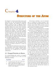
lateral and intercalary (Fig. 6.2). New cells<br />
produced by meristem are initially like those<br />
of meristem itself, but as they grow and<br />
mature, their characteristics slowly change<br />
and they become differentiated as<br />
components of other tissues.<br />
Apical meristem<br />
Jar 1 Jar 2<br />
Fig. 6.1: Growth of roots in onion bulbs<br />
Intercalary meristem<br />
Activity ______________ 6.1<br />
• Take two glass jars and fill them with<br />
water.<br />
• Now, take two onion bulbs and place<br />
one on each jar, as shown in<br />
Fig. 6.1.<br />
• Observe the growth of roots in both the<br />
bulbs for a few days.<br />
• Measure the length of roots on day 1,<br />
2 and 3.<br />
• On day 4, cut the root tips of the onion<br />
bulb in jar 2 by about 1 cm. After this,<br />
observe the growth of roots in both the<br />
jars and measure their lengths each<br />
day for five more days and record the<br />
observations in tables, like the table<br />
below:<br />
Length Day 1 Day 2 Day 3 Day 4 Day 5<br />
Jar 1<br />
Jar 2<br />
Lateral meristem<br />
Fig. 6.2: Location of meristematic tissue in plant body<br />
Apical meristem is present at the growing<br />
tips of stems and roots and increases the<br />
length of the stem and the root. The girth of<br />
the stem or root increases due to lateral<br />
meristem (cambium). Intercalary meristem is<br />
the meristem at the base of the leaves or<br />
internodes (on either side of the node)<br />
on twigs.<br />
TISSUES 69
As the cells of this tissue are very active,<br />
they have dense cytoplasm, thin cellulose<br />
walls and prominent nuclei. They lack<br />
vacuoles. Can we think why they would lack<br />
vacuoles? (You might want to refer to the<br />
functions of vacuoles in the chapter on cells.)<br />
6.2.2 PERMANENT TISSUE<br />
What happens to the cells formed by<br />
meristematic tissue? They take up a specific<br />
role and lose the ability to divide. As a result,<br />
they form a permanent tissue. This process<br />
of taking up a permanent shape, size, and a<br />
function is called differentiation. Cells of<br />
meristematic tissue differentiate to form<br />
different types of permanent tissue.<br />
• Now, answer the following on the basis<br />
of your observation:<br />
1. Are all cells similar in structure?<br />
2. How many types of cells can<br />
be seen?<br />
3. Can we think of reasons why there<br />
would be so many types of cells?<br />
• We can also try to cut sections of plant<br />
roots. We can even try cutting sections<br />
of root and stem of different plants.<br />
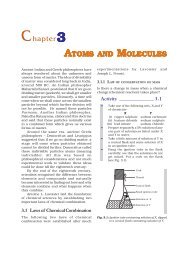
6.2.2 (i) SIMPLE PERMANENT TISSUE<br />
A few layers of cells form the basic packing<br />
tissue. This tissue is parenchyma, a type of<br />
permanent tissue. It consists of relatively<br />
unspecialised cells with thin cell walls. They<br />
are live cells. They are usually loosely packed,<br />
Trichome<br />
Mucilaginous canal<br />
Cuticle<br />
Epidermis<br />
Hypodermis<br />
Cortex<br />
Endodermis<br />
Pericycle<br />
Phloem<br />
Cambium<br />
Metaxylem<br />
Protoxylem<br />
Medullary ray<br />
Xylem<br />
Vascular bundle<br />
Pith<br />
Fig. 6.3: Section of a stem<br />
Activity ______________ 6.2<br />
• Take a plant stem and with the help<br />
of your teacher cut into very thin slices<br />
or sections.<br />
• Now, stain the slices with safranin.<br />
Place one neatly cut section on a slide,<br />
and put a drop of glycerine.<br />
• Cover with a cover-slip and observe<br />
under a microscope. Observe the<br />
various types of cells and their<br />
arrangement. Compare it with Fig. 6.3.<br />
so that large spaces between cells<br />
(intercellular spaces) are found in this tissue<br />
[Fig. 6.4 a(i)]. This tissue provides support to<br />
plants and also stores food. In some<br />
situations, it contains chlorophyll and<br />
performs photosynthesis, and then it is called<br />
chlorenchyma. In aquatic plants, large air<br />
cavities are present in parenchyma to give<br />
buoyancy to the plants to help them float.<br />
Such a parenchyma type is called<br />
aerenchyma. The parenchyma of stems and<br />
roots also stores nutrients and water.<br />
70<br />
SCIENCE
The flexibility in plants is due to another<br />
permanent tissue, collenchyma. It allows<br />
easy bending in various parts of a plant (leaf,<br />
stem) without breaking. It also provides<br />
mechanical support to plants. We can find<br />
this tissue in leaf stalks below the epidermis.<br />
The cells of this tissue are living, elongated<br />
and irregularly thickened at the<br />
corners. There is very little intercellular<br />
space (Fig. 6.4 b).<br />
Intercellular spaces<br />
Wall thickenings<br />
Nucleus<br />
Vacuole<br />
Cell wall<br />
a (i)<br />
b (i)<br />
Cytoplasm<br />
Nucleus<br />
Middle lamella<br />
Chloroplast<br />
Vacuole<br />
Intercellular space<br />
Primary cell wall<br />
End wall<br />
Primary cell wall<br />
(thickened at corners)<br />
Chloroplast<br />
Nucleus<br />
Vacuole<br />
Cytoplasm<br />
Intercellular space<br />
a (ii)<br />
b (ii)<br />
Simple<br />
pit pair<br />
Narrow lumen<br />
Lignified<br />
thick wall<br />
c (i)<br />
c (ii)<br />
Fig. 6.4: Various types of simple tissues: (a) Parenchyma (i) transverse section, (ii) longitudinal section;<br />
(b) Collenchyma (i) transverse section, (ii) longitudinal section; (c) Sclerenchyma (i) transverse section,<br />
(ii) longitudinal section.<br />
TISSUES 71
Yet another type of permanent tissue is<br />
sclerenchyma. It is the tissue which makes<br />
the plant hard and stiff. We have seen the<br />
husk of a coconut. It is made of<br />
sclerenchymatous tissue. The cells of this<br />
tissue are dead. They are long and narrow as<br />
the walls are thickened due to lignin (a<br />
chemical substance which acts as cement and<br />
hardens them). Often these walls are so thick<br />
that there is no internal space inside the cell<br />
(Fig. 6.4 c). This tissue is present in stems,<br />
around vascular bundles, in the veins of<br />
leaves and in the hard covering of seeds and<br />
nuts. It provides strength to the plant parts.<br />
Activity ______________ 6.3<br />
• Take a freshly plucked leaf of Rheo.<br />
• Stretch and break it by applying<br />
pressure.<br />
• While breaking it, keep it stretched<br />
gently so that some peel or skin<br />
projects out from the cut.<br />
• Remove this peel and put it in a petri<br />
dish filled with water.<br />
• Add a few drops of safranin.<br />
• Wait for a couple of minutes and then<br />
transfer it onto a slide. Gently place a<br />
cover slip over it.<br />
• Observe under microscope.<br />
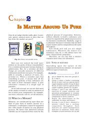
Guard<br />
cells<br />
Epidermal<br />
cell<br />
(a)<br />
Fig. 6.5: Guard cells and epidermal cells: (a) lateral<br />
view, (b) surface view<br />
(b)<br />
Stomata<br />
Guard<br />
cell<br />
What you observe is the outermost layer<br />
of cells, called epidermis. The epidermis is<br />
usually made of a single layer of cells. In some<br />
plants living in very dry habitats, the<br />
epidermis may be thicker since protection<br />
against water loss is critical. The entire<br />
surface of a plant has this outer covering of<br />
epidermis. It protects all the parts of the plant.<br />
Epidermal cells on the aerial parts of the plant<br />
often secrete a waxy, water-resistant layer on<br />
their outer surface. This aids in protection<br />
against loss of water, mechanical injury and<br />
invasion by parasitic fungi. Since it has a<br />
protective role to play, cells of epidermal<br />
tissue form a continuous layer without<br />
intercellular spaces. Most epidermal cells are<br />
relatively flat. Often their outer and side walls<br />
are thicker than the inner wall.<br />
We can observe small pores here and there<br />
in the epidermis of the leaf. These pores are<br />
called stomata (Fig. 6.5). Stomata are<br />
enclosed by two kidney-shaped cells called<br />
guard cells. They are necessary for<br />
exchanging gases with the atmosphere.<br />
Transpiration (loss of water in the form of<br />
water vapour) also takes place through<br />
stomata.<br />
Think about which gas may be required<br />
for photosynthesis.<br />
Find out the role of transpiration in plants.<br />
Epidermal cells of the roots, whose<br />
function is water absorption, commonly bear<br />
long hair-like parts that greatly increase the<br />
total absorptive surface area.<br />
In some plants like desert plants,<br />
epidermis has a thick waxy coating of cutin<br />
(chemical substance with waterproof quality)<br />
on its outer surface. Can we think of a reason<br />
for this?<br />
Is the outer layer of a branch of a tree<br />
different from the outer layer of a young stem?<br />
As plants grow older, the outer protective<br />
tissue undergoes certain changes. A strip of<br />
secondary meristem replaces the epidermis<br />
of the stem. Cells on the outside are cut off<br />
from this layer. This forms the several-layer<br />
thick cork or the bark of the tree. Cells of<br />
cork are dead and compactly arranged<br />
without intercellular spaces (Fig. 6.6). They<br />
also have a chemical called suberin in their<br />
walls that makes them impervious to gases<br />
and water.<br />
72<br />
SCIENCE
Cork cells<br />
Ruptured epidermis<br />
parts of the plant. Except for phloem fibres,<br />
phloem cells are living cells.<br />
Fig. 6.6: Protective tissue<br />
6.2.2 (ii) COMPLEX PERMANENT TISSUE<br />
The different types of tissues we have<br />
discussed until now are all made of one type<br />
of cells, which look like each other. Such<br />
tissues are called simple permanent tissue.<br />
Yet another type of permanent tissue is<br />
complex tissue. Complex tissues are made of<br />
more than one type of cells. All these cells<br />
coordinate to perform a common function.<br />
Xylem and phloem are examples of such<br />
complex tissues. They are both conducting<br />
tissues and constitute a vascular bundle.<br />
Vascular or conductive tissue is a distinctive<br />
feature of the complex plants, one that has<br />
made possible their survival in the terrestrial<br />
environment. In Fig. 6.3 showing a section of<br />
stem, can you see different types of cells in<br />
the vascular bundle?<br />
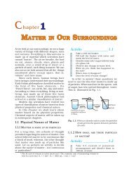
Xylem consists of tracheids, vessels,<br />
xylem parenchyma (Fig. 6.7 a,b,c) and xylem<br />
fibres. The cells have thick walls, and many<br />
of them are dead cells. Tracheids and vessels<br />
are tubular structures. This allows them to<br />
transport water and minerals vertically. The<br />
parenchyma stores food and helps in the<br />
sideways conduction of water. Fibres are<br />
mainly supportive in function.<br />
Phloem is made up of four types of<br />
elements: sieve tubes, companion cells,<br />
phloem fibres and the phloem parenchyma<br />
[Fig. 6.7 (d)]. Sieve tubes are tubular cells with<br />
perforated walls. Phloem is unlike xylem in<br />
that materials can move in both directions in<br />
it. Phloem transports food from leaves to other<br />
Pit<br />
Phloem<br />
Pits<br />
Xylem<br />
Sieve plate<br />
Sieve tube<br />
Phloem<br />
parenchyma<br />
Companion cell<br />
Nucleus<br />
Cytoplasm<br />
(a) Tracheid (b) Vessel (c) Xylem parenchyma<br />
(d) Section of phloem<br />
Fig. 6.7: Types of complex tissue<br />
TISSUES 73
Questions<br />
1. Name types of simple tissues.<br />
2. Where is apical meristem found?<br />
3. Which tissue makes up the husk<br />
of coconut?<br />
4. What are the constituents of<br />
phloem?<br />
6.3 Animal Tissues<br />
When we breathe we can actually feel the<br />
movement of our chest. How do these body<br />
parts move? For this we have specialised cells<br />
called muscle cells (Fig. 6.8). The contraction<br />
and relaxation of these cells result in<br />
movement.<br />
During breathing we inhale oxygen. Where<br />
does this oxygen go? It is absorbed in the<br />
lungs and then is transported to all the body<br />
cells through blood. Why would cells need<br />
oxygen? The functions of mitochondria we<br />
studied earlier provide a clue to this question.<br />
Blood flows and carries various substances<br />
from one part of the body to the other. For<br />
example, it carries oxygen and food to all cells.<br />
It also collects wastes from all parts of the<br />
body and carries them to the liver and kidney<br />
for disposal.<br />
Blood and muscles are both examples of<br />
tissues found in our body. On the basis of<br />
the functions they perform we can think of<br />
different types of animal tissues, such as<br />
epithelial tissue, connective tissue, muscular<br />
tissue and nervous tissue. Blood is a type of<br />
connective tissue, and muscle forms<br />
muscular tissue.<br />
6.3.1 EPITHELIAL TISSUE<br />
Smooth muscle fibres<br />
Nucleus<br />
Smooth muscle fibre<br />
(Cell)<br />
Fig. 6.8: Location of muscle fibres<br />
The covering or protective tissues in the<br />
animal body are epithelial tissues. Epithelium<br />
covers most organs and cavities within the<br />
body. It also forms a barrier to keep different<br />
body systems separate. The skin, the lining<br />
of the mouth, the lining of blood vessels, lung<br />
alveoli and kidney tubules are all made of<br />
epithelial tissue. Epithelial tissue cells are<br />
tightly packed and form a continuous sheet.<br />
They have only a small amount of cementing<br />
material between them and almost no<br />
intercellular spaces. Obviously, anything<br />
entering or leaving the body must cross at<br />
least one layer of epithelium. As a result, the<br />
permeability of the cells of various epithelia<br />
play an important role in regulating the<br />
exchange of materials between the body and<br />
the external environment and also between<br />
different parts of the body. Regardless of the<br />
type, all epithelium is usually separated from<br />
the underlying tissue by an extracellular<br />
fibrous basement membrane.<br />
Different epithelia (Fig. 6.9) show differing<br />
structures that correlate with their unique<br />
functions. For example, in cells lining blood<br />
vessels or lung alveoli, where transportation<br />
of substances occurs through a selectively<br />
74<br />
SCIENCE
permeable surface, there is a simple flat kind<br />
of epithelium. This is called the simple<br />
(a) Squamous<br />
(b) Cuboidal<br />
(c) Columnar (Ciliated)<br />
squamous epithelium. Simple squamous<br />
epithelial cells are extremely thin and flat and<br />
form a delicate lining. The oesophagus and<br />
the lining of the mouth are also covered with<br />
squamous epithelium. The skin, which<br />
protects the body, is also made of squamous<br />
epithelium. Skin epithelial cells are arranged<br />
in many layers to prevent wear and tear. Since<br />
they are arranged in a pattern of layers, the<br />
epithelium is called stratified squamous<br />
epithelium.<br />
Where absorption and secretion occur, as<br />
in the inner lining of the intestine, tall<br />
epithelial cells are present. This columnar<br />
(meaning ‘pillar-like’) epithelium facilitates<br />
movement across the epithelial barrier. In the<br />
respiratory tract, the columnar epithelial<br />
tissue also has cilia, which are hair-like<br />
projections on the outer surfaces of epithelial<br />
cells. These cilia can move, and their<br />
movement pushes the mucus forward to clear<br />
it. This type of epithelium is thus ciliated<br />
columnar epithelium.<br />
Cuboidal epithelium (with cube-shaped<br />
cells) forms the lining of kidney tubules and<br />
ducts of salivary glands, where it provides<br />
mechanical support. Epithelial cells often<br />
acquire additional specialisation as gland<br />
cells, which can secrete substances at the<br />
epithelial surface. Sometimes a portion of the<br />
epithelial tissue folds inward, and a<br />
multicellular gland is formed. This is<br />
glandular epithelium.<br />
6.3.2 CONNECTIVE TISSUE<br />
(d) Stratified squamous<br />
Fig. 6.9: Different types of epithelial tissues<br />
Blood is a type of connective tissue. Why<br />
would it be called ‘connective’ tissue? A clue<br />
is provided in the introduction of this chapter!<br />
Now, let us look at this type of tissue in some<br />
more detail. The cells of connective tissue are<br />
loosely spaced and embedded in an<br />
intercellular matrix (Fig. 6.10). The matrix<br />
may be jelly like, fluid, dense or rigid. The<br />
nature of matrix differs in concordance with<br />
the function of the particular connective<br />
tissue.<br />
Take a drop of blood on a slide and observe<br />
different cells present in it under a microscope.<br />
TISSUES 75
Reticular fibre<br />
Macrophage<br />
Collagen fibre<br />
Haversian canal<br />
(contains blood vessels<br />
and nerve fibres)<br />
(c)<br />
Red blood<br />
corpuscle<br />
Different white<br />
blood corpuscles<br />
Mast cell<br />
Fat droplet<br />
Fibroblast<br />
(a)<br />
Adipocyte<br />
(b)<br />
Nucleus<br />
Chondrocyte<br />
Hyaline matrix<br />
Canaliculus (contains<br />
slender process of bone<br />
cell or osteocyte)<br />
Neutrophil Eosinophil Basophil<br />
(polynuclear<br />
leucocyte)<br />
Lymphocyte Monocyte<br />
Cytoplasm<br />
Plasma cell<br />
Nucleus<br />
Platelets<br />
(e)<br />
Fig. 6.10: Types of connective tissues: (a) areolar<br />
tissue, (b) adipose tissue, (c) compact<br />
bone, (d) hyaline cartilage, (e) types of<br />
blood cells<br />
(d)<br />
Blood has a fluid (liquid) matrix called<br />
plasma, in which red blood cells (RBCs), white<br />
blood cells (WBCs) and platelets are<br />
suspended. The plasma contains proteins,<br />
salts and hormones. Blood flows and<br />
transports gases, digested food, hormones<br />
and waste materials to different parts of the<br />
body.<br />
Bone is another example of a connective<br />
tissue. It forms the framework that supports<br />
the body. It also anchors the muscles and<br />
supports the main organs of the body. It is a<br />
strong and nonflexible tissue (what would be<br />
the advantage of these properties for bone<br />
functions?). Bone cells are embedded in a<br />
hard matrix that is composed of calcium and<br />
phosphorus compounds.<br />
Two bones can be connected to each other<br />
by another type of connective tissue called<br />
the ligament. This tissue is very elastic. It has<br />
considerable strength. Ligaments contain<br />
very little matrix. Tendons connect bones to<br />
muscles and are another type of connective<br />
tissue. Tendons are fibrous tissue with great<br />
strength but limited flexibility.<br />
Another type of connective tissue,<br />
cartilage, has widely spaced cells. The solid<br />
matrix is composed of proteins and sugars.<br />
Cartilage smoothens bone surfaces at joints<br />
and is also present in the nose, ear, trachea<br />
and larynx. We can fold the cartilage of the<br />
ears, but we cannot bend the bones in our<br />
arms. Think of how the two tissues are<br />
different!<br />
Areolar connective tissue is found between<br />
the skin and muscles, around blood vessels<br />
and nerves and in the bone marrow. It fills<br />
the space inside the organs, supports internal<br />
organs and helps in repair of tissues.<br />
Where are fats stored in our body? Fatstoring<br />
adipose tissue is found below the skin<br />
and between internal organs. The cells of this<br />
tissue are filled with fat globules. Storage of<br />
fats also lets it act as an insulator.<br />
6.3.3 MUSCULAR TISSUE<br />
Muscular tissue consists of elongated cells,<br />
also called muscle fibres. This tissue is<br />
responsible for movement in our body.<br />
76<br />
SCIENCE
Muscles contain special proteins called<br />
contractile proteins, which contract and relax<br />
to cause movement.<br />
Spindle shaped<br />
muscle cell<br />
Nuclei<br />
(a)<br />
(b)<br />
Nucleus<br />
Striations<br />
Striations<br />
Nuclei<br />
to bones and help in body movement. Under<br />
the microscope, these muscles show alternate<br />
light and dark bands or striations when<br />
stained appropriately. As a result, they are<br />
also called striated muscles. The cells of this<br />
tissue are long, cylindrical, unbranched and<br />
multinucleate (having many nuclei).<br />
The movement of food in the alimentary<br />
canal or the contraction and relaxation of<br />
blood vessels are involuntary movements. We<br />
cannot really start them or stop them simply<br />
by wanting to do so! Smooth muscles [Fig.<br />
6.11(b)] or involuntary muscles control such<br />
movements. They are also found in the iris of<br />
the eye, in ureters and in the bronchi of the<br />
lungs. The cells are long with pointed ends<br />
(spindle-shaped) and uninucleate (having a<br />
single nucleus). They are also called<br />
unstriated muscles – why would they be<br />
called that?<br />
The muscles of the heart show rhythmic<br />
contraction and relaxation throughout life.<br />
These involuntary muscles are called cardiac<br />
muscles [Fig. 6.11(c)]. Heart muscle cells are<br />
cylindrical, branched and uninucleate.<br />
Compare the structures of different types<br />
of muscular tissues. Note their shape,<br />
number of nuclei and position of nuclei within<br />
the cell.<br />
6.3.4 NERVOUS TISSUE<br />
(c)<br />
Fig. 6.11: Types of muscles fibres: (a) striated<br />
muscle, (b) smooth muscle, (c) cardiac<br />
muscle<br />
We can move some muscles by conscious<br />
will. Muscles present in our limbs move when<br />
we want them to, and stop when we so decide.<br />
Such muscles are called voluntary muscles<br />
[Fig. 6.11(a)]. These muscles are also called<br />
skeletal muscles as they are mostly attached<br />
All cells possess the ability to respond to<br />
stimuli. However, cells of the nervous tissue<br />
are highly specialised for being stimulated<br />
and then transmitting the stimulus very<br />
rapidly from one place to another within the<br />
body. The brain, spinal cord and nerves are<br />
all composed of the nervous tissue. The cells<br />
of this tissue are called nerve cells or neurons.<br />
A neuron consists of a cell body with a<br />
nucleus and cytoplasm, from which long thin<br />
hair-like parts arise (Fig. 6.12). Usually each<br />
neuron has a single long part, called the axon,<br />
and many short, branched parts called<br />
dendrites. An individual nerve cell may be up<br />
to a metre long. Many nerve fibres bound<br />
together by connective tissue make up<br />
a nerve.<br />
TISSUES 77
Nucleus<br />
Cell body<br />
Dendrite<br />
Axon<br />
Nerve ending<br />
Fig. 6.12: Neuron-unit of nervous tissue<br />
Nerve impulses allow us to move our<br />
muscles when we want to. The functional<br />
combination of nerve and muscle tissue is<br />
fundamental to most animals. This<br />
combination enables animals to move rapidly<br />
in response to stimuli.<br />
Questions<br />
1. Name the tissue responsible for<br />
movement in our body.<br />
2. What does a neuron look like?<br />
3. Give three features of cardiac<br />
muscles.<br />
4. What are the functions of areolar<br />
tissue?<br />
What<br />
you have<br />
learnt<br />
• Tissue is a group of cells similar in structure and function.<br />
• Plant tissues are of two main types – meristematic and<br />
permanent.<br />
• Meristematic tissue is the dividing tissue present in the growing<br />
regions of the plant.<br />
• Permanent tissues are derived from meristematic tissue once<br />
they lose the ability to divide. They are classified as simple and<br />
complex tissues.<br />
• Parenchyma, collenchyma and sclerenchyma are three types<br />
of simple tissues. Xylem and phloem are types of complex<br />
tissues.<br />
• Animal tissues can be epithelial, connective, muscular and<br />
nervous tissue.<br />
• Depending on shape and function, epithelial tissue is classified<br />
as squamous, cuboidal, columnar, ciliated and glandular.<br />
• The different types of connective tissues in our body include<br />
areolar tissue, adipose tissue, bone, tendon, ligament, cartilage<br />
and blood.<br />
• Striated, unstriated and cardiac are three types of muscle<br />
tissues.<br />
• Nervous tissue is made of neurons that receive and conduct<br />
impulses.<br />
78<br />
SCIENCE
Exercises<br />
1. Define the term “tissue”.<br />
2. How many types of elements together make up the xylem tissue?<br />
Name them.<br />
3. How are simple tissues different from complex tissues in plants?<br />
4. Differentiate between parenchyma, collenchyma and<br />
sclerenchyma on the basis of their cell wall.<br />
5. What are the functions of the stomata?<br />
6. Diagrammatically show the difference between the three types<br />
of muscle fibres.<br />
7. What is the specific function of the cardiac muscle?<br />
8. Differentiate between striated, unstriated and cardiac muscles<br />
on the basis of their structure and site/location in the body.<br />
9. Draw a labelled diagram of a neuron.<br />
10. Name the following.<br />
(a) Tissue that forms the inner lining of our mouth.<br />
(b) Tissue that connects muscle to bone in humans.<br />
(c) Tissue that transports food in plants.<br />
(d) Tissue that stores fat in our body.<br />
(e) Connective tissue with a fluid matrix.<br />
(f) Tissue present in the brain.<br />
11. Identify the type of tissue in the following: skin, bark of tree,<br />
bone, lining of kidney tubule, vascular bundle.<br />
12. Name the regions in which parenchyma tissue is present.<br />
13. What is the role of epidermis in plants?<br />
14. How does the cork act as a protective tissue?<br />
15. Complete the table:<br />
TISSUES 79