Formation and color properties of vanadium doped ZrSiO4 ceramic ...
Formation and color properties of vanadium doped ZrSiO4 ceramic ...
Formation and color properties of vanadium doped ZrSiO4 ceramic ...

Create successful ePaper yourself
Turn your PDF publications into a flip-book with our unique Google optimized e-Paper software.
Pyon a , Kyong-Sop Han b <strong>and</strong> Byung-Ha Lee a, *<br />
a Kyu-Ri<br />
<strong>of</strong> Material Science & Engineering, Myongji University, YongIn, Korea<br />
Department<br />
b Department <strong>of</strong> Material, KIST, Seoul, Korea<br />
<strong>doped</strong> zircon pigments were prepared by a <strong>ceramic</strong> method from a mixture <strong>of</strong> monoclinic zirconia <strong>and</strong> silica with<br />
Vanadium<br />
fluoride as a mineralizer <strong>and</strong> the <strong>color</strong> <strong>properties</strong> <strong>of</strong> the pigments were also investigated. The raw composition was<br />
sodium<br />
in order to optimize the synthesis <strong>of</strong> the final pigments. The <strong>color</strong> <strong>of</strong> synthesized pigments observed in the absence <strong>and</strong><br />
varied<br />
presence <strong>of</strong> the NaF was green <strong>and</strong> blue, respectively. They were characterized by X-ray diffraction, UV-Vis <strong>and</strong> Raman<br />
the<br />
The <strong>color</strong> changes produced in the samples during heating are discussed in terms <strong>of</strong> the valence <strong>of</strong> the<br />
spectroscopies.<br />
species in the zircon matrix <strong>and</strong> the yield <strong>of</strong> the zircon phase. In these zircon pigments, evidence <strong>of</strong> the intermediate<br />
<strong>vanadium</strong><br />
+4 ) in an initial step in the synthesis process is observed by Raman spectroscopy. V -ZrSiO 4<br />
pigments using rice<br />
phase(NaVO 3 o<br />
give rise to a blue <strong>color</strong>ation at a low temperature (750 C) <strong>and</strong> they became a more intense blue <strong>color</strong> with holding<br />
ash husk<br />
structures are frequently found in <strong>ceramic</strong><br />
Zircon-<strong>doped</strong><br />
<strong>and</strong> are closely related to <strong>properties</strong>, such as low<br />
pigments<br />
dissolution resistance <strong>and</strong> tinctorial strength that<br />
toxicity,<br />
desirable for a wide range <strong>of</strong> industrial applications.<br />
are<br />
frist commercially introduced zircon pigment was<br />
The<br />
blue(DCMA 14-42-2)” containing <strong>vanadium</strong> as<br />
“turquoise<br />
chromophore cation. A yellow zircon pigment containing<br />
a<br />
(DCMA 14-43-4) <strong>and</strong> a pink zircon pigment<br />
praseodymium<br />
14-44-5) containing iron are also commercially<br />
(DCMA<br />
available[1-6].<br />
forms solutions <strong>and</strong> incorporates into<br />
Vanadium 4+ solid<br />
lattice with a form <strong>of</strong> V ions that is the origin<br />
zircon the<br />
the blue <strong>color</strong> <strong>of</strong> this pigment. But there are some contradictory<br />
<strong>of</strong><br />
reports about the limit <strong>of</strong> the solid solution, the<br />
location <strong>and</strong> stabilization <strong>of</strong> the blue <strong>color</strong> [7]. Eppler<br />
valance,<br />
the mechanism <strong>of</strong> formation to elucidate the origin<br />
examined<br />
the <strong>color</strong>ing by zircon pigments, <strong>and</strong> also proposed the<br />
<strong>of</strong><br />
played by mineralizers in the reaction kinetics through<br />
role<br />
ion model <strong>and</strong> a vapor transport model [8].<br />
an 4+ diffusion<br />
al. [9] confirmed the existence <strong>of</strong> V in the ZrO 8<br />
et Demiray<br />
in zircon structure using crystal field theory<br />
position 4+ the<br />
spectrum analysis <strong>and</strong> concluded that V ions<br />
optical through<br />
the octahedral sites since the unit cell parameters in<br />
occupy<br />
zircon are the same as those in un<strong>doped</strong> zircon.<br />
V-<strong>doped</strong><br />
research <strong>of</strong> the V-ZrSiO 4<br />
system indicates<br />
Structural 4+<br />
[10]<br />
V cation which forms a solid solution with the<br />
the that<br />
author:<br />
*Corresponding<br />
: +82-31-330-6461<br />
Tel<br />
+82-31-330-6457<br />
Fax:<br />
da5raa@naver.com<br />
E-mail:<br />
flux increased the solubility <strong>and</strong> thermal<br />
<strong>and</strong> 4+<br />
additions(NaF)<br />
V in zircon but did not affect the valence<br />
<strong>of</strong> stability<br />
coordination. Raman spectra have been proposed<br />
<strong>and</strong><br />
to try to obtain information concerning the position<br />
[11-13]<br />
<strong>vanadium</strong>(IV) in the zircon lattice. De Waal et al. [11]<br />
<strong>of</strong><br />
for the calculation <strong>of</strong> V-O bond distances in<br />
used<br />
compounds equation <strong>of</strong> Hardcastle <strong>and</strong><br />
<strong>vanadium</strong>(IV) 4+ the<br />
<strong>and</strong> indicated that the Si (4b) positions<br />
[13], Waches<br />
4+ chromophores as V from Raman spectra.<br />
accommodate the<br />
previous study [14], rice husk ash was used as a<br />
a In<br />
<strong>of</strong> silica in the preparation <strong>of</strong> pr-ZrSiO 4<br />
solid<br />
source<br />
to produce a yellow powder in which a degree<br />
solutions<br />
high reactivity allows a significant increase in the<br />
<strong>of</strong><br />
<strong>of</strong> the process at a lower temperature. Currently<br />
efficiency<br />
Korea, the estimated production <strong>of</strong> rice husk is approximately<br />
in<br />
800,000 tons per year. Research is continuing to<br />
the feasibility <strong>of</strong> using rice husk ash which remains<br />
determine<br />
waste after burning, as an industrial raw material [15-17].<br />
as<br />
the present study, the raw composition <strong>of</strong> <strong>vanadium</strong><br />
In<br />
<strong>and</strong> firing conditions were varied in order to<br />
contents<br />
the synthesis <strong>of</strong> the final pigments <strong>and</strong> the Raman<br />
optimize<br />
<strong>of</strong> un<strong>doped</strong> zircon powder was compared to that<br />
spectrum<br />
the V-<strong>doped</strong> zircon pigments using rice husk ash <strong>and</strong><br />
<strong>of</strong><br />
2<br />
as a source <strong>of</strong> silica in order to elucidate the state <strong>of</strong><br />
SiO<br />
ions in the formation process by means <strong>of</strong> a<br />
<strong>vanadium</strong><br />
temperature processing route.<br />
low<br />
preparation<br />
Sample<br />
preparation <strong>of</strong> the <strong>vanadium</strong>-<strong>doped</strong> zircon pigments<br />
The<br />
carried out using traditional <strong>ceramic</strong> procedures. Highpurity<br />
was<br />
reagent-grade monoclinic zirconia (Yakuri), rice<br />
J O U R N A L O F<br />
Journal <strong>of</strong> Ceramic Processing Research. Vol. 12, No. 3, pp. 279~288 (2011)<br />
Ceramic<br />
Processing Research<br />
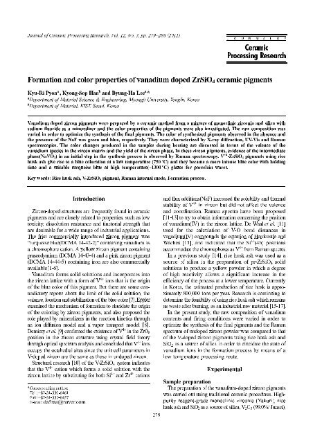
<strong>Formation</strong> <strong>and</strong> <strong>color</strong> <strong>properties</strong> <strong>of</strong> <strong>vanadium</strong> <strong>doped</strong> ZrSiO 4 <strong>ceramic</strong> pigments<br />
time <strong>and</strong> a suitable turquoise blue at high temperature(~1300 o C) glazes for porcelain wares.<br />
Key words: Rice husk ash, V-ZrSiO 4 pigment, Raman internal mode, <strong>Formation</strong> process.<br />
Introduction<br />
Experimental<br />
zircon lattice by substituting for both Si 4+ <strong>and</strong> Zr 4+ cations<br />
husk ash <strong>and</strong> SiO 2<br />
as a source <strong>of</strong> silica, V 2<br />
O 5<br />
(99.0%/ Junsei),<br />
279
well as a mineralizer NaF (Duksan) were selected as the<br />
as<br />
materials in the preparation <strong>of</strong> the pigments. The rice<br />
raw<br />
ash used was obtained by the slowest possible burning<br />
husk<br />
1000 o C. After burning it was washed repeatedly with<br />
at<br />
to remove as much alkali as possible. The average<br />
water<br />
size was 0.56 µm. ZrO 2 <strong>and</strong> SiO 2 were mixed to<br />
particle<br />
equimolar concentration. 0.1~0.2 mol% <strong>of</strong> V 2 O 5 ,<br />
an<br />
mol% <strong>of</strong> NaF were added to the mixture <strong>and</strong> this was<br />
0.5<br />
milled in acetone. The resulting dried powders were<br />
then<br />
homogenized in an agate mortar <strong>and</strong> samples <strong>of</strong><br />
finally<br />
mixture were placed in alumina crucibles. In order to<br />
the<br />
the V-ZrSiO 4 pigment to be formed in a solid-state<br />
allow<br />
the dried powders were fired in an electric furnace<br />
reaction,<br />
temperatures ranging between 600 <strong>and</strong> 1000 o C for<br />
at<br />
h. To produce an intense blue pigment <strong>and</strong> lower the<br />
3<br />
temperature, the firing conditions were designed<br />
synthesis<br />
holding schedules which were varied from 3 h to<br />
with<br />
h at 750 o C. Also to investigate the formation process<br />
10<br />
this pigment, the holding schedules were varied from<br />
in<br />
h to 10 h at 700 o C.<br />
0<br />
<strong>of</strong> V-ZrSiO 4 pigments<br />
Characteristics<br />
measure the silica content in the rice husk ash, an<br />
To<br />
was carried out using XRF (Shimadzu, XRF -1800).<br />
analysis<br />
particle size analyzer (Shimadzu, SALD-7101) was used<br />
A<br />
an analysis <strong>of</strong> the particle size. Characterization <strong>of</strong> the<br />
for<br />
samples was done using an X-ray diffractometer<br />
calcined<br />
Ni-filtered Cu Kα radiation (Shimadzu, XRD 7000).<br />
with<br />
a qualitative analysis, the patterns were recorded on<br />
For<br />
samples in the 10-70 2θ range at room temperature<br />
ground<br />
a scanning rate <strong>of</strong> 10 o /minute <strong>and</strong> a step size <strong>of</strong> 0.2 o .<br />
at<br />
wt% <strong>of</strong> corundum was added to all samples as an internal<br />
10<br />
The fraction <strong>of</strong> ZrSiO 4 (αZrSiO 4 = I ZS(200) +<br />
st<strong>and</strong>ard.<br />
M(11 ) + I M(111) + I T(111) ) was obtained based on the relative<br />
I<br />
calculated using the four peak areas <strong>of</strong> ZrSiO 4<br />
intensity<br />
2 (111) that appeared in the 2θ range <strong>of</strong> 26-32 o in<br />
t-ZrO<br />
X-ray diffraction patterns <strong>of</strong> the sample powders<br />
the<br />
at each temperature [18].<br />
heat-treated<br />
oxidation state <strong>of</strong> the <strong>vanadium</strong> in the pigments<br />
The<br />
at various calcination temperatures was analyzed<br />
synthesized<br />
UV-Vis spectra (Shimadzu, UV-2410 PC). Assignment<br />
using<br />
the zircon frequency <strong>and</strong> <strong>doped</strong> V-O bonding frequency<br />
<strong>of</strong><br />
confirmed using Raman spectroscopy (Dimention<br />
was<br />
Lambda Solution, Inc, U.S.A.). The Raman spectra<br />
D2,<br />
excited with a semiconductor laser operating at<br />
were<br />
nm <strong>and</strong> vertical scattered reflection light was analyzed<br />
532<br />
a CCD dispersive Raman spectrometer at a spectral<br />
using<br />
<strong>of</strong> 3 cm −1 . Raman spectra were recorded at room<br />
resolution<br />
using a vector Raman probe (RP532-US).<br />
temperature<br />
UV-Vis spectra were obtained in the absorption mode<br />
The<br />
the range <strong>of</strong> 200-800 nm. The <strong>color</strong>s <strong>of</strong> the synthesized<br />
in<br />
<strong>and</strong> their <strong>color</strong>ing in a lime-barium glaze were<br />
pigments<br />
via UV-Vis spectra. The results were also obtained<br />
analyzed<br />
by means <strong>of</strong> UV PC optical <strong>color</strong> analysis<br />
numerically<br />
(Shimadzu, P/N 206-67449) using the CIE L*a*b*<br />
s<strong>of</strong>tware<br />
<strong>of</strong> glazes under high-temperature firing<br />
Application<br />
scope <strong>of</strong> application <strong>of</strong> the V-ZrSiO 4 pigments<br />
The<br />
in the present study is the range <strong>of</strong> the <strong>ceramic</strong>s<br />
synthesized<br />
by high-temperature firing (SK 8-10). For the final<br />
created<br />
<strong>of</strong> the <strong>color</strong>ing <strong>of</strong> the synthesized pigments,<br />
evaluation<br />
were conducted by applying the pigments<br />
experiments<br />
lime-barium glazes used in a <strong>ceramic</strong> made by hightemperature<br />
to<br />
firing. Its Seger formula is as follows:<br />
KNaO<br />
0.297<br />
CaO 0.734 Al 2 O 3 4.682 SiO 2<br />
0.157<br />
MgO<br />
0.018<br />
BaO 0.528<br />
<strong>of</strong> the synthesized pigments was added to limebarium<br />
Each<br />
glazes (10%) by wet-mixing. Firing was done in<br />
electric furnace by raising the temperature to 1265 o C<br />
an<br />
30 minuite at a rate <strong>of</strong> 3 K·minuite −1 . The samples<br />
for<br />
then cooled. The characteristics <strong>of</strong> the <strong>color</strong>s <strong>of</strong> the<br />
were<br />
fired glazes using the synthesized<br />
high-temperature<br />
4 pigments were measured by UV-Vis spectroscopy,<br />
V-ZrSiO<br />
these values were then expressed numerically based<br />
<strong>and</strong><br />
<strong>of</strong> raw samples<br />
Characterization<br />
results <strong>of</strong> the chemical analysis <strong>of</strong> the rice husk<br />
The<br />
are shown in Table 1. The loss by ignition <strong>of</strong> the ground<br />
ash<br />
husk ash was 25.75% while that <strong>of</strong> the silica content<br />
rice<br />
58%. Additionally, the content <strong>of</strong> the Fe 2 O 3 , which<br />
was<br />
cloth <strong>color</strong>ing, was extremely low at 1.89%. This<br />
affects<br />
<strong>of</strong> synthesis parameters<br />
Optimization<br />
changes depending on different calcination tem-<br />
Phase<br />
peratures<br />
the synthesis <strong>of</strong> V-<strong>doped</strong> blue zircon pigment<br />
Regarding<br />
rice husk ash, an investigation was carried out<br />
using<br />
the relation <strong>of</strong> the formation <strong>of</strong> zircon crystals<br />
between<br />
the <strong>color</strong>ing <strong>of</strong> the pigment according to the calcination<br />
<strong>and</strong><br />
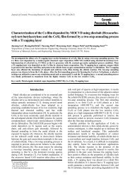
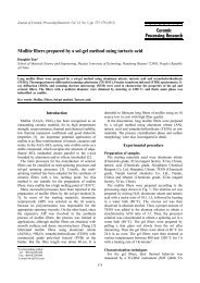
Fig. 1 shows the calcination temperature-<br />
temperature.<br />
changes <strong>of</strong> the crystal phases <strong>of</strong> the V-ZrSiO 4<br />
dependent<br />
synthesized with the addition <strong>of</strong> 0.2 mol% <strong>of</strong><br />
pigment<br />
Table 1. Chemical analysis <strong>of</strong> the rice husk ash as received<br />
280 Kyu-Ri Pyon, Kyong-Sop Han <strong>and</strong> Byung-Ha Lee<br />
on the CIE L*a*b* <strong>color</strong> order system.<br />
Results <strong>and</strong> Discussion<br />
value for the content <strong>of</strong> the alkali was 4.04%.<br />
1<br />
(monoclinic) m-ZrO 2 (11 , 111) <strong>and</strong> (tetragonal)<br />
1<br />
(200),<br />
V 2 O 5 . The holding time at each temperature zone for the<br />
Oxide Composition (%) Oxide Composition (%)<br />
SiO 2 58.16 K 2 O 3.90<br />
Al 2 O 3 3.23 P 2 O 5 2.31<br />
Fe 2 O 3 1.89 MnO 0.26<br />
MgO 1.23<br />
CaO 2.40 Ig.loss 25.75<br />
Na2O 0.14<br />
<strong>color</strong> system.
1. XRD patterns <strong>of</strong> samples formed at different temperatures<br />
Fig.<br />
V-<strong>doped</strong> ZrSiO 4 pigments.(V 2 O 5 0.2, NaF 0.5 mol%) (600, 700,<br />
in<br />
<strong>of</strong> pigment was 3 hours. In the case where<br />
synthesis o the<br />
temperature was 600 C, the main phase<br />
calcination the<br />
were observed (JCPDS : 30-1259). This observation<br />
NaF<br />
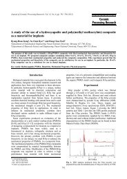
in agreement with the Raman analysis. Raman analysis<br />
is<br />
carried in order to more clearly confirm the presence<br />
was o out<br />
3<br />
produced at 600 C, which therefore had been<br />
NaVO <strong>of</strong><br />
before zircon was produced; <strong>and</strong> the result is<br />
produced<br />
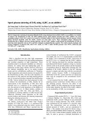
in Fig. 2. The st<strong>and</strong>ard in Fig. 2 represents the result<br />
shown<br />
o analysis <strong>of</strong> the NaVO 3<br />
synthesized at 650 C using<br />
<strong>of</strong> the<br />
mixture <strong>of</strong> V 2<br />
O 5<br />
<strong>and</strong> NaF. The result showed<br />
equimolar an<br />
o pigment synthesized at 600 C showed the appearance<br />
that the<br />
characteristic NaVO 3<br />
b<strong>and</strong>s at 952, 915, 637,<br />
<strong>of</strong><br />
−1 248, 225 <strong>and</strong> 180 cm . Thus it was confirmed<br />
506, 342,<br />
3<br />
is the intermediate phase, the first reaction<br />
NaVO that<br />
during the process <strong>of</strong> the pigment. Also it was<br />
product<br />
that during the initial period <strong>of</strong> reaction, <strong>vanadium</strong><br />
confirmed<br />
5+ the intermediate phase V , which is a yellow<br />
exists as<br />
reaction between m-ZrO 2<br />
<strong>and</strong> silica began at<br />
The powder.<br />
o to initiate the active production <strong>of</strong> zircon crystals.<br />
700 o C<br />
temperature reached 750 C, zircon became<br />
the When<br />
o phase <strong>and</strong> at 800 C it showed a rapid growth<br />
the main<br />
the relative reduction <strong>of</strong> the M-ZrO 2<br />
to a<br />
by accompanied<br />
The Hedvall effect [19] has been well known: that<br />
trace.<br />
the synthesis <strong>of</strong> zircon, the temperature zone for the<br />
during<br />
phase transition or quartz-cristoballite<br />
monoclinic-tetragonal<br />
transition allows a very reactive state, maximizing<br />
phase<br />
reaction rate, <strong>and</strong> thus resulting in an increase <strong>of</strong> zircon<br />
the<br />
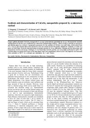
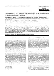
Fig. 3 shows the changes in the yields <strong>of</strong> zircon<br />
formation.<br />
m-ZrO 2<br />
according to varying the calcination temperature<br />
<strong>and</strong><br />
when a V-ZrSiO 4<br />
pigment is synthesized with<br />
<strong>and</strong> M-ZrO 2 yields with different temperatures <strong>of</strong><br />
Zircon<br />
V <strong>doped</strong> ZrSiO 4 pigments.<br />
the<br />
o C, thereby maximizing the reaction so as to increase<br />
600<br />
the next step the yield <strong>of</strong> zircon by the Hedvall effect.<br />
in<br />
the synthesis <strong>of</strong> zircon pigment, the temperature at which<br />
In<br />
begins to be synthesized as the main phase is very<br />
zircon<br />
because at this stage <strong>color</strong>ing <strong>of</strong> the pigment<br />
important<br />
as the chromophore is included into the host<br />
begins<br />
The <strong>of</strong> zircon was shown to be highest (94.7%)<br />
matrix. o yield<br />
C. The optimum calcination temperature for V<br />
800 at<br />
o <strong>doped</strong> blue pigment was 800 C for 3 h <strong>and</strong> the<br />
0.2 mol%<br />
was synthesized at this temperature as<br />
pigment best<br />
in Fig. 3. The <strong>color</strong> property <strong>of</strong> this pigment when<br />
shown<br />
to a glaze was L* 62.66 a* -14.66 b*-9.12(H<br />
applied<br />
changes depending on the content <strong>of</strong> V 2 O 5 <strong>and</strong><br />
Phase<br />
holding times<br />
different<br />
<strong>Formation</strong> <strong>and</strong> <strong>color</strong> <strong>properties</strong> <strong>of</strong> <strong>vanadium</strong> <strong>doped</strong> ZrSiO 4 <strong>ceramic</strong> pigments 281<br />
Fig. 2. Raman spectra <strong>of</strong> <strong>vanadium</strong> 0.2 mol% <strong>doped</strong> zircon blue<br />
pigments using rice husk ash at 600 o C.<br />
: 3 h keeping) Z : C o 800,1000 750, , 2 ZrO monoclinic : m zircon,<br />
3 NaVO : N quartz, : Q cristobalite. : C<br />
m-ZrO 2<br />
with traces <strong>of</strong> unreacted silica <strong>and</strong> NaVO 3<br />
was<br />
<strong>vanadium</strong> oxide), which is a eutectic <strong>of</strong> V 2<br />
O 5<br />
<strong>and</strong><br />
(sodium<br />
Fig. 3.<br />
<strong>of</strong> NaF allowed the phase transition <strong>of</strong> SiO 2<br />
addition<br />
→ cristoballite) at the very low temperature <strong>of</strong><br />
(quartz<br />
2.0B V/C 6.2/3.8 ).<br />
the addition <strong>of</strong> 0.2 mol% V 2<br />
O 5<br />
. The result showed that the<br />
In order to find the optimum V 2<br />
O 5<br />
content for the synthesis
4. XRD patterns <strong>of</strong> content change <strong>of</strong> V 2 O 5 at 800 o C(3 h) in<br />
Fig.<br />
ZrSiO 4 pigments.(V 2 O 5 0.2, NaF 0.5 mol%) Z : zircon,<br />
V-<strong>doped</strong><br />
V-<strong>doped</strong> blue zircon pigment using rice husk ash,<br />
<strong>of</strong><br />
was carried out at 800 o C for 3 hours with the<br />
calcination<br />
<strong>of</strong> 0.05, 0.1, 0.2 <strong>and</strong> 0.25 mol% V 2 O 5 . The results<br />
addition<br />
shown in Fig. 4. During the synthesis <strong>of</strong> zircon pigment,<br />
are<br />
oxide first forms a eutectic by reacting with NaF<br />
<strong>vanadium</strong><br />
it also plays a role as a mineralizer to promote the<br />
while<br />
<strong>of</strong> zircon. In all the samples the main phase was<br />
formation<br />
but the presence <strong>of</strong> unreacted M-ZrO 2 exerted a subtle<br />
zircon,<br />
on the completion <strong>of</strong> pigment synthesis as well as<br />
effect<br />
the intensity <strong>of</strong> <strong>color</strong>ing. The addition <strong>of</strong> more than<br />
on<br />
mol% V 2 O 5 allowed a good production <strong>of</strong> zircon during<br />
0.1<br />
resulting in the blue <strong>color</strong>ing <strong>of</strong> the synthesized<br />
calcination,<br />
In this research, the optimum amount <strong>of</strong> V 2 O 5 that<br />
pigment.<br />
in the best blue <strong>color</strong>ing <strong>of</strong> the pigment was<br />
resulted<br />
mol% when this pigment was applied to a glaze, the<br />
0.2<br />
values were L* 62.66, a* −14.66, b* −9.12 (H 2.0B<br />
<strong>color</strong><br />
6.2/3.8).<br />
V/C<br />
holding time at the final temperature has been<br />
The<br />
to be an important synthetic factor that can lower<br />
reported<br />
calcination temperature by reducing the unnecessary<br />
the<br />
nd phase <strong>and</strong> by stabilizing the growth <strong>of</strong> the 1 st phase<br />
2<br />
<strong>and</strong> that can also increase the intensity <strong>of</strong> <strong>color</strong>ing<br />
zircon<br />
When pigment samples synthesized at various calcination<br />
[14].<br />
temperatures were applied to a glaze the best<br />
was shown by the sample calcined at 800 o C for<br />
<strong>color</strong>ing<br />
hours. Whereas the sample calcined at 750 o C for 3 hours<br />
3<br />
a reduction <strong>of</strong> the 2 nd phase (M-ZrO 2 ) as the zircon<br />
showed<br />
Therefore, in order to obtain the optimum calcination<br />
grew.<br />
<strong>of</strong> 3, 5 <strong>and</strong> 10 hours. Fig. 5 shows the change <strong>of</strong><br />
times<br />
phases according to the change <strong>of</strong> holding time.<br />
crystal<br />
results showed that the highest intensity <strong>of</strong> zircon<br />
The<br />
obtained from the sample calcined at 750 o C for 5 hours,<br />
was<br />
with the best <strong>color</strong>ing: L* 58.38, a* −12.87, b* −10.63<br />
also<br />
3.7B V/C 5.7/3.9). The best condition for the synthesis<br />
(H<br />
5. XRD patterns for different holding times at 750 o C in V-<br />
Fig.<br />
ZrSiO 4 pigments.(V 2 O 5 0.2, NaF 0.5 mol%) Z : zircon,<br />
<strong>doped</strong><br />
the V-ZrSiO 4 pigment using rice husk ash was calcination<br />
<strong>of</strong><br />
750 o C for 5 hours with the addition <strong>of</strong> 0.2 mol% V 2 O 5<br />
at<br />
0.5 mol% NaF. This is because the holding time at<br />
<strong>and</strong> o C can stabilize the growth <strong>of</strong> zircon crystals as well<br />
750<br />
the inclusion <strong>of</strong> V ions, resulting in an improvement<br />
as<br />
the <strong>color</strong>ing.<br />
<strong>of</strong><br />
<strong>properties</strong> <strong>of</strong> pigments<br />
Color<br />
analysis <strong>of</strong> pigments with different calcination<br />
UV-Vis<br />
temperatures<br />
pigments synthesized at 600-1000 o C with the<br />
The<br />
<strong>of</strong> 0.2 mol% V 2 O 5 were added to a lime-barium<br />
addition<br />
at a concentration <strong>of</strong> 10%, for an analysis <strong>of</strong> the<br />
glaze<br />
by UV-Vis spectroscopy <strong>and</strong> the results are<br />
<strong>color</strong>ing<br />
in Fig. 6. <strong>and</strong> Table 2. The pigment synthesized<br />
shown<br />
600 o C gave a yellow <strong>color</strong>. There was a change in the<br />
at<br />
spectra from 700 o C, the onset temperature <strong>of</strong><br />
UV-Vis<br />
6. Optical absorption spectra <strong>of</strong> glazed samples with V-<br />
Fig.<br />
ZrSiO 4 pigments.(V 2 O 5 0.2, NaF 0.5 mol%) synthesized at<br />
<strong>doped</strong><br />
282 Kyu-Ri Pyon, Kyong-Sop Han <strong>and</strong> Byung-Ha Lee<br />
m : monoclinic ZrO 2 .<br />
m : monoclinic ZrO 2 , Q : low quartz, C : cristobalite, N : NaVO 3 .<br />
at 750 o C, samples containing 0.2 mol% V 2 O 5<br />
condition<br />
0.5 mol% NaF were calcined for varying holding<br />
<strong>and</strong><br />
different temperatures.
2. Lab parameters <strong>of</strong> glazed tiles with synthesized samples<br />
Table<br />
V 0.2mol% <strong>doped</strong> blue pigments with different temperatures<br />
<strong>of</strong><br />
at which active production <strong>of</strong> zircon begins.<br />
synthesis<br />
is, the characteristic absorption b<strong>and</strong> for a yellow<br />
That<br />
at 435-480 nm moved towards short wavelengths<br />
<strong>color</strong><br />
the b<strong>and</strong> at long wavelengths around 650 nm<br />
while<br />
o intensive. From above 700 C, the characteristic<br />
became 4+ more<br />
b<strong>and</strong> <strong>of</strong> V at 640 nm [20] was observed<br />
absorption<br />
with a clear change in the <strong>color</strong>ing to blue <strong>of</strong> the<br />
along<br />
applied to the glaze <strong>and</strong> the blue <strong>color</strong>ing was<br />
pigment<br />
enhanced this characteristic b<strong>and</strong> became more<br />
more 5+ as<br />
is due to the conversion <strong>of</strong> the V species,<br />
This intense.<br />
is in the initial step <strong>of</strong> pigment synthesis,<br />
which 4+<br />
prevalent<br />
in the final step. The <strong>color</strong>ing <strong>of</strong> the pigment<br />
V to<br />
o increase <strong>of</strong> the blue <strong>color</strong> property from 750 C<br />
showed an<br />
begins to grow rapidly <strong>and</strong> the best blue<br />
zircon when<br />
o obtained at 800 C with a b* value <strong>of</strong> −9.12.<br />
<strong>color</strong> was<br />
been literature reporting that the characteristic<br />
has There<br />
absorption b<strong>and</strong>s <strong>of</strong> V 4+<br />
nm. They reported that the b<strong>and</strong> [21] at 290 nm was<br />
640<br />
to 2B 1 → 2A (the dodecahedral transition) <strong>and</strong> that<br />
due<br />
b<strong>and</strong> at 640 nm is related to 2B 1 → 2 E (the dodecahedral<br />
the<br />
tetrahedral transition) this is in agreement with the<br />
<strong>and</strong><br />
<strong>of</strong> the present research. It has been confirmed that in<br />
result<br />
synthesis <strong>of</strong> the V-<strong>doped</strong> zircon pigment using rice<br />
the<br />
4+ the characteristic absorption b<strong>and</strong>s <strong>of</strong> V which<br />
husk ash<br />
major cause for <strong>color</strong>ing appears at 290 <strong>and</strong> 640 nm<br />
the is<br />
to a valence electron transition.<br />
due<br />
7 <strong>and</strong> Table 2 show the L*a*b* values. When<br />
Fig.<br />
Fig. 7. a*-b* plots <strong>of</strong> the glazed sample with V-<strong>doped</strong> ZrSiO 4<br />
to lime-barium glaze the pigments synthesized<br />
added o a<br />
C with the addition <strong>of</strong> 0.2 mol% V 2 O 5 showed<br />
600-1000 at<br />
following The ion in the pigment<br />
the o <strong>color</strong>ing. 5+ <strong>vanadium</strong><br />
C is in the state <strong>of</strong> V (NaVO 3 ) which<br />
600 at synthesized<br />
a yellow <strong>color</strong> so when it was applied to a glaze the<br />
has<br />
value was 19.53 giving a grayish yellow <strong>color</strong>. The<br />
b*<br />
o the pigment synthesized at 700 C to a glaze<br />
application <strong>of</strong><br />
a negative a* value (−13.59) <strong>and</strong> b* value<br />
in resulted<br />
giving weak blue <strong>color</strong>. Whereas the application<br />
(−5.08), o a<br />
pigment synthesized at 750 C to a glaze resulted in<br />
the <strong>of</strong><br />
<strong>and</strong> values that are similar to those <strong>of</strong> the pigment<br />
a* o b*<br />
700 C giving a light bluish green <strong>color</strong> but<br />
at synthesized<br />
an increase <strong>of</strong> the L* value to 65.99. As shown in<br />
with<br />
3, the pigment synthesized at a higher calcination<br />
Fig.<br />
showed higher yield <strong>of</strong> zircon resulting<br />
temperature o a<br />
<strong>color</strong> property. The pigment synthesized at 800 C<br />
bluer in<br />
the highest yield <strong>of</strong> zircon <strong>and</strong> when applied to<br />
showed<br />
glaze it also showed the highest −b* value <strong>of</strong> −9.12.<br />
a<br />
the mean time, its H/VC value was 2.0B 6.2/3.8 giving<br />
In<br />
best greenish blue <strong>color</strong>. The pigment synthesized at<br />
the<br />
o C showed a somewhat poor yield <strong>of</strong> zircon with<br />
1000<br />
re-increase <strong>of</strong> the M-ZrO 2 yield the <strong>color</strong>ing <strong>of</strong> the<br />
a<br />
applied to a glaze also became poor to a pale<br />
pigment<br />
<strong>color</strong>.<br />
green<br />
the results <strong>of</strong> applying the pigments synthesized<br />
Thus<br />
various calcination temperatures to glazes showed that<br />
at<br />
o condition was 800 C for 3 hours. The<br />
the optimum<br />
under this condition showed the best<br />
synthesized pigment<br />
blue <strong>color</strong>(L* 62.66, a* −14.66, b* −9.12, Hue<br />
greenish<br />
2.0B).<br />
value<br />
changes depending on the content <strong>of</strong> V 2 O 5 <strong>and</strong><br />
Color<br />
holding times<br />
different<br />
o synthesized at 800 C for 3 hours with<br />
The pigments<br />
2 O 5 contents (0.1, 0.15, 0.2 <strong>and</strong> 0.25 mol%)<br />
V varying<br />
added to a lime-barium glaze for an analysis by<br />
were<br />
spectroscopy the result is shown in Fig. 8. The<br />
UV-Vis<br />
8. a*-b* plots <strong>of</strong> glazed samples with V-<strong>doped</strong> ZrSiO 4 pigments<br />
Fig.<br />
0.5 mol%) with different V 2 O 5 contents(mol%) at 800 o C <strong>and</strong><br />
(NaF<br />
<strong>Formation</strong> <strong>and</strong> <strong>color</strong> <strong>properties</strong> <strong>of</strong> <strong>vanadium</strong> <strong>doped</strong> ZrSiO 4 <strong>ceramic</strong> pigments 283<br />
Samples L* a* b* H V/C Color<br />
1000(3 h)68.29 0−7.30 01.78 8.2G0 6.7/1.2 Pale Green<br />
1800(3 h)62.66 −14.66 −9.12 2.0B0 6.2/3.8 Greenish Blue<br />
1750(3 h)65.99 −13.27 −4.31 8.5BG 6.5/2.9 Light Bluish Green<br />
1700(3 h)71.61 −13.59 −5.08 9.5BG 7.1/3.0 Light Bluish Green<br />
1600(3 h)78.93 0−2.69 19.53 5.0Y0 7.8/2.6 Grayish Yellow<br />
appeared at 290 nm <strong>and</strong><br />
with different holding times at 750 o C (V 2 O 5 0.2, NaF 0.5 mol%).<br />
pigments.(V 2 O 5 0.2, NaF 0.5 mol%) different temperatures.
sample synthesized with the addition <strong>of</strong> 0.1 mol%<br />
pigment<br />
2 O 5 <strong>and</strong> applied to a glaze gave a grayish yellow green<br />
V<br />
Where the V 2 O 5 content was increased to 0.15 mol%<br />
<strong>color</strong>.<br />
was a remarkable increase <strong>of</strong> the blue <strong>color</strong> property<br />
there<br />
with a decrease <strong>of</strong> the b* value to 1.08. however,<br />
along<br />
L* value was still 70.88 giving a pale green <strong>color</strong> <strong>of</strong> a<br />
the<br />
bright <strong>and</strong> weak tone. The application <strong>of</strong> the pigment<br />
fairly<br />
with the addition <strong>of</strong> 0.2 mol% V 2 O 5 to a<br />
synthesized<br />
resulted in a big change <strong>of</strong> <strong>color</strong>. That is, the <strong>color</strong><br />
glaze<br />
darker along with an increase <strong>of</strong> the blue <strong>color</strong><br />
became<br />
resulting in a light greenish blue <strong>color</strong>. In the case<br />
property<br />
0.25 mol% V 2 O 5 the <strong>color</strong> became rather brighter to<br />
<strong>of</strong><br />
light blue <strong>color</strong>. Thus in the experiment with varying V 2 O 5<br />
a<br />
the optimum V 2 O 5 content for an excellent blue<br />
contents,<br />
was 0.2mol% which is also in agreement with<br />
<strong>color</strong>ing<br />
result <strong>of</strong> XRD in Fig. 1.<br />
the<br />
pigments synthesized with the addition <strong>of</strong> 0.2 mol%<br />
The<br />
2 O 5 at 750 o C for various holding times <strong>of</strong> 3, 5, 7, <strong>and</strong><br />
V<br />
hours were applied to a lime-barium glaze for an analysis<br />
10<br />
UV-Vis spectroscopy <strong>and</strong> the results are is shown in<br />
by<br />
8 <strong>and</strong> 9. In the experiment with varying holding times<br />
Figs.<br />
750 o C, the sample with 5 hours holding time showed a<br />
at<br />
blue <strong>color</strong> property than the sample with 3 hours<br />
better<br />
time at the same temperature <strong>and</strong> also than the<br />
holding<br />
with 3 hours holding time at 800 o C. The sample<br />
sample<br />
7 hours holding time showed almost the same <strong>color</strong>ing<br />
with<br />
the sample with 5 hours holding time but with a<br />
as<br />
for higher brightness resulting in a L* value<br />
propensity<br />
61.16 <strong>and</strong> when the holding time was 10 hours, there<br />
<strong>of</strong><br />
again an increase <strong>of</strong> the b* value to −4.25, resulting<br />
was<br />
an even brighter <strong>color</strong>. The result <strong>of</strong> the experiments<br />
in<br />
varying holding times showed that the optimum<br />
with<br />
for pigment synthesis was 750 o C with 5 hours<br />
condition<br />
time. The effect <strong>of</strong> holding time on the synthesis<br />
holding<br />
not so great in the case <strong>of</strong> the V-ZrSiO 4 pigment<br />
was<br />
to the ZrSiO 4 pigment, however it was possible<br />
compared<br />
lower the highest calcination temperature from 800 o C<br />
to<br />
to 750 o C as well as to more effectively improve<br />
down<br />
blue <strong>color</strong> property.<br />
the<br />
could be understood as shown in Fig. 9 by the<br />
This<br />
stabilization <strong>of</strong> the V ion to the state <strong>of</strong> 4+ which<br />
further<br />
the blue <strong>color</strong>ing <strong>of</strong> the pigment. In these<br />
maximizes<br />
the optimum calcination conditions for the<br />
experiments,<br />
<strong>of</strong> the zircon pigment with the addition <strong>of</strong> 0.2 mol%<br />
synthesis<br />
2 O 5 was 750 o C for 5 hours <strong>and</strong> application <strong>of</strong> this pigment<br />
V<br />
a glaze gave the most vivid <strong>and</strong> intense light greenish<br />
to<br />
Raman spectra <strong>of</strong> ZrSiO 4 :V 4+<br />
analysis was carried out in order to investigate<br />
Raman<br />
effect <strong>of</strong> rice husk ash <strong>and</strong> the calcination temperature<br />
the<br />
the zircon frequency <strong>and</strong> <strong>doped</strong> V-O bonding frequency<br />
on<br />
the synthesis <strong>of</strong> V- ZrSiO 4 pigment <strong>and</strong> the results<br />
during<br />
shown in Fig. 10 <strong>and</strong> Table 3. Assignments were<br />
are<br />
with the results <strong>of</strong> the researches conducted by<br />
made<br />
et al. [22] <strong>and</strong> Knittle <strong>and</strong> Williams [23] as well as<br />
Syme<br />
the zircon <strong>and</strong> V-O vibration Raman modes <strong>of</strong> the<br />
with<br />
9. Optical absorption spectrum <strong>of</strong> a glazed sample with V-<strong>doped</strong><br />
Fig.<br />
4 pigments.(V2O5 0.2, NaF 0.5 mol%) different holding time<br />
ZrSiO<br />
spectra <strong>of</strong> <strong>vanadium</strong> 0.2 mol% <strong>doped</strong> zircon blue<br />
Raman<br />
using SiO 2 <strong>and</strong> RHA at different temperatures. Z : Zircon.<br />
pigments<br />
synthesized in this research.<br />
pigments<br />
10 shows the Raman spectra <strong>of</strong> the V-ZrSiO 4<br />
Fig.<br />
synthesized using rice husk ash <strong>and</strong> SiO 2 . Table 4<br />
pigment<br />
the symmetry species mode assignment for each<br />
shows<br />
When rice husk ash was used to synthesize<br />
frequency.<br />
the optimum condition was 750 o C for 5 hours<br />
pigments<br />
zircon was produced at lower temperatures than when<br />
<strong>and</strong><br />
2 was used. As shown by the comparison in Table 4<br />
SiO<br />
majority <strong>of</strong> the b<strong>and</strong>s presented in Fig. 10 are the<br />
the<br />
b<strong>and</strong>s <strong>of</strong> ZrSiO 4 . In the case <strong>of</strong> the pigment<br />
characteristic<br />
using rice husk ash at 750 o C, the characteristic<br />
synthesized<br />
b<strong>and</strong>s including the most intensive zircon B 1g mode<br />
zircon<br />
almost the same peak intensity as in the case <strong>of</strong><br />
showed<br />
pigment synthesized using SiO 2 at 800 o C. This is due<br />
the<br />
the higher reaction rate <strong>of</strong> amorphous rice husk ash.<br />
to<br />
result <strong>of</strong> the Raman analysis on commercial ZrSiO 4<br />
The<br />
<strong>and</strong> the zircon powder synthesized by Syme et al.<br />
powder<br />
showed almost the same pure zircon assignment at<br />
[22]<br />
g , A 1g <strong>and</strong> B 1g . In the case <strong>of</strong> the <strong>vanadium</strong>-<strong>doped</strong> pigments,<br />
E<br />
vibrational modes are shown although weak at<br />
unique<br />
284 Kyu-Ri Pyon, Kyong-Sop Han <strong>and</strong> Byung-Ha Lee<br />
at 750 o C.<br />
Fig. 10.<br />
blue <strong>color</strong> (L* 58.38, a* −12.87, b* −10.63).<br />
pigments
4 single crystal<br />
V-ZrSiO<br />
et al.[11])<br />
(Wall<br />
ZrSiO 4 Pigment<br />
V-<strong>doped</strong><br />
(RHA)<br />
−1<br />
882, 498 <strong>and</strong> 473 cm . The low intensity b<strong>and</strong>s<br />
950, 912,<br />
Fig. 10 were not observed in the pure zircon<br />
in presented<br />
That is, they are the b<strong>and</strong>s that are presumed<br />
powder.<br />
be the V-O vibration modes <strong>of</strong> the <strong>vanadium</strong> in the<br />
to<br />
VO 4<br />
coordination in the zircon structure. As<br />
tetrahedral<br />
in Table 4, these b<strong>and</strong>s were also found in other<br />
shown<br />
2<br />
O 5<br />
. De Waal et al. [11] reported that the new internal<br />
V<br />
not observed with a pure zircon powder were caused<br />
modes<br />
ZrSiO 4 Pigment<br />
V-<strong>doped</strong><br />
2 ) (SiO<br />
ZrSiO 4<br />
Commercial<br />
Powder<br />
4 ZrSiO<br />
et al.[22])<br />
(Syme<br />
υ/cm −1 υ/cm −1 υ/cm −1 υ/cm −1 υ/cm −1 υ/cm −1<br />
1007 1003 1005 1005 1009 Zircon B 1g<br />
974 971 971 971 975 Zircon A 1g<br />
953 950 950 V-O a<br />
925 - - 923 925 Zircon E g<br />
918 912 910 V-O a<br />
912 882 876 V-O a<br />
V-O tetrahedral 11)<br />
1 (A1) V<br />
streching<br />
Symmetric<br />
2 (E) V<br />
Bending<br />
3 (F 2 ) V<br />
streching<br />
Asymmetric<br />
4 ( F 2 ) V<br />
Bending<br />
2 B<br />
E<br />
2 B<br />
E<br />
625 625 640 641 Zircon B 1g<br />
473 473 V-O a<br />
434 433 437 439 Zircon A 1g<br />
389 389 408 393 Zircon E g<br />
V-O a<br />
353 353 353 357 Zircon E g<br />
219 219 223 225 Zircon E g<br />
211 214 Zircon B 1g<br />
201 201 201 202 Zircon E g<br />
1g<br />
917<br />
B<br />
908<br />
g E<br />
1g<br />
503<br />
B<br />
473<br />
g E<br />
912<br />
882<br />
498<br />
473<br />
915<br />
-<br />
506<br />
-<br />
699<br />
-<br />
481<br />
-<br />
the D 2d<br />
site in the zircon lattice. That is, as shown in<br />
<strong>of</strong><br />
4 the peaks at 950, 912, 882, 498, 473 cm −1 observed<br />
Table<br />
the V-ZrSiO 4<br />
pigment synthesized using rice husk ash<br />
with<br />
confirmed to be the V-O vibration modes (V 1<br />
(A 1<br />
)<br />
were<br />
V 3<br />
(F 2<br />
) asymmetric stretching, <strong>and</strong> V 4<br />
(F 2<br />
)<br />
stretching,<br />
that are related to Raman active modes at each<br />
bending)<br />
group (A 1g<br />
, A 1g,<br />
B 1g,<br />
E g,<br />
B 1g<br />
<strong>and</strong> E g<br />
). Therefore the<br />
factor<br />
ions <strong>of</strong> the zircon pigment synthesized using<br />
<strong>vanadium</strong><br />
husk which are responsible for the blue <strong>color</strong>ing,<br />
rice 4+ ash<br />
to be located at the Si sites <strong>of</strong> D 2d<br />
symmetry<br />
considered are<br />
<strong>Formation</strong> <strong>and</strong> <strong>color</strong> <strong>properties</strong> <strong>of</strong> <strong>vanadium</strong> <strong>doped</strong> ZrSiO 4 <strong>ceramic</strong> pigments 285<br />
Table 3. Raman b<strong>and</strong> wavenumber(υ) observed for the V-<strong>doped</strong> pigment using RHA(750/5 h), SiO 2 (800/3 h) <strong>and</strong> single crystal (Wall et<br />
al.[11]) compared with those observed for commercial zircon powder, together with assignments from pure zircon by Syme et al.[22]<br />
Assignment<br />
852 851 867 -<br />
788 798 815 -<br />
615 621 -<br />
571<br />
499 V-O a<br />
498<br />
181 181 -<br />
/cm −1 ) <strong>of</strong> synthesized pigments are compared with other compounds in which<br />
Table 4. Raman Internal vibrations <strong>and</strong> wavenumber(<br />
ṽ<br />
<strong>vanadium</strong> has tetrahedral coordination.<br />
group 11) Factor group 11) ZrSiO 4 :V Powder 11) ZrSiO 4 : V Powder (RHA) NaVO 3 synthsized V 2 O 5 commercial<br />
Site<br />
d D 2d D 4h /cm −1 /cm −1 /cm −1 /cm −1<br />
T<br />
ṽ ṽ ṽ ṽ<br />
A 1 A 1g 950 950 953 991<br />
A 1 A 1g 378 - 342 301<br />
with a tetrahedral coordination (the α-NaVO 3<br />
compounds<br />
in the present research as well as commercial<br />
synthesized<br />
by the presence <strong>of</strong> the <strong>vanadium</strong> located at the silicon site<br />
in the tetravalent state.
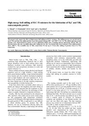
process <strong>of</strong> V-Zircon pigments<br />
<strong>Formation</strong><br />
pigment was synthesized in an alumina crucible<br />
When<br />
synthesized pigment formed 2-3 layers as shown in<br />
the<br />
11. The result <strong>of</strong> the XRD analysis on the individual<br />
Fig.<br />
is shown in Table 5 <strong>and</strong> the results <strong>of</strong> the Raman<br />
layers<br />
in Fig. 12. As shown in Table 5, 2 layers <strong>of</strong> brown<br />
analysis<br />
yellow were observed in the pigments synthesized<br />
<strong>and</strong> o <strong>color</strong>s o<br />
C <strong>and</strong> 650 C whereas 3 layers <strong>of</strong> yellow, brown<br />
600 at<br />
blue were observed in the pigment synthesized<br />
<strong>and</strong> o <strong>color</strong>s<br />
C. According to the result <strong>of</strong> the XRD analysis on<br />
700 at<br />
o o at 600 C <strong>and</strong> 650 C, in all the<br />
the pigments synthesized<br />
was a detection <strong>of</strong> unreacted M-ZrO 2,<br />
there formed layers<br />
cristobalite <strong>and</strong> NaVO 3<br />
with a similar intensity. In<br />
quartz,<br />
Raman spectra however, there was a distinct difference<br />
the<br />
the intensity. This is because the Raman frequency allows<br />
in<br />
detection as peaks <strong>of</strong> the vibration modes for inter-<br />
the<br />
interactions, if the degree <strong>of</strong> crystallization<br />
molecular o even<br />
In the case <strong>of</strong> the pigment synthesized at 600 C<br />
low. is<br />
3 hours, the yellow layer which was formed above<br />
for<br />
brown layer, showed a higher Raman intensity <strong>of</strong> the<br />
the<br />
Fig. 11. Layers <strong>of</strong> pigment synthesized at 700 o C for 3 h.<br />
5. XRD phase study according to each layers <strong>of</strong> V-<strong>doped</strong><br />
Table<br />
pigments using RHA at different temperature <strong>and</strong> holding time.<br />
zircon<br />
o C) 600 (3 h) 650 (3 h)<br />
Temp.(<br />
h) 700(10<br />
Yellow<br />
layer<br />
Brown<br />
layer<br />
M(vs),<br />
C(m),<br />
Q(w)<br />
N(t)<br />
M(vs),<br />
C(m),<br />
Q(w)<br />
N(t)<br />
M(vs),<br />
C(m),<br />
Q(w)<br />
N(t)<br />
M(vs),<br />
C(m),<br />
Q(w)<br />
N(t)<br />
Blue<br />
layer<br />
Brown<br />
layer<br />
Yellow<br />
layer<br />
Z(vs)<br />
M(w)<br />
M(vs)<br />
C(m)<br />
Q(w)<br />
Z(w)<br />
M(vs)<br />
C(m)<br />
Q(w)<br />
N(t)<br />
Z(vs)<br />
M(t)<br />
M(vs)<br />
C(m)<br />
Q(w)<br />
Z(w)<br />
M(vs)<br />
C(m)<br />
Q(w)<br />
N(t)<br />
Z(vs)<br />
M(t)<br />
M(vs)<br />
C(m)<br />
Q(w)<br />
Z(w)<br />
: Zircon, M : m-zro2, Q : quartz, C : cristobalite, N : NaVO 3<br />
*Z<br />
: very strong, m : medium, w : weak, t : trace<br />
*vs<br />
12. Raman spectra <strong>of</strong> <strong>vanadium</strong> 0.2 mol% <strong>doped</strong> zircon blue<br />
Fig.<br />
using RHA at different temperatures <strong>and</strong> times. Z : Zircon,<br />
pigments<br />
showed a remarkable increase in the Raman intensity<br />
also<br />
NaVO 3<br />
compared to the brown layer.<br />
<strong>of</strong><br />
o calcination temperature was 700 C, the result<br />
Where the<br />
XRD analysis showed the detection <strong>of</strong> almost the<br />
the <strong>of</strong><br />
phase regardless <strong>of</strong> the holding time. In this case,<br />
same<br />
main phase <strong>of</strong> the blue layer was zircon while a trace<br />
the<br />
zircon was detected in the brown layer. In the yellow<br />
<strong>of</strong><br />
there was an observation <strong>of</strong> M-ZrO 2<br />
, quartz, cristo-<br />
layer,<br />
<strong>and</strong> NaVO 3<br />
which were undergoing a reaction.<br />
balite<br />
to the result <strong>of</strong> the Raman analysis in Fig. 12,<br />
According<br />
o layer <strong>of</strong> the pigment synthesized at 700 C<br />
the yellow<br />
remarkable decrease in the Raman intensity <strong>of</strong><br />
a shows<br />
3<br />
to the yellow layer <strong>of</strong> the pigment synthesized<br />
NaVO o compared<br />
600 C. This is due to the movement <strong>of</strong> <strong>vanadium</strong><br />
at<br />
into the zircon lattice as zircon begins to be formed.<br />
ions<br />
the case <strong>of</strong> the pigment synthesized under the calcination<br />
In<br />
o 700 C for 7 hours, the yellow layer at the<br />
conditions <strong>of</strong><br />
as the blue layer broadened thus<br />
disappeared bottom<br />
2 layers but there was no big change in the<br />
forming<br />
phase formed. This means that the synthetic<br />
crystalline<br />
<strong>of</strong> the pigment has a tendency <strong>of</strong> spreading<br />
reaction<br />
that is, in the vertical direction. The pigment<br />
downwards<br />
o the calcination condition <strong>of</strong> 700 C for<br />
synthesized under<br />
formed only one layer <strong>of</strong> a V-ZrSiO 4<br />
blue pigment<br />
hours 10<br />
was found to be the single phase <strong>of</strong> zircon.<br />
which<br />
formation process <strong>of</strong> V-ZrSiO 4<br />
blue pigment using<br />
The<br />
husk ash was determined by XRD <strong>and</strong> Raman analyses.<br />
rice<br />
based on the results, a schematic model <strong>of</strong> the reaction<br />
And<br />
o mineralizer NaF at 600 C to produce the intermediate<br />
with the<br />
NaVO 3<br />
which is a eutectic <strong>and</strong> that in this<br />
phase<br />
a <strong>of</strong> SiF 4<br />
into the M-ZrO 2<br />
lattice to synthesize<br />
as o form<br />
the temperature reaches 750 C at which point<br />
until zircon<br />
286 Kyu-Ri Pyon, Kyong-Sop Han <strong>and</strong> Byung-Ha Lee<br />
M : Monoclinic ZrO 2 , N : NaVO 3 .<br />
main phase M-ZrO 2<br />
as shown in Fig. 12. The yellow layer<br />
Powder <strong>color</strong> <strong>of</strong> layers / Phase present<br />
700 (3 h) 700 (5 h) 700 (7 h)<br />
Z(vs)<br />
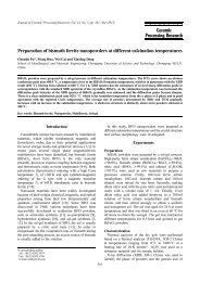
is shown in Fig. 13. It was confirmed that V 2<br />
O 5<br />
process<br />
is responsible for the pigment <strong>color</strong>ing, first reacts<br />
which<br />
N(t)<br />
N(t)<br />
N(t)<br />
5+ exists in the state <strong>of</strong> V . In this case, SiO 2<br />
way <strong>vanadium</strong><br />
to the vapor phase <strong>of</strong> SiF 4<br />
[8]. SiO 2<br />
diffuses<br />
converted is<br />
the reaction is completed. It was determined that <strong>vanadium</strong>
Fig. 13. Schematic model for formation <strong>of</strong> the <strong>vanadium</strong> zircon <strong>ceramic</strong> pigment.<br />
are included into the zircon structure in the state <strong>of</strong><br />
ions +<br />
resulting in the blue <strong>color</strong>ing.<br />
4<br />
o C<br />
*500-700<br />
2 +4NaF→ SiF 4(g) +2Na 2 O [24]<br />
SiO<br />
2 +4NaF→ SiF 4(g) +Na 2 SiO 3 [24]<br />
2SiO<br />
2 O 5 +4NaF+O 2 → 4NaVO 3 +2F 2<br />
2V<br />
2 +2F 2 → SiF 4(g) +O 2<br />
SiO<br />
o C<br />
*700-800<br />
4 +ZrO 2 +O 2 → ZrSiO 4 +2F 2 [24]<br />
SiF<br />
3 +ZrO 2 +SiO 2 → ZrSiO 4 +2Na 2 O<br />
4NaVO<br />
2 → ZrSiO 4 M-ZrO<br />
Conclusions<br />
In the case where rice husk ash was used to<br />
(1)<br />
a V-ZrSiO 4 pigment, the best <strong>color</strong>ing was<br />
synthesize<br />
o synthesis was carried out at 750 C for 5<br />
obtained when<br />
the addition <strong>of</strong> O.2 mol% <strong>of</strong> V 2 O 5 <strong>and</strong> 0.5 mol%<br />
with hours<br />
NaF; the resulting <strong>color</strong> values were L* 58.38, a* −12.87<br />
<strong>of</strong><br />
b* −10.63.<br />
<strong>and</strong><br />
The process the pigment synthesis is<br />
(2) o formation o <strong>of</strong><br />
500 C to 700 C, the temperature for<br />
From follows: as<br />
active reaction there is the initial step reaction caused<br />
an<br />
the phase transition <strong>of</strong> α-quartz →β-cristobalite involving<br />
by<br />
reaction <strong>of</strong> V 2 O 5 with NaF to produce the intermediate<br />
the<br />
NaVO 3 <strong>and</strong> SiF 4 . At the final step in the temperature<br />
phase<br />
o o C to 800 C, zircon is synthesized as<br />
range from 700<br />
into the M-ZrO 2 lattice. As soon as it is<br />
diffuses 4 SiF<br />
4+ is incorporated into the mother crystal<br />
produced, V<br />
a turquoise blue <strong>color</strong>ing <strong>of</strong> the pigment.<br />
in resulting<br />
As for the valence <strong>of</strong> the <strong>vanadium</strong> ions in the<br />
(3)<br />
pigment, the result <strong>of</strong> UV-Vis analysis showed<br />
synthesized<br />
4+ absorption b<strong>and</strong>s <strong>of</strong> V at 290 nm<br />
the characteristic<br />
→ 2A 1 ) <strong>and</strong> 640 nm (2B 1 → 2 E ).<br />
1 (2B<br />
Based on the result <strong>of</strong> the Raman spectral analysis<br />
(4)<br />
the synthesized pigment, new assignments were made<br />
<strong>of</strong><br />
the internal modes at 1003, 971, 950, 625, 434, 389,<br />
<strong>of</strong> −1 zircon<br />
201 <strong>and</strong> 181 cm as well as the V-O vibration<br />
219, 353,<br />
<strong>Formation</strong> <strong>and</strong> <strong>color</strong> <strong>properties</strong> <strong>of</strong> <strong>vanadium</strong> <strong>doped</strong> ZrSiO 4 <strong>ceramic</strong> pigments 287<br />
α-quartz →β-cristobalite<br />
internal modes at 950, 912, 882, 498 <strong>and</strong> 473 cm −1 .<br />
Acknowledgments<br />
This work was supported by Korea Science <strong>and</strong>
(KOSEF) grant funded by Korean Government<br />
Engineering<br />
(No. ROA-2006-000-10442-0).<br />
(MOST)<br />
A. Beltran, S. Bohm, A. Flores-Riveros, J.A. Igualada, G.<br />
1.<br />
<strong>and</strong> J. Andres, J. Phys. Chem. 97[11] (1993) 2555-<br />
Monros<br />
2559.<br />
C.A. Seabright <strong>and</strong> U.S Patent. 2,441,447 (1948).<br />
2.<br />
K.R. Pyon <strong>and</strong> B.H. Lee, J. Ceram. Soc. Jpn. 117[3] (2009)<br />
3.<br />
258-263.<br />
M. Llusar, J.B. Vicent, J. Badenes, M.A. Tena <strong>and</strong> G.<br />
4.<br />
J. Euro. Ceram. Soc. 19[15] (1999) 2647-2657.<br />
Monros,<br />
G. Monros, J. Carda, M. A. Tena <strong>and</strong> P. Escribano, J. Euro.<br />
5.<br />
Soc. 11 (1993) 77-86.<br />
Ceram.<br />
M. Shoyama, H. Nasu <strong>and</strong> K. Kamiya, J. Ceram. Soc. Jpn.<br />
6.<br />
(1998) 279-284.<br />
106[3]<br />
G. Mnros, J. Carda, M. A. Tena, P. Escribano, V. Cantavella<br />
7.<br />
J. Alarcon, Mat. Res. Bull. 27[6] (1992) 753-760.<br />
<strong>and</strong><br />
R.A. Eppler, J. Am. Ceram. Soc. 53[8] (1970) 457-462.<br />
8.<br />
T. Demiray, D.K. Nath <strong>and</strong> F.A. Hummel, J. Am. Ceram.<br />
9.<br />
53[1] (1970) 1-4.<br />
Soc.<br />
M. Ocana <strong>and</strong> A.R. Gonzalez-Elipe, J. Am. Ceram. Soc.<br />
10.<br />
(1998) 395-409.<br />
81[2]<br />
D. De Waal, A.M. Heyns <strong>and</strong> G. Pretorius, J. Raman. Spec.<br />
11.<br />
P. Ch<strong>and</strong>ley, R.J.H Clark, J. Crystal Growth. 116[1/2]<br />
12.<br />
151-157.<br />
(1992)<br />
F. D. Hardcastle, I. E. Wachs, J.Phys. Chem. 95[13] (1991)<br />
13.<br />
657-662.<br />
K. R. Pyon <strong>and</strong> B. H. Lee, J. Kor. Ceram. Soc. 46[4] (2009)<br />
14.<br />
397-404.<br />
S. J. Park, M. H. Kim, <strong>and</strong> Y. K. Choi, J. Biosystem Eng.<br />
15.<br />
(2006) 33-38.<br />
31[1]<br />
F. Bondioli, F. Andreola, L. Barbieri, T. Manfredini, <strong>and</strong> A.<br />
16.<br />
Ferrari, J. Euro. Ceram. Soc. 27[12] (2007) 3483-3488.<br />
M.<br />
C. S. Prasad, K. N. Maiti, <strong>and</strong> R. Venugopal, Ceram. Inter.<br />
17.<br />
(2001) 629-635.<br />
27[6]<br />
Y. Kanno, J. Mater. Sci. 24[7] (1989) 2415-2420.<br />
18.<br />
K.V.G.K. Gokhale, S.V. Ramani <strong>and</strong> E.C. Subbarao, J. Mater.<br />
19.<br />
4 , Letter (1969) 468-469.<br />
Sci.<br />
G. Monoros, M.C. Marti, J. Carda, M.A. Tena, V. Cantavella<br />
20.<br />
J. Alarcon, Br. Ceram. Trans. 92[3] (1993) 120-127.<br />
<strong>and</strong><br />
G. Monoros, M.C. Marti, J. Carda, M.A. Tena, P. Escribano,<br />
21.<br />
Canatavella <strong>and</strong> J. Alarcon, Mat. Res. Bull. 27 (1992)<br />
V.<br />
753-760.<br />
R.W.G Syme, D.J. Lockwood <strong>and</strong> H.J. Kerr, J. Phys. C:<br />
22.<br />
State Phys. 10 (1977) 1335-1348.<br />
Solid<br />
E. Knittle <strong>and</strong> Q. Williams, American Mineralogist 78<br />
23.<br />
245-252.<br />
(1993)<br />
288 Kyu-Ri Pyon, Kyong-Sop Han <strong>and</strong> Byung-Ha Lee<br />
References<br />
24. M. Trojan, Dyes <strong>and</strong> Pigments 9 (1988) 261-273.<br />
27 (1996) 657-662.