Laparoscopic Treatment of Deep Infiltrating Endometriosis ... - Sobracil
Laparoscopic Treatment of Deep Infiltrating Endometriosis ... - Sobracil
Laparoscopic Treatment of Deep Infiltrating Endometriosis ... - Sobracil

You also want an ePaper? Increase the reach of your titles
YUMPU automatically turns print PDFs into web optimized ePapers that Google loves.
Original Article<br />
Vol. Brazilian 6, Nº 1Journal<br />
<strong>Laparoscopic</strong> <strong>Treatment</strong> <strong>of</strong> <strong>Deep</strong> <strong>Infiltrating</strong> <strong>Endometriosis</strong> <strong>of</strong> the Intestine - Technical Aspects 23<br />
<strong>of</strong> Videoendoscopic<br />
Surgery<br />
<strong>Laparoscopic</strong> <strong>Treatment</strong> <strong>of</strong> <strong>Deep</strong> <strong>Infiltrating</strong><br />
<strong>Endometriosis</strong> <strong>of</strong> the Intestine - Technical Aspects<br />
Tratamento Cirúrgico de Endometriose Pr<strong>of</strong>unda Infiltrativa Intestinal<br />
por Laparoscopia - Aspectos Técnicos<br />
WILLIAM KONDO 1,2 ; MONICA TESSMANN ZOMER 1,2 ; REITAN RIBEIRO 2 ; CARLOS TRIPPIA 3 ; MARCO<br />
AURÉLIO PINHO DE OLIVEIRA 4,5 ; CLAUDIO PEIXOTO CRISPI 4<br />
1.<br />
Sugisawa Hospital and Medical Center, Curitiba; 2. Vita Batel Hospital, Curitiba; 3. Instituto Roentgen<br />
Diagnóstico/ Roentgen Diagnostic Institute, Curitiba; 4. Endometrosis <strong>Treatment</strong> Center <strong>of</strong> Rio de Janeiro;<br />
5.<br />
Pedro Ernesto University Hospital, University <strong>of</strong> the State <strong>of</strong> Rio de Janeiro (UERJ).<br />
ABSTRACT<br />
Surgery remains the best long-term treatment for deep infiltrating endometriosis affecting the bowel. Several surgical<br />
procedures (rectal shaving, mucosal skinning, discoid resection, and segmental bowel resection) have been performed<br />
with heterogeneous outcomes. In this article we discuss the indications <strong>of</strong> each procedure as well as the technical<br />
details.<br />
Key words: <strong>Deep</strong> infiltrating endometriosis. Laparoscopy. Surgery. Colorectal. Bowel.<br />
Braz. J. Video-Sur, 2012, v. 5, n. 2: 023-039<br />
Accepted after revision: march, 13, 2012.<br />
INTRODUCTION<br />
<strong>Endometriosis</strong> is a chronic inflammatory disease<br />
characterized by the growth <strong>of</strong> stroma and/or<br />
endometrial glands in areas outside the endometrial<br />
cavity. The prevalence <strong>of</strong> endometriosis in women <strong>of</strong><br />
reproductive age is estimated at about 10%. 1<br />
Infiltrative forms <strong>of</strong> the disease, called deep<br />
infiltrating endometriosis (DIE), are responsible<br />
for painful symptoms whose intensity is correlated to<br />
the depth <strong>of</strong> lesions 2 and seem to affect about 20% <strong>of</strong><br />
women with endometriosis. 3<br />
One <strong>of</strong> the basic characteristics <strong>of</strong> DIE is<br />
the multifocal pattern <strong>of</strong> distribution <strong>of</strong> lesions, which<br />
is <strong>of</strong> fundamental importance in both the diagnosis<br />
and treatment <strong>of</strong> the disease. There is a<br />
“preference” <strong>of</strong> the disease for the posterior pelvic<br />
compartment (uterosacral ligaments, retrocervical<br />
region, posterior vaginal fornix, rectosigmoid and<br />
ureter) over the anterior pelvic compartment (round<br />
ligament, anterior uterine wall/anterior peritoneal<br />
reflection, and bladder) and the remainder <strong>of</strong> the<br />
abdomen (small bowel, omentum, right colon and<br />
appendix). 4,5 In addition, there is a direct association<br />
between the presence <strong>of</strong> cystic lesions <strong>of</strong><br />
endometriosis in the ovary (ovarian endometrioma)<br />
and DIE. 6<br />
Intestinal DIE is defined as a lesion <strong>of</strong><br />
endometriosis that infiltrates at least the muscularis<br />
propria layer <strong>of</strong> the intestinal wall 7 and has great<br />
preference for the rectosigmoid. 5,8,9 There appears<br />
to be intestinal involvement in about 45% to 56% <strong>of</strong><br />
patients with DIE 5,8 and 57.1% <strong>of</strong> women with<br />
ovarian endometrioma. 6 When it affects the<br />
rectosigmoid colon, the intestinal involvement usually<br />
occurs by contiguity and, over time, successive<br />
inflammatory processes triggered by cyclic bleeding<br />
<strong>of</strong> the lesions promote the “infiltration” <strong>of</strong> the lesion<br />
into the intestinal wall. Some characteristic findings<br />
<strong>of</strong> DIE in intestinal imaging are due this<br />
pathophysiological process, which promotes<br />
proliferation <strong>of</strong> the smooth muscle tissue and a<br />
fibrotic reaction, resulting in the formation <strong>of</strong> a solid<br />
nodule. 10<br />
The high rate <strong>of</strong> intestinal involvement by DIE<br />
has lead to the popularization and ongoing improvement<br />
<strong>of</strong> surgical techniques used to address this disease.<br />
In this article we describe in detail the surgical<br />
principles and operative tactics used by our group<br />
when approaching intestinal DIE.<br />
23
24 Kondo et al.<br />
Braz. J. Video-Sur., January / March 2013<br />
PRE-OPERATIVE DIAGNOSIS<br />
The preoperative evaluation <strong>of</strong> patients with<br />
endometriosis begins with a good history and a<br />
thorough physical examination.<br />
Symptoms that should raise suspicion <strong>of</strong> the<br />
likely presence <strong>of</strong> the endometriosis include<br />
dysmenorrhea, dyspareunia, and chronic pelvic pain.<br />
Beside these, it is important to investigate digestive<br />
symptoms and menstrual or peri-menstrual urinary<br />
symptoms. In a retrospective study, FAUCONNIER<br />
and cols. 11 demonstrated that painful symptomatology<br />
was specific to an anatomic site or organ affected by<br />
the DIE implant.<br />
Dyspareunia was associated with DIE<br />
involving the uterosacral ligaments; painful bowel<br />
movements during menstruation were associated with<br />
DIE involving the posterior wall <strong>of</strong> the vagina; noncyclic<br />
pelvic pain and functional bowel symptoms were<br />
associated with intestinal involvement; and functional<br />
urinary symptoms were associated with DIE<br />
involvement <strong>of</strong> the bladder. In a prospective study <strong>of</strong><br />
CHAPRON and cols. 12 observed that painful<br />
defecation during menses and severe dyspareunia are<br />
specifically related to the presence <strong>of</strong> DIE<br />
compromising the posterior pelvic compartment.<br />
Recently, BALLESTER and cols. 13 have<br />
shown that there is also an increased incidence <strong>of</strong><br />
urinary symptoms in women with posterior DIE and<br />
that parametrial involvement is associated with<br />
changes in urinary function. In the case <strong>of</strong> intestinal<br />
DIE, there is greater likelihood <strong>of</strong> the presence <strong>of</strong><br />
symptoms such as cyclic painful defecation, cyclical<br />
constipation, and longer evacuation time. 14<br />
These symptoms, however, may be absent;<br />
thus infiltrative lesions should be sought in all women<br />
who present with complaints <strong>of</strong> severe dysmenorrhea<br />
that alter their quality <strong>of</strong> life significantly (causing them<br />
to miss work, school, etc.) or that require use <strong>of</strong> potent<br />
analgesics. The questioning <strong>of</strong> patients regarding<br />
menstrual history during adolescence can identify<br />
markers associated with DIE. 15 Patients with DIE<br />
have a positive family history <strong>of</strong> endometriosis,<br />
increased absenteeism from school during<br />
menstruation, and greater use <strong>of</strong> oral contraceptives<br />
for the treatment <strong>of</strong> severe primary dysmenorrhea.<br />
The physical examination is a fundamental step<br />
in the preoperative evaluation <strong>of</strong> deep endometriosis<br />
lesions (Figure 1). On vaginal examination, one looks<br />
for a zone that is hardened, retracted, and extremely<br />
Figure 1 - Speculum examination showing an endometriotic gland<br />
in the posterior vaginal fornix.<br />
tender in the posterior fornix <strong>of</strong> the vagina and/or in the<br />
topography <strong>of</strong> the uterosacral ligaments. One should<br />
assess the volume and limits (inferior, posterior, and lateral)<br />
<strong>of</strong> the lesion. On rectal examination, one must<br />
determine the relationships between the lesion and the<br />
wall <strong>of</strong> the rectum, assessing the mobility <strong>of</strong> the layers<br />
<strong>of</strong> the rectal wall.<br />
Vaginal examination alone, however, may be<br />
insufficient to accurately detect endometriosis prior<br />
to laparoscopy. The use <strong>of</strong> transvaginal pelvic<br />
ultrasound, performed by a well-trained radiologist,<br />
improves the diagnostic accuracy <strong>of</strong> the examination,<br />
especially in patients with ovarian endometrioma or<br />
DIE involving the uterosacral ligaments, the bladder,<br />
or the rectosigmoid. 16 Several studies have shown<br />
that transvaginal pelvic ultrasound is <strong>of</strong> utmost<br />
importance for the preoperative mapping <strong>of</strong><br />
endometriosis involving the ovaries, the region<br />
retrocervical region, or the rectosigmoid. 6,8,10,16-19<br />
The objective <strong>of</strong> preoperative imaging,<br />
therefore, is to map more precisely the areas affected<br />
by the disease, identifying especially those lesions that<br />
affect the gastrointestinal and urinary tracts.<br />
Specifically in the case <strong>of</strong> intestinal DIE lesions, the<br />
preoperative mapping should contain the following<br />
information: 10,18,19
Vol. 6, Nº 1<br />
<strong>Laparoscopic</strong> <strong>Treatment</strong> <strong>of</strong> <strong>Deep</strong> <strong>Infiltrating</strong> <strong>Endometriosis</strong> <strong>of</strong> the Intestine - Technical Aspects 25<br />
· size <strong>of</strong> the lesion;<br />
· the depth <strong>of</strong> infiltration <strong>of</strong> the lesion in the<br />
layers <strong>of</strong> the intestinal wall;<br />
· distance <strong>of</strong> the lesion from the anal verge;<br />
· percentage <strong>of</strong> intestinal circumference<br />
involved;<br />
· presence <strong>of</strong> multifocal lesions.<br />
Ultrasonography <strong>of</strong> the urinary tract is also<br />
indispensable during the preoperative evaluation <strong>of</strong><br />
bulky (> 30 mm) retrocervical DIE lesions and <strong>of</strong> lateral<br />
DIE lesions to exclude ureteral involvement by<br />
the disease. 20,21<br />
Some authors 22,23 have shown that magnetic<br />
resonance imaging (MRI) <strong>of</strong> the pelvis with<br />
opacification <strong>of</strong> the vagina and rectum with ultrasound<br />
gel has a high sensitivity for detection <strong>of</strong> DIE lesions.<br />
In our experience, MRI is important for assessing<br />
ovarian endometriomas and multifocal intestinal DIE<br />
lesions; it does not, however, replace ultrasound. The<br />
depth <strong>of</strong> infiltration <strong>of</strong> a lesion in the intestinal wall is<br />
best assessed by ultrasonography (Figure 2).<br />
Some groups have experience with the use<br />
<strong>of</strong> barium enema as part <strong>of</strong> the preoperative evaluation<br />
and surgical planning <strong>of</strong> patients with intestinal DIE, 24<br />
but transvaginal pelvic ultrasound has a higher<br />
sensitivity in detecting the presence <strong>of</strong> posterior DIE<br />
lesions. 25 As the predilection <strong>of</strong> the DIE implants is for<br />
the outer layers <strong>of</strong> the intestinal wall, 26 colonoscopy –<br />
which better evaluates the mucosa <strong>of</strong> the intestine –<br />
has a limited role in identifying intestinal DIE lesions.<br />
Still, colonoscopy may be able to identify extrinsic<br />
compression by deep endometriosis nodules and<br />
involvement <strong>of</strong> the intestinal mucosa in cases with<br />
complete infiltration <strong>of</strong> all intestinal layers (Figure 3).<br />
patients reporting substantial reduction or complete<br />
relief <strong>of</strong> pain symptoms.<br />
Figure 2 - Transvaginal pelvic ultrasound showing a cross section<br />
<strong>of</strong> the intestinal wall, with all its layers. From outside to inside:<br />
serosa, muscularis propria, submucosa, muscularis mucosa, and<br />
mucosa.<br />
CLINICAL TREATMENT<br />
Medical management has an important role<br />
in controlling the painful symptoms resulting from DIE<br />
involving the bowel. In the review by VERCELLINI<br />
and cols. 27 , pharmacological treatment <strong>of</strong> patients with<br />
rectovaginal endometriosis achieved good pain relief,<br />
reduced lesions during therapy, improved healthrelated<br />
quality <strong>of</strong> life, and patient satisfaction. The<br />
analgesic effect <strong>of</strong> the medications evaluated (GnRH<br />
agonists, danazol, intrauterine contraceptive with<br />
progestin, oral progestin, and combined oral<br />
contraceptive) was significant for the entire treatment<br />
period (from 6 to 12 months), with 60% to 90% <strong>of</strong><br />
Figure 3 - Four DIE lesions causing extrinsic compression and<br />
infiltration <strong>of</strong> the intestinal wall.
26 Kondo et al.<br />
Braz. J. Video-Sur., January / March 2013<br />
FERRERO and cols. 28 evaluated six women<br />
with colorectal endometriosis infiltrating at least the<br />
muscular layer <strong>of</strong> the bowel, without stenosis<br />
exceeding 60% <strong>of</strong> the lumen, with painful symptoms<br />
and infections. They were treated with letrozole (2.5<br />
mg/d) and norethisterone acetate (2.5 mg/d)<br />
continuously for six months and had improvement <strong>of</strong><br />
pain symptoms, non-cyclic pelvic pain, deep<br />
dyspareunia, dyschezia, intestinal colic, abdominal<br />
distension, and the presence <strong>of</strong> mucus in the stool.<br />
Gastrointestinal symptoms improved in 67% <strong>of</strong><br />
women.<br />
Recently a prospective study 29 evaluated the<br />
efficacy <strong>of</strong> continuous use <strong>of</strong> low-dose oral<br />
contraceptive (15ìg 60ìg ethinyl estradiol and<br />
gestodene for 12 months) in the treatment <strong>of</strong> pain and<br />
other symptoms associated with colorectal<br />
endometriosis nodules. Twenty-six women <strong>of</strong><br />
reproductive age with colorectal nodules infiltrating<br />
at least the intestinal muscularis proprio and without<br />
intestinal stenosis exceeding 50% were evaluated. A<br />
significant improvement in the intensity <strong>of</strong> all symptoms<br />
(dysmenorrhea, non-menstrual pelvic pain, deep<br />
dyspareunia, and painful defecation) rated using a visual<br />
analog scale was observed. There was a<br />
reduction in diameter (mean reduction 26%) and volume<br />
(mean reduction <strong>of</strong> 62%) <strong>of</strong> the nodules after a<br />
period <strong>of</strong> 12 months <strong>of</strong> treatment.<br />
Although the aforementioned studies show<br />
that medical management has an important role in<br />
controlling pain symptoms, it is important to understand<br />
that there may be progression <strong>of</strong> the lesion over the<br />
long term, even when patients’ pain symptoms are<br />
controlled. When opting for medical management <strong>of</strong><br />
endometriosis, monitoring with annual imaging studies<br />
should be performed, because the presence <strong>of</strong><br />
obstruction <strong>of</strong> the urinary tract or gastrointestinal tract<br />
is an absolute indication for surgery. Furthermore,<br />
medical management has no benefit in the treatment<br />
<strong>of</strong> endometriosis-related infertility.<br />
SURGICAL TREATMENT<br />
Surgical treatment <strong>of</strong> deep infiltrating<br />
endometrial lesions <strong>of</strong> the intestinal is reserved for<br />
patients who have failed clinical treatment, have<br />
recurrent symptoms after stopping medical treatment,<br />
infertility related to endometriosis, obstruction <strong>of</strong> the<br />
urinary tract or gastrointestinal tract, or the presence<br />
<strong>of</strong> cyclic rectorrhagia.<br />
We describe our surgical approach for DIE<br />
lesions located in the rectum and sigmoid colon, which<br />
represent more than 80% <strong>of</strong> DIE bowel lesions. 4 For<br />
didactic purposes, we discuss the lesions affecting the<br />
rectum and rectosigmoid (up to 15cm from the anal<br />
margin) separately from those affecting the sigmoid.<br />
Lesions <strong>of</strong> the rectum and rectosigmoid<br />
Intestinal DIE involving the rectum and<br />
rectosigmoid can be treated in variety <strong>of</strong> ways. 30-36<br />
Surgical planning is performed in accordance with the<br />
preoperative diagnostic imaging findings as described<br />
above. 10,18,19<br />
The techniques described in the literature to<br />
address these intestinal lesions <strong>of</strong> DIE include rectal<br />
shaving, rectal shaving with mucosal skinning, discoid<br />
resection and segmental resection. Each technique has<br />
its peculiarities in terms <strong>of</strong> surgical principles, risks <strong>of</strong><br />
intraoperative and postoperative complications,<br />
recurrence rate, and postoperative fertility.<br />
Rectal Shaving and mucosal skinning<br />
Rectal shaving and mucosal skinning require<br />
finding – in the thickness <strong>of</strong> the muscular layer <strong>of</strong> the<br />
intestinal wall – a clean plane beyond which there are<br />
no more endometriosis lesions. This procedure is<br />
performed accepting a risk <strong>of</strong> opening the intestinal<br />
lumen. According to Donnez and cols., 37 this resection<br />
should avoid as much as possible the risk <strong>of</strong> opening<br />
the intestinal lumen. For others, the resection should<br />
be complete (mucosal skinning = preservation only <strong>of</strong><br />
the intact mucosal layer with manual suturing <strong>of</strong> the<br />
sectioned bowel wall), accepting the risk <strong>of</strong> an opening<br />
the lumen <strong>of</strong> the intestine in a certain number <strong>of</strong><br />
patients, 38 at the expense <strong>of</strong> systematic attainment <strong>of</strong><br />
a segmental resection <strong>of</strong> the intestine. 39,40<br />
Two surgical tactics can be used: 35<br />
· Tradicional technique;<br />
· Reverse technique.<br />
Traditionally, gynecologists first separate the<br />
posterior surface <strong>of</strong> the nodule from the anterior wall<br />
<strong>of</strong> the rectum, and then perform the separation <strong>of</strong> the<br />
lesion from the posterior vaginal fornix and from the<br />
retrocervical region (Figure 4). It is important to<br />
remember that the lower the dissection, the more anterior<br />
the axis <strong>of</strong> scissors should be to avoid injuries to<br />
the rectum.<br />
The retraction induced by the nodule causes<br />
the rectum to envelop the nodule; therefore the<br />
axis <strong>of</strong> the cut is not constant throughout the
Vol. 6, Nº 1<br />
<strong>Laparoscopic</strong> <strong>Treatment</strong> <strong>of</strong> <strong>Deep</strong> <strong>Infiltrating</strong> <strong>Endometriosis</strong> <strong>of</strong> the Intestine - Technical Aspects 27<br />
Figure 4 - (A) <strong>Deep</strong> endometriotic lesion involving the right uterosacral ligament, posterior vaginal fornix and the anterior wall <strong>of</strong> the<br />
rectum (to the muscle layer). (B) The arrow identifies the superior retraction <strong>of</strong> the rectum caused by the endometriosis lesion. (C and D)<br />
Opening <strong>of</strong> the fibers <strong>of</strong> the muscle layer <strong>of</strong> the rectum (arrows) for the removal <strong>of</strong> the endometriosis lesion. (E) Suturing with 3-0<br />
polypropylene <strong>of</strong> the area <strong>of</strong> the defect in the muscle layer <strong>of</strong> the rectum (arrows). (E) Sutura da área de defeito na camada muscular do<br />
reto (setas) com fio de polipropileno 3-0. (F) Rectal suture (yellow arrow) and the area to be dissected in the rectovaginal septum (green<br />
arrows). (G to I) Separation <strong>of</strong> the endometriosis lesion from the retrocervical region and from the rectovaginal septum, and resection <strong>of</strong><br />
the posterior vaginal fornix.<br />
dissection. The surgeon should follow the contour<br />
<strong>of</strong> the rectum around the endometriotic nodule. The<br />
goal is to free the nodule completely from the bowel<br />
wall to identify the healthy vagina distal to the<br />
nodule. This dissection can be aided by a digital<br />
rectal and vaginal examination, by positioning a<br />
gauze compress or a malleable valve in the posterior<br />
vaginal fornix or by the insertion <strong>of</strong> a rectal<br />
probe. After freeing the posterior face <strong>of</strong> the<br />
nodule, it should be freed from the posterior aspect<br />
<strong>of</strong> the uterus, from the base <strong>of</strong> the broad ligament,<br />
and from the vagina.<br />
When the case requires resection <strong>of</strong> the vagina,<br />
cold scissors or fine (1 mm diameter) monopolar<br />
tip with pue cutting current is used. Whenever opening<br />
a cyst containing chocolate-colored contents or if you<br />
see the appearance <strong>of</strong> small blackened points,<br />
resection is not sufficient, and the excision should be
28 Kondo et al.<br />
Braz. J. Video-Sur., January / March 2013<br />
expanded. Avoid the loss <strong>of</strong> CO 2<br />
during this surgical<br />
time by placing a surgical glove containing two or three<br />
gauzes into the vagina.<br />
Alternatively, one may use a surgical approach<br />
called a “reverse” (Figure 5). In this technique, first<br />
the anterior surface <strong>of</strong> the nodule is freed from the<br />
posterior surface <strong>of</strong> the uterus and vagina. The mobility<br />
obtained after freeing the anterior face <strong>of</strong> the nodule<br />
allows better exposure <strong>of</strong> the nodule during the<br />
dissection <strong>of</strong> the most difficult area to be treated,<br />
which is in contact with the rectum. When the nodule<br />
infiltrates the vagina, the vagina is opened at it superior<br />
segment, near the cervix, and then laterally to<br />
disease-free zones. The distal limit <strong>of</strong> vaginal<br />
sectioning is identified through the vagina, distal to the<br />
nodule. After sectioning the distal limit <strong>of</strong> the lesion,<br />
one identifies a healthy rectovaginal plane. In this<br />
way the nodule can easily be pulled with a grasping<br />
forceps to expose its posterior surface adhered to the<br />
anterior wall <strong>of</strong> the rectum, followed by the progressive<br />
Figure 5 – (A) DIE lesion involving the intestine (in yellow). (B) Resection <strong>of</strong> the right uterosacral ligament and separation <strong>of</strong> lesion from<br />
the retrocervical region (arrows). (C and D) Resection <strong>of</strong> the posterior vaginal fornix with identification <strong>of</strong> the healthy area in rectovaginal<br />
septum. (E to H) Separation <strong>of</strong> DIE lesion (arrows) from the rectal wall progressively sectioning the muscle layer from the intestinal wall.<br />
(I) Thin appearance <strong>of</strong> the rectum (arrows).
Vol. 6, Nº 1<br />
<strong>Laparoscopic</strong> <strong>Treatment</strong> <strong>of</strong> <strong>Deep</strong> <strong>Infiltrating</strong> <strong>Endometriosis</strong> <strong>of</strong> the Intestine - Technical Aspects 29<br />
separation <strong>of</strong> the lesion from the anterior wall <strong>of</strong> the<br />
rectum.<br />
At the end <strong>of</strong> the procedure, if the shaving<br />
was deeper, with opening <strong>of</strong> the muscular layer or<br />
below, suturing is done with 3-0 or 4-0 mon<strong>of</strong>ilament<br />
(polypropylene) suture in a single plane with<br />
continuous or separated stitches.<br />
Discoid Resection<br />
Discoid Resection entails wedge resection <strong>of</strong><br />
the anterior wall <strong>of</strong> the rectum, in total thickness. Here<br />
we describe two techniques that can be used: 32,41<br />
· Resection <strong>of</strong> the endometrial nodule with<br />
the cold scissors, followed by rectal repair by manual<br />
suturing;<br />
· Resection <strong>of</strong> the endometrial nodule using a<br />
circular stapler inserted transanally.<br />
Most authors who use this technique reserve<br />
it for single lesions <strong>of</strong> DIE located on the anterior wall<br />
<strong>of</strong> the rectosigmoid colon, with deep infiltration beyond<br />
the muscle layer, that are smaller than 30mm in<br />
diameter and occupying less than one-third <strong>of</strong> the intestinal<br />
circumference. 41 Some authors, however,<br />
have described an alternative technique <strong>of</strong> double<br />
stapling for resection <strong>of</strong> bulkier lesions, with promising<br />
results. 42 In the technique with manual suturing, two<br />
repair sutures are placed adjacent to the lesion, in<br />
healthy intestinal wall, and the anterior intestinal wall<br />
is resected in a wedge with a transverse incision into<br />
the intestinal lumen using cold scissors or ultrasonic<br />
energy. The defect area in the anterior intestinal wall<br />
is then repaired in two planes : one, the total plane,<br />
encompasses the full wall thickness with continuous<br />
sutures ; and the other, the sero-muscular plane, uses<br />
continuous or separated 3-0 or 4-0 mon<strong>of</strong>ilament<br />
(polypropylene) sutures. 42<br />
We prefer to perform the discoid resection<br />
using the 29 or 33 mm circular intraluminal stapler<br />
(Ethicon Endosurgery Inc., Cincinnati, OH, USA)<br />
introduced transanally because the technique is<br />
quicker and there is no contact between the intestinal<br />
lumen and the abdominal-pelvic cavity during the<br />
procedure (Figure 6). After freeing the intestinal lesion<br />
from the retrocervical region, from the posterior vaginal<br />
fornix, and from the rectovaginal septum, as<br />
described above for the reverse technique, the<br />
endometriotic lesion remain attached only to the anterior<br />
wall <strong>of</strong> the rectum. A superficial shaving is<br />
performed to separate the posterior wall <strong>of</strong> the lesion<br />
from the anterior wall <strong>of</strong> the rectum, leaving only the<br />
fibrotic area <strong>of</strong> deep infiltration by the endometriosis<br />
in the intestinal wall.<br />
A mon<strong>of</strong>ilament (2-0 mononylon) suture is<br />
passed into the lesion laparoscopically and left free in<br />
the cavity with the long ends. Guided laparoscopically,<br />
the circular stapler is inserted transanally, until it is<br />
beyond the lesion. It is gently opened, observing the<br />
formation <strong>of</strong> a groove between the anvil and the<br />
stapler. The two ends <strong>of</strong> the suture are grasped with<br />
a needle holder and pulled vertically downward in order<br />
to introduce the fibrotic area <strong>of</strong> the anterior wall <strong>of</strong><br />
the rectum into the groove <strong>of</strong> the circular stapler.<br />
When the lesion is ideally positioned, the circular<br />
stapler is closed with the anterior wall <strong>of</strong> the<br />
rectum inside. During this closure, the stapler should<br />
be lowered so that its end is anteriorized, thereby<br />
avoiding the stapling <strong>of</strong> the posterior intestinal wall.<br />
The stapler is fired, and the anterior wall <strong>of</strong> the rectum<br />
is stapled and cut. After careful removal <strong>of</strong> the stapler,<br />
one can view the discoid <strong>of</strong> anterior rectal wall inside<br />
the circular stapler.<br />
In attempting to expand the use <strong>of</strong> discoid<br />
resection several authors 42 have developed the same<br />
technique with double stapling (Figure 7). In this way<br />
single lesions with lengths <strong>of</strong> up to 60mm and involving<br />
up to 40% the intestinal circumference can be removed.<br />
In this technique, the first suture is passed from the<br />
proximal free edge to the middle <strong>of</strong> the lesion. The first<br />
firing <strong>of</strong> the circular stapler removes part <strong>of</strong> the intestinal<br />
lesion. Another suture is passed including the staple<br />
line <strong>of</strong> the intestine and distal free edge <strong>of</strong> the lesion.<br />
The second firing <strong>of</strong> the circular stapler results in the<br />
removal <strong>of</strong> the rest <strong>of</strong> the intestinal DIE lesion.<br />
We can call the three above-mentioned<br />
surgical techniques (rectal shaving, mucosal skinning,<br />
and discoid resection) “nodulectomies,” since all have<br />
the goal <strong>of</strong> removing only the intestinal DIE nodule<br />
from the intestinal wall.<br />
Segmental resection <strong>of</strong> the intestine<br />
Segmental resection <strong>of</strong> the bowel entails<br />
resecting the segment <strong>of</strong> the bowel affected by DIE.<br />
In theory, this technique <strong>of</strong>fers “certainty” – once the<br />
margins <strong>of</strong> the resection are free <strong>of</strong> disease – that the<br />
lesion has been completely resected, 39,40 but it has an<br />
immediate operative morbidity, and a more significant<br />
long-term morbidity. 30,34,43,44 For some authors, this<br />
action should be systematic whenever the DIE<br />
infiltrates the muscle. 39,45
30 Kondo et al.<br />
Braz. J. Video-Sur., January / March 2013<br />
Figure 6 - (A and B) Separation <strong>of</strong> the lesion (in yellow) from the retrocervical region, leaving it attached to the anterior wall <strong>of</strong> the rectum.<br />
(C and D) Separation <strong>of</strong> the DIE lesion from the anterior wall <strong>of</strong> the rectum. (E) Residual area <strong>of</strong> infiltration <strong>of</strong> DIE in the intestinal wall<br />
(yellow arrows). (F) Passing the suture through healthy edges <strong>of</strong> the lesion. (G to I) Adjustment <strong>of</strong> the lesion within the circular stapler<br />
introduced transanally, finalizing the discoid resection.<br />
One <strong>of</strong> the important arguments in favor <strong>of</strong><br />
this more aggressive “attitude” would be the certainty<br />
<strong>of</strong> complete treatment <strong>of</strong> the disease, as<br />
REMORGIDA and cols. 46 showed that the histological<br />
examination <strong>of</strong> the surgical specimen <strong>of</strong> the intestine<br />
resected after a “nodulectomy” was performed found<br />
residual lesions <strong>of</strong> endometriosis involving the muscle<br />
layer <strong>of</strong> the intestine in 43.8% <strong>of</strong> cases. This is a<br />
strong argument in favor <strong>of</strong> intestinal segmental<br />
resection, about which we can also <strong>of</strong>fer some<br />
counter-arguments:<br />
· The “nodulectomy” performed in cases<br />
where one knows that the segmental resection <strong>of</strong><br />
the intestine will be performed during the same<br />
surgical procedure is probably not the same<br />
“nodulectomy” that gets performed under other<br />
circumstances;<br />
· The segmental resection is not always a<br />
guarantee <strong>of</strong> disease-free margins. There is<br />
histological evidence <strong>of</strong> positive margins after<br />
segmental resection for treatment <strong>of</strong> intestinal DIE in<br />
up to 22% <strong>of</strong> cases; 47,48
Vol. 6, Nº 1<br />
<strong>Laparoscopic</strong> <strong>Treatment</strong> <strong>of</strong> <strong>Deep</strong> <strong>Infiltrating</strong> <strong>Endometriosis</strong> <strong>of</strong> the Intestine - Technical Aspects 31<br />
Figure 7 - Length <strong>of</strong> the DIE implant in the rectum. (B and C) Passing the first suture through the proximal end <strong>of</strong> the lesion. (D and E)<br />
Stapling <strong>of</strong> the proximal end <strong>of</strong> the lesion. (F and G) Passing the second suture encompassing the previous stapling area and the distal<br />
end <strong>of</strong> the lesion. (H and I) Second stapling, finalizing the double discoid resection.<br />
· There is not always evidence <strong>of</strong> intestinal<br />
DIE (involvement beyond the muscle layer) in surgical<br />
specimens <strong>of</strong> segmental resections; 0.8% <strong>of</strong> the time<br />
the intestinal segment may not have histological<br />
evidence <strong>of</strong> transmural invasion by the disease; 47<br />
· It is not clear that the radicality <strong>of</strong> resecting<br />
the full thickness <strong>of</strong> the rectal muscle confers a real<br />
clinical benefit to patients. Recently, MABROUK and<br />
cols. 49 demonstrated that there is no statistical<br />
difference in terms <strong>of</strong> recurrence, pain symptoms, or<br />
improvement in quality <strong>of</strong> life in patients with and<br />
without positive margins after segmental resection <strong>of</strong><br />
the bowel.<br />
We reserve the segmental resection <strong>of</strong> the<br />
intestine for cases <strong>of</strong> bulky lesions <strong>of</strong> the rectosigmoid<br />
exceeding 30mm, stenotic lesions (that block advancing<br />
the circular stapler into position), lesions in the sigmoid<br />
colon (which are beyond the reach <strong>of</strong> the circular<br />
stapler), and multifocal lesions.<br />
The opening <strong>of</strong> the left parietocolic gutter in<br />
Toldt’s fascia allows mobilization <strong>of</strong> the descending<br />
colon (Figure 8). Rarely, it is necessary to completely
32 Kondo et al.<br />
Braz. J. Video-Sur., January / March 2013<br />
Figure 8 - (A and B) Opening <strong>of</strong> the mesorectum adjacent to the sacral promontory and dissection in a caudal direction. (C) Opening the<br />
peritoneal layer lateral to the rectum on the right side. (D) Nerve preservation is identified by yellow arrows. (E) Identification <strong>of</strong> the<br />
healthy area in the rectovaginal septum, distal to the intestinal DIE lesion. (F) Cranial dissection <strong>of</strong> mesorectum and mobilization <strong>of</strong> left<br />
parietocolic gutter. You can identify retroperitoneal structures: aorta, left ureter, and left gonadal vein. (G) Stapling distal to the intestinal<br />
DIE lesion. (H and I) End-to-end anastomosis using a circular stapler inserted transanally.<br />
mobilize the left colon by the opening the gastrocolic<br />
ligament, entering the epiploic retrocavity, freeing <strong>of</strong><br />
the splenic flexure <strong>of</strong> the colon, and separated the<br />
colon from the omentum.<br />
The rectosigmoid is pulled vertically by the<br />
assistant, up and toward the left pelvic wall, exposing<br />
the mesorectum and the sacral promontory. The<br />
mesorectum is dissected over the sacral promontory,<br />
first caudally to the level <strong>of</strong> the elevators and then<br />
cranially. The peritoneum layer lateral to the rectum<br />
is incised bilaterally and the peritoneal reflection <strong>of</strong><br />
the posterior fornix is opened.<br />
The dissection proceeds until a healthy area<br />
in rectovaginal septum is reached, which has been<br />
previously dissected using the reverse technique<br />
mentioned above. The rectum is mobilized at least 2
Vol. 6, Nº 1<br />
<strong>Laparoscopic</strong> <strong>Treatment</strong> <strong>of</strong> <strong>Deep</strong> <strong>Infiltrating</strong> <strong>Endometriosis</strong> <strong>of</strong> the Intestine - Technical Aspects 33<br />
cm below the endometriosis nodule. The 45mm<br />
articulated linear cutting endostapler (Ethicon<br />
Endosurgery, Inc., Cincinnati, OH, USA) is introduced<br />
into the cavity through a 12mm trocar positioned in<br />
right iliac fossa and the rectum is cut distal to the lesion<br />
(Figure 8G). At this point, there are some options for<br />
resection <strong>of</strong> the rectum or the rectosigmoid:<br />
· A transverse minilaparotomy is performed<br />
in suprapubic region (Pfannenstiel). After extraction<br />
<strong>of</strong> the intestine through the incision, the rectum or<br />
rectosigmoid (depending on the length <strong>of</strong> the lesion)<br />
proximal to the lesion is cut using a cold scalpel. A<br />
purse string suture is performed on the end <strong>of</strong> the<br />
sigmoid with 2-0 mononylon and the anvil <strong>of</strong> the 29<br />
mm or 33 mm intraluminal circular stapler (Ethicon<br />
Endosurgery, Inc., Cincinnati, OH, USA) is positioned<br />
and secured with the previously placed suture. The<br />
intestine is repositioned inside the abdominal cavity,<br />
the abdominal wall is sutured, the pneumoperitoneum<br />
is reestablished, and the transanal end-to-end<br />
colorectal anastomosis is performed guided<br />
laparoscopically;<br />
· In cases where it was necessary to resect<br />
the posterior vaginal fornix or perform a hysterectomy,<br />
the rectosigmoid can be extracted transvaginally. 50 The<br />
resection <strong>of</strong> the rectum or rectosigmoid is performed,<br />
followed by purse-string suturing <strong>of</strong> the end <strong>of</strong> the<br />
sigmoid and positioning <strong>of</strong> the anvil <strong>of</strong> the intraluminal<br />
circular stapler. The sigmoid is reintroduced into the<br />
abdominal cavity, a glove containing air and gauzes is<br />
introduced into the vagina to prevent the leakage <strong>of</strong><br />
CO 2<br />
, the pneumoperitoneum is reestablished, and the<br />
transanal end-to-end colorectal anastomosis is<br />
performed (Figure 9).<br />
Figure 9 - (A) Positioning <strong>of</strong> trocars for segmental resection <strong>of</strong> the intestine with transvaginal extraction <strong>of</strong> the surgical specimen. (B) Transvaginal<br />
extraction <strong>of</strong> the rectosigmoid. (C) Sectioning <strong>of</strong> the sigmoid colon. (D) Purse string suturing and positioning the anvil inside the sigmoid colon.
34 Kondo et al.<br />
Braz. J. Video-Sur., January / March 2013<br />
SURGICAL TREATMENT OF<br />
ENDOMETRIOSIS LESIONS OF THE<br />
SIGMOID COLON<br />
In cases <strong>of</strong> high lesions <strong>of</strong> the sigmoid colon<br />
(more than 15 cm from the anal margin), without<br />
evidence <strong>of</strong> multifocal lesions involving the rectum or<br />
rectosigmoid, one can opt for an isolated resection <strong>of</strong><br />
intestinal DIE implant.<br />
Because a circular stapler cannot be used to<br />
perform an anastomosis more than 15 cm from the<br />
anal margin, we avoid an extensive bowel resection<br />
performing the intestinal resection through a mini-<br />
Pfannenstiel incision (3 to 4 cm). In this way we can<br />
perform an economical bowel resection (remember<br />
we are dealing with a benign disease!) and preserve<br />
the rectum, which can reduce the chance <strong>of</strong> functional<br />
bowel changes postoperatively.<br />
After treatment <strong>of</strong> the pelvic endometriosis<br />
lesions and complete mobilization <strong>of</strong> the sigmoid colon,<br />
a mini-Pfannenstiel incision <strong>of</strong> approximately 4 to 5<br />
cm is made and the sigmoid colon is exteriorized<br />
through the incision. Two procedures can be<br />
performed:<br />
Discoid recession<br />
Discoid resection allows the maintenance<br />
<strong>of</strong> vascularization along the entire mesenteric margin<br />
<strong>of</strong> the intestinal wall (Figure 10). Two clamps are<br />
positioned on the loop <strong>of</strong> the sigmoid colon, one<br />
proximal and one distal to the lesion. The intestinal<br />
DIE lesion is resected with electrocautery (full<br />
thickness) giving rise to a defect in the antimesenteric<br />
margin <strong>of</strong> the sigmoid colon wall. The<br />
intestinal wall is sutured in two layers using<br />
continuous absorbable sutures <strong>of</strong> 3-0 polyglactin 910<br />
(Vicryl ® ) or nonabsorbable 3-0 polypropylene<br />
(Prolene ® ).<br />
Segmental resection <strong>of</strong> the intestine<br />
Two intestinal clamps are positioned on the<br />
loop <strong>of</strong> the sigmoid colon, one proximal and one distal<br />
to the lesion. The vessels <strong>of</strong> the meso-sigmoid are<br />
ligated close to the intestinal wall with 3-0 cotton suture<br />
and the intestinal segment to be removed is cut with<br />
the cold scalpel. The end-to-end anastomosis is made<br />
in two planes: (1) through the total thickness <strong>of</strong> the<br />
bowel wall with a running suture using absorbable 3-<br />
0 polyglactin 910 (Vicryl ® ) or nonabsorbable 3-0<br />
polypropylene (Prolene ® ), and (2) through the<br />
seromuscular layers using running or separate sutures<br />
using the same suture material (Figure 11).<br />
After the segmental resection <strong>of</strong> the intestine,<br />
the sigmoid colon is repositioned within the abdominal<br />
cavity. The incision in the aponeurosis is sutured with<br />
0 or 1 polyglactin 910 (Vicryl ® ) and the<br />
pneumoperitoneum is restored. Hemostasis is verified,<br />
and the test <strong>of</strong> tubal permeability is performed using<br />
methylene blue, when indicated. Both in the setting<br />
<strong>of</strong> discoid resection and in the case <strong>of</strong> segmental<br />
resection, we usually do not test the integrity <strong>of</strong> the<br />
anastomosis with the “tire-fitter’s” manuever because<br />
the air injected transanally normally cannot reach the<br />
area <strong>of</strong> the anastomosis.<br />
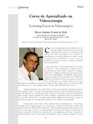
DISCUSSION<br />
As previously mentioned, DIE involving the<br />
intestine can be treated using different surgical<br />
techniques. The choice <strong>of</strong> surgical technique depends<br />
on a number <strong>of</strong> factors including: the patient’s<br />
symptoms, characteristics <strong>of</strong> the endometriosis lesions,<br />
the surgical team’s experience with the different<br />
procedures, the patient’s aspirations to have children,<br />
and the patient’s consent after being informed <strong>of</strong> the<br />
risks <strong>of</strong> each procedure. All authors agree that an<br />
indication for surgery exists when the stenosis exceed<br />
50% <strong>of</strong> the intestinal lumen, the urinary tract is<br />
obstructed, or in the presence <strong>of</strong> rectorrhagia. 21,51,52<br />
With nodulectomy or the segmental resection<br />
<strong>of</strong> the intestine there is the possibility <strong>of</strong> complications.<br />
The postoperative results <strong>of</strong> the different surgical<br />
techniques have been compared for various services<br />
and there seems to be a tendency to choose less<br />
aggressive techniques when possible. In a prospective<br />
analysis <strong>of</strong> 500 cases <strong>of</strong> DIE nodules treated by rectal<br />
shaving, 31 major complications included 7 cases (1.4%)<br />
<strong>of</strong> rectal perforation, 4 cases (0.8%) <strong>of</strong> ureteral injury,<br />
one case (0.2%) where bleeding exceeded 300 ml,<br />
and urinary retention in 4 patients (0.8%). Of the 388<br />
women who wanted to get pregnant, 221 (57%),<br />
conceived naturally, and 107 (28%) through in vitro<br />
fertilization. The recurrence rate was 8% and was<br />
significantly lower (p
Vol. 6, Nº 1<br />
<strong>Laparoscopic</strong> <strong>Treatment</strong> <strong>of</strong> <strong>Deep</strong> <strong>Infiltrating</strong> <strong>Endometriosis</strong> <strong>of</strong> the Intestine - Technical Aspects 35<br />
Figure 10 - (A) Presence <strong>of</strong> several endometriosis implants in the posterior fornix and uterosacral ligaments. (B) Intestinal DIE lesion<br />
involving the sigmoid colon. (C) Mobility test <strong>of</strong> the sigmoid to evaluate the possibility extraction through Pfannenstiel incision. (D)<br />
Transparietal extraction <strong>of</strong> sigmoid colon. (E and F) Resection <strong>of</strong> the lesion from the anterior and lateral walls <strong>of</strong> the sigmoid colon. (G)<br />
Appearance <strong>of</strong> the sigmoid wall defect created after resection <strong>of</strong> an intestinal DIE implant. (H) Suture <strong>of</strong> the sigmoid colon in the transverse<br />
direction. (I) Appearance after returning the sigmoid colon inside the abdominal cavity.<br />
postoperative period with segmental resection <strong>of</strong> the<br />
intestine than with isolated shaving (92% vs. 80%,<br />
respectively; p
36 Kondo et al.<br />
Braz. J. Video-Sur., January / March 2013<br />
Figure 11 - (A) Large intestinal DIE lesion involving the sigmoid colon. (B) The rectum and the rectosigmoid transition do not have other<br />
DIE lesions. (C) Extraction <strong>of</strong> the sigmoid colon through Pfannenstiel incision. (D) Ligation <strong>of</strong> the vessels <strong>of</strong> the meso-sigmoid. (E and F)<br />
Clamping proximal and distal to the sigmoid colon in preparation for the resection <strong>of</strong> the area affected by intestinal DIE. (G and H) Endto-end<br />
anastomosis in two planes. (I) <strong>Laparoscopic</strong> appearance <strong>of</strong> the suturing after the repositioning <strong>of</strong> the sigmoid colon inside the<br />
abdominal cavity.<br />
postoperative complications requiring surgical<br />
intervention in the discoid resection group was 4.16%<br />
and the recurrence rate at 33 months <strong>of</strong> median followup<br />
was 10.4%.<br />
Recently, two other studies 44,53 compared<br />
intra- and post-operative nodulectomy outcomes and<br />
results <strong>of</strong> segmental resection for the treatment <strong>of</strong><br />
intestinal DIE. In the first study, 53 “nodulectomy” was<br />
performed by discoid resection using a circular stapler.<br />
The mean operative time was shorter, blood loss was<br />
less, hospital stays shorter, and the anastomosis<br />
stenosis rate was lower. In the second study, 44 the<br />
“nodulectomy” was performed with an Ultracision<br />
harmonic scalpel (Ethicon Endosurgery, Cincinnati,<br />
OH, USA). Resulting partial or full thickness defects<br />
in the rectal wall were sutured laparoscopically. In<br />
the group that underwent segmental resection 18%<br />
had bladder atony, 24% experienced chronic<br />
constipation, 6% developed an anastomotic fistula, and<br />
13% had acute compartment syndrome with a
Vol. 6, Nº 1<br />
<strong>Laparoscopic</strong> <strong>Treatment</strong> <strong>of</strong> <strong>Deep</strong> <strong>Infiltrating</strong> <strong>Endometriosis</strong> <strong>of</strong> the Intestine - Technical Aspects 37<br />
disturbance <strong>of</strong> peripheral nerve sensation. In the group<br />
that underwent excision <strong>of</strong> the nodule, 4% experienced<br />
transient motor paralysis <strong>of</strong> the right obturator nerve.<br />
The improvement <strong>of</strong> pain symptoms appeared to be<br />
equivalent in the two groups. 54<br />
These complication rates notwithstanding, at<br />
centers with extensive experience in segmental<br />
resection for the treatment <strong>of</strong> intestinal DIE,<br />
complication rates tend to decrease. RUFFO and<br />
cols., 36 report a case series <strong>of</strong> 750 women underwent<br />
laparoscopic resection <strong>of</strong> the mid or lower rectum for<br />
DIE. The median operative time was 255 minutes,<br />
with intraoperative blood loss <strong>of</strong> 150 ml. A blood<br />
transfusion was required in 7% <strong>of</strong> cases; conversion<br />
to laparotomy occurred in 1.6% <strong>of</strong> cases. The rate <strong>of</strong><br />
temporary ileostomy was 14.5%. The rate <strong>of</strong><br />
anastomotic fistula, rectovaginal fistula and intra-abdominal<br />
bleeding was 3%, 2% and 1.2%, respectively.<br />
Re-operation was required in 5.5% <strong>of</strong> patients.<br />
FINAL CONSIDERATIONS<br />
With improvements in imaging techniques,<br />
deep infiltrating endometriosis with involving the<br />
intestine has been increasingly diagnosed worldwide.<br />
There is a global trend to approach this disease as<br />
conservatively as possible, because it is a benign<br />
disease. Regardless <strong>of</strong> the technique used, the ultimate<br />
goal <strong>of</strong> treatment is to improve the quality <strong>of</strong> life <strong>of</strong><br />
patients. Prospective randomized studies are still<br />
needed to define the ideal surgical technique to address<br />
DIE <strong>of</strong> the intestine.<br />
RESUMO<br />
A cirurgia permanece o melhor tratamento a longo prazo para a endometriose pr<strong>of</strong>unda infiltrativa com comprometimento<br />
intestinal. Várias técnicas cirúrgicas têm sido aplicadas (shaving retal, mucosal skinning, ressecção em disco e<br />
ressecção segmentar) com resultados variados. Neste artigo abordamos as indicações de cada procedimento bem<br />
como os detalhes técnicos das mesmas.<br />
Palavras chave: Endometriose pr<strong>of</strong>unda infiltrativa. Laparoscopia. Cirurgia. Colo-retal. Intestino.<br />
REFERENCES<br />
1. Eskenazi B, Warner ML. Epidemiology <strong>of</strong> endometriosis.<br />
Obstet Gynecol Clin North Am 1997; 24: 235-58.<br />
2. Ripps BA, Martin DC. Correlation <strong>of</strong> focal pelvic tenderness<br />
with implant dimension and stage <strong>of</strong> endometriosis. J Reprod<br />
Med 1992; 37: 620-4.<br />
3. Chapron C, Dubuisson JB, Fritel X, Fernandez B, Poncelet<br />
C, Béguin S, et al. Operative management <strong>of</strong> deep<br />
endometriosis infiltrating the uterosacral ligaments. J Am<br />
Assoc Gynecol Laparosc 1999; 6: 31-7.<br />
4. Chapron C, Chopin N, Borghese B, Foulot H, Dousset B,<br />
Vacher-Lavenu MC, et al. <strong>Deep</strong>ly infiltrating endometriosis:<br />
pathogenetic implications <strong>of</strong> the anatomical distribution. Hum<br />
Reprod 2006; 21: 1839-45.<br />
5. Kondo W, Ribeiro R, Trippia C, Zomer MT. <strong>Deep</strong> infiltrating<br />
endometriosis: anatomical distribution and surgical treatment.<br />
Rev Bras Ginecol Obstet 2012; 34: 278-84.<br />
6. Kondo W, Ribeiro R, Trippia C, Zomer MT. Association<br />
between ovarian endometrioma and deep infiltrating<br />
endometriosis. Rev Bras Ginecol Obstet 2012; in press.<br />
7. Chapron C, Fauconnier A, Vieira M, Barakat H, Dousset B,<br />
Pansini V, et al. Anatomical distribution <strong>of</strong> deeply infiltrating<br />
endometriosis: surgical implications and proposition for a<br />
classification. Hum Reprod 2003; 18: 157-61.<br />
8. Piketty M, Chopin N, Dousset B, Millischer-Bellaische AE,<br />
Roseau G, Leconte M, et al. Preoperative work-up for<br />
patients with deeply infiltrating endometriosis: transvaginal<br />
ultrasonography must definitely be the first-line imaging<br />
examination. Hum Reprod 2009; 24: 602-7.<br />
9. Vercellini P, Chapron C, Fedele L, Gattei U, Daguati R,<br />
Crosignani PG. Evidence for asymmetric distribution <strong>of</strong> lower<br />
intestinal tract endometriosis. BJOG 2004; 111: 1213-7.<br />
10. Kondo W, Zomer MT, Pinto EP, Ribeiro R, Ribeiro MFC,<br />
Trippia CR, et al. <strong>Deep</strong> infiltrating endometriosis: imaging<br />
features and laparoscopic correlation. Journal <strong>of</strong><br />
<strong>Endometriosis</strong> 2011; 3: 197-212.<br />
11. Fauconnier A, Chapron C, Dubuisson JB, Vieira M, Dousset<br />
B, Bréart G. Relation between pain symptoms and the<br />
anatomic location <strong>of</strong> deep infiltrating endometriosis. Fertil<br />
Steril 2002; 78: 719-26.<br />
12. Chapron C, Barakat H, Fritel X, Dubuisson JB, Bréart G,<br />
Fauconnier A. Presurgical diagnosis <strong>of</strong> posterior deep<br />
infiltrating endometriosis based on a standardized<br />
questionnaire. Hum Reprod 2005; 20: 507-13.<br />
13. Ballester M, Santulli P, Bazot M, Coutant C, Rouzier R,<br />
Daraï E. Preoperative evaluation <strong>of</strong> posterior deep-infiltrating<br />
endometriosis demonstrates a relationship with urinary<br />
dysfunction and parametrial involvement. J Minim Invasive<br />
Gynecol 2011; 18: 36-42.
38 Kondo et al.<br />
Braz. J. Video-Sur., January / March 2013<br />
14. Roman H, Ness J, Suciu N, Bridoux V, Gourcerol G, Leroi<br />
AM, et al. Are digestive symptoms in women presenting<br />
with pelvic endometriosis specific to lesion localizations? A<br />
preliminary prospective study. Hum Reprod 2012; in press.<br />
15. Chapron C, Lafay-Pillet MC, Monceau E, Borghese B, Ngô<br />
C, Souza C, et al. Questioning patients about their adolescent<br />
history can identify markers associated with deep infiltrating<br />
endometriosis. Fertil Steril 2011; 95: 877-81.<br />
16. Hudelist G, Ballard K, English J, Wright J, Banerjee S,<br />
Mastoroudes H, et al. Transvaginal sonography vs. clinical<br />
examination in the preoperative diagnosis <strong>of</strong> deep infiltrating<br />
endometriosis. Ultrasound Obstet Gynecol 2011; 37: 480-<br />
7.<br />
17. Abrao MS, Gonçalves MO, Dias JA Jr, Podgaec S, Chamie<br />
LP, Blasbalg R. Comparison between clinical examination,<br />
transvaginal sonography and magnetic resonance imaging for<br />
the diagnosis <strong>of</strong> deep endometriosis. Hum Reprod 2007; 22:<br />
3092-7.<br />
18. Goncalves MO, Podgaec S, Dias JA Jr, Gonzalez M, Abrao<br />
MS. Transvaginal ultrasonography with bowel preparation<br />
is able to predict the number <strong>of</strong> lesions and rectosigmoid<br />
layers affected in cases <strong>of</strong> deep endometriosis, defining<br />
surgical strategy. Hum Reprod 2010; 25: 665-71.<br />
19. Chamié LP, Pereira RM, Zanatta A, Serafini PC. Transvaginal<br />
US after bowel preparation for deeply infiltrating<br />
endometriosis: protocol, imaging appearances, and<br />
laparoscopic correlation. Radiographics 2010; 30: 1235-49.<br />
20. Donnez J, Nisolle M, Squifflet J. Ureteral endometriosis: a<br />
complication <strong>of</strong> rectovaginal endometriotic (adenomyotic)<br />
nodules. Fertil Steril 2002; 77: 32-7.<br />
21. Kondo W, Branco AW, Trippia CH, Ribeiro R, Zomer MT.<br />
Retrocervical deep infiltrating endometriotic lesions larger<br />
than 30mm are associated with an increased rate <strong>of</strong> ureteral<br />
involvement. J Minim Invasive Gynecol 2012; in press.<br />
22. Takeuchi H, Kuwatsuru R, Kitade M, Sakurai A, Kikuchi I,<br />
Shimanuki H, et al. A novel technique using magnetic<br />
resonance imaging jelly for evaluation <strong>of</strong> rectovaginal<br />
endometriosis. Fertil Steril 2005; 83: 442-7.<br />
23. Chassang M, Novellas S, Bloch-Marcotte C, Delotte J,<br />
Toullalan O, Bongain A, et al. Utility <strong>of</strong> vaginal and rectal<br />
contrast medium in MRI for the detection <strong>of</strong> deep pelvic<br />
endometriosis. Eur Radiol 2010; 20: 1003-10.<br />
24. Landi S, Barbieri F, Fiaccavento A, Mainardi P, Ruffo G,<br />
Selvaggi L, et al. Preoperative double-contrast barium enema<br />
in patients with suspected intestinal endometriosis. J Am<br />
Assoc Gynecol Laparosc 2004; 11: 223-8.<br />
25. Savelli L, Manuzzi L, Coe M, Mabrouk M, Di Donato N,<br />
Venturoli S, et al. Comparison <strong>of</strong> transvaginal sonography<br />
and double-contrast barium enema for diagnosing deep<br />
infiltrating endometriosis <strong>of</strong> the posterior compartment.<br />
Ultrasound Obstet Gynecol 2011; 38: 466-71.<br />
26. Levitt MD, Hodby KJ, van Merwyk AJ, Glancy RJ. Cyclical<br />
rectal bleeding in colorectal endometriosis. Aust N Z J Surg<br />
1989; 59: 941-3.<br />
27. Vercellini P, Crosignani PG, Somigliana E, Berlanda N, Barbara<br />
G, Fedele L. Medical treatment for rectovaginal<br />
endometriosis: what is the evidence? Hum Reprod 2009; 24:<br />
2504-14.<br />
28. Ferrero S, Camerini G, Ragni N, Venturini PL, Biscaldi E,<br />
Seracchioli R, et al. Letrozole and norethisterone acetate in<br />
colorectal endometriosis. Eur J Obstet Gynecol Reprod Biol<br />
2010; 150: 199-202.<br />
29. Ferrari S, Persico P, DI Puppo F, Vigano’ P, Tandoi I,<br />
Garavaglia E, et al. Continuous low-dose oral contraceptive<br />
in the treatment <strong>of</strong> colorectal endometriosis evaluated by<br />
rectal endoscopic ultrasonography. Acta Obstet Gynecol<br />
Scand 2012; 91: 699-703.<br />
30. Mohr C, Nezhat FR, Nezhat CH, Seidman DS, Nezhat CR.<br />
Fertility considerations in laparoscopic treatment <strong>of</strong><br />
infiltrative bowel endometriosis. JSLS 2005; 9: 16-24.<br />
31. Donnez J, Squifflet J. Complications, pregnancy and<br />
recurrence in a prospective series <strong>of</strong> 500 patients operated<br />
on by the shaving technique for deep rectovaginal<br />
endometriotic nodules. Hum Reprod 2010; 25: 1949-58.<br />
32. Fanfani F, Fagotti A, Gagliardi ML, Ruffo G, Ceccaroni M,<br />
Scambia G, et al. Discoid or segmental rectosigmoid resection<br />
for deep infiltrating endometriosis: a case-control study. Fertil<br />
Steril 2010; 94: 444-9.<br />
33. Kössi J, Setälä M, Enholm B, Luostarinen M. The early<br />
outcome <strong>of</strong> laparoscopic sigmoid and rectal resection for<br />
endometriosis. Colorectal Dis 2010; 12: 232-5.<br />
34. Kondo W, Bourdel N, Tamburro S, Cavoli D, Jardon K,<br />
Rabischong B, et al. Complications after surgery for deeply<br />
infiltrating pelvic endometriosis. BJOG 2011; 118: 292-<br />
8.<br />
35. Kondo W, Bourdel N, Jardon K, Tamburro S, Cavoli D,<br />
Matsuzaki S, et al. Comparison between standard and reverse<br />
laparoscopic techniques for rectovaginal endometriosis. Surg<br />
Endosc 2011; 25: 2711-7.<br />
36. Ruffo G, Sartori A, Crippa S, Partelli S, Barugola G, Manzoni<br />
A, et al. <strong>Laparoscopic</strong> rectal resection for severe<br />
endometriosis <strong>of</strong> the mid and low rectum: technique and<br />
operative results. Surg Endosc 2012; 26: 1035-40.<br />
37. Donnez J, Nisolle M, Gillerot S, Smets M, Bassil S, Casanas-<br />
Roux F. Rectovaginal septum adenomyotic nodules: a series<br />
<strong>of</strong> 500 cases. Br J Obstet Gynaecol 1997; 104: 1014-8.<br />
38. Koninckx PR, Ussia A, Adamyan L, Wattiez A, Donnez J.<br />
<strong>Deep</strong> endometriosis: definition, diagnosis, and treatment.<br />
Fertil Steril 2012; 98: 564-71.<br />
39. Darai E, Ackerman G, Bazot M, Rouzier R, Dubernard G.<br />
<strong>Laparoscopic</strong> segmental colorectal resection for<br />
endometriosis: limits and complications. Surg Endosc 2007;<br />
21: 1572-7.<br />
40. Leconte M, Chapron C, Dousset B. Surgical treatment <strong>of</strong><br />
rectal endometriosis. J Chir (Paris) 2007; 144: 5-10.<br />
41. Woods RJ, Heriot AG, Chen FC. Anterior rectal wall excision<br />
for endometriosis using the circular stapler. ANZ J Surg 2003;<br />
73: 647-8.
Vol. 6, Nº 1<br />
<strong>Laparoscopic</strong> <strong>Treatment</strong> <strong>of</strong> <strong>Deep</strong> <strong>Infiltrating</strong> <strong>Endometriosis</strong> <strong>of</strong> the Intestine - Technical Aspects 39<br />
42. Crispi CP, Schor E, Oliveira MAP, Abraão M, Ribeiro PAAG.<br />
Endometriose. In: Crispi CP, Oliveira FMM, Damian Jr JC,<br />
Oliveira MAP, Ribeiro PAGR, editors. Tratado de endoscopia<br />
ginecológica: cirurgia minimamente invasiva. 3rd ed. Rio de<br />
Janeiro: Revinter; 2012. p.233-79.<br />
43. Daraï E, Bazot M, Rouzier R, Houry S, Dubernard G.<br />
Outcome <strong>of</strong> laparoscopic colorectal resection for<br />
endometriosis. Curr Opin Obstet Gynecol 2007; 19: 308-<br />
13.<br />
44. Roman H, Rozsnayi F, Puscasiu L, Resch B, Belhiba H,<br />
Lefebure B, et al. Complications associated with two<br />
laparoscopic procedures used in the management <strong>of</strong> rectal<br />
endometriosis. JSLS 2010; 14: 169-77.<br />
45. Keckstein J, Wiesinger H. <strong>Deep</strong> endometriosis, including<br />
intestinal involvement—the interdisciplinary approach.<br />
Minim Invasive Ther Allied Technol 2005; 14: 160-6.<br />
46. Remorgida V, Ragni N, Ferrero S, Anserini P, Torelli P,<br />
Fulcheri E. How complete is full thickness discoid resection<br />
<strong>of</strong> bowel endometriotic lesions? A prospective surgical and<br />
histological study. Hum Reprod 2005; 20: 2317-20.<br />
47. Meuleman C, Tomassetti C, D’Hoore A, Van Cleynenbreugel<br />
B, Penninckx F, Vergote I, et al. Surgical treatment <strong>of</strong> deeply<br />
infiltrating endometriosis with colorectal involvement. Hum<br />
Reprod Update 2011; 17: 311-26.<br />
48. Meuleman C, Tomassetti C, D’Hoore A, Buyens A, Van<br />
Cleynenbreugel B, Fieuws S, et al. Clinical outcome after<br />
CO‚ laser laparoscopic radical excision <strong>of</strong> endometriosis<br />
with colorectal wall invasion combined with laparoscopic<br />
segmental bowel resection and reanastomosis. Hum Reprod<br />
2011; 26: 2336-43.<br />
49. Mabrouk M, Spagnolo E, Raimondo D, D’Errico A, Caprara<br />
G, Malvi D, et al. Segmental bowel resection for colorectal<br />
endometriosis: is there a correlation between histological<br />
pattern and clinical outcomes? Hum Reprod 2012; 27: 1314-<br />
9.<br />
50. Boni L, Tenconi S, Beretta P, Cromi A, Dionigi G, Rovera F,<br />
et al. <strong>Laparoscopic</strong> colorectal resections with transvaginal<br />
specimen extraction for severe endometriosis. Surg Oncol<br />
2007;16 Suppl 1: S157-60.<br />
51. Abrão MS, Podgaec S, Dias JA Jr, Averbach M, Silva LF,<br />
Marino de Carvalho F. <strong>Endometriosis</strong> lesions that<br />
compromise the rectum deeper than the inner muscularis<br />
layer have more than 40% <strong>of</strong> the circumference <strong>of</strong> the rectum<br />
affected by the disease. J Minim Invasive Gynecol 2008; 15:<br />
280-5.<br />
52. Canis M, Botchorishvili R, Slim K, Pezet D, Pouly JL,<br />
Wattiez A, et al. Bowel endometriosis. Eight cases <strong>of</strong> colorectal<br />
resection. J Gynecol Obstet Biol Reprod (Paris) 1996; 25:<br />
699-709.<br />
53. Moawad NS, Guido R, Ramanathan R, Mansuria S, Lee T.<br />
Comparison <strong>of</strong> laparoscopic anterior discoid resection and<br />
laparoscopic low anterior resection <strong>of</strong> deep infiltrating<br />
rectosigmoid endometriosis. JSLS 2011; 15: 331-8.<br />
54. Roman H, Loisel C, Resch B, Tuech JJ, Hochain P, Leroi<br />
AM, et al. Delayed functional outcomes associated with<br />
surgical management <strong>of</strong> deep rectovaginal endometriosis with<br />
rectal involvement: giving patients an informed choice. Hum<br />
Reprod 2010; 25: 890-9.<br />
Correspondent author:<br />
DR. WILLIAM KONDO<br />
Avenida Getulio Vargas 3163 Apt 21<br />
Curitiba, PR 80240-041 Brazil<br />
Telefone: 55 (41) 3259-6723<br />
E-mail: williamkondo@yahoo.com<br />
Site: www.endoscopiaginecologica.med.br<br />
www.drwilliamkondo.site.med.br<br />
Brazilian Journal <strong>of</strong> Videoendoscopic Surgery - v. 6 - n. 1 - Jan./Mar. 2013 - Subscription: + 55 21 3325-7724 - E-mail: revista@sobracil.org.br<br />
ISSN 1983-9901: (Press) ISSN 1983-991X: (on-line) - SOBRACIL - Press Graphic & Publishing Ltd. Rio de Janeiro, RJ-Brasil