Part 2
Part 2
Part 2

Create successful ePaper yourself
Turn your PDF publications into a flip-book with our unique Google optimized e-Paper software.
Short Right Leg Syndrome 143<br />
Gluteus<br />
medius<br />
Gluteus<br />
minimus<br />
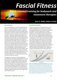
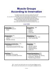
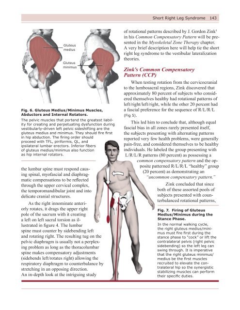
Fig. 6. Gluteus Medius/Minimus Muscles,<br />
Abductors and Internal Rotators.<br />
The pelvic muscles that portend the greatest liability<br />
for creating and perpetuating dysfunction during<br />
vestibularly-driven left pelvic sideshifting are the<br />
gluteus medius and minimus. They should fire first<br />
in hip abduction. The firing order should<br />
proceed with TFL, piriformis, QL, and<br />
ipsilateral lumbar erectors. Inferior fibers<br />
of gluteus medius/minimus also function<br />
as hip internal rotators.<br />
the lumbar spine must respond causing<br />
spinal, myofascial and diaphragmatic<br />
compensations to be reflected<br />
through the upper cervical complex,<br />
the temporomandibular joint and into<br />
delicate cranial structures.<br />
As the right innominate anteriorly<br />
rotates, it drags the upper right<br />
pole of the sacrum with it creating<br />
a left on left sacral torsion as illustrated<br />
in figure 4. The lumbar<br />
spine must counter by sidebending left<br />
and rotating right. The resulting tug on the<br />
pelvic diaphragm is usually not a perplexing<br />
problem as long as the thoracolumbar<br />
spine makes compensatory adjustments<br />
(sidebends left/rotates right) allowing the<br />
respiratory diaphragm to counterbalance by<br />
stretching in an opposing direction.<br />
An in-depth look at the intriguing study<br />
of rotational patterns described by J. Gordon Zink 5<br />
in his Common Compensatory Pattern will be presented<br />
in the Myoskeletal Zone Therapy chapter.<br />
A very brief description here will help tie the short<br />
right leg syndrome to the vestibular lateralization<br />
theories.<br />
Zink’s Common Compensatory<br />
Pattern (CCP)<br />
When testing rotation from the cervicocranial<br />
to the lumbosacral regions, Zink discovered that<br />
approximately 80 percent of subjects who considered<br />
themselves healthy had rotational patterns of<br />
left/right/left/right, while the other 20 percent had<br />
a fascial preference for the sequence of R/L/R/L<br />
(Fig 5).<br />
This led him to conclude that, although equal<br />
fascial bias in all zones rarely presented itself,<br />
the subjects presenting with alternating patterns<br />
reported very few health problems, were generally<br />
pain-free, and considered themselves to be healthy<br />
individuals. He labeled the group presenting with<br />
L/R/L/R patterns (80 percent) as possessing a<br />
common compensatory pattern and the opposite<br />
patterned R/L/R/L “healthy” group<br />
(20 percent) as demonstrating an<br />
“uncommon compensatory pattern.”<br />
Zink concluded that since<br />
both of these assorted pools of<br />
subjects presented with counterbalanced<br />
rotational patterns,<br />
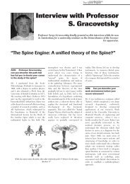
Fig. 7. Firing of Gluteus<br />
Medius/Minimus during the<br />
Stance Phase.<br />
In the normal walking cycle,<br />
the right gluteus medius/minimus<br />
must fire first during the<br />
stance phase to “cock” or lift the<br />
contralateral pelvis (right pelvic<br />
sidebending) so the left leg can<br />
swing through. It is imperative<br />
that the right gluteus minimus/<br />
medius be the first muscles<br />
recruited to elevate the contralateral<br />
hip so the synergistic<br />
stabilizing muscles can perform<br />
their specific duties.
144 Short Right Leg Syndrome<br />
they were more adaptive, healthier and better able<br />
to ward off stress and disease.<br />
Effects of spinal decompensation in the four<br />
transitional zones and resulting altered diaphragmatic<br />
function is discussed in greater detail in the<br />
following two chapters.<br />
Counterintuitive Possibilities<br />
The central theme in Myoskeletal Zone Therapy<br />
® focuses on the influence cerebral lateralization<br />
– due to predictable fetal positioning during the<br />
third trimester – has on embryologic development.<br />
Eighty percent of vertex births are in a left fetal<br />
lie. In this position, the head is flexed and turned<br />
left. During the walking cycle, as the mother’s<br />
belly moves anteriorly (maternal acceleration),<br />
fetal inertia forces the left side of the baby’s head<br />
posteriorly. It is theorized that repeated left-sided<br />
stimulation affects the baby’s utricle which, in<br />
turn, perpetuates early development of not only<br />
the utricle but the entire vestibular system. The<br />
utricle is considered the most influential organ<br />
of the vestibular system. Its duty is to supply a<br />
steady stream of updated data concerning position<br />
and movement of a person’s head. Other vital inner<br />
ear sensing structures include the semicircular<br />
canals – anterior, posterior and lateral which lie<br />
anatomically in different planes with each intricately<br />
placed at right angles to the others. Thus,<br />
the combined functioning of this elaborate vestibular<br />
apparatus efficiently deals with various head<br />
movements: up and down, side to side, and tilting<br />
from one side to the other.<br />
Interestingly, cerebral lateralization appears<br />
to be a primary influential factor in addressing<br />
the perplexing, but fascinating question of limb<br />
length discrepancies observed in our offices each<br />
day. An in-depth discussion of these theories is<br />
presented in the Myoskeletal Zone Therapy chapter<br />
but an introductory overview is also necessary.<br />
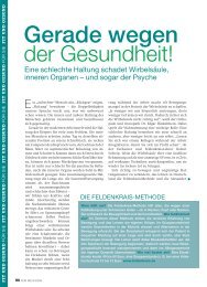
Cervicocranial<br />
S R R L<br />
Cervicothoracic<br />
S R R R<br />
Thoracolumbar<br />
S L R R<br />
Lumbosacral<br />
S R R L<br />
8b<br />
8A<br />
Figs. 8 A & B Pic. # A is Aphrodites Statue and beside it<br />
(#B) is Posterior view of lady.<br />
Figure A and B depict the Common Compensatory Pattern. Although the<br />
Aphrodites was sculpted in Greece around 350 BC, it beautifully demonstrates<br />
the incredible eye of the early sculptors in describing commonly<br />
seen postural patterns. Notice how the left side-shifted, posterior/superiorly<br />
rotated pelvis creates a compensatory scoliosis causing the left<br />
shoulder to drop. Figure B shows the associated spinal biomechanical<br />
adaptations inherent in J Gordon Zink, and Janda’s postural models.
Short Right Leg Syndrome 145<br />
Personal Perspectives on Muscle<br />
Substitutions Patterns<br />
In an attempt to combine many of my favorite<br />
researchers’ postural models (Zink, Janda, Previc,<br />
Geschwind, Pope, Greenman, etc.) into some<br />
sort of reasonable therapeutic protocol, I have<br />
come to recognize certain predictable substitution<br />
patterns that develop from vestibular and<br />
motor dominant neurological adaptations. In my<br />
opinion, the pelvic muscles that hold the greatest<br />
liability for creating and perpetuating dysfunction<br />
during vestibularly-driven left pelvic sideshifting<br />
are…the gluteus medius and minimus. My respect<br />
for these often neglected muscles has grown with<br />
increased observation and study. I now place them<br />
on a level with the other distinguished antigravity<br />
structures such as: (Fig. 6)<br />
• Transversus abdominis: Through its<br />
intimate connection with the thoracolumbar<br />
fasciae, multifidus and sacrospinous ligaments,<br />
the transversus should not only brace<br />
the lumbar spine during forward bending, but<br />
also help lift the ribcage off the pelvic girdle<br />
with each step.<br />
• Hip flexors/extensors: Allow the lower<br />
quadrant to spring forward when hip<br />
extension firing order patterns are<br />
functioning properly. Optimum hip extension<br />
firing order during the walking cycle<br />
should be: rectus femoris (extend the knee),<br />
ipsilateral hamstrings, ipsilateral gluteus<br />
maximus, contralateral lumbar erectors,<br />
and ipsilateral erectors. Anterior hip capsule<br />
adhesions often inhibit the antigravity<br />
function of these muscles resulting in<br />
disruptive firing order substitution patterns.<br />
The proclamation concerning the importance of<br />
gluteus medius/minimus as primary antigravity<br />
structures deserves further explanation.<br />
Abduction Firing Order<br />
Substitution Patterns<br />
As discussed in an earlier chapter on firing<br />
order patterns, during hip abduction (raising the<br />
extended top leg toward the ceiling while in a<br />
sidelying position), gluteus medius and minimus<br />
should fire first followed by tensor fasciae latae,<br />
piriformis, quadratus lumborum, and ipsilateral<br />
lumbar erectors.<br />
To test the firing order in hip abduction,<br />
simply assume a left sidelying position and raise<br />
(abduct) the fully extended right leg toward the<br />
ceiling. The gluteus medius/minimus should fire<br />
first followed by their synergistic muscles listed<br />
above.<br />
However, during weight-bearing, the gluteus<br />
medius/minimus perform a completely different<br />
function. During the normal walking cycle, the<br />
right gluteus medius/minimus must fire during<br />
the stance phase to “cock” or lift the contralateral<br />
pelvis (right pelvic sidebending) so the left<br />
leg can swing through (Fig. 7). It is imperative<br />
that the right gluteus minimus/medius be the first<br />
muscles recruited to elevate the contralateral hip<br />
so the synergistic stabilizing muscles can perform<br />
their specific duties.<br />
Tensor<br />
Fascia<br />
Latae<br />
Fig. 9. TFL and<br />
Iliotibial Tract.<br />
A common substitution<br />
pattern for stretch-weakened<br />
gluteus medius/minimus<br />
is the TFL. The brain<br />
frequently recruits TFL to<br />
help in abduction efforts.<br />
However, the TFL eventually<br />
overpowers the gluteals,<br />
becomes hypertonic<br />
and short, and begins an<br />
unmerciful pull on the IT<br />
band occasionally leading to<br />
IT-band friction syndrome.
146 Short Right Leg Syndrome<br />
All sorts of aberrant muscle substitution patterns<br />
can be singled out during the “hip abduction<br />
sidelying test” and through keen observation of a<br />
client’s gait. These distorted patterns indicate loss<br />
of primary antigravity function throughout all<br />
lumbopelvic structures. Although some substitution<br />
patterns wreak more havoc that others, all<br />
create biomechanical breakdown in people whose<br />
bodies are unable to compensate at the four<br />
transitional zones (lumbosacral, thoracolumbar,<br />
cervicothoracic, and cervicocranial), as described<br />
by Zink.<br />
Here’s the Catch<br />
During prolonged standing, the client’s body<br />
weight routinely shifts over the vestibularly dominant<br />
left leg eventually creating stretch weakness<br />
in the left gluteus medius/minimus. This pattern<br />
can be easily tested in your own body to offer<br />
clues as to your pattern, e.g., are you ideal (equal<br />
fascial bias in all four zones), do you follow the<br />
L/R/L/R common compensatory pattern, or does<br />
your structure follow a R/L/R/L uncommon<br />
compensatory pattern?<br />
Stand with all your weight on the left leg<br />
while the fingers of your left hand palpate the<br />
left acetabulum (lateral hip) and relax your body.<br />
(You may have to hold on to something to reproduce<br />
the feeling of prolonged relaxed stance.) Do<br />
you feel the hip pop out laterally against your<br />
fingers? Now test the right side to see if it pops<br />
out more during normal weight bearing. If the<br />
acetabulum on the right “gives” more, it is likely<br />
that your structure follows an uncommon compensatory<br />
pattern (UCCP).<br />
Those that feel a stretch weakness on the right<br />
side probably fall within the 20 percent that Zink<br />
defined as uncommon compensatory pattern.<br />
Some will not feel stretch weakness in the gluteus<br />
medius/minimus during prolonged standing. This<br />
small percentage of the population where there<br />
is equal fascial bias at all four transitional zones<br />
would be considered “ideal”, indicating no rotational<br />
preference at the lumbosacral, thoracolumbar,<br />
cervicothoracic, or cervicocranial junctions.<br />
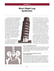
1. Upslipped Innominate Left<br />
2. Cephalad Pubes Left<br />
3. Lumbar - FSR(L) L4 / L5<br />
- S(L)R(R) L5<br />
4. Sacral Torsion L on L<br />
Post &<br />
Caudal ILA<br />
Sulci Deep<br />
Pronated Foot Left<br />
Supinated Foot Right<br />
Fig. 10. Most Common Pattern Resulting<br />
from Short Right Leg.<br />
This illustration depicts the following predictable<br />
pattern: Short right leg, descended right pubis, anterior/inferior<br />
rotated right ilium, left posterior/superior<br />
ilium, substitution muscle imbalance patterns<br />
in hip extension and abduction, left-on-left forward<br />
sacral torsion, functional right convex lumbar scoliosis,<br />
various type 1 group compensatory curves and<br />
non-neutral spinal fixations finally compensating in<br />
the upper cervical complex to level the eyes.<br />
Those with a supine anteriorly/inferiorly rotated<br />
right ilium (most common) should feel the left<br />
acetabulum pop out more to the left during left<br />
leg loading indicating weakness in these primary<br />
abductor muscles. Pay attention to people in public,<br />
such as grocery checkers, hairdressers, assembly<br />
workers, etc., as they stand in prolonged positions<br />
with weight load-bearing on one leg. Recall<br />
that the acetabulum will slightly protrude left as<br />
the gluteus medius/minimus give to the ipsilateral<br />
side during stance. An excellent example is shown<br />
in Figures 8 a & b. These perfectly represent the
Short Right Leg Syndrome 147<br />
Fig. 11. Feedback Mechanisms that Contribute<br />
to Proper CNS Functioning.<br />
Typically, the body’s innate wisdom immediately<br />
begins correcting the limb length strain pattern with<br />
information gleaned from proprioceptors on the<br />
bottom of the feet that sense weight imbalance and<br />
body sway. The antigravity function of posture relies<br />
on these cutaneous receptors which are joined by<br />
visual, vestibular, and other somatosensory systems<br />
that constantly inform the CNS as to the body’s<br />
position and movement.<br />
biomechanics of Zink’s common compensatory<br />
pattern as seen in the famous sculpture of Aphrodites<br />
and the accompanying illustration depicting<br />
a posterior view of the identical pattern. Notice<br />
how the Aphrodites’ pelvic side-shifts over the<br />
left vestibularly dominant left leg creating compensations<br />
reflected throughout her entire body.<br />
On A Personal Note<br />
Those of us old enough to remember the<br />
famous Marilyn Monroe walk can visualize how<br />
her pelvis shifted side to side rather than in an<br />
ideal smooth anterior/posterior patterned gait.<br />
Weight-bearing during the stance phase caused<br />
Marilyn’s pelvis to sideshift toward the weightsupporting<br />
side. In this instance, her weight was<br />
greater on the short leg side (Leaning Tower of<br />
Pisa) during gait which is usually the case in<br />
people presenting with a true short leg. However<br />
during prolonged standing, if Marilyn followed<br />
a left vestibularly dominant pattern, one would<br />
expect the weight-bearing left hip to pop out<br />
laterally.<br />
This dysfunctional gait is easy to recognize<br />
when viewing old Monroe films. Her right acetabulum<br />
would protrude laterally with each step.<br />
A dear friend, Peter Lawford, enjoyed telling<br />
the story of how Marilyn concocted this unique<br />
walk. Apparently, she began by first cutting one<br />
inch off her left high heel shoe and walked in the<br />
unbalanced heels for a few weeks until the left<br />
gluteus medius/minimus overstretched allowing<br />
the hip to pop out and swing laterally. After<br />
a couple of weeks she would switch shoes and<br />
remove an inch from the opposite heel. Marilyn<br />
gradually increased the amount cut off each heel<br />
and continued with the experiment, switching<br />
back and forth between shoes, until she finally<br />
created an aberrant muscle imbalance pattern that<br />
would evolve into the famous Marilyn Monroe<br />
“hip-swing.”<br />
And the beat goes on…<br />
During gait with body weight shifted over<br />
the short right leg, the right gluteus medius/minimus<br />
and associated hip abductors really have to<br />
be strong to develop enough contractile force to<br />
“hike” the contralateral left hip high enough to<br />
allow the long left leg to swing through. Usually<br />
the brain aids in the process by recruiting the<br />
right tensor fasciae latae (TFL), sometimes in<br />
conjunction with piriformis, to help elevate the<br />
left hip. Of course, the down side of this substitution<br />
pattern is that the TFL eventually overpowers<br />
the gluteals, becomes hypertonic and short,<br />
and begins an unmerciful pull on the iliotibial<br />
tract (Fig. 9). I have found this common, but<br />
abnormal TFL firing order substitution pattern<br />
as a major contributor to many conditions such<br />
as iliotibial band friction syndrome, especially in<br />
amateur and competitive athletes.
148 Short Right Leg Syndrome<br />
Conversely, on the left side a prevalent hip abduction<br />
substitution pattern develops as the brain is<br />
forced to recruit the QL muscle to fire first due to<br />
an extremely stretch-weakened gluteus medius/<br />
minimus muscle group. This pervasive and devastating<br />
pattern causes further ipsilateral posterior<br />
innominate rotation, flattening of lumbar lordosis,<br />
left sidebending of the lumbars, tractioning of the<br />
12th rib, and eventual back pain. These folks are<br />
easily recognized as they sidebend their torso left<br />
to allow the right leg to swing through. Therapists<br />
often mis-assess this pattern since it appears that<br />
the right side is doing all the work of pulling up<br />
the right hip and leg. If the trunk does not left<br />
sidebend during the left stance phase of gait, it is<br />
possible that they are lifting with their right side.<br />
Try the sidelying hip abduction test to determine<br />
if you are one of many who are left vestibular<br />
dominant and follow the common compensatory<br />
pattern. Then have someone skilled<br />
in measuring anatomic landmarks check your<br />
structure while in a supine position to see if you<br />
fit the common compensatory pattern which is<br />
reflected in a short right leg and anterior/inferior<br />
ipsilaterally rotated ilium.<br />
During the left sidelying hip abduction test,<br />
frequently the gluteus medius/minimus, TFL and<br />
piriformis will all fire together, indicating that<br />
they are combining forces due to weakness in the<br />
gluteus medius/minimus. During the right sidelying<br />
hip abduction test, look for the quadratus to<br />
fire first (indicated by dipping in the 12th rib area)<br />
followed by either TFL or the inhibited gluteus<br />
medius/minimus. This substitution pattern is a<br />
major pain generator and suggests gluteal weakness<br />
from excessive weight bearing during the<br />
stance phase.<br />
The observations described above are only<br />
meant as an overview of a particular muscle<br />
imbalance pattern I have found interesting to<br />
work with in my practice and I remain unaware<br />
of any studies performed to verify these findings.<br />
Therefore, these conclusions may or may<br />
not prove to be accurate in all cases, e.g. clients<br />
presenting with fixed scoliotic patterns, sacralizations,<br />
hemipelvis, etc. Test your clients using hip<br />
hyperextension and hip abduction tests presented<br />
in Myoskeletal Alignment Techniques Volume I<br />
and see what correlations (if any) you find using<br />
this neuromyoskeletal theory.<br />
The Pronated Foot<br />
The foot is abducted,<br />
dorsally flexed and<br />
everted.<br />
1. Internally rotates the<br />
lower extremity<br />
2. Shortens the<br />
Lower extremity<br />
The Supinated Foot<br />
The foot is abducted,<br />
plantar flexed and<br />
inverted.<br />
1. Externally rotates the<br />
lower extremity<br />
2. Lengthens the<br />
Lower extremity<br />
Fig. 12. Rigid, Supinated and Hyperpronated Flat Foot.<br />
As specialized receptors inform the brain of pelvic imbalance, signals are sent to supinate the foot on the<br />
short leg side in an attempt to lift and balance the anterior/inferior rotated ilium. Regrettably, prolonged<br />
supination strains the myofascia and metatarsals due to excessive weight-bearing on the lateral arch. The<br />
brains attempt to lift the ilium often causes the foot to function in an equinus position to prevent dorsiflexion.<br />
The opposite pattern typically occurs on the long-leg side causing hyperpronation or flattening of the medial<br />
arch to lower the high ilium. If the antigravity (springing) function of the left foot is lost, the dropped arch<br />
becomes a precursor to foot/hip/knee and back pain.
Short Right Leg Syndrome 149<br />
Patterns Most Commonly<br />
Encountered<br />
Now that a brief overview of vestibular lateralization,<br />
firing order and muscle substitution<br />
patterns have been discussed, the short leg theories<br />
resulting from a posterior/superior rotated<br />
ilium can be revisited. Although all the long-held<br />
assumptions suggesting that a long leg develops<br />
as the hip drops (anterior/inferior) seems to make<br />
perfect sense, these are not patterns I commonly<br />
see in my practice. The following is a structural<br />
formula I look for and expect to see in a majority<br />
of new clients presenting for an initial structural<br />
evaluation (Fig. 10):<br />
• Short right leg (supine);<br />
• Descended right pubis;<br />
• Right anterior/inferior ilium (supine);<br />
• Left posterior/superior ilium<br />
(spring test prone);<br />
• Substitution muscle imbalance patterns in hip<br />
extension and abduction;<br />
• Left-on-left forward sacral torsion;<br />
• Functional lumbar scoliosis convex right;<br />
• Various compensatory type I group curves<br />
resolving at O-A to level the eyes;<br />
• Scattered non-neutral dysfunctions (facet<br />
joints stuck open or closed), and<br />
• Associated muscle/visceral/diaphragmatic<br />
imbalance patterns.<br />
The degree of compensation may largely<br />
depend on:<br />
• Degree of leg length discrepancy;<br />
• Functional or true leg length problem;<br />
• Associated traumas: acute, collective, or<br />
degenerative;<br />
• Presence of tonic neck reflexes;<br />
• Cranial deviations causing re-patterning from<br />
the top down; and<br />
• Viscerosomatic dysfunction.<br />
Short Functional Right Leg, Anteriorly<br />
Tilted Ilium and a Low Femoral Head?<br />
The question I have asked myself for years is<br />
this: “What biomechanical mechanism is acting<br />
on the iliosacral joint in the presence of an anterior/inferior<br />
right rotated ilium and a functional<br />
short right leg?” While hanging out in clinic one<br />
day with a friend, colleague and manipulative<br />
osteopath, Ross Pope, he offered an interesting<br />
viewpoint that helped me better visualize the<br />
possible development of this common aberrant<br />
postural pattern. While viewing various postural<br />
radiographic films, I posed this simple question,<br />
“What altered pelvic mechanics do you believe<br />
are involved in your patients presenting with low<br />
femoral heads and functional short legs?”<br />
Pope answered with these statements: “As you<br />
see in this film, the patient presents with a low<br />
femoral head and accompanying anatomic short<br />
right leg when standing. However, a functional leg<br />
length discrepancy is noted upon clinical examination<br />
when the patient is in a supine position. In<br />
this case the leg itself appears shorter as viewed<br />
by comparing the medial malleoli. The ilium on<br />
the “functionally” short side is anteriorly rotated<br />
which places the femoral head in a more cephalad<br />
position in an off-weighted position.”<br />
Bottom line he states: “In the overwhelming<br />
majority of cases, the leg that appears to be functionally<br />
short and the leg that is actually short are<br />
the same. So, yes there is usually a low femoral<br />
head on the right (standing) with a short right leg<br />
(supine). The corollary to this is a low left femoral<br />
head with a functionally short left leg, which is<br />
also prevalent but less so.”<br />
He paused, reflected on what he was about to<br />
say, and continued with this stipulation: “On the<br />
other hand, there are exceptions to, or disparities<br />
in these typical findings. For example, on occasion<br />
you will see a low femoral head height on the<br />
left with a functionally short right leg. This can<br />
occur in a patient with an otherwise normal pelvis<br />
and is probably due to right motor dominance<br />
combined with left vestibular dominance. In other
150 Short Right Leg Syndrome<br />
words when you have a right-handed person with<br />
a short left leg – the muscular component overrides<br />
the anatomic. In these cases the short left<br />
leg is probably either congenital or secondary to<br />
trauma. On other occasions you will find a normal<br />
pelvis radiographically, i.e. level sacral base and<br />
equal leg lengths, and the patient will display a<br />
functional short right leg when supine. This again<br />
shows muscular dominance and is typical for<br />
right-handed (CCP) people. The main reason for<br />
the exceptions is either fixed scoliosis or a cranial<br />
asymmetry (usually lost vertical dimension on<br />
one side of the bite).”<br />
The key phrase for me was: “The ilium on<br />
the “functionally” short side is anteriorly rotated<br />
which places the femoral head in a more cephalad<br />
position in an off-weighted position.” Although<br />
this was precisely the picture I had in my mind<br />
concerning positioning of the femoral head and<br />
acetabulum, I was unable to verify these findings<br />
using palpatory evaluations, I simply had to see it.<br />
Working from an<br />
Assessment Baseline<br />
One assessment protocol I have found helpful<br />
when performing pain management structural<br />
work is to first develop a baseline of aberration.<br />
Working from a baseline simply means that I<br />
strive to view the body as a whole while maintaining<br />
certain expectations of what patterns I am<br />
inclined to see. For example, I ask myself:<br />
• What predictable structural patterns are<br />
represented;<br />
• Which alternate patterns typically accompany<br />
the aberration I am seeing;<br />
• Is there a “key” neuromyoskeletal disorder<br />
triggering the primary dysfunction;<br />
• Am I seeing an upper or lower crossed or<br />
common compensatory pattern; and<br />
• What fascial, skeletal, or diaphragmatic tissue<br />
is responsible for their primary decompensation?<br />
With all the new research surfacing on predictable<br />
patterns, it is now possible to begin combining<br />
various formulas to help develop a clearer<br />
picture of common postural asymmetries. This<br />
synergy greatly enhances visual and hands-on<br />
evaluations.<br />
PLUMB LINE<br />
Fig. 13. Short Right Leg Sideshifts the Pelvis,<br />
Resulting in Compensatory Scoliosis.<br />
The most common postural compensation for leg<br />
length discrepancy is a functional scoliosis. A general<br />
rule has been suggested which summarizes<br />
the type of scoliosis present in relation to the limb<br />
length discrepancy. If the leg length discrepancy<br />
is less than 1cm, a ‘C’ curve will be present with<br />
the shoulder on the short side being the higher of<br />
the two. Conversely, an ‘S’ curve will be observed if<br />
the limb length discrepancy is more than 1cm. This<br />
increased leg length inequality causes the shoulder<br />
on the shorter side to appear lower. Typically, the<br />
pelvis will be more inferior on the short side and the<br />
thoracic spine will have a type I group curve convex<br />
left with the shoulder and arm hanging lower on the<br />
long leg side (left).
Short Right Leg Syndrome 151<br />
Searching for the “Tie That Binds”<br />
Based on research from innovators such as<br />
Janda, Rolf, Zink, Greenman, Mitchell, Previc,<br />
Pope, and others, I often begin my visual and<br />
hands-on analysis with a preconceived notion of<br />
certain predictable muscle/joint strain patterns<br />
and then record non-adapting or decompensated<br />
patterns. I find this approach more interesting,<br />
less confusing and more practical than myopically<br />
investigating a single area and then attempting to<br />
relate it to the rest of the structure. Although the<br />
process demands an open mind and heart, it is<br />
still exciting to search for early embryologic and<br />
CCP clues attempting to determine where the client<br />
either follows or departs from the norm. When<br />
clients come in hurting, I always ask myself:<br />
“What key dysfunction is driving this aberrant<br />
posturally-initiated pain pattern and does the root<br />
cause of this disorder seem to be based in genetic<br />
influences, early embryologic development,<br />
trauma, habitual patterns, gravitational exposure,<br />
psychosocial, or vestibular/motor dominance?”<br />
Unless I am dealing with acute injuries, such<br />
as tennis elbow, ankle or knee sprains, etc., I typically<br />
observe for common dysfunctional postural<br />
patterns (upper and lower crossed, common compensatory)<br />
rather than focus evaluation on each<br />
individual body segment. Sometimes it works;<br />
and other times I simply get lost in all the overlapping<br />
embedded aberrant strain patterns. At this<br />
point, I begin at square one and untangle the mess<br />
until familiar patterns begin to emerge.<br />
Back to the Basics…<br />
Short Right Leg<br />
Limb length discrepancy is simply defined as<br />
a condition where one leg is shorter than the other.<br />
When a substantial difference exists, disruptive<br />
effects on gait and posture can occur.<br />
As discussed earlier, leg length discrepancy<br />
can be divided into two etiological groups;<br />
1. Structural: True shortening of the skeleton<br />
from congenital, traumatic, or diseased<br />
origins.<br />
2. Functional: Develops as a result of altered<br />
mechanics of the lower extremities (foot<br />
hyperpronation) or pelvic obliquity due to<br />
upper quadrant muscle imbalances such<br />
as tonic neck reflexes, poor trunk stabilization,<br />
protective lumbar muscle guarding,<br />
deep fascial strain patterns, etc.<br />
For efficient locomotion, a symmetrical and<br />
well aligned body is necessary. If symmetry is<br />
distorted, particular by limb length discrepancies,<br />
then gait and posture are disrupted. Consequently,<br />
a diversity of symptoms can prevail, and without<br />
adequate treatment, often manifests as chronic<br />
sources of pain and dysfunction.<br />
The Body’s Innate Compensatory<br />
Capability<br />
Simply stated, innate compensations occur to<br />
lengthen the short leg and shorten the long leg.<br />
Typically, the body’s innate wisdom immediately<br />
begins the pelvic-balancing correction process<br />
using information gleaned from proprioceptors<br />
embedded in fascia, muscles and joints located<br />
on the bottom of the feet. Millions of these highly<br />
sensitive receptors sense weight imbalance and<br />
body sway (Fig. 11). Over a period of weeks, it is<br />
intriguing to observe how the brain slowly raises<br />
(supinates) the medial arch on the short leg side in<br />
an attempt to balance the anterior/inferior rotated<br />
innominate. Regrettably, prolonged supination<br />
often strains and sometimes fractures the metatarsals,<br />
talus or calcaneus bones due to excessive<br />
lateral arch weight-bearing. As the brain succeeds<br />
in elevating the anterior/inferior ilium, the foot is<br />
forced to function in an equinus position to prevent<br />
dorsiflexion.<br />
The opposite pelvic-balancing pattern typically<br />
occurs on the long-leg side as the brain<br />
hyperpronates or flattens the medial arch in an<br />
effort to lower the high ilium. Excessive medial<br />
wear on the client’s left shoe is a dead-giveaway<br />
that hyperpronation is at work. Long-term hyperpronation<br />
not only diminishes the body’s natural<br />
antigravity springing system, but is also a precursor<br />
to foot, ankle, knee, and hip pain.
152 Short Right Leg Syndrome<br />
As a result of these foot compensations, the<br />
shorter leg may be prone to stress fractures due to<br />
the non-shock absorbing nature of the supinated<br />
foot (Fig. 12). Likewise, hyperpronation of the<br />
long leg may cause medial knee pain as the tibia<br />
internally rotates.<br />
In the lower limbs, compensations at each<br />
level can be summarized as follows;<br />
• Ankle instability due to foot supination on<br />
the short side;<br />
• Knee hyperextension on the short side and the<br />
knee flexed on the long side;<br />
• Externally rotated leg on the short side; and<br />
• Circumduction of the long limb.<br />
Trunk and head compensations<br />
Compensatory scoliosis is commonly reflected<br />
as a low shoulder on the high ilium side. Since the<br />
head typically will not tilt to maintain the eyes<br />
parallel to the horizon, a short “C” curve is common<br />
in the cervical vertebrae. Elbow and hand<br />
positions may appear shorter on the shorter leg<br />
side, with the opposing arm swinging more on the<br />
shorter leg side.<br />
As mentioned earlier, some authors suggest<br />
that there is a rotation of the pelvis towards the<br />
long leg side, possibly due to hyperpronation and<br />
medial leg rotation. 6 They describe a typical gait<br />
where the short leg steps down and the long leg<br />
compensates by “vaulting” This pattern of walking<br />
on toes on the short side and flexing the knee<br />
of the long side seems to be a fairly consistent<br />
compensatory gait. Unfortunately, as the center<br />
of gravity unevenly shifts, the smooth sinusoidal<br />
motion during the walking cycle is disrupted.<br />
Thus, cosmetic effects of gait can also contribute<br />
to compensation mechanisms and eventual tissue<br />
injury. For example walking on the toes may lead<br />
to a contracture of the Achilles and calf muscles<br />
leading to conditions such as Achilles tendinitis<br />
and plantar fasciitis.<br />
Other muscle compensations of the CCP<br />
include left-sided QL shortening (long left-leg<br />
side) accompanied by contralateral compensatory<br />
shortening of levator scapulae, sternocleidomastoid,<br />
upper trapezius and middle/lateral scalene<br />
muscles to counterbalance the low left shoulder<br />
and to maintain the head in a more erect position<br />
(Fig. 10). Regrettably, asymmetrical myofascial<br />
torsioning slowly sinks its insidious tentacles into<br />
associated spinal joint structures setting off neuroreflexive<br />
muscle guarding responses.<br />
That Hitch in your Get-Along<br />
The presence of a limb length discrepancy is<br />
usually easily recognizable during gait by observing<br />
for:<br />
• Shoulder tilting to one side;<br />
• Unequal arm swing;<br />
• Pelvic tilt;<br />
• Foot supinated on the short side and pronated<br />
on the long side;<br />
• Ankle plantarflexed on the short side; and<br />
• Knee flexed on the long side.<br />
Note: During running, it has been suggested that<br />
limb length discrepancy makes no real difference<br />
due to the fact that only one foot strikes the<br />
ground at any given time. However, Blustein and<br />
D’Amico’s extensive research finds that leg length<br />
discrepancy is the third most common cause of<br />
running injuries. 7<br />
Posture and Limb Length Discrepancy<br />
The most common postural compensation for<br />
leg length discrepancy is a functional scoliosis.<br />
Scoliotic patterns that are noted in both standing<br />
and flexion indicate the presence of a structural or<br />
fixed scoliosis (Fig. 13).<br />
A general rule has been suggested which summarizes<br />
the type of scoliosis present in relation<br />
to the limb length discrepancy. If the leg length<br />
discrepancy is less than 1cm, a “C” curve will be<br />
present with the shoulder on the short side being<br />
the higher of the two. Conversely, an “S” curve<br />
will be observed if the limb length discrepancy<br />
is more than 1cm. With this increased leg length
Short Right Leg Syndrome 153<br />
distortion, the shoulder on the shorter side should<br />
appear lower. 8<br />
Typically, the pelvis will be more inferior on<br />
the short side and the thoracic spine will have a<br />
type I group curve convex left with the shoulder<br />
and arm hanging lower on the long leg side (left).<br />
Conclusion<br />
The importance of limb length discrepancy<br />
cannot be ignored, and is often the key feature in<br />
lower limb and back pathologies. Measuring the<br />
limbs in conjunction with gait and posture analysis<br />
is vital. Thus, developing advanced visual and<br />
anatomic client evaluation skills are paramount<br />
in helping structurally-minded somatic therapists<br />
distinguish between functional and structural<br />
limb length inequalities. If in doubt about your<br />
ability to effectively and consistently distinguish<br />
leg length asymmetry, refer the client to manual<br />
medicine physicians for a radiographic screening.<br />
Proper limb measurement is essential; unfortunately<br />
there is no single hands-on method that<br />
proves to be completely reliable in its own right.<br />
It is for this reason that following a holistic approach<br />
that includes recognizing and eliminating<br />
aberrant strain patterns, correcting aberrant firing<br />
order patterns, and searching for embryologic<br />
clues to key posturally-initiated pain issues may<br />
boost your success and empower your practice.<br />
The compensations which are part of limb length<br />
discrepancy have been discussed. Although presentations<br />
do differ from client to client, most of<br />
the patterning theories presented will prove accurate.<br />
The most important feature for the beginning<br />
therapist to recognize is that asymmetry exists…<br />
from there more specific details will emerge with<br />
experience.<br />
Integral parts to treatment of the condition are<br />
identification, comprehension of each individual’s<br />
compensatory adaptations, and their relationship<br />
to resultant symptomatology. Today’s therapist<br />
must be aware of the fundamental importance of<br />
limb inequalities—particularly the short right leg<br />
phenomenon. Keep an open mind, look for structural<br />
relationships, and have fun when assessing<br />
and treating asymmetrical leg length patterns and<br />
resulting compensations.<br />
References:<br />
1. Garson JG. Inequality in length of lower limbs.<br />
Journal of Anatomical Physiology. 1897. pp 502-507.<br />
2. Hasse C, Dehner, Arch. Etiology and Pathophysiology<br />
of Leg Length Discrepancies. Anatomical Entwickl.<br />
1893.<br />
3. Denslow J, Chase I, et al. Mechanical stresses<br />
in the human lumbar spine and pelvis. 1962. In:<br />
Postural Balance and Imbalance. Peterson B, ed.<br />
Indianapolis: American Academy of Osteopathy, pp.<br />
76-82, 1983.<br />
4. Juhl J, Prevalence of Frontal Plane Pelvic Postural<br />
Asymmetry, Journal of the American Osteopathic<br />
Association, Volume 104. 2004.<br />
5. Zink JG, Lawson W: “An osteopathic structural<br />
examination and functional interpretation of the<br />
soma.” Osteopathic Annals, 7:12, Dec. 1979.<br />
6. Blake, R.L. and Ferguson, H. (1992) Limb length<br />
discrepancy. JAPMA 82 (1) pp 33-38.<br />
7. Blustein, S.M. and D’Amico, J.C. (1985). Limb<br />
length discrepancy: identification, clinical significance<br />
and management. JAPMA 75(4) pp200-206.<br />
8. Chambers, M.R.C. (1996). Limb length inequality:<br />
types, etiologies’, pathomechanics, values and<br />
incidence. Journal of British Podiatry Medicine<br />
51(5) pp74-80.