Tissue refractive index as marker of disease - Quantitative Light ...
Tissue refractive index as marker of disease - Quantitative Light ...
Tissue refractive index as marker of disease - Quantitative Light ...

You also want an ePaper? Increase the reach of your titles
YUMPU automatically turns print PDFs into web optimized ePapers that Google loves.
<strong>Tissue</strong> <strong>refractive</strong> <strong>index</strong> <strong>as</strong> <strong>marker</strong> <strong>of</strong> dise<strong>as</strong>e<br />
Zhuo Wang 1 , Krishnarao Tangella 2 , Andre Balla 3 , and Gabriel Popescu 1 *<br />
1 <strong>Quantitative</strong> <strong>Light</strong> Imaging Laboratory<br />
Department <strong>of</strong> Electrical and Computer Engineering, Beckman Institute for Advanced Science<br />
& Technology, University <strong>of</strong> Illinois at Urbana-Champaign, Urbana, IL 61801<br />
2 Department <strong>of</strong> Pathology, Christie Clinic and University <strong>of</strong> Illinois at Urbana-Champaign,<br />
Urbana, IL 61801<br />
3 Department <strong>of</strong> Pathology, University <strong>of</strong> Illinois at Chicago, Chicago, IL 60612<br />
*gpopescu@illinois.edu<br />
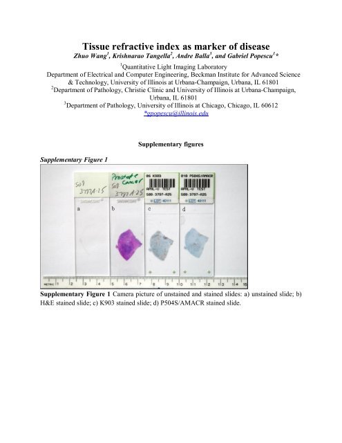
Supplementary Figure 1<br />
Supplementary figures<br />
Supplementary Figure 1 Camera picture <strong>of</strong> unstained and stained slides: a) unstained slide; b)<br />
H&E stained slide; c) K903 stained slide; d) P504S/AMACR stained slide.
Supplementary Figure 2<br />
Supplementary Figure 2 Lymphocytes identification; objective is 10X/0.3. Scale bar: 100μm.<br />
(a) SLIM image; color bar indicates optica path length in nm. (b) H&E stained image. The high<br />
<strong>refractive</strong> <strong>index</strong> cells in (a) turn out to be blue in H&E. (c) CD45 stained image. CD45 antibody<br />
reacts with both alloantigens and all is<strong>of</strong>orms <strong>of</strong> the CD45 leukocyte common antigen (LCA),<br />
and is used <strong>as</strong> a reporter <strong>of</strong> lymphocytes in prostate tissues. Lymphocytes were confirmed with<br />
CD45 staining.
Supplementary Figure 3<br />
Supplementary Figure 3 Stromal cells identification; objective is 10X/0.3. Scale bar: 100μm.<br />
(a) SLIM image; color bar indicates optical path length in nm. (b) H&E stained image. The<br />
high <strong>refractive</strong> <strong>index</strong> cells in (a) are black in H&E. (c) P63 stained image. p63 is a recently<br />
characterized p53-homolog that is consistently expressed by b<strong>as</strong>al/somatic stem cells <strong>of</strong> stratified<br />
epithelia, myoepithelial cells <strong>of</strong> the bre<strong>as</strong>t and salivary glands, and proliferative compartment <strong>of</strong><br />
g<strong>as</strong>tric mucosa. The cells are negative and thus not myoepithelial cells. (d) CD45 stained image.<br />
CD45 antibody reacts with both alloantigens and all is<strong>of</strong>orms <strong>of</strong> the CD45 leukocyte common<br />
antigen (LCA), and is used <strong>as</strong> a reporter <strong>of</strong> lymphocytes in prostate tissues. The cells are<br />
negative and thus not lymphocytes. (e) pan-CK stained image. pan-CK is intended for laboratory<br />
use to identify epithelial cells using light microscopy. The cells are negative and thus not <strong>of</strong><br />
epithelial origin.
Supplementary Figure 4<br />
Supplementary Figure 4 Eleven c<strong>as</strong>es used for the prostate cancer study summarized in Fig. 4.<br />
First column: SLIM images, color bar in nm; second column: H&E stained images; third<br />
column: ls map, color bar in μm; fourth column: K903 stained images; fifth column:<br />
P504S/AMACR stained images. Out <strong>of</strong> all the eleven c<strong>as</strong>es, seven c<strong>as</strong>es are rated Gle<strong>as</strong>on grade<br />
6/10, two c<strong>as</strong>es are rated Gle<strong>as</strong>on grade 7/10, one c<strong>as</strong>e is Gle<strong>as</strong>on grade 9/10 and one c<strong>as</strong>e is<br />
benign.
Supplementary Figure 5<br />
Supplementary Figure 5 Statistical parameters for 49 cancerous are<strong>as</strong> and 51 benign are<strong>as</strong> from<br />
11 biopsies. (a) Standard deviation vs. Mean; (b) Skewness vs. Mean; (c) Kurtosis vs. Mean; (d)<br />
Mode vs. Mean; (e) Standard deviation/Mean vs. standard deviation; (f) Mode vs. Standard<br />
deviation/Mean.