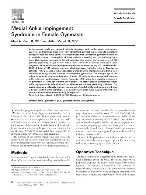
Medial Ankle Impingement Syndrome in Female Gymnasts
Medial Ankle Impingement Syndrome in Female Gymnasts
Medial Ankle Impingement Syndrome in Female Gymnasts

Create successful ePaper yourself
Turn your PDF publications into a flip-book with our unique Google optimized e-Paper software.
<strong>Medial</strong> <strong>Ankle</strong> <strong>Imp<strong>in</strong>gement</strong><br />
<strong>Syndrome</strong> <strong>in</strong> <strong>Female</strong> <strong>Gymnasts</strong><br />
Mark A. Vann, II, MD,* and Arthur Manoli, II, MD †<br />
In the current study we reviewed patients diagnosed with medial ankle imp<strong>in</strong>gement<br />
syndrome and identified those <strong>in</strong>volved <strong>in</strong> competitive gymnastics present<strong>in</strong>g to our referral<br />
orthopedic foot and ankle center. We hypothesized that competitive gymnastics would be<br />
a relatively common characteristic of those patients present<strong>in</strong>g with this pathologic process.<br />
Chart review was used <strong>in</strong> this retrospective case series. Our review <strong>in</strong>volved 789<br />
patients present<strong>in</strong>g to our center with a chief compla<strong>in</strong>t of medial-sided ankle pa<strong>in</strong>,<br />
diagnosed with medial ankle imp<strong>in</strong>gement syndrome between January 2001 and December<br />
2007. A total of 115 patients met our <strong>in</strong>itial age-based <strong>in</strong>clusion criteria. Twenty-two<br />
patients (19%) present<strong>in</strong>g with a diagnosis of medial ankle imp<strong>in</strong>gement syndrome were<br />
identified as be<strong>in</strong>g actively <strong>in</strong>volved <strong>in</strong> competitive gymnastics. The average age of this<br />
subset of patients at presentation was 19 years. All patients were treated with an open<br />
ankle arthrotomy and tenosynovectomy. Inspection of the ankle jo<strong>in</strong>t revealed evidence of<br />
19 patients (86%) with concomitant ankle lesions. The predilection of symptomatic medial<br />
ankle imp<strong>in</strong>gement <strong>in</strong> def<strong>in</strong>ed athletic populations has not been previously published. Our<br />
series suggests a relatively common occurrence of medial ankle imp<strong>in</strong>gement syndrome,<br />
with concomitant ankle pathology, <strong>in</strong> competitive gymnasts. After surgical <strong>in</strong>tervention, a<br />
return to competitive gymnastics may be expected.<br />
Oper Tech Sports Med 18:50-52 © 2010 Elsevier Inc. All rights reserved.<br />
KEYWORDS ankle, gymnastics, pa<strong>in</strong>, gymnasts, female, imp<strong>in</strong>gement<br />
<strong>Medial</strong> imp<strong>in</strong>gement syndrome of the anterior tibiotalar<br />
fascicle of the deltoid ligament was first described by<br />
Mosier-LaClair et al 1 <strong>in</strong> 1998. This syndrome was noted to<br />
occur after <strong>in</strong>version ankle spra<strong>in</strong>s and fractures, talar neck,<br />
and body fractures with the pathomechanics described as<br />
imp<strong>in</strong>gement of the anterior tibiotalar fascicle of the deltoid<br />
ligament on the anteromedial aspect of the talus dur<strong>in</strong>g ankle<br />
dorsiflexion. Associated anteromedial talar osteophytes or<br />
chondral lesions have frequently been identified.<br />
The purpose of the current retrospective case series is to<br />
review those patients present<strong>in</strong>g with symptomatic medial<br />
ankle imp<strong>in</strong>gement syndrome, necessitat<strong>in</strong>g operative <strong>in</strong>tervention,<br />
and their participation <strong>in</strong> competitive gymnastics.<br />
Methods<br />
Patients exam<strong>in</strong>ed <strong>in</strong> our referral orthopedic foot and ankle<br />
cl<strong>in</strong>ic with anteromedial ankle pa<strong>in</strong> diagnosed with medial<br />
*Gulf Coast Medical Center, Wharton, TX.<br />
†Michigan International Foot and <strong>Ankle</strong> Center, Pontiac, MI.<br />
Address repr<strong>in</strong>t requests to Mark A. Vann, II, MD, PLLC. P.O. Box 60<br />
Richmond, TX 77406. E-mail: orthovann@yahoo.com<br />
imp<strong>in</strong>gement syndrome were identified us<strong>in</strong>g our database of<br />
surgical patients treated between 2001 and 2007. 789 subjects<br />
were identified who had undergone open ankle arthrotomy<br />
and tenosynovectomy (CPT: 27,626). Our exclusion<br />
criteria were developed <strong>in</strong> an effort to focus on the population<br />
of competitive young gymnasts seen <strong>in</strong> our practice. Due to<br />
the relatively short duration of a competitive gymnasts’ career,<br />
we chose a maximum age of 25 years as the cut-off for<br />
<strong>in</strong>clusion <strong>in</strong>to our study.<br />
A total of 115 patients met our <strong>in</strong>clusion criteria. All patients<br />
<strong>in</strong> this study had undergone open ankle arthrotomy<br />
with tenosynovectomy by the senior author.<br />
Operative Technique<br />
The <strong>in</strong>itial sk<strong>in</strong> <strong>in</strong>cision is determ<strong>in</strong>ed by palpation of superficial<br />
landmarks about the ankle. The medial malleolus is<br />
palpated along with the anterior most aspect of the deltoid<br />
ligament. The tibiotalar articulation is then palpated. In<br />
larger patients, identification of this jo<strong>in</strong>t may be facilitated<br />
by plantar and dorsiflexion of the ankle jo<strong>in</strong>t. Plac<strong>in</strong>g the sk<strong>in</strong><br />
<strong>in</strong>cision just lateral to the anterior-most fibers of the deltoid<br />
50 1060-1872/10/$-see front matter © 2010 Elsevier Inc. All rights reserved.<br />
doi:10.1053/j.otsm.2009.11.003
<strong>Medial</strong> ankle imp<strong>in</strong>gement syndrome 51<br />
Figure 1 Intraoperative photograph demonstrat<strong>in</strong>g a large talar osteophyte<br />
along anteromedial border of talar dome.<br />
Figure 3 Intraoperative photograph demonstrat<strong>in</strong>g the use of plantarflexion<br />
<strong>in</strong> visualiz<strong>in</strong>g the extent of pathology <strong>in</strong>volved <strong>in</strong> medial<br />
ankle imp<strong>in</strong>gement.<br />
ligament facilitates the approach to the offend<strong>in</strong>g pathology.<br />
The proximal extent of the sk<strong>in</strong> <strong>in</strong>cision should be to the level<br />
of the tibiotalar articulation and the distal extent should<br />
reach the region of the talar neck.<br />
Blunt dissection is performed down to the level of the<br />
ankle capsule. Care must be taken when perform<strong>in</strong>g the ankle<br />
capsulotomy, as the tissue is typically th<strong>in</strong>. Sharp penetration<br />
may damage the underly<strong>in</strong>g talar cartilage. The capsulotomy<br />
is carried to the medial corner of the ankle mortise<br />
proximally with visualization of the tibial plafond. Distally,<br />
the capsulotomy is carried to the talar neck. This exposure<br />
provides excellent visualization of all possible aspects of the<br />
offend<strong>in</strong>g pathology <strong>in</strong> this imp<strong>in</strong>gement syndrome (Fig. 1).<br />
The anteromedial aspects of the talar dome and neck are<br />
<strong>in</strong>spected for the offend<strong>in</strong>g osteophytes or chondral defects<br />
seen <strong>in</strong> this syndrome (Fig. 2). Dorsiflexion and plantarflexion<br />
of the ankle may be necessary to fully visualize the pathology<br />
(Fig. 3). A narrow nose rongeur (synovial rongeur) is<br />
used to debride the lesions. The anterior fascicles of the deltoid<br />
ligament are then <strong>in</strong>spected for evidence of <strong>in</strong>flammation<br />
and/or hypertrophy. These fascicles may be debrided or<br />
sharply excised as necessary if imp<strong>in</strong>gement is noted. The<br />
anteromedial ankle mortise is <strong>in</strong>spected for evidence of osteophytes<br />
along the anterior most borders of the medial malleolus<br />
and/or the medial aspect of the anterior tibial plafond. Care must<br />
be taken to visualize and subsequently debride osteophytes <strong>in</strong><br />
this region us<strong>in</strong>g the narrow rongeur, as this is a major potential<br />
site of bony imp<strong>in</strong>gement dur<strong>in</strong>g extremes of dorsiflexion.<br />
The rema<strong>in</strong>der of the tibiotalar articulation is then visualized<br />
and carefully <strong>in</strong>spected for any evidence of concomitant<br />
pathology <strong>in</strong>clud<strong>in</strong>g chondral lesions, loose bodies, or synovitis.<br />
Manual jo<strong>in</strong>t distraction, plantarflexion, and dorsiflexion<br />
may be used to <strong>in</strong>spect the rema<strong>in</strong>der of the ankle. Concurrent<br />
pathology may be addressed dur<strong>in</strong>g this portion of<br />
the procedure.<br />
Irrigation of the jo<strong>in</strong>t is then performed followed by capsular<br />
closure us<strong>in</strong>g an absorbable 2-0 suture. Sk<strong>in</strong> closure is<br />
performed us<strong>in</strong>g nonabsorbable 2-0 suture.<br />
Figure 2 Gross specimen with marker <strong>in</strong>dicat<strong>in</strong>g common location<br />
of talar osteophytes or chondral defects seen <strong>in</strong> medial ankle imp<strong>in</strong>gement.<br />
Table 1 High-Impact Activities Reported by Patients, not Involved<br />
<strong>in</strong> Gymnastics, Treated for <strong>Medial</strong> <strong>Ankle</strong> <strong>Imp<strong>in</strong>gement</strong><br />
<strong>Syndrome</strong><br />
Activity<br />
Number of Patients<br />
Soccer 3<br />
Cross-country 2<br />
Basketball 2<br />
Tennis 1<br />
Pole-vault<strong>in</strong>g 1<br />
Track 1<br />
March<strong>in</strong>g band 1<br />
Dance 1<br />
Ski<strong>in</strong>g 1
52 M.A. Vann, II and A. Manoli, II<br />
Table 2 Concomitant <strong>Ankle</strong> Lesions Seen <strong>in</strong> <strong>Female</strong> <strong>Gymnasts</strong><br />
Undergo<strong>in</strong>g Surgery for <strong>Medial</strong> <strong>Ankle</strong> <strong>Imp<strong>in</strong>gement</strong><br />
Concomitant Lesion<br />
Number of Patients<br />
Kiss<strong>in</strong>g lesion of tibia 8<br />
Lateral ligamentous laxity 6<br />
Osteochondritis dessicans 5<br />
Bassett’s ligament 2 4<br />
Loose bodies 2<br />
Results<br />
Of the 115 patients meet<strong>in</strong>g our <strong>in</strong>clusion criteria, 22 patients<br />
(19%) were noted to be actively <strong>in</strong>volved <strong>in</strong> competitive<br />
gymnastics at the time of symptom onset. Thirteen patients<br />
(14%) were actively <strong>in</strong>volved <strong>in</strong> high-impact activities<br />
other than gymnastics, with soccer, cross-country runn<strong>in</strong>g,<br />
and basketball, account<strong>in</strong>g for the majority of patients <strong>in</strong> this<br />
group (Table 1).<br />
All subjects actively <strong>in</strong>volved <strong>in</strong> gymnastics were female<br />
with a mean age of 19 years and a range of 15-23 years.<br />
Concomitant ankle pathology was noted <strong>in</strong> 19 (86%) of the<br />
gymnasts <strong>in</strong> our study group. Six patients had 2 or more<br />
concomitant lesions (Table 2).<br />
After surgical <strong>in</strong>tervention, 14 patients (64%) returned to<br />
competitive gymnastics. The average age at time of return was<br />
20 years. A review of 6 patients with 2 or more concomitant<br />
lesions (<strong>in</strong> addition to imp<strong>in</strong>gement) revealed 4 patients<br />
(66%) who were able to return to competitive gymnastics.<br />
Discussion<br />
The preponderance of young gymnasts seen with a medial<br />
imp<strong>in</strong>gement syndrome has not previously been reported <strong>in</strong><br />
the English literature. Although the diagnosis of this syndrome<br />
has been primarily based on physical exam<strong>in</strong>ation<br />
f<strong>in</strong>d<strong>in</strong>gs, Tol et al 3 have evaluated the use of oblique foot<br />
radiographs and found <strong>in</strong>creased visualization of osteophytes<br />
along the anteromedial border of the talus (Fig. 4). Computed<br />
tomography and magnetic resonance imag<strong>in</strong>g have<br />
been used to further visualize the extent of the imp<strong>in</strong>g<strong>in</strong>g site,<br />
and may be useful when a diagnosis is not readily determ<strong>in</strong>ed<br />
by exam<strong>in</strong>ation.<br />
Our group has treated this syndrome with surgical debridement<br />
of the anterior tibiotalar fascicle of the deltoid<br />
through an open arthrotomy. Intraoperative f<strong>in</strong>d<strong>in</strong>gs typically<br />
<strong>in</strong>clude thicken<strong>in</strong>g of the fascicle, localized synovitis,<br />
and talar osteophytes, which may be addressed through use<br />
of a narrow rongeur. Work by Tol and van Dijk 4 furthered<br />
our understand<strong>in</strong>g of this imp<strong>in</strong>gement complex with the<br />
identification of their “kiss<strong>in</strong>g lesion” seen on the correspond<strong>in</strong>g<br />
anteromedial aspect of the tibia. Their work dispelled<br />
the belief that these tibial osteophytes were due to<br />
traction on the anterior capsule (“traction osteophytes”), 5 by<br />
not<strong>in</strong>g that the distal tibial <strong>in</strong>sertion of the capsule was well<br />
superior to the level of the osteophytes <strong>in</strong> cadaveric specimens.<br />
Our knowledge of and experience with this syndrome has<br />
permitted us to effectively treat young gymnasts with symptomatic<br />
ankles, allow<strong>in</strong>g a high rate of return to competitive<br />
gymnastics. This study provides a guidel<strong>in</strong>e by which physicians<br />
may engage <strong>in</strong> an educated and <strong>in</strong>formed discussion<br />
with this young, athletic population regard<strong>in</strong>g their likelihood<br />
of return to competition after surgical <strong>in</strong>tervention.<br />
We have also shown that a thorough physical exam<strong>in</strong>ation<br />
and detailed <strong>in</strong>traoperative <strong>in</strong>spection is essential to avoid<br />
miss<strong>in</strong>g any concomitant pathology that may be present <strong>in</strong><br />
the vast majority of patients present<strong>in</strong>g with this medial ankle<br />
imp<strong>in</strong>gement syndrome.<br />
Conclusions<br />
Our series suggests a relatively common occurrence of symptomatic<br />
medial ankle imp<strong>in</strong>gement syndrome <strong>in</strong> competitive<br />
gymnasts. Physical exam<strong>in</strong>ation rema<strong>in</strong>s an effective diagnostic<br />
technique for the diagnosis of this pathologic process.<br />
After surgical <strong>in</strong>tervention, a return to competitive gymnastics<br />
may be expected, and the presence of 2 or more<br />
concomitant lesions does not predict an impaired prognosis<br />
of return<strong>in</strong>g to athletics.<br />
Figure 4 Oblique radiograph demonstrat<strong>in</strong>g large osteophyte along<br />
anteromedial talar dome.<br />
References<br />
1. Mosier-La Clair S, Monroe M, Manoli A: <strong>Medial</strong> imp<strong>in</strong>gement syndrome<br />
of the anterior tibiotalar fascicle of the deltoid ligament on the talus. Foot<br />
<strong>Ankle</strong> Int 21:385-391, 2000<br />
2. Bassett FH, Gates HS, Billys JB, et al: Talar imp<strong>in</strong>gement by the antero<strong>in</strong>ferior<br />
tibiofibular ligament. A cause of chronic pa<strong>in</strong> <strong>in</strong> the ankle after<br />
<strong>in</strong>version spra<strong>in</strong>. J Bone Jo<strong>in</strong>t Surg Am 72-A:55-59, 1990<br />
3. Tol J, Verhagen RA, Krips R, et al: The anterior ankle imp<strong>in</strong>gement<br />
syndrome: Diagnostic value of oblique radiographs. Foot <strong>Ankle</strong> Int 25:<br />
63-68, 2004<br />
4. Tol J, van Dijk N: Etiology of the anterior ankle imp<strong>in</strong>gement syndrome:<br />
A descriptive anatomical study. Foot <strong>Ankle</strong> Int 25:382-386, 2004<br />
5. O’Donoghue D: <strong>Imp<strong>in</strong>gement</strong> exostoses of the talus and tibia. J Bone<br />
Jo<strong>in</strong>t Surg Am 39-A:835-852, 1957