32. Health and Hygiene - The National Institute of Open Schooling
32. Health and Hygiene - The National Institute of Open Schooling
32. Health and Hygiene - The National Institute of Open Schooling

You also want an ePaper? Increase the reach of your titles
YUMPU automatically turns print PDFs into web optimized ePapers that Google loves.
<strong>Health</strong> <strong>and</strong> <strong>Hygiene</strong><br />
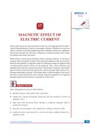
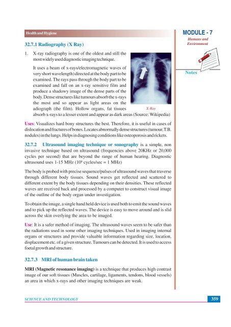
<strong>32.</strong>7.1 Radiography (X Ray)<br />
1. X-ray radiography is one <strong>of</strong> the oldest <strong>and</strong> still the<br />
most widely used diagnostic imaging technique.<br />
It uses a beam <strong>of</strong> x-rays/electromagnetic waves <strong>of</strong><br />
very short wavelength) directed at the body part to be<br />
examined. <strong>The</strong> rays pass through the body part to be<br />
examined <strong>and</strong> fall on an x-ray sensitive film <strong>and</strong><br />
produce a shadowy image <strong>of</strong> the dense parts <strong>of</strong> the<br />
body. Dense structures like tumours absorb the x-rays<br />
the most <strong>and</strong> so appear as light areas on the<br />
adiograph (the film). Hollow organs, fat tissues<br />
X-Ray<br />
absorb x-rays to a lesser extent <strong>and</strong> appear as dark areas (Source: Wikipedia)<br />
MODULE - 7<br />
Humans <strong>and</strong><br />
Environment<br />
Notes<br />
Uses: Visualizes hard bony structures the best. <strong>The</strong>refore, it is useful in cases <strong>of</strong><br />
dislocation <strong>and</strong> fractures <strong>of</strong> bones. Locates abnormally dense structures (tumour, T.B.<br />
nodules) in the lungs. Helps in diagnosing conditions like osteoporosis <strong>and</strong> rickets.<br />
<strong>32.</strong>7.2 Ultrasound imaging technique or sonography is a simple, non<br />
invasive technique based on ultrasound (frequencies above 20KHz or 20,000<br />
cycles per second) that are beyond the range <strong>of</strong> human hearing. Diagnostic<br />
ultrasound uses 1-15 MHz (10 6 cycles/sec = 1 MHz)<br />
<strong>The</strong> body is probed with precise sequence/pulses <strong>of</strong> ultrasound waves that traverse<br />
through different body tissues. Sound waves get reflected <strong>and</strong> scattered to<br />
different extent by the body tissues depending on their densities. <strong>The</strong>se reflected<br />
waves are received back <strong>and</strong> processed by a computer to construct visual image<br />
<strong>of</strong> the outline <strong>of</strong> the body organ under investigation.<br />
To obtain the image, a single h<strong>and</strong> held device is used both to emit the sound waves<br />
<strong>and</strong> to pick up the reflected waves. <strong>The</strong> device is easy to move around <strong>and</strong> is slid<br />
across the skin overlying the area to be imaged.<br />
Use: It is a safer method <strong>of</strong> imaging. <strong>The</strong> ultrasound waves seem to be safer than<br />
the radiations used in some other imaging techniques. Used in imaging internal<br />
organs or structures <strong>and</strong> provide valuable information regarding size, location,<br />
displacement etc. <strong>of</strong> a given structure. Tumours can be detected. It is used to access<br />
foetal growth <strong>and</strong> structure.<br />
<strong>32.</strong>7.3 MRI <strong>of</strong> human brain taken<br />
MRI (Magnetic resonance imaging) is a technique that produces high contrast<br />
image <strong>of</strong> our s<strong>of</strong>t tissues (Muscles, cartilage, ligaments, tendons, blood vessels)<br />
an area in which x-rays <strong>and</strong> other imaging techniques are weak.<br />
SCIENCE AND TECHNOLOGY<br />
359