STRUCTURE OF THE HUMAN BODY Vertebral Levels
STRUCTURE OF THE HUMAN BODY Vertebral Levels
STRUCTURE OF THE HUMAN BODY Vertebral Levels

You also want an ePaper? Increase the reach of your titles
YUMPU automatically turns print PDFs into web optimized ePapers that Google loves.
<strong>STRUCTURE</strong> <strong>OF</strong> <strong>THE</strong> <strong>HUMAN</strong> <strong>BODY</strong><br />
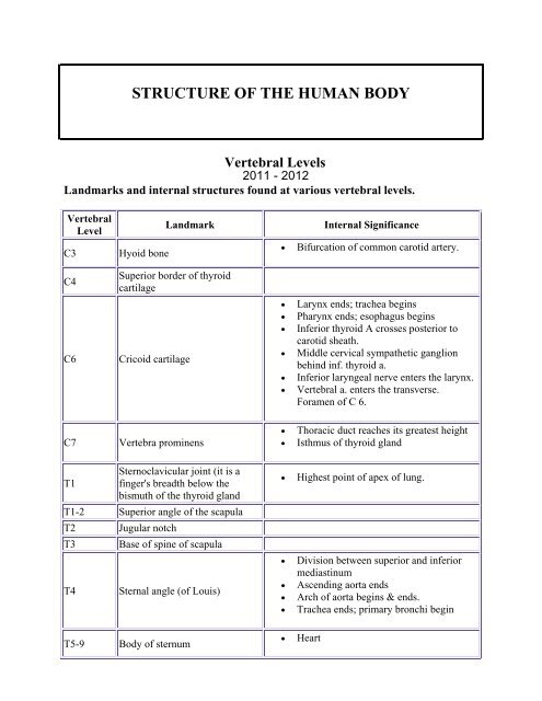
<strong>Vertebral</strong> <strong>Levels</strong><br />
2011 - 2012<br />
Landmarks and internal structures found at various vertebral levels.<br />
<strong>Vertebral</strong><br />
Level<br />
C3<br />
C4<br />
C6<br />
Hyoid bone<br />
Landmark<br />
Superior border of thyroid<br />
cartilage<br />
Cricoid cartilage<br />
Internal Significance<br />
• Bifurcation of common carotid artery.<br />
• Larynx ends; trachea begins<br />
• Pharynx ends; esophagus begins<br />
• Inferior thyroid A crosses posterior to<br />
carotid sheath.<br />
• Middle cervical sympathetic ganglion<br />
behind inf. thyroid a.<br />
• Inferior laryngeal nerve enters the larynx.<br />
• <strong>Vertebral</strong> a. enters the transverse.<br />
Foramen of C 6.<br />
C7<br />
Vertebra prominens<br />
• Thoracic duct reaches its greatest height<br />
• Isthmus of thyroid gland<br />
T1<br />
Sternoclavicular joint (it is a<br />
finger's breadth below the<br />
bismuth of the thyroid gland<br />
T1-2 Superior angle of the scapula<br />
T2<br />
T3<br />
T4<br />
Jugular notch<br />
Base of spine of scapula<br />
Sternal angle (of Louis)<br />
T5-9 Body of sternum<br />
• Highest point of apex of lung.<br />
• Division between superior and inferior<br />
mediastinum<br />
• Ascending aorta ends<br />
• Arch of aorta begins & ends.<br />
• Trachea ends; primary bronchi begin<br />
• Heart
T7<br />
T8<br />
T9<br />
T9-L3<br />
T10<br />
T12<br />
Inferior angle of scapula<br />
Xiphisternal junction<br />
Costal margin<br />
• Inferior vena cava passes through<br />
diaphragm<br />
• Costal slips of diaphragm<br />
• Esophagus through diaphragm<br />
• Aorta through diaphragm<br />
• Thoracic duct through diaphragm<br />
• Azygos V. through diaphragm<br />
L1<br />
Tran pyloric plane: Found at the<br />
midpoint between the jugular<br />
notch and the pubic symphysis<br />
• Pyloris of stomach immediately above<br />
and to the right of the midline.<br />
• Duodenojejunal flexure to the left of<br />
midline and immediately below it<br />
• Pancreas on a line with it<br />
• Origin of Superior Mesenteric artery<br />
• Hilum of kidneys: left is above and right<br />
is below.<br />
• Celiac a. originates just above and renal<br />
arteries originate just below this line.<br />
L2<br />
• Thoracic duct begins<br />
• Azygos and hemiazygos begin<br />
L4/L5<br />
Iliac crest<br />
• Aorta bifurcates into common iliac<br />
arteries<br />
• Inferior vena cava formed from common<br />
iliac veins<br />
S2<br />
S3<br />
Posterior superior iliac spine<br />
(dimple)<br />
Posterior inferior iliac spine<br />
• End of dural sac<br />
• Middle of sacroiliac joint<br />
• Pelvic colon ends and rectum begins;<br />
important landmark in surgery of recto<br />
sigmoid carcinoma