HIDA Scans:
HIDA Scans:
HIDA Scans:

You also want an ePaper? Increase the reach of your titles
YUMPU automatically turns print PDFs into web optimized ePapers that Google loves.
SWCSNM 2011<br />
<strong>HIDA</strong> <strong>Scans</strong>:<br />
Hepatobiliary Scintigraphy<br />
Mike Middleton, MD, FACNM<br />
Professor of Radiology<br />
Division of Nuclear Radiology & Advanced<br />
Molecular Imaging<br />
Scott & White Clinic/TAMUHSC
OUTLINE<br />
• Functional/Morphologic Hepatic Imaging<br />
Dependent on the Radiopharmaceutical Utilized<br />
(TABLE 1)<br />
• Biliary <strong>Scans</strong> and agents<br />
• Brief History<br />
• Scheduling<br />
• Physiology/Preparation<br />
• Techniques<br />
• Interpretation<br />
• Interventions
NM Scheduling Program
NM Scheduling Program
Common Hepatic<br />
Studies on a typical<br />
day utilizing the Scott<br />
& White NM<br />
Scheduling Program
Hepatobiliary Scintigraphy
Scheduling a <strong>HIDA</strong><br />
scan with CCK
General<br />
• Biliary iv agents are rapidly excreted by<br />
hepatocytes and delivered to bile<br />
unconjugated<br />
• The liver, biliary system, bowel, and<br />
sometimes portions of the stomach are<br />
normally visualized
Objectives<br />
• Detect cystic duct obstruction (ie. As in<br />
acute cholecystitis)<br />
• Detect CBD obstruction<br />
• Detect biliary leaks<br />
• Detect GB dysfunction<br />
• Detect or assess post-op GI tract<br />
complications<br />
• Assess neonatal jaundice
Technique/Preparation<br />
• Pharmaceuticals<br />
• 5mCi Tc-99m Disofenin<br />
• 5mCi Tc-99m Mebrofenin<br />
• Patient Preparation<br />
• Fasting >4hrs but 24hrs, administer CCK to contract GB<br />
(0.02ug/kg Sincalide, slowly iv)
Technique<br />
• LFOV Camera, ant views<br />
• Dynamic 1 minute (summed 5 min images)<br />
• Extra views may help distinguish GB from bowel<br />
prn, ie LAO, Rt lat.<br />
• (Note to Techs: if in doubt do a R lat for your doc<br />
!!!!)<br />
• Shielding with lead may be helpful if trying to<br />
bring out a faintly visualized GB or gut<br />
• Exam tailored to question being asked
Technique<br />
• After imaging 1 hr, images checked<br />
• Interventions made until examination<br />
considered complete<br />
• Sometimes delayed or ultra-delayed views<br />
are required
Technique<br />
• Interventions<br />
• Water po to wash out duodenal activity to distinguish it<br />
from GB<br />
• MSO4 to increase sphincter of Oddi to accelerate GB<br />
visualization (.02ug/kg slow iv)<br />
• Milk, fatty snack, or CCK (sincalide) to increase<br />
contraction of GB and increase bowel visualization<br />
• Peds: Repeat c/ Phenobarbitol to induce liver enzyme<br />
activity for several days in a neonate if nonvisualization<br />
of Bowel by 24 hrs
SAMPLE FLOW<br />
SCHEME:<br />
<strong>HIDA</strong> <strong>Scans</strong>
• A normal scan<br />
Interpretation<br />
• GB and bowell seen within 1 hr<br />
• Normal response to sincalide with a GBEF ><br />
35%
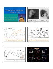
Interpretation<br />
• Nonvisualization of GB at 4hrs<br />
• Activity normally enters the GB passively from<br />
the ductal system. In acute cholecystitis, there<br />
is usually cystic duct obstruction preventing<br />
bile from entering the GB (90-95% of pts)<br />
• DDX: acute cholecystitis, s/p cholecystectomy,<br />
prolonged fast or recent fatty meal, chronic<br />
chlocecystitis (rare)
First Hour
4<br />
hours
Interpretation<br />
• “Rim” or “Stripe” Sign<br />
• Nonvisualization of the GB with increased<br />
activity at the liver margin near the GB fossa<br />
• Seen in pts with sever acute cholecystittus and<br />
has a 20-40% incidence of grangrenous<br />
cholecystitus
Interpretation<br />
• Delayed visualization of the GB<br />
• After 1 hr by “convention”, sometimes<br />
considered if >30 minutes after bowel has been<br />
confirmed, (even if within the 1 hr)<br />
• Most commonly seen with chronic cholecystitis
Interpretation<br />
• Delayed visualization of Gut with GB<br />
visualization<br />
• “NON specific”<br />
• DDX: chronic cholecystitis, post-opiate<br />
administration, partial CBD obstruction, other<br />
inflammatory bowel processes (including<br />
pancreatitis, etc)
Interpretation<br />
• Leak<br />
• Most often seen post-op (ie lap<br />
cholecystectomy) or penetrating trauma<br />
• Usually seen as intense activity nor conforming<br />
to normal anatomical structures…..multiple<br />
views help (ie right pericolic gutter activity)
Interpretation<br />
• Hepatic uptake only (nonvisualization of<br />
the ducts, GB, and gut)<br />
• Acute CBD obstruction vs cholestatic<br />
hepatitis….however the clinical presentations<br />
of these generally differ<br />
• Hepatic uptake may be a clue to interpretation,<br />
being more diminished in hepatitis.<br />
• NOTE: In complete CBD obstruction – it is<br />
rare to see GB activity because of backpressure<br />
in the biliary system
Interpretation<br />
• Bowel distortions<br />
• Pacreatitis or masses may compress the<br />
duodenum and cause non-vis. of the 3 rd portion<br />
• Prior bowel surgery<br />
• Cut-off CBD sign<br />
• Stone impacted in the CBD?
Interpretation<br />
• Stomach visualized – enterogastric reflux<br />
but sometimes normal?
Interpretation<br />
• Parenchymal defects<br />
• Any lesion which displaces hepatocytes may<br />
cause a liver defect on early images (ie mets,<br />
abscess, cyst, tumor)<br />
• Hepatomas and adenomas occasionally show<br />
delayed uptake of <strong>HIDA</strong> compared with<br />
surrounding liver
Interpretation<br />
• SPECIAL PROCEDURES<br />
• GB Ejection Fraction (GBEF) Commonly Performed (<br />
more common now than emergent <strong>HIDA</strong> scans)<br />
• When chronic acalculous cholecystitis or a biliary dyskinetic<br />
syndrome is suspected for chronic RUQ pain (not really an<br />
indication for an emergency study)<br />
• GBEF of >35% considered normal at 20 minutes post<br />
CCK (sincalide) administration<br />
•
Pediatric Applications of <strong>HIDA</strong><br />
<strong>Scans</strong>
Pediatrics<br />
• In the neonatal setting, the <strong>HIDA</strong> scan be helpful<br />
in distinguishing Neonatal Hepatitis from Biliary<br />
Atresia<br />
• Imaging should be performed up to 4 and 24 hours<br />
if needed<br />
• Visualization of bowel excludes biliary atresia<br />
• Suggest -Repeat with Phenobarbitol if no bowel<br />
• Visualization of bowel by 4-24 hrs more typical of<br />
neonatal hepatitis
Pediatrics<br />
BILIARY ATRESIA: NO bowel by 24 hours
CASE STUDY<br />
What’s the diagnosis?
The End<br />
Thank You For Your Attention