Early miscarriage after single and double blastocyst transfer â An ...
Early miscarriage after single and double blastocyst transfer â An ...
Early miscarriage after single and double blastocyst transfer â An ...

You also want an ePaper? Increase the reach of your titles
YUMPU automatically turns print PDFs into web optimized ePapers that Google loves.
Izvirni članek/Original article<br />
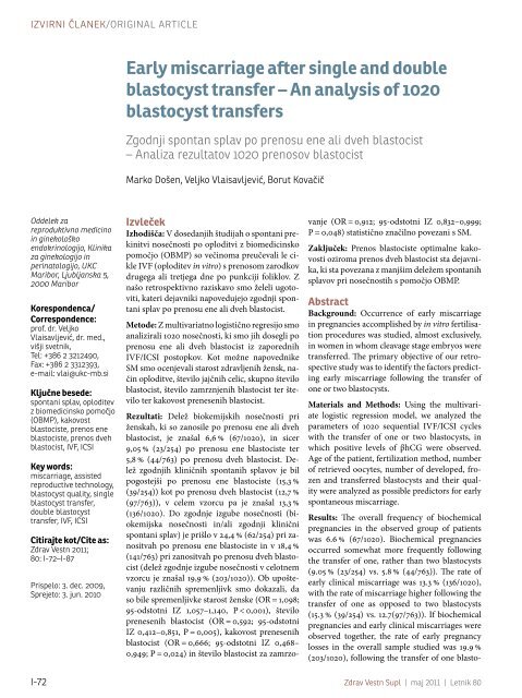
<strong>Early</strong> <strong>miscarriage</strong><br />
<strong>after</strong> <strong>single</strong> <strong>and</strong><br />
<strong>double</strong> <strong>blastocyst</strong><br />
<strong>transfer</strong><br />
<strong>Early</strong> <strong>miscarriage</strong> <strong>after</strong> <strong>single</strong> <strong>and</strong> <strong>double</strong><br />
<strong>blastocyst</strong> <strong>transfer</strong> – <strong>An</strong> analysis of 1020<br />
<strong>blastocyst</strong> <strong>transfer</strong>s<br />
Zgodnji spontan splav po prenosu ene ali dveh blastocist<br />
– <strong>An</strong>aliza rezultatov 1020 prenosov blastocist<br />
Marko Došen, Veljko Vlaisavljević, Borut Kovačič<br />
Oddelek za<br />
reproduktivno medicino<br />
in ginekološko<br />
endokrinologijo, Klinika<br />
za ginekologijo in<br />
perinatologijo, UKC<br />
Maribor, Ljubljanska 5,<br />
2000 Maribor<br />
Korespondenca/<br />
Correspondence:<br />
prof. dr. Veljko<br />
Vlaisavljević, dr. med.,<br />
višji svetnik,<br />
Tel: +386 2 3212490,<br />
Fax: +386 2 3312393,<br />
e-mail: vlai@ukc-mb.si<br />
Ključne besede:<br />
spontani splav, oploditev<br />
z biomedicinsko pomočjo<br />
(OBMP), kakovost<br />
blastociste, prenos ene<br />
blastociste, prenos dveh<br />
blastocist, IVF, ICSI<br />
Key words:<br />
<strong>miscarriage</strong>, assisted<br />
reproductive technology,<br />
<strong>blastocyst</strong> quality, <strong>single</strong><br />
<strong>blastocyst</strong> <strong>transfer</strong>,<br />
<strong>double</strong> <strong>blastocyst</strong><br />
<strong>transfer</strong>, IVF, ICSI<br />
Citirajte kot/Cite as:<br />
Zdrav Vestn 2011;<br />
80: I-72–I-87<br />
Prispelo: 3. dec. 2009,<br />
Sprejeto: 3. jun. 2010<br />
Izvleček<br />
Izhodišča: V dosedanjih študijah o spontani prekinitvi<br />
nosečnosti po oploditvi z biomedicinsko<br />
pomočjo (OBMP) so večinoma preučevali le cikle<br />
IVF (oploditev in vitro) s prenosom zarodkov<br />
drugega ali tretjega dne po punkciji foliklov. Z<br />
našo retrospektivno raziskavo smo želeli ugotoviti,<br />
kateri dejavniki napovedujejo zgodnji spontani<br />
splav po prenosu ene ali dveh blastocist.<br />
Metode: Z multivariatno logistično regresijo smo<br />
analizirali 1020 nosečnosti, ki smo jih dosegli po<br />
prenosu ene ali dveh blastocist iz zaporednih<br />
IVF/ICSI postopkov. Kot možne napovednike<br />
SM smo ocenjevali starost zdravljenih žensk, način<br />
oploditve, število jajčnih celic, skupno število<br />
blastocist, število zamrznjenih blastocist ter število<br />
ter kakovost prenesenih blastocist.<br />
Rezultati: Delež biokemijskih nosečnosti pri<br />
ženskah, ki so zanosile po prenosu ene ali dveh<br />
blastocist, je znašal 6,6 % (67/1020), in sicer<br />
9,05 % (23/254) po prenosu ene blastociste ter<br />
5,8 % (44/763) po prenosu dveh blastocist. Delež<br />
zgodnjih kliničnih spontanih splavov je bil<br />
pogostejši po prenosu ene blastociste (15,3 %<br />
(39/254)) kot po prenosu dveh blastocist (12,7 %<br />
(97/763)), v celem vzorcu pa je znašal 13,3 %<br />
(136/1020). Do zgodnje izgube nosečnosti (biokemijska<br />
nosečnosti in/ali zgodnji klinični<br />
spontani splav) je prišlo v 24,4 % (62/254) pri zanositvah<br />
po prenosu ene blastociste in v 18,4 %<br />
(141/763) pri zanositvah po prenosu dveh blastocist<br />
(delež zgodnje izgube nosečnosti v celotnem<br />
vzorcu je znašal 19,9 % (203/1020)). Ob upoštevanju<br />
različnih spremenljivk smo dokazali, da<br />
so bile spremenljivke starost ženske (OR = 1,098;<br />
95-odstotni IZ 1,057–1,140, P < 0,001), število<br />
prenesenih blastocist (OR = 0,592; 95-odstotni<br />
IZ 0,412–0,851, P = 0,005), kakovost prenesenih<br />
blastocist (OR = 0,666; 95-odstotni IZ 0,468–<br />
0,949; P = 0,024) in število blastocist za zamrzovanje<br />
(OR = 0,912; 95-odstotni IZ 0,832–0,999;<br />
P = 0,048) statistično značilno povezani s SM.<br />
Zaključek: Prenos blastociste optimalne kakovosti<br />
oziroma prenos dveh blastocist sta dejavnika,<br />
ki sta povezana z manjšim deležem spontanih<br />
splavov pri nosečnostih s pomočjo OBMP.<br />
Abstract<br />
Background: Occurrence of early <strong>miscarriage</strong><br />
in pregnancies accomplished by in vitro fertilisation<br />
procedures was studied, almost exclusively,<br />
in women in whom cleavage stage embryos were<br />
<strong>transfer</strong>red. The primary objective of our retrospective<br />
study was to identify the factors predicting<br />
early <strong>miscarriage</strong> following the <strong>transfer</strong> of<br />
one or two <strong>blastocyst</strong>s.<br />
Materials <strong>and</strong> Methods: Using the multivariate<br />
logistic regression model, we analyzed the<br />
parameters of 1020 sequential IVF/ICSI cycles<br />
with the <strong>transfer</strong> of one or two <strong>blastocyst</strong>s, in<br />
which positive levels of βhCG were observed.<br />
Age of the patient, fertilization method, number<br />
of retrieved oocytes, number of developed, frozen<br />
<strong>and</strong> <strong>transfer</strong>red <strong>blastocyst</strong>s <strong>and</strong> their quality<br />
were analyzed as possible predictors for early<br />
spontaneous <strong>miscarriage</strong>.<br />
Results: The overall frequency of biochemical<br />
pregnancies in the observed group of patients<br />
was 6.6 % (67/1020). Biochemical pregnancies<br />
occurred somewhat more frequently following<br />
the <strong>transfer</strong> of one, rather than two <strong>blastocyst</strong>s<br />
(9.05 % (23/254) vs. 5.8 % (44/763)). The rate of<br />
early clinical <strong>miscarriage</strong> was 13.3 % (136/1020),<br />
with the rate of <strong>miscarriage</strong> higher following the<br />
<strong>transfer</strong> of one as opposed to two <strong>blastocyst</strong>s<br />
(15.3 % (39/254) vs. 12.7(97/763)). If biochemical<br />
pregnancies <strong>and</strong> early clinical <strong>miscarriage</strong>s were<br />
observed together, the rate of early pregnancy<br />
losses in the overall sample studied was 19.9 %<br />
(203/1020), following the <strong>transfer</strong> of one blasto-<br />
I-72 Zdrav Vestn Supl | maj 2011 | Letnik 80
Izvirni članek/Original article<br />
cyst 24.4 % (62/254) <strong>and</strong> following the <strong>transfer</strong> of<br />
two 18.4 % (141/763). Using the multivariate logistic<br />
regression, we demonstrated that statistically<br />
significant predictors for early spontaneous<br />
<strong>miscarriage</strong>s were the patient’s age (OR = 1.098;<br />
95 % CI 1.057–1.140, P < 0.001), the number of<br />
<strong>blastocyst</strong>s <strong>transfer</strong>red (OR = 0.592; 95 % CI<br />
0.412–0.851, P = 0.005), the quality of <strong>blastocyst</strong>s<br />
<strong>transfer</strong>red (OR=0.666; 95 % CI 0.468–0.949;<br />
P = 0.024), as well as the number of <strong>blastocyst</strong>s<br />
frozen (OR = 0.912; 95 % CI 0.832–0.999;<br />
P = 0.048).<br />
Conclusion: The <strong>transfer</strong> of optimal quality<br />
<strong>blastocyst</strong>(s) <strong>and</strong> <strong>transfer</strong> of two <strong>blastocyst</strong>s were<br />
associated with a reduced rate of spontaneous<br />
<strong>miscarriage</strong>s in pregnancies achieved by ART.<br />
Table 1: Age of female patients.<br />
Introduction<br />
<strong>Early</strong> <strong>miscarriage</strong> (early spontaneous<br />
abortion) is a very common event in early<br />
pregnancy.1 This high frequency of early<br />
<strong>miscarriage</strong> is characteristic of our species.1-5<br />
According to the results of several studies<br />
which evaluated frequency of <strong>miscarriage</strong> in<br />
the first trimester of pregnancy <strong>after</strong> spontaneous<br />
conception, approximatelly 30 % of<br />
total fertilized ova were lost prior to the next<br />
expected menstruation (i. e. prior to the first<br />
measurement of the level of βHCG 14 days<br />
<strong>after</strong> ovulation–the so-called premenstrual<br />
losses), another 30 % were spontaneously<br />
miscarried prior to the ultrasound confirmation<br />
of successful implantation (biochemical<br />
pregnancies), <strong>and</strong> only 10–15 % of all losses<br />
were spontaneous <strong>miscarriage</strong>s recognized<br />
<strong>after</strong> the first ultrosonographic confirmation<br />
of viable intrauterine pregnancy.1-7,51-54<br />
Ackowledged unmodifiable risk factors for<br />
<strong>miscarriage</strong> in general population are maternal<br />
age, previous history of spontaneous<br />
<strong>miscarriage</strong>s, certain anatomical, endocrine<br />
<strong>and</strong> autoimmune disorders, trombophilia<br />
<strong>and</strong> infections. Modifiable risk factors are<br />
pronounced maternal obesity, cigarette<br />
smoking, use of alcohol, recreational drugs<br />
<strong>and</strong> high coffein intake.3,7‐10 The most common<br />
cause of <strong>miscarriage</strong> <strong>after</strong> spontaneous<br />
conception is a chromosomal abnormality<br />
of the conceptus, which has been detected<br />
in approximatelly 50 % of cytogenetically<br />
examined miscarried fetuses.11-12,24 It has<br />
been observed that this high aneuploidy rate<br />
Variable SBT group DBT group Overall<br />
Patient’s age<br />
(mean ± SD; range)<br />
31.2 ± 4.3<br />
(21–41)<br />
32.3 ± 4.3<br />
(20–44)<br />
* SBT–<strong>single</strong> <strong>blastocyst</strong> <strong>transfer</strong>; DBT–<strong>double</strong> <strong>blastocyst</strong> <strong>transfer</strong><br />
32.0 ± 4.3<br />
(22–44)<br />
is mainly the consequence of high incidence<br />
of meiotic errors (mostly chromosome nondisjunction)<br />
in oocytes of older mothers.13<br />
The precise mechanism of meiotic nondisjuction<br />
is not completely elucidated, but<br />
it has been noted that there is significantly<br />
less chiasmata in oocyte chromosomes of<br />
older women than in oocyte chromosomes<br />
of younger ones.14,44<br />
Miscarriage in pregnancies achieved by<br />
assisted reproductive technology (ART)<br />
procedures is slightly more frequent than in<br />
general population <strong>and</strong> occurs in 16–30 % of<br />
patients with confirmed pregnancy.15-20 If<br />
one considers only losses of embryos prior<br />
to the first measurement of bHCG 14 days<br />
<strong>after</strong> embryo <strong>transfer</strong>, the difference between<br />
pregnancies achieved by ART <strong>and</strong><br />
those <strong>after</strong> spontaneous conception is even<br />
more pronounced–the rate of premenstrual<br />
losses is 50 % <strong>after</strong> embryo <strong>transfer</strong> <strong>and</strong> only<br />
30 % <strong>after</strong> spontaneous conception.21 One<br />
explanation for this increase of early pregnancy<br />
loss rate may be the intense follow<br />
up of ART pregnancies. <strong>An</strong>other reason of<br />
this phenomenon could be that risk factors<br />
for infertility may considerably overlap with<br />
those for <strong>miscarriage</strong>.15 Risk factors for early<br />
<strong>miscarriage</strong> <strong>after</strong> ART are age of the mother,15,25-26<br />
age of the father,27 number of previous<br />
spontaneous <strong>miscarriage</strong>s,7,15,25-26,28<br />
pronounced obesity, cigarette smoking,29-31<br />
alcohol consumption,8,32-33 use of recreational<br />
drugs29 <strong>and</strong> high doses of caffeine.34<br />
<strong>An</strong>other group of risk factors for early <strong>miscarriage</strong><br />
in ART pregnancies originates from<br />
ART itself. The characteristics of stimulated<br />
cycles, which could be risk factors for early<br />
<strong>miscarriage</strong> <strong>after</strong> ART, are method of fertilisation<br />
(IVF/ICSI),15,40,49 protocol of stimulation48<br />
<strong>and</strong> the level of controlled ovarian<br />
hyperstimulation (estradiol level at oocyte<br />
retrieval, ocurrence of ovarian hyperstimu-<br />
Zdrav Vestn Supl | <strong>Early</strong> <strong>miscarriage</strong> <strong>after</strong> <strong>single</strong> <strong>and</strong> <strong>double</strong> <strong>blastocyst</strong> <strong>transfer</strong> I-73
Izvirni članek/Original article<br />
Table 2: Etiology of infertility in the examined cohort.<br />
Etiology of infertility in the examined cohort Frequency (%)<br />
Tubal factor 36.6 % (371)<br />
Idiopathic female infertility 20.0 % (203)<br />
<strong>An</strong>ovulation 11.3 % (115)<br />
Uterine factors 5.9 % (60)<br />
Infertility due to male factor 21.3 % (216)<br />
Other main diagnosis of infertility 4.8 % (49)<br />
Overall 100.0 % (1020)<br />
lation syndrome, number of developed/<br />
punctured follicules, number of retrieved<br />
oocytes). Important parameters of the developing<br />
embryos, which could be risk factors<br />
for early <strong>miscarriage</strong> <strong>after</strong> ART, are total<br />
number of developed embryos, developmental<br />
stage of <strong>transfer</strong>red embryos, number<br />
of <strong>transfer</strong>red embryos <strong>and</strong> their quality,<br />
number of embryos suitable for freezing <strong>and</strong><br />
freezing/thawing of embryos.40,45-50 As in<br />
pregnancies <strong>after</strong> spontaneous conception,<br />
the most common cause of early <strong>miscarriage</strong><br />
in ART pregnancies is chromosomal abnormality<br />
of the embryo,22-24 which has been<br />
detected by citogenetic methods in 55 % of<br />
miscarried fetuses.24 The distribution of abnormalities<br />
among affected chromosomes<br />
in the studied miscarried fetuses is virtually<br />
the same in both spontaneous <strong>and</strong> assisted<br />
conception group of patients.24<br />
Risk factors for early <strong>miscarriage</strong> in ART<br />
pregnancies were evaluated almost exclusively<br />
in cycles in which cleavage stage embryos<br />
had been <strong>transfer</strong>red (embryo <strong>transfer</strong><br />
on the second or third day following follicle<br />
aspiration). The primary objective of our<br />
study was to identify factors predicting early<br />
<strong>miscarriage</strong> <strong>after</strong> <strong>transfer</strong> of one or two <strong>blastocyst</strong>s.<br />
Material <strong>and</strong> methods<br />
Patients<br />
Retrospective clinical study encompassed<br />
1020 stimulated IVF or ICSI cycles,<br />
performed at the Department of Reproductive<br />
Medicine <strong>and</strong> Gynecological Endocrinology<br />
at the University Medical Centre of<br />
Maribor, Slovenia. The data was gathered<br />
from January 2001 to December 2005.<br />
Patients included were under 43 years of<br />
age <strong>and</strong>, prior to entering an IVF/ICSI treatment,<br />
underwent all tests prescribed by the<br />
protocol for clinical examination of infertile<br />
couples.<br />
Only the patients meeting the following<br />
criteria were included in the study:<br />
1. embryo <strong>transfer</strong> was performed on day<br />
4 or 5 <strong>after</strong> follicle aspiration (<strong>blastocyst</strong><br />
stage embryos);<br />
2. only one or two fresh <strong>blastocyst</strong>s were<br />
<strong>transfer</strong>red (<strong>single</strong> <strong>blastocyst</strong> <strong>transfer</strong>–<br />
SBT or <strong>double</strong> <strong>blastocyst</strong> <strong>transfer</strong>–DBT);<br />
3. serum βhCG levels exceeded 15 IU/l on<br />
the fourteenth day following the <strong>blastocyst</strong><br />
<strong>transfer</strong>; <strong>and</strong><br />
4. information on the pregnancy outcome<br />
was known <strong>and</strong> documented.<br />
From the group of pregnant patients<br />
meeting these criteria, those in whom the<br />
pregnancy ended as an extrauterine pregnancy,<br />
spontaneous abortion <strong>after</strong> week 13<br />
or delivery were excluded from the study.<br />
Stimulation protocols<br />
Patients were most frequently stimulated<br />
according to the protocol involving<br />
gonadotrophin-releasing hormone agonists<br />
(GnRH-a) (86 %) (almost exclusively using<br />
the long protocol). In the remaining patients,<br />
the protocol with gonadotrophin-releasing<br />
hormone antagonists (GnRH-ant) was applied.35<br />
GnRH agonists used were triptorelin<br />
(Diphereline®, Ipsen Pharma Biotech,<br />
France) (48 %), gosereline (Zoladex®, Zeneca<br />
Pharmaceuticals, Engl<strong>and</strong>) (23 %) or busereline<br />
(Suprefact®, Sanofi Aventis, France)<br />
(15 %). Cetorelix (3 mg) (Cetrotide®, Merck<br />
Serono, Switzerl<strong>and</strong>) was used as GnRH antagonist.<br />
Follicle growth was predominantly<br />
stimulated by recombinant FSH (Gonal F®,<br />
Merck Serono, Switzerl<strong>and</strong>), while human<br />
menopausal gonadotrophin (HMG) (Menopur®,<br />
Ferring Pharmaceuticals, Switzerl<strong>and</strong>)<br />
was used infrequently. On the day when at<br />
least two follicles reached an average diameter<br />
of 18 mm, final maturation of the oocyte<br />
was stimulated by the urinary human HCG<br />
(Profasi®, Merck Serono, Switzerl<strong>and</strong>, using<br />
I-74 Zdrav Vestn Supl | maj 2011 | Letnik 80
Izvirni članek/Original article<br />
Table 3: Etiology of infertility in patients’ partners.<br />
Etiology of infertility in partners of the patients Frequency (%)<br />
Oligozoospermia 29.9 % (304)<br />
Azoospermia 10.2 % (104)<br />
Normoastenozoospermia 7.3 % (74)<br />
Oligoastenozoospermia 5.7 % (58)<br />
Oligoastenoteratozoospermia 3.6 % (37)<br />
Oligoteratozoospermia 1.1 % (11)<br />
Normoastenoteratozoospermia 0.8 % (8)<br />
Normoteratozoospermia 0.6 % (6)<br />
Female infertility 37.3 % (379)<br />
Other main diagnosis of infertility 3.8 % (39)<br />
Overall 100.0 % (1020)<br />
a dose of 10,000 IU) or human recombinant<br />
HCG (Ovitrelle®, Merck Serono, Switzerl<strong>and</strong>,<br />
250 mg dose). A detailed description<br />
of the laboratory procedures can be found<br />
elsewhere.36 Approximately 36 hours (36 ± 1)<br />
following the administration of HCG, oocytes<br />
were recovered by ultrasound-guided<br />
trans-vaginal follicle aspiration. Fertilization<br />
was performed through IVF or ICSI.<br />
Medicult® media (MediCult, Denmark)<br />
were used for oocyte culturing. Pursuant<br />
to the protocol of our centre, only one or<br />
maximally two <strong>blastocyst</strong>s were <strong>transfer</strong>red<br />
on the fifth, exceptionally on the fourth day<br />
(1.8 %) following follicle aspiration. Labotec®<br />
catheter (Labotec, Germany) was used for<br />
<strong>blastocyst</strong> <strong>transfer</strong>. According to the legislation<br />
in force at the time of the study, the<br />
couple was allowed to decide on the number<br />
of embryos to be <strong>transfer</strong>red. Embryos were<br />
<strong>transfer</strong>red only <strong>after</strong> both partners signed<br />
the official consent form for the <strong>transfer</strong> of<br />
embryos. A day <strong>after</strong> the follicle aspiration,<br />
all patients started receiving didrogesterone<br />
(30 mg/day) (Dabroston®, Belupo, Croatia)<br />
or micronized progesterone (600 mg/day)<br />
(Utrogestan®, Laboratories Besins International,<br />
France) for luteal support.<br />
Blastocyst quality evaluation system<br />
The quality of <strong>transfer</strong>red <strong>blastocyst</strong>s was<br />
evaluated by a <strong>blastocyst</strong> classification system<br />
based on morphological criteria, devel-<br />
oped by our Centre.37 This classification is<br />
a modification of the <strong>blastocyst</strong> evaluation<br />
system introduced by Gardner <strong>and</strong> Schoolcraft.38<br />
The classification used in our laboratory<br />
takes into consideration four parameters:<br />
blastocoel expansion, inner cellular<br />
mass (ICM) form, morphology <strong>and</strong> cohesion<br />
of the trophoectoderm (TE) as well as<br />
the degree of embryo fragmentation.<br />
B1 class <strong>blastocyst</strong>s were those in which<br />
blastocoel was completely exp<strong>and</strong>ed, ICM<br />
was round or oval, TE was seen as cohesive<br />
epithelium <strong>and</strong> there were no excluded<br />
blastomeres or cytoplasmatic fragments. B2<br />
class <strong>blastocyst</strong>s were exp<strong>and</strong>ed with optimal<br />
ICM, but suboptimal TE. In B3 class<br />
were grouped morphologically optimal unexp<strong>and</strong>ed<br />
<strong>blastocyst</strong>s <strong>and</strong> compact morulae<br />
(embryos formed from more than 10 blastomeres<br />
tightly connected into a compact<br />
formation). Exp<strong>and</strong>ed <strong>blastocyst</strong>s with normal<br />
TE, but suboptimal ICM (ICM smaller<br />
in size than the blastomere of a correctly<br />
divided 16-cell embryo) were arranged in<br />
the B4 class. All exp<strong>and</strong>ed <strong>blastocyst</strong>s with<br />
suboptimal ICM <strong>and</strong> with fragments or necrotic<br />
foci in the TE were included in B5<br />
class. Morulae <strong>and</strong> early <strong>blastocyst</strong>s, which<br />
had up to 20 % blastomeres or fragments excluded<br />
in the periviteline space were classified<br />
as B6 class. B7 class comprised necrotic<br />
<strong>blastocyst</strong>s (more than 50 % of embryonic<br />
material was necrotic, <strong>and</strong> abnormal ICM<br />
was present; <strong>blastocyst</strong>s with normal ICM<br />
but necrotic TE were included in B2 class).<br />
Small <strong>blastocyst</strong>s <strong>and</strong> morulae (more than<br />
20 % of excluded blastomeres) <strong>and</strong> also very<br />
fragmented embryos in which some sign of<br />
blastocoel or ellipsoid TE cells were noted<br />
were included in B8 class. Non-compact<br />
morulae <strong>and</strong> fewer than 12-cell embryos on<br />
day 5 were considered as arrested embryos<br />
<strong>and</strong> had never been used for <strong>transfer</strong>.<br />
Pregnancy confirmation<br />
Fourteen days <strong>after</strong> the <strong>transfer</strong> of embryos,<br />
serum βhCG levels were determined<br />
in all patients. Pregnancy was considered<br />
confirmed if the level exceeded 15 UI/l. Total<br />
βhCG levels were determined by Architect<br />
i2000 (Abbott diagnostics, USA) analyzer,<br />
Zdrav Vestn Supl | <strong>Early</strong> <strong>miscarriage</strong> <strong>after</strong> <strong>single</strong> <strong>and</strong> <strong>double</strong> <strong>blastocyst</strong> <strong>transfer</strong> I-75
Izvirni članek/Original article<br />
Table 4: Pregnancy outcome in the observed group of patients.<br />
Variable SBT group DBT group Overall<br />
Delivery rate (livebirths) (%)<br />
<strong>single</strong>tons 75.6 % (192/254) 45.3 % (347/766) 52.8 % (539/1020)<br />
twins 0 34.9 % (267/766) 26.2 % (267/1020)<br />
triplets 0 1.0 % (8/766) 0.8 % (8/1020)<br />
Spontaneous <strong>miscarriage</strong>s (%)<br />
biochemical pregnancy 9.05 % (23/254) 5.8 % (44/763) 6.6 % (67/1020)<br />
early clinical spont <strong>miscarriage</strong> 15.3 % (39/254) 12.7 % (97/763) 13.3 % (136/1020)<br />
Overall spont. <strong>miscarriage</strong>s 24.4 % (62/254) 18.4 % (141/763) 19.9 % (203/1020)<br />
* SBT–<strong>single</strong> <strong>blastocyst</strong> <strong>transfer</strong>; DBT–<strong>double</strong> <strong>blastocyst</strong> <strong>transfer</strong><br />
using immunochemical hemiluminescent<br />
methods (CMIA: acridine; analytical sensitivity<br />
< 1.2 IU/l).<br />
Definitions of <strong>miscarriage</strong><br />
<strong>and</strong> exclusion criteria<br />
<strong>Early</strong> <strong>miscarriage</strong> (early spontaneous<br />
abortion in earlier nomenclature) is generally<br />
defined as an unprovoked termination<br />
of pregnancy prior to the end of first trimester<br />
of pregnancy (13 +6 weeks of gestation).<br />
A distinction should be drawn among premenstrual<br />
pregnancy loss (loss of conceptus<br />
prior to the first measurement of βhCG level<br />
14 days <strong>after</strong> ovulation or embryo <strong>transfer</strong>),<br />
biochemical pregnancy (loss of conceptus<br />
<strong>after</strong> the first measurement of βhCG level<br />
but before the ultrasound (US) confirmation<br />
of implantation) <strong>and</strong> early clinical <strong>miscarriage</strong><br />
(pregnancy loss <strong>after</strong> US confirmation<br />
of viable pregnancy but before the beginning<br />
of the second trimester). Biochemical<br />
pregnancies <strong>and</strong> early clinical <strong>miscarriage</strong>s<br />
are commonly identified together as early<br />
pregnancy losses (EPL).<br />
The definition of spontaneous <strong>miscarriage</strong>,<br />
which was used in our study, included<br />
the following clinical outcomes of pregnancy<br />
following its confirmation by positive serum<br />
βhCG levels:<br />
1. a decrease in serum βhCG levels compared<br />
to the values found at the first<br />
measurement prior to the first US control<br />
(biochemical pregnancy);<br />
2. presence of a gestational sac without<br />
an embryo at the first ultrasound (US)<br />
control 14 days <strong>after</strong> the positive results<br />
of the serum βhCG levels (anembrionic<br />
pregnancy);<br />
3. presence of a gestational sac with an embryo<br />
with no cardiac action, <strong>after</strong> the<br />
previous US control has confirmed heart<br />
action (missed abortion);<br />
4. spontaneous expulsion of the conception<br />
products from the uterus, following an<br />
US confirmation of the presence of a gestational<br />
sac with or without an embryo<br />
(complete or incomplete clinical <strong>miscarriage</strong>).<br />
In the case of an anembryonic pregnancy<br />
<strong>and</strong> missed abortion, it was noted that the<br />
<strong>miscarriage</strong> occurred on the day that the<br />
definitive clinical diagnosis was made by<br />
trans-vaginal ultrasound examination, regardless<br />
of when the expulsion of the conception<br />
products (spontaneous or induced)<br />
actually occurred.<br />
The following outcomes of pregnancy<br />
were excluded from the definition of <strong>miscarriage</strong>:<br />
1. clinically confirmed extra uterine pregnancy;<br />
2. <strong>miscarriage</strong> <strong>after</strong> 13 +6 week of gestation<br />
(late <strong>miscarriage</strong>); <strong>and</strong><br />
3. spontaneous loss (spontaneous reduction)<br />
of one embryo with the survival of<br />
the other, in case of twin pregnancy, determined<br />
at the first ultrasound control.<br />
Late <strong>miscarriage</strong>s (those occurring <strong>after</strong><br />
13 +6 week of gestation) were excluded<br />
I-76 Zdrav Vestn Supl | maj 2011 | Letnik 80
Izvirni članek/Original article<br />
Table 5: Characteristics of the observed cycles–method of fertilization.<br />
Method of fertilization (%) SBT group DBT group Overall<br />
IVF 23.2 % (59/254) 25.3 % (194/766) 24.8 % (253/1020)<br />
ICSI 76.8 % (195/254) 74.7 % (572/766) 75.2 % (767/1020)<br />
* SBT–<strong>single</strong> <strong>blastocyst</strong> <strong>transfer</strong>; DBT–<strong>double</strong> <strong>blastocyst</strong> <strong>transfer</strong><br />
from the analysis due to the specific nature<br />
of their etiology, which is significantly different<br />
from those of early <strong>miscarriage</strong>s. In<br />
addition, we were unable to include in our<br />
study pregnancy losses which occurred <strong>after</strong><br />
the <strong>transfer</strong> of <strong>blastocyst</strong> into the uterus, but<br />
prior to the determination of serum βhCG<br />
14 days <strong>after</strong> the <strong>blastocyst</strong> <strong>transfer</strong> (the socalled<br />
premenstrual <strong>miscarriage</strong>s).<br />
Pregnancies in which the <strong>blastocyst</strong><br />
spontaneously divided into two embryos<br />
(monozygotic twins) <strong>after</strong> the <strong>transfer</strong> of<br />
one or two <strong>blastocyst</strong>s were excluded from<br />
the analysis.<br />
Our centre’s database did not record the<br />
number of previous <strong>miscarriage</strong>s, so that<br />
repeated <strong>miscarriage</strong>s could not be included<br />
into the multivariate statistical analysis<br />
model.<br />
Data quality<br />
Data used in this analysis were received<br />
from the centre’s database on couples whose<br />
infertility was treated by medically assisted<br />
fertilization techniques. If there was any<br />
data missing in the database for any variable,<br />
the patient’s documentation (paper records)<br />
was checked. If it was still impossible to find<br />
the missing data, the patient was excluded<br />
from further analysis.<br />
The <strong>miscarriage</strong>s were either confirmed<br />
clinically at our centre or were reported by<br />
patients in the form of a questionnaire that<br />
we routinely sent to each patient whose<br />
pregnancy was confirmed by the first test of<br />
serum βhCG.<br />
Statistical analysis<br />
We examined the following parameters<br />
as potential factors predicting the occurrence<br />
of an early spontaneous <strong>miscarriage</strong><br />
following medically assisted fertilization:<br />
patient’s age, method of fertilization (IVF or<br />
ICSI), number of oocytes retrieved by follicle<br />
aspiration, total number of <strong>blastocyst</strong>s,<br />
number of <strong>blastocyst</strong>s frozen, number of<br />
<strong>blastocyst</strong>s <strong>transfer</strong>red <strong>and</strong> quality of <strong>transfer</strong>red<br />
<strong>blastocyst</strong>s.<br />
Quantitative variables were presented in<br />
the form of mean value ± st<strong>and</strong>ard deviation<br />
(SD) <strong>and</strong> category variables were presented<br />
as ratios.<br />
Bivariate analysis of the correlation between<br />
risk factors <strong>and</strong> <strong>miscarriage</strong>s was performed<br />
using the logistic regression model.<br />
In addition, an analysis using multivariate<br />
logistic regression model was also conducted.<br />
All risk factors were first entered into the<br />
log-likelihood fitting model unconditionally.<br />
Backward stepwise logistic regression was<br />
used subsequently at the removal probability<br />
of P=0.1. The results were interpreted as<br />
the odds ratios (OR) for the observed event,<br />
with a confidence interval (CI) of 95 %.<br />
The study had a 95 % power to detect a<br />
difference of 10 % (27 % vs. 17 %) in the rate<br />
of <strong>miscarriage</strong>s in the analyzed sample of<br />
709 cycles with the <strong>transfer</strong> of optimal quality<br />
<strong>blastocyst</strong>s <strong>and</strong> 311 cycles with the <strong>transfer</strong><br />
of suboptimal quality <strong>blastocyst</strong>s.<br />
P value of under 0.05 was considered to<br />
be statistically significant. Statistical analysis<br />
was performed using STATISTICA® software,<br />
version 8.0 (StatSoft Inc., OK, USA).<br />
Results<br />
A. Demographic <strong>and</strong> clinical<br />
characteristics of the patients<br />
In the period covered by our research,<br />
pregnancy was confirmed in 1020 patients<br />
who underwent IVF/ICSI cycles with the<br />
<strong>transfer</strong> of one or two <strong>blastocyst</strong>s.<br />
Average age of the patients in the studied<br />
group was 32 years (32.0 ± 4.3; the youngest<br />
patient was 22 <strong>and</strong> the oldest 44 years old),<br />
with no significant difference in age between<br />
Zdrav Vestn Supl | <strong>Early</strong> <strong>miscarriage</strong> <strong>after</strong> <strong>single</strong> <strong>and</strong> <strong>double</strong> <strong>blastocyst</strong> <strong>transfer</strong> I-77
Izvirni članek/Original article<br />
Table 6: Characteristics of the observed cycles–number of retrieved oocytes, number of developed <strong>and</strong><br />
frozen <strong>blastocyst</strong>s.<br />
Variable SBT group DBT group Overall<br />
Number of retrieved oocytes (mean ± SD; range) 11.5 ± 6.7<br />
(1–37)<br />
Number of developed <strong>blastocyst</strong>s (mean ± SD; range) 4.5 ± 2.6<br />
(1–19)<br />
Number of frozen <strong>blastocyst</strong>s (mean ± SD; range) 3.1 ± 3.4<br />
(0–16)<br />
9.9 ± 4.2<br />
( 2- 27)<br />
4.7 ± 3.8<br />
(1–19)<br />
1.7 ± 2.0<br />
(0–9)<br />
10.3 ± 5.0<br />
(1–37)<br />
4.5 ± 2.1<br />
(1–11)<br />
2.1 ± 2.5<br />
(0–16)<br />
* SBT–<strong>single</strong> <strong>blastocyst</strong> <strong>transfer</strong>; DBT–<strong>double</strong> <strong>blastocyst</strong> <strong>transfer</strong><br />
patients in whom one <strong>blastocyst</strong> was <strong>transfer</strong>red<br />
vs. those in whom two <strong>blastocyst</strong>s<br />
were <strong>transfer</strong>red (Table 1).<br />
Main diagnoses in patients <strong>and</strong> their<br />
partners are presented in Tables 2 <strong>and</strong> 3.<br />
In 254 (24.9 %) patients a <strong>single</strong> <strong>blastocyst</strong><br />
was <strong>transfer</strong>red, while in 766 (75.1 %)<br />
two <strong>blastocyst</strong>s were <strong>transfer</strong>red. The <strong>blastocyst</strong>s<br />
were <strong>transfer</strong>red almost exclusively on<br />
the fifth, <strong>and</strong> only exceptionally (in 1.8 % of<br />
cases) on the fourth day <strong>after</strong> the retrieval of<br />
oocytes.<br />
In the studied group of women in whom<br />
<strong>single</strong> <strong>blastocyst</strong> <strong>transfer</strong> was performed, in<br />
75.6 % of the cases (192/254) the pregnancy<br />
resulted in live birth <strong>and</strong> in 24.4 % of the cases<br />
(62/254) in an early spontaneous <strong>miscarriage</strong><br />
(as a biochemical pregnancy in 9.05 %<br />
(23/254) <strong>and</strong> as an early clinical spontaneous<br />
<strong>miscarriage</strong> in 15.3 % (39/254)). Spontaneous<br />
division of the <strong>blastocyst</strong> into two embryos<br />
(monozygotic twins) was not observed in<br />
any patient following the <strong>transfer</strong> of one<br />
<strong>blastocyst</strong> (Table 4).<br />
In patients in whom two <strong>blastocyst</strong>s<br />
were <strong>transfer</strong>red, the pregnancy resulted in<br />
live birth in 81.5 % (622/763) patients <strong>and</strong> in<br />
a <strong>miscarriage</strong> in 18.4 % (141/763) (as a biochemical<br />
pregnancy in 5.8 % (44/763) <strong>and</strong> as<br />
an early clinical spontaneous <strong>miscarriage</strong> in<br />
12.7 % (97/763)) (data on three patients were<br />
incomplete). Spontaneous division of the<br />
<strong>blastocyst</strong> into two embryos (monozygotic<br />
twins) was observed in 8 cases following the<br />
<strong>transfer</strong> of two <strong>blastocyst</strong>s (the rate of triplet<br />
pregnancies in this subgroup was 1.0 %<br />
(8/763)) (Table 4).<br />
In most patients (approximately 75 % of<br />
the women) ICSI was used as the fertilization<br />
technique, while IVF was used in the<br />
remaining cases. There was no significant<br />
difference in the frequency of a certain<br />
method of fertilization between SBT <strong>and</strong><br />
DBT subgroups (Table 5).<br />
Values of the remaining parameters examined<br />
(except <strong>blastocyst</strong> quality, which is<br />
described separately, for the sake of clarity)<br />
are presented in Table 6.<br />
B. Bivariate analysis<br />
Prior to multivariate analysis, bivariate<br />
logistic regression was used for a preliminary<br />
assessment of the predictive value of<br />
each of the risk factors for early spontaneous<br />
<strong>miscarriage</strong> following medically assisted<br />
conception.<br />
Quality of <strong>blastocyst</strong>s <strong>and</strong> early<br />
spontaneous <strong>miscarriage</strong><br />
Within the bivariate analysis, special attention<br />
was given to the examination of the<br />
correlation between the quality of <strong>transfer</strong>red<br />
<strong>blastocyst</strong>s <strong>and</strong> early spontaneous<br />
pregnancy loss, due to the great theoretical<br />
importance of this parameter for the occurrence<br />
of <strong>miscarriage</strong>. Information on <strong>blastocyst</strong><br />
quality were analyzed in the whole<br />
group (both SBT <strong>and</strong> DBT subgroups were<br />
included). Therefore, the data on the quality<br />
of <strong>blastocyst</strong>s expressed using the previously<br />
presented system of <strong>blastocyst</strong> quality<br />
classification (categories B1–B8) had to be<br />
transformed. This transformation was performed<br />
in two steps. First, the <strong>blastocyst</strong>s<br />
from B1 category were designated as optimal<br />
quality <strong>blastocyst</strong>s, while those in categories<br />
B2–B8 were classified as being of suboptimal<br />
quality. In the second step, DBT subgroup in<br />
which <strong>blastocyst</strong>s of defferent quality were<br />
I-78 Zdrav Vestn Supl | maj 2011 | Letnik 80
Izvirni članek/Original article<br />
Table 7: Quality of <strong>transfer</strong>red <strong>blastocyst</strong>s <strong>after</strong> final conversion of quality grade.<br />
Quality of <strong>blastocyst</strong> SBT group DBT group Overall<br />
Optimal 65.7 % (167/254) 70.8 % (542/766) 69.5 % (709/1020)<br />
Suboptimal 34.3 % (87/254) 29.2 % (224/766) 30.5 % (311/1020)<br />
* SBT–<strong>single</strong> <strong>blastocyst</strong> <strong>transfer</strong>; DBT–<strong>double</strong> <strong>blastocyst</strong> <strong>transfer</strong><br />
Techniques of assisted reproductive<br />
technology (ART) are multiphase procedures.<br />
The outcome of ART also depends on<br />
factors that are highly specific to these techniques.<br />
Partial or complete modification of<br />
these factors is possible in a large number of<br />
cases, which is why their identification <strong>and</strong><br />
study represents an opportunity for the improvement<br />
of ART success.<br />
Ability to predict the outcome of ART<br />
procedures based on the value of numerous<br />
parameters related to the stimulation of fol<strong>transfer</strong>red,<br />
was merged with the subgroup<br />
in which two optimal quality <strong>blastocyst</strong>s<br />
were <strong>transfer</strong>red. This transformation, i. e.<br />
data regrouping was performed in line with<br />
the assumption that in those cases in which<br />
multiple embryos of different quality were<br />
<strong>transfer</strong>red <strong>and</strong> only one of them was implanted,<br />
the higher quality embryo (so-called<br />
leading embryo) had the highest probability<br />
of implantation. Data on <strong>blastocyst</strong> quality<br />
following an additional transformation of<br />
the data is shown in Table 7.<br />
Application of a logistic regression in bivariate<br />
model showed that there was a high<br />
statistically significant correlation between<br />
the quality of <strong>transfer</strong>red <strong>blastocyst</strong>s, defined<br />
in the aforementioned manner (<strong>blastocyst</strong><br />
quality designated as optimal or suboptimal<br />
in the whole group) <strong>and</strong> the rate of<br />
early spontaneous <strong>miscarriage</strong>s (OR = 0.560;<br />
95 % CI 0.407–0.770, P < 0.001) (Table 8).<br />
Other studied parameters<br />
<strong>and</strong> <strong>miscarriage</strong><br />
Association of each of the examined parameters<br />
(patient’s age, fertilization method,<br />
number of oocytes retrieved, total number<br />
of <strong>blastocyst</strong>s, number of frozen <strong>blastocyst</strong>s,<br />
number of <strong>transfer</strong>red <strong>blastocyst</strong>s <strong>and</strong> the<br />
quality of <strong>transfer</strong>red <strong>blastocyst</strong>s) was examined<br />
using the logistic regression. Except for<br />
the number of oocytes, all other parameters<br />
showed a statistically significant correlation<br />
with the occurrence of early spontaneous<br />
<strong>miscarriage</strong>. The results of the bivariate logistic<br />
regression for the studied parameters<br />
are shown in Table 8.<br />
Multivariate analysis<br />
All parameters that were shown to have<br />
a statistically significant association with<br />
the occurrence of early spontaneous <strong>miscarriage</strong><br />
by the analysis in bivariate model,<br />
were subsequently included in the multivariate<br />
logistic regression model. The only<br />
exception was the parameter total number<br />
of <strong>blastocyst</strong>s, which was excluded due to<br />
its statistically significant correlation with<br />
the parameter number of <strong>blastocyst</strong>s frozen<br />
(r = 0,966). We decided to include the parameter<br />
number of <strong>blastocyst</strong>s frozen into the<br />
multivariate model, since this variable was<br />
more strongly associated with the frequency<br />
of early spontaneous <strong>miscarriage</strong>.<br />
In the first step, all selected variables were<br />
included in the multivariate analysis model.<br />
The results were then confirmed using the<br />
backward elimination model, as a stepwise<br />
method for the selection of variables.<br />
The analysis showed that statistically significant<br />
factors predictive of the early <strong>miscarriage</strong><br />
in the studied group were the patient’s<br />
age (OR = 1.098; 95 % CI 1.057–1.140,<br />
P < 0.001), the number of <strong>blastocyst</strong>s <strong>transfer</strong>red<br />
(OR = 0.592; 95 % CI 0.412–0.851,<br />
P = 0.005), the quality of <strong>blastocyst</strong>s <strong>transfer</strong>red<br />
(OR = 0.666; 95 % CI 0.468–0.949;<br />
P = 0.024), as well as the number of <strong>blastocyst</strong>s<br />
frozen (OR = 0.912; 95 % CI 0.832–<br />
0.999; P = 0.048) (Table 9).<br />
Discussion<br />
Frequency of early <strong>miscarriage</strong><br />
Zdrav Vestn Supl | <strong>Early</strong> <strong>miscarriage</strong> <strong>after</strong> <strong>single</strong> <strong>and</strong> <strong>double</strong> <strong>blastocyst</strong> <strong>transfer</strong> I-79
Izvirni članek/Original article<br />
liculogenesis <strong>and</strong> <strong>transfer</strong> of embryos was<br />
the subject of much study. Previous research<br />
included almost exclusively those patients<br />
in whom the cleavage stage embryos were<br />
<strong>transfer</strong>red (i.e. embryos on the third day<br />
<strong>after</strong> follicle aspiration). The main objective<br />
of our study was to assess the risks of early<br />
<strong>miscarriage</strong> as well as to determine the predictive<br />
value of different risk factors for the<br />
occurrence of early <strong>miscarriage</strong>s in women<br />
who conceived <strong>after</strong> the ART procedures involving<br />
the <strong>transfer</strong> of <strong>blastocyst</strong>(s).<br />
When comparing data from different<br />
studies, we must be very mindful of the precise<br />
definition of pregnancy <strong>and</strong> <strong>miscarriage</strong>.<br />
We should differentiate among premenstrual<br />
pregnancy loss, biochemical pregnancy,<br />
early <strong>and</strong> late clinical <strong>miscarriage</strong> (definitions<br />
were given in previous section).<br />
Premenstrual pregnancy loss<br />
Unfortunately, we were unable to assess<br />
the frequency of premenstrual pregnancies<br />
(i.e. pregnancies lost prior to the first measurement<br />
of βhCG level). In the literature<br />
published so far, information on this form<br />
of early pregnancy loss is very scarce. Taking<br />
into consideration that the patients must<br />
frequently submit their blood or urine for<br />
analysis around the period immediately<br />
<strong>after</strong> the ovulation in spontaneous conception,<br />
i.e. following the <strong>transfer</strong> of embryos<br />
in ART procedures, it is easy to underst<strong>and</strong><br />
why these studies are so few. In one of these<br />
rare researches in this field, it was shown<br />
that the rate of premenstrual pregnancy<br />
losses was significantly higher following the<br />
<strong>transfer</strong> of embryos than following spontaneous<br />
conception (50 % vs. 30 %).21<br />
Biochemical pregnancies<br />
In our study, the total frequency of biochemical<br />
pregnancies was 6.6 % (67/1020)<br />
(Table 4). Biochemical pregnancies were<br />
somewhat more frequent following the<br />
<strong>transfer</strong> of one than following the <strong>transfer</strong><br />
of two <strong>blastocyst</strong>s (9.05 % (23/254) vs. 5.8 %<br />
(44/763)). In most studies published so far,<br />
these early pregnancy losses were not analyzed<br />
separately.<br />
In most studies published so far, biochemical<br />
pregnancies were either excluded<br />
from the final analysis,15 or more common-<br />
ly, they were included in the same group as<br />
early clinical <strong>miscarriage</strong>s.26,40,46,48-49 There<br />
were, however, studies that addressed the<br />
problem of biochemical pregnancy separately<br />
from other forms of early <strong>miscarriage</strong>.<br />
For example, Tummers et al.47 observed the<br />
frequency of biochemical pregnancies in<br />
patients following in vitro fertilization techniques<br />
of 5.8 %, while De Neubourg et al.49<br />
observed this outcome in 8.1 % of the studied<br />
pregnant patients following the <strong>transfer</strong><br />
of the highest quality embryos. The frequency<br />
of biochemical pregnancy observed in<br />
our study was comparable to the results of<br />
these researchers.<br />
<strong>Early</strong> clinical <strong>miscarriage</strong><br />
The rate of early clinical <strong>miscarriage</strong>s<br />
in the studied group of women was 13.3 %<br />
(136/1020). The frequency of early clinical<br />
<strong>miscarriage</strong>s was higher following the<br />
<strong>transfer</strong> of one than following the <strong>transfer</strong><br />
of two <strong>blastocyst</strong>s (15.3 % (39/254) vs. 12.7 %<br />
(97/763)) (Table 4).<br />
Our results were comparable to the observation<br />
of Wang et al. who examined the<br />
occurrence of early clinical <strong>miscarriage</strong>s in<br />
a group of women that had conceived following<br />
assisted fertilization, as well as in<br />
two cohorts of pregnant women who had<br />
conceived spontaneously (the so-called<br />
Ford’s <strong>and</strong> Treloar’s cohort).15 Biochemical<br />
pregnancies <strong>and</strong> <strong>miscarriage</strong>s prior to the<br />
6th week of gestation were excluded from<br />
the analysis. It was shown that there was a<br />
small, but statistically significant additional<br />
risk for the occurrence of an early <strong>miscarriage</strong><br />
in women who had undergone ART<br />
procedures. Specifically, in the first trimester<br />
of gestation, the observed frequency of<br />
early <strong>miscarriage</strong> was 16.5 % following assisted<br />
conception vs. the frequency of 14.0 %<br />
(Ford’s cohort) or 11.3 % (Treloar’s cohort)<br />
following spontaneous conception.15<br />
<strong>Early</strong> pregnancy loss<br />
In our research, early pregnancy loss<br />
(EPL–defined as biochemical pregnancy <strong>and</strong><br />
clinical <strong>miscarriage</strong> taken together), was observed<br />
in 19.9 % of the patients (203/1020).<br />
This outcome of pregnancy was more common<br />
following the <strong>transfer</strong> of one (24.4 %<br />
(62/254)) than following the <strong>transfer</strong> of two<br />
I-80 Zdrav Vestn Supl | maj 2011 | Letnik 80
Izvirni članek/Original article<br />
<strong>blastocyst</strong>s (18.4 % (141/763)). EPL rate remained<br />
constant throughout the study period.<br />
The results of published studies, which<br />
included biochemical pregnancies <strong>and</strong> early<br />
<strong>miscarriage</strong>s together showed that early<br />
pregnancy loss <strong>after</strong> ART occurred with a<br />
frequency of 15–30 %.15,26,40,47,49-50 There<br />
were, however, significant differences between<br />
these studies in the definition of early<br />
<strong>miscarriage</strong>s, as well as in the number of observed<br />
patients.<br />
Winter et al40 recorded the frequency<br />
of early <strong>miscarriage</strong> of 16.0 % in a group of<br />
1196 pregnancies following in vitro fertilization<br />
(the definition of an early <strong>miscarriage</strong><br />
included biochemical pregnancies <strong>and</strong> early<br />
clinical <strong>miscarriage</strong>s up to weeks 6–7 of<br />
gestation; extra-uterine pregnancies were<br />
excluded). Hourvitz et al.26 analyzed 1471<br />
patients who conceived following the <strong>transfer</strong><br />
of cleavage stage embryos <strong>and</strong> found<br />
a frequency of early <strong>miscarriage</strong> of 31.7 %<br />
(definition of an early <strong>miscarriage</strong> included<br />
biochemical pregnancies, extra uterine<br />
pregnancies <strong>and</strong> clinical <strong>miscarriage</strong>s up to<br />
12 +6 weeks of gestation). In patients under<br />
35 years of age, early <strong>miscarriage</strong> frequency<br />
was 28.5 %, while in those 35 years of age <strong>and</strong><br />
older it amounted to as high as 38.6 %. Tummers<br />
et al.47 analyzed 1597 <strong>single</strong> <strong>and</strong> twin<br />
pregnancies following in vitro fertilization.<br />
They reported that the frequency of early<br />
<strong>miscarriage</strong> in the whole group was 17.7 %,<br />
with the EPLs significantly more frequent<br />
in women with <strong>single</strong> pregnancies than in<br />
those with twin pregnancies (21.8 % vs. 5.1 %)<br />
(definition of an early <strong>miscarriage</strong> included<br />
biochemical pregnancies <strong>and</strong> clinical <strong>miscarriage</strong>s<br />
up to 11 +6 weeks of gestation; extra<br />
uterine pregnancies were excluded). Schieve<br />
et al.55 studied the occurrence of <strong>miscarriage</strong>s<br />
in the total of 48043 patients to whom<br />
fresh embryos, conceived by the fertilization<br />
of their own oocytes, were <strong>transfer</strong>red; they<br />
found the frequency of <strong>miscarriage</strong> of 14.5 %<br />
(definition of an early <strong>miscarriage</strong> included<br />
the early <strong>and</strong> the late clinical <strong>miscarriage</strong>;<br />
biochemical <strong>and</strong> extra-uterine pregnancies<br />
were excluded). In the subgroup of patients<br />
from 20–29 years of age, the <strong>miscarriage</strong><br />
rate was 10 %, while in the 40–47 year age<br />
subgroup, this rate amounted to 39.3 %.55 La<br />
Sala et al.50 analyzed a group of 407 pregnant<br />
women following in vitro fertilization<br />
<strong>and</strong> reported the frequency of early <strong>miscarriage</strong><br />
of 23.1 % (their definition of early<br />
<strong>miscarriage</strong> included the losses detected up<br />
to the ultrasound control in the second trimester,<br />
while biochemical <strong>and</strong> extra-uterine<br />
pregnancies were excluded).50 De Neubourg<br />
et al.49 studied a group of 370 patients who<br />
conceived <strong>after</strong> an elective <strong>transfer</strong> of a <strong>single</strong><br />
top quality embryo <strong>and</strong> reported the rate<br />
of early <strong>miscarriage</strong>s of 29.7 % (definition of<br />
early <strong>miscarriage</strong> encompassed biochemical<br />
pregnancies, early clinical <strong>miscarriage</strong> <strong>and</strong><br />
extra-uterine pregnancies).<br />
Whether we take into account solely the<br />
frequency of early clinical <strong>miscarriage</strong>s, or<br />
the frequency of EPL (early clinical <strong>miscarriage</strong>s<br />
<strong>and</strong> biochemical pregnancies together),<br />
the rate of <strong>miscarriage</strong>s in the sample<br />
we observed was smaller than the one reported<br />
in literature <strong>and</strong> showed no practical<br />
discrepancy from that observed following<br />
spontaneous conception.<br />
Age of the mother<br />
While controlling the mutual influences<br />
of potential risk factors for the occurrence<br />
of early <strong>miscarriage</strong> using the multivariate<br />
analysis model, in our study we showed that<br />
the mother’s age was the most significant<br />
positive predictor of sporadic early <strong>miscarriage</strong><br />
(OR = 1.098; 95 % CI 1.057–1.140,<br />
P < 0.001). The results observed were in<br />
complete accordance with the findings of<br />
other researchers.15,40,25-26<br />
Cytogenetic studies provided explanation<br />
of this strong association between<br />
patients’ age <strong>and</strong> <strong>miscarriage</strong> rate. It was<br />
observed that older women had higher incidence<br />
of meiotic errors than younger ones<br />
<strong>and</strong> that these errors were the cause of a<br />
higher incidence of chromosomal abnormalities<br />
in miscarried fetuses <strong>and</strong> livebirth<br />
children of older women.20-21,25 It was also<br />
shown that chromosomal abnormalities<br />
of the fetus were the most common cause<br />
of early <strong>miscarriage</strong>.22-24 Taking into account<br />
all these data, the pathophysiologic<br />
mechanism, which explains the influence of<br />
Zdrav Vestn Supl | <strong>Early</strong> <strong>miscarriage</strong> <strong>after</strong> <strong>single</strong> <strong>and</strong> <strong>double</strong> <strong>blastocyst</strong> <strong>transfer</strong> I-81
Izvirni članek/Original article<br />
mother’s age on the frequency of early <strong>miscarriage</strong><br />
is quite straightforward.<br />
Number of <strong>blastocyst</strong>s <strong>transfer</strong>red<br />
In our study, we also showed that the<br />
number of <strong>transfer</strong>red <strong>blastocyst</strong>s was a<br />
significant negative predictor for the occurrence<br />
of early <strong>miscarriage</strong> (OR = 0.592; 95 %<br />
CI 0.412–0.851, P = 0.005). The rate of early<br />
<strong>miscarriage</strong> following SBT was 24.4 % vs.<br />
18.4 % following DBT.<br />
Research results on the association between<br />
the number of embryos <strong>transfer</strong>red<br />
<strong>and</strong> the frequency of early <strong>miscarriage</strong> published<br />
to date are also scarce. Our results<br />
confirm the findings of the study by Balen et<br />
al.45 These researchers showed that the rate<br />
of early <strong>miscarriage</strong> was statistically more<br />
significant in the group of pregnant women<br />
in whom one or two embryos were <strong>transfer</strong>red<br />
than in those in whom three or four<br />
embryos were <strong>transfer</strong>red. It should be noted<br />
that this study was performed at a time<br />
when the <strong>transfer</strong> of multiple embryos was a<br />
common practice. The rate of early <strong>miscarriage</strong><br />
following the <strong>transfer</strong> of one embryo<br />
was 37.7 %, following the <strong>transfer</strong> of two embryos<br />
34.6 %, following the <strong>transfer</strong> of three<br />
embryos 22.5 %, <strong>and</strong> following the <strong>transfer</strong><br />
of four embryos 25.2 %.45 Similar results<br />
were observed by La Sala et al.,50 in their<br />
analysis of a group of 962 pregnant women<br />
who had 1 to 4 embryos <strong>transfer</strong>red. Results<br />
of this research indicated that the frequency<br />
of <strong>miscarriage</strong> declines with an increase in<br />
the number of embryos <strong>transfer</strong>red. Following<br />
the <strong>transfer</strong> of one embryo, the frequency<br />
of <strong>miscarriage</strong> was 25.8 %, following<br />
the <strong>transfer</strong> of two embryos 11.5 %, following<br />
the <strong>transfer</strong> of three embryos 8.1 % <strong>and</strong> following<br />
the <strong>transfer</strong> of four embryos 11.8 %.<br />
Unfortunately, these investigators did not<br />
statistically analysed the significance of the<br />
association between the number of embryos<br />
<strong>transfer</strong>red <strong>and</strong> the frequency of early <strong>miscarriage</strong>.<br />
Contrary to these results, in the aforementioned<br />
study by Hourvitz et al.,26 multivariate<br />
logistic regression model showed no<br />
association between the number of embryos<br />
<strong>and</strong> the frequency of early <strong>miscarriage</strong>.<br />
Theoretically, according to our results,<br />
<strong>transfer</strong> of two <strong>blastocyst</strong>s could be used as<br />
a tool for reducing early <strong>miscarriage</strong> rate.<br />
But we should be very cautious about this<br />
assumption. Transfer of two <strong>blastocyst</strong>s of<br />
optimal quality in younger women carries<br />
a significant risk for twin or even triplet<br />
pregnancy.58 The ideal goal of an ART procedure<br />
should be to get healthy child <strong>after</strong><br />
uncomplicated pregnancy, which ends up as<br />
vaginal delivery at term. Keeping in mind<br />
this goal, reduction of early <strong>miscarriage</strong> rate<br />
on the account of growth of risky multifetal<br />
pregnancy rate is not justifiable. There are,<br />
however, special groups of patients (for example,<br />
older women, patients with embryos<br />
of poor quality or ones with a history of repeated<br />
early <strong>miscarriage</strong>s) who could benefit<br />
of this approach.<br />
Blastocyst quality<br />
In the case of <strong>single</strong> <strong>blastocyst</strong> <strong>transfer</strong>,<br />
analysis of the association between its quality<br />
<strong>and</strong> the frequency of early <strong>miscarriage</strong><br />
was a relatively simple task. In patients who<br />
have two <strong>blastocyst</strong>s <strong>transfer</strong>red, the situation<br />
is somewhat more complicated. First,<br />
morphological grades of <strong>blastocyst</strong> quality<br />
are categorical variables, so in the case of the<br />
<strong>transfer</strong> of two or more <strong>blastocyst</strong>s of different<br />
quality, mean values of their morphological<br />
quality grades cannot be taken into consideration.<br />
Second, in the case of <strong>transfer</strong><br />
of two <strong>blastocyst</strong>s of different qualities, in<br />
which only one of these eventually implants,<br />
it is impossible to determine exactly which<br />
one of them has implanted.<br />
The <strong>transfer</strong> of two (or more) <strong>blastocyst</strong>s<br />
is (still) a common clinical practice. If we<br />
were to exclude, for the sake of simplicity of<br />
the analysis, the information on patients in<br />
whom two <strong>blastocyst</strong>s were <strong>transfer</strong>red, we<br />
would reduce the number of cycles observed<br />
significantly, while at the same time, investigation<br />
of the influence of other significant<br />
variables (such as the number of <strong>transfer</strong>red<br />
embryos) would be impossible.<br />
Researchers who have come across with<br />
this issue tried to overcome it in different<br />
ways. Lambers et al.48 used a cumulative<br />
embryo score, previously introduced<br />
I-82 Zdrav Vestn Supl | maj 2011 | Letnik 80
Izvirni članek/Original article<br />
Table 8: Evaluation of the association between the observed variables <strong>and</strong> spontaneous <strong>miscarriage</strong> by bivariate logistic regression.<br />
Variable B St<strong>and</strong>ard<br />
Error (SE)<br />
Level of signi<br />
ficance (P)<br />
Odds ratio<br />
(OR)<br />
95 % Confidence interval (CI)<br />
for OR<br />
Lower<br />
Upper<br />
Patient’s age 0.088 0.019 < 0.001 1.092 1.053 1.133<br />
Method of fertilization (IVF / ICSI) 0.439 0.197 0.026 1.551 1.055 2.280<br />
No. of retrieved oocytes -0.006 0.016 0.693 0.994 0.964 1.025<br />
No. of developed <strong>blastocyst</strong>s -0.071 0.032 0.027 0.932 0.875 0.992<br />
No. of frozen <strong>blastocyst</strong>s -0.083 0.035 0.017 0.920 0.860 0.985<br />
No. of <strong>transfer</strong>red <strong>blastocyst</strong>s (SBT or DBT) -0.359 0.173 0.038 0.699 0.297 0.981<br />
Quality of <strong>transfer</strong>red <strong>blastocyst</strong>s -0.580 0.163 < 0.001 0.560 0.407 0.770<br />
* IVF–in vitro fertilization; ICSI–intracytoplasmic sperm injection; SBT–<strong>single</strong> <strong>blastocyst</strong> <strong>transfer</strong>; DBT–<strong>double</strong> <strong>blastocyst</strong> <strong>transfer</strong><br />
by Steer et al.56 Cumulative embryo score<br />
was defined as an additive parameter (i.e.<br />
following the <strong>transfer</strong> of two embryos with<br />
scores of 1 <strong>and</strong> 3, the total score of embryos<br />
<strong>transfer</strong>red was 4). Winter et al.40 assessed<br />
embryo quality with relation to the number<br />
of embryos <strong>transfer</strong>red <strong>and</strong> the possibility of<br />
elective <strong>transfer</strong>. According to this system,<br />
embryos were scored 1 in the case of an elective<br />
<strong>transfer</strong> of one or two embryos (highest<br />
score); elective <strong>transfer</strong> of 3 embryos yielded<br />
a score of 2; if two or three embryos had<br />
been <strong>transfer</strong>red non-electively, the score<br />
was 3, <strong>and</strong> if only a <strong>single</strong> embryo was <strong>transfer</strong>red<br />
non-electively, it was scored 4 (worst<br />
score). La Sala et al.46 excluded from their<br />
study patients in whom embryos with different<br />
quality scores had been <strong>transfer</strong>red.<br />
In their study, Hourvitz et al.26 assumed<br />
that in cases in which more than one embryo<br />
was <strong>transfer</strong>red, the highest quality<br />
embryo (the so-called leading embryo) had<br />
the highest likelihood for implantation. This<br />
assumption was based on the well documented<br />
association between embryo quality<br />
<strong>and</strong> implantation rate suggesting that the<br />
implanting embryo was the best quality embryo<br />
<strong>transfer</strong>red with a high level of probability.49,57<br />
We considered this approach<br />
to be more logical <strong>and</strong> better documented<br />
than the others. We also did not accept the<br />
approach used by La Sala et al.,46 i. e. to investigate<br />
only cycles involving exclusively<br />
the <strong>transfer</strong> of embryos of the same quality,<br />
since these situations were rare in clinical<br />
practice.<br />
In our study, we demonstrated that the<br />
quality of <strong>blastocyst</strong>s was a statistically significant<br />
negative predictor for the occurrence<br />
of early <strong>miscarriage</strong> (OR = 0.666; 95 %<br />
CI 0.468–0.949; P = 0.024).<br />
Our results confirmed the findings of<br />
the aforementioned study by Hourvitz et<br />
al.26 These authors used the number of cells<br />
in the leading embryo <strong>and</strong> the degree of its<br />
fragmentation as indicators of embryo quality,<br />
but found that only the number of cells<br />
had a significant association with the rate of<br />
early <strong>miscarriage</strong>s. Similarly, Winter et al.40<br />
reported that the <strong>transfer</strong> of lowest quality<br />
embryos lead to a three-fold increase in the<br />
occurrence of early <strong>miscarriage</strong>s, compared<br />
to the group in which only the highest quality<br />
embryos were <strong>transfer</strong>red. On the other<br />
h<strong>and</strong>, Lambers et al.48 failed to show a statistically<br />
significant association between the<br />
early <strong>miscarriage</strong> frequency <strong>and</strong> the quality<br />
of embryos, expressed as a cumulative embryo<br />
score. This result was unexpected for<br />
the researchers themselves, who assumed<br />
that the cause was in the small number of<br />
patients in the multiple pregnancy group,<br />
combined with the even lower early <strong>miscarriage</strong><br />
rate in this group. Also, there was<br />
a possibility that cumulative embryo score<br />
wasn’t the most appropriate method to express<br />
embryo quality. Association between<br />
<strong>miscarriage</strong> rate <strong>and</strong> quality of the <strong>transfer</strong>red<br />
embryos was not found by De Neubourg<br />
et al.49 in their study on the <strong>transfer</strong><br />
of exclusively one highest quality embryo,<br />
as the model of early pregnancy outcome.<br />
Zdrav Vestn Supl | <strong>Early</strong> <strong>miscarriage</strong> <strong>after</strong> <strong>single</strong> <strong>and</strong> <strong>double</strong> <strong>blastocyst</strong> <strong>transfer</strong> I-83
Izvirni članek/Original article<br />
Table 9: Evaluation of the relationship between the observed variables <strong>and</strong> spontaneous <strong>miscarriage</strong> by multivariable logistic<br />
regression.<br />
Variable B value St<strong>and</strong>ard<br />
Error (SE)<br />
Level of<br />
significance<br />
(P)<br />
Odds ratio<br />
(OR)<br />
95 % Confidence interval (CI)<br />
for OR<br />
Lower<br />
Upper<br />
Patient’s age 0.093 0.019 < 0.001 1.098 1.057 1.140<br />
Method of fertilization (IVF / ICSI) 0.325 0.202 0.108 1.384 0.931 2.057<br />
No. of retrieved oocytes 0.028 0.020 0.163 1.028 0.989 1.069<br />
No. of frozen <strong>blastocyst</strong>s -0.092 0.047 0.048 0.912 0.832 0.999<br />
No. of <strong>transfer</strong>red <strong>blastocyst</strong>s<br />
(SBT or DBT)<br />
-0.524 0.185 0.005 0.592 0.412 0.851<br />
Quality of <strong>transfer</strong>red <strong>blastocyst</strong>s -0.406 0.180 0.024 0.666 0.468 0.949<br />
* IVF–in vitro fertilization; ICSI–intracytoplasmic sperm injection; SBT–<strong>single</strong> <strong>blastocyst</strong> <strong>transfer</strong>; DBT–<strong>double</strong> <strong>blastocyst</strong> <strong>transfer</strong><br />
These authors concluded that the quality of<br />
embryos, based on morphological criteria,<br />
was a good predictor for the implantation<br />
rate, but not for the early <strong>miscarriage</strong> frequency.<br />
24<br />
We think that embryo quality determined<br />
by morphological criteria is a useful<br />
tool for predicting pregnancy outcome.<br />
Which one of different approaches to express<br />
embryo quality in the case of <strong>transfer</strong><br />
of more than one embryo/<strong>blastocyst</strong> is the<br />
most useful <strong>and</strong> successful remains an open<br />
question <strong>and</strong> it should be the focus of future<br />
studies.<br />
Number of <strong>blastocyst</strong>s frozen<br />
We also showed that the number of<br />
frozen <strong>blastocyst</strong>s represented a significant<br />
negative predictor for early <strong>miscarriage</strong><br />
(OR = 0.912; 95 % CI 0.832–0.999;<br />
P = 0.048). Similar results were found by<br />
Lambers et al.,48 who reported that positive<br />
predictors for the continuation of pregnancy<br />
were the number of frozen embryos <strong>and</strong> the<br />
younger age of the mother. We agree with<br />
the assumption of these authors that the big<br />
number of embryos/<strong>blastocyst</strong>s suitable for<br />
freezing represents an indirect sign of developmental<br />
potential of the obtained embryos,<br />
but also of a reproductive potential of a given<br />
patient.<br />
Fertilization method (IVF or ICSI)<br />
In our study, we were unable to demonstrate<br />
a statistically significant associa-<br />
tion between the fertilization method (IVF<br />
or ICSI) <strong>and</strong> the rate of early <strong>miscarriage</strong><br />
(OR = 1.384; 95 % CI 0.931–2.057, P = 0.108).<br />
These results confirmed the findings of<br />
practically all other studies dealing with this<br />
issue.15,26,46,49-50<br />
Conclusion<br />
In the analyzed group of pregnant women,<br />
the total rate of biochemical pregnancies<br />
was 6.6 % (67/1020). Biochemical pregnancies<br />
were somewhat more frequent following<br />
the <strong>transfer</strong> of one than following the<br />
<strong>transfer</strong> of two <strong>blastocyst</strong>s (9.05 % (23/254)<br />
vs. 5.8 % (44/763)). <strong>Early</strong> clinical <strong>miscarriage</strong><br />
rate was 13.3 % (136/1020). The frequency of<br />
early clinical <strong>miscarriage</strong> was higher following<br />
the <strong>transfer</strong> of one than following the<br />
<strong>transfer</strong> of two <strong>blastocyst</strong>s (15.3 % (39/254)<br />
vs. 12.7 % (97/763)). If both biochemical<br />
pregnancies <strong>and</strong> early clinical <strong>miscarriage</strong>s<br />
are included in the definition of early <strong>miscarriage</strong>s,<br />
the rate of EPL in the whole examined<br />
group was 19.9 % (203/1020), following<br />
the <strong>transfer</strong> of one <strong>blastocyst</strong> 24.4 % (62/254)<br />
<strong>and</strong> following the <strong>transfer</strong> of two <strong>blastocyst</strong>s<br />
18.4 % (141/763).<br />
By applying the multivariate analysis<br />
model, we showed that statistically significant<br />
factors that predict the occurrence<br />
of early <strong>miscarriage</strong>s were the patient’s<br />
age (OR = 1.098; 95 % CI 1.057–1.140,<br />
P < 0.001), the number of <strong>blastocyst</strong>s <strong>transfer</strong>red<br />
(OR = 0.592; 95 % CI 0.412–0.851,<br />
P = 0.005), the quality of the <strong>transfer</strong>red<br />
I-84 Zdrav Vestn Supl | maj 2011 | Letnik 80
Izvirni članek/Original article<br />
<strong>blastocyst</strong>s (OR = 0.666; 95 % CI 0.468–<br />
0.949; P = 0.024), as well as the number<br />
of frozen <strong>blastocyst</strong>s (OR = 0.912; 95 % CI<br />
0.832–0.999; P = 0.048). In addition, we<br />
demonstrated that the number of oocytes<br />
retrieved by follicle aspiration (OR = 1.028;<br />
95 % CI 0.989–1.069, P = 0.163) <strong>and</strong> fertilization<br />
method (IVF or ICSI) (OR = 1.384; 95 %<br />
CI 0.931–2.057, P = 0.108) were not statistically<br />
significantly associated with the rate of<br />
early <strong>miscarriage</strong>s in the observed group of<br />
patients.<br />
Transfer of a <strong>single</strong>, optimal quality <strong>blastocyst</strong>,<br />
as well as the <strong>transfer</strong> of two <strong>blastocyst</strong>s,<br />
were associated with a lower frequency<br />
of early <strong>miscarriage</strong>s in pregnancies<br />
achieved by in vitro fertilization.<br />
Acknowledgements<br />
Data used in this analysis was received<br />
from the database of the Department for<br />
Reproductive Medicine <strong>and</strong> Gynaecological<br />
Endocrinology of the University Clinical<br />
Centre in Maribor (Maribor, Slovenia).<br />
We would especially like to express our<br />
sincerest thanks to our colleagues from the<br />
Department for Reproductive Medicine <strong>and</strong><br />
Gynaecological Endocrinology of the University<br />
Clinical Centre in Maribor. Without<br />
their persistent efforts in collecting the data,<br />
this analysis would not have been possible.<br />
We owe particular gratitude to our colleague,<br />
Mojca Čižek–Sajko, who has been an<br />
associate to the team in the field of clinical<br />
embryology for many years, for her advice<br />
on research methodology <strong>and</strong> her assistance<br />
in statistical analyses.<br />
We would also like to thank Miss Marija<br />
Majkić, translator, for her advice on linguistic<br />
processing of this text.<br />
References<br />
4. Roberts CJ, Lowe CR. Where have all the conceptions<br />
gone? Lancet 1975; 305: 498–9.<br />
5. Leslie PW, Campbell KL, Little MA. Pregnancy<br />
loss in nomadic <strong>and</strong> settled women in Turkana,<br />
Kenya: a prospective study. Hum Biol 1993; 65:<br />
237–54.<br />
6. Regan L, Rai R. Epidemiology <strong>and</strong> medical causes<br />
of <strong>miscarriage</strong>. Bailliere‘s Clin Obstet Gynecol<br />
2000; 14: 839–54.<br />
7. Holman DJ, Wood JW. Pregnancy loss <strong>and</strong> fecundability<br />
in women. In: Ellison PT, ed. Reproductive<br />
ecology <strong>and</strong> human evolution. Hawthorne, NY:<br />
Aldine de Gruyter; 2001. p. 15–38.<br />
8. Macklon NS, Geredts JPM, Fauser BCJM. Conception<br />
to ongoing pregnancy: the „black box“ of<br />
early pregnancy loss. Hum Reprod Update 2002;<br />
8: 333–43.<br />
9. Wilcox AJ, Weinberg CR, O’Connor JF, Baird DD,<br />
Schlatterer JP, Canfield RE, et al. Incidence of early<br />
loss of pregnancy. New Engl J Med 1988; 319:<br />
189–94.<br />
10. Regan L, Braude PB, Trembath PL. Influence of<br />
past reproductive performance on risk of spontaneous<br />
abortion. Br Med J 1989; 299: 541–45.<br />
11. Henriksen TB, Hjollund NH, Jensen TK, Bonde<br />
JP, <strong>An</strong>dersson AM, Kolstad H, et al. Alcohol<br />
consumption <strong>and</strong> the time of consumption <strong>and</strong><br />
spontaneous abortion. Am J Epidemiol 2004; 160:<br />
661–7.<br />
12. Nybo <strong>An</strong>dersen AM, Wohlfahrt J, Christens P,<br />
Olsen J, Melbye M. Maternal age <strong>and</strong> fetal loss:<br />
population based register linkage study. Br Med J<br />
2000; 320: 1708–12.<br />
13. De La Rochebrochard E, Thonneau P. Paternal age<br />
<strong>and</strong> maternal age are risk factors for <strong>miscarriage</strong>;<br />
results of a multicentre European study. Hum Reprod<br />
2002; 17: 1649–56.<br />
14. Boue J, Boue A, Lazar P. Retrospective <strong>and</strong> prospective<br />
epidemiological studies of 1500 karyotyped<br />
spontaneous human abortions. Teratology<br />
1975, 12: 11–26<br />
15. Hassold T, Chen N, Funkhouser J, Jooss T, Manuel<br />
B, Matsuura J, et al. A cytogenetic study of 1000<br />
abortions. <strong>An</strong>n Hum Genet 1980, 44: 151–164<br />
16. Haasold T, Hunt P. To err (meiotically) is human:<br />
the genesis of human aneuploidy. Nat Rev Genet<br />
2001; 2: 280–91.<br />
17. Plachot M. Chromosomal abnormalities in oocytes.<br />
Moll Cell Endocrinol 2001; 183: S59–S63.<br />
18. Wang J, Norman RJ, Wilcox AJ. Incidence of spontaneous<br />
abortion among pregnancies produced by<br />
assisted reproductive technology. Hum Reprod<br />
2004; 19: 272–7.<br />
19. Liu HC, Jones GS, Jones HW Jr, Rosenwaks Z.<br />
Mechanisms <strong>and</strong> factors of early pregnancy wastage<br />
in in vitro fertilization–embryo <strong>transfer</strong> patients.<br />
Fertil Steril 1988; 50: 95–101.<br />
20. FIVNAT (French In Vitro National). Pregnancies<br />
<strong>and</strong> births resulting from in vitro fertilization:<br />
French national registry, analysis of data 1986 to<br />
1990. Fertil Steril 1995; 64: 746–56.<br />
21. Breart G, Mouzon J. Assisted reproduction vigilance.<br />
Bull Acad Natl Med 1995; 179: 1759–64.<br />
Zdrav Vestn Supl | <strong>Early</strong> <strong>miscarriage</strong> <strong>after</strong> <strong>single</strong> <strong>and</strong> <strong>double</strong> <strong>blastocyst</strong> <strong>transfer</strong> I-85
Izvirni članek/Original article<br />
22. Ezra Y, Schenker J. Abortion rate in assisted reproduction–true<br />
increase? <strong>Early</strong> Pregnancy 1995;<br />
1: 171–5.<br />
23. Simmon C, L<strong>and</strong>eras J, Zuzuarregui J, Martin J,<br />
Remohi J, Pellice A. <strong>Early</strong> pregnancy losses in in<br />
vitro fertilization <strong>and</strong> oocyte donation. Fertil Steril<br />
1999; 72: 1061–5.<br />
24. Boomsma CM, Kavelaars A, Eijkemans MJ,<br />
Lentjes EG, Fauser BC, Heijnen CJ, et al. Endometrial<br />
secretion analysis identifies a cytokine profile<br />
predictive of pregnancy in IVF. Human Reproduction<br />
2009; 24: 1427–35.<br />
25. Gianaroli L, Magli MC, Ferraretti AP, Fortini D,<br />
Tabanelli C, Gergolet M. Gonadal activity <strong>and</strong><br />
chromosomal constitution of in vitro generated<br />
embryos. Mol Cell Endocrinol 2000; 30: 111–6.<br />
26. Vidal F, Rubio C, Simón C, Giménez C, Mínguez<br />
Y, Pellicer A, et al. Is there a place for preimplantation<br />
genetic diagnosis screening in recurrent <strong>miscarriage</strong><br />
patients? J Reprod Fertil 2000; 55(suppl):<br />
143–6.<br />
27. Martínez MC, Méndez C, Ferro J, Nicolás M,<br />
Serra V, L<strong>and</strong>eras J. Cytogenetic analysis of early<br />
nonviable pregnancies <strong>after</strong> assisted reproduction<br />
treatment. Fertil Steril 2010; 93: 289–92.<br />
28. Nugent D, Balen AH. The effects of female age on<br />
fecundity <strong>and</strong> pregnancy outcome. Hum Fertil<br />
2001; 4: 43–8.<br />
29. Hourvitz A, Lerner-Geva L, Elizur SE, Baum M,<br />
Levron J, David B, et al. Role of embryo quality in<br />
predicting early pregnancy loss following assisted<br />
reproductive technology. RBM Online 2006;<br />
13(4): 504–9.<br />
30. Slama R, Bouyer J, Windham G, Fenster L, Werwatz<br />
A, Swan SH. Influence of paternal age on<br />
the risk of spontaneous abortion. Am J Epidemiol<br />
2005; 161: 816–23.<br />
31. Nybo <strong>An</strong>dersen AM, Wohlfahrt J, Christens P,<br />
Olsen J, Melbye M. Maternal age <strong>and</strong> fetal loss:<br />
population based register linkage study. Br Med J<br />
2000; 320: 1708–12.<br />
32. Ness RB, Grisso JA, Hirschinger N, Markovic N,<br />
Shaw LM, Day NL, et al. Cocaine <strong>and</strong> tobacco use<br />
<strong>and</strong> the risk of spontaneous abortion. N Engl J<br />
Med 1999; 340: 333–9.<br />
33. Mishra GD, Dobson AJ, Schofield MJ. Cigarette<br />
smoking menstrual symptoms <strong>and</strong> <strong>miscarriage</strong><br />
among young women. Aust N Z J Public Health<br />
2000; 24: 413–20.<br />
34. Chatenoud L, Parazzini F, di Cintio E, Zanconato<br />
G, Benzi G, Bortolus R, et al. Paternal <strong>and</strong> maternal<br />
smoking habits before conception <strong>and</strong> during<br />
the first trimester: relationship to spontaneous<br />
abortion. <strong>An</strong>n Epidemiol 1998; 8: 520–6.<br />
35. Windham GC, Von Behren J, Fenster L, Schaefer<br />
C, Swan SH. Moderate maternal alcohol consumption<br />
<strong>and</strong> risk of spontaneous abortion. Epidemiol<br />
1997; 8: 509–14.<br />
36. Henriksen TB, Hjollund NH, Jensen TK, Bonde<br />
JP, <strong>An</strong>dersson AM, Kolstad H, et al. Alcohol consumption<br />
at the time of conception <strong>and</strong> spontaneous<br />
abortion. Am J Epidemiol 2004; 160: 661–7.<br />
37. Parazzini F, Chatenoud L, Di Cintio E, Mezzopane<br />
R, Surace M, Zanconato G, et al. Coffee consumption<br />
<strong>and</strong> risk of hospitalized <strong>miscarriage</strong> before 12<br />
weeks of gestation. Hum Reprod 1998; 13: 2286–91.<br />
38. Vlaisavljevic V, Reljic M, Gavric Lovrec V, Kovacic<br />
B. Comparable effectiveness using flexible <strong>single</strong>dose<br />
GnRH antagonist (cetrorelix) <strong>and</strong> <strong>single</strong>dose<br />
long GnRH agonist (goserelin) protocol for<br />
in-vitro fertilization cycles–a prospective, r<strong>and</strong>omized<br />
study. RBM Online 2003; 7: 301–8.<br />
39. Kovacic B, Cizek Sajko M, Vlaisavljevic V. A prospective,<br />
r<strong>and</strong>omized trial on the effect of atmospheric<br />
versus reduced oxygen concentration on<br />
the outcome of intracytoplasmic sperm injection<br />
cycles. Fertil Steril 2009; in press.<br />
40. Kovacic B, Vlaisavljevic V, Reljic M, Cizek-Sajko<br />
M. Developmental capacity of different morphological<br />
types of day 5 human morulae <strong>and</strong> <strong>blastocyst</strong>s.<br />
RBM Online 2004; 8: 687–94.<br />
41. Gardner DK, Schoolcraft WB. In vitro culture of<br />
human <strong>blastocyst</strong>s. In: Jansen R, Mortimer D, eds.<br />
Toward Reproductive Certainty: Fertility <strong>and</strong> Genetics<br />
Beyond. Carnforth: Partenon Publishing<br />
UK; 1999. p. 378–88.<br />
42. Chard T. Frequency of implantation <strong>and</strong> early<br />
pregnancy loss in natural cycles. Baill Clin Obstet<br />
Gynecol 1991; 5: 179–89.<br />
43. Winter E, Wang J, Davies MJ, Norman R. <strong>Early</strong><br />
pregnancy loss following assisted reproductive<br />
technology treatment. Hum Reprod 2002; 17:<br />
3220–3.<br />
44. Hook EB. Rates of chromosome abnormalities at<br />
different maternal ages. Obstet Gynecol 1981; 58:<br />
282–5.<br />
45. Simpson JL, Bombard AT. Chromosomal abnormalities<br />
in spontaneous abortion: frequency,<br />
pathology <strong>and</strong> genetic counseling. In: Edmonds<br />
KB, ed. Spontaneous abortion. Oxford: Blackwell;<br />
1987. p. 51–76.<br />
46. Plachot M, M<strong>and</strong>elbaum J, Junca AM et al. Cytogenetic<br />
analysis <strong>and</strong> developmental capacity of<br />
normal <strong>and</strong> abnormal embryos <strong>after</strong> IVF. Hum<br />
Reprod 1989; 4 (Suppl): 99–103.<br />
47. <strong>An</strong>gell RR. First-meiotic division nondisjunction<br />
in human oocytes. Am J Hum Genet 1997; 61:<br />
23–32.<br />
48. Balen AH, McDougall J, Tan SL. The influence<br />
of the number of embryos <strong>transfer</strong>red in 1060<br />
in-vitro fertilization pregnancies on <strong>miscarriage</strong><br />
rates <strong>and</strong> pregnancy outcome. Hum Reprod 1993;<br />
8: 1324–8.<br />
49. La Sala GB, Nicoli A, Villani MT, Gallinelli A,<br />
Nucera G, Blickstein I. Spontaneous embryonic<br />
loss rates in twin <strong>and</strong> <strong>single</strong>ton pregnancies <strong>after</strong><br />
<strong>transfer</strong> of top- versus intermediate-quality embryos.<br />
Fertil Steril 2005; 84: 1602–5.<br />
50. Tummers P, De Sutter P, Dhont M. Risk of spontaneous<br />
abortion in <strong>single</strong>ton <strong>and</strong> twin pregnancies<br />
<strong>after</strong> IVF/ICSI. Hum Reprod 2003; 18: 1720–3.<br />
51. Lambers MJ, Mager E, Goutbeek J, McDonnell J,<br />
Homburg R, Schats R, et al. Factors determining<br />
early pregnancy loss in <strong>single</strong>ton <strong>and</strong> multiple implantations.<br />
Hum Reprod 2007; 22: 275–9.<br />
52. De Neubourg D, Gerris J, Mangelschots K, Van<br />
Royen E, Vercruyssen M, Elseviers M. Single top<br />
quality embryo <strong>transfer</strong> as a model for prediction<br />
of early pregnancy outcome. Hum Reprod 2004;<br />
19: 1476–9.<br />
53. La Sala GB, Nucera G, Gallinelli A, Nicoli A, Villani<br />
MT, Blickstein I. Spontaneous embryonic loss<br />
<strong>after</strong> in vitro fertilization with <strong>and</strong> without intra-<br />
I-86 Zdrav Vestn Supl | maj 2011 | Letnik 80
Izvirni članek/Original article<br />
cytoplasmatic sperm injection. Fertil Steril 2004;<br />
82: 1536–9.<br />
54. Ellish NJ, Saboda K, O’Connor J, Nasca PC, Stanek<br />
EJ, Boyle C. A prospective study of early pregnancy<br />
loss. Hum Reprod 1996; 11: 406–12.<br />
55. Zinaman MJ, Clegg ED, Brown CC, O’Connor J,<br />
Selevan SG. Estimates of human fetility <strong>and</strong> pregnancy<br />
loss. Fertil Steril 1996; 65: 503–9.<br />
56. Wang X, Chen C, Wang L, Chen D, Guang W,<br />
French J. Conception, early pregnancy loss <strong>and</strong><br />
time to clinical pregnancy: a population-based<br />
prospective study. Fertil Steril 2003; 79: 577–84.<br />
57. Chard T. Frequency of implantation <strong>and</strong> early<br />
pregnancy loss in natural cycles. Baill Clin Obstet<br />
Gynecol 1991; 5: 179–89.<br />
58. Schieve LA, Tatham L, Peterson HB, Toner J, Jeng<br />
G. Spontaneous abortion among pregnancies<br />
concieved using assisted reproductive technology<br />
in the United States. Obstet Gynecol 2003; 101:<br />
959–67.<br />
59. Steer CV, Mills CL, Tan SL, Campbell S, Edwards<br />
RG. The cumulative embryo score: a predictive<br />
embryo scoring technique to select the optimal<br />
number of embryos to <strong>transfer</strong> in an in-vitro fertilization<br />
<strong>and</strong> embryo <strong>transfer</strong> programme. Hum<br />
Reprod 1992; 7: 117–9.<br />
60. Hunault CC, Eijkemans MJ, Pieters MH, te Velde<br />
ER, Habbema JD, Fauser BC, et al. A prediction<br />
model for selecting patients undergoing in vitro<br />
fertilization for elective <strong>single</strong> embryo <strong>transfer</strong>.<br />
Fertil Steril 2002; 77: 725–32.<br />
61. Mullin CM, Fino ME, Talebian S, Krey LC, Licciardi<br />
F, Grifo JA. Comparison of pregnancy outcomes<br />
in elective <strong>single</strong> <strong>blastocyst</strong> <strong>transfer</strong> versus<br />
<strong>double</strong> <strong>blastocyst</strong> <strong>transfer</strong> stratified by age. Fertil<br />
Steril 2010; 93: 1837–43.<br />
Zdrav Vestn Supl | <strong>Early</strong> <strong>miscarriage</strong> <strong>after</strong> <strong>single</strong> <strong>and</strong> <strong>double</strong> <strong>blastocyst</strong> <strong>transfer</strong> I-87