Bacterial colonisation in the gut of Phlebotomus ... - ResearchGate
Bacterial colonisation in the gut of Phlebotomus ... - ResearchGate
Bacterial colonisation in the gut of Phlebotomus ... - ResearchGate

Create successful ePaper yourself
Turn your PDF publications into a flip-book with our unique Google optimized e-Paper software.
FOLIA PARASITOLOGICA 49: 73-77, 2002<br />
<strong>Bacterial</strong> <strong>colonisation</strong> <strong>in</strong> <strong>the</strong> <strong>gut</strong> <strong>of</strong> <strong>Phlebotomus</strong> duboscqi (Diptera:<br />
Psychodidae): transtadial passage and <strong>the</strong> role <strong>of</strong> female diet<br />
Petr Volf 1 , Alena Kiewegová 1 and Alexandr Nemec 2<br />
1 Department <strong>of</strong> Parasitology, Charles University, V<strong>in</strong>ičná 7, 128 44 Prague 2, Czech Republic;<br />
2 National Institute <strong>of</strong> Public Health, Šrobárova 48, 100 42 Prague 10, Czech Republic<br />
Key words: sandfly, bacteria, Ochrobactrum, Leishmania, lect<strong>in</strong>, transtadial transmission<br />
Abstract. Bacteria isolated from <strong>the</strong> <strong>gut</strong> <strong>of</strong> different developmental stages <strong>of</strong> <strong>Phlebotomus</strong> duboscqi Neveu-Lemaire, 1906<br />
belonged almost all to aerobic or facultatively anaerobic gram-negative rods. In females, <strong>the</strong> highest bacterial counts were<br />
observed two days after bloodfeed<strong>in</strong>g; seven days after bloodfeed<strong>in</strong>g <strong>the</strong> bacterial counts returned to pre-feed<strong>in</strong>g levels. Most<br />
isolates were identified phenotypically as Ochrobactrum sp. The dist<strong>in</strong>ctiveness and homogeneity <strong>of</strong> <strong>the</strong> phenotypic and<br />
genotypic characteristics <strong>of</strong> Ochrobactrum isolates <strong>in</strong>dicated that <strong>the</strong>y belonged to a s<strong>in</strong>gle stra<strong>in</strong> (designated AK). This stra<strong>in</strong><br />
was acquired by larvae from food and passaged transtadially: it was isolated from <strong>the</strong> <strong>gut</strong>s <strong>of</strong> fourth-<strong>in</strong>star larvae shortly before<br />
pupation, from pupae as well from newly emerged females. Most o<strong>the</strong>r bacteria found <strong>in</strong> females were acquired from <strong>the</strong> sugar<br />
solution fed to adults. To determ<strong>in</strong>e if <strong>the</strong> mid<strong>gut</strong> lect<strong>in</strong> activity may serve as antibacterial agent females were membrane-fed on<br />
blood with addition <strong>of</strong> <strong>in</strong>hibitory carbohydrates. No significant differences <strong>in</strong> bacterial <strong>in</strong>fections were found between<br />
experimental and control groups and we suppose that <strong>the</strong> lect<strong>in</strong> activity has no effect on gram-negative bacteria present <strong>in</strong> sandfly<br />
<strong>gut</strong>.<br />
Bacteria occurr<strong>in</strong>g <strong>in</strong> <strong>the</strong> <strong>gut</strong> <strong>of</strong> haematophagous<br />
<strong>in</strong>sects may have an important role <strong>in</strong> epidemiology <strong>of</strong><br />
human <strong>in</strong>fectious diseases. Such bacteria may <strong>in</strong>terfere<br />
with <strong>the</strong> development <strong>of</strong> medically important pathogens.<br />
For example, high mid<strong>gut</strong> counts <strong>of</strong> gram-negative<br />
bacteria are known to significantly reduce oocyst<br />
numbers <strong>in</strong> Plasmodium-<strong>in</strong>fected mosquitoes (Seitz et<br />
al. 1987, Beier et al. 1994, Pumpuni et al. 1996). A high<br />
prevalence <strong>of</strong> microbial <strong>in</strong>fection <strong>in</strong> <strong>the</strong> digestive tract<br />
<strong>of</strong> wild-caught <strong>Phlebotomus</strong> females was suggested to<br />
have a negative effect on Leishmania transmission <strong>in</strong><br />
endemic areas (Schle<strong>in</strong> et al. 1985).<br />
Sandfly females may <strong>in</strong>gest bacteria while feed<strong>in</strong>g on<br />
blood or sugar-meal. Blood-meals are usually sterile,<br />
while sugar-meals from different sources may conta<strong>in</strong> a<br />
variety <strong>of</strong> contam<strong>in</strong>at<strong>in</strong>g microorganisms. The present<br />
study has two parts. The aim <strong>of</strong> <strong>the</strong> first part was to<br />
exam<strong>in</strong>e <strong>the</strong> prevalence <strong>of</strong> bacteria <strong>in</strong> <strong>the</strong> <strong>gut</strong> <strong>of</strong> various<br />
developmental stages <strong>of</strong> <strong>Phlebotomus</strong> duboscqi, Neveu-<br />
Lemaire, 1906 and compare <strong>the</strong> number <strong>of</strong> bacteria<br />
present <strong>in</strong> females before and after bloodfeed<strong>in</strong>g.<br />
Interest<strong>in</strong>gly, prelim<strong>in</strong>ary experiments detected <strong>the</strong><br />
presence <strong>of</strong> bacteria <strong>in</strong> newly emerged females that had<br />
never fed. This f<strong>in</strong>d<strong>in</strong>g drew our attention to <strong>the</strong><br />
possibility <strong>of</strong> transtadial transmission s<strong>in</strong>ce <strong>the</strong> passage<br />
<strong>of</strong> bacteria from larvae to adults was not previously<br />
reported <strong>in</strong> sandflies.<br />
The second part <strong>of</strong> <strong>the</strong> work was aimed at elucidat<strong>in</strong>g<br />
<strong>the</strong> role <strong>of</strong> mid<strong>gut</strong> lect<strong>in</strong> activity <strong>in</strong> bacterial <strong>in</strong>fections.<br />
Arthropod lect<strong>in</strong>s are known to be employed <strong>in</strong> immune<br />
and defence reactions based on recognition <strong>of</strong> self/nonself<br />
and <strong>the</strong>y can act as opson<strong>in</strong>s participat<strong>in</strong>g <strong>in</strong> phagocytosis<br />
<strong>of</strong> microorganisms (for review see Olafsen<br />
1996). In vectors <strong>of</strong> <strong>in</strong>fectious diseases lect<strong>in</strong>s have a<br />
regulatory role <strong>in</strong> <strong>the</strong> refractor<strong>in</strong>ess/susceptibility to<br />
transmitted trypanosomatid parasites (for review see<br />
Welburn and Maudl<strong>in</strong> 1999). Studies carried out on<br />
various sandfly species demonstrated <strong>the</strong> presence <strong>of</strong><br />
<strong>gut</strong> lect<strong>in</strong> activity specific for am<strong>in</strong>osugars and hepar<strong>in</strong><br />
(for review see Grubh<strong>of</strong>fer et al. 1997). The activity<br />
<strong>the</strong>oretically serves as one <strong>of</strong> factors affect<strong>in</strong>g sandfly<br />
susceptibility to Leishmania <strong>in</strong>fections (Volf et al.<br />
1998); however, no <strong>in</strong>formation is available about its<br />
role <strong>in</strong> bacterial <strong>in</strong>fections. To determ<strong>in</strong>e if <strong>the</strong> mid<strong>gut</strong><br />
lect<strong>in</strong> activity may serve as an antibacterial agent,<br />
females were membrane-fed on blood with addition <strong>of</strong><br />
<strong>in</strong>hibitory carbohydrates.<br />
MATERIALS AND METHODS<br />
Sandfly colonies. The Senegal stra<strong>in</strong> <strong>of</strong> <strong>Phlebotomus</strong><br />
duboscqi Neveu-Lemaire, 1906 was obta<strong>in</strong>ed from Pr<strong>of</strong>.<br />
Killick-Kendrick <strong>in</strong> 1994. The colony was ma<strong>in</strong>ta<strong>in</strong>ed at 26°C<br />
and LD 14:10 photoperiod. Adults had a permanent access to<br />
cotton wool soaked by 50% sucrose. The stock solution <strong>of</strong><br />
sucrose was sterilized by boil<strong>in</strong>g <strong>in</strong> a microwave oven and <strong>the</strong><br />
cotton wool was changed four times a week. Once a week<br />
females were allowed to feed for 1 h on anes<strong>the</strong>tized mice.<br />
Larvae were fed on mixture <strong>of</strong> dried rabbit faeces and rabbit<br />
food. This mixture was aged <strong>in</strong> humid and aerobic conditions<br />
for three weeks, <strong>the</strong>n dried, ground and stored <strong>in</strong> plastic<br />
conta<strong>in</strong>ers at –20°C until use.<br />
Address for correspondence: P. Volf, Department <strong>of</strong> Parasitology, Charles University, V<strong>in</strong>ičná 7, 128 44 Prague 2, Czech Republic. Phone:<br />
++420 2 21953196; Fax: ++420 2 24919704; E-mail: volf@cesnet.cz<br />
73
Effect <strong>of</strong> bloodfeed<strong>in</strong>g and addition <strong>of</strong> <strong>in</strong>hibitory<br />
sugars. Six- to eight-day-old female sandflies were ei<strong>the</strong>r fed<br />
on anes<strong>the</strong>tized mice or membrane-fed on blood with addition<br />
<strong>of</strong> carbohydrate <strong>in</strong>hibitors <strong>of</strong> lect<strong>in</strong> activity as described<br />
previously by Volf et al. (1998). Briefly, sterile stock solutions<br />
<strong>of</strong> galactosam<strong>in</strong>e or galactose (250 mM) and hepar<strong>in</strong> (2500<br />
U/ml) were made up <strong>in</strong> phosphate buffered sal<strong>in</strong>e, pH 7.4<br />
(PBS). Females were fed through chick-sk<strong>in</strong> membrane on<br />
citrated and heat-<strong>in</strong>activated human blood with added sugar<br />
solutions. In <strong>the</strong> first experiment, galactosam<strong>in</strong>e was used as<br />
an <strong>in</strong>hibitor and galactose served as a non<strong>in</strong>hibitory control.<br />
The f<strong>in</strong>al concentration was 50 mM for both monosaccharides.<br />
In <strong>the</strong> second experiment one group <strong>of</strong> females was fed on<br />
hepar<strong>in</strong> (f<strong>in</strong>al concentration 500 U/ml) while <strong>in</strong> controls PBS<br />
alone was added <strong>in</strong>to <strong>the</strong> blood-meal. Each feed<strong>in</strong>g experiment<br />
was repeated twice.<br />
Engorged females were ma<strong>in</strong>ta<strong>in</strong>ed separately <strong>in</strong> clean<br />
cages at 26°C and dissected 48 h and seven days after feed<strong>in</strong>g.<br />
Before dissection, females were anes<strong>the</strong>tized by chill<strong>in</strong>g on ice<br />
and surface-sterilized for 1 m<strong>in</strong> <strong>in</strong> 70% ethanol. Each<br />
specimen was <strong>the</strong>n washed <strong>in</strong> sterile isotonic sal<strong>in</strong>e and<br />
transferred to drop <strong>of</strong> sal<strong>in</strong>e on sterile slide. The <strong>gut</strong> (h<strong>in</strong>d<strong>gut</strong><br />
and mid<strong>gut</strong>) with Malpighian tubules was dissected out us<strong>in</strong>g<br />
sterile needles, transferred <strong>in</strong>to sterile Eppendorf tube with<br />
500 µl <strong>of</strong> sal<strong>in</strong>e and homogenized under sterile conditions<br />
us<strong>in</strong>g a motorized pellet pestle.<br />
To determ<strong>in</strong>e <strong>the</strong> number <strong>of</strong> colony form<strong>in</strong>g units (CFU),<br />
<strong>the</strong> sample was serially diluted ten times (from 1 : 10 to 1 :<br />
100 000) <strong>in</strong> sterile sal<strong>in</strong>e. From each dilution, 100 µl <strong>of</strong> <strong>the</strong><br />
sample was spread onto <strong>the</strong> surface <strong>of</strong> Petri dishes (90-mm<br />
diameter) conta<strong>in</strong><strong>in</strong>g trypton soya agar (TSA) (Bridson 1995).<br />
The dishes were <strong>in</strong>cubated for 3-5 days at 30°C. Total colony<br />
counts were recorded <strong>in</strong> each dilution and <strong>the</strong> average for<br />
every sample was calculated. Representative colonies were<br />
subcultured on TSA and stored <strong>in</strong> glycerol broth at –80°C for<br />
fur<strong>the</strong>r identification.<br />
Prevalence <strong>of</strong> bacteria <strong>in</strong> various developmental stages.<br />
The follow<strong>in</strong>g developmental stages <strong>of</strong> P. duboscqi were<br />
exam<strong>in</strong>ed for bacterial presence: fourth-<strong>in</strong>star larvae actively<br />
feed<strong>in</strong>g (with <strong>gut</strong> filled with larval diet), fourth-<strong>in</strong>star larvae<br />
ready to moult (with “milky” <strong>gut</strong> because <strong>of</strong> defecated mid<strong>gut</strong><br />
content), pupae before emerg<strong>in</strong>g (with dark brown eyes), and<br />
newly emerged females (less than 12 h old). Bacteria were not<br />
quantified <strong>in</strong> this experiment.<br />
Before dissection, larvae and pupae were surface-sterilized<br />
for 10 m<strong>in</strong> <strong>in</strong> 96% ethanol; adult females were knocked down<br />
on ice and surface-sterilized for 1 m<strong>in</strong> <strong>in</strong> 70% ethanol. Each<br />
specimen was <strong>the</strong>n washed <strong>in</strong> sterile isotonic sal<strong>in</strong>e and<br />
transferred to drop <strong>of</strong> sal<strong>in</strong>e on sterile slide. The <strong>gut</strong>s were<br />
dissected and <strong>gut</strong> homogenates were prepared as described<br />
above. In larvae with food-filled <strong>gut</strong>s <strong>the</strong> suspension was<br />
diluted 1 : 100 because <strong>of</strong> high bacterial numbers present. One<br />
hundred µl <strong>of</strong> suspension was spread onto <strong>the</strong> Petri dish with<br />
TSA and <strong>in</strong>cubated as described above. Total colony counts<br />
were recorded. Representative colonies were subcultured and<br />
stored for identification under conditions described above.<br />
Possible sources <strong>of</strong> contam<strong>in</strong>ation were studied by isolation<br />
<strong>of</strong> bacteria from larval food, stock solution <strong>of</strong> sucrose used as<br />
a sugar-meal for adults and from <strong>the</strong> chicken sk<strong>in</strong> prepared for<br />
experimental membrane-feed<strong>in</strong>g.<br />
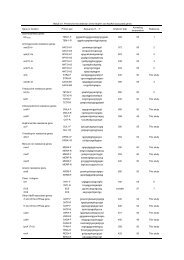
<strong>Bacterial</strong> identification. Bacteria were identified us<strong>in</strong>g <strong>the</strong><br />
standard bacteriological methods (Lennette et al. 1985).<br />
Biochemical properties <strong>of</strong> <strong>the</strong> isolates belong<strong>in</strong>g to<br />
Enterobacteriaceae and glucose nonferment<strong>in</strong>g gram-negative<br />
bacteria were <strong>in</strong>vestigated by <strong>the</strong> API20E and API20NE<br />
systems (BioMerieux), respectively. The <strong>in</strong>terpretation <strong>of</strong><br />
<strong>the</strong>se results was based on numerical probabilistic<br />
identification us<strong>in</strong>g ApiLab Plus 3.3.3 s<strong>of</strong>tware package<br />
(BioMerieux). The stra<strong>in</strong> identity <strong>of</strong> Ochrobactrum isolates<br />
was <strong>in</strong>vestigated by random amplified polymorphic DNA<br />
analysis (RAPD) us<strong>in</strong>g primer DAF4 (Grundmann et al.<br />
1997).<br />
RESULTS<br />
<strong>Bacterial</strong> identification. With a few exceptions all<br />
isolates belonged to aerobic or facultatively anaerobic<br />
gram-negative rod-shaped bacteria. Us<strong>in</strong>g <strong>the</strong> API20NE<br />
system, <strong>the</strong> majority <strong>of</strong> <strong>the</strong> isolates were allocated to<br />
Ochrobactrum anthropi with high identification<br />
probability (99.9%). All <strong>the</strong>se isolates shared dist<strong>in</strong>ctive<br />
and homogeneous biochemical (API20NE code<br />
1243315) and morphological properties (uniform colony<br />
morphology). In addition, four isolates obta<strong>in</strong>ed at<br />
different time <strong>in</strong>tervals yielded almost identical DAF4-<br />
RAPD f<strong>in</strong>gerpr<strong>in</strong>ts (data not shown). Based on <strong>the</strong>se<br />
results, <strong>the</strong> O. anthropi-like isolates were considered as<br />
a population <strong>of</strong> a s<strong>in</strong>gle stra<strong>in</strong> designated AK. Stra<strong>in</strong> AK<br />
was <strong>the</strong>n submitted to <strong>the</strong> Czech Collection <strong>of</strong> Microogranisms<br />
(Brno, Czech Republic), where <strong>the</strong> genus<br />
identity <strong>of</strong> this stra<strong>in</strong> was confirmed but species identity<br />
rema<strong>in</strong>ed elusive. These organisms are <strong>the</strong>refore designated<br />
as stra<strong>in</strong> AK <strong>of</strong> Ochrobactrum sp. <strong>in</strong> <strong>the</strong> present<br />
study.<br />
Bacteria <strong>of</strong> various sandfly stages. Results <strong>of</strong><br />
bacterial isolations from forth-<strong>in</strong>star larvae, pupae and<br />
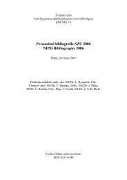
freshly emerged females are listed <strong>in</strong> Fig. 1. <strong>Bacterial</strong><br />
<strong>in</strong>fections were isolated from <strong>gut</strong>s <strong>of</strong> all 40 fourth-<strong>in</strong>star<br />
larvae studied. In actively feed<strong>in</strong>g larvae, whose <strong>gut</strong> was<br />
filled with larval food, a great variety <strong>of</strong> bacterial<br />
species was found. Stra<strong>in</strong> AK was present <strong>in</strong> about two<br />
thirds <strong>of</strong> <strong>in</strong>dividuals, <strong>of</strong>ten accompanied by o<strong>the</strong>r<br />
bacteria. On <strong>the</strong> o<strong>the</strong>r hand, <strong>in</strong> larvae with empty <strong>gut</strong>s,<br />
stra<strong>in</strong> AK was <strong>the</strong> only bacterium identified; it was<br />
isolated <strong>in</strong> 95% <strong>of</strong> larvae (Fig. 1). In pupae, most <strong>in</strong>dividuals<br />
ei<strong>the</strong>r harboured stra<strong>in</strong> AK (41%) or were sterile<br />
(41%). Isolates from <strong>the</strong> rest <strong>of</strong> <strong>the</strong> pupae <strong>in</strong>cluded<br />
Pseudomonas sp., Oligella sp. and few specimens which<br />
could not be identified by methods used (Fig. 1).<br />
Experiments showed that most sandflies are not free<br />
<strong>of</strong> bacterial flora upon emergence. In <strong>the</strong> <strong>gut</strong> <strong>of</strong> newly<br />
emerged females, <strong>the</strong> diversity <strong>of</strong> bacterial <strong>in</strong>fections<br />
was similar to that <strong>in</strong> pupae: almost two thirds <strong>of</strong><br />
<strong>in</strong>dividuals (64%) harboured stra<strong>in</strong> AK and 30% were<br />
sterile (Fig. 1). In 6% <strong>of</strong> females, o<strong>the</strong>r gram-negative<br />
bacteria (Enterobacter gergoviae and Sph<strong>in</strong>gobacterium<br />
spiritivorum) were found. In most cases <strong>the</strong> <strong>in</strong>fection<br />
was present <strong>in</strong> both regions <strong>of</strong> <strong>the</strong> <strong>gut</strong>, <strong>in</strong> <strong>the</strong> pylorus<br />
74
Volf et al.: Bacteria <strong>in</strong> sandfly <strong>gut</strong><br />
Fig. 1. Prevalence <strong>of</strong> bacteria <strong>in</strong> <strong>the</strong> <strong>gut</strong> <strong>of</strong> various developmental<br />
stages <strong>of</strong> <strong>Phlebotomus</strong> duboscqi: fourth-<strong>in</strong>star larvae<br />
actively feed<strong>in</strong>g (L4a), fourth-<strong>in</strong>star larvae ready to moult<br />
(L4b), pupae just before hatch<strong>in</strong>g (Pup) and freshly hatched<br />
females (Fem).<br />
CFU<br />
(log 10<br />
)<br />
7<br />
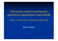
Fig. 2. <strong>Bacterial</strong> numbers <strong>in</strong> <strong>the</strong> <strong>gut</strong> <strong>of</strong> <strong>Phlebotomus</strong> duboscqi<br />
females that did not take blood (day 0) and <strong>in</strong> females that<br />
took blood two and seven days ago (day 2 and day 7,<br />
respectively). <strong>Bacterial</strong> numbers are given is colony-form<strong>in</strong>g<br />
units (CFU).<br />
CFU<br />
(log 10 )<br />
6<br />
5<br />
4<br />
3<br />
2<br />
1<br />
8<br />
7<br />
6<br />
5<br />
4<br />
3<br />
2<br />
1<br />
day 0 day 2 day 7<br />
Gal NGal Hep PBS<br />
Gal NGal Hep PBS<br />
day 2 day 7<br />
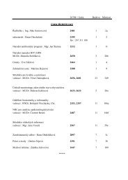
Fig. 3. <strong>Bacterial</strong> numbers (CFU) <strong>in</strong> <strong>the</strong> <strong>gut</strong> <strong>of</strong> <strong>Phlebotomus</strong><br />
duboscqi females fed on blood with addition <strong>of</strong> carbohydrates<br />
diluted <strong>in</strong> PBS: 50 mM galactose (Gal), 50 mM galactosam<strong>in</strong>e<br />
(NGal), hepar<strong>in</strong> 500 U/ml (Hep). PBS alone served as a<br />
control.<br />
region with Malpighian tubules as well as <strong>in</strong> <strong>the</strong> mid<strong>gut</strong>.<br />
In three cases, however, stra<strong>in</strong> AK was cultivated<br />
from <strong>the</strong> pylorus region only.<br />
Study on possible sources <strong>of</strong> bacterial <strong>in</strong>fection<br />
revealed <strong>the</strong> presence <strong>of</strong> Serratia marcescens, Citrobacter<br />
sp. and Stenotrophomonas maltophilia <strong>in</strong> sucrose<br />
solutions. Various bacteria, <strong>in</strong>clud<strong>in</strong>g high number <strong>of</strong><br />
colonies <strong>of</strong> stra<strong>in</strong> AK were found <strong>in</strong> diluted larval food.<br />
Few uniform colonies isolated from chick-sk<strong>in</strong><br />
membrane did not correspond with any bacteria identified<br />
from female mid<strong>gut</strong>s.<br />
Effect <strong>of</strong> bloodfeed<strong>in</strong>g. The prevalence <strong>of</strong> bacterial<br />
species isolated from P. duboscqi females which did not<br />
take blood and those dissected two and seven days after<br />
bloodfeed<strong>in</strong>g is summarised <strong>in</strong> Table 1: 42% to 69% <strong>of</strong><br />
female sandflies were found to conta<strong>in</strong> bacteria. Differences<br />
between groups <strong>of</strong> females were not statistically<br />
significant (χ 2 = 1.9, P = 0.38). In four cases, two<br />
different bacterial stra<strong>in</strong>s were isolated from <strong>the</strong> same<br />
female.<br />
The average bacterial numbers for all unfed and<br />
bloodfed females dissected are shown <strong>in</strong> Fig. 2. The<br />
highest counts were observed two days after bloodfeed<strong>in</strong>g,<br />
differences between groups, however, were not<br />
statistically significant (χ 2 =1.12, P = 0.35) because <strong>of</strong><br />
<strong>the</strong> wide variation <strong>in</strong> bacterial numbers present <strong>in</strong><br />
<strong>in</strong>dividually dissected <strong>gut</strong>s. Seven days after bloodfeed<strong>in</strong>g,<br />
<strong>the</strong> bacterial counts were roughly back to <strong>the</strong><br />
pre-feed<strong>in</strong>g levels.<br />
Effect <strong>of</strong> carbohydrate <strong>in</strong>hibitors. Females fed on<br />
blood with added carbohydrates were dissected two or<br />
seven days after feed<strong>in</strong>g. The relative prevalence <strong>of</strong><br />
bacterial species found is summarised <strong>in</strong> Table 2. Aga<strong>in</strong>,<br />
stra<strong>in</strong> AK and o<strong>the</strong>r gram-negative rods were present.<br />
The number <strong>of</strong> females with a sterile <strong>gut</strong> did not significantly<br />
differ between galactosam<strong>in</strong>e and galactose<br />
groups (χ 2 = 3.0, P = 0.39). In experiments with hepar<strong>in</strong>,<br />
significant differences were found on day 2 only; higher<br />
bacterial numbers were found <strong>in</strong> <strong>the</strong> control group fed<br />
on blood with PBS (χ 2 = 5.6, P = 0.02).<br />
<strong>Bacterial</strong> numbers are given <strong>in</strong> Fig. 3. There was no<br />
statistical difference between pairs <strong>of</strong> groups. However,<br />
higher bacterial numbers were observed on day 2 after<br />
bloodfeed<strong>in</strong>g <strong>in</strong> all four groups.<br />
Table 1. Number <strong>of</strong> bacterial isolates from <strong>the</strong> <strong>gut</strong> <strong>of</strong><br />
<strong>Phlebotomus</strong> duboscqi females <strong>of</strong> various bloodfeed<strong>in</strong>g status:<br />
unfed on blood, two and seven days after bloodfeed<strong>in</strong>g on<br />
mice.<br />
Females<br />
(n)<br />
unfed<br />
(12)<br />
day 2<br />
(13)<br />
day 7<br />
(13)<br />
Infected 42% 69% 54%<br />
Ochrobactrum sp., stra<strong>in</strong> AK 5 9 6<br />
Serratia marcescens 1 0 0<br />
Stenotrophomonas maltophilia 0 1 1<br />
Nonidentified 0 1 1<br />
75
Table 2. Number <strong>of</strong> bacterial isolates from <strong>the</strong> <strong>gut</strong> <strong>of</strong> females membrane-fed on blood with carbohydrate <strong>in</strong>hibitors <strong>of</strong> lect<strong>in</strong><br />
activity: galactosam<strong>in</strong>e (NGal) and hepar<strong>in</strong> (Hep). Controls <strong>in</strong>cluded non<strong>in</strong>hibitory carbohydrate galactose (Gal) and PBS.<br />
Females were dissected 2 and 7 days after feed<strong>in</strong>g.<br />
day 2 day 7<br />
Females fed on<br />
(n)<br />
NGal<br />
(12)<br />
Gal<br />
(11)<br />
Hep<br />
(9)<br />
PBS<br />
(10)<br />
NGal<br />
(16)<br />
Gal<br />
(16)<br />
Hep<br />
(13)<br />
PBS<br />
(12)<br />
Infected (%) 58 73 56 100 69 44 31 56<br />
Ochrobactrum sp., stra<strong>in</strong> AK 7 6 3 6 8 6 1 4<br />
Serratia marcescens 3 4 1 1<br />
Stenotrophomonas maltophilia 1<br />
Citrobacter sp. 1 2<br />
Nonidentified 1 3 2 1<br />
DISCUSSION<br />
<strong>Bacterial</strong> isolations revealed <strong>the</strong> frequent presence <strong>of</strong><br />
gram-negative bacteria <strong>in</strong> <strong>the</strong> <strong>gut</strong> <strong>of</strong> various developmental<br />
stages <strong>of</strong> <strong>Phlebotomus</strong> duboscqi. Aerobic and<br />
facultatively anaerobic gram-negative rods are ubiquitous<br />
bacteria grow<strong>in</strong>g easily on <strong>the</strong> type <strong>of</strong> medium used<br />
which may support <strong>the</strong>ir higher prevalence <strong>in</strong> sandfly<br />
mid<strong>gut</strong>. Our results fit with those obta<strong>in</strong>ed by o<strong>the</strong>r<br />
studies <strong>of</strong> <strong>the</strong> mid<strong>gut</strong> tissue <strong>of</strong> colonised as well as wildcaught<br />
mosquitoes and sandflies. The frequent presence<br />
<strong>of</strong> gram-negative rods belong<strong>in</strong>g mostly to Enterobacteriaceae<br />
was demonstrated <strong>in</strong> adults <strong>of</strong> Culex (Chao<br />
and Wistreich 1959, 1960, Ferguson and Micks 1961) as<br />
well as Anopheles species (DeMaio et al. 1996,<br />
Pumpuni et al. 1996). Similarly, <strong>in</strong> laboratory-bred and<br />
wild-caught sandflies all predom<strong>in</strong>ant species identified<br />
by Dillon et al. (1996) were Enterobacteriaceae. On <strong>the</strong><br />
o<strong>the</strong>r hand, gram-positive bacteria were less frequently<br />
found <strong>in</strong> sandflies (Schle<strong>in</strong> et al. 1985, Dillon et al.<br />
1996). It appears likely that <strong>the</strong> antimicrobial activity<br />
effective aga<strong>in</strong>st Micrococcus luteus and Bacillus<br />
subtilis, recently demonstrated by our group <strong>in</strong> mid<strong>gut</strong><br />
extracts <strong>of</strong> Lutzomyia and <strong>Phlebotomus</strong> species<br />
(Kiewegová 1999), may render <strong>the</strong>se <strong>in</strong>sects less<br />
susceptible to <strong>colonisation</strong> by gram-positive bacteria.<br />
Dillon et al. (1996) showed that <strong>the</strong> number <strong>of</strong> bacteria<br />
present <strong>in</strong> <strong>the</strong> <strong>gut</strong> <strong>of</strong> <strong>Phlebotomus</strong> papatasi changes<br />
dur<strong>in</strong>g <strong>the</strong> lifetime <strong>of</strong> a female. Here we studied <strong>the</strong><br />
effect <strong>of</strong> bloodfeed<strong>in</strong>g status: <strong>in</strong> both types <strong>of</strong> bloodfeed<strong>in</strong>g,<br />
on anes<strong>the</strong>tized mice and on chick-sk<strong>in</strong> membrane,<br />
<strong>the</strong> highest bacterial counts were observed two<br />
days after blood <strong>in</strong>gestion. The prote<strong>in</strong>-rich bolus <strong>of</strong> <strong>the</strong><br />
blood presumably caused rapid growth <strong>of</strong> mid<strong>gut</strong><br />
bacteria and when digestion was completed (on day 4-5)<br />
most bacteria were defecated with blood rema<strong>in</strong>s.<br />
Similar trends were found <strong>in</strong> mosquitoes where mid<strong>gut</strong><br />
bacterial counts peaked 48 h after blood <strong>in</strong>gestion and<br />
<strong>the</strong>n decreased to pre-blood-meal levels as a result <strong>of</strong><br />
digestion and excretion (DeMaio et al. 1996, Pumpuni<br />
et al. 1996).<br />
The addition <strong>of</strong> carbohydrate <strong>in</strong>hibitors (galactosam<strong>in</strong>e<br />
and hepar<strong>in</strong>) <strong>in</strong>to <strong>the</strong> blood-meal did not affect<br />
bacterial counts <strong>in</strong> sandfly mid<strong>gut</strong>. Therefore, we<br />
suppose that <strong>the</strong> mid<strong>gut</strong> lect<strong>in</strong> activity <strong>of</strong> sandflies has<br />
no effect on bacterial <strong>in</strong>fections.<br />
Specific attention was paid to <strong>the</strong> most prevalent<br />
stra<strong>in</strong> AK <strong>of</strong> <strong>the</strong> genus Ochrobactrum. The nonfermentative<br />
gram-negative bacteria Ochrobactrum anthropi is<br />
known as a potential human pathogen (Kern et al.<br />
1993). Stra<strong>in</strong> AK was isolated from <strong>the</strong> <strong>gut</strong> <strong>of</strong> fourth<strong>in</strong>star<br />
larvae just before pupation, <strong>the</strong> <strong>gut</strong> <strong>of</strong> females<br />
ready to emerge from pupae as well from mid<strong>gut</strong> and<br />
h<strong>in</strong>d<strong>gut</strong> <strong>of</strong> newly emerged adult females. Study on<br />
possible sources <strong>of</strong> <strong>in</strong>fection showed that larvae acquire<br />
this stra<strong>in</strong>, toge<strong>the</strong>r with o<strong>the</strong>r bacteria, from <strong>the</strong><br />
environment with larval food. Contrary to o<strong>the</strong>r<br />
bacteria, stra<strong>in</strong> AK was able to survive <strong>the</strong> mechanical<br />
shedd<strong>in</strong>g <strong>of</strong> <strong>the</strong> larval mid<strong>gut</strong> shortly before pupation<br />
and was transmitted transtadially. Its presence <strong>in</strong> <strong>the</strong> <strong>gut</strong><br />
<strong>of</strong> <strong>in</strong>dividuals ready to emerge from pupa was<br />
confirmed by <strong>in</strong>direct fluorescence <strong>in</strong> sections; specific<br />
polyclonal antibodies raised <strong>in</strong> rabbits gave a positive<br />
reaction with bacteria localised <strong>in</strong> <strong>the</strong> <strong>gut</strong> lumen<br />
(Kiewegová 1999). On TSA <strong>the</strong> stra<strong>in</strong> AK yielded<br />
mucoid colonies which <strong>in</strong>dicates an extensive<br />
production <strong>of</strong> glycocalyx. This may enable this stra<strong>in</strong> to<br />
colonise <strong>the</strong> sandfly <strong>gut</strong> and survive dur<strong>in</strong>g transtadial<br />
passage.<br />
In Diptera, most extracellular bacteria present <strong>in</strong> <strong>the</strong><br />
<strong>gut</strong> are not able to survive transtadial passage, ma<strong>in</strong>ly<br />
because <strong>of</strong> drastic changes <strong>in</strong> <strong>the</strong> <strong>gut</strong> environment and<br />
<strong>in</strong>creased secretion <strong>of</strong> antibacterial compounds dur<strong>in</strong>g<br />
pupal stage. However, <strong>the</strong> study carried out on<br />
laboratory-reared Anopheles mosquitoes showed that<br />
newly emerged adults harboured bacteria <strong>in</strong> <strong>the</strong>ir<br />
mid<strong>gut</strong> and transtadial transmission was demonstrated<br />
by successfully passag<strong>in</strong>g Escherichia coli from <strong>the</strong><br />
larval to <strong>the</strong> adult stage (Pumpuni et al. 1996).<br />
While stra<strong>in</strong> AK was passaged to adults transtadially<br />
most o<strong>the</strong>r bacteria found <strong>in</strong> adults were acquired from<br />
<strong>the</strong> sugar-meal. Some <strong>of</strong> <strong>the</strong>m, like Serratia sp., are<br />
known to be pathogenic for <strong>in</strong>sects (Seitz et al. 1987)<br />
and we found <strong>the</strong>m <strong>in</strong> high numbers <strong>in</strong> dead or dy<strong>in</strong>g<br />
blood-fed females (data not shown). Moreover, <strong>the</strong><br />
microbial <strong>colonisation</strong> <strong>of</strong> <strong>the</strong> <strong>gut</strong> is known to adversely<br />
affect <strong>the</strong> development <strong>of</strong> transmitted parasites (Seitz et<br />
76
Volf et al.: Bacteria <strong>in</strong> sandfly <strong>gut</strong><br />
al. 1987, Beier et al. 1994, Pumpuni et al. 1996). In<br />
sandflies, this po<strong>in</strong>t was raised for <strong>the</strong> first time by<br />
Adler and Theodor (1929) and later studied on laboratory-reared<br />
P. papatasi by Schle<strong>in</strong> et al. (1985), who<br />
demonstrated that fungi-<strong>in</strong>fected sandfly females were<br />
refractory to <strong>in</strong>fections with Leishmania major. Our<br />
study suggests that bacteria passaged transtadially or<br />
acquired from <strong>the</strong> sugar-meal <strong>of</strong>fered to adults <strong>in</strong><br />
laboratory conditions may overgrow shortly after bloodfeed<strong>in</strong>g<br />
and cause <strong>the</strong> mortality <strong>of</strong> females or negatively<br />
affect experimental <strong>in</strong>fection by Leishmania parasites.<br />
Acknowledgements. We thank Ms. Květa Gavurová for<br />
skilled technical assistance and A. Richardson for l<strong>in</strong>guistical<br />
improvement <strong>of</strong> <strong>the</strong> manuscript. The study was supported by<br />
<strong>the</strong> M<strong>in</strong>istry <strong>of</strong> Education (projects VS 96142 and<br />
J13/981131-B4) and <strong>the</strong> Grant Agency <strong>of</strong> <strong>the</strong> Czech Republic<br />
(project 206/99/1389).<br />
REFERENCES<br />
ADLER S., THEODOR O. 1929: Attempts to transmit<br />
Leishmania tropica by bite: <strong>the</strong> transmission <strong>of</strong> L. tropica<br />
by <strong>Phlebotomus</strong> sergenti. Ann. Trop. Med. Parasitol. 23:<br />
1-18.<br />
BEIER M.S., PUMPUNI C.B., BEIER J.C., DAVIS J.R.<br />
1994: Effects <strong>of</strong> para-am<strong>in</strong>obenzoic acid, <strong>in</strong>sul<strong>in</strong>, and<br />
gentamyc<strong>in</strong> on Plasmodium falciparum development <strong>in</strong><br />
anophel<strong>in</strong>e mosquitoes (Diptera: Culicidae). J. Med.<br />
Entomol. 31: 561-565.<br />
BRIDSON E.I. 1995: The Oxoid Manual. Unipath Limited,<br />
Hampshire.<br />
CHAO J., WISTREICH G.A. 1959: Microbial isolations from<br />
<strong>the</strong> mid-<strong>gut</strong> <strong>of</strong> Culex tarsalis Coquillet. J. Insect Pathol. 1:<br />
311-318.<br />
CHAO J., WISTREICH G.A. 1960: Microorganisms from <strong>the</strong><br />
mid-<strong>gut</strong> <strong>of</strong> larval and adult Culex qu<strong>in</strong>quefasciatus Say.<br />
J. Insect Pathol. 2: 220-224.<br />
DeMAIO J., PUMPUNI C.B., KENT M., BEIER J.C. 1996:<br />
The mid<strong>gut</strong> bacterial flora <strong>of</strong> wild Äedes triseriatus, Culex<br />
pipiens and Psorophora columbiae mosquitoes. Am. J.<br />
Trop. Med. Hyg. 54: 219-223.<br />
DILLON R.J., ELKORDY E., SHEHATA M., LANE R.P.<br />
1996: The prevalence <strong>of</strong> a microbiota <strong>in</strong> <strong>the</strong> digestive tract<br />
<strong>of</strong> <strong>Phlebotomus</strong> papatasi. Ann. Trop. Med. Parasitol. 90:<br />
669-673.<br />
FERGUSON M., MICKS D.W. 1961: Microorganisms<br />
associated with mosquitoes: I. Bacteria isolated from <strong>the</strong><br />
mid-<strong>gut</strong> <strong>of</strong> adult Culex fatigans Wiedemann. J. Insect<br />
Pathol. 3: 112-119.<br />
GRUBHOFFER L., HYPŠA V., VOLF P. 1997: Lect<strong>in</strong>s<br />
(hemagglut<strong>in</strong><strong>in</strong>s) <strong>in</strong> <strong>the</strong> <strong>gut</strong> <strong>of</strong> <strong>the</strong> important disease<br />
vectors. Parasite 4: 203-216.<br />
GRUNDMANN H.J., TOWNER K.J., DIJKSHOORN L.,<br />
GERNER-SMIDT P., MAHER M., SEIFERT H.,<br />
VANEECHOUTTE M. 1997. Multicenter study us<strong>in</strong>g<br />
standardized protocols and reagents for evaluation <strong>of</strong><br />
reproducibility <strong>of</strong> PCR-based f<strong>in</strong>gerpr<strong>in</strong>t<strong>in</strong>g <strong>of</strong> Ac<strong>in</strong>etobacter<br />
spp. J. Cl<strong>in</strong>. Microbiol. 35: 3071-3077.<br />
KERN W.V., OETHINGER M., KAUFHOLD A.,<br />
ROZDZINSKI E., MARRE R. 1993. Ochrobactrumanthropi<br />
bacteremia – report <strong>of</strong> 4 cases and short review.<br />
Infection 21: 306-310.<br />
KIEWEGOVÁ A. 1999: Antimikrobiální aktivity <strong>Phlebotomus</strong><br />
duboscqi. Unpubl. M.Sc. Thesis, Charles<br />
University, Prague, 81 pp.<br />
LENNETTE E.H., BALOWS A., HAUSLER W.J.,<br />
SHADOMY H.J. 1985: Manual <strong>of</strong> Cl<strong>in</strong>ical Micro-biology.<br />
Fourth Edition. American Society for Microbiol-ogy,<br />
Wash<strong>in</strong>gton D.C., 1150 pp.<br />
OLAFSEN J.A. 1996: Lect<strong>in</strong>s: models <strong>of</strong> natural and <strong>in</strong>duced<br />
molecules <strong>in</strong> <strong>in</strong>vertebrates. In: E.L. Cooper (Ed.),<br />
Advances <strong>in</strong> Comparative and Environmental Physiology.<br />
Vol. 24. Spr<strong>in</strong>ger-Verlag, Berl<strong>in</strong>, Heidelberg, pp. 49-76.<br />
PUMPUNI C.B., DeMAIO J., KENT M., DAVIS J.R., BEIER<br />
J.C. 1996: <strong>Bacterial</strong> population dynamics <strong>in</strong> three<br />
anophel<strong>in</strong>e species: impact on Plasmodium sporogonic<br />
development. Am. J. Trop. Med. Hyg. 54: 214-218.<br />
SCHLEIN Y., POLACHECK I., YUVAL B. 1985: Mycoses,<br />
bacterial <strong>in</strong>fections and antibacterial activity <strong>in</strong> sandflies<br />
(Psychodidae) and <strong>the</strong>ir possible role <strong>in</strong> <strong>the</strong> transmission<br />
<strong>of</strong> leishmaniasis. Parasitology 90: 57-66.<br />
SEITZ H.M., MAIER W.A., ROTTOK M., BECKER-<br />
FELDMANN H. 1987: Concomitant <strong>in</strong>fections <strong>of</strong><br />
Anopheles stephensi with Plasmodium berghei and<br />
Serratia marcescens: additive detrimental effects. Zbl.<br />
Bakt. Hyg. 266: 155-166.<br />
VOLF P., KIEWEGOVÁ A., SVOBODOVÁ M. 1998:<br />
Sandfly mid<strong>gut</strong> lect<strong>in</strong>: effect <strong>of</strong> galactosam<strong>in</strong>e on<br />
Leishmania major <strong>in</strong>fections. Med. Vet. Entomol. 12: 151-<br />
154.<br />
WELBURN S.C., MAUDLIN I. 1999: Tsetse-trypanosome<br />
<strong>in</strong>teractions: rites <strong>of</strong> passage. Parasitol. Today 15: 399-<br />
403.<br />
Received 16 January 2001 Accepted 13 June 2001<br />
77