crea.lign download pdf - Bredent.co.uk
crea.lign download pdf - Bredent.co.uk
crea.lign download pdf - Bredent.co.uk

Create successful ePaper yourself
Turn your PDF publications into a flip-book with our unique Google optimized e-Paper software.
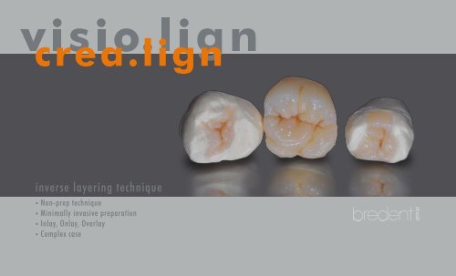
<strong>crea</strong>.<strong>lign</strong><br />
inverse layering technique<br />
• Non-prep technique<br />
• Minimally invasive preparation<br />
• Inlay, Onlay, Overlay<br />
• Complex case
Properties <strong>crea</strong>.<strong>lign</strong><br />
<strong>crea</strong>.<strong>lign</strong> is a light-curing <strong>co</strong>mposite,<br />
which <strong>co</strong>nsists of 50% opalescent ceramic particles and a high-strength oligomer matrix. <strong>crea</strong>.<strong>lign</strong><br />
veneering material <strong>co</strong>ntains only nanofillers and does not have any ground glass fillers. A special<br />
manufacturing process eliminates any agglomerates or lumping and a homogeneous and dense<br />
surface is achieved with the particle size of 40 nm.<br />
The omission of harder glass fillers which lead to embrittlement in <strong>co</strong>mposites results in excellent<br />
polishing properties and a high resistance to plaque and abrasion.<br />
The special gel-like <strong>co</strong>nsistency and homogeneity of the material allows for the adaption of elasticity<br />
and hardness of the <strong>co</strong>mposite to various substructure materials. The <strong>co</strong>mbination of opalescent ceramic<br />
and crack-resistant <strong>co</strong>mposite matrix transforms the properties of a liquid ceramic to <strong>crea</strong>.<strong>lign</strong>.<br />
Using a simple procedure, restorations perfectly matching natural teeth can be produced.<br />
Ac<strong>co</strong>rdingly, <strong>crea</strong>.<strong>lign</strong> can be used to easily produce long-term restorations with shade stability and<br />
plaque resistance in the laboratory. It can also be used chairside, which <strong>co</strong>uld previosuly only be<br />
achieved with veneering ceramic. <strong>crea</strong>.<strong>lign</strong> is not only suitable for crowns and bridges but also for the<br />
„additional veneer technique“, as described in this brochure.<br />
2<br />
<strong>crea</strong>.
Contents<br />
Preface......................................................................................................... 4<br />
inverse layering technique.................................................... 5<br />
Non-prep technique....................................................................... 6<br />
Minimally invasive preparation................................... 12<br />
Inlay, Onlay, Overlay............................................................... 20<br />
Complex case..................................................................................... 38<br />
Vincenzo Musella ........................................................................ 54<br />
<strong>lign</strong><br />
3
Preface<br />
“Non-prep technique” and<br />
“minimally invasive preparation technique”<br />
Preparation of the tooth has always been <strong>co</strong>nsidered to be an essential pre<strong>co</strong>ndition for the production<br />
of a prosthetic restoration. In the preparation process material is removed to <strong>crea</strong>te space for<br />
build ups or inserting dental restorations. The removal of natural tooth structure results in <strong>co</strong>nsiderable<br />
loss of biological material. Loss of structure can be limited through the application use of<br />
adhesive bonding technique, which is used when luting/cementing veneers. In such cases, however,<br />
the preparation results in irreversible - yet limited - damage. In recent years a new type of prosthetic<br />
therapy has been endorsed, which enables esthetic and/or functional <strong>co</strong>rrection of the tooth through<br />
the use of very thin veneers. The veneers are luted directly to the etched enamel without using a<br />
substructure.<br />
Numerous advantages can be observed:<br />
• <strong>co</strong>mplete reversibility of the prosthetic treatment<br />
• no destruction of hard tooth structure (biological loss) caused by preparation<br />
• excellent adhesive bonding, directly to the enamel<br />
The indication range includes cases in which the intended dimension of the tooth is larger than the<br />
existing dimension. Such „additional veneers“, are based on a <strong>co</strong>mplex procedure, which depends<br />
highly on the dental technician‘s skills. Moreover it is possible to expand the indication range of these<br />
„additional veneers“ by preparing the tooth in a minimally invasive manner. If parts of a tooth to be<br />
restored represent a potential <strong>co</strong>nstraint to the use of an „additional veneer“, minimal preparation<br />
is suitable to change the shape of the tooth by slightly reducing the tooth structure without the need<br />
for full preparation of the tooth. In this case, minimal removal of tooth surface is also based on the<br />
<strong>co</strong>ncept of reversibility and the advantage of direct bonding to the enamel is maintained.<br />
4<br />
<strong>crea</strong>.
inverse layering technique<br />
“inverse layering technique“<br />
Traditionally, <strong>co</strong>mposite restorations are produced using a direct additive technique.<br />
This method has fundamentally changed through the use of “inverse layering technique” in which the<br />
restoration is produced by using a reverse approach - the so-called “inverse layering technique”.<br />
The wax-up is of utmost significance when using this technique since a successful restoration depends<br />
on a carefuly and precise wax work.<br />
In the step-by-step description of this technique, a key made of transparent sili<strong>co</strong>ne (shore hardness:<br />
60) is prepared after waxing up. This key needs to reproduce all details of the model in a very accurate<br />
manner.<br />
Once the transparent key is <strong>co</strong>mpleted, inverse layering is carried out; transparent materials and first<br />
dentine are applied followed by dentine materials with different levels of chroma.<br />
When using this technique, it is important to keep in mind that the use of <strong>co</strong>nventional, flowable<br />
<strong>co</strong>mposites poses certain risks due to their limited stability.<br />
Therefore it is essential to select a suitable <strong>co</strong>mposite, which is less susceptible to fracture (brittle) and<br />
features high mechanical strength.<br />
This technique does not exclude any type of restoration and is suitable for the following indications:<br />
• NON-PREP TECHNIQUE<br />
• MINIMALLY INVASIVE PREPARATION<br />
• INLAY-ONLAY-OVERLAY<br />
• COMPLEX CASES WITH OR WITHOUT SUBSTRUCTURE<br />
(ALLOY-ZIRCONIUM OXIDE-GLASS FIBER-PEEK)<br />
<strong>lign</strong><br />
5
Non-prep technique<br />
1 2<br />
Non-prep tec<br />
Initial situation<br />
Final result<br />
Due to low cracking properties, the „additional veneering technique“ with <strong>co</strong>mposite material is especially suitable for cases that do not require preparation:<br />
Eventhough the patient does not identify any specific esthetic problems, it is possible to improve the function and shape of the respective teeth<br />
without an invasive procedure.<br />
6
hnique<br />
3<br />
The prepared master<br />
model with removable<br />
dies.<br />
4<br />
Morphological wax-up<br />
of anterior teeth<br />
(wax) for the “additional<br />
veneers”..<br />
5<br />
Investment (plaster)<br />
of the separated dies,<br />
including wax-up, forthe<br />
manufacture of the sili<strong>co</strong>ne<br />
key (<strong>co</strong>unterpart) for<br />
the subsquent processing<br />
steps of inverse layering.<br />
6<br />
The transparent sili<strong>co</strong>ne<br />
key. Hardening<br />
in the pressure pot<br />
at 2.5 bar to achieve<br />
accurate reproduction<br />
of the model.<br />
7
Non-prep technique<br />
7<br />
Non-prep tec<br />
Inverse layering starts<br />
with the application of<br />
the incisal material with a<br />
high lightness value (E2)<br />
in the key.<br />
8<br />
The material is applied<br />
with a small brush.<br />
9<br />
Pre-curing for 2-3 sec.<br />
with the bre.lux hand<br />
lamp or a suitable light<br />
source.<br />
10<br />
Application of the<br />
effect materials to <strong>crea</strong>te<br />
<strong>co</strong>ntrasts.<br />
Pre-curing: 2-3 sec.<br />
8
hnique<br />
11<br />
Opalescent effects with<br />
the Incisal opal and Incisal<br />
blue materials,<br />
pre-curing: 2-3 sec.<br />
12<br />
Then a thin dentine<br />
layer (shade A1)<br />
is applied with the<br />
syringe.<br />
13<br />
Application of the dentine<br />
with a small brush.<br />
Placing the key onto the<br />
plaster base to <strong>co</strong>mplete<br />
polymerization of the<br />
<strong>co</strong>mposite.<br />
14<br />
Final curing - 360 sec.<br />
in the bre.Lux Power<br />
Unit.<br />
9
15<br />
Non-prep technique<br />
Non-prep tec<br />
16<br />
Result after light curing and removal of the key.<br />
The translucency of the <strong>crea</strong>.<strong>lign</strong> <strong>co</strong>mposite restauration..<br />
17 18 Try-in of the veneers 19<br />
prior to insertion with<br />
a rubber dam. To check<br />
the fit, it is re<strong>co</strong>mmended<br />
to initially use a paste (try-in) to insert<br />
the teeth. The suitable shade of the adhesive<br />
<strong>co</strong>mposite is selected.<br />
The <strong>co</strong>mpleted, finished and polished veneers on the master model.<br />
Veneers in situ after definitive bonding/luting.<br />
10
hnique<br />
1 2<br />
Initial situation<br />
Pronounced diastema between teeth 11 and 21.<br />
Final result<br />
Indirect restoration made of <strong>co</strong>mposite and closure of diastema.<br />
As shown in the photos above, the „additional veneer technique“ is also suitable for patients with diastemas without preparation of the tooth.<br />
11
inimally invasiv<br />
Minimally invasive preparation<br />
1 2 3<br />
Initial situation<br />
Minimally invasive preparation for indirect restoration<br />
with veneers (11-21-22).<br />
Fabrication of the master model with removable dies<br />
made of Exakto-Form polyurethane resin.<br />
Wax-up: The shape and texture of the wax model<br />
needs to include all details of the final restoration in<br />
order to reproduce them accurately in the <strong>co</strong>mpacted<br />
sili<strong>co</strong>ne key (using <strong>co</strong>mposite).<br />
12
e preparation<br />
4 5<br />
6<br />
Fabrication of the key made of transparent sili<strong>co</strong>ne<br />
visio.sil (shore hardness 60). An optimal hardness sili<strong>co</strong>ne<br />
should be selected in order to avoid damaging<br />
the wax model. The sili<strong>co</strong>ne key should be hardened<br />
at a pressure of 2.5 bar to avoid inclusion of air and<br />
ensure maximum precision of the duplicate. Preserving<br />
the wax model enables the assessment of the<br />
layer thickness of wax to <strong>co</strong>mposite during inverse<br />
layering.<br />
Once the sili<strong>co</strong>ne has hardenend and the individual<br />
dies have been adequately insulated, the inverse<br />
layering technique is carried out using <strong>crea</strong>.<strong>lign</strong>.<br />
Depending on the type of re<strong>co</strong>nstruction, layering<br />
starts at the incisal edge. The effect materials are<br />
applied with a small brush. The Bleach material, for<br />
example, is re<strong>co</strong>mmended for young teeth.<br />
A pre-curing cycle (2-3 sec.) using a hand lamp is required<br />
after each individual layer to avoid individual<br />
effects mixing with other materials or with each other.<br />
13
inimally invasiv<br />
Minimally invasive preparation<br />
7 8 9<br />
Layering can be individualized with transparent and<br />
opal effect materials ac<strong>co</strong>rding to the lightness values<br />
of the teeth. If individualization is not carried out,<br />
layering is <strong>co</strong>ntinued with the enamel materials, as<br />
shown in photo 10.<br />
The use of stains (internal stains) is another type of<br />
possible individualization. In order to imitate a crack<br />
line, for example, the use of white stain material is<br />
suggested.<br />
Pre-curing of the incisal effect materials.<br />
14
e preparation<br />
10 11<br />
12<br />
In this step, the effect materials are applied in ac<strong>co</strong>rdance<br />
with the desired <strong>co</strong>lor gradient.<br />
Pre-curing cycle<br />
Application of dentine: while taking the layer<br />
thickness into ac<strong>co</strong>unt, it is possible to use dentine<br />
materials with different chroma levels to achieve an<br />
enhanced effect of depth.<br />
For restorations with in<strong>crea</strong>sing thickness, first lighter<br />
dentine materials are applied initially to the enamel<br />
materials ( Layering scheme, page 56 ).<br />
15
inimally invasiv<br />
Minimally invasive preparation<br />
13 14 15<br />
Once the key is positioned on the model, the dentine<br />
is pre-cured.<br />
Final curing of the re<strong>co</strong>nstruction in the polymerization<br />
unit.<br />
After final polymerization, the sili<strong>co</strong>ne key enables<br />
clear re<strong>co</strong>gnition of the interdental separations,<br />
which can be reworked appropriately.<br />
16
e preparation<br />
16 17<br />
18<br />
Adequate preparation of the surface is important<br />
for subsequent high-gloss polishing. Use the yellow<br />
Abraso-Fix brush and at a low speed is re<strong>co</strong>mmended<br />
to avoid overheating the <strong>co</strong>mposite and „flattening“<br />
the texture.<br />
Prepolishing with a soft brush made of white goat<br />
hair and Acrypol prepolishing paste.<br />
Finally, high-gloss polishing is carried out with a soft<br />
<strong>co</strong>tton map (at low speed) and Abraso-Starglanz<br />
polishing paste.<br />
17
inimally invasiv<br />
Minimally invasive preparation<br />
19 20<br />
Photo of the restoration on the model taken with transmitted light.<br />
The translucency of the <strong>co</strong>mposite is stunning.<br />
The final result after high gloss polishing.<br />
18
e preparation<br />
21 22<br />
Black and white photo of the restoration to evaluate the shape.<br />
The <strong>co</strong>mpleted restoration in situ after adhesive bonding/luting.<br />
19
nlay Onlay Overl<br />
Inlay Onlay Overlay<br />
1 2 3<br />
Initial situation<br />
Preparation of the teeth.<br />
MOD inlay on tooth 25, full crown on tooth 26 and<br />
inlay on tooth 27.<br />
Details of the polyether impression.<br />
20
ay<br />
4 5<br />
6<br />
Blocking out the impression: this step is required<br />
since the dies are made of polyurethane resin which<br />
has a low vis<strong>co</strong>sity (highly liquid properties) during<br />
pouring.<br />
To prepare a bubble-free model, it is re<strong>co</strong>mmended<br />
to fill the cavities with Exakto Form using a small<br />
brush.<br />
The model can be removed from the mould and<br />
adjusted after 45 minutes.<br />
21
nlay Onlay Overl<br />
Inlay Onlay Overlay<br />
7 8 9<br />
Once the <strong>co</strong>mplete model has been poured, the individual<br />
dies are separated and prepared adequately.<br />
The individual, prepared dies are insulated.<br />
The prepared and insulated dies are repositioned in<br />
the impression, which is then filled with stone for the<br />
fabrication of the master model.<br />
22
ay<br />
10 11<br />
12<br />
The master model prepared in this way maintains<br />
the biological height of the emergence profiles and<br />
enables easy removal of the respective dies made of<br />
Exakto-Form polyurethane resin.<br />
Anatomical wax model of the indirect restorations:<br />
MOD inlay on tooth 25, full crown on tooth 26 and<br />
inlay on tooth 27.<br />
When waxing-up the occlusal surfaces, the fissures<br />
are deeper and larger to individualize layering with<br />
stains in the final finish.<br />
( page 27 )<br />
A separate plaster base is <strong>crea</strong>ted for the individual<br />
dies with anatomical wax-up. This transfer base is<br />
invested for the injection of the transparent sili<strong>co</strong>ne.<br />
The sili<strong>co</strong>ne is then <strong>co</strong>mpacted at a pressure of 2.5<br />
bar to reproduce all details of the wax model in a<br />
highly accurate manner.<br />
23
nlay Onlay Overl<br />
Inlay Onlay Overlay<br />
13 14<br />
Application of a very thin dentine layer to the die, which is subsequently <strong>co</strong>ated with<br />
the „Caramel“ modifier to achieve an enhanced effect of depth and to mask the<br />
light <strong>co</strong>re build-up.<br />
Application of the bleach material into the key to „emphasize“ the marginal ridges<br />
of the tooth.<br />
24
ay<br />
15 16<br />
In<strong>crea</strong>sing the opal effects with the Incisal blue or Incisal opal materials.<br />
After pre-curing the Bleach material and the Incisal blue material, apply a small<br />
quantity of E2 incisal material.<br />
25
nlay Onlay Overl<br />
Inlay Onlay Overlay<br />
17 18<br />
Dentine layering: Depending on the total thickness of the restoration, two or three<br />
different dentine shades are applied to in<strong>crea</strong>se the effect of depth of the tooth ( <br />
Layering scheme, page 56 ).<br />
Initially, dentine materials A1 and A2 are applied as primary dentine.<br />
Application of the se<strong>co</strong>ndary dentine of the selected tooth shade A3 with higher<br />
chroma.<br />
26
ay<br />
19 20<br />
Repositioning the sili<strong>co</strong>ne key on the plaster base with dies and pre-curing.<br />
After pre-curing, the <strong>co</strong>mposite restorations are characterized with stains to „intensify“<br />
fissures and pits of the occlusal surfaces. In the main fissure, for example, brown<br />
and orange stains are used to achieve an enhanced effect of depth. <strong>crea</strong>.<strong>lign</strong> stains<br />
can be directly applied and do not need to be overlayed (<strong>co</strong>ated). The restoration is<br />
finished and polished as shown on page 17 after final curing in the bre.Lux Power<br />
Unit (360 sec.).<br />
27
Inlay Onlay<br />
Inlay<br />
Overlay<br />
Onlay Ov<br />
21<br />
28
erlay<br />
22<br />
Completed and polished restorations on the master model.<br />
29
nlay Onlay Overl<br />
Inlay Onlay Overlay<br />
23 24 25<br />
The indirect restorations are sandblasted<br />
with aluminium oxide (50 µm) prior to adhesive<br />
bonding/luting to the teeth.<br />
Careful cleaning of the indirect restorations with<br />
methylated spirit (ethanol <strong>co</strong>lorless).<br />
Drying the indirect restorations with air.<br />
30
ay<br />
26 27<br />
28<br />
A gel-like film is used for marginal protection of the<br />
part of the tooth not to be restored.<br />
Sandblasting the preparations using aluminium<br />
oxide (50 µm).<br />
Careful cleaning to remove any residual aluminium<br />
oxide.<br />
31
nlay Onlay Overl<br />
Inlay Onlay Overlay<br />
29 30 31<br />
Selective etching of the tooth enamel for 15 sec. Dentine etching for 15 sec. Careful cleaning with water to remove any acid<br />
residues.<br />
32
ay<br />
32 33<br />
34<br />
Application of the adhesive to the preparations. Each<br />
adhesive features specific processing guidelines need<br />
to be strictly adhered to.<br />
Polymerization (light curing) of the adhesive.<br />
Optionally, a primer / bonder is applied thinly to the<br />
restoration. This is only necessary if the restoration<br />
was <strong>co</strong>mpleted more than 24 hours ago. The <strong>crea</strong>.<strong>lign</strong><br />
Modelling Liquid can be used for this purpose.<br />
33
nlay Onlay Overl<br />
Inlay Onlay Overlay<br />
35 36<br />
During the processing steps described below, it is re<strong>co</strong>mmended to protect the restorations<br />
with the applied adhesive against light to avoid premature curing.<br />
Application of the incisal material to the marginal areas of the restoration.<br />
34
ay<br />
37 38<br />
Apply dentine-<strong>co</strong>lored adhesive (bonding) material to the central area of the<br />
restoration.<br />
Positioning the indirect restorations on the preparations.<br />
35
Inlay Onlay Overlay<br />
39<br />
nlay Onlay Overl<br />
Final polymerization of the bonded indirect restorations.<br />
36
40<br />
ay<br />
The final result after bonding.<br />
37
Comp<br />
Complex case with immediate loading<br />
Initial situation<br />
38
lex case<br />
Temporary restoration prior to preparation<br />
A temporary wax-up with guided registration is the basis for determining the temporary restoration.<br />
39
Comp<br />
Complex case with immediate loading<br />
1 2 3<br />
Lower impression made of polyether. Upper impression made of polyether. Models are mounted in the articulator in ac<strong>co</strong>rdance with the<br />
bite registration obtained from duplicating the temporary.<br />
40
lex case<br />
4 5<br />
6<br />
Completed diagnostic wax-up.<br />
Before preparing the sili<strong>co</strong>ne keys of the model, it is necessary<br />
to notch the models to obtain the exact position of the<br />
keys.<br />
Drilling templates with transfer keys. They are used for<br />
positioning the implants during surgery and for the transfer<br />
of the implant position to the original model.<br />
41
Comp<br />
Complex case with immediate loading<br />
7 8 9<br />
Surgical phase with extraction of the teeth and insertion of<br />
the blueSKY implants.<br />
After insertion of the implants, the drilling template is<br />
prepared for fixation of the impression abutments and used<br />
as a transfer key for subsequent, accurate positioning of the<br />
laboratory analogs on the original model.<br />
Resin is used for fixation of the impression abutments on the<br />
drilling template with transfer key.<br />
42
lex case<br />
10<br />
11<br />
The upper transfer key<br />
with the impression abutments<br />
fixed with resin.<br />
The lower transfer key<br />
with the impression abutments<br />
fixed with resin.<br />
12<br />
13<br />
The upper transfer key<br />
with screwed laboratory<br />
analogs.<br />
The lower transfer key<br />
with screwed laboratory<br />
analogs.<br />
43
Comp<br />
Complex case with immediate loading<br />
14 15 16<br />
The plaster model is ground with a bur in order to place the<br />
laboratory analogs into the exact position (passive fit).<br />
Accurate preparation of the two models for positioning of the drilling templates as transfer keys (with screwed laboratory analogs).<br />
44
lex case<br />
17 18<br />
19<br />
The upper transfer key accurately placed on the model.<br />
The areas of the previously ground upper and lower plaster<br />
models are filled with class IV stone, Thixo-Rock, using a<br />
syringe.<br />
Front view of the two models mounted in the articulator with<br />
screwed <strong>co</strong>nical abutments of the SKY fast & fixed system.<br />
45
Comp<br />
Complex case with immediate loading<br />
20 21 22<br />
With a previously prepared key, the soft gingival mask is produced using pink sili<strong>co</strong>ne.<br />
Front view of the two models mounted in the articulator with<br />
the accurately transferred implant positions and the restoration<br />
of the soft tissue from the presurgical phase.<br />
46
lex case<br />
23 24<br />
25<br />
The wax-up is duplicated with burn-out Pi-Ku-Plast HP36<br />
resin to obtain two models. While taking the layer thickness<br />
into ac<strong>co</strong>unt, the resin model is reduced appropriately. A<br />
dimensionally accurate resin framework is obtained for the<br />
casting technique.<br />
The two metal substructures are carefully finished and fitted to the implant abutments.<br />
47
Comp<br />
Complex case with immediate loading<br />
26 27 28<br />
29<br />
30<br />
The cast and finished substructures<br />
are prepared for chemical<br />
bonding with Silano-<br />
Pen [Fig. 26]. Sandblasting<br />
with aluminium oxide (110<br />
µm), then surface treatment<br />
with the Silano-Pen flame,<br />
which leaves silicate particles<br />
on the surface (silica <strong>co</strong>ating)<br />
[Fig. 27]. After a few minutes the primer can be applied to the treated surface to activate the ceramic layer (silanization) [Fig. 28 and 29]. <strong>co</strong>mbo.<strong>lign</strong> opaque material can than be applied [Fig. 30].<br />
48
lex case<br />
31<br />
Inverse layering of the <strong>co</strong>mposite<br />
starts in the sili<strong>co</strong>ne key of<br />
the wax-up. The Bleach dentine<br />
material BL3 is applied to the<br />
marginal ridges in the posterior<br />
region and in the anterior<br />
region to accentuate the incisal<br />
ridges.<br />
Pre-curing: 2-3 sec.<br />
32<br />
E2 incisal material and<br />
incisal opal material can<br />
than be applied.<br />
Pre-curing: 2-3 sec.<br />
33<br />
Application of the internal<br />
stains and effect materials, such<br />
as Crack-line, in the anterior<br />
region<br />
Pre-curing: 2-3 sec.<br />
34<br />
Application of E3 incisal<br />
material.<br />
Pre-curing: 2-3 sec.<br />
49
Comp<br />
Complex case with immediate loading<br />
Application of A2 dentine material<br />
Pre-curing: 2-3 sec.<br />
A3 dentine material is applied almost to<br />
the top of the key to achieve an enhanced<br />
effect of depth and the gingiva is layered<br />
with gum materials (rose, pink and light).<br />
Pre-curing: 2-3 sec. After each layer of<br />
approx. 1 mm, intermediate curing is<br />
carried out in the bre.Lux Power Unit<br />
(180 sec.).<br />
Once layering is <strong>co</strong>mpleted, the key is<br />
repositioned on the model and the last<br />
curing cycle (360 sec.) is carried out.<br />
35 36 37<br />
50
lex case<br />
38<br />
Completed, finished<br />
and polished restoration<br />
on the model.<br />
51
Comp<br />
Complex case with immediate loading<br />
39<br />
41<br />
40<br />
52<br />
Lateral view of the restoration. Front view of the restoration with immediate loading 48<br />
hours after surgery.
lex case<br />
42 43<br />
53
Vincenzo Musella<br />
The Italian dental technician Vincenzo Musella whose interest in dental techniques<br />
was aroused by Prof. Mario Martignoni, received his diploma from the renowned<br />
Instute Galvani in the province of Reggio Emilia (Italy) and has been running a laboratory<br />
in Modena since 1988. During his career he enhanced his skills and knowlege in numerous<br />
training events, which brought him together with Giuseppe Zuppardi (dental technician) in<br />
1996.<br />
Today, they both share a long-standing friendship and closely <strong>co</strong>operate in the development<br />
of various possibilities with ceramic restorations. Furthermore he <strong>co</strong>operates with his dear<br />
friend Dr. Dario Castellani and in 1999 he initiated (together with Giuseppe Zuppardi) a<br />
<strong>co</strong>urse program for ceramic restoration especially tailored towards the needs and requirements<br />
of young people.<br />
V i n c e n z o<br />
54
In the following years he worked with Prof. Jeffrey Okeson, the head of the<br />
Center of Orofacial Pain at the University of Kentucky until 2002.<br />
He also actively <strong>co</strong>operated with Prof. Angelo Putignano in the development of<br />
new materials and <strong>co</strong>mpletion of patient cases.<br />
He is currently studying dentistry and dental prosthetics at Università Politecnica<br />
delle Marche in An<strong>co</strong>na (Italy).<br />
M u s e l l a<br />
Thanks to Dr. Dario Castellani, Dr. Luca Cantoni, Dr. Alessandro Agnini, Dr. Cinzia Barbieri and Prof. Pierangelo Oliveri<br />
for their valuable clinical support.<br />
Special thanks go to our dear friend Pier Paolo Goldoni for his <strong>co</strong>ntinued efforts and support of our work.<br />
55
Scheme<br />
of inverse layering<br />
<strong>crea</strong>.<strong>lign</strong> layering materials are perfectly suitable<br />
for minimally invasive layering and veneering<br />
technique in cases of limited available space. The<br />
dentine shades feature somewhat higher chroma<br />
than dental <strong>co</strong>mposites for the veneer technique<br />
and the incisal materials are a bit lighter and<br />
highly opalescent.<br />
It is favourable to have different levels of chroma<br />
which would mean that the selected tooth shade<br />
of the classic A-D shade guide would be layered as<br />
se<strong>co</strong>ndary dentine, e.g. A3 and then equal quantities<br />
of A2 and A1 dentine materials are applied<br />
over the top, if the layer thickness is more than 0.5<br />
mm. The layer thickness of these primary dentine<br />
materials, however, should always be lower than<br />
the thickness of the se<strong>co</strong>ndary dentine material.<br />
Significant <strong>co</strong>lor depth and <strong>co</strong>ntrast can be<br />
achieved, especially in the veneer technique, so<br />
even devitalized teeth can be <strong>co</strong>vered in a minimally<br />
invasive manner.<br />
As shown in the case described, the incisal material<br />
selected in the posterior region is always one<br />
shade lighter than in the anterior region (for A3:<br />
E2 instead of E3).<br />
The opal enamel material is the only non-fluorescent<br />
material. If enamel materials need to appear<br />
more transparent, it is re<strong>co</strong>mmended to mix the<br />
respective enamel material with <strong>crea</strong>.<strong>lign</strong><br />
Modelling Liquid in a ratio of max. 30%.<br />
BL 3<br />
Incisal<br />
Dentine A1<br />
Dentine A2<br />
Dentine A3<br />
Caramel<br />
1.0 mm<br />
0.8 mm<br />
0.5 mm<br />
art director: Andrea Fedrizzi<br />
photographer: Vincenzo Musella<br />
GmbH & Co.KG · Weissenhorner Str. 2 · 89250 Senden · Germany · Tel. (+49) 0 73 09 / 8 72-4 40 · Fax (+49) 0 73 09 / 8 72-4 44<br />
www.bredent.<strong>co</strong>m · e-mail info@bredent.<strong>co</strong>m<br />
06/12 482 GB 2,5<br />
Subject to changes