Paterson Institute for Cancer Research Scientific Report 2009
Paterson Institute for Cancer Research Scientific Report 2009
Paterson Institute for Cancer Research Scientific Report 2009

You also want an ePaper? Increase the reach of your titles
YUMPU automatically turns print PDFs into web optimized ePapers that Google loves.
<strong>Paterson</strong><br />
<strong>Institute</strong><br />
<strong>for</strong> <strong>Cancer</strong><br />
<strong>Research</strong><br />
<strong>Scientific</strong> <strong>Report</strong> <strong>2009</strong>
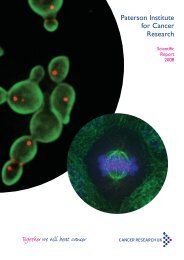
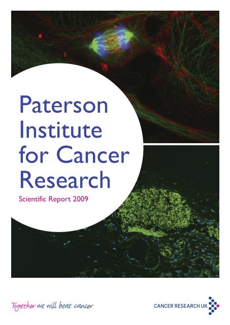
Cover images<br />
Top<br />
Mitotic BPAE cells in anaphase. F-actin is labelled with<br />
Texas Red-x phalloidin. Microtubules, in green, are labelled<br />
with mouse anti-α-tubulin BODIPY FL goat antimouse<br />
IgG. Blue nuclear staining with DAPI. Imaged<br />
on the Spinning Disk Confocal microscope.<br />
Image provided by Achille Dunn, Advanced Imaging Facility.<br />
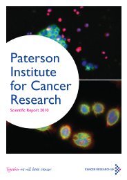
Bottom<br />
Immunostaining demonstrating blood vessels<br />
surrounding a tumour. Glut1 immunostaining (red)<br />
specifically labels veinous structures whereas arterial<br />
structures are Glut1 negative. Red blood cells in the<br />
vessels were detected by inherent autofluorescence<br />
(green) and cell nuclei were labelled with DAPI (blue).<br />
Image provided by Darren Roberts, Clinical and Experimental<br />
Pharmacology Group.
<strong>Scientific</strong> <strong>Report</strong> <strong>2009</strong><br />
<strong>Paterson</strong> <strong>Institute</strong><br />
<strong>for</strong> <strong>Cancer</strong> <strong>Research</strong>
Contents<br />
Director’s Introduction 5<br />
<strong>Research</strong> Highlights 8<br />
Drug Discovery in the Manchester 12<br />
<strong>Cancer</strong> <strong>Research</strong> Centre<br />
Georges Lacaud 38<br />
Stem Cell Biology Group<br />
Valerie Kouskoff 40<br />
Stem Cell and Haematopoiesis Group<br />
Akira Orimo 42<br />
Stromal-Tumour Interaction Group<br />
<strong>Research</strong> Groups – <strong>Paterson</strong> <strong>Institute</strong><br />
Crispin Miller 16<br />
Applied Computational Biology and<br />
Bioin<strong>for</strong>matics Group<br />
Geoff Margison 18<br />
Carcinogenesis Group<br />
Karim Labib 20<br />
Cell Cycle Group<br />
Iain Hagan 22<br />
Cell Division Group<br />
Nic Jones 24<br />
Cell Regulation Group<br />
Angeliki Malliri 26<br />
Cell Signalling Group<br />
Caroline Dive and Malcolm Ranson 28<br />
Clinical and Experimental Pharmacology Group<br />
<strong>Research</strong> Groups – The University of Manchester<br />
School of <strong>Cancer</strong> and Enabling Sciences<br />
Robert E Hawkins and Peter L Stern 46<br />
Biological, Immune and Gene Therapy Group<br />
Vaskar Saha 48<br />
Children’s <strong>Cancer</strong> Group<br />
Tim Illidge 50<br />
Targeted Therapy Group<br />
Catharine M.L. West 52<br />
Translational Radiobiology Group<br />
Robert Hawkins 54<br />
Medical Oncology: Cell Therapy Group<br />
Gordon Jayson 56<br />
Medical Oncology: Translational<br />
Anti-Angiogenesis Group<br />
Ivan Ahel 30<br />
DNA Damage Response Group<br />
<strong>Research</strong> Services 58<br />
Peter L Stern 32<br />
Immunology Group<br />
<strong>Research</strong> Publications 66<br />
Nullin Divecha 34<br />
Inositide Laboratory<br />
Tim Somervaille 36<br />
Leukaemia Biology Group<br />
Seminar Series <strong>2009</strong> 74<br />
Postgraduate Education 76
Operations 78<br />
<strong>Cancer</strong> <strong>Research</strong> UK’s 84<br />
Local Engagement and Development<br />
Acknowledgement <strong>for</strong> Funding 86<br />
of the <strong>Paterson</strong> <strong>Institute</strong><br />
Career Opportunities at the 87<br />
<strong>Paterson</strong> <strong>Institute</strong><br />
Contact Details 88
4 | <strong>Paterson</strong> <strong>Institute</strong> <strong>for</strong> <strong>Cancer</strong> <strong>Research</strong> <strong>Scientific</strong> <strong>Report</strong> <strong>2009</strong>
Director’s introduction<br />
Welcome to the <strong>2009</strong> <strong>Paterson</strong> <strong>Institute</strong> Annual <strong>Scientific</strong><br />
<strong>Report</strong>. It has been a particularly important year with the<br />
completion of the <strong>Institute</strong>’s Quinquennial Review but also<br />
with the initiation of the new Drug Discovery Centre.<br />
Nic Jones<br />
The <strong>Institute</strong> is very privileged to receive such<br />
strong financial core support from <strong>Cancer</strong><br />
<strong>Research</strong> UK. In return the <strong>Institute</strong> has to<br />
ensure that it is undertaking research of the<br />
highest international quality and that the<br />
research that we do will impact significantly<br />
towards achievement of CR-UK’s major research<br />
goals as outlined in their 5-year strategic plan.<br />
There are a number of ways of measuring our<br />
success which includes periodic review of all the<br />
research programmes. However, perhaps the<br />
most important process is the Quinquennial<br />
<strong>Institute</strong> Review which is commissioned by CR-<br />
UK and involves assembling a team of worldleading<br />
scientists experienced in running<br />
research organisations or institutes. The review<br />
team visits the <strong>Institute</strong> over a two-day period<br />
and assesses in depth the <strong>Institute</strong>’s per<strong>for</strong>mance<br />
over the last five years and also reviews the<br />
strategic direction and plans <strong>for</strong> the next fiveyear<br />
period. For CR-UK, such reviews are<br />
essential <strong>for</strong> them to ensure that the ‘return on<br />
investment’ is meeting or exceeding their<br />
expectations and thereby justifies the long-term<br />
commitment they make to the <strong>Institute</strong> and to<br />
the <strong>Institute</strong> Director. For us, it is a strong<br />
reminder of the competitive environment we<br />
work in and the duty we have to deliver highquality<br />
and relevant research.<br />
The review took place at the end of June with a<br />
very successful outcome. The review party<br />
praised the continuing progress we had made<br />
over the last five years and were especially<br />
supportive of the establishment of the<br />
Manchester <strong>Cancer</strong> <strong>Research</strong> Centre (MCRC),<br />
the progress it had made in such a short-period<br />
of time since it was initiated and the significant<br />
role the <strong>Institute</strong> plays in delivering the MCRC’s<br />
goals and ambitions. I was obviously very<br />
pleased that the <strong>Institute</strong> received such strong<br />
support and that developments over the last few<br />
years were positively recognised. However,<br />
these reviews are not just about judgement of<br />
past activities but are also about assessing and<br />
advising on future directions and in this respect<br />
Director’s Introduction | 5
the review was also supportive and provided<br />
valuable, constructive input into the<br />
development of future programmes which will<br />
ensure that we can build on the progress we<br />
have made over the next five years. <strong>Cancer</strong><br />
research is at a very exciting stage and driven by<br />
new technologies, new and exciting avenues are<br />
opening up especially at the laboratory/clinical<br />
interface. Given our proximity to The Christie<br />
NHS Foundation Trust and our involvement in<br />
the MCRC, we are well placed to take advantage<br />
of these new opportunities.<br />
An exciting development that really began to<br />
take off this last year is the Drug Discovery<br />
Centre, an initiative that is a key component of<br />
CR-UK’s strategic plan to increase its capability<br />
and activity in the development of small<br />
molecule drugs. Two new centres are being<br />
developed linked to the <strong>Paterson</strong> <strong>Institute</strong> and<br />
our sister institute in Glasgow, the Beatson<br />
<strong>Institute</strong>. CR-UK is providing significant new<br />
funding in order to develop the centre at the<br />
<strong>Paterson</strong> <strong>Institute</strong>. Linking the centre to the<br />
<strong>Institute</strong> has a number of advantages including<br />
the potential interactions with already existing<br />
groups involved in biomarker research and the<br />
testing of new therapies in early clinical trials, the<br />
exploitation of the cancer biology research<br />
ongoing with the <strong>Institute</strong> and the wider<br />
interactions facilitated through the MCRC. There<br />
are real opportunities <strong>for</strong> academic drug<br />
discovery programmes to exploit and add value<br />
to the research that we do and to consider<br />
areas of real clinical need that <strong>for</strong> a variety of<br />
reasons might not be attractive to the<br />
pharmaceutical industry. Developing new drugs<br />
is very challenging and takes many years but the<br />
benefits to us of having such a Centre within the<br />
<strong>Institute</strong> will be tangible from the start – it will<br />
greatly enhance the multidisciplinary<br />
environment of the <strong>Institute</strong>, provide<br />
leading–edge chemical tools to enhance our<br />
research ef<strong>for</strong>ts and instil within the <strong>Institute</strong> a<br />
drug hunting culture. Donald Ogilvie joined us<br />
in February <strong>2009</strong> to lead this new development.<br />
He previously acquired considerable experience<br />
in leading drug discovery programmes at<br />
AstraZeneca having overseen the development<br />
of at least eight candidates from target<br />
identification to clinical trials. A chemistry lead<br />
(Allan Jordan) has also been recruited as well as<br />
a number of chemistry and biology team<br />
members and refurbishment of laboratory<br />
facilities suitable <strong>for</strong> this type of activity has just<br />
been completed. Thus in 2010 a number of<br />
discovery programmes will be initiated and we<br />
look <strong>for</strong>ward to experiencing the success of the<br />
Centre over the years to come.<br />
Recruitment and retention of internationally<br />
competitive scientific leaders is essential <strong>for</strong><br />
maintaining research excellence and building<br />
areas of research strength. Many of our recruits<br />
are at the Junior Group Leader level and after<br />
six years they undergo a rigorous evaluation to<br />
consider promotion to Senior Group Leader<br />
level and as a consequence long-term and<br />
increased commitment to research support. This<br />
is a very important quality-control step and the<br />
opinions of outside experts in the field are<br />
crucial to the decision that is made. Only those<br />
leaders who have a demonstrable international<br />
profile and have contributed significantly to the<br />
field are expected to be successful. During the<br />
last year, two group leaders were evaluated <strong>for</strong><br />
such promotion – Georges Lacaud and Valerie<br />
Kouskoff. We are delighted that both were<br />
successful and will continue their careers and<br />
productive research programmes in the <strong>Institute</strong>.<br />
They both work on the differentiation of<br />
embryonic stem cells especially down the<br />
haematopoietic lineage. Understanding in detail<br />
how this process is regulated is important to<br />
understanding a number of haematological<br />
malignancies. Another indicator of research<br />
6 | <strong>Paterson</strong> <strong>Institute</strong> <strong>for</strong> <strong>Cancer</strong> <strong>Research</strong> <strong>Scientific</strong> <strong>Report</strong> <strong>2009</strong>
success and reputation is winning individual<br />
awards and this year two of our group leaders<br />
received an award. Iain Hagan was elected as an<br />
EMBO member. Membership is a lifelong<br />
honour with new members nominated and<br />
elected annually based on proven excellence in<br />
research. Karim Labib was awarded the Hooke<br />
Medal by the British Society <strong>for</strong> Cell Biology. The<br />
medal is awarded to an emerging leader in cell<br />
biology and will be presented to Karim at the<br />
annual spring meeting in 2010.<br />
We continue to develop our research services –<br />
they are a vital component of the infrastructure<br />
of the <strong>Institute</strong> providing cutting-edge capabilities<br />
and technologies. The quality of these services<br />
was especially praised during the site visit and<br />
there is no doubt that their availability<br />
profoundly changes the nature of the<br />
experimental approaches that can be adopted<br />
within the various research programmes. As<br />
part of our continuing investment we established<br />
a next generation sequencing plat<strong>for</strong>m and the<br />
additional computation and analysis<br />
infrastructure that supports this technology. This<br />
technology is incredibly powerful and is being<br />
used to address a number of important<br />
biological questions and will increasingly in the<br />
future be essential <strong>for</strong> addressing questions of<br />
high clinical importance. This will be an area that<br />
will require continuous investment over the<br />
coming years as the applications of the<br />
technology grows and new generation plat<strong>for</strong>ms<br />
developed.<br />
Inevitably the MCRC was an important theme<br />
considered by the <strong>Institute</strong> review party and<br />
there was great enthusiasm <strong>for</strong> the partnership<br />
and in particular the potential that is has <strong>for</strong><br />
promoting translational research and ensuring<br />
that research funding can ultimately benefit<br />
cancer patients. Much progress within the<br />
MCRC has been made over the last year:<br />
investment in breast cancer research continued<br />
with the appointment to the University Medical<br />
School of Professor Jonas Bergh from the<br />
Karolinska <strong>Institute</strong>; further investment was made<br />
to the tumour biobank initiative recognising its<br />
impressive success; investment in biomarker<br />
research by AstraZeneca through the AZ/MCRC<br />
alliance was doubled in a new three-year<br />
agreement; development of a strategic plan <strong>for</strong><br />
investment and development of lung cancer<br />
research. These are just a few examples of<br />
developments in the MCRC. We are building <strong>for</strong><br />
the future and <strong>2009</strong> saw great progress in the<br />
development of a new clinical treatment centre<br />
by The Christie NHS Foundation Trust. A third<br />
of this new £35 million facility will be devoted to<br />
early phase clinical trials leading to one of the<br />
biggest dedicated trial centres of its kind<br />
worldwide. Completion of this exciting<br />
development is expected in 2010. In addition,<br />
work is expected to begin soon on the detailed<br />
planning of a new MCRC research building cofunded<br />
by CR-UK and The University of<br />
Manchester. This will provide great opportunities<br />
to increase our overall research ef<strong>for</strong>ts in key<br />
areas of cancer research. So there is much to<br />
look <strong>for</strong>ward to. I hope you enjoy reading this<br />
annual report and seeing the advances we are<br />
making.<br />
Director’s Introduction | 7
<strong>Research</strong> Highlights<br />
In this section we are highlighting some research publications<br />
from <strong>2009</strong> which report significant advances in specific areas.<br />
The selected papers demonstrate the breadth and the quality<br />
of the research being undertaken by <strong>Cancer</strong> <strong>Research</strong> UKfunded<br />
groups in the <strong>Paterson</strong> <strong>Institute</strong>.<br />
Woodcock, S.A., Rooney, C., Liontos, M.,<br />
Connolly, Y., Zoumpourlis, V., Whetton, A.D.,<br />
Gorgoulis, V.G. and Malliri, A.<br />
SRC-induced disassembly of adherens junctions<br />
requires localized phosphorylation and<br />
degradation of the rac activator tiam1.<br />
Mol Cell <strong>2009</strong>; 33: 639-653.<br />
The Rac activator Tiam1 is required <strong>for</strong> adherens<br />
junction (AJ) maintenance and its depletion<br />
results in AJ disassembly. Conversely, the<br />
oncoprotein Src potently induces AJ disassembly<br />
and epithelial–mesenchymal transition (EMT). In<br />
this study it was shown that Tiam1 is<br />
phosphorylated on Y384 by Src. This occurs<br />
predominantly at AJs, is required <strong>for</strong> Src-induced<br />
AJ disassembly and cell migration, and creates a<br />
docking site on Tiam1 <strong>for</strong> Grb2. It was found<br />
that Tiam1 is associated with ERK. Following<br />
recruitment of the Grb2-Sos1 complex, ERK<br />
becomes activated and triggers the localised<br />
degradation of Tiam1 at AJs, likely involving<br />
calpain proteases. Furthermore it was<br />
demonstrated that in human tumours Y384<br />
phosphorylation positively correlates with Src<br />
activity, while total Tiam1 levels are inversely<br />
correlated. There<strong>for</strong>e, these data implicated<br />
Tiam1 phosphorylation and consequent<br />
degradation in Src-mediated EMT and resultant<br />
cell motility, and established a new paradigm <strong>for</strong><br />
regulating local concentrations of Rho-GEFs.<br />
Tubbs, J.L., Latypov, V., Kanugula, S., Butt, A.,<br />
Melikishvili, M., Kraehenbuehl, R., Fleck, O.,<br />
Marriott, A., Watson, A.J., Verbeek, B., McGown,<br />
G., Thorncroft, M., Santibanez-Koref, M.F.,<br />
Millington, C., Arvai, A.S., Kroeger, M.D.,<br />
Peterson, L.A., Williams, D.M., Fried, M.G.,<br />
Margison, G.P., Pegg, A.E. and Tainer, J.A.<br />
Flipping of alkylated DNA damage bridges base<br />
and nucleotide excision repair. Nature <strong>2009</strong>;<br />
459: 808-813.<br />
A few years ago, the Carcinogenesis Group<br />
discovered a new family of proteins that<br />
recognise certain types of damage in DNA<br />
bases. Collaborating with groups in Newcastle,<br />
Sheffield, Bangor, Hershey, Minneapolis, Lexington<br />
and La Jolla, the crystal structure of the protein<br />
from Schizosaccharomyces pombe, bound to a<br />
short oligonucleotide containing such damage,<br />
has now been published in Nature. The protein<br />
clamps around the damaged base and flips it out<br />
of the helix into a binding pocket, generating a<br />
kink in the DNA. This results in the elimination<br />
of the lesion from DNA, but defining the<br />
detailed molecular mechanism of this process is<br />
proving rather a challenge. Nevertheless, if a<br />
similar mechanism occurred in human cells, it<br />
could have important implications not only in<br />
cancer causation, but also in cancer<br />
chemotherapy, where the sensitivity of normal<br />
cells to the toxic side effects of treatment, and<br />
the resistance of tumour cells to drugs, are<br />
recurrent problems. The search is on.<br />
Ivanov, A., Beers, S.A., Walshe, C.A.,<br />
Honeychurch, J., Alduaij, W., Cox, K.L., Potter,<br />
K.N., Murray, S., Chan, C.H., Klymenko, T.,<br />
Erenpreisa, J., Glennie, M.J., Illidge, T.M. and<br />
Cragg, M.S.<br />
Monoclonal antibodies directed to CD20 and<br />
HLA-DR can elicit homotypic adhesion followed<br />
by lysosome-mediated cell death in human<br />
lymphoma and leukemia cells.<br />
J Clin Invest <strong>2009</strong>; 119: 2143-2159.<br />
After the initial success with Rituximab (anti-<br />
CD20) monoclonal antibody (mAb) which has<br />
improved outcomes <strong>for</strong> patients with in B cell<br />
8 | <strong>Paterson</strong> <strong>Institute</strong> <strong>for</strong> <strong>Cancer</strong> <strong>Research</strong> <strong>Scientific</strong> <strong>Report</strong> <strong>2009</strong>
malignancies, mAb are increasingly utilized in the<br />
treatment of many cancers. Although the Fc-<br />
FcgR interactions with recruitment of immune<br />
effector cells such as macrophages and NK cells<br />
are thought to explain much of the therapeutic<br />
effect seen with some mAb like Rituximab, this<br />
does not explain why certain mAb specificities<br />
are more potent than others. An additional<br />
effector mechanism available to mAb is the<br />
direct induction of cell death. Previously, we<br />
demonstrated that Type II anti-CD20 mAb were<br />
able to evoke a non-apoptotic mode of cell<br />
death that appeared linked with the induction of<br />
homotypic adhesion and furthermore was able<br />
to overcome resistance to apoptosis in tumour<br />
cells. In this publication we reveal that peripheral<br />
re-localization of actin is critical <strong>for</strong> the adhesion<br />
and cell death induced by both Type II anti-CD20<br />
mAb and HLA DR Class II mAb in both<br />
lymphoma cell lines and primary CLL cells. The<br />
mode of cell death engaged is rapid, nonapoptotic,<br />
non-autophagic and dependent on<br />
both the integrity of plasma membrane<br />
cholesterol and activation of the V-type ATPase.<br />
This cytoplasmic cell death involves lysosomes<br />
which swell and then disperse their contents,<br />
including cathepsin B, into the cytoplasm and<br />
surrounding environment. The resulting loss of<br />
plasma membrane integrity occurs in the<br />
absence of DNA fragmentation and is<br />
independent of caspase and Bcl-2 control. These<br />
experiments provide new insights into how two<br />
clinically relevant mAb elicit cell death and show<br />
<strong>for</strong> the first time that this occurs through a<br />
previously unrecognized lysosome-dependent<br />
pathway.<br />
Somervaille, T.C., Matheny, C.J., Spencer, G.J.,<br />
Iwasaki, M., Rinn, J.L., Witten, D.M., Chang, H.Y.,<br />
Shurtleff, S.A., Downing, J.R. and Cleary, M.L.<br />
Hierarchical maintenance of MLL myeloid<br />
leukemia stem cells employs a transcriptional<br />
program shared with embryonic rather than<br />
adult stem cells. Cell Stem Cell <strong>2009</strong>; 4:<br />
129-140.<br />
Highlighted in:<br />
Cell Stem Cell Preview.<br />
Cell Stem Cell <strong>2009</strong>; 4: 97-98.<br />
An important question in the biology of acute<br />
myeloid leukaemia is whether the leukaemia<br />
stem cells (LSCs) that drive expansion of the<br />
disease and which trigger relapse are closer in<br />
nature to normal haematopoietic stem cells<br />
(HSCs) or alternatively more like downstream<br />
myeloid lineage cells that have inappropriately<br />
acquired an ability to undergo self-renewal. In a<br />
mouse model of human leukaemia initiated by<br />
MLL fusion oncogenes LSCs have biological<br />
properties quite distinct from HSCs: they are<br />
metabolically active, proliferating, aberrantly selfrenewing,<br />
downstream myeloid cells which have<br />
a transcriptional programme more akin to that<br />
of embryonic stem cells than adult tissue stem<br />
cells. This observation suggests that genes and<br />
pathways important in LSCs could be selectively<br />
targeted by therapies that spare normal HSCs.<br />
Patel, N., Krishnan, S., Offman, M.N., Krol, M.,<br />
Moss, C.X., Leighton, C., van Delft, F.W.,<br />
Holland, M., Liu, J., Alexander, S., Dempsey, C.,<br />
Ariffin, H., Essink, M., Eden, T.O., Watts, C.,<br />
Bates, P.A. and Saha, V.<br />
A dyad of lymphoblastic lysosomal cysteine<br />
proteases degrades the antileukemic drug L-<br />
asparaginase. J Clin Invest <strong>2009</strong>; 119:<br />
1964-1973.<br />
We are now in an unprecedented era where<br />
~90% of children with acute lymphoblastic<br />
leukaemia can be cured with combination<br />
cytotoxic chemotherapy. The drugs used are<br />
non-specific in action, show a wide interpatient<br />
variability and associated with considerable<br />
toxicity. This makes tailoring therapy difficult. This<br />
paper shows <strong>for</strong> the first time how leukaemic<br />
cells from some patients produce proteases that<br />
degrade and inactivate a key antileukaemic drug<br />
L-Asparaginase, suggesting that early screening<br />
may identify patients who do not benefit from<br />
this drug. By pinpointing and then modifying the<br />
exact sites of cleavage, the investigators were<br />
able to produce a protease resistant active L-<br />
Asparaginase. In the process, they identified key<br />
structural details that will allow the engineering<br />
of a safer and better drug <strong>for</strong> all patients. While<br />
current focus is on identifying smart molecules<br />
<strong>for</strong> targeted therapy, this paper shows there is<br />
still life in the old drug yet.<br />
<strong>Research</strong> Highlights | 9
Morohashi, H., Maculins, T. and Labib, K.<br />
The Amino-Terminal TPR Domain of Dia2<br />
Tethers SCF(Dia2) to the Replisome Progression<br />
Complex. Curr Biol <strong>2009</strong>;19: 1943-1949.<br />
E3 ligases <strong>for</strong> ubiquitin and Sumo play a key<br />
role in preserving genome stability during<br />
chromosome replication, by activating or<br />
repressing particular pathways of DNA repair.<br />
This study reported that a specific <strong>for</strong>m of the<br />
SCF ubiquitin ligase associates with the<br />
replisome in budding yeast. All eukaryotes have<br />
multiple <strong>for</strong>ms of the SCF E3 ligase, distinguished<br />
from each other by different ‘F-box’ subunits<br />
that target the ligase to specific substrates. In<br />
addition to the substrate-binding domain,<br />
around one third of F-box proteins have<br />
additional domains at the amino terminus of<br />
unknown function. The association of SCFDia2<br />
with the replisome was found to be mediated<br />
by a unique TPR domain at the amino terminus<br />
of Dia2, which binds two particular components<br />
of the replisome. The TPR domain of Dia2<br />
tethers SCFDia2 to the replisome, probably<br />
increasing the local concentration of the ligase<br />
at <strong>for</strong>ks. This represents a novel <strong>for</strong>m of<br />
regulation of SCF E3 ligases, and becomes<br />
important when cells accumulate a specific class<br />
of stalled <strong>for</strong>k. It now seems likely that the<br />
amino terminal domains of other F-box proteins<br />
might also control the localisation of their<br />
cognate SCF ligases.<br />
Lawrence, C.L., Jones, N. and Wilkinson, C.R.<br />
Stress-Induced Phosphorylation of S. pombe Atf1<br />
Abrogates Its Interaction with F Box Protein<br />
Fbh1. Curr Biol <strong>2009</strong>; 19: 1907-1911.<br />
The Atf1 transcription factor is critical <strong>for</strong><br />
directing stress-induced gene expression in<br />
fission yeast. Previously we found that upon<br />
exposure to stress, Atf1 is hyper-phosphorylated<br />
by the MAP kinase, Sty1, which results in its<br />
stabilization. The resulting increase in Atf1 is vital<br />
<strong>for</strong> a robust response to stress. Here, we<br />
investigated the mechanism by which<br />
phosphorylation stabilizes Atf1 and found that<br />
this protein is a target <strong>for</strong> the ubiquitinproteasome<br />
system with its degradation<br />
dependent upon an SCF E3 ligase containing the<br />
F-box protein Fbh1. F-box proteins usually<br />
target phosphorylated substrates <strong>for</strong><br />
ubiquitination. However, stress-induced<br />
phosphorylation serves to inhibit the binding of<br />
Atf1 to Fbh1, thus representing a novel means of<br />
regulating the interaction between an F-box<br />
protein and its substrate. Atf1 is the first<br />
example of a substrate <strong>for</strong> any SCFFbh1<br />
complex but it seems likely that Fbh1, in<br />
common with other F-box proteins, will direct<br />
multiple targets <strong>for</strong> ubiquitination via the<br />
SCFFbh1. Potential substrates are proteins<br />
involved in the homologous recombination<br />
pathway of DNA repair, as others have shown<br />
that Fbh1 acts downstream of Rad51 in this<br />
process. Moreover, the mechanism we have<br />
described <strong>for</strong> regulating Atf1-Fbh1 binding may<br />
apply to other substrates of Fbh1.<br />
Dean, E., Jodrell, D., Connolly, K., Danson, S.,<br />
Jolivet, J., Durkin, J., Morris, S., Jowle, D., Ward,<br />
T., Cummings, J., Dickinson, G., Aarons, L.,<br />
Lacasse, E., Robson, L., Dive, C. and Ranson, M.<br />
Phase I trial of AEG35156 administered as a 7-<br />
day and 3-day continuous intravenous infusion in<br />
patients with advanced refractory cancer.<br />
J Clin Oncol <strong>2009</strong>; 27: 1660-1666.<br />
This paper demonstrates synergistic working<br />
between the DCU Early Clinical Trials Unit and<br />
the Clinical and Experimental Pharmacology<br />
Group and reports the ‘first into man’ study of<br />
AEG35156, a second generation antisense to X-<br />
linked inhibitor of apoptosis protein (XIAP). The<br />
clinical hypothesis tested was that XIAP<br />
inhibition reduces the threshold <strong>for</strong> apoptosis in<br />
tumour, exploiting inherent cellular stresses in<br />
the tumour micro-environment. This CR-UK<br />
sponsored trial was the first undertaken<br />
worldwide <strong>for</strong> a XIAP targeted drug. We<br />
determined the maximum tolerated dose of<br />
AEG35156, and examined a number of<br />
pharmacodynamic circulating and imaging<br />
biomarkers. Knock down of XIAP mRNA was<br />
demonstrated in PBMCs and drug-induced<br />
changes in circulating cell death biomarkers were<br />
observed. The study showed that the drug was<br />
well tolerated with clinical evidence of activity in<br />
refractory lymphoma, melanoma and breast<br />
cancer. Dr Dean has since taken up a Clinical<br />
Lectureship to continue her research on<br />
apoptosis targeted drugs.<br />
Lancrin, C., Sroczynska, P., Stephenson, C., Allen,<br />
T., Kouskoff, V. and Lacaud, G.<br />
The haemangioblast generates haematopoietic<br />
cells through a haemogenic endothelium stage.<br />
Nature <strong>2009</strong>; 457: 892-895.<br />
Highlighted in:<br />
Nature News & Views.<br />
Nature <strong>2009</strong>; 457: 801-803.<br />
Cell Stem Cell Preview.<br />
Cell Stem Cell <strong>2009</strong>; 4:189-190.<br />
Nature <strong>Report</strong>s Stem Cells <strong>2009</strong>; Mar 12;<br />
doi:10.1038/stemcells.<strong>2009</strong>.35.<br />
selected by F1000<br />
The cellular origin of blood cells is controversial.<br />
One first model proposes that haematopoietic<br />
and endothelial cells arise from a common<br />
mesodermal precursor called the<br />
haemangioblast. A conflicting theory instead<br />
associates the first haematopoietic cells to a<br />
10 | <strong>Paterson</strong> <strong>Institute</strong> <strong>for</strong> <strong>Cancer</strong> <strong>Research</strong> <strong>Scientific</strong> <strong>Report</strong> <strong>2009</strong>
differentiated endothelial cell with<br />
haematopoietic potential, i.e. a haemogenic<br />
endothelium. In this paper, we demonstrated<br />
that the emergence of blood cells from the<br />
haemangioblast precursor proceeds through a<br />
haemogenic endothelium intermediate. These<br />
results unite the two theories on the origin of<br />
haematopoietic development into a single linear<br />
developmental process. This finding strongly<br />
supports the endothelial origin of some, if not all,<br />
haematopoietic cells.<br />
Gandillet, A., Serrano, A.G., Pearson, S., Lie,<br />
A.L.M., Lacaud, G. and Kouskoff, V.<br />
Sox7-sustained expression alters the balance<br />
between proliferation and differentiation of<br />
hematopoietic progenitors at the onset of blood<br />
specification. Blood <strong>2009</strong>; 114: 4813-4822.<br />
The molecular mechanisms that regulate the<br />
balance between proliferation and differentiation<br />
of precursors at the onset of haematopoiesis<br />
specification are poorly understood. We show in<br />
this study that Sox7 is transiently expressed at<br />
the onset of blood specification. While Sox7<br />
knockdown decreases the <strong>for</strong>mation of<br />
haematopoietic progenitors, the en<strong>for</strong>ced<br />
expression of this transcription factor promotes<br />
the maintenance of multi-potency and selfrenewal.<br />
Our data demonstrate that the<br />
sustained expression of Sox7 is sufficient to<br />
completely alter the balance between<br />
proliferation and differentiation of<br />
haematopoietic precursors. Removal of Sox7-<br />
en<strong>for</strong>ced expression fully restores this<br />
equilibrium and leads to the efficient<br />
differentiation of haematopoietic progenitors.<br />
This represent a very attractive characteristic of<br />
Sox7 function and might in the future become a<br />
powerful molecular tool to allow the expansion<br />
of haematopoietic progenitors to be used <strong>for</strong><br />
potential cell replacem-ent therapy. From a<br />
fundamental perspective, it will be very<br />
interesting to explore the molecular programme<br />
that is either maintained or initiated by<br />
Sox7 expression.<br />
<strong>Research</strong> Highlights | 11
Drug Discovery in the Manchester<br />
<strong>Cancer</strong> <strong>Research</strong> Centre<br />
by Donald Ogilvie and Allan Jordan<br />
In their recent strategy review, <strong>Cancer</strong> <strong>Research</strong> UK decided to<br />
increase significantly their long term investment in small<br />
molecule drug discovery and to align this additional resource<br />
with the core-funded cancer research institutes in Glasgow<br />
(Beatson) and Manchester (<strong>Paterson</strong>).<br />
Donald Ogilvie &<br />
Allan Jordan<br />
The purpose of co-locating these activities is, of<br />
course, to maximise the opportunity <strong>for</strong><br />
translating the ground-breaking basic cancer<br />
research from these centres of excellence into<br />
novel therapeutic opportunities.<br />
In this article, we will outline our vision <strong>for</strong> the<br />
drug discovery centre in Manchester and how<br />
we intend to deliver maximum value from this<br />
new investment.<br />
Our ultimate aim is to identify novel drug<br />
therapies to satisfy the unmet clinical needs of<br />
cancer patients. However, drug discovery and<br />
clinical development are long and complex<br />
processes and we will need to engage with many<br />
partners to achieve this goal.<br />
The first key partner is of course <strong>Cancer</strong><br />
<strong>Research</strong> UK who are providing the crucial<br />
funding - £8 million <strong>for</strong> the first five years. But<br />
<strong>Cancer</strong> <strong>Research</strong> UK is more than just a source<br />
of funding <strong>for</strong> this new venture. As well as<br />
individual programme grants, <strong>Cancer</strong> <strong>Research</strong><br />
UK already supports major drug discovery<br />
centres in London, Sutton and Newcastle<br />
providing a broad portfolio of projects. The new<br />
centres in Manchester and Glasgow will be<br />
seeking to complement one another in<br />
extending this portfolio into new areas of<br />
breaking cancer science and drug discovery<br />
technology. The leaders of these drug discovery<br />
centres are now meeting regularly to share<br />
expertise, coordinate their activities and identify<br />
areas of cooperation and collaboration in order<br />
to maximise the effectiveness of <strong>Cancer</strong><br />
<strong>Research</strong> UK drug discovery. As one example of<br />
this cooperation, we will be accessing the<br />
compound collection and screening technology<br />
in the London centre to support our hit<br />
identification projects.<br />
Another important part of the <strong>Cancer</strong> <strong>Research</strong><br />
UK “family” is <strong>Cancer</strong> <strong>Research</strong> Technology<br />
(CRT) who provide us with intellectual property<br />
and business development support. This is<br />
particularly important <strong>for</strong> the protection of drug<br />
discovery inventions and, in the longer term, <strong>for</strong><br />
identification of partners to take our candidate<br />
drugs into clinical trials.<br />
Our major source of local partnerships is the<br />
Manchester <strong>Cancer</strong> <strong>Research</strong> Centre (MCRC).<br />
Within this environment there is a rich pool of<br />
basic and translational cancer science, cutting<br />
edge technology and clinical expertise.<br />
A key component of the MCRC is the breadth<br />
of clinical and drug development expertise at<br />
The Christie Hospital. This provides direct<br />
insight into the areas of unmet clinical need and<br />
the hypotheses to address them but also brings<br />
a tangible connection with our ultimate<br />
customer, the cancer patient. At the other end<br />
of the MCRC spectrum are the basic scientists in<br />
the <strong>Paterson</strong> <strong>Institute</strong> and more broadly in<br />
Manchester University who provide insights into<br />
the mechanisms of cancer and how to measure<br />
these in preclinical models. In the middle are the<br />
translational scientists and clinicians, particularly<br />
in the Clinical and Experimental Pharmacology<br />
Group at the <strong>Paterson</strong>, who provide the<br />
roadmap <strong>for</strong> initial clinical development,<br />
particularly in the validation of novel biomarkers.<br />
We are also exploring opportunities to access<br />
other key technologies (e.g. biophysical and<br />
computational chemistry, biochemistry and<br />
protein structural analysis) through experts in<br />
Manchester University.<br />
Since drug discovery and development takes<br />
such a long time (10+ years) and many projects<br />
do not make it to the clinic we need to need to<br />
be able to demonstrate that we are making<br />
12 | <strong>Paterson</strong> <strong>Institute</strong> <strong>for</strong> <strong>Cancer</strong> <strong>Research</strong> <strong>Scientific</strong> <strong>Report</strong> <strong>2009</strong>
progress in the shorter term. In the first five<br />
years, this will be primarily through the<br />
generation of a unique (within <strong>Cancer</strong><br />
<strong>Research</strong> UK) portfolio of attractive drug<br />
discovery projects.<br />
During the last six months we have spent a lot<br />
of time developing our target selection strategy<br />
into a “roadshow” that we have been presenting<br />
to groups of cancer researchers in the MCRC.<br />
These presentations have been followed up with<br />
more detailed target discussions and this has<br />
identified the highest priority projects that are<br />
already underway (through collaborations).<br />
Target review will be an ongoing activity so that<br />
we can keep abreast of new developments in<br />
cancer science and fuel the drug discovery<br />
“pipeline” with the best opportunities.<br />
Once a target is identified, the aim of the drug<br />
discovery process is to identify compounds that<br />
modulate its activity in order to deliver clinical<br />
benefit. This is an iterative process involving the<br />
identification of initial chemical “hits”, the<br />
exploration of their drug potential to create<br />
“leads” and then the optimisation of these leads<br />
to create a clinical candidate <strong>for</strong> testing in<br />
cancer patients.<br />
In parallel with our target selection activities, we<br />
have designed, built and equipped a new<br />
laboratory and have recruited a highly skilled<br />
team of drug discovery biologists and chemists,<br />
all of whom have had industrial experience in<br />
the large or smaller (Biotech) pharmaceutical<br />
sectors. This core of expertise will enable us to<br />
hit the ground running when the new laboratory<br />
opens in January 2010. An unusual but<br />
deliberate feature of the new facility is the colocation<br />
of chemistry and biological science<br />
activities in the same laboratory. We believe that<br />
this will foster closer teamwork between those<br />
who design and make novel compounds and<br />
those who test their activity.<br />
In <strong>2009</strong> we laid the foundations of this new and<br />
exciting venture. By the end of 2010 we will<br />
have a fully functioning team and laboratory and<br />
will have started our first home-grown MCRC<br />
drug discovery projects.<br />
Figure 1<br />
The newly completed Drug<br />
Discovery laboratories<br />
Drug Discovery in the Manchester <strong>Cancer</strong> <strong>Research</strong> Centre | 13
14 | <strong>Paterson</strong> <strong>Institute</strong> <strong>for</strong> <strong>Cancer</strong> <strong>Research</strong> <strong>Scientific</strong> <strong>Report</strong> <strong>2009</strong>
<strong>Research</strong> groups<br />
<strong>Paterson</strong> <strong>Institute</strong> <strong>for</strong> <strong>Cancer</strong> <strong>Research</strong><br />
<strong>Research</strong> Groups - <strong>Paterson</strong> <strong>Institute</strong> <strong>for</strong> <strong>Cancer</strong> <strong>Research</strong> | 15
Applied Computational Biology and<br />
Bioin<strong>for</strong>matics Group<br />
http://www.paterson.man.ac.uk/bioin<strong>for</strong>matics<br />
Group Leader<br />
Crispin Miller<br />
Postdoctoral Fellows<br />
James Brad<strong>for</strong>d<br />
John Hall (joint with Translational<br />
Radiobiology Group)<br />
Hui Sun Leong<br />
Yaoyong Li<br />
Carla Möller-Levet (joint with<br />
Translational Radiobiology<br />
Group; to September <strong>2009</strong>)<br />
<strong>Scientific</strong> Officer<br />
Paul Scutt<br />
<strong>Research</strong> Applications<br />
Programmers<br />
Tim Yates<br />
Chris Wirth<br />
Graduate Students<br />
Danny Bitton<br />
Sharmin Naaz (joint with Stem<br />
Cell and Haematopoeisis Group)<br />
Andrzej Rutkowski (joint with<br />
Immunology Group)<br />
System Administrator<br />
Zhi Cheng Wang (joint with IT<br />
department)<br />
The Applied Computational Biology and Bioin<strong>for</strong>matics group<br />
is a computational genomics group focused on developing a<br />
better understanding of the genome and the role it plays in<br />
cancer. Much of the group’s work is directed at exploring the<br />
complexities that arise through processes such as alternative<br />
splicing and the expression of non-coding RNAs. We do this<br />
through a combination of bench science, computer science,<br />
mathematics and statistics, and our work is highly dependent<br />
on analysing and integrating the data arising from technologies<br />
such as next generation sequencing, microarrays and<br />
proteomics.<br />
Alternative Splicing<br />
Although generally the most well characterised<br />
parts of the genome, many protein-coding loci<br />
are still not fully understood, not least because of<br />
the additional complexities caused by alternative<br />
splicing. This is the process by which cells can<br />
selectively remove different sections of premRNA<br />
during RNA processing. It allows the<br />
expression of a set of closely related, but<br />
different transcripts from a single locus, is<br />
prevalent, and tightly controlled. The majority of<br />
human genes are alternatively spliced, increasing<br />
the molecular repertoire of a cell substantially.<br />
Given its prevalence, it is not surprising that it is<br />
intimately involved in many of the key processes<br />
associated with cancer, including angiogenesis,<br />
differentiation and apoptosis, and it has been<br />
shown to be disrupted in many cancers.<br />
Until relatively recently, it has been impossible to<br />
study alternative splicing in a systematic manner,<br />
due to our inability to generate global surveys of<br />
transcription at sufficient levels of detail.<br />
However, advances in technology have now<br />
started to make this possible. Affymetrix Exon<br />
1.0ST arrays aim, <strong>for</strong> example, to separately<br />
target every known and predicted exon in the<br />
entire genome by featuring individual probesets<br />
placed at strategic intervals across each gene. In<br />
collaboration with Professor Adrian Harris in<br />
Ox<strong>for</strong>d and the Translational Radiobiology<br />
Group at The University of Manchester, we have<br />
been using these arrays to consider changes in<br />
splicing as a consequence of tumour hypoxia in<br />
Head and Neck Squamous Cell Carcinomas<br />
(HNSCCs). To do this, Carla Möller-Levet has<br />
developed novel algorithms <strong>for</strong> analysing the<br />
signals from each individual exon probeset<br />
targeting a given gene in order to identify<br />
differential splicing events. This work has built on<br />
earlier ef<strong>for</strong>ts in the group to develop<br />
annotation databases (http://xmap.picr.man.ac.uk;<br />
Yates et al., Nucleic Acids Res 2008; D780) and<br />
analysis software in R/BioConductor (exonmap;<br />
Okoniewski et al., Genome Biol 2007; 8: R79).<br />
Through these studies, we were able to identify<br />
a set of characteristic splicing events, a subset of<br />
which were subsequently validated using real<br />
time PCR (Guy Betts; Translational Radiobiology).<br />
This included an iso<strong>for</strong>m of the gene Laminin α3,<br />
LAMA3-A, which we were able to show was<br />
prognostic <strong>for</strong> overall survival, while an alternate<br />
iso<strong>for</strong>m of the same gene, LAMA3-B, was not<br />
(Möller-Levet et al., <strong>2009</strong>).<br />
Massively Parallel Nucleotide Sequencing<br />
(MPNS) and RNA-Seq<br />
A substantial amount of the group’s ef<strong>for</strong>t has<br />
been directed at handling the billions of<br />
nucleotides generated each week by our AB<br />
SOLiD Next Generation Sequencing plat<strong>for</strong>m.<br />
James Brad<strong>for</strong>d has been exploring how it can<br />
be used to generate global surveys of<br />
transcription through the analysis of total RNA<br />
and generating, in collaboration with Yaoyong Li,<br />
16 | <strong>Paterson</strong> <strong>Institute</strong> <strong>for</strong> <strong>Cancer</strong> <strong>Research</strong> <strong>Scientific</strong> <strong>Report</strong> <strong>2009</strong>
Figure 1<br />
RNA-Seq data generated from AB<br />
SOLiD and displayed in the X:Map<br />
genome browser. Millions of short<br />
50mer RNA sequences were<br />
generated using the AB SOLiD<br />
sequencer, and aligned to the<br />
genome (green peaks) in order to<br />
provide a very fine-grained measure<br />
of gene expression. These data can<br />
be placed alongside genome<br />
annotation using the X:Map browser.<br />
The blue box in the figure<br />
corresponds to a gene, with individual<br />
exons shown by the smaller red<br />
boxes. The 3’ UTR is shown at the<br />
end of the gene in white, and the<br />
locations of known protein domains<br />
are shown in the orange box below.<br />
Below this, protein domain<br />
annotations are shown.<br />
the analysis techniques and statistical filters<br />
needed to routinely use the plat<strong>for</strong>m <strong>for</strong> RNA-<br />
Seq applications. This has involved extensive use<br />
of genome annotation supplied through our<br />
database, X:Map, and BioConductor package,<br />
exonmap.<br />
Non coding RNAs<br />
The human genome consists of approximately 3<br />
billion nucleotides, of which only about 2%<br />
actually code <strong>for</strong> proteins. This raises a<br />
fundamental question as to how much of the<br />
remaining 98% of the genome is functional, and<br />
the additional roles it might play within a cell.<br />
Recent technological developments including<br />
tiling microarrays and next-generation<br />
sequencing have led to a substantial increase in<br />
our understanding of the non protein-coding<br />
complement of the human genome, and it is<br />
now known that the majority of it is actually<br />
transcribed. Many of these loci are now known<br />
to express RNA sequences that are functional<br />
within their own right, even though they are<br />
never translated into proteins. A key focus of<br />
the group is to develop a better understanding<br />
of these non-coding RNAs (ncRNAs) and the<br />
roles they play in cancer. We are currently using<br />
MPNS technology to generate RNA-Seq<br />
datasets to support these analyses.<br />
Genome annotation<br />
As technology advances further, a common<br />
theme is the ability to generate unbiased surveys<br />
of the entire genome at increasingly fine levels of<br />
granularity. In order to make sense of these<br />
data, it is necessary to have access to genome<br />
annotation in a <strong>for</strong>m that makes it amenable to<br />
algorithmic analysis and the application of<br />
appropriately robust statistics. We have been<br />
developing annotation tools that make these<br />
data available in the statistical software R, and<br />
have been extending this work to provide<br />
integrated access to DNA, RNA and protein<br />
level annotation.<br />
A further challenge with MPNS datasets is the<br />
need to visualise the results of an experiment,<br />
which in its raw <strong>for</strong>m consists of millions of<br />
individual short nucleotide sequences. We are<br />
developing extensions to our Google Maps<br />
based genome browser (Tim Yates, X:Map;<br />
http://xmap.picr.man.ac.uk) that allows MPNS<br />
data to be presented alongside genome<br />
annotations (see Figure).<br />
Formalin-Fixed Paraffin-Embedded Tissue<br />
Vast archives of well-annotated clinical material<br />
exist as Formalin-Fixed Paraffin-Embedded<br />
(FFPE) tissue. However, this approach to tissue<br />
preservation, which was developed be<strong>for</strong>e<br />
techniques such as microarrays were invented,<br />
poses significant challenges if these samples are<br />
to be used <strong>for</strong> expression profiling experiments.<br />
In collaboration with Kim Linton (The University<br />
of Manchester) and Stuart Pepper (Molecular<br />
Biology Core Facility) we have been able to<br />
show that the material can be successfully<br />
profiled on Affymetrix Exon arrays (Linton et al.,<br />
<strong>2009</strong>), and work is also underway in<br />
collaboration with the Translational Radiobiology<br />
Group to apply these arrays to the analysis of<br />
cervix tumours (see also the Translational<br />
Radiobiology report).<br />
Publications listed on page 66<br />
Applied Computational Biology and Bioin<strong>for</strong>matics Group | 17
Carcinogenesis Group<br />
http://www.paterson.man.ac.uk/carcinogenesis<br />
Group Leader<br />
Geoff Margison<br />
Postdoctoral Fellow<br />
Vitaly Latypov<br />
<strong>Scientific</strong> Officers<br />
Gail McGown<br />
Mary Thorncroft<br />
Mandy Watson<br />
Graduate Student<br />
Andrew Marriott<br />
We have been involved in investigating the mechanism of the<br />
biological effects of a class of chemical agents called the<br />
alkylating agents. Our interest is based on the observations<br />
that agents of this type are mutagenic, and are probably human<br />
carcinogens, and on the fact that they are toxic, a characteristic<br />
that is exploited in their use in the treatment of certain types<br />
of cancer. Both mutation and toxicity can be explained by the<br />
reaction of these agents with the purine and pyrimidine bases<br />
in DNA. Although there are more than a dozen known types<br />
of DNA damage that can be generated, one of these, O 6 -<br />
alkylguanine, which often constitutes only about 6% of the total<br />
damage, seems to be the most important. Our current focus<br />
is on how this damage is processed and the impact that this<br />
has on the biological effects of these agents.<br />
Undergraduate Students<br />
Alison Bennett (From Sept)<br />
James Ding (June-Aug)<br />
Michael Morten (July-Sept)<br />
Sonia McNichol (From Aug)<br />
Volunteer worker<br />
Jonathan Doyle (to Feb)<br />
Background<br />
The simplest representatives of the alkylating<br />
agents are the methylating agents. These include<br />
potent toxins and mutagens such as N-methyl-<br />
N-nitrosoguanidine (MNNG) and<br />
chemotherapeutic agents such as dacarbazine,<br />
which is used in the treatment of malignant<br />
melanoma and the <strong>Cancer</strong> <strong>Research</strong> UK drug<br />
Temozolomide, which is used in the treatment of<br />
melanoma and glioma. All of these agents<br />
generate O 6 -methylguanine in DNA and this<br />
appears to be responsible <strong>for</strong> their biological<br />
effects. Our current perception of the<br />
mechanisms of these effects is summarised in<br />
Figure 1. The most critical factor in whether or<br />
not these effects are manifested is probably the<br />
damage reversal protein O 6 -methylguanine-DNA<br />
methyltransferase (MGMT), which can simply<br />
remove the methyl group and restore the DNA<br />
to its predamaged state (Figure 1) in a reaction<br />
that also results in the inactivation of the protein.<br />
If this does not happen, the DNA can be<br />
replicated and a mispair, either O 6 meG:T or<br />
O 6 meG:C, can be generated. If the <strong>for</strong>mer<br />
undergoes further replication, a G:C to A:T<br />
transition mutation is generated, and this is the<br />
most characteristic mutational hallmark of these<br />
agents. However, both mismatches can be<br />
recognised by the post replication mismatch<br />
repair system, which results in a series of events<br />
that can culminate in cell death or DNA<br />
recombination. MGMT can there<strong>for</strong>e protect<br />
cells against both the mutagenic (both point<br />
mutations and recombinations) and toxic effects<br />
of alkylating agents.<br />
In relation to cancer chemotherapy, that some<br />
tumours do not respond to dacarbazine or<br />
Temozolomide treatment has been attributed to<br />
the protective effect of MGMT. In our attempts<br />
to circumvent this, we have previously described<br />
the drug Lomeguatrib (LM), originally known as<br />
PaTrin-2 and one of the products of a very<br />
fruitful collaboration with Prof Brian McMurry<br />
and the late Dr Stanley McElhinney (and their<br />
group at the Chemistry Department, Trinity<br />
College, Dublin). LM is a very potent inactivator<br />
of MGMT and in a range of preclinical studies it<br />
effectively sensitised human cells and human<br />
tumour xenografts to the killing effect of<br />
Temozolomide and other agents of that type.<br />
Clinical trials of LM in combination with<br />
Temozolomide have been completed and the<br />
dose required to inactivate MGMT in several<br />
tumour types has been established.<br />
18 | <strong>Paterson</strong> <strong>Institute</strong> <strong>for</strong> <strong>Cancer</strong> <strong>Research</strong> <strong>Scientific</strong> <strong>Report</strong> <strong>2009</strong>
Figure 1<br />
Possible fates of O 6 -methylguanine in<br />
DNA. SCE, sister chromatid<br />
exchange; TDG, thymine-DNA<br />
glycosylase; BER, base excision repair;<br />
MGMT, O 6 -methylguanine-DNA<br />
methyltransferase; me, methyl group;<br />
Top1, indicates binding of<br />
topoisomerase 1 to O 6 -<br />
methylguanine present in Top1<br />
cleavage sites; HR, homologous<br />
recombination; NHEJ, nonhomologous<br />
end joining; Y-family,<br />
translesion DNA polymerases; N,<br />
Cytosine or Thymine; p ,<br />
phosphorylation; S1, S2, first and<br />
second S-phase following exposure to<br />
methylating agents and <strong>for</strong>mation of<br />
DNA damage; Exo1, Exonuclease 1.<br />
Black and red horizontal dashed<br />
lines generally indicate parent and<br />
template DNA strands. Question<br />
marks indicate a degree of<br />
uncertainty.<br />
Some organisms do not have an MGMT gene,<br />
and based on our original amino acid sequence<br />
homology searches we have established that<br />
these possess a different mechanism <strong>for</strong> dealing<br />
with O 6 -alkylguanine damage in their DNA.<br />
Ef<strong>for</strong>ts directed towards the characterisation of<br />
this novel repair pathway are ongoing.<br />
Clinical trials of Lomeguatrib<br />
Lomeguatrib, has now completed phase II clinical<br />
trials in combination with Temozolomide in<br />
malignant melanoma. Un<strong>for</strong>tunately, LM did not<br />
improve the response of melanoma to<br />
Temozolomide, but actually this is not unusual,<br />
melanoma being one of the most difficult and<br />
chemotherapy-resistant tumours to treat<br />
successfully. Further work in this area has<br />
involved defining the dose of LM that is<br />
necessary to inactivate MGMT in other human<br />
tumour types including glioma, prostate and<br />
colorectal tumours. This study involved<br />
administration of LM to patients who were<br />
about to undergo surgical removal of their<br />
primary tumour. Resected tumours were<br />
analysed <strong>for</strong> active and inactivated MGMT and<br />
this allowed calculation of the amount of MGMT<br />
that was inactivated. In subsequent patients,<br />
doses were escalated until complete inactivation<br />
was obtained. In principle this knowledge can<br />
now be exploited to design phase II trials in<br />
these diseases.<br />
In addition, we have published an assessment of<br />
the levels of MGMT in peripheral blood cells in<br />
relation to the toxic effects of Temozolomide in<br />
the bone marrow. This has highlighted the<br />
possibility that blood samples might be used to<br />
indicate which patients may be more affected by<br />
the myelosuppressive effects of Temozolomide,<br />
and hence be more closely monitored, but also<br />
which patients should be more resistant, and<br />
hence might tolerate higher doses of the drug.<br />
These possibilities have yet to be put into<br />
practice clinically.<br />
Alkyltransferase-like proteins<br />
MGMT genes are present in prokaryotes, archea<br />
and eukaryotes and are characterised by the<br />
presence in the active site domain of a cysteine<br />
residue that accepts the alkyl group from the O 6 -<br />
position of guanine. A few years ago we<br />
reported the presence of what we called<br />
alkyltransferase-like (ATL) proteins in<br />
prokaryotes and some simple eukaryotes. The<br />
key difference is that ATL proteins do not have<br />
cysteine, but usually tryptophan in this position.<br />
The evolution of these proteins is itself intriguing,<br />
inasmuch as E. coli expresses in fact two MGMTlike<br />
and one ATL protein, that we have named<br />
eATL, whereas Saccharomyces cerevisiae<br />
expresses only an MGMT protein and<br />
Schizosaccharomyces pombe expresses only an<br />
ATL protein, that we have named Atl1. ATL<br />
proteins are much smaller than MGMT proteins<br />
and the tryptophan residue is not the only<br />
reason <strong>for</strong> their inability to transfer alkyl groups:<br />
in our initial studies, we showed that mutation of<br />
tryptophan to cysteine in the Atl1 protein did<br />
not confer MGMT activity.<br />
Work in this area continues and in one<br />
collaboration with David Williams (University of<br />
Sheffield) and John Tainer (Scripp’s <strong>Research</strong><br />
<strong>Institute</strong>, La Jolla), crystal structures of Atl1<br />
bound to short duplex oligonucleotides<br />
containing O 6 -methylguanine or O 6 -<br />
pyridyloxobutylguanine have been obtained.<br />
From these, the similarity in the ways that both<br />
Atl1 and MGMT bind to DNA, flip out the O 6 -<br />
alkylguanine from the base stack using an<br />
arginine “finger” and accommodate the base in a<br />
binding pocket are very clear. We have also<br />
provided further evidence, in the <strong>for</strong>m of<br />
additional epistasis analysis, to support our<br />
previous suggestion that processing of the<br />
damage proceeds following the binding of Atl1,<br />
via the nucleotide excision repair pathway, and<br />
not the base excision repair or double strand<br />
break repair pathways. Thus there is increasing<br />
evidence that Atl1 is a damage sensing protein<br />
that signals to downstream factors. We are<br />
currently exploring if this is the global genome<br />
or transcription-coupled branch of this<br />
mechanism and, using various methodologies,<br />
what proteins might be involved in these<br />
interactions.<br />
Publications listed on page 66<br />
Carcinogenesis Group | 19
Cell Cycle Group<br />
http://www.paterson.man.ac.uk/cellcycle<br />
Group Leader<br />
Karim Labib<br />
Postdoctoral Fellows<br />
Giacomo de Piccoli<br />
Luis Garcia-Rodriguez<br />
Alberto Sanchez-Diaz<br />
Sugopa Sengupta<br />
<strong>Scientific</strong> Officers<br />
Frederick van Deursen<br />
Pedro Junior Nkosi<br />
Graduate Students<br />
Asli Devrekanli<br />
Magdalena Foltman<br />
Tim Maculins<br />
Our group studies the mechanisms that drive the eukaryotic<br />
cell cycle. During <strong>2009</strong> we described novel aspects of the<br />
structure and function of the eukaryotic replisome, the multiprotein<br />
machine that mediates chromosome replication at<br />
DNA replication <strong>for</strong>ks. Co-ordination between the replicative<br />
helicase that unwinds the parental duplex, and the DNA<br />
polymerases that act on the leading and lagging strands, is<br />
important to minimise the exposure of single-strand DNA and<br />
thus preserve genome integrity. We found that the Ctf4<br />
protein and the GINS complex play a key role in coupling the<br />
helicase complex to DNA polymerase alpha that acts on the<br />
lagging strand. In addition, we found that two proteins<br />
associated with the helicase at <strong>for</strong>ks serve to recruit a<br />
particular E3 ubiquitin ligase, which acts to preserve genome<br />
integrity during chromosome replication.<br />
Chromosome replication is a highly complex<br />
process in eukaryotic cells, about which much<br />
remains to be discovered. Part of the<br />
complexity comes from the fact that replication<br />
is regulated very carefully to try to ensure that a<br />
single perfect copy of the genome is made in<br />
each round of the cell cycle. Additional<br />
complexity comes from the fact that DNA<br />
synthesis at replication <strong>for</strong>ks is coupled to other<br />
interesting processes such as the reproduction of<br />
epigenetic chromatin marks throughout the<br />
genome, the establishment of cohesion between<br />
the nascent sister chromatids, and the activation<br />
of checkpoint signaling pathways in response to<br />
problems at <strong>for</strong>ks (e.g. caused by DNA damage).<br />
By analogy with prokaryotes it seems very likely<br />
that a subset of replication factors acting at<br />
DNA replication <strong>for</strong>ks will interact to <strong>for</strong>m a<br />
large multi-protein machine called the replisome.<br />
The <strong>for</strong>mation of the replisome ensures that<br />
unwinding of the DNA duplex by the replicative<br />
helicase is co-ordinated with synthesis of the<br />
leading and lagging strands. This co-ordination is<br />
very important as it serves to reduce the<br />
exposure of single-strand DNA that might<br />
otherwise be attacked by nucleases and lead to<br />
breakage of chromosomes during replication.<br />
The eukaryotic replisome is poorly<br />
characterised, and is likely to be significantly<br />
more complex than its prokaryotic counterparts.<br />
It seems likely that the <strong>for</strong>mation of the<br />
replisome will involve factors that physically link<br />
the MCM2-7 helicase to the three replicative<br />
polymerases that mediate DNA synthesis on the<br />
leading and lagging strands.<br />
Previously we found that a set of regulatory<br />
factors assemble around the MCM2-7 helicase at<br />
replication <strong>for</strong>ks to <strong>for</strong>m what we called the<br />
Replisome Progression Complex or RPC<br />
(Gambus et al., Nat Cell Biol 2006; 8: 358).<br />
Formation and maintenance of the RPC requires<br />
the GINS complex, which together with Cdc45<br />
is likely to be an essential component of the<br />
active MCM2-7 helicase (Cdc45-MCM-GINS<br />
together <strong>for</strong>m the CMG complex). This year we<br />
reported that the Ctf4 protein plays a key role<br />
in connecting MCM2-7 helicase at <strong>for</strong>ks to DNA<br />
polymerase alpha that acts on the lagging strand<br />
(Gambus et al., <strong>2009</strong>). We found that Ctf4 binds<br />
directly to the amino terminus of the catalytic<br />
subunit of DNA polymerase alpha, as well as<br />
binding directly to the GINS complex that is<br />
20 | <strong>Paterson</strong> <strong>Institute</strong> <strong>for</strong> <strong>Cancer</strong> <strong>Research</strong> <strong>Scientific</strong> <strong>Report</strong> <strong>2009</strong>
Figure 1<br />
A complex of Ctf4 and GINS plays a<br />
key role in connecting DNA<br />
polymerase alpha to the MCM2-7<br />
helicase at DNA replication <strong>for</strong>ks.<br />
The replisome is still poorly<br />
understood in eukaryotic cells and it<br />
is likely that further components<br />
remain to be characterised (indicated<br />
in grey in the figure). See text <strong>for</strong><br />
further details.<br />
associated at <strong>for</strong>ks with the MCM2-7 helicase<br />
(Figure 1). In the absence of Ctf4, DNA<br />
polymerase alpha is no longer able to associate<br />
stably with the replisome. Under such<br />
conditions cells are still viable but have a greatly<br />
increased rate of genome instability.<br />
Simultaneous removal of Ctf4 and Mrc1<br />
(another component of the RPC) causes a<br />
sustained and lethal DNA damage response<br />
during chromosome replication. These findings<br />
indicate that replisome <strong>for</strong>mation is needed not<br />
simply to allow DNA synthesis to proceed when<br />
the parental DNA duplex is unwound, but is also<br />
crucial to allow cells to survive the process of<br />
chromosome replication without incurring<br />
permanent damage to the chromosomes.<br />
By isolating the RPC from budding yeast cells we<br />
also found that it is associated with a specific E3<br />
ubiquitin ligase, called SCF Dia2 (Morohashi et al.,<br />
<strong>2009</strong>). All eukaryotes have multiple <strong>for</strong>ms of the<br />
SCF ligase (SCF = Skp1, Cullin, F-box protein),<br />
which are distinguished from each other by<br />
different F-box proteins that represent the<br />
substrate binding subunits of the SCF. Each F-<br />
box protein is connected to the rest of the<br />
5'<br />
3'<br />
MCM<br />
Cdc45<br />
Tof1<br />
Top1 Mrc1<br />
Csm3<br />
GINS<br />
Ctf4<br />
FACT<br />
ligase by the F-box motif, on the carboxyterminal<br />
side of which is located the substrate<br />
binding domain. In addition, around a third of<br />
the 20 F-box proteins encoded by the budding<br />
yeast genome contain an additional domain of<br />
unknown function at the amino terminus of the<br />
protein. We found that a unique TPR domain at<br />
the amino terminal end of Dia2 links SCF Dia2 to<br />
the Mrc1 and Ctf4 components of the RPC,<br />
thereby tethering SCF Dia2 to the replisome<br />
(Figure 2). It thus appears that the TPR domain<br />
controls the localisation of SCF Dia2 , probably<br />
increasing the local concentration of the ligase at<br />
DNA replication <strong>for</strong>ks. We found that the TPR<br />
domain is required in cells that accumulate<br />
stalled DNA replication <strong>for</strong>ks at sites in the<br />
genome where non-nucleosomal proteins are<br />
bound very tightly to DNA. Such paused <strong>for</strong>ks<br />
do not activate a checkpoint response, and<br />
tethering of SCF Dia2 might help the ligase interact<br />
more effectively with substrates under such<br />
conditions. As many other F-box proteins have<br />
amino terminal domains of unknown function, it<br />
now seems likely that these too might regulate<br />
the localisation of the cognate <strong>for</strong>ms of the SCF.<br />
Publications listed on page 67<br />
Primase<br />
3'<br />
5'<br />
Leading strand<br />
Figure 2<br />
SCF Dia2 associates with the<br />
Replisome Progression Complex at<br />
DNA replication <strong>for</strong>ks. The amino<br />
terminal TPR domain of Dia2 binds<br />
Ctf4 and Mrc1, thereby tethering<br />
SCF Dia2 to the RPC at DNA<br />
replication <strong>for</strong>ks. For the sake of<br />
simplicity, other replisome<br />
components are not included in the<br />
figure.<br />
5'<br />
3'<br />
MCM<br />
Cdc45<br />
Tof1<br />
Top1 Mrc1<br />
Csm3<br />
GINS<br />
Ctf4<br />
FACT<br />
TPR<br />
F<br />
Dia2<br />
Skp1<br />
Cdc53 (cullin)<br />
Lagging strand<br />
3'<br />
5'<br />
3'<br />
5'<br />
Leading strand<br />
SCF Dia2<br />
Rbx1<br />
/Hrt1<br />
Lagging strand<br />
3'<br />
5'<br />
Cell Cycle Group | 21
Cell Division Group<br />
http://www.paterson.man.ac.uk/celldivision<br />
Group Leader<br />
Iain Hagan<br />
Postdoctoral Fellows<br />
Marisa Alonso-Nuñez<br />
Marisa Madrid<br />
<strong>Scientific</strong> Officer<br />
Torsten Geerlings<br />
Graduate Students<br />
Elvan Boke<br />
Dorota Feret<br />
Avinash Patel<br />
Yisu Wang<br />
Development and health rely upon the controlled balance of<br />
cell growth and division. The size of each organ at each stage<br />
of our lives is a product of the number of cells in that tissue<br />
and the size of each cell. <strong>Cancer</strong> arises from an imbalance of<br />
cell proliferation. It is becoming increasingly apparent that<br />
errors in the ability to integrate growth control and cell<br />
division can lead to this imbalance. Thus, understanding the<br />
co-ordination between growth and division lies at the heart of<br />
understanding the basis of many cancers. Because the<br />
regulatory networks that control the timing of cell division and<br />
chromosome segregation are highly conserved, studying the<br />
complexities of cell division in the relatively simple unicellular<br />
yeasts greatly accelerates the analysis of the more complex<br />
issue of the control of cell division in man.<br />
Activation of Mitosis Promoting Factor (MPF)<br />
regulates the timing of cell division<br />
We study cell division in the fission yeast<br />
Schizosaccharomyces pombe because it is a<br />
simple, unicellular organism with excellent<br />
genetics that is cheap to grow and divides<br />
rapidly. Commitment to mitosis in S. pombe, as<br />
in all eukaryotes, is regulated by the activity of a<br />
protein kinase complex called MPF. MPF is<br />
composed of a catalytic sub-unit encoded by the<br />
cdc2 + gene and a regulatory sub-unit called<br />
cyclin B. Prior to mitosis MPF is inhibited via<br />
phosphorylation by the protein kinase Wee1 on<br />
a residue (tyrosine 15) that lies in the ATP<br />
binding pocket of Cdc2. This phosphate can be<br />
removed by a protein phosphatase encoded by<br />
the cdc25 + gene. The balance of activity<br />
between Cdc25 and Wee1 is the critical factor in<br />
determining when MPF will be activated to drive<br />
mitotic commitment. Once a critical threshold<br />
level of MPF is activated a positive feedback loop<br />
is promoted to boost Cdc25 activity and<br />
suppress Wee1 activity, thereby driving full-scale<br />
commitment to mitosis (figure 1). Fully activated<br />
MPF then activates a number of highly<br />
conserved kinases that are named after the<br />
founder members of each group Polo, aurora<br />
and NIMA. These kinases participate in the<br />
positive feedback loop to promote the further<br />
activation of Cdc25 and inhibition of Wee1 to<br />
drive complete commitment to mitosis.<br />
Chromosome segregation by the mitotic<br />
spindle<br />
In addition to their role in the feedback<br />
activation of MPF, the mitotic kinases promote<br />
the assembly of the bipolar mitotic spindle that<br />
physically segregates the duplicated genomes<br />
into each daughter cell. The principal<br />
components of the mitotic spindle are the two<br />
sets of microtubules that extend from the two<br />
spindle poles. After attachment to spindle<br />
microtubules, the chromosomes align midway<br />
between the two poles (figure 2) be<strong>for</strong>e each<br />
chromosome splits in two and each half moves<br />
to either pole. Subsequent ingression of the<br />
plasma membrane between these segregated<br />
genomes (figure 3) completes cell division as<br />
each daughter cell inherits one genome and one<br />
spindle pole.<br />
22 | <strong>Paterson</strong> <strong>Institute</strong> <strong>for</strong> <strong>Cancer</strong> <strong>Research</strong> <strong>Scientific</strong> <strong>Report</strong> <strong>2009</strong>
Figure 1<br />
Figure 2<br />
Microtubules extend from the two<br />
spindle poles (purple spheres) to<br />
either interdigitate with microtubules<br />
extending from the opposite poles<br />
(black lines) or bind to specialised<br />
regions on the chromosomes (brown<br />
bars) called kinetochores (green<br />
circles).<br />
Positive feedback loop<br />
T14 Y15<br />
Cdc25<br />
Cdc2<br />
Cdc2<br />
cyclinB<br />
cyclinB<br />
Wee1<br />
Positive feedback loop<br />
Figure 1 Figure 2<br />
Figure 3<br />
After the chromosomes have moved<br />
towards the poles along kinetochore<br />
associated microtubules the plasma<br />
membrane (thick blue line) is pulled<br />
in by the cytokinetic, actomyosin ring<br />
(green hoops) to initiate the<br />
separation of the cell into two<br />
daughters.<br />
Characterisation of Cut12 suggests that events<br />
on the spindle pole regulate MPF activation<br />
during mitotic commitment<br />
The characterisation of the cut12.1 mutation in<br />
our laboratory a number of years ago uncovered<br />
an unanticipated link between the spindle pole<br />
and MPF activation. We found that this loss of<br />
function mutation in the spindle pole component<br />
Cut12 blocked spindle <strong>for</strong>mation as cells <strong>for</strong>med<br />
monopolar, rather than bipolar spindles.<br />
Surprisingly, we also found that the cut12 + gene<br />
was identical to the stf1 + gene. The gain-offunction<br />
stf1 mutations had been identified<br />
because they enabled cells to live without the<br />
mitotic promoter Cdc25 (suppressor of twenty<br />
five). Subsequent work in our laboratory had<br />
established that this influence of Cut12 upon<br />
mitotic control involved modulation of the<br />
activity of the feedback loop kinase polo at the<br />
spindle pole, however, the basis <strong>for</strong> the cut12.1<br />
monopolar spindle phenotype has remained<br />
obscure. By exploiting the light- and electronmicroscopy<br />
core facilities within the <strong>Paterson</strong><br />
<strong>Institute</strong> we have established that cut12.1 cells<br />
are unable to <strong>for</strong>m a bipolar spindle because<br />
they fail to activate the newer of the two mitotic<br />
spindle poles. Thus, the cut12.1 loss-of-function<br />
phenotype arises from a local deficiency in MPF<br />
activation while the gain-of-function stf1<br />
mutations lead to inappropriate global activation<br />
of MPF throughout the entire cell. These<br />
observations put the Cut12/Stf1 molecule and<br />
the spindle pole at the heart of the controls that<br />
determine the timing of cell division.<br />
From yeast to man<br />
Our findings in fission yeast suggest that the<br />
feedback loop that provides the priming impetus<br />
to promote global MPF activation emanates<br />
from the spindle pole. The demonstration that<br />
active MPF first appears on human spindle poles<br />
in human cells by Jon Pines’ group (CR-UK<br />
funded, Gurdon <strong>Institute</strong> Cambridge) supports<br />
the view that the networks we are studying in<br />
yeast are representative of those operating in<br />
man. Thus, continued exploitation of the<br />
malleability of fission yeast to interrogate Cut12<br />
function will shed light on conserved<br />
mechanisms operating in the more intractable<br />
human cells.<br />
Lessons from yeast<br />
The ability to manipulate genes at will in a simple<br />
organism whose primary purpose is simply to<br />
grow and divide is enabling us to explore the<br />
finer points of the pathways that co-ordinate<br />
growth with spatial and environmental cues. This<br />
in<strong>for</strong>mation in<strong>for</strong>ms studies in higher systems<br />
that, in turn, raise models that can be most<br />
readily tested in yeast. This re-iterative cycle of<br />
comparative studies ensures that great strides<br />
are being made in understanding the molecular<br />
basis of cell division and growth.<br />
Publications listed on page 67<br />
Cell Division Group | 23
Cell Regulation Group<br />
http://www.paterson.man.ac.uk/groups/cellreg.jsp<br />
Group Leader<br />
Nic Jones<br />
The AP-1 transcription factor is activated in response to many<br />
extracellular signals including growth factors, cytokines and<br />
various stress conditions. As a result it is essential <strong>for</strong> a wide<br />
variety of biological activities which in mammalian cells range<br />
from cell proliferation and differentiation to regulation of<br />
apoptosis. Deregulation of AP-1 activity has been associated<br />
with numerous disease conditions such as inflammation and<br />
cancer.<br />
Associate Scientists<br />
Wolfgang Breitwieser<br />
Caroline Wilkinson<br />
Postdoctoral Fellows<br />
Yujun Di<br />
Clare Lawrence (until Sept)<br />
<strong>Scientific</strong> Officers<br />
Keren Dawson<br />
Steve Lyons<br />
Graduate Students<br />
Orestis Mavroudis-Chocholis<br />
(until Dec)<br />
Malgorzata Gozdecka<br />
Emily Holmes (from Oct)<br />
Jacek Walczynski<br />
Lu Zhang<br />
The AP-1 factor comprises a diverse array of<br />
homo- and heterodimeric complex combinations<br />
involving proteins from the Jun, Fos, ATF and Maf<br />
transcription factor families. These combinations<br />
vary from one cell type to another and different<br />
combinations recognise distinct DNA elements<br />
and are differentially regulated. Much of the<br />
work in our laboratory has focused on two of<br />
these AP-1 proteins, the transcription factors<br />
ATF2 and ATF7, which are both activated by<br />
phosphorylation mediated by the stress activated<br />
MAP (mitogen activated protein) Kinases p38<br />
and JNK. ATF2 and ATF7 have been specifically<br />
implicated in stress responses, cell cycle<br />
progression, apoptosis and DNA damage<br />
response. Germ line mutation of Atf2 leads to<br />
post-natal lethality and simultaneous deletion of<br />
both Atf2 and Atf7 leads to embryonic lethality<br />
as a result of massive apoptosis in the embryonic<br />
liver involving both developing hepatocytes and<br />
haematopoietic cells.<br />
Evidence is accumulating that ATF2 can promote<br />
oncogene activation or tumour suppression<br />
activity depending on the tissue context. We are<br />
using different mouse models to investigate the<br />
potential role of ATF2 and ATF7 in<br />
tumourigenesis. A tumour suppressor role has<br />
been revealed in a skin tumourigenesis model<br />
using a mutant mouse where ATF2 is specifically<br />
deleted in keratinocytes (in collaboration with Z<br />
Ronai, Burnham <strong>Institute</strong> <strong>for</strong> Medical <strong>Research</strong>).<br />
Upon tumour initiation and promotion, the<br />
mutant animals demonstrate a significantly earlier<br />
onset of papillomas as well as greater numbers.<br />
Likewise we have shown that irradiation of mice<br />
with ATF2 specifically deleted in T-cells results in<br />
earlier onset of T-cell lymphomas.<br />
We are characterising the potential role of ATF2<br />
in B-cell lymphomas since a number of reports<br />
have shown that JNK is highly active in cultured<br />
B lymphoma cell lines, and that JNK is critical <strong>for</strong><br />
tumour cell growth and survival. Interestingly we<br />
found that the levels of active ATF2 is also<br />
elevated in B lymphoma lines (e.g. Burkitt’s<br />
lymphoma, Follicular lymphoma) and to address<br />
the functional significance of this increased<br />
activity we currently analyse a set of tumour cell<br />
lines in which ATF2 has been targeted by RNA<br />
knockdown. In a complementary approach we<br />
have generated a B-cell specific ATF2 knockout<br />
mouse and crossed them to transgenic mice that<br />
express the B-cell tumour inducing Eμ-Myc<br />
transgene. Differences in the number of Myc<br />
induced lymphomas or the timing of lymphoma<br />
onset will establish the importance of ATF2 in<br />
this tumour type.<br />
We had previously established that ATF2 and<br />
ATF7 were essential <strong>for</strong> the survival of<br />
hepatoblasts in the developing embryo through<br />
coordinating negative regulating feedback<br />
mechanisms that restrict the activity of the stress<br />
activated kinase p38. Cultured hepatoblasts can<br />
be used to study the onset of hepatocellular<br />
carcinoma (HCC) through trans<strong>for</strong>mation with<br />
oncogenes and reintroduction into recipient<br />
livers via orthotopic transplantation. We utilised<br />
this technique to address a possible role <strong>for</strong><br />
ATF2 and ATF7 in HCC. We found that double<br />
knockout hepatoblasts trans<strong>for</strong>med with the<br />
HRas oncogene (HRasG12D) produced more<br />
and significantly larger tumours in recipient livers<br />
compared to hepatoblasts that were normal <strong>for</strong><br />
ATF2. In addition, deletion of ATF2 and ATF7 in<br />
cells isolated from established liver tumours<br />
24 | <strong>Paterson</strong> <strong>Institute</strong> <strong>for</strong> <strong>Cancer</strong> <strong>Research</strong> <strong>Scientific</strong> <strong>Report</strong> <strong>2009</strong>
Figure 1<br />
Bioluminescence visualisation of H-<br />
Ras trans<strong>for</strong>med hepatoblasts<br />
orthotopically transplanted into donor<br />
livers.<br />
result in accelerated growth in graft models. The<br />
results there<strong>for</strong>e support a potential role of<br />
these factors in suppressing tumour induction or<br />
tumour cell growth. The significance of these<br />
findings will be further explored.<br />
Homologues of the AP-1 family are found in all<br />
eukaryotic organisms and their involvement in<br />
the stress response is highly conserved. In fission<br />
yeast stress responses are coordinated through<br />
the Sty1 signalling pathway which is analogous to<br />
the mammalian p38 pathway. Furthermore,<br />
many of the changes in the transcriptional profile<br />
of cells following stress is orchestrated through<br />
the Atf1 and Pap1 transcription factors which<br />
are related to mammalian ATF2 and cJun<br />
respectively.<br />
Given the central role that Atf1 plays in the<br />
stress response, we have characterised in detail<br />
its regulation and its interaction with the Sty1<br />
kinase. Previously we found that upon exposure<br />
to stress, Atf1 is hyper-phosphorylated by the<br />
MAP kinase Sty1 which results in its stabilization.<br />
Further studies show that Atf1 is a target <strong>for</strong> the<br />
ubiquitin-proteasome system and that its<br />
degradation is dependent upon an SCF E3 ligase<br />
containing the F-box protein Fbh1. This F-box<br />
protein is unique in that it has its own intrinsic<br />
enzymatic activity - in addition to containing an<br />
F-box motif, it is also a DNA helicase. Turnover<br />
of Atf1 requires an intact F-box but not DNA<br />
helicase activity of Fbh1. Atf1 and Fbh1 interact<br />
under basal conditions but this binding is lost<br />
upon stress-dependent phosphorylation of Atf1.<br />
Most F-box protein-substrate interactions<br />
described to date are mediated positively by<br />
phosphorylation. Thus our findings represent a<br />
novel means of regulating the interaction<br />
between an F-box protein and its substrate.<br />
Moreover, Atf1 is the first target described, in any<br />
organism, <strong>for</strong> the Fbh1 F-box protein. However<br />
in addition to Atf1, it seems likely that Fbh1, in<br />
common with other F-box proteins, will direct<br />
multiple targets <strong>for</strong> ubiquitination via the SCF Fbh1 .<br />
Potential substrates are proteins involved in the<br />
homologous recombination pathway of DNA<br />
repair, as others have demonstrated that Fbh1<br />
plays a role in this process, downstream of the<br />
key recombination factor, Rad51.<br />
Publications listed on page 67<br />
Figure 2<br />
Model explaining the regulation of<br />
Atf1 levels by Fbh1. D represents<br />
putative degrons in Atf1 which<br />
interact with Fbh1. Upon stress, the<br />
degrons become phosphorylated<br />
which abrogates the interaction with<br />
Fbh1 thus promoting stability of Atf1.<br />
Skp1<br />
atf1-11M<br />
Cullin 1 Rbx1<br />
D<br />
Fbh1<br />
D<br />
Atf1-11M<br />
E2<br />
Ub<br />
Ub<br />
Ub<br />
Ub<br />
Ub<br />
Ub<br />
Cullin 1 Rbx1<br />
E2<br />
Skp1<br />
Ub<br />
Ub<br />
Ub<br />
Fbh1 P D<br />
D Atf1<br />
26S<br />
proteasome<br />
Stress<br />
Cullin 1 Rbx1<br />
E2<br />
Skp1<br />
Fbh1<br />
X<br />
P P D P<br />
D Atf1<br />
P P<br />
P<br />
P<br />
Accumulation<br />
Robust response<br />
to stress<br />
Cell Regulation Group | 25
Cell Signalling Group<br />
http://www.paterson.man.ac.uk/cellsignalling<br />
Group Leader<br />
Angeliki Malliri<br />
Postdoctoral Fellows<br />
Sonia Castillo-Lluva<br />
Claire Rooney (until March<br />
<strong>2009</strong>)<br />
Helen Rushton<br />
Simon Woodcock<br />
<strong>Scientific</strong> Officer<br />
Gavin White<br />
Tumour initiation and progression result from inappropriate<br />
activation of intracellular signalling cascades. Rho-like GTPases<br />
are molecular switches in signalling pathways that regulate<br />
cytoskeletal and junctional organisation, as well as gene<br />
transcription. In this way, Rho proteins influence cell<br />
morphology, adhesion, motility, as well as cell cycle progression<br />
and cell survival. Rho proteins are trans<strong>for</strong>ming in vitro and are<br />
essential <strong>for</strong> Ras-mediated in vitro trans<strong>for</strong>mation. Moreover,<br />
data have emerged to directly implicate Rho proteins in<br />
tumour initiation and progression in vivo. Our group<br />
investigates how the activities of certain regulators of the Rho<br />
protein Rac are controlled. We are also identifying signalling<br />
events and cellular processes downstream of Rac that<br />
modulate tumour susceptibility and disease progression.<br />
Graduate Students<br />
Lucy Dalton (until October<br />
<strong>2009</strong>)<br />
Natalie Reeves<br />
Chong Tan<br />
Tiam1 (<strong>for</strong> T-lymphoma invasion and metastasis<br />
protein) is a guanine nucleotide exchange factor<br />
(GEF) that selectively activates Rac. Tiam1-<br />
deficient cells are resistant to Ras-induced<br />
cellular trans<strong>for</strong>mation (Malliri et al., Nature<br />
2002; 417: 867). Mice deficient <strong>for</strong> Tiam1 are<br />
resistant to the <strong>for</strong>mation of skin tumours<br />
induced by chemical carcinogens and consequent<br />
oncogenic activation of the c-Ha-Ras gene<br />
(Malliri et al., Nature 2002; 417: 867).<br />
Nonetheless, the few skin tumours arising in<br />
Tiam1-deficient mice progressed more<br />
frequently to malignancy than those in wild-type<br />
mice, suggesting that Tiam1 deficiency promotes<br />
malignant conversion (Malliri et al., Nature 2002;<br />
417: 867). Thus, while Tiam1/Rac co-operate<br />
with Ras in establishing tumours, they antagonize<br />
Ras during tumour invasion.<br />
Tiam1/Rac signalling is targeted by Src during<br />
the epithelial–mesenchymal transition.<br />
One mechanism by which Tiam1 and Rac<br />
suppress malignant progression is through<br />
promoting cell–cell adhesion. In vitro studies<br />
have shown that over-expression of activated<br />
Rac or Tiam1 promotes the <strong>for</strong>mation of<br />
adherens junctions (AJs) and the accompanying<br />
induction of an epithelial-like phenotype in a<br />
number of mesenchymal cell lines (Malliri &<br />
Collard, Curr Opin Cell Biol 2003; 15: 583).<br />
Moreover, using both RNA interference and cells<br />
derived from Tiam1-deficient mice, it has been<br />
shown that endogenous Tiam1 is required <strong>for</strong><br />
both the <strong>for</strong>mation as well as the maintenance<br />
of cadherin-based adhesions (Malliri et al., J Biol<br />
Chem 2004; 279: 30092). The oncoprotein Src,<br />
a non-receptor tyrosine kinase implicated in<br />
malignant progression, potently induces<br />
epithelial–mesenchymal transition (EMT) by<br />
targeting AJs <strong>for</strong> dissassembly. We recently<br />
showed that direct phosphorylation of Tiam1 by<br />
Src is required <strong>for</strong> the initial stages of Srcinduced<br />
EMT. Moreover, we identified a novel<br />
post-translational mechanism of regulating Tiam1<br />
levels. We showed that Src phosphorylates<br />
Tiam1 on tyrosine 384 (Y384). This occurs<br />
predominantly at AJs during the initial stages of<br />
Src-induced EMT and creates a docking site on<br />
Tiam1 <strong>for</strong> Grb2. We found that Tiam1 is<br />
constitutively associated with extracellular signalregulated<br />
kinase (ERK). Following recruitment of<br />
the Grb2-Sos1 complex, ERK becomes activated<br />
and triggers the localised degradation of Tiam1<br />
at AJs through activating calpain proteases.<br />
Significantly, we demonstrated that in human<br />
lung, colon, and head and neck cancers<br />
26 | <strong>Paterson</strong> <strong>Institute</strong> <strong>for</strong> <strong>Cancer</strong> <strong>Research</strong> <strong>Scientific</strong> <strong>Report</strong> <strong>2009</strong>
Figure 1<br />
MDCK cells treated with sodium<br />
pervanadate (PV), an irreversible<br />
protein-tyrosine phosphatase<br />
inhibitor, display endogenous Tiam1<br />
phosphorylated at Tyrosine 384<br />
specifically at cell-cell adhesions.<br />
phosphorylation of Y384 of Tiam1 positively<br />
correlated with Src activity, while total levels of<br />
Tiam1 were inversely correlated with Src activity,<br />
consistent with the above-mentioned posttranslational<br />
regulatory mechanism operating in<br />
malignancies. Abrogating Tiam1 phosphorylation<br />
and degradation suppressesed Src-induced AJ<br />
disassembly. As a consequence, cells expressing<br />
a non-phosphorylatable Tiam1 showed a marked<br />
decrease in wound closure in response to Src<br />
(Woodcock et al., <strong>2009</strong>b).<br />
A distinct role <strong>for</strong> the homologue of Tiam1,<br />
STEF, in regulating focal adhesions.<br />
The mechanisms underlying focal adhesion<br />
disassembly, required <strong>for</strong> optimal cell migration,<br />
are poorly understood. Microtubules are critical<br />
mediators of this process; direct targeting of<br />
focal adhesions by microtubules coincides with<br />
their disassembly. Re-growth of microtubules,<br />
induced by removal of the microtubule<br />
destabiliser nocodazole, activates the Rho-like<br />
GTPase Rac, concomitant with focal adhesion<br />
disassembly. Recently we have shown that the<br />
Rac guanine nucleotide exchange factor (GEF)<br />
STEF (<strong>for</strong> Sif and Tiam1-like exchange factor) is<br />
responsible <strong>for</strong> activation of Rac during<br />
microtubule re-growth. Importantly we also<br />
showed that STEF is required <strong>for</strong> multiple<br />
targeting of focal adhesions by microtubules. As<br />
a result, focal adhesions in STEF knock-down<br />
cells have a reduced rate of disassembly and are<br />
consequently enlarged. This leads to a reduced<br />
speed of migration in these cells. Taken together,<br />
these findings reveal a novel role <strong>for</strong> the Rac-<br />
GEF STEF in focal adhesion disassembly and cell<br />
migration via microtubule-mediated mechanisms.<br />
Tiam1 interacting proteins.<br />
It is increasingly apparent that Rho GEFs do<br />
more than simply activate Rho molecules; several<br />
studies now point to their role in influencing the<br />
choice of biological response elicited by a given<br />
Rho protein. GEFs have been shown to bind to<br />
effectors directly or to scaffold proteins that<br />
complex with components of effector pathways.<br />
Thus Tiam1 interacts with IB2/JIP2, a scaffold that<br />
promotes Rac activation of p38 kinase cascade<br />
over JNK MAP kinase cascade (Buchsbaum et al.,<br />
Mol Cell Biol 2002; 22: 4073), and also with<br />
spinophilin, a scaffold that promotes Rac<br />
activation of p70 S6K over Pak1, a different Rac<br />
effector (Buchsbaum et al., J Biol Chem 2003;<br />
278: 18833). In our lab, we are using<br />
biochemical approaches to identify Rac and Rac<br />
GEF interacting proteins involved in different<br />
aspects of trans<strong>for</strong>mation including malignant<br />
progression (acquisition of invasiveness). Toward<br />
this end, we recently reported a modified<br />
tandem affinity purification method that enriches<br />
<strong>for</strong> transient protein interactions. Using this<br />
technique, we identified 14-3-3 proteins as Tiam1<br />
binding partners. The interaction of Tiam1 with<br />
14-3-3 proteins was largely dependent on the<br />
N-terminal region of Tiam1; within this region,<br />
there are four putative phospho-serinecontaining<br />
14-3-3 binding motifs, and we<br />
confirmed that two of them (Ser172 and<br />
Ser231) are phosphorylated in cells using mass<br />
spectrometry. Moreover, we showed that<br />
phosphorylation at three of these motifs<br />
(containing Ser60, Ser172 and Ser231) is<br />
required <strong>for</strong> the binding of 14-3-3 proteins to<br />
this region of Tiam1. We showed also that<br />
phosphorylation of these sites does not affect<br />
Tiam1 activity; significantly however, we<br />
demonstrated that phosphorylation of the<br />
Ser60-containing motif is required <strong>for</strong> the<br />
degradation of Tiam1 (Woodcock et al., <strong>2009</strong>a).<br />
Publications listed on page 67<br />
Figure 2<br />
Model: Src phosphorylates Tiam1 at<br />
sites of cell-cell adhesions.<br />
Phosphorylated Tiam1 recruits the<br />
Grb2-Sos complex and, via MEK,<br />
increases activation of the ERK<br />
associated with Tiam1, and hence<br />
the local activation of calpain<br />
proteases at cell–cell adhesions.<br />
Calpain mediated proteolysis of<br />
Tiam1 results in its inactivation,<br />
reducing the activity of Rac that is<br />
necessary to maintain cadherin<br />
adhesions.<br />
Cell Signalling Group | 27
Clinical and Experimental Pharmacology Group<br />
http://www.paterson.man.ac.uk/cep<br />
Group Leader<br />
Caroline Dive and<br />
Malcolm Ranson<br />
Disease Focus Team Leaders<br />
Fiona Blackhall<br />
Guy Makin<br />
Andrew Renehan<br />
Staff Scientists<br />
Jeff Cummings<br />
Tim Ward<br />
Associate Scientist<br />
Kathryn Simpson<br />
Postdoctoral Fellows<br />
Ivona Baricevic-Jones<br />
Luke Harrison<br />
Sarah Holt (AZ secondment)<br />
Jian Mei Hou<br />
Tetanya Klymenko<br />
Flavia Moreira Leite<br />
Lee Lancashire<br />
David Moore<br />
Christopher Morrow<br />
Darren Roberts<br />
Clinical <strong>Research</strong> Fellows<br />
Kyaw Aung<br />
Ruth Board<br />
Emma Dean<br />
Alastair Greystoke<br />
Sarah Hughes<br />
Matthew Krebs<br />
Leila Khoja<br />
Gireesh Kumaran<br />
Kalena Marti Marti<br />
Development of molecularly targeted anticancer drugs<br />
mandates parallel development of biomarkers to achieve the<br />
right drug at the right dose <strong>for</strong> the right patient. CEP research<br />
is predicated on novel agents entering clinical trial within the<br />
Derek Crowther Early Clinical Trials Unit (DCU) at The<br />
Christie Hospital. DCU typically supports c100-120 trials with<br />
c6400 patient visits p.a. A new £35M <strong>Cancer</strong> Treatment<br />
Centre will open in 2010 to provide comprehensive clinical<br />
trials facilities, one of the largest early clinical trials centres<br />
worldwide, and the incorporated and enhanced biomarker<br />
laboratory facilities will strengthen further the CEP-DCU axis.<br />
In <strong>2009</strong>, biomarker research highlights included investigations of<br />
the collective migration of circulating tumour cells, the utility of<br />
circulating cell death biomarkers in B lymphoma, drug<br />
combination studies <strong>for</strong> BH-3 mimetic drug that target<br />
apoptosis control, and developments in clinical proteomics,<br />
tissue biomarkers and biostatistics approaches related to<br />
biomarker driven clinical trials.<br />
Circulating Tumour Cells (CTCs).<br />
Within the Lung <strong>Cancer</strong> Disease Focus Group<br />
(Lead, F Blackhall), a filter-based approach was<br />
tested to isolate CTCs. CTCs were found<br />
circulating both as tumour microemboli (clusters<br />
of cells, Figure 1) and as single cells that<br />
expressed the lung diagnostic biomarker TTF1.<br />
Considerable cellular heterogeneity was<br />
apparent regarding epithelial (cytokeratins) and<br />
mesenchymal (vimentin, N-cadherin) markers in<br />
both single CTCs and microemboli suggestive of<br />
partial Epithelial Mesenchymal Transition (EMT).<br />
Initial analysis suggests that CTC survival is<br />
enhanced during collective migration (as<br />
microemboli) and the relevance of this to<br />
patient’s response to therapy and survival is<br />
being sought.<br />
CK18 based circulating biomarkers of cell<br />
death in lymphoma<br />
Despite recent improvement in lymphoma<br />
treatment, chemoresistance and toxicity risk are<br />
hard to predict. In collaboration with Prof John<br />
Rad<strong>for</strong>d and Dr Kim Linton, circulating<br />
biomarkers of cell death (nucleosomal DNA<br />
(nDNA) and cytokeratin 18 (CK18)), together<br />
with circulating FLT3 ligand, a potential<br />
myelosuppression biomarker, were assessed at<br />
baseline and serially after standard<br />
chemotherapy. CK18 is not expressed in<br />
lymphoma cells and thus reports drug-induced<br />
epithelial toxicity. Baseline nDNA level was<br />
higher in all lymphoma subtypes compared to<br />
healthy controls and was prognostic <strong>for</strong> overall<br />
survival in Diffuse Large B Cell Lymphoma.<br />
Baseline nDNA also predicted extent of tumour<br />
shrinkage post chemotherapy. Circulating CK18<br />
increased within 48h of chemotherapy and was<br />
significantly higher in patients experiencing<br />
epithelial toxicity graded >3 by CTCAE scoring<br />
criteria. FLT3 ligand was elevated within 3-8<br />
days of chemotherapy and predicted subsequent<br />
development of neutropenic sepsis. These early<br />
promising data suggest that inexpensive<br />
circulating biomarkers could contribute useful<br />
in<strong>for</strong>mation regarding tumour response and<br />
28 | <strong>Paterson</strong> <strong>Institute</strong> <strong>for</strong> <strong>Cancer</strong> <strong>Research</strong> <strong>Scientific</strong> <strong>Report</strong> <strong>2009</strong>
<strong>Scientific</strong> Officers<br />
Karen Brookes<br />
Damien Brown<br />
Fouziah Butt<br />
Martin Dawson<br />
Olive Denneny<br />
Maciej Dolniak<br />
Martin Greaves<br />
Grace Hampson<br />
Cassandra Hodgkinson<br />
Matthew Lancashire<br />
Daniel Morris<br />
Karen Morris<br />
Lyndsey Priest<br />
Robert Sloane<br />
Nigel Smith<br />
Hannah Turpin<br />
Zaira Yunis<br />
Graduate Students<br />
Martin Brandenburg<br />
Cristina Martin-Fernandez<br />
Dimitra Micha<br />
Elizabeth Hitchman<br />
Shaun Villa<br />
<strong>Scientific</strong> Administrator<br />
Aileen Jardine<br />
Figure 1<br />
The collective migration of SCLC<br />
circulating tumour microemboli<br />
(CTM) identified using ISET<br />
technology. Dark circles are pores in<br />
the filter, brown staining <strong>for</strong> CD45<br />
identifies contaminant white blood<br />
cells (WBC).<br />
toxicity <strong>for</strong> lymphoma patients on standard<br />
chemotherapy and have potential utility in<br />
development of mechanism-based therapeutics.<br />
These biomarkers are being incorporated within<br />
multicentre phase III trials in order to progress to<br />
biomarker qualification.<br />
The Colorectal (CRC) Disease Focus Group<br />
Our focus on CRC (lead: Dr Andrew Renehan)<br />
has developed in <strong>2009</strong> with respect to<br />
biomarker-driven trials (and their statistical<br />
considerations) and the influence of obesity on<br />
anti-cancer therapy response. Key landmarks<br />
were commencement of the biomarker-linked E-<br />
SCOUT trial of cetuximab combination therapy<br />
(encompassing CTCs, detection of KRAS<br />
mutations in circulating free DNA and circulating<br />
biomarkers of cell death); expansion of our<br />
blood and tissue collection portfolio (to include<br />
Stepping Hill Hospital); appointment of a CR-UK<br />
AZ clinical fellow with specific interest in antiangiogenic<br />
therapies and biomarkers in CRC;<br />
and establishment of the Manchester-Birmingham<br />
Biostatistical Biomarkers Collaboration (MBBBC)<br />
with Professors Cindy Billingham and Philip<br />
Johnson. With the appointment of a further<br />
postdoctoral scientist, studies commenced using<br />
an in vitro model of K-Ras dependent<br />
chemoresistance to chronic insulin exposure and<br />
of CRC progression in obese mice.<br />
Clinical proteomics<br />
In collaboration with Prof Tony Whetton, the<br />
clinical proteomics team substantially improved<br />
its capability to detect and relatively quantify, low<br />
abundance proteins in human plasma following<br />
upgrade of our MALDI-ToFToF mass<br />
spectrometer. The new ABI 5800 instrument<br />
enables 10x faster data acquisition with<br />
improved resolution and sensitivity. Various<br />
operating methods involved in ion selection and<br />
data processing were optimised <strong>for</strong> maximal<br />
protein identification with minimal false discovery<br />
rates. The throughput of MALDI plate spotting<br />
was increased and processing of 8-channel<br />
iTRAQ samples of human plasma is now twice<br />
as fast with 2-3 times the number of protein<br />
identifications. Per<strong>for</strong>ming at internationally<br />
competitive levels to detect and quantify ~400<br />
proteins in human plasma, we now detect<br />
proteins in the low abundance range, a probable<br />
source of tissue leakage molecules that includes<br />
disease-specific biomarkers. Our first clinical<br />
study assessing the intra-and inter-person<br />
reproducibility and experimental variation of<br />
samples from the PACER-TRANS clinical trial in<br />
pancreatic cancer is underway in collaboration<br />
with Prof. Catherine West.<br />
BH-3 Mimetics, Hypoxia and Drug<br />
Combinations<br />
The preclinical pharmacology team examines<br />
effects of tumour micro-environment on drug<br />
sensitivity and evaluates drug combinations, with<br />
a particular emphasis on apoptosis-targeted<br />
drugs. Hypoxia occurs in regions of solid<br />
tumours distant from blood supply where cells<br />
exhibit reduced sensitivity to standard<br />
chemotherapy and radiotherapy. However,<br />
hypoxia sensitised CRC and SCLC cells to<br />
apoptosis induced by the Bcl-2 family targeted<br />
BH-3 mimetic ABT-737 via reduced expression<br />
of Mcl-1, an established ABT-737 resistance<br />
factor. Increased ABT-737-induced apoptosis<br />
occurred in hypoxic regions of SCLC tumour<br />
xenografts. This suggests that, if delivered<br />
sufficiently, ABT-737 could target hypoxic tumour<br />
cells that are chemo-refractive that in patients<br />
cause tumour repopulation during relapse. In<br />
CRC cancer cells, ABT-737 induced cell death<br />
was also enhanced when combined with<br />
inhibitors of PI3-kinase signalling. This was not<br />
recapitulated by inhibition of PKB or mTOR that<br />
act downstream of PI3-kinase. PI3-kinase<br />
inhibition was associated with increased binding<br />
of the pro-apoptotic BH-3 only protein BIM to<br />
the ABT-737 target Bcl-x L , suggesting a possible<br />
mechanism <strong>for</strong> this drug interaction.<br />
Tissue Biomarkers<br />
Our validated tissue biomarker assay portfolio<br />
increased in <strong>2009</strong> to include Ca IX, GLUT-1 1<br />
and pimonidazole to report hypoxia. With tissue<br />
biomarker studies increasing, automated<br />
histology has been incorporated into the GCLP<br />
laboratory base. With ECMC funding, an Ariol<br />
image analysis plat<strong>for</strong>m (Applied Imaging<br />
Genetix) has been installed and is being<br />
customised <strong>for</strong> CEP GCLP activity allowing highthroughput<br />
and automated image analysis <strong>for</strong><br />
biomarker quantification. This system supports<br />
‘slide-link’ a software package linking serial tissue<br />
sections. CEP has scanned and analysed >800<br />
slides from a number of preclinical in vivo studies<br />
This approach allows analysis of co-localised<br />
multiple markers without the need <strong>for</strong> complex<br />
multiple staining techniques and was used to this<br />
approach to quantify cell death biomarkers in<br />
both normoxic and hypoxic regions of tumour<br />
xenografts treated with ABT-737.<br />
Publications listed on page 67<br />
Clinical and Experimental Pharmacology Group | 29
DNA Damage Response Group<br />
http://www.paterson.man.ac.uk/dnadamage<br />
Group Leader<br />
Ivan Ahel<br />
Postdoctoral Fellows<br />
Dragana Ahel<br />
Rolf Kraehenbuehl<br />
Marianna Rossi<br />
The genetic material (DNA) is constantly exposed to damage,<br />
both from endogenous and exogenous sources, and the living<br />
organisms had to evolve a variety of DNA repair mechanisms<br />
to maintain the genome stability. Inadequate or abnormal<br />
DNA repair can cause diseases that are in man associated with<br />
cancer, neurodegeneration, immunodeficiency or<br />
developmental abnormalities. Our laboratory is studying<br />
molecular pathways employed in mammalian DNA damage<br />
response in order to gain a better understanding of the<br />
mechanisms underlying human disease, which could potentially<br />
provide a basis <strong>for</strong> the development of new therapies.<br />
<strong>Scientific</strong> Officer<br />
Ria Weston<br />
Graduate Student<br />
Pawan Mehrotra<br />
Genomic instability is the driving <strong>for</strong>ce of cancer<br />
development, which requires multiple DNA<br />
mutations resulting in loss of cellular growth<br />
control. To accelerate the accumulation of<br />
genetic changes, cancers often sacrifice specific<br />
DNA repair pathways, which in turn can be<br />
exploited as an Achilles heel of cancer. This<br />
means that the genetic damage that builds up in<br />
cancer cells makes them susceptible to<br />
treatment with certain DNA-damaging agents, or<br />
to inhibitors that block alternative DNA repair<br />
pathways, while normal cells with a full repair<br />
repertoire are readily much less sensitive. One<br />
specific class of anticancer drugs that work in this<br />
way are inhibitors of family of enzymes called<br />
poly(ADP-ribose) polymerases (PARPs). PARPs<br />
use cellular NAD as a substrate to synthesize a<br />
peculiar type of posttranslational protein<br />
modification consisting of long chains of<br />
repeating ADP-ribose units (poly(ADP-ribose) or<br />
PAR) and regulate a variety of important<br />
processes including DNA repair, chromatin<br />
structure, mitosis, transcription, checkpoint and<br />
apoptosis (Hakme et al., EMBO rep <strong>2009</strong>; 9:<br />
1094).<br />
Poly(ADP-ribosyl)ation in regulation of<br />
DNA repair<br />
The role of poly(ADP-ribosyl)ation is best<br />
understood in the regulation of DNA repair,<br />
which is controlled by the two PARPs responsive<br />
to DNA strand breaks (PARP1 and PARP2).<br />
Poly(ADP-ribose) arising at the sites of damaged<br />
DNA serves as a plat<strong>for</strong>m <strong>for</strong> specific<br />
recruitment and scaffolding of DNA repair<br />
complexes. In addition, the damage-induced<br />
poly(ADP-ribosyl)ation has been known to have<br />
a role in relaxation of the chromatin structure, as<br />
well as in apoptotic signalling. The recent<br />
development of potent PARP1/2 inhibitors<br />
provided powerful tools to study pathways<br />
regulated by poly(ADP-ribose), as well as<br />
providing one of the very promising class of<br />
drugs <strong>for</strong> cancer treatment. Specifically, selective<br />
inhibition of the single-strand break repair<br />
pathway using permeable PARP inhibitors has<br />
been proven highly effective against breast and<br />
ovarian cancers (Bryant et al., Nature 2005; 434:<br />
913). Thus, understanding the molecular basis of<br />
PAR-dependent DNA repair processes is likely<br />
of vital importance <strong>for</strong> selecting and developing<br />
efficient therapies.<br />
Identification and characterization of novel<br />
poly(ADP-ribose)-regulated factors<br />
Our laboratory is particularly interested in<br />
identification of new pathways and protein<br />
functions regulated by poly(ADP-ribosyl)ation.<br />
Recently, in screening <strong>for</strong> proteins with the ability<br />
to bind poly(ADP-ribose), we discovered a<br />
poly(ADP-ribose)-binding zinc finger motif<br />
(PBZ). PBZ is a structurally distinctive, atypical<br />
type of zinc finger that is associated with several<br />
proteins involved in response to DNA damage<br />
(Ahel et al., Nature, 2008; 451: 81) (Figure 1).<br />
One of the human proteins containing a PBZ<br />
30 | <strong>Paterson</strong> <strong>Institute</strong> <strong>for</strong> <strong>Cancer</strong> <strong>Research</strong> <strong>Scientific</strong> <strong>Report</strong> <strong>2009</strong>
Figure 1<br />
NMR structure of APLF poly(ADPribose)-binding<br />
zinc finger.<br />
poly(ADP-ribosyl)ated PARP1, which allows<br />
APLF’s timely localization to the sites of DNA<br />
damage (Figure 2).<br />
motif is a protein called Checkpoint protein with<br />
FHA and RING domains (CHFR). CHFR is an<br />
ubiquitin ligase frequently inactivated in human<br />
epithelial tumours, which acts as a key regulator<br />
of the poorly understood early mitotic<br />
checkpoint that transiently delays chromosome<br />
condensation and nuclear envelope breakdown<br />
in response to variety of stresses. The<br />
elucidation of the function of the PBZ motif gave<br />
us a vital clue to discover that the CHFRdependent<br />
checkpoint is regulated by PARPs and<br />
that the PBZ motif in CHFR protein is critical <strong>for</strong><br />
checkpoint activation. Another PBZ-regulated<br />
protein we are studying is a protein called<br />
Aprataxin-PNK-like factor (APLF). APLF is a<br />
protein of unknown function, which uses tandem<br />
PBZ repeats <strong>for</strong> direct interaction with<br />
Another class of proteins in focus of our<br />
research are macro-domain proteins. Macrodomain<br />
is another module with the capacity to<br />
bind poly(ADP-ribose) and we recently identified<br />
two human macro-domain chromatin-associated<br />
factors that are recruited to broken DNA ends<br />
in a poly(ADP-ribose)-dependent manner. One<br />
of these factors is a histone H2A variant called<br />
macroH2A, which has been previously implicated<br />
in heterochromatin maintenance, but its function<br />
in DNA damage response is still unclear.<br />
Another macro-domain factor we are studying is<br />
a chromatin remodeler of the Snf2 helicase<br />
family called ALC1 (Amplified in Liver <strong>Cancer</strong>;<br />
also know as CHD1L). ALC1 is frequently<br />
overexpressed in human hepatocellular<br />
carcinoma (HCC) and it has been shown that<br />
ALC1 misregulation triggers tumorigenesis<br />
(Chen et al., Plos One, <strong>2009</strong>; 4: e6727). Our<br />
recent data show that ALC1 chromatin<br />
remodeling activity is critical <strong>for</strong> efficiency of<br />
DNA repair reactions and that this activity is<br />
stimulated by the interaction with poly(ADPribosyl)ated<br />
PARP1 (Ahel et al., <strong>2009</strong>).<br />
Furthermore, we showed that overexpression of<br />
ALC1 results in specific sensitivity to certain<br />
types of DNA-damaging drugs, which suggests a<br />
potential basis <strong>for</strong> developing a targeted therapy<br />
<strong>for</strong> ALC1-overexpressing tumours.<br />
Publications listed on page 68<br />
Figure 2<br />
Recruitment of APLF to repair foci<br />
after DNA damage. Immunostaining<br />
of endogenous APLF in untreated<br />
U20S cells (left panel) and in 5-Gy<br />
irradiated U20S cells (right panel).<br />
DNA is stained with DAPI.<br />
DNA Damage Response Group | 31
Immunology Group<br />
http://www.paterson.man.ac.uk/immunology<br />
Group Leader<br />
Peter L. Stern<br />
Postdoctoral Fellows<br />
Fernanda Castro<br />
Owen McGinn<br />
Tom Southgate<br />
Aurelia Gallego (July-September)<br />
Clinical <strong>Research</strong> Fellow<br />
Christy Ralph (with Robert<br />
Hawkins)<br />
The basic research of the Immunology Group is directed at<br />
understanding and exploiting the function and/or expression of<br />
5T4 oncofoetal molecules in the context of cancer associated<br />
changes in motility and adhesion contributing to metastasis.<br />
We also have significant collaborations within the MCRC<br />
focused on more translational objectives <strong>for</strong> 5T4-directed<br />
immunotherapies with the Cellular Therapy Group (RE<br />
Hawkins), Gynaecological Oncology (HC Kitchener, St Mary’s<br />
Hospital) and the Children’s <strong>Cancer</strong> Group (V Saha). Some of<br />
our studies relating to successful translation of 5T4 directed<br />
immunotherapies are reported elsewhere (BIGT). This report<br />
will focus on our work utilizing our 5T4 KO mouse and the<br />
investigation of modulation of immune regulatory processes in<br />
5T4 tumour immunity.<br />
<strong>Scientific</strong> Officers<br />
Kate Mulryan<br />
Debbie Burt (seconded to<br />
CIMML)<br />
Graduate Students<br />
Mariam Al-Muftah (with David<br />
Gilham)<br />
Andrzej Rutkowski (with Crispin<br />
Miller)<br />
Georgi Marinov<br />
Undergraduate student<br />
Stefanie Frick (October to<br />
December)<br />
5T4 knock out (KO) mouse<br />
We have constructed a 5T4 KO mouse by<br />
replacing the second exon of 5T4, which<br />
encodes the entire protein, with an IRES-<br />
LacZneo reporter gene in ES cells. These cells<br />
were used to produce chimaeric mice and<br />
germline progeny; 5T4 KO heterozygote mice<br />
were backcrossed to the C57BL/6 background<br />
(> 10 generations). KO 5T4 C57BL/6 animals<br />
are viable but adult animals show some<br />
structural disorganization within the brain and<br />
exhibit a high frequency of hydrocephalus (13%,<br />
at median age 49 days). Preliminary studies<br />
suggest that lack of 5T4 expression in vivo can<br />
influence the rate of wound healing and that<br />
polyp <strong>for</strong>mation by null 5T4-MIN mice is<br />
significantly reduced. Previous studies have<br />
shown a role <strong>for</strong> 5T4 influencing the<br />
organization of the actin cytoskeleton relating to<br />
altered motility relevant in development and<br />
cancer. We have now used primary KO 5T4<br />
mouse embryonic fibroblasts (MEF) from day 13<br />
embryos as a tool <strong>for</strong> further investigation of<br />
5T4 function. For example, Figure 1 shows that<br />
in 5T4KO MEFs, the organization of the actin<br />
cytoskeleton is disrupted compared to the WT-<br />
MEF. Current studies are focused on the<br />
molecular mechanisms underpinning 5T4<br />
influence on motility in embryonic and<br />
cancer cells.<br />
5T4 immunotherapy<br />
We have utilized the 5T4 KO mice to generate<br />
several monoclonal antibodies specific <strong>for</strong> m5T4<br />
(B3F1 (IgG 2a ); B5C9 (IgG 1 ); B1C3 (IgG 2a ); P1C9<br />
(IgG 2b ) and P1H10 (IgG 2b ), mapped the location<br />
of the different epitopes recognized within the<br />
m5T4 glycoprotein and measured their affinities.<br />
One aim is to assess the functional and possibly<br />
therapeutic impact of treatment with these<br />
antibodies in different autologous tumour<br />
therapy models (e.g. m5T4B16, CT26). The<br />
focus on antibodies has been prompted by<br />
results from several clinical studies of the 5T4-<br />
MVA vaccine (TroVax) which have associated<br />
prolonged survival with an antibody response to<br />
5T4, and an immunotherapy based on targeting<br />
of a modified bacterial superantigen with<br />
antibodies against 5T4 with efficacy in renal<br />
carcinoma patients (Elkord et al., <strong>2009</strong>b). Model<br />
systems have also been established investigating<br />
chimaeric immune receptor (CIR) T-cells directed<br />
to human 5T4 in combination with vaccination<br />
(Jiang et al., J Immunol 2006; 177: 4288). We are<br />
now exploring aspects of T-cell and antibody<br />
therapy in our C57BL/6 m5T4 tumour model<br />
system utilizing the 5T4 null mouse to generate<br />
32 | <strong>Paterson</strong> <strong>Institute</strong> <strong>for</strong> <strong>Cancer</strong> <strong>Research</strong> <strong>Scientific</strong> <strong>Report</strong> <strong>2009</strong>
cytotoxic T lymphocytes and the KO mAbs <strong>for</strong><br />
direct therapy and <strong>for</strong> construction of CIR T<br />
cells. To generate gene modified m5T4-specific T-<br />
cells, single chain variable fragments (ScFvs) were<br />
isolated from two hybridoma cell-lines producing<br />
anti-m5T4 mAbs and the sequences were<br />
validated. CIR T-cell populations specific <strong>for</strong><br />
m5T4 will be generated using retroviral<br />
construct transfection of polyclonally activated T-<br />
cells. For comparison, natural m5T4-specific T-<br />
cells are primed in 5T4 KO mice by<br />
immunization with Adm5T4 and expanded in<br />
vitro by weekly restimulation with MHC class I or<br />
class II restricted m5T4 peptides <strong>for</strong> CD4 or<br />
CD8 T cells respectively and IL2/IL7. The goal is<br />
to examine any potential toxicity and relative<br />
efficacy of B16m5T4 tumour therapies based on<br />
either 5T4 specific antibody or T-cell treatments<br />
in C57BL/6 compared to 5T4 KO mice. One<br />
potential limitation to the T-cell therapeutic<br />
approaches is our demonstration that during T-<br />
cell development, high affinity 5T4 specific T-cells<br />
are functionally deleted in the WT animals unlike<br />
the 5T4KO mice which are able to evoke strong<br />
immunity against 5T4. We have explored the<br />
hypothesis that an important mechanism<br />
controlling natural recognition of 5T4 is the<br />
presence of 5T4 specific T regulatory cells (T<br />
reg). We have now shown that WT mice have<br />
greater 5T4 specific T reg activity than 5T4 KO<br />
mice. It is apparent that treatment with<br />
antibodies targeting immuno-modulatory<br />
proteins on T reg can augment natural and/or<br />
vaccine induced tumour immunity in murine<br />
models and this has led to a number of early<br />
phase clinical trials. Cytotoxic T lymphocyte<br />
antigen 4 (CTLA4) is a key negative regulator of<br />
T-cell activation and can be targeted by antibody<br />
to release potentially useful anti-tumour activity.<br />
Folate receptor 4 (FR4) is a new cell surface<br />
marker of T reg where specific antibody can be<br />
used to modulate T reg activity in vivo. We have<br />
used anti-CTLA4 or anti-FR4 antibodies <strong>for</strong><br />
modulating controlling T reg activity in C57Bl/6<br />
mice in order to potentiate 5T4 specific<br />
immunity following Adm5T4 vaccination and<br />
assessment of autologous m5T4 tumour therapy.<br />
FR4 antibodies were shown to improve the<br />
efficacy of the 5T4 vaccination protocol in our<br />
B16m5T4 tumour model but anti-CTLA4 failed<br />
to improve tumour therapy in the Balb/c CT26<br />
tumour model. An anti-CTLA4 antibody<br />
(tremelimumab) has been also been investigated<br />
in a phase II trial in patients with advanced<br />
gastric and oesophageal adenocarcinoma.<br />
Anti-CTLA4 Clinical Trial<br />
We conducted a phase II trial of tremelimumab<br />
as second-line treatment <strong>for</strong> patients with<br />
metastatic gastric and oesophageal<br />
adenocarcinomas. Tremelimumab was given<br />
every 3 months until symptomatic disease<br />
progression. Safety, clinical efficacy and<br />
immunologic activity were evaluated. Eighteen<br />
patients received tremelimumab. Most drugrelated<br />
toxicity was mild, but there was a single<br />
toxic death due to bowel per<strong>for</strong>ation<br />
complicating colitis. Four patients had stable<br />
disease with clinical benefit; a single patient<br />
achieved a partial response after 8 cycles (25.4<br />
months) and remains well on study at 32.7<br />
months. Markers of T reg phenotype, <strong>for</strong>khead<br />
box protein 3 (FoxP3) and CTLA4, doubled<br />
transiently in CD4 + CD25 high lymphocytes in the<br />
first month after tremelimumab be<strong>for</strong>e returning<br />
to baseline. In contrast CTLA4 increased in<br />
CD4 + CD25 low/negative lymphocytes throughout the<br />
cycle of treatment. De novo or boosted<br />
proliferative responses were detected to<br />
tumour-associated antigens (TAA) 5T4 (8 of 18<br />
patients) and CEA (5 of 13). Patients with a<br />
post-treatment CEA proliferative response had<br />
median survival of 17.1 months compared to 4.7<br />
months <strong>for</strong> non-responders (p=0.004). Despite<br />
the disappointing objective response rate of<br />
tremelimumab, one patient has remarkably<br />
durable benefit <strong>for</strong> this poor prognosis disease.<br />
In vitro evidence of enhanced proliferative<br />
responses to relevant TAA suggests that<br />
combining CTLA4 blockade with antigentargeted<br />
therapy may warrant further<br />
investigation in this challenging setting.<br />
Publications listed on page 69<br />
Figure 1<br />
In differentiating 5T4KO ES cells, the<br />
F-actin filaments are principally at<br />
the cell periphery compared to the<br />
typical radial cytoskeletal pattern in<br />
WT differentiating ES cells. F-Actin<br />
was labelled using Phaloidin-633<br />
Alexa Fluor (Invitrogen) and nuclei by<br />
DAPI. The images are presented as<br />
3D surparsed projections.<br />
Immunology Group | 33
Inositide Laboratory<br />
http://www.paterson.man.ac.uk/inositide<br />
Group Leader<br />
Nullin Divecha<br />
Associate Scientist<br />
David Jones<br />
Postdoctoral Fellows<br />
Daniel Fitzgerald (from<br />
November)<br />
Maria Carla Motta<br />
Iman van den Bout<br />
<strong>Scientific</strong> Officer<br />
Yvette Bultsma<br />
Graduate Students<br />
Julian Blaser (Joint with Tim<br />
Somervaille)<br />
Xiaowen Hu<br />
Willem-Jan Keune<br />
Lilly Sommer<br />
Phosphoinositides are a family of lipid second messengers that<br />
are regulated by a network of kinases and phosphatases in<br />
response to environmental changes. Alterations in<br />
phosphoinositide levels can regulate many different cancerrelevant<br />
pathways including survival, proliferation, migration, cell<br />
substratum interactions and transcription. PtdIns(4,5)P 2 is at<br />
the heart of phosphoinositide signalling in cancer as it is the<br />
substrate <strong>for</strong> phosphatidylinositol-3-kinase (PI3K) and<br />
phospholipase C (PIC) both of which are deregulated in<br />
human tumours. Furthermore PtdIns(4,5)P 2 is itself a<br />
regulator of cytoskeletal dynamics, cell survival and cell polarity.<br />
PIP5Ks and PtdIns(4,5)P 2<br />
PtdIns(4,5)P 2 is present in the plasma membrane<br />
and in the nucleus where each pool can be<br />
regulated separately. PtdIns(4,5)P 2 can be<br />
synthesised by two different families of kinases<br />
using two different substrates (see figure 1). It is<br />
likely that PIP5Ks are the major regulators of<br />
PtdIns(4,5)P 2 synthesis while the PIP4Ks regulate<br />
PtdIns5P levels and perhaps a minor pool of<br />
PtdIns(4,5)P 2.<br />
There are four genes coding <strong>for</strong> PIP5K (α, β and<br />
γ and L) of which α, β and γ are active while L is<br />
inactive but can interact with and may regulate<br />
the localisation and activity of other PIP5K<br />
iso<strong>for</strong>ms. We are using RNAi mediated<br />
suppression of PIP5Ks to define iso<strong>for</strong>m specific<br />
regulation of PtdIns(4,5)P 2 pools and whether<br />
PIP5K iso<strong>for</strong>ms are linked to specific downstream<br />
pathways. For example suppression of α or β<br />
expression in N1E-115 neuroblastoma cells leads<br />
to their differentiation and a cessation in<br />
proliferation, while the suppression of γ does<br />
not. Furthermore suppression of α reduced<br />
PtdIns(4,5)P 2 levels by approximately 50% while<br />
suppression of β and γ had little effect on cellular<br />
PtdIns(4,5)P 2 levels. How a cell maintains<br />
different pools of PtdIns(4,5)P 2 and how these<br />
pools signal differentially is not clear. PIP5Kβ is<br />
upregulated in a variety of cancer cell lines and<br />
overexpression of PIP5Kβ can induce dramatic<br />
changes in cell morphology, increase migratory<br />
capacity and attenuate apoptosis in response to<br />
cellular stressors. PIP5Kβ is modified by<br />
phosphorylation at multiple sites and one of<br />
these sites is phosphorylated in response to PKC<br />
activation, although it is not clear if this is direct.<br />
PKC is stimulated in response to PLC signalling,<br />
suggesting that phosphorylation of PIP5K may<br />
regulate the its activity or localisation<br />
downstream of phospholipase signalling. To<br />
understand how PIP5K iso<strong>for</strong>ms regulate specific<br />
signalling pathways we are identifying proteins<br />
that interact with them using affinity purification<br />
and mass spectrometry and yeast two hybrid<br />
analysis. To define how PtdIns(4,5)P 2 regulates<br />
cellular function we have identified nuclear and<br />
cytosolic proteins, that interact specifically with<br />
this lipid (collaboration with Dr. C. D’Santos CRI<br />
Cambridge) and will study how their interaction<br />
with PtdIns(4,5)P 2 is able to modulate their<br />
function in vivo.<br />
PIP4K and PtdIns5P<br />
There are three iso<strong>for</strong>ms of PIP4Ks (α, β and γ)<br />
of which α is cytosolic, β is cytosolic and nuclear<br />
and γ localises to internal membrane<br />
compartments. PIP4Ks can regulate the cellular<br />
levels of PtdIns5P however in vitro studies<br />
measuring their activity showed that<br />
α>>>β>>>>γ. Analysis of the molecular size<br />
of PIP4Kβ showed that it exists as a dimer.<br />
PIP4Kβ <strong>for</strong>ms homodimers and heterodimers<br />
with the α iso<strong>for</strong>m and heterodimerisation can<br />
target the α iso<strong>for</strong>m to the nucleus.<br />
Furthermore PIP4Kα is required <strong>for</strong> functions<br />
34 | <strong>Paterson</strong> <strong>Institute</strong> <strong>for</strong> <strong>Cancer</strong> <strong>Research</strong> <strong>Scientific</strong> <strong>Report</strong> <strong>2009</strong>
PKC<br />
Figure 1<br />
There are two pathways <strong>for</strong><br />
PtdIns(4,5)P2 synthesis however the<br />
major pathway is probably through<br />
PIP5K. PIP4K probably regulates the<br />
levels of PtdIns5P. CRT359 is an<br />
inhibitor of PIP5K which should inhibit<br />
the PI3K and the phospholipase<br />
(PLC) pathway. Diacylglycerol (DAG)<br />
activates protein kinase C (PKC)<br />
which regulates the phosphorylation<br />
of PIP5K.<br />
PtdIns4P<br />
DAG<br />
PIP5K<br />
CRT359-<br />
PLC<br />
PtdIns(4,5)P2<br />
PtdIns5P<br />
PIP4K<br />
PI3K<br />
Cell survival, proliferation migration<br />
PIP3<br />
previously associated with PIP4Kβ. Interestingly<br />
heterodimerisation is more prevalent in the<br />
nucleus than in the cytosol suggesting that it may<br />
be a regulated process.<br />
We have developed an antibody to PIP4Kβ, and<br />
in collaboration with Prof Landberg<br />
(Breakthrough Breast Biology and University of<br />
Manchester) have interrogated a tissue array of<br />
advanced breast tumour samples. Our initial<br />
study shows that PIP4Kβ is often upregulated in<br />
human breast tumours (figure 2). This study will<br />
be extended to a larger tissue array which will<br />
enable us to correlate PIP4Kβ overexpression<br />
with specific human tumour profiles.<br />
In response to oxidative stress the cellular levels<br />
of PtdIns5P are dramatically increased through<br />
the modulation of a variety of important<br />
regulatory circuits within the cell. To deduce<br />
how PtdIns5P modulates responses to oxidative<br />
stress we have generated a cell line in which<br />
increased PtdIns5P can be specifically attenuated.<br />
Signalling pathways and gene expression profiles<br />
are being interrogated to uncover PtdIns5P<br />
specific signalling. To define how PtdIns5P<br />
regulates cellular and nuclear function, proteins<br />
that specifically interact with PtdIns5P have been<br />
purified and identified (collaboration with Dr. C.<br />
D’Santos CRI Cambridge). For example<br />
PtdIns5P interacts with the PHD finger of<br />
inhibitor of growth protein 2 (ING2) and<br />
modulates the ability of ING2 to regulate p53<br />
acetylation and transcriptional activity. PHD<br />
fingers can also interact with histones, both<br />
methylated and non methylated, although the<br />
relationship between phosphoinositide<br />
interaction and histone binding is not clear. We<br />
have identified a variety of other PHD finger<br />
containing proteins that interact with<br />
phosphoinositides, one of which is involved in<br />
the regulation of the basal transcription. We<br />
have generated mutants that no longer interact<br />
with phosphoinositides which will delineate the<br />
role of phosphoinositides in basal transcription.<br />
In collaboration with Prof. Z. Avramova (UNL<br />
Center <strong>for</strong> Biotechnology and Plant Science<br />
Initiative, UNL, Lincoln, NE, USA) we have<br />
analysed the function of ATX1, a PHD finger<br />
containing plant homologue of a mammalian<br />
trithorax gene. ATX1 controls developmental<br />
transcription programmes through its ability to<br />
control histone methylation. We have found that<br />
the nuclear localisation of ATX1 and there<strong>for</strong>e its<br />
nuclear activity is regulated by interaction with<br />
PtdIns5P. Mammalian trithorax genes also<br />
contain PHD fingers that can interact with<br />
phosphoinositides and these genes often<br />
undergo chromosomal translocations and<br />
deregulation that can induce human leukaemias.<br />
PIP5K as a target <strong>for</strong> drug development.<br />
As PtdIns(4,5)P 2 is the substrate <strong>for</strong> both the PI-<br />
3-kinase which regulates the oncogenic activity<br />
of PKB and <strong>for</strong> PLC which regulates calcium<br />
dynamics and PKC activity, inhibition of<br />
PtdIns(4,5)P 2 synthesis may be useful to inhibit<br />
cancer cell growth (figure 1). PIP5K inhibitors,<br />
identified and developed by <strong>Cancer</strong> <strong>Research</strong><br />
Technology (CRT), attenuate PKB signalling and<br />
are able to inhibit cell growth. Migration of<br />
cancer cells from the tumour and their<br />
dissemination into new tissues (metastasis) is the<br />
major cause of death in human cancers. We have<br />
found that both RNAi mediated suppression of<br />
PIP5K iso<strong>for</strong>ms or treatment with CRT359<br />
inhibits migration.<br />
Publications listed on page 69<br />
Figure 2<br />
A tissue array of advanced human<br />
breast tumours (180 samples) was<br />
stained <strong>for</strong> PIP4Kβ. The figure shows<br />
specific staining (brown area) of the<br />
tumour but not in the stroma.<br />
Inositide Laboratory | 35
Leukaemia Biology Group<br />
http://www.paterson.man.ac.uk/leukaemia<br />
Group Leader<br />
Tim Somervaille<br />
Postdoctoral Fellows<br />
Xu Huang<br />
James Lynch<br />
<strong>Scientific</strong> Officer<br />
Gary Spencer<br />
Clinical <strong>Research</strong> Fellow<br />
Brigit Greystoke<br />
Graduate Students<br />
William Harris<br />
Filippo Ciceri<br />
Julian Blaser (co-supervised with<br />
Nullin Divecha)<br />
The development of malignant disease is thought to require<br />
the accumulation of multiple collaborating genetic mutations in<br />
a long lived cell. In leukaemogenesis, one hypothesis is that<br />
mutations accumulate over time in the self-renewing<br />
haematopoietic stem cell (HSC) compartment, perhaps<br />
conferring a selective proliferative or survival advantage in the<br />
mutated HSCs by comparison with unmutated ones. This<br />
eventually results in full-fledged leukaemia stem cells (LSCs)<br />
that share many of the properties of their normal HSC<br />
counterparts. A complementary hypothesis is that<br />
leukaemogenic mutations confer the capacity to undergo selfrenewal<br />
on progenitor cells downstream of the HSC, which<br />
under normal circumstances are irreversibly committed to<br />
terminal differentiation. Aberrantly self-renewing downstream<br />
progenitor cells, which may proliferate more frequently that<br />
the relatively quiescent HSC pool, represent an important<br />
potential reservoir <strong>for</strong> the acquisition and fixation of further<br />
cooperating mutations required <strong>for</strong> development of full<br />
potency LSCs.<br />
In work published in Cell Stem Cell earlier this<br />
year, members of the Leukaemia Biology Group<br />
(Tim Somervaille and Gary Spencer) reported<br />
findings pertinent to this question. A retroviral<br />
transduction and transplantation model that<br />
faithfully recapitulates many of the pathological<br />
features of human acute myeloid leukaemias<br />
(AMLs) associated with chromosomal<br />
translocations of the MLL gene was used in the<br />
study. The MLL gene is mutated by<br />
chromosomal translocation in approximately 5-<br />
10% of human acute leukaemias. The study lead<br />
to the conclusion that MLL fusion oncogenes<br />
convert a normal haematopoietic hierarchy into<br />
a leukaemia cell hierarchy, sustained at its apex<br />
by a population of LSCs most similar in nature<br />
to inappropriately self-renewing downstream<br />
myeloid cells (Figure 1). Critically, MLL LSCs<br />
retain the capacity to undergo spontaneous<br />
differentiation in vivo to terminally differentiated<br />
neutrophils, emphasising that the differentiation<br />
block is incomplete. Interestingly terminal<br />
differentiation of MLL trans<strong>for</strong>med cells takes<br />
place in the presence of continued high level<br />
expression of Hoxa/Meis1 suggesting that, while<br />
inappropriate expression of these genes may be<br />
sufficient to establish the leukaemia cell hierarchy,<br />
alternative mechanisms may regulate exit of<br />
LSCs from the self-renewing compartment. It is<br />
interesting to speculate that the cellular effect of<br />
Hoxa/Meis1 expression in MLL leukaemias may<br />
be to introduce at the level of the GMP or<br />
myeloblast a finite probability of a self-renewal<br />
division where none previously existed, although<br />
the molecular circuitry of this phenomenon<br />
remains unclear. The accumulation of an<br />
inappropriately self-renewing, proliferating<br />
population of pre-LSC downstream of the HSC<br />
would provide a long-lived target population of<br />
cells receptive to secondary mutations in, <strong>for</strong><br />
example, Ras or Flt3 which are often mutated in<br />
human MLL leukaemias, or <strong>for</strong> accretion of<br />
36 | <strong>Paterson</strong> <strong>Institute</strong> <strong>for</strong> <strong>Cancer</strong> <strong>Research</strong> <strong>Scientific</strong> <strong>Report</strong> <strong>2009</strong>
Figure 1<br />
MLL fusion oncogenes convert a<br />
normal haematopoietic hierarchy into<br />
a leukaemia cell hierarchy sustained<br />
at its apex by self-renewing<br />
leukaemia stem cells.<br />
Figure 2<br />
Human acute myeloid leukaemia<br />
cells may engraft immune deficient<br />
mice. FACS plots show bone marrow<br />
cells from NSG mice. Human AML<br />
cells (in blue) share the marrow<br />
space with recipient normal<br />
haematopoiesis (in red) six to eight<br />
weeks following transplantation<br />
(experimental work per<strong>for</strong>med by Xu<br />
Huang).<br />
epigenetic dysfunction, which may permit<br />
adaptation of the LSC to the prevailing<br />
microenvironment (e.g. Somervaille and Cleary,<br />
<strong>Cancer</strong> Cell 2006; 10: 257).<br />
Comparison of the relative frequencies of LSCs<br />
in mice with AML initiated by distinct MLL fusion<br />
oncogenes has enabled the derivation of a<br />
transcriptional program closely associated with<br />
the LSC (Somervaille et al., <strong>2009</strong>). The program<br />
is concordantly regulated between high and low<br />
LSC frequency populations, both within and<br />
between individual leukaemias. Interestingly, the<br />
global transcriptional profile of MLL LSCs is<br />
much more akin to that of ESCs rather than<br />
adult stem cells, emphasising that even though<br />
MLL LSCs share some transcriptional features<br />
with HSCs, substantial differences exist. Within<br />
the core gene set that regulates the self-renewal<br />
of the LSC, Myb, Cbx5 (HP1α) and Hmgb3 are<br />
critical components, as demonstrated by<br />
abrogation of LSC potential upon genetic<br />
knockdown. Conversely, when over expressed<br />
together Myb, Cbx5 and Hmgb3 robustly<br />
immortalise murine haematopoietic stem and<br />
progenitor cells without inducing up regulation<br />
of Hoxa or Meis1. This suggests that<br />
downstream of the critical MLL/Hoxa/Meis1<br />
leukaemia initiation program lies a distinct LSC<br />
maintenance program that regulates exit of LSCs<br />
from the self-renewing compartment, and which<br />
is influenced but not directly controlled by the<br />
prevailing levels of Hoxa or Meis1 (Figure 1).<br />
Now the Leukaemia Biology Laboratory is fully<br />
staffed, new experimental approaches are being<br />
developed and refined, all with the aim of trying<br />
to define the genes, pathways and cellular<br />
mechanisms that sustain leukaemia stem cells in<br />
a self-renewing state. These include experiments<br />
using adult or neonatal immune deficient mice<br />
where the capacity of human AML cells to<br />
initiate xenografts can be tested (Figure 2). Our<br />
close proximity to The Christie and links with the<br />
Department of Haematology facilitate both the<br />
acquisition of human tissue and the development<br />
of such experimental approaches.<br />
Publications listed on page 69<br />
Leukaemia Biology Group | 37
Stem Cell Biology Group<br />
http://www.paterson.man.ac.uk/stemcellbiology<br />
Group Leader<br />
Georges Lacaud<br />
Postdoctoral Fellows<br />
Cristina Ferreras<br />
Christophe Lancrin<br />
Michael Lie-A-Ling<br />
Flor Perez-Campo<br />
<strong>Scientific</strong> Officer<br />
Ting Zheng<br />
Graduate Students<br />
Patrycja Sroczynska<br />
Olga Tsoulaki<br />
Monika Antkiewicz<br />
Milena Mazan<br />
The major interest of our lab is to decipher the cellular and<br />
molecular mechanisms controlling the development and<br />
maintenance of the haematopoietic system. In this context, we<br />
study the functions of the transcription factor AML1/RUNX1<br />
and the transcriptional co-activator MOZ. AML1/RUNX1 is<br />
one of the most frequent targets of gene rearrangements and<br />
mutations in acute leukaemia. Similarly the gene MOZ is<br />
involved in myeloid chromosomal translocations.<br />
Understanding the function of these transcription regulators<br />
during normal haematopoiesis should result in a better<br />
comprehension of how perturbations of their functions lead<br />
the development of leukaemia.<br />
Generation of blood cells<br />
The earliest site of blood cell development in<br />
the mouse embryo is the yolk sac where blood<br />
islands, consisting of haematopoietic cells<br />
surrounded by a layer of angioblasts, develop at<br />
approximately day 7.5 of gestation. The parallel<br />
development of these two lineages in close<br />
association provided the basis <strong>for</strong> the hypothesis<br />
that they arise from a common precursor, a cell<br />
called the haemangioblast. A conflicting theory<br />
however associates the first haematopoietic cells<br />
to a phenotypically differentiated endothelial cell<br />
with haematopoietic potential, i.e. a haemogenic<br />
endothelium. Support <strong>for</strong> the haemangioblast<br />
concept was initially provided by the<br />
identification during embryonic stem (ES) cells<br />
differentiation of a clonal precursor, the blast<br />
colony-<strong>for</strong>ming cell (BL-CFC), which gives rise<br />
after 4 days to blast colonies with both<br />
endothelial and haematopoietic potential.<br />
Although recent studies have now provided<br />
evidence <strong>for</strong> the presence of this bipotential<br />
precursor in vivo, the precise mechanism of<br />
generation of haematopoietic cells from<br />
the haemangioblast still remains<br />
completely unknown.<br />
We per<strong>for</strong>med a series of studies to determine<br />
the cellular and molecular events leading to the<br />
generation of blast colony from BL-CFC. Our<br />
data demonstrate that the haemangioblast<br />
generates haematopoietic cells through the<br />
<strong>for</strong>mation of a haemogenic endothelium<br />
intermediate, providing the first direct link<br />
between these two precursor populations. This<br />
haemogenic endothelial cell population is<br />
transiently generated during blast development<br />
and is also detected in gastrulating embryos. At<br />
the molecular level, we have demonstrated that<br />
the transcription factor SCL/TAL1 is<br />
indispensable <strong>for</strong> the establishment of this<br />
haemogenic endothelium cell population from<br />
the haemangioblast whereas RUNX1/AML1 is<br />
critical <strong>for</strong> the generation of haematopoietic cells<br />
from this haemogenic endothelium. These<br />
results indicate that the two a priori conflicting<br />
theories on the origin of haematopoietic<br />
development, haemangioblast and haemogenic<br />
endothelium, can be merged into a single<br />
linear developmental process leading to<br />
the <strong>for</strong>mation of the first committed<br />
haematopoietic precursors.<br />
Transcriptional targets of<br />
Runx1/AML1:AI467606<br />
Our initial studies revealed a profound defect in<br />
the potential of the Runx1 -/- ES cells to generate<br />
blast colonies and there<strong>for</strong>e RUNX1 is likely to<br />
regulate the expression of an important set of<br />
genes at this stage of development. To identify<br />
these genes, we compared gene expression in<br />
haemangioblast-derived cell populations<br />
38 | <strong>Paterson</strong> <strong>Institute</strong> <strong>for</strong> <strong>Cancer</strong> <strong>Research</strong> <strong>Scientific</strong> <strong>Report</strong> <strong>2009</strong>
Haemangioblast<br />
Flk1+<br />
Runx1 independent<br />
Figure 1<br />
Model of sequential activation of<br />
Runx1 promoters.<br />
Haemogenic<br />
Endothelium Ⅰ<br />
Proximal+<br />
Tie2+<br />
cKit+<br />
CD41−<br />
Distal−<br />
Figure 2<br />
Binding of Runx1 to the promoter of<br />
AI467606 detected by ChIP-seq.<br />
Haemogenic<br />
Endothelium Ⅱ<br />
Proximal+<br />
Tie2+<br />
cKit+<br />
CD41+<br />
Distal−<br />
Runx1 dependent<br />
Definitive<br />
Haematopoietic<br />
Progenitors<br />
Proximal+<br />
Tie2 low<br />
cKit+<br />
CD41+<br />
Distal+<br />
generated from either Runx1 deficient or Runx1<br />
competent ES cells. Such comparisons<br />
highlighted a candidate, AI467606 (AI), a<br />
previously uncharacterized gene, <strong>for</strong> further<br />
studies. This novel gene encodes a protein of<br />
225 amino acids with a putative trans-membrane<br />
domain. We first demonstrated by chromatin<br />
immuno-precipitation the binding of Runx1 to<br />
the promoter of AI during blast colony<br />
development and by luciferase reporter assay,<br />
the regulation of AI transcription by Runx1.<br />
Furthermore we observed that re-expression of<br />
Runx1 in Runx1 -/- cells led to a rapid upregulation<br />
of AI expression. Altogether these results<br />
suggest that AI is a direct transcriptional target of<br />
Runx1. To track the cells expressing AI, we<br />
generated a transgenic ES cell line with a<br />
modified bacterial artificial chromosome (BAC)<br />
containing a GFP reporter gene knocked-in the<br />
AI locus. We demonstrated in vivo and in vitro<br />
the emergence of AI expressing cells from the<br />
CD41 + cell population. In foetal and adult mice,<br />
AI was specifically and highly expressed in<br />
haematopoietic organs as indicated by immunohistochemistry<br />
and flow cytometry analysis.<br />
Furthermore, the AI + cell population contained<br />
all haematopoietic potential. Altogether these<br />
results suggest that AI expression marks<br />
haematopoietic cells throughout development,<br />
from their emergence in the yolk sac, through<br />
foetal liver and finally to adult haematopoietic<br />
organs. We are currently establishing knock-out<br />
ES cells and conditional knock-out mouse line to<br />
evaluate the potential function of this gene<br />
during haematopoiesis.<br />
Transcriptional activities of Runx1 promoters<br />
In vertebrates, the transcription of the Runx1<br />
gene is under the control of two alternative<br />
promoters, distal P1 and proximal P2, which<br />
generate specific Runx1 transcripts. We<br />
investigated the activities of distal and proximal<br />
Runx1 promoters at the single cell level and<br />
tracked the cell populations expressing the two<br />
respective iso<strong>for</strong>ms. We demonstrated that at<br />
the onset of haematopoiesis both in vitro and in<br />
vivo the activity of the proximal promoter marks<br />
a haemogenic endothelium cell population,<br />
whereas the subsequent activation of the distal<br />
Runx1 promoter defines fully committed<br />
definitive haematopoietic progenitors.<br />
Interestingly, haematopoietic commitment in<br />
distal Runx1 knock-out embryos appears normal,<br />
suggesting that the proximal iso<strong>for</strong>m plays a<br />
critical role in the generation of haematopoietic<br />
cells from haemogenic endothelium. Altogether,<br />
our data demonstrate that the differential<br />
activities of the two Runx1 promoters define<br />
milestones of haematopoietic development.<br />
Identification and access to the discrete stages<br />
of haematopoietic development defined by the<br />
activities of the Runx1 promoters will provide<br />
the opportunity to further explore the<br />
cellular and molecular mechanisms of<br />
blood development.<br />
Chromatin remodelling and Runx1 at the onset<br />
of haematopoietic development<br />
We have examined in collaboration with the<br />
group of Prof. Bonifer (<strong>Institute</strong> <strong>for</strong> Molecular<br />
Medicine, Leeds University) the molecular events<br />
mediated by Runx1 leading to the transcriptional<br />
activation of two master myeloid genes Pu.1 and<br />
Csf1r. We demonstrated that although Runx1<br />
binds to both loci, it is sufficient to initiate<br />
chromatin remodelling events at the Pu.1 locus<br />
but not at the Csf1r locus. We also found that<br />
Runx1 directly interacts with its target sequences<br />
in haemangioblasts, but that this interaction is<br />
initially transient (“hit and run” fashion).<br />
Accordingly, a single pulse of Runx1 expression in<br />
developing Runx1 -/- blast colony is sufficient to<br />
rescue the expression of these genes and the<br />
development of definitive haematopoietic<br />
precursors. These results suggest that Runx1 is<br />
crucial <strong>for</strong> the initial chromatin remodeling<br />
events at loci encoding transcription factors that<br />
are regulators of specific blood cell lineages.<br />
Our data suggest that once these transcription<br />
factors are expressed, stable transcription factor<br />
complexes are <strong>for</strong>med on these genes, active<br />
chromatin is maintained and an epigenetic<br />
memory <strong>for</strong> active gene expression is<br />
established. Once this has occurred, Runx1<br />
becomes less important and is used only in<br />
certain genomic contexts as one of many<br />
other transcription factors. This could explain<br />
why Runx1 is not generally essential <strong>for</strong><br />
adult haematopoiesis.<br />
Publications listed on page 69<br />
Stem Cell Biology Group | 39
Stem Cell and Haematopoiesis Group<br />
http://www.paterson.man.ac.uk/sch<br />
Group Leader<br />
Valerie Kouskoff<br />
Postdoctoral Fellows<br />
Arnaud Gandillet<br />
Alicia Gonzalez-Serrano<br />
<strong>Scientific</strong> Officer<br />
Stella Pearson<br />
Graduate Students<br />
Katalin Boros<br />
Guilherme Costa<br />
Sarah Lewis<br />
Andrzej Mazan<br />
Understanding the molecular mechanisms that control the<br />
<strong>for</strong>mation of blood precursors from the mesodermal germ<br />
layer is the major focus of our laboratory. Several lines of<br />
evidence suggest that leukemogenesis may result from the<br />
re-initiation of an embryonic programme during adult life or<br />
from the inappropriate expression of genes controlling critical<br />
steps of the haematopoietic embryonic programme. A clear<br />
understanding of the molecular mechanisms orchestrating the<br />
onset of haematopoietic specification should help us to better<br />
define the basis of de-regulated proliferation and differentiation<br />
observed in haematological malignancies.<br />
Sox7-sustained expression alters the balance<br />
between proliferation and differentiation at the<br />
onset of blood development<br />
Based on its differential expression profile<br />
between mesoderm and haemangioblast, we<br />
selected Sox7 as a potential candidate gene<br />
involved in the regulation of blood lineage<br />
<strong>for</strong>mation. A thorough analysis of Sox7<br />
expression at the onset of blood specification<br />
revealed that this transcription factor was<br />
transiently expressed in Flk1 + precursors as they<br />
underwent specification to the haematopoietic<br />
programme. To identify the cells that transiently<br />
express Sox7, we established an ES cell line<br />
carrying a bacterial artificial chromosome (BAC)<br />
transgene that contains the complete Sox7 locus<br />
with the first exon of Sox7 replaced by a GFPreporter<br />
cassette. Upon the in vitro<br />
differentiation of these ES cells, we were able to<br />
track and analyse Sox7-expressing cells. Results<br />
obtained using this ES cell line revealed that Sox7<br />
was transiently expressed in a small subset of<br />
Flk1 + cells that subsequently became committed<br />
to CD41 + haematopoietic precursors of both<br />
primitive and definitive lineages. The expression<br />
of CD41 and Sox7 was inversely correlated as<br />
Sox7 expression was down regulated as soon as<br />
the cells acquired CD41 expression. Using a<br />
mouse transgenic line derived from these Sox7-<br />
GFP BAC ES cells, we also showed that a small<br />
subpopulation of Flk1 + cells co-expressed Sox7<br />
transiently in gastrulating embryos. Altogether,<br />
these data suggest that Sox7 expression defines a<br />
narrow window of development between<br />
haemangioblast specification and haematopoietic<br />
commitment.<br />
We also undertook classical loss- and gain-offunction<br />
approaches to define a possible role <strong>for</strong><br />
Sox7 in haematopoiesis. As a mean to achieve<br />
loss of function, we made use of the shRNA<br />
technology to knockdown Sox7 expression in<br />
vitro. Knocking down Sox7 expression during the<br />
specification of mesodermal precursors to<br />
haematopoiesis significantly decreased the<br />
<strong>for</strong>mation of haematopoietic progenitors as well<br />
as endothelial progenitors. Sox7 knockdown led<br />
to a substantial increase in precursor cell death,<br />
pointing toward a possible role <strong>for</strong> Sox7 in cell<br />
survival during haematopoiesis specification.<br />
Sox7 expression is naturally down regulated as<br />
haematopoietic differentiation progresses to the<br />
<strong>for</strong>mation mature blood lineages. It seemed<br />
there<strong>for</strong>e of interest to address the biological<br />
relevance of this down-regulation <strong>for</strong><br />
haematopoietic maturation. Gain-of-function<br />
studies were per<strong>for</strong>med using an inducible<br />
system to generate ES cell lines in which the<br />
expression of Sox7 was inducible upon<br />
doxycycline addition. Surprisingly, Sox7-sustained<br />
expression in the earliest committed<br />
haematopoietic precursors resulted in the<br />
maintenance of their multi-potent and selfrenewing<br />
status. Sox7-expressing cells rapidly<br />
accumulated in the culture, showing extensive<br />
proliferative potential but no sign of maturation.<br />
40 | <strong>Paterson</strong> <strong>Institute</strong> <strong>for</strong> <strong>Cancer</strong> <strong>Research</strong> <strong>Scientific</strong> <strong>Report</strong> <strong>2009</strong>
Removal of this differentiation block driven by<br />
Sox7-en<strong>for</strong>ced expression led to the efficient<br />
differentiation of haematopoietic progenitors to<br />
all erythroid and myeloid lineages.<br />
Altogether, our study identify Sox7 as a novel and<br />
important player in the molecular regulation of<br />
the first committed blood precursors. Our data<br />
demonstrate that decreased Sox7 expression<br />
affects the <strong>for</strong>mation of blood precursors while<br />
its sustained expression is sufficient to alter<br />
drastically the balance between proliferation and<br />
differentiation.<br />
Contrasting effects of Sox17 and Sox18-<br />
sustained expression at the onset of blood<br />
specification<br />
Given the known redundant roles of Sox7, Sox17<br />
and Sox18 in cardiovascular development and<br />
the importance of Sox7 at the onset of<br />
haematopoiesis, we decided to explore how<br />
Sox17 and Sox18 en<strong>for</strong>ced expression may<br />
influence blood specification. To determine the<br />
impact of Sox17 and Sox18 ectopic expression<br />
on blood precursor development, we established<br />
ES clones carrying either Sox17 or Sox18<br />
inducible constructs using the doxycycline<br />
inducible system described <strong>for</strong> Sox7. Without<br />
doxycycline, cells derived from day 5 EB <strong>for</strong>med<br />
primitive and definitive haematopoietic colonies,<br />
whereas with doxycycline few primitive or<br />
definitive colonies could be detected. Upon<br />
Sox18-en<strong>for</strong>ced expression, a large number of<br />
colonies, blastic in appearance, developed<br />
whereas very few blastic colonies were<br />
observed upon Sox17-en<strong>for</strong>ced expression.<br />
Little evidence of maturation was detected in<br />
Sox18-expressing blast colonies, with most cells<br />
retaining an immature immuno-phenotype and<br />
morphology. Upon removal of doxycycline and<br />
down-regulation of Sox18 expression, individual<br />
Sox18 + colonies gave rise to all erythroid and<br />
myeloid lineages. Overall, the outcome of<br />
Sox18-en<strong>for</strong>ced expression was reminiscent of<br />
the phenotype observed upon Sox7-en<strong>for</strong>ced<br />
expression: in both cases, we observed a block in<br />
blood lineage maturation and the accumulation<br />
of blast-like colonies containing immature multipotent<br />
blood progenitors.<br />
Considering the high degree of homology among<br />
the three Sox F genes and their known<br />
functional redundancy, it was surprising that<br />
Sox17-en<strong>for</strong>ced expression led to an almost<br />
complete absence of colonies, either mature or<br />
immature. Intrigued by these results, we set out<br />
to define the basis <strong>for</strong> such a difference. We<br />
previously showed that Sox7-en<strong>for</strong>ced<br />
expression led to the enhanced proliferation of<br />
blood precursors. Similarly, Sox18 induced the<br />
massive proliferation of CD41 + blood<br />
precursors. In contrast, Sox17 did not confer any<br />
proliferative advantage. Indeed, cell death<br />
analysis revealed that Sox17-en<strong>for</strong>ced expression<br />
caused an increase in apoptosis. However, this<br />
apoptosis was cell-context dependent and<br />
specific to early haematopoietic precursors, as<br />
Sox17 induction in undifferentiated ES cells did<br />
not impair proliferation or increased apoptosis.<br />
To compare and contrast en<strong>for</strong>ced-expression<br />
phenotype to expression pattern, we analysed<br />
endogenous expression of Sox7, Sox17 and<br />
Sox18 at the onset of haematopoietic<br />
specification. Absolute quantification of the three<br />
transcripts during haemangioblast differentiation<br />
revealed that Sox7 was the most abundant<br />
transcript in Flk1 + cells and at day 1 of blast<br />
development. Sox18 expression was<br />
predominant at day 2 and 3 while Sox17<br />
expression was only marginally present at all<br />
time points analyzed.<br />
Overall, our data suggest that despite the<br />
redundant roles generally observed <strong>for</strong> Sox F<br />
factors in several aspects of cardiovascular<br />
development, their expression patterns and<br />
function upon en<strong>for</strong>ced expression at the onset<br />
of blood development are rather different. Sox7<br />
and Sox18 have partially overlapping expression<br />
patterns and their sustained expression<br />
promotes the proliferation of early blood<br />
precursors while blocking further differentiation.<br />
In contrast, Sox17 expression is marginal at the<br />
onset of blood development and its ectopic<br />
expression appears detrimental to the survival of<br />
these early precursors.<br />
Publications listed on page 70<br />
Figure 1<br />
Mesoderm<br />
Smooth<br />
muscle<br />
Endothelium<br />
Primitive<br />
Erythrocytes<br />
Haemangioblast<br />
Haemogenic<br />
Endothelium<br />
Macrophages<br />
Mast<br />
Sox7<br />
Definitive<br />
Precursors<br />
Definitive<br />
Erythrocytes<br />
Sox18<br />
Stem Cell and Haematopoiesis Group | 41
Stromal-Tumour Interaction Group<br />
http://www.paterson.man.ac.uk/stromal/<br />
Group Leader<br />
Akira Orimo<br />
Postdoctoral Fellows<br />
Yasushi Kojima<br />
Urszula Polanska<br />
<strong>Scientific</strong> Officer<br />
Kieran Mellody<br />
Graduate Student<br />
Ahmet Acar<br />
Human tumours are highly complex tissues and the nonneoplastic<br />
cell compartment of tumours, which is often termed<br />
the “stroma”, is itself quite complex histologically. Carcinoma<br />
cells initially recruit and/or activate these various stromal nonneoplastic<br />
cells, including fibroblasts, myofibroblasts, immune<br />
cells, endothelial cells and bone marrow-derived cells. The<br />
resulting stromal cells reciprocate by fostering carcinoma cell<br />
growth and survival during the course of tumour progression.<br />
Studying the heterotypic interactions between the neoplastic<br />
cells and the supporting stroma is believed to be essential <strong>for</strong><br />
understanding nature of a bulk of carcinoma mass. We focus<br />
on studying 1) how tumour-associated stromal fibroblasts<br />
become altered and co-evolve with tumour cells during the<br />
course of tumour progression, 2) how the stromal fibroblasts<br />
facilitate tumour progression, and 3) what specific stromaderived<br />
signal is crucial in promoting tumour invasion<br />
and metastasis.<br />
Tumour-promoting roles of carcinomaassociated<br />
fibroblasts (CAFs)<br />
Neoplastic epithelial cells coexist in carcinomas<br />
with a stroma composed of various types of<br />
mesenchymal cells as well as extracellular matrix<br />
(ECM), both of which create the complexity of<br />
the tumour microenvironment. Noticeable<br />
numbers of myofibroblasts, which are<br />
characterized by their production of α-smooth<br />
muscle actin (α-SMA), have been observed<br />
repeatedly in the stroma of the majority of<br />
invasive human breast cancers (Figure 1).<br />
However, the specific contributions of these cells<br />
to tumour progression are poorly defined.<br />
Myofibroblasts also exist in areas of wound<br />
healing and chronic inflammation, and are often<br />
portrayed as “activated fibroblasts” that play<br />
crucial roles in wound repair; myofibroblasts<br />
possess greatly increased contractile ability,<br />
promote angiogenesis, and stimulate epithelial<br />
cell growth through the production of ECM and<br />
the secretion of growth factor and cytokines.<br />
The striking histological resemblance is observed<br />
between tumour stroma and the stroma present<br />
in sites of wound healing, both of which contain<br />
considerable numbers of myofibroblasts.<br />
Stromal fibroblasts and myofibroblasts,<br />
collectively termed carcinoma-associated<br />
fibroblasts (CAFs), were extracted from various<br />
human carcinomas. CAFs, in comparison with<br />
their control fibroblasts, when coinjected with<br />
carcinoma cells into immunodeficient mice, are<br />
known to substantially promote carcinoma<br />
growth and neoangiogenesis.<br />
Evolution of tumour stromal myofibroblasts in<br />
tumour<br />
CAFs retain their myofibroblastic properties and<br />
tumour-promoting phenotypes, after they have<br />
been passaged <strong>for</strong> ten population doublings<br />
(PDs) in vitro in the absence of ongoing contact<br />
with carcinoma cells. Accordingly, even though<br />
the CAFs appear to have initially acquired their<br />
unique phenotypes under the influence of<br />
carcinoma cells, once it is acquired, they might<br />
42 | <strong>Paterson</strong> <strong>Institute</strong> <strong>for</strong> <strong>Cancer</strong> <strong>Research</strong> <strong>Scientific</strong> <strong>Report</strong> <strong>2009</strong>
Figure 1.<br />
Tumour-associated stroma includes<br />
large numbers of α-smooth muscle<br />
actin (α-SMA)-positive myofibroblasts<br />
in human breast cancers.<br />
Immunostaining of human breast<br />
tissues by an anti-α-SMA antibody<br />
(a, c) and also staining with H&E (b,<br />
d). The tumour region (c, d) and<br />
non-tumour region (a, b) dissected<br />
from the breast tissue of the human<br />
breast cancer patient are shown.<br />
Myofibroblasts (indicated by an<br />
arrow) in the tumour region (c) and<br />
myoepithelial cells in non-tumour<br />
breast region (a) are strongly positive<br />
<strong>for</strong> α-SMA. Scale bar, 75 μm.<br />
display this trait independent of further signalling<br />
from the carcinoma cells. Unanswered by these<br />
observations are (i) how do CAFs acquire and<br />
maintain their activated, tumour-enhancing<br />
phenotypes? (ii) might CAFs harbour genetic<br />
and/or epigenetic alterations that act to confer<br />
the unique phenotypes?<br />
Some reports indicate that stromal regions<br />
microdissected from human breast cancers<br />
exhibit a high frequency of genetic alterations,<br />
such as chromosomal regions of loss of<br />
heterozygosity (LOH) and somatic mutations.<br />
A recent report also suggests that stromal<br />
fibroblasts that have undergone p53 loss are<br />
clonally selected during tumour progression,<br />
yielding a highly proliferative stroma. However,<br />
others indicate that tumour-associated stroma<br />
and CAFs exhibit no detectable genetic<br />
alterations, as gauged by array CGH and SNP<br />
array analyses; this suggests that any stably<br />
maintained phenotype may depend on<br />
epigenetic modifications of the genome, such as<br />
DNA methylation. Alternatively, the stabilization<br />
of their phenotype may depend on some type<br />
of positive-feedback signalling of the sort created<br />
by autocrine signalling loops. We note that our<br />
CAFs show no detectable aneuploidy as<br />
determined by karyotype analysis, no anchorageindependent<br />
growth in culture, and no<br />
tumourigenicity in vivo. Moreover, some of the<br />
CAFs begin to senesce after 15 population<br />
doublings (PDs) in culture, similar to the<br />
behaviour of normal human stromal fibroblasts.<br />
Various cell types are thought to act as a source<br />
<strong>for</strong> the emergence of tumour-promoting CAF<br />
myofibroblasts (Figure 2). Resident fibroblasts<br />
are likely a major source of myofibroblasts, whilst<br />
other mesenchymal cell types including<br />
endothelial cells, pericytes, smooth muscle cells<br />
and preadipocytes are also capable of converting<br />
into myofibroblasts. Bone marrow-derived<br />
progenitors, such as fibrocytes and mesenchymal<br />
stem cells (MSCs), are also reported to<br />
differentiate into myofibroblasts within the<br />
tumour. It is also possible that a small number of<br />
myofibroblasts present within the normal<br />
fibroblast populations are clonally expanded in<br />
response to selective pressure imposed by the<br />
tumour microenvironment. In addition, the<br />
stromal cells that have acquired epigenetic or<br />
genetic alterations may differentiate into<br />
myofibroblasts and these cells are then also likely<br />
to undergo clonal expansion.<br />
The tumour stroma continues to remodel itself<br />
during tumour progression and actively recruits<br />
various cell types into the tumour mass where<br />
they act as different sources <strong>for</strong> myofibroblasts.<br />
It is not known, however, if different cells of<br />
origins in the myofibroblast populations in the<br />
stroma of tumour exhibit different tumourpromoting<br />
properties. Once generated,<br />
myofibroblasts could maintain their ability to<br />
substantially promote tumour growth and<br />
progression in many aspects via their interactions<br />
with carcinoma cells and other host stromal cells.<br />
Studying cross-talk between tumour cells and<br />
mesenchymal cells during tumour progression<br />
could help understand nature of biology of<br />
human carcinomas and facilitate to develop<br />
novel stroma-targeted therapeutic approaches.<br />
Publications listed on page 70<br />
Figure 2.<br />
Schematic representation of cellular<br />
origins of CAF myofibroblasts during<br />
tumour progression; 1) myofibroblasts<br />
are proposed to be derived from<br />
various mesenchymal cell types, such<br />
as pre-existing fibroblasts, pericytes<br />
and/or endothelial cells through<br />
trans-differentiation. 2) Bone<br />
marrow-derived progenitor cells<br />
(BMDCs) including mesenchymal<br />
stem cells (MSCs) and fibrocytes also<br />
differentiate into myofibroblasts. 3<br />
and 4) A small population of residual<br />
myofibroblasts or mesenchymal cells<br />
that carry genetic alterations may be<br />
clonally selected during tumour<br />
progression. The genetically altered<br />
mesenchymal cells may or may not<br />
differentiate into myofibroblasts.<br />
Stromal-Tumour Interaction Group | 43
44 | <strong>Paterson</strong> <strong>Institute</strong> <strong>for</strong> <strong>Cancer</strong> <strong>Research</strong> <strong>Scientific</strong> <strong>Report</strong> <strong>2009</strong>
<strong>Research</strong> groups<br />
The University of Manchester School of <strong>Cancer</strong><br />
and Enabling Sciences<br />
<strong>Research</strong> Groups - The University of Manchester School of <strong>Cancer</strong> and Enabling Sciences | 45
Biological, Immune and Gene Therapy Group<br />
The Biological Immune and Gene Therapy (BIGT) group<br />
focuses on translational and clinical research in this field. The<br />
Clinical Immune and Molecular Monitoring Laboratory<br />
(CIMML) in TRF2 was established at the end of 2008 and is<br />
now functional in respect to GCLP evaluation. A range of<br />
validated immune and molecular based assays <strong>for</strong> monitoring<br />
clinical trials have been established including primary, secondary<br />
and research related assays in several different studies. There is<br />
a separate but linked development of a GMP cell processing<br />
facility which will open in March 2010 at the Incubator Building.<br />
Group Leader<br />
Robert E Hawkins<br />
and Peter L Stern<br />
Senior Clinical <strong>Research</strong> Fellow<br />
Fiona Thistlethwaite<br />
Senior <strong>Research</strong> Fellow<br />
David Gilham<br />
Postdoctoral Fellows<br />
Eyad Elkord (Cellular<br />
Immunology)<br />
Dominic Rothwell (Molecular<br />
Monitoring)<br />
Ryan Guest (at GMP Cellular<br />
Therapy Unit)<br />
Clinical <strong>Research</strong> Fellows<br />
Christy Ralph (to Dec <strong>2009</strong>)<br />
Alaaeldin Shablak<br />
Saladin Sawan (with Catherine<br />
Holland)<br />
Smita Sharma (from Oct <strong>2009</strong>)<br />
<strong>Research</strong> Student<br />
Sameena Khan<br />
GCLP validation<br />
The CIMML was built through Christie Gene<br />
Therapy appeal funding and the staffing is<br />
supported by CR-UK following our 2005 site<br />
visit. The GCLP compliance of CIMML was<br />
audited in November <strong>2009</strong> by CR-UK with an<br />
extremely positive outcome. The GCLP primary<br />
and secondary assays of the ongoing gene<br />
modified T-cell clinical trials have now been<br />
transferred from the National Blood Service.<br />
Molecular assays developed include DNA/RNA<br />
isolation from clinical samples, T-cell<br />
spectratyping, real-time PCR, LAM-PCR, SNP<br />
analysis, mutational analysis and gene expression<br />
studies. Immunological assays developed include<br />
PBMC isolation, multicolour flow cytometric<br />
assays <strong>for</strong> detection of surface and intracellular<br />
markers, ELISPOT, cellular proliferation, cellular<br />
cytotoxicity, ELISA and cytokine release assays.<br />
These are broadly applicable to a range of trials<br />
and are critical <strong>for</strong> engineered T-cell therapy trials<br />
(see below).<br />
Trials targeting 5T4<br />
5T4 is a trophoblast antigen found to be<br />
selectively expressed on many common cancers.<br />
It was discovered by Professor Stern and he and<br />
the BIGT group have been involved at all stages<br />
of pre-clinical and clinical development. This<br />
work links with companies who have licensed<br />
the antigen from CR-UK. There are two current<br />
approaches to targeting 5T4:<br />
• TroVax ® is a recombinant viral vaccine based<br />
on MVA delivering 5T4<br />
• Anyara ® is an antibody targeted super-antigen<br />
which acts to target T cells to tumours<br />
Both are progressing through Phase II to Phase III<br />
studies and the BIGT group continues to play a<br />
major role in their evaluation. Both studies were<br />
adopted by the UK NCRN.<br />
Vaccination of metastatic renal cancer patients<br />
with TroVax given alongside interferon-alpha<br />
We conducted an open-label phase I/II trial in<br />
which TroVax was administered alongside<br />
interferon-alpha (IFNα) to 11 patients with<br />
metastatic renal cell carcinoma. Antigen-specific<br />
cellular and humoral responses were monitored<br />
throughout the study, and clinical responses were<br />
assessed by measuring the changes in tumor<br />
burden. Treatment with TroVax plus IFNα was<br />
well tolerated with no serious adverse events<br />
attributed to TroVax. All 11 patients mounted<br />
5T4-specific antibody responses and 5 (45%)<br />
mounted cellular responses. No objective<br />
tumour responses were seen, but the overall<br />
median time to progression of 9 months was<br />
longer than expected <strong>for</strong> IFNα alone and the<br />
median overall survival was >26 months which is<br />
also much longer than expected. The high<br />
frequency of 5T4-specific immune responses and<br />
apparent long term benefit provided part of the<br />
justification <strong>for</strong> a large Phase III trial (TRIST).<br />
46 | <strong>Paterson</strong> <strong>Institute</strong> <strong>for</strong> <strong>Cancer</strong> <strong>Research</strong> <strong>Scientific</strong> <strong>Report</strong> <strong>2009</strong>
<strong>Scientific</strong> Officers<br />
Debbie Burt (seconded from<br />
Immunology)<br />
Hayley Batha<br />
Natalia Krillova (at GMP Cellular<br />
Therapy Unit)<br />
Sam Mowbray (at GMP Cellular<br />
Therapy Unit)<br />
Lidan Chrisitie (at GMP Cellular<br />
Therapy Unit)<br />
<strong>Research</strong> Nurse<br />
Andrea Byatt<br />
Administration<br />
Anvi Wadke – Clinical Trials<br />
Administration<br />
Nicola Hudson –<br />
ATTACK/ATTRACT project<br />
manager<br />
Figure 1<br />
Specifically designed isolator being<br />
installed in the Cellular Therapy Unit<br />
at The University of Manchester<br />
Incubator Building. The innovative<br />
design of the unit, devised with<br />
MHRA and CR-UK involvement, will<br />
facilitate the ambitious plans to<br />
develop a core multi-user facility<br />
which can deliver multiple cellular<br />
therapy products in an efficient<br />
manner.<br />
A randomised, double-blind, placebo-controlled<br />
phase III study of MVA-5T4 (TroVax) in<br />
metastatic renal cancer patients<br />
The TRIST trial (TroVax Renal Immunotherapy<br />
Survival Trial; 2006-001246-13), which<br />
investigated whether TroVax added to first-line<br />
standard of care, prolonged the survival of<br />
patients with metastatic clear cell renal cancer,<br />
has completed recruitment. 700 patients were<br />
randomised 1:1 to receive up to 13<br />
immunisations of MVA-5T4 or placebo in<br />
combination with either Sunitinib, low-dose IL-2<br />
or IFN-α. Only preliminary results are available<br />
at present but Professor Hawkins presented<br />
these <strong>for</strong> the first time at ECCO <strong>2009</strong> in Berlin.<br />
The treatment was generally well tolerated with<br />
no significant toxicity related to the vaccine.<br />
Early survival analysis demonstrated no significant<br />
improvement in overall survival (20.1 vs 19.2<br />
months; HR: 1.07; 95% CI: 0.86, 1.33; p = 0.55).<br />
However, a prospectively planned analysis<br />
demonstrated a significant survival advantage in<br />
good prognosis patients treated with IL-2 +<br />
Trovax (not reached vs 19.5 months; HR: 0.54;<br />
95% CI: 0.30, 0.98; p = 0.04). Antibody<br />
responses against 5T4 were induced in most<br />
MVA-5T4-treated patients and were associated<br />
with enhanced survival. The group of good<br />
prognosis patients comprises those who benefit<br />
most from immunotherapy and consideration is<br />
being given how to take this positive finding<br />
<strong>for</strong>ward in the context of changing standard<br />
treatments <strong>for</strong> renal cancer.<br />
5T4 antibody targeted superantigen therapy<br />
A Phase II/III study of ABR-217620 (ANYARA)<br />
combined with IFN-α compared to IFN-α alone<br />
in patients with advanced renal cell cancer has<br />
been undertaken. The interim analysis satisfied<br />
pre-defined criteria and the study was expanded<br />
from a Phase II to a Phase III study. Recruitment<br />
is now completed and CIMML has completed<br />
the validation and undertaken an extended<br />
immune analysis of a subset the patients treated<br />
with ANYARA and IFNα. The analysis includes<br />
flow cytometric assays <strong>for</strong> the expansion of<br />
specific Vβ TCR cells driven by the ANYARA, and<br />
also the evaluation of different lymphocyte<br />
subpopulations.<br />
Cellular Therapy<br />
Adoptive cellular therapy <strong>for</strong> cancer is a very<br />
promising and potentially general approach to<br />
the treatment of cancer. The Manchester Group<br />
is the leading European Centre and co-ordinates<br />
two large European programmes in this area and<br />
is the only centre with two engineered cell<br />
therapy trials on-going. The ATTACK consortium<br />
(www.ATTACK-cancer.org; Coordinator: Robert<br />
Hawkins) is a €12M FP6 preclinical project which<br />
has had a major impact in technology<br />
development in this field. An FP7 training<br />
network (ATTRACT) is commencing (end <strong>2009</strong>)<br />
with a more clinical/translational focus. The<br />
overall aim is to efficiently evaluate this promising<br />
approach to cancer treatment.<br />
Engineered T-cell Trials<br />
Two products developed by the Cellular Therapy<br />
Group are in CR-UK-NAC and Kay Kendall<br />
programme funded Phase I/II trials targeting CEA<br />
in gastrointestinal and CD19 in B-cell<br />
malignancies. They both combine<br />
preconditioning chemotherapy and IL-2 in<br />
addition to cell therapy as pre-clinical models<br />
and clinical trials at the NCI in melanoma<br />
patients suggest this is advantageous. The trial<br />
targeting CD19 is at early stage but already<br />
encouraging clinical effects have been seen and<br />
treatment is well tolerated. The trial targeting<br />
CEA is nearing the maximum doses and we<br />
hope to expand these cohorts to look <strong>for</strong><br />
efficacy and to optimize the cellular production<br />
methods. These trials are clinically complex and<br />
NHS R&D funding supports ward facilities where<br />
these types of specialist treatment can be<br />
undertaken. The clinical team led by Professor<br />
Hawkins and Dr Fiona Thistlethwaite is<br />
supported by experienced research nurse/trials<br />
coordinators to ensure effective management of<br />
these complex patients. Recruitment to these<br />
trials will continue next year.<br />
GMP Cell Therapy Unit<br />
To further develop this area we are developing a<br />
new GMP Cell Therapy Unit. This is being fitted<br />
and equipped with funding (£700,000) from The<br />
Christie Charity and has University and EU<br />
support via the ATTRACT training network as<br />
well as specific clinical trials grants. The unit is<br />
anticipated to produce its first GMP product <strong>for</strong><br />
the current CR-UK trial in March 2010.<br />
Based on the strategy defined at our ATTACK<br />
organized international workshop we are also<br />
developing clinical trials in melanoma linking with<br />
the NCI. After initial national feasibility studies<br />
multi-centre European studies are planned. Initial<br />
melanoma studies should commence mid 2010<br />
and already key preliminary studies have been<br />
completed with the melanoma group.<br />
Publications listed on page 70<br />
Figure 1<br />
Biological, Immune and Gene Therapy Group | 47
Children’s <strong>Cancer</strong> Group<br />
Group Leader<br />
Vaskar Saha<br />
Postdoctoral Fellows<br />
John Bridgeman<br />
Clare Dempsey<br />
Mark Holland<br />
Jizhong Liu<br />
Our research focuses on understanding the biological basis <strong>for</strong><br />
the differences in the therapeutic response in children with<br />
acute lymphoblastic leukaemia (ALL). Our clinical data is<br />
obtained from a number of international clinical trials which we<br />
coordinate and we obtain clinical material nationwide from<br />
children with ALL undergoing therapy on national protocols.<br />
The material linked with clinical data, provides the basis <strong>for</strong> our<br />
laboratory research. This has been a busy year <strong>for</strong> us as the<br />
group is finally at full strength. We moved into new premises<br />
last summer and the laboratory is now fully kitted and<br />
expanding.<br />
Clinical <strong>Research</strong> Fellows<br />
Shekhar Krishnan<br />
Ashish Masurekar<br />
<strong>Scientific</strong> Officers<br />
Seema Alexander<br />
Naina Patel<br />
Graduate Student<br />
Eva Diffner<br />
Clinical Trials Manager<br />
Catriona Parker<br />
Administrator<br />
Charlotte O’Horo<br />
Clinical Trials<br />
We reported on the results of the clinical trials,<br />
managed by Catriona Parker, last year. While<br />
therapy is highly successful in childhood ALL,<br />
relapses within the central nervous system<br />
(CNS) are a poorly understood peculiarity of<br />
the disease. Opinion is divided as to the<br />
mechanisms of these relapses. As this feature is<br />
therapy related, Shekhar Krishnan investigated<br />
the temporal changes and incidence in the<br />
pattern of CNS relapses in children with ALL<br />
treated on four consecutive national trials from<br />
1985 – 2001. During this period we abandoned<br />
cranial irradiation and progressively intensified<br />
systemic therapy. Shekhar’s critical analysis<br />
showed that not only have overall relapses<br />
halved but both isolated and combined (with<br />
bone marrow) CNS relapses have dramatically<br />
decreased since 1985 (Krishnan et al., Leukaemia<br />
in press). This suggests that systemic therapy<br />
rather than directed CNS therapy has been the<br />
key to the decline in all types of relapses.<br />
However, over time the proportionate number<br />
of CNS relapses has increased and bone<br />
marrow relapses have declined. This supports<br />
the view that cells which have an increased<br />
ability to migrate into extramedullary sites may<br />
have a survival advantage giving rise to relapses<br />
later on. These findings have influenced the<br />
design of the <strong>for</strong>thcoming frontline trial,<br />
ALL2010.<br />
Laboratory<br />
We reported last year lymphoblasts from<br />
patients with high risk ALL express the lysosomal<br />
cysteine proteases Cathepsin B (CTSB) and<br />
asparaginyl endopeptidase (AEP) and degrade<br />
the drug asparaginase (2). Ashish Masurekar is<br />
investigating this in context of a prospective<br />
national biomarker trial in patients undergoing<br />
therapy. AEP has been reported to be<br />
overexpressed at the leading edge of epithelial<br />
cancers of poor prognosis with increased<br />
invasive and metastatic potential. In our<br />
observations active AEP was localised to a<br />
discrete endocytic compartment contiguous with<br />
the plasma membrane (Patel et al., <strong>2009</strong>). We<br />
speculated that lymphoblast adhesion and<br />
invasion into the CNS could be facilitated by<br />
AEP. In our initial experiments we found in vitro<br />
evidence of increased invasive potential in AEP<br />
producing leukaemic cell lines. However, such<br />
invasion was only modestly inhibited by an AEP<br />
inhibitor or by knocking down AEP. To further<br />
investigate the novel changes in the plasma<br />
membrane proteome of invasive cells, Mark<br />
Holland used a comparative semiquantitative<br />
mass spectrometry (MS) approach. Almost half<br />
the proteins on the plasma membrane of the<br />
invasive cell type were associated with adhesion,<br />
invasion and cytoskeleton re-organisation.<br />
Pathway analyses suggested a pivotal role <strong>for</strong><br />
RAC. Seema Alexander showed that specific<br />
inhibition of RAC completely abrogated invasion<br />
48 | <strong>Paterson</strong> <strong>Institute</strong> <strong>for</strong> <strong>Cancer</strong> <strong>Research</strong> <strong>Scientific</strong> <strong>Report</strong> <strong>2009</strong>
Figure 1<br />
Progressive improvement in outcome<br />
on consecutive clinical trials in<br />
childhood ALL. The trials were<br />
UKALL X (1985-90); UKALL XI<br />
(1991-7); ALL97 (1997-99); ALL<br />
99(1999-2002) and the current trial<br />
ALL2003 (2003 till present).<br />
Figure 2<br />
A murine model of CNS disease in<br />
childhood ALL. Histological sections<br />
through the eye (A and B) and brain<br />
(C) of mouse with CNS leukaemia<br />
after engraftment with an invasive<br />
cell line. Human cells are GFP<br />
positive and stain brown. (A) Shows<br />
infiltration into the choroid with no<br />
evidence of disease within the<br />
vitreous or retina. (B) A close up of<br />
A. Disease lies within the layers of<br />
the choroid and could only have<br />
tracked along the optic nerve. (C)<br />
Shows heavy infiltration in the<br />
meninges and presence of disease<br />
within vessels supplying the cortex.<br />
Percentage of Relapses<br />
40<br />
35<br />
30<br />
25<br />
20<br />
15<br />
10<br />
5<br />
0<br />
and that the invasive cell lines had a wellorganised<br />
filamentous actin structure. Along<br />
with Fernanda Castro (Peter Stern’s Immunology<br />
Group), she has also shown that subpopulations<br />
of leukaemic blast cells carry proteomic<br />
signatures similar to that identified in invasive cell<br />
lines. Using a NOD-SCID model Fernanda and<br />
Mark were then able to show that while noninvasive<br />
cell lines produced leukaemia in mice,<br />
the invasive cell lines led not only to leukaemia<br />
but the establishment of CNS disease, which was<br />
partially inhibited by a RAC inhibitor. Overall the<br />
work suggests a model by which cells migrate to<br />
extramedullary sites, adhere to the host cells<br />
then are able to invade across the blood and<br />
CSF brain barriers into the central nervous<br />
system.<br />
Overall<br />
Non CNS<br />
X XI 97 99/01 2003<br />
Isolated CNS<br />
Combined CNS<br />
Observations by others and our own<br />
experiments suggest that the ability to overcome<br />
the chemotherapy is not innate to all<br />
lymphoblasts, but that stromal and mesenchymal<br />
cells, which are relatively chemoresistant, are also<br />
able to protect leukaemic cells. Using the 3D in<br />
vitro system we described last year, Jizhong Liu<br />
has now demonstrated that the mesenchymal<br />
cells produce soluble factor(s) that are able to<br />
protect leukaemic cells, not in physical contact<br />
with the stroma. It is highly likely that the<br />
soluble factor(s) induce this by signalling through<br />
receptors identified in our previous proteomic<br />
analysis. Such signalling could well be processed<br />
by the increased endosomal/lysosomal trafficking<br />
we have observed in lymphoblasts. John<br />
Bridgeman is developing techniques <strong>for</strong> the<br />
subfractionation and proteomic analyses of these<br />
subcellular organelles. The poor risk CALM-<br />
AF10 cytogenetic sub type of childhood ALL<br />
disrupts clathrin mediated endocytosis. Clare<br />
Dempsey along with Eva Diffner have introduced<br />
variations of this fusion gene into lentiviral<br />
vectors to create stable cell lines. They are now<br />
in the process of transducing early<br />
haematopoietic cell progenitors to study the<br />
changes in behaviour in both in vitro and in vivo<br />
experiments.<br />
Overall as a translational research group, we<br />
continue to conduct hypothesis directed<br />
researched based on observations made on<br />
patients undergoing clinical trials. In <strong>2009</strong> we bid<br />
farewell to Naina Patel who came up with the<br />
group from London and was principally<br />
responsible <strong>for</strong> re-establishing the laboratory<br />
from scratch and supervising its subsequent<br />
expansion. We wish her well in her new job at<br />
University College London. We are pleased to<br />
report that both Hany Ariffin and David Samuel<br />
successfully defended their doctoral dissertations<br />
in <strong>2009</strong>.<br />
Publications listed on page 71<br />
Children’s <strong>Cancer</strong> Group | 49
Targeted Therapy Group<br />
Group Leader<br />
Tim Illidge<br />
Preclinical Group<br />
Postdoctoral Fellows<br />
Jamie Honeychurch<br />
Ellie Cheadle<br />
Academic Clinical Fellow<br />
Nick Brown<br />
Graduate Students<br />
Waleed Alduaij<br />
Monique Melis<br />
Senior Lecturer<br />
Yong Du<br />
Clinical Radioimmunotherapy<br />
Group<br />
Senior Clinical Scientist<br />
Maureen Zivanovic<br />
Clinical Scientist (ECMC<br />
funded)<br />
Jill Tipping<br />
<strong>Research</strong> Nurses<br />
Susan Neeson<br />
Caroline Hamer<br />
The past year has been a highly successful one <strong>for</strong> the Targeted<br />
Therapy Group. The highlights have included a successful<br />
quinquennial review of the CR-UK programme grant, the<br />
award of a Leukaemia <strong>Research</strong> Fund project grant, high<br />
impact factor publications, the appointment of two new postdoctoral<br />
research fellows namely Drs Ellie Cheadle and Simon<br />
Dovedi (arriving in January 2010) and the development of<br />
CR-UK/AZ clinical training fellowships in conjunction with<br />
Professor Strat<strong>for</strong>d (School of Pharmacy).<br />
The overarching goal of the Targeted Therapy<br />
Group is to define the optimal way to combine<br />
radiotherapy (RT) with immunotherapy in the<br />
treatment of cancer by enhancing our<br />
understanding of the underlying mechanisms of<br />
action. The specific objectives of the group are i)<br />
to investigate the mechanisms of action of<br />
radioimmunotherapy ii) to investigate how the<br />
recognition of RT-induced tumour cell death by<br />
different antigen presenting cells in the tumour<br />
microenvironment can impact on the ensuing<br />
immune response; iii) to investigate the role of<br />
myeloid derived suppressor cells (MDSC) in<br />
tumour regrowth after RT and to develop<br />
strategies to enhance RT tumour control by<br />
modifying host immune response after RT and<br />
iii) to translate our experimental research<br />
findings into developing early phase clinical trials.<br />
Aspects of progress made with these research<br />
projects are outlined below.<br />
Mechanisms of action of radioimmunotherapy<br />
Our recent work, in collaboration with Dr Mark<br />
Cragg’s group in Southampton, has investigated a<br />
new <strong>for</strong>m of mAb induced cell death in B-cell<br />
leukaemias and lymphomas. Using both<br />
lymphoma cell lines and primary chronic<br />
lymphocytic leukaemia (CLL) cells, we have<br />
demonstrated <strong>for</strong> the first time the importance<br />
of lysosome-mediated cell death <strong>for</strong> antibody<br />
therapy elicited using clinically relevant mAb<br />
directed against two different target antigens<br />
namely CD20 and HLA DR. We have<br />
determined that death is preceded by<br />
homotypic adhesion with both adhesion and<br />
death being dependent upon actin<br />
redistribution (Figure 1).<br />
The ability of functionally different mAb targeted<br />
to different antigens namely, CD20 and HLA DR<br />
antigens suggests that the phenomena observed<br />
may be a more general mechanism of lymphoma<br />
cell killing by activating antibodies. The latter is<br />
potentially of great interest as it provides a<br />
potential means to bypass the often dysregulated<br />
apoptotic death pathways of tumour cells<br />
allowing <strong>for</strong> effective tumour cell killing in the<br />
presence of apoptotic inhibition. Interestingly<br />
both of the mAbs studied evoke lysosomal nonapoptotic<br />
cell death pathway and this is likely to<br />
go some way to explain their efficacy in vivo. This<br />
work was recently published in the Journal of<br />
Clinical Investigation. (Ivanov et al., <strong>2009</strong>). We<br />
have built on this success recently and Waleed<br />
Alduaij in collaboration with Andrei Ivanov and<br />
Mark Cragg’s group has focused on the newgeneration<br />
humanised anti-CD20 Monoclonal<br />
Antibody (GA101). GA101 has been shown to<br />
initiate large amounts of this non-apoptotic<br />
mode of cell death in B-lymphoma cell lines in<br />
contrast to rituximab. Inhibitors of actin<br />
polymerization (latrunculin B and cytochalasin D)<br />
inhibited cell death elicited by GA101. The role<br />
of lysosomal activity in GA101-induced<br />
programmed cell death (PCD) was assessed<br />
using the inhibitor of the lysosomal protease,<br />
cathepsin B, which significantly inhibited cell<br />
death induced by GA101 (Figure 2). To confirm<br />
that this mode of death is non-apoptotic, we<br />
50 | <strong>Paterson</strong> <strong>Institute</strong> <strong>for</strong> <strong>Cancer</strong> <strong>Research</strong> <strong>Scientific</strong> <strong>Report</strong> <strong>2009</strong>
Figure 1<br />
Actin relocalization and cell death<br />
following treatment with GA101<br />
Figure 2<br />
GA101 induced-PCD is dependent<br />
on lysosomes<br />
demonstrated that GA101-induced PCD was<br />
independent of the antiapoptotic BCL-2<br />
oncoprotein and caspases. Taken together, these<br />
findings demonstrate that GA101 is the first<br />
humanized anti-CD20 mAb with Type II<br />
properties, potently eliciting a novel mode of cell<br />
death in B-cell malignancies, which potentially can<br />
lead to improved B-cell depletion over rituximab.<br />
We are currently investigating this in vivo using<br />
human CD20 transgenic mice and this work will<br />
be presented as an oral presentation at the<br />
American Society of Hematologists <strong>2009</strong>.<br />
Immune response to RT-induced dying<br />
tumour cells<br />
Our recent work in this area has focused on<br />
understanding the nature of the host immune<br />
response to RT-induced tumour cell death. We<br />
have focused our attentions on two types of<br />
Antigen Presenting Cells (APC), namely<br />
macrophages (MΦ) and Dendritic cells (DC).<br />
This work, carried out by Jamie Honeychurch,<br />
has demonstrated that by manipulating MΦ<br />
within the tumour microenvironment protective<br />
anti-tumour CD8 T-cell responses with anti-<br />
CD40 against irradiated lymphoma cells can be<br />
induced. In these studies the potential<br />
importance of MΦ in cellular vaccination has<br />
been demonstrated. Depletion of MΦ using<br />
clodronate-encapsulated liposomes has been<br />
shown to considerably enhance primary<br />
vaccination efficacy in the presence of adjuvant<br />
anti-CD40 mAb. Our results demonstrate that<br />
in order to induce a protective immune<br />
response, additional host immune stimulation<br />
is required and that depletion of MΦ<br />
populations can improve tumour cellular<br />
vaccination strategies.<br />
Monique Melis in collaboration with Kathryn<br />
Simpson (Clinical Experimental Pharmacology)<br />
has developed a Doxycycline-regulated Caspase-<br />
3 death switch in a number of tumour models.<br />
These models will enable us to titrate the<br />
proportion of ‘death switch’ cells and define the<br />
levels of apoptotic cell death required in tumour<br />
to provoke immune responses as well as<br />
assessing the T-cell responses to apoptotic cell<br />
death and how this changes with depletion of<br />
selective APC.<br />
Clinical translational applications of the<br />
laboratory research programme<br />
There have been a number of major successes in<br />
translational research that have resulted directly<br />
from, or are closely related to this CR-UK<br />
laboratory programme of work.<br />
A serum rituximab ELISA assay has now been<br />
validated to GCLP in collaboration with the<br />
Clinical and Experimental Pharmacology Group<br />
(CEP), through a CR-UK TRICC grant (in<br />
collaboration with Professor Dive) to fund Grace<br />
Hampson. A robust, reliable and reproducible<br />
ELISA which can accurately establish serum<br />
rituximab concentrations accurately has been<br />
established as a national reference laboratory<br />
resource (Hampson et al., – submitted). Work is<br />
ongoing analyzing serum rituximab levels from<br />
samples collected as part of the NCRI Phase II<br />
SCHRIFT study.<br />
Early phase clinical trials of<br />
radioimmunotherapy (RIT)<br />
The clinical RIT group has made considerable<br />
progress over the last few years in leading early<br />
phase clinical trial design both nationally and<br />
internationally, and has a substantial portfolio of<br />
early phase clinical trials. A highlight of the year<br />
was the publication of the Phase I/II dose<br />
escalation RIT study using 131 -rituximab. This<br />
study was the first to investigate the effect that<br />
induction therapy (4 weekly infusions of 375<br />
mg/m 2 rituximab) and the subsequent efficacy<br />
and toxicity of anti-CD20 RIT in relapsed<br />
indolent B cell Lymphoma (Illidge et al., <strong>2009</strong>).<br />
Induction therapy with rituximab was found to<br />
significantly increase the effective half-life of 131 I-<br />
rituximab. An important observation from this<br />
work was that induction therapy with multiple<br />
doses of rituximab did not compromise the<br />
clinical efficacy or increase toxicity of subsequent<br />
131<br />
I-rituximab RIT. The overall response rate<br />
(ORR) was 94%, with a complete response (CR)<br />
rate 50%. The median time to progression was<br />
20 months, significantly longer than <strong>for</strong> the last<br />
qualifying chemotherapy with ongoing durable<br />
remission of more than 60 months.<br />
The phase II trial of Fractionated Zevalin (FIZZ<br />
study) is the first study to be per<strong>for</strong>med using<br />
two fractions of RIT in previously untreated<br />
follicular lymphoma. It is a multicentre<br />
investigator-led study using fractionated RIT and<br />
is progressing well and 54 of the 70 patients<br />
required have been recruited. A further novel<br />
investigator-led trial uses abbreviated<br />
chemotherapy followed by RIT in relapsed<br />
follicular lymphoma. The SCHRIFT study (Short<br />
CHemotherapy Radioimmunotherapy In<br />
Follicular lymphoma Trial) is a NCRI lymphoma<br />
group study and has recruited 32 of the 60<br />
required patients with widespread interest across<br />
the UK. The SCHRIFT study is the first RIT study<br />
ever to be conducted across the UK making RIT<br />
widely available to more patients than has ever<br />
been previously possible within the UK.<br />
Translational science associated with all of these<br />
trials include the measurement of rituximab<br />
pharmacokinetics, Human Anti-Mouse Antibody<br />
(HAMA) responses and circulating biomarkers of<br />
cell death nucleosomal DNA (nDNA) in<br />
collaboration with Professor Dive’s CEP group.<br />
Publications listed on page 71<br />
Targeted Therapy Group | 51
Translational Radiobiology Group<br />
Group Leader<br />
Catharine M.L.<br />
West<br />
Postdoctoral Fellows<br />
John Hall (joint with<br />
Applied Computational Biology<br />
& Bioin<strong>for</strong>matics)<br />
Carla Möller Levet (joint with<br />
Applied Computational Biology<br />
& Bioin<strong>for</strong>matics; to September<br />
<strong>2009</strong>)<br />
Fabian Zanella (from August<br />
<strong>2009</strong>)<br />
Clinical <strong>Research</strong> Fellows<br />
Guy Betts<br />
Ahmad Mirza (from February<br />
<strong>2009</strong>)<br />
<strong>Scientific</strong> Officers<br />
Joely Irlam-Jones<br />
Helen Valentine<br />
Graduate Student<br />
Stephanie Donaldson (joint with<br />
Imaging Science)<br />
<strong>Scientific</strong> Administrator<br />
Rebecca Elliott<br />
The expansion of knowledge through sequencing the human<br />
genome has been accompanied by the growth of high<br />
throughput technologies <strong>for</strong> genome-wide analyses. These<br />
developments increase the possibility of future personalised<br />
therapy based on molecular profiling. The Translational<br />
Radiobiology Group aims to exploit high throughput<br />
technologies to develop molecular profiles that predict the<br />
response of cancer patients to radiation therapy.<br />
Tumour radiosensitivity<br />
Some tumours respond well to radiotherapy,<br />
whereas others do not. The underlying biology<br />
that account <strong>for</strong> differences in response to<br />
radiotherapy is poorly understood. There is<br />
evidence that intrinsic sensitivity to radiation,<br />
hypoxia and proliferation are important. Work<br />
carried out by the group several years ago<br />
showed measurements of tumour radiosensitivity<br />
determined as the surviving fraction of cells after<br />
2 Gy of irradiation (SF2) in an in vitro clonogenic<br />
assay was an independent prognostic factor <strong>for</strong><br />
the outcome of radiotherapy. However, the<br />
clonogenic assay took four weeks to per<strong>for</strong>m<br />
and was technically demanding, precluding its use<br />
as a clinical test. John Hall is carrying out a<br />
project exploring the potential of using<br />
Affymetrix Human Exon 1.0 arrays in<br />
conjunction with archival specimens (aged 15-23<br />
years) to derive a molecular profile associated<br />
with tumour radiosensitivity (SF2). In<br />
collaboration with Carla Möller-Levet, a tumour<br />
radiosensitivity associated gene expression<br />
signature was generated using 49 <strong>for</strong>malin-fixed,<br />
paraffin-embedded (FFPE) cervix tumours <strong>for</strong><br />
which SF2 data were available. Differential<br />
expression analysis compared 11 samples with<br />
high and 11 with low SF2 values. A signature<br />
was derived comprising 1073 probesets<br />
targeting 1025 genes (false discovery rate, FDR<br />
0.1; figure A).<br />
One of the genes predicted to be linked with<br />
radiosensitive tumours from the array analysis<br />
(p=0.001, FDR-adj.) was verified at the protein<br />
level by immunohistochemistry in 74 cervix<br />
tumours (Helen Valentine). High protein<br />
expression of the gene was associated with low<br />
SF2 (p=0.0038) and a good outcome following<br />
radiotherapy (p=0.024). This work<br />
demonstrates that RNA extracted from archival<br />
FFPE samples (up to 23 years old) can be used<br />
to accurately identify genes which are<br />
differentially expressed at the protein level.<br />
These initial findings endorse the methods used<br />
and allay fears concerning sample degradation.<br />
Continuing work is focussing on validating the<br />
gene signature and investigating the role<br />
candidate genes play in determining the intrinsic<br />
radiosensitivity of cervix tumours.<br />
Tumour hypoxia<br />
In collaboration with Prof Adrian Harris and Dr<br />
Francesca Buffa in Ox<strong>for</strong>d and the Applied<br />
Computational Biology and Bioin<strong>for</strong>matics<br />
(ACBB) Group, a tumour hypoxia-associated<br />
gene signature was derived. Last year the<br />
signature was streamlined and reduced from 99<br />
to 26 genes, and an MRC grant obtained to<br />
validate and qualify the multiplex 26-gene<br />
hypoxia biomarker. Guy Betts liaised with<br />
Applied Biosystems to produce customised 384-<br />
well TaqMan Low Density Array (TLDA) cards<br />
<strong>for</strong> a quantitative real-time PCR (qRT-PCR)<br />
application. An endogenous control (18S<br />
ribosomal RNA) and five reference genes are<br />
included with the signature so that four samples<br />
can be run in triplicate per array. One of the<br />
first experiments carried out by Guy Betts and<br />
Fabian Zanella showed an excellent correlation<br />
between TLDA and Affymetrix HGU133 Plus 2<br />
array data (r=0.91; figure B) in a series of human<br />
head and neck squamous cell carcinomas<br />
(HNSCCs). An achievement <strong>for</strong> <strong>2009</strong> was<br />
52 | <strong>Paterson</strong> <strong>Institute</strong> <strong>for</strong> <strong>Cancer</strong> <strong>Research</strong> <strong>Scientific</strong> <strong>Report</strong> <strong>2009</strong>
setting up Good Clinical Laboratory Practice<br />
(GCLP) methods to take the work <strong>for</strong>ward<br />
(Joely Irlam-Jones) and starting RNA extraction<br />
from FFPE blocks from the BCON bladder<br />
cancer trial that randomised patients to receive<br />
radiotherapy alone or with hypoxia modification.<br />
Work by the group deriving gene signatures to<br />
predict the response of cancer patients to<br />
radiotherapy is also increasing knowledge of the<br />
biological processes that underlie variation in<br />
radioresponse in cancer patients. Alternative<br />
splicing is an increasingly recognised method <strong>for</strong><br />
obtaining condition-specific differential protein<br />
expression. Although several hypoxia-associated<br />
genes are known to be alternatively spliced (e.g.<br />
CA9, VEGF, SLC2A1), no genes are known to be<br />
alternatively spliced in response to hypoxia.<br />
Again in collaboration with the ACCB group, 10<br />
of the 59 HNSCC samples arrayed previously<br />
using Affymetrix HGU133 Plus 2 arrays were<br />
interrogated using exon arrays. Five samples had<br />
high and five low hypoxia scores as determined<br />
by the median RNA expression of 99 hypoxiaassociated<br />
genes. Analysis by Carla Möller-Levet<br />
identified individual gene transcripts (exons)<br />
likely to be differentially expressed in response<br />
to hypoxia and Guy Betts validated the potential<br />
alternatively spliced transcripts using qRT-PCR.<br />
Using a high stringency level <strong>for</strong> detecting<br />
alternative splicing associated with hypoxia, four<br />
genes were identified: LAMA3, WDR66, SLCO1B3<br />
and COL4A6. Of the four genes, LAMA3 (laminin<br />
α-3) was selected <strong>for</strong> validation due to the<br />
availability of discriminatory, reliable RT-PCR<br />
primers. The Exon 1.0 array findings <strong>for</strong> LAMA3<br />
were confirmed using qRT-PCR. SLC2A1 (Glut-<br />
1) was used as a control as there was no<br />
evidence <strong>for</strong> hypoxia-associated alternative<br />
splicing from the Exon 1.0 array analysis but it is<br />
known to be highly induced under hypoxia.<br />
SLC2A1 served not only as a positive control<br />
detecting hypoxic transcriptional activation, but<br />
also as a negative control showing no differential<br />
transcript expression between two primers<br />
targeting different transcripts of SLC2A1.<br />
Subsequent interrogation of the Affymetrix<br />
HGU133 Plus 2 microarray data (n=59) showed<br />
specific microarray probes <strong>for</strong> both transcripts of<br />
LAMA3 (LAMA3-A and LAMA3-B). Differences<br />
in individual probe expression within one gene<br />
would not have been identified using<br />
conventional microarray analysis. Outcome<br />
analysis, based on different transcript expression,<br />
was per<strong>for</strong>med on the series of 59 HNSCCs.<br />
LAMA3-B had no prognostic significance (figure<br />
C) but expression of LAMA3-A was prognostic<br />
(figure D) <strong>for</strong> overall survival. The work shows<br />
that splice variants may be more phenotype<br />
specific than generic gene assays and are<br />
thus an important consideration in<br />
biomarker development.<br />
Normal tissue radiosensitivity<br />
The Translational Radiobiology group (Rebecca<br />
Elliott) co-ordinates the UK RAPPER study<br />
(Radiogenomics: Assessment of Polymorphisms<br />
<strong>for</strong> Predicting the Effects of Radiotherapy). The<br />
multi-centre study has collected over 2,600<br />
blood samples from radiotherapy patients and<br />
the first genotyping work was carried out <strong>2009</strong>.<br />
Collaborators in Cambridge (Drs Gill Barnett<br />
and Alison Dunning) looked at polymorphisms in<br />
the TGFB1 gene in relation to toxicity in 778<br />
breast cancer patients included in the RAPPER<br />
study. The large study failed to confirm<br />
previously reported associations in smaller<br />
studies, which highlights the need to analyse<br />
sufficient sample sizes to have rigorous statistical<br />
power. Highlights during <strong>2009</strong> were securing<br />
funding to carry out a genome wide association<br />
study on the RAPPER samples and the group<br />
leading the organisation of a meeting in<br />
Manchester that established an international<br />
Radiogenomics Consortium.<br />
Publications listed on page 72<br />
Figure A<br />
Derivation of a gene signature<br />
associated with tumour<br />
radiosensitivity.<br />
Figure B<br />
Validation of a 26-gene hypoxia<br />
biomarker: relationship between<br />
TLDA and Affymetrix array data in<br />
HNSCC.<br />
Figure C,D<br />
Prognostic significance of LAMA3-B<br />
(C) and LAMA3-A (D) in 59<br />
HNSCC. Patients were stratified by<br />
highest quartile of RNA expression<br />
(0.75-1.00) versus lowest three<br />
quartiles (0-0.75).<br />
Translational Radiobiology Group | 53
Medical Oncology: Cell Therapy Group<br />
Group Leader<br />
Robert Hawkins<br />
Senior <strong>Research</strong> Fellow<br />
David Gilham<br />
Postdoctoral Fellows<br />
Vivien Hanson<br />
John Bridgeman (until October)<br />
Eleanor Cheadle (until October)<br />
Jennifer Loconto (until October)<br />
<strong>Scientific</strong> Officers<br />
Vicky Sheard<br />
Marzieh Kamjoo (from<br />
November)<br />
Allison Robinson (until October)<br />
Graduate Students<br />
Grazyna Lipowska-Bhalla<br />
Erik Alcantar Orozco<br />
Mariam Al-Muftah (with<br />
Immunology)<br />
Anissa Cucchi (visiting student)<br />
ATTACK Project Officer<br />
Nikki Hudson<br />
Tumours actively avoid recognition by the immune system.<br />
This avoidance takes many <strong>for</strong>ms but includes the loss of key<br />
immune cell recognition proteins on the tumour cell surface<br />
effectively making the cancer invisible to T-cells, a critical part of<br />
the immune system. We have been working on methods to<br />
modify T-cells so that they can recognise and then specifically<br />
target tumours.<br />
Chimeric Antigen Receptors<br />
Our central approach has been the expression<br />
of a chimeric antigen receptor (CAR) on T-cells<br />
which uses antibody-based technology to target<br />
tumour cells directly thereby circumventing some<br />
of the immune avoidance mechanisms employed<br />
by the tumour. The CAR also consists of a T-cell<br />
signalling domain which, as a result of binding of<br />
the antibody to tumour, results in activation of<br />
the T-cell and subsequent immune mediated<br />
destruction of the target tumour cell. Whilst the<br />
approach is feasible and now being tested in<br />
clinical trials in Manchester (see BIGT report),<br />
there are significant improvements that are<br />
required in order to increase the therapeutic<br />
potential of this approach. Delivering these<br />
improvements is the focus of the research<br />
activity of the Cell Therapy group.<br />
Improving the expression of CAR’s in T-cells:<br />
Gene-transfer and vector engineering.<br />
For the CAR T-cell approach to succeed, the<br />
genetic material encoding the receptor needs to<br />
be efficiently introduced and strongly expressed<br />
in primary T-cells. The most commonly used<br />
approach involves retroviral gene transfer.<br />
However, to achieve high level genetic<br />
modification, T-cells need to be activated – a<br />
process that may reduce the ability of the T-cell<br />
to optimally function within the patient.<br />
Lentiviral vectors can transduce non-dividing<br />
cells; however, these vectors are less efficient in T-<br />
cells. During the past year, we have optimised<br />
methods that permit lentiviral vectors to<br />
transduce primary mouse T-cells (Gilham et al., J<br />
Gene Med, in press) thereby allowing us to fully<br />
explore the benefits of this vector in model<br />
systems. As an alternative approach, we are also<br />
testing non-viral nucleofection systems <strong>for</strong> their<br />
ability to modify primary T-cells. Our preliminary<br />
studies indicate that the approach is feasible and<br />
T-cells expressing CAR’s can be generated using<br />
this approach.<br />
In order to select the gene-modified T-cells, we<br />
have employed multi-gene expression systems.<br />
To this end, a selection marker (e.g. the<br />
truncated CD34 protein) and the CAR are coexpressed<br />
from a single vector by means of bicistronic<br />
expression motifs. We have tested the<br />
effect that one such motif (the Foot and Mouth<br />
Disease virus 2A proteocleavage sequence) and<br />
identified that the orientation of the transgenes<br />
relative to the 2A sequence may be important<br />
and especially so with respect to secreted<br />
proteins (Rothwell et al., submitted to Hum<br />
Gene Ther). Further optimisation of the 2A and<br />
CAR sequence has now generated vectors that<br />
drive higher levels of protein expression and<br />
these are now being tested in primary T-cells to<br />
determine whether this enhanced expression<br />
correlates with improved tumour killing.<br />
Identification of CAR – host protein<br />
interactions on the T-cell surface: implications<br />
<strong>for</strong> optimal CAR function.<br />
In last years report, we documented our<br />
preliminary observations showing that the CAR<br />
associates with the host T-cell receptor and that<br />
this interaction was important <strong>for</strong> the optimal<br />
function <strong>for</strong> CAR’s bearing a CD3ζ signalling<br />
domain. These observations have been<br />
strengthened during this past year (Bridgeman et<br />
al., J Immunol, under revision) with the technical<br />
development of a novel bead-based flow<br />
cytometry immuno-precipitation method<br />
(Bridgeman, Blaylock et al., Cytometry part A, in<br />
press; the result of a collaborative ef<strong>for</strong>t with<br />
54 | <strong>Paterson</strong> <strong>Institute</strong> <strong>for</strong> <strong>Cancer</strong> <strong>Research</strong> <strong>Scientific</strong> <strong>Report</strong> <strong>2009</strong>
Figure 1<br />
In vivo imaging of mice bearing<br />
systemic A20 B-cell lymphoma<br />
labelled with a luciferase marker<br />
gene (day 0) were treated with a<br />
single of CD19z specific T-cells. Six<br />
days later, imaging confirmed that<br />
the B-cell lymphoma had been<br />
eradicated due to targeted T-cell<br />
activity.<br />
Morgan Blaylock) being critical to proving the<br />
biochemical interaction of CAR and T-cell<br />
receptor.<br />
Mouse models of CAR T-cell function: the role<br />
of the autologous antigen.<br />
In order to understand how CAR T-cells function<br />
and to prove that the modifications we<br />
introduce result in an increased anti-tumour<br />
potency, mouse models are critical. We have<br />
previously shown that human T-cells can<br />
challenge the growth of B-cell lymphoma in<br />
immuno-compromised models when combined<br />
with chemotherapy. Furthermore, mouse T-cells<br />
bearing a CD19 specific CAR can eradicate a 13<br />
day established mouse B-cell lymphoma<br />
expressing human CD19 (Cheadle et al., <strong>2009</strong>).<br />
However, these systems do not accurately model<br />
the natural situation where the tumour antigen<br />
may be expressed on normal healthy tissues.<br />
We have generated a mouse CD19 specific<br />
CAR and used this to target mouse cell<br />
lymphoma in the immuno-compromised mouse.<br />
In this system, the tumour antigen is naturally<br />
expressed at high level on host B-cells. As such,<br />
this model questions whether the CAR T-cells<br />
can target tumour cells while examining any<br />
potential side-effects on normal cells. Similar to<br />
our previous studies, these T-cells can efficiently<br />
reject long-term (13 day) established syngeneic<br />
lymphoma; however, the CAR T-cells persist<br />
within the animal’s peripheral circulation <strong>for</strong> a<br />
period of 40 – 50 days after which they<br />
disappear. During this time, the number of<br />
circulating B-cells remains depressed but returns<br />
to normal levels shortly after the CAR T-cells<br />
disappear (Cheadle et al., J Immunol, in press,<br />
Figure 1). Taken together, these observations<br />
suggest that the CAR T-cells are eliminating host<br />
B-cells and finally become exhausted in the face<br />
of the continuous re-population of B-cells by the<br />
host and are themselves eliminated. In this<br />
model, there is no apparent toxicity associated<br />
with this transient absence of B-cells. Questions<br />
remain concerning the potential impact of CAR<br />
T-cells that possess more powerful signalling<br />
domains and whether targeting of tumour<br />
antigens that are expressed on ‘more sensitive’<br />
organs by CAR T-cells results in more severe<br />
toxicities remains an important area to<br />
investigate. As such, the development of these<br />
autologous model systems is important and a<br />
major focus of the group’s work.<br />
Malignant Melanoma: pre-clinical development<br />
of cell therapy.<br />
Following the lead of workers in the USA (Dr S<br />
Rosenberg), we are looking to develop cell<br />
therapy protocols to treat malignant melanoma<br />
in Manchester. Our initial work focused upon<br />
isolating tumour infiltrating lymphocytes (TIL) <strong>for</strong><br />
use in adoptive cell therapy and this work is now<br />
moving on to clinical development with<br />
proposals to test the approach both at a small<br />
scale local level and also to test TIL therapy in<br />
multi-centre EU phase III trials as the major<br />
impetus of a major EU grant initiative lead by<br />
our group. We are also working on a gene<br />
therapy approach using a T-cell receptor<br />
provided by Dr Rosenberg to re-direct T-cells to<br />
melanoma tumours.<br />
The ‘ATTACK’ consortium<br />
The ATTACK consortium is an EU FP6 funded<br />
integrated project (lead by Professor Hawkins)<br />
involving 16 laboratories focusing upon the preclinical<br />
optimisation of gene-modified T-cell<br />
therapy. The second ATTACK-organised ‘Cellular<br />
Therapy of <strong>Cancer</strong>’ symposium was held in<br />
Milan, Italy this year and attended by over 150<br />
delegates from the EU, USA and Australia. A<br />
report summarised the meeting has been<br />
published (Bridgeman et al., <strong>2009</strong>); however, a<br />
critical output of the meeting was a clinically<br />
focused discussion meeting where it was agreed<br />
that multi-centre clinical trials of cell therapy<br />
were clearly needed in order to test the ability<br />
to deliver and the potency of cell therapies in<br />
the wider context than that currently achieved<br />
within small-scale phase I clinical trials. All<br />
participants agreed that this was an important<br />
focus <strong>for</strong> the community to work towards<br />
delivering in the near future.<br />
Publications listed on page 72<br />
Medical Oncology: Cell Therapy Group | 55
Medical Oncology: Translational<br />
Anti-Angiogenesis Group<br />
Group Leader<br />
Gordon Jayson<br />
Senior Fellow<br />
Egle Avizienyte<br />
Heparan sulfate (HS) is essential <strong>for</strong> the biological activity of<br />
the majority of angiogenic cytokines. Using novel synthetic<br />
chemistry we have generated a range of oligosaccharides that<br />
have allowed us to determine the structures that inhibit the<br />
activity of several angiogenic growth factors. One issue that is<br />
critical to the development of these molecules is the<br />
elucidation of proof of mechanism biomarkers and to that end<br />
we are developing novel assays of HS and HS proteoglycans,<br />
which will complement our existing portfolio of serological and<br />
imaging biomarkers that have been implemented in the clinic.<br />
Postdoctoral fellows<br />
Alison Backen<br />
Claire Cole<br />
Steen Hansen<br />
Gavin Miller<br />
Clinical <strong>Research</strong> Fellows<br />
Ying Kiat Zee<br />
Gireesh Kumaran<br />
Claire Mitchell<br />
Nish Murukesh<br />
<strong>Scientific</strong> Officers<br />
Graham Rushton<br />
Karen Brookes<br />
Heparan sulfate and angiogenesis<br />
Heparan sulfate (HS) is a linear<br />
glycosaminoglycan that is essential <strong>for</strong> the<br />
biological activity of most angiogenic cytokines.<br />
Previously we demonstrated that heparin<br />
oligosaccharides inhibited angiogenesis in vivo and<br />
there<strong>for</strong>e undertook an organic chemistry<br />
programme to synthesise HS oligosaccharides to<br />
determine structure-activity relationships. We<br />
have generated a range of HS oligosaccharides<br />
containing up to 12 saccharide residues and have<br />
demonstrated that specific sulfation patterns are<br />
associated with a broader range of activity<br />
against several angiogenic cytokines that have<br />
been implicated in ovarian cancer (see figure).<br />
These molecules demonstrate the capacity to<br />
inhibit cytokine-induced endothelial migration,<br />
proliferation and tube <strong>for</strong>mation and further in<br />
vivo evaluation is planned.<br />
FGF and ovarian cancer<br />
We previously demonstrated that human<br />
epithelial ovarian cancer (EOC) endothelium<br />
expresses heparan sulfate that can activate FGF2.<br />
We there<strong>for</strong>e undertook a comprehensive<br />
evaluation of FGFs in EOC and have identified a<br />
receptor isotype switch where trans<strong>for</strong>mation is<br />
associated with the expression of FGFR2 IIIb; an<br />
iso<strong>for</strong>m that confers on EOC cells the ability to<br />
respond to FGFs 3, 7 and 10. Further in vitro<br />
and subsequently in vivo evaluation<br />
demonstrated that RNAi-mediated knock down<br />
of FGFR2 IIIb in human EOC xenografts<br />
impaired tumour growth and significantly<br />
enhanced the sensitivity of the xenografts to<br />
platinum chemotherapy through enhanced<br />
apoptosis and reductions in the number of cells<br />
entering the cell cycle. These data identify<br />
FGFR2IIIb and its ligands as new targets <strong>for</strong> the<br />
treatment of EOC. However, in preliminary<br />
studies we have demonstrated that inhibition of<br />
other FGF receptors can have the converse<br />
effect implying that caution should be exhibited<br />
when introducing broad spectrum FGF receptor<br />
inhibitors into the clinic.<br />
Imaging biomarkers<br />
As the above programme moves towards the<br />
clinic it is critical that we develop biomarkers<br />
that can detect proof of mechanism and<br />
principle in phase I evaluation. In a longstanding<br />
programme that aims to identify imaging<br />
biomarkers that could be used to evaluate antiangiogenic<br />
agents we have demonstrated in<br />
several different clinical trials and clinical settings<br />
that the vascular enhancing fraction is of<br />
prognostic significance. We there<strong>for</strong>e sought to<br />
determine whether anti-angiogenic agents impact<br />
on enhancing fraction and have demonstrated<br />
that bevacizumab does reduce enhancing<br />
fraction and subsequently that cediranib<br />
maintains a reduction in enhancing fraction in<br />
patients who continue to benefit from VEGF<br />
inhibitors.<br />
We have reported the potential of oxygenenhanced<br />
imaging as a novel biomarker <strong>for</strong> the<br />
function of the vasculature in tumours allowing<br />
56 | <strong>Paterson</strong> <strong>Institute</strong> <strong>for</strong> <strong>Cancer</strong> <strong>Research</strong> <strong>Scientific</strong> <strong>Report</strong> <strong>2009</strong>
Figure 1<br />
HUVEC coated beads were<br />
embedded in fibrin gels and treated<br />
with either FGF2 (a: 10 ng/ml) or<br />
VEGF (c: 20 ng/ml) in the absence<br />
(a, c) or presence of defined<br />
oligosaccharides (b: FGF2 +<br />
oligosaccharide 50μg/ml. d: VEGF +<br />
oligosaccharide 50μg/ml). Tubules<br />
were visualised by staining with<br />
Calcein AM.<br />
us to build on our recently published time<br />
course trial of the impact of bevacizumab on<br />
several imaging parameters in patients with<br />
colorectal cancer liver metastases. These data<br />
led to the construction of the trial described<br />
below where we will test the predictive value of<br />
imaging parameters in patients with metastatic<br />
colon cancer.<br />
Having evaluated a series of dynamic contrast<br />
enhanced magnetic resonance imaging<br />
parameters over the last few years we have<br />
demonstrated that the findings from this<br />
methodology compare well with those derived<br />
from dynamic contrast-enhanced computed<br />
tomography and, <strong>for</strong> the first time, evaluated the<br />
relationship between tumour growth and<br />
dynamic imaging in patients with residual ovarian<br />
cancer following initial treatment. This study was<br />
complemented by simultaneous evaluation of<br />
serological biomarkers that revealed novel<br />
relationships between imaging and circulating<br />
parameters. The combination of imaging and<br />
circulating parameters demonstrated that at very<br />
low levels of vp (an index of tumour perfusion),<br />
higher concentrations of VEGFR1 and 2 were<br />
found in plasma suggesting that hypoxia was<br />
driving this relationship.<br />
of angiogenesis-related cytokines in the samples<br />
will be determined towards the end of this year.<br />
Additional international studies will be facilitated<br />
through our role as the lead site <strong>for</strong> translational<br />
research <strong>for</strong> ICON7.<br />
The combination of imaging and serological<br />
biomarkers is a powerful means of biologically<br />
evaluating anti-angiogenic agents. We have<br />
there<strong>for</strong>e initiated a trial in colorectal cancer<br />
where we are combining serological and imaging<br />
biomarkers to determine which patients with<br />
metastatic colorectal cancer benefit from<br />
bevacizumab. The trial, which will run over the<br />
next two years, will bring state of the art<br />
biomarker studies to address this question and<br />
will incorporate novel statistical analyses to<br />
identify the critical suite of biomarkers.<br />
Publications listed on page 73<br />
Serological biomarkers<br />
In collaboration with colleagues at the University<br />
of Leeds (Banks, Hall) and the MRC Clinical Trials<br />
Unit we have completed the first academic<br />
international sample collection within the<br />
context of a randomised trial (ICON7). The trial<br />
examines the potential value of bevacizumab in<br />
patients with EOC receiving platinum based<br />
chemotherapy. The concentration of a number<br />
Medical Oncology: Translational Anti-Angiogenesis Group | 57
<strong>Research</strong> Services<br />
http://www.paterson.man.ac.uk/research/<br />
The quality of our <strong>Research</strong> Services was rightly highlighted <strong>for</strong><br />
praise during the <strong>Institute</strong> Site Visit in <strong>2009</strong> and the enthusiasm<br />
and knowledge of all the service unit heads was acknowledged.<br />
The <strong>Research</strong> Services underpin the investigations of all groups<br />
in the <strong>Paterson</strong>, and we continue to invest in both the staff and<br />
equipment to ensure the services are properly resourced.<br />
Head of <strong>Research</strong> Services<br />
Jenny Varley<br />
Figure 1<br />
Mitotic BPAE cells in anaphase.<br />
F-actin is labelled with Texas<br />
Red-x phalloidin. Microtubules,<br />
in green, are labelled with<br />
mouse anti-α-tubulin BODIPY<br />
FL goat anti-mouse IgG. Blue<br />
nuclear staining with DAPI.<br />
Imaged on the Spinning Disk<br />
Confocal microscope.<br />
Advanced Imaging Facility<br />
Head: Steve Bagley<br />
The Advanced Imaging Facility provides access to<br />
state-of-the-art imaging tools, analysis of images,<br />
training researchers and consultancy in<br />
experimental design, all of which is undertaken<br />
with an array of microscopes and computational<br />
tools <strong>for</strong> both imaging live cellular processes and<br />
tissues. To achieve this staff within the facility<br />
develop and integrate a range of techniques <strong>for</strong><br />
detecting cellular and molecular interactions, to<br />
improve the spatial and temporal resolution of<br />
the microscope and to progress environmental<br />
control so that cells remain in a physiologically<br />
viable state whilst imaging.<br />
One of the major advances this year has been to<br />
investigate and refine how the cells under study<br />
are presented to the microscope. There is a<br />
requirement <strong>for</strong> changing the aqueous<br />
environment around the cells which is essential<br />
when setting up drug screening investigations or<br />
when considering how the cells respond to<br />
changes in the surrounding environment, <strong>for</strong><br />
example when they undergo a stress response.<br />
Conventionally, to image biological activity over<br />
time, cells in media are imaged between two<br />
glass coverslips, on a multi-well plate, Petri dish<br />
or culture flask. The coverslip/cell chamber<br />
method, which is by far the most accurate<br />
method of both imaging and environmental<br />
control, has a major flaw in that if a liquid such as<br />
cell media is introduced over time, a small<br />
change in pressure occurs which leads to one of<br />
the coverslips acting as a diaphragm. This axial<br />
movement of the coverslip leads to the object<br />
under study going in and out of focus which in<br />
turn requires the biological sample to be<br />
sampled more frequently. As biology occurs in<br />
four dimensions, in 3D space over time, cell<br />
chamber systems are not ideal as they are not<br />
amenable to allowing the researcher to alter the<br />
environment around the cell whilst maintaining<br />
constant focus of the protein or cell structure<br />
under study. Over this year, micro-fluidic systems<br />
have been examined as a viable technique <strong>for</strong><br />
the researcher to change the liquid phase<br />
around cells <strong>for</strong> tasks such as cell maintenance<br />
and drug screening. Both the cell media and<br />
treatment can be altered around the cell in a<br />
controlled manner whilst maintaining a fixed<br />
point of focus. These techniques are in the<br />
process of being introduced into the laboratory<br />
<strong>for</strong> both short and long term time lapse and are<br />
suitable <strong>for</strong> quantifying changes in cell content<br />
and response in both mammalian cell culture<br />
and model cell systems.<br />
The Förster Resonance Energy Transfer (FRET)<br />
technique allows the researcher to investigate<br />
molecular interactions at the level of<br />
protein/protein interaction (4-6nm resolution).<br />
58 | <strong>Paterson</strong> <strong>Institute</strong> <strong>for</strong> <strong>Cancer</strong> <strong>Research</strong> <strong>Scientific</strong> <strong>Report</strong> <strong>2009</strong>
Currently in the laboratory, filters are utilised to<br />
record three wave<strong>for</strong>ms and then a calculation is<br />
made of the level of interaction between two<br />
proteins. An examination of spectral unmixing<br />
techniques <strong>for</strong> the analysis of FRET has been<br />
undertaken; a spectrophotometer attached to a<br />
microscope records across a field of view, not<br />
just <strong>for</strong> intensity as with camera based systems,<br />
but also <strong>for</strong> wavelength (from 500-1000nm), and<br />
then by gating the resultant wavelengths a level<br />
of the amount of interaction between two<br />
proteins can be assessed. Over the coming<br />
year this will be developed further to allow<br />
standardisation of the technique and<br />
development of computation analysis. The<br />
spectrophotometer is also be utilised <strong>for</strong><br />
quantum dot multiplexing and <strong>for</strong> spectrally<br />
unmixing fluorescent proteins which will<br />
allow <strong>for</strong> the visualisation of up to five<br />
molecular interactions.<br />
Over the last year several approaches have been<br />
made to improve both the image quality and the<br />
standardisation of the equipment. With the<br />
appointment of a new member of staff it is now<br />
possible <strong>for</strong> all of the equipment to be<br />
monitored on a weekly basis <strong>for</strong> defects in the<br />
way the equipment is set up that would lead to<br />
changes in photo-sensitivity, faithfulness of the<br />
imaging method and highlights any problems that<br />
could arise in the future. Additional checks are<br />
also made to the quality of data being produced<br />
by histological imaging. As many of the<br />
techniques utilised as standard are becoming<br />
more complex and multifaceted, support and<br />
advice at the microscope is now easier to obtain.<br />
The facility is working closer than ever with the<br />
IT department, consequently storage and archive<br />
are now maintained and managed in a more<br />
suitable fashion. In <strong>2009</strong> 15-17 Terabytes of raw<br />
data were generated. Analysis of images<br />
(volumes of data over time) is becoming more<br />
prevalent and at the end of the year a separate<br />
computer room has been provided with 64-bit<br />
workstations running a variety of software tools<br />
to achieve visualisation and numerical analysis.<br />
Due to the amount of data being generated<br />
there are problems that ensue when trying to<br />
locate data and to marry both the image with<br />
the in<strong>for</strong>mation about how the cells were<br />
treated in the laboratory, consequently image<br />
database techniques have been examined as a<br />
technique <strong>for</strong> the archival of raw data and<br />
laboratory method. Over the coming there<br />
will be a trial of the software in the<br />
laboratory environment.<br />
The advances in techniques, equipment and<br />
training have allowed more scientists to use the<br />
microscopes as a tool in their research. Over<br />
the last year <strong>for</strong>ty five scientists have been<br />
trained to use the microscopes and to analyse<br />
their results in an appropriate manner. Ten<br />
research papers have been published by<br />
members of the <strong>Institute</strong> where equipment from<br />
the facility has been integral part of the study<br />
which cover diverse subjects such as defects in<br />
cell division, proliferation, differentiation,<br />
haematopoiesis, hypoxia, drug resistance, cell<br />
adhesion and cell migration.<br />
During <strong>2009</strong>, 150 members of the public been<br />
shown the imaging techniques used in the<br />
laboratory. Over two days, school leavers and<br />
sixth <strong>for</strong>m students have used the microscopes<br />
to investigate cell division and had hands on<br />
experience in the analysis of histological images.<br />
Over the coming year priority will be given to<br />
refinement of the FRET technique, introduction<br />
of micro-fluidics as a routine practice, controlling<br />
lasers <strong>for</strong> inducing DNA damage in live cells to<br />
study the DNA repair and stress responses, and<br />
developing analysis software.<br />
Biological Resources Unit<br />
The <strong>Paterson</strong> animal facility is now running at full<br />
capacity with all 3000-plus cages in use, and we<br />
have seen a steady increase in both the<br />
transgenic and experimental areas which<br />
supports 13 project licence holders and 65<br />
personal licence holders.<br />
Transgenic production remains at the <strong>for</strong>efront<br />
with approximate 140 lines in active breeding<br />
programmes. To ensure that stocks are available<br />
<strong>for</strong> experimental use, transgenic mice are<br />
produced under a centralised service licence<br />
which provides strict control with respect to<br />
minimum wastage, batch production and optimal<br />
animal husbandry.<br />
The facility is regulated by the Home Office in<br />
strict accordance with the Animal <strong>Scientific</strong><br />
Procedures Act 1986 and locally the Ethical<br />
Review Process is managed through a<br />
committee which reviews new applications,<br />
amendments and grant applications.<br />
Transgenic Services<br />
During <strong>2009</strong> we have been developing<br />
techniques <strong>for</strong> in vitro fertilisation (IVF) using<br />
fresh sperm. We are achieving a 50-60% success<br />
rate which is comparable with the Jackson<br />
Laboratories. We have also continued to<br />
optimise the IVF technique using cryopreserved<br />
sperm – a technique which can be problematic.<br />
When using cryopreserved sperm the thaw rate<br />
of the straws is critical as is the use of the<br />
additive methyl-β-cyclodextrin (MBCD) in the<br />
human tubule fluid (HTF) media. MBCD is used<br />
to sequester cholesterol from the sperm<br />
<strong>Research</strong> Services | 59
membrane which aids capacitation be<strong>for</strong>e the<br />
addition of the oocytes to the IVF culture dish.<br />
Chemical thinning of the oocyte zona pellucida<br />
and selection of motile sperm is also employed<br />
to increase fertilisation rates. Through<br />
refinement of these techniques our<br />
cryopreserved sperm fertilisation rates have<br />
achieved rates as high as 40% which is<br />
comparable to other transgenic facilities within<br />
the UK.<br />
The microinjection service has carried out over<br />
25 Embryonic Stem cell injections and associated<br />
techniques such as critically timed superovulation<br />
regimes in female mice, and the production of<br />
sterile male mice. To date eleven genetically<br />
altered mouse strains have been produced, with<br />
some others still in the pipeline.<br />
Resource sharing of genetically altered animals<br />
has resulted in a number of lines being<br />
transferred and received to facilitate the research<br />
both within the UK and worldwide. Sharing<br />
strains in this way ensures the application of the<br />
3R’s and as such the IVF technique will lead to a<br />
major refinement, reduction and replacement <strong>for</strong><br />
archiving of strains.<br />
The rederivation process of new strains at the<br />
<strong>Paterson</strong> has included the following<br />
• MC4R and tubby strains. These are both well<br />
characterised models of obesity and will be<br />
studied to better understand the biological<br />
mechanisms underpinning the clinical<br />
observation that obesity is associated with<br />
increased cancer risk and adverse treatment<br />
outcome<br />
• MLL conditional mice. This strain will be used<br />
to ascertain whether MLL is required <strong>for</strong><br />
leukemogenesis by MLL fusion oncogenes or<br />
other leukaemia-initiating oncogenes<br />
• B6 FVB Tg (ITGAM) mice. These provide a<br />
model system <strong>for</strong> the conditional ablation of<br />
macrophages and will be used to understand<br />
how macrophages influence the immune<br />
response to radiation-induced tumour cell<br />
death and contribute to the activity of anti-<br />
CD20 monoclonal antibody therapy<br />
• Rt TA2S-M2 mice. This line ubiquitously<br />
expresses rtTA which is controlled by an 8kb<br />
genomic fragment from the methylation-free<br />
CpG island of the human hnRNPA2B1-CBX3<br />
housekeeping gene locus. This strain will be<br />
crossed with a number of established lines that<br />
carry the tetracycline reverse transcriptional<br />
activator rtTA<br />
A dedicated Quarantine area using flexible film<br />
isolators running at negative pressure<br />
accommodates ‘live mice’ with unknown health<br />
status to ensure the specific pathogen free (SPF)<br />
status of the facility is maintained.<br />
Experimental Services<br />
In vivo technical support has been provided<br />
offering a range of surgical and non surgical<br />
procedures. Many of these procedures are well<br />
established, and range from routine oral dosing,<br />
subcutaneous cell implantation, venepuncture via<br />
the lateral tail vein, intra-tumoural injections,<br />
radiolabelled antibody delivery, intramuscular<br />
injections and X-ray irradiations. Health and<br />
welfare <strong>for</strong> animals under procedure remains<br />
paramount and extensive monitoring has been<br />
per<strong>for</strong>med including calliper measurements <strong>for</strong><br />
subcutaneous tumours, body weights, abdominal<br />
palpations and routine tissue sampling post<br />
mortem at the completion of studies.<br />
New in vivo techniques developed have included,<br />
• Intra-facial vein injection of mouse neonates<br />
following irradiation<br />
• Bone marrow cells transplanted to <strong>for</strong>m<br />
heterotopic ossicles<br />
• Subcutaneous wound healing<br />
Equipment Purchase<br />
• Two replacement cage changing stations<br />
<strong>for</strong> carrying out routine husbandry tasks and<br />
animal manipulation<br />
<strong>Cancer</strong> <strong>Research</strong> UK GeneChip Microarray<br />
Service<br />
Head: Stuart Pepper<br />
The Molecular Biology Core Facility at the<br />
<strong>Paterson</strong> hosts a microarray service which is<br />
available not just to staff on site but to all<br />
<strong>Cancer</strong> <strong>Research</strong> UK funded groups. The facility<br />
has two full Affymetrix systems which allow us<br />
to handle a high throughput of samples. Despite<br />
the appearance of competing technologies,<br />
demand <strong>for</strong> expression profiling has remained<br />
high and this year we have processed several<br />
hundred samples.<br />
The main difference we have seen over the last<br />
year is that more samples require special<br />
handling: small samples resulting from FACS<br />
protocols have become more common as have<br />
archival samples extracted from <strong>for</strong>malin-fixed<br />
paraffin-embedded material.<br />
Last year we reported a collaboration with Dr<br />
Kim Linton (Christie Hospital) determining gene<br />
expression profiles in archival sarcoma samples.<br />
This year we have published a follow-up paper<br />
showing that by using exon arrays and newly<br />
released reagents, it is now possible to obtain<br />
better results than previous methods allowed<br />
(Linton et al., Biotechniques <strong>2009</strong>; 47: 587).<br />
60 | <strong>Paterson</strong> <strong>Institute</strong> <strong>for</strong> <strong>Cancer</strong> <strong>Research</strong> <strong>Scientific</strong> <strong>Report</strong> <strong>2009</strong> <strong>Research</strong> Services | 60
Figure 2<br />
A snapshot of sorting in action -<br />
each of the droplets contains a single<br />
cell, the sorter decides on their fate.<br />
Since this work was completed we have had<br />
several large projects exploiting this application<br />
brought to the facility.<br />
Samples derived from FACS sorting are often<br />
limited in the number of cells available. Using<br />
the latest amplification protocols we have been<br />
able to return good quality data to users where<br />
populations have been limited to a few hundred<br />
cells rather than the more usual several million<br />
used as a start point. Ideally we would want to<br />
offer single-cell profiling, but whilst this is possible<br />
there is no currently available protocol that<br />
would allow it to be offered as a routine service.<br />
In the meantime we will continue to evaluate<br />
any approaches to single-cell profiling that<br />
become available.<br />
Flow Cytometry Facility<br />
Head: Morgan Blaylock<br />
The Flow Cytometry Facility at the <strong>Paterson</strong><br />
<strong>Institute</strong> provides state-of-the-art<br />
instrumentation, education and expert technical<br />
assistance to investigators <strong>for</strong> the successful<br />
per<strong>for</strong>mance of flow cytometry-based studies.<br />
The goal of the facility is both to support<br />
current research applications and to continuously<br />
extend the repertoire of flow cytometric<br />
methods available to users to facilitate<br />
researchers in finding answers <strong>for</strong> the treatment,<br />
prevention and understanding of cancer.<br />
Flow cytometry can be viewed as a specialised<br />
<strong>for</strong>m of fluorescence microscopy and is a means<br />
of measuring the physical and chemical<br />
characteristics of cells or particles. Any aspect of<br />
a cell which can be labelled or detected with a<br />
fluorescent marker can be identified and<br />
quantified by flow cytometry. We can assess: cell<br />
phenotype by looking <strong>for</strong> expression of cell<br />
surface, cytoplasmic or nuclear antigens, cellular<br />
DNA or RNA content, fluorescent protein<br />
expression, functional aspects of the cell such as<br />
enzyme activity, apoptotic status, ion flux or pH.<br />
In addition, any population identified on an<br />
analytical flow cytometer can be retrieved by<br />
using a cell sorter which has the ability to<br />
physically separate cells of interest from a mixed<br />
population.<br />
Analytical Cytometry<br />
Early this year we relocated analytical cytometry<br />
to our new dedicated lab which offers a very<br />
stable optimal environment <strong>for</strong> the systems. The<br />
facility currently has four bench top cytometers<br />
including one plate-based bead reader. These<br />
are all user-operated systems which we offer<br />
basic training in a group setting which is<br />
supplemented with one to one training <strong>for</strong><br />
specific applications.<br />
BD FACScan - 3 colour single laser (blue)<br />
BD FACSCalibur - 4 colours dual laser (blue<br />
and red)<br />
BD FACSArray - 4 colours, dual laser (green<br />
and red)<br />
BD LSRII - 17 colours, quadruple laser (UV,<br />
violet, blue and red)<br />
Cell sorters<br />
With the removal of the analytical cytometers<br />
from the main cytometry lab we have<br />
established a sorting suite which has minimised<br />
the traffic through the room providing a much<br />
more stable environment <strong>for</strong> the sorters. The<br />
sorting suite currently houses three cell sorters<br />
which are able to retrieve up to four specifically<br />
defined populations so that cells may be<br />
recovered <strong>for</strong> further study including re-culture,<br />
RNA or DNA extraction or use in functional cell<br />
assays. The cell sorters are operated solely by<br />
the Flow Cytometry team on a daily basis<br />
BD FACSVantage SE - 2 way sorting, 5 colours,<br />
dual laser (blue and red)<br />
BD FACSAria - 4 way sorting 12 colours, triple<br />
laser (violet, blue and red)<br />
BD InFlux - 4 way sorting, 14 colours, quadruple<br />
laser (UV, violet, blue red or orange)<br />
Other services<br />
Our lab offers a full range of educational and<br />
cytometric services. We are able to advise on a<br />
wide variety of cytometry related subjects<br />
including experimental design, selection of<br />
reagents, data analysis and interpretation, we can<br />
act as a beta test site <strong>for</strong> novel cytometry<br />
applications and we also advise on data<br />
<strong>Research</strong> Services | 61
Figure 3<br />
The photomicrographs show<br />
normal human breast tissue<br />
be<strong>for</strong>e (upper panel) and after<br />
(lower panel) laser capture<br />
microdissection of epithelial<br />
cells. The frozen section has<br />
been stained with Gills<br />
haematoxylin which highlights<br />
the terminal ductal lobular unit<br />
(centre of view) from which<br />
epithelial cells are captured <strong>for</strong><br />
RNA isolation in order to study<br />
gene expression.<br />
presentation. The latter is becoming more and<br />
more important as journals require cytometric<br />
data to be more transparent.<br />
We have been involved in the continuation and<br />
development of a number of projects this year.<br />
We have continued our collaboration with the<br />
Leukaemia Biology Group, who are studying the<br />
developmental control of human haematopoietic<br />
stem cells and their progenitors. They are using<br />
human bone marrow to isolate different<br />
populations of human haematopoietic cells.<br />
They have been using flow cytometry to<br />
immunophenotype and sort cells, producing<br />
purified populations highly enriched <strong>for</strong><br />
previously defined cellular potentials. By applying<br />
microarray and proteomic analysis to these cells<br />
they are expecting to generate a detailed gene<br />
and protein expression map in human<br />
haematopoiesis. In addition we have been<br />
involved in studies which have yielded novel<br />
applications of flow cytometry such as work<br />
conducted with John Bridgeman and David<br />
Gilham. Together we have developed a flow<br />
cytometric immunoprecipitation method capable<br />
of investigating protein-protein interactions<br />
specifically the analysis of the T-cell receptor.<br />
Histology<br />
Head: Garry Ashton<br />
Workloads have continued to increase, but the<br />
recruitment and training of a new scientific<br />
officer has given the unit the capacity to<br />
continue to offer a comprehensive and flexible<br />
service in all of our heavily used key services.<br />
We have also been able to continue to focus on<br />
the unit’s development over the last twelve<br />
months.<br />
In early <strong>2009</strong> the existing Arcturus PixCell II laser<br />
capture microdissection (LCM) system was<br />
replaced with the Leica LMD6000 system. The<br />
system relies on gravity <strong>for</strong> sample collection and<br />
a UV optics driven laser. Evaluation of various<br />
sample preparation techniques has continued.<br />
At present the Clinical and Experimental<br />
Pharmacology Group are looking at the feasibility<br />
of utilising LCM <strong>for</strong> the purification of circulating<br />
tumour cells (CTCs) enriched by either the<br />
CellSearch or ISET technologies, and the<br />
subsequent RNA evaluation <strong>for</strong> gene expression<br />
analysis. Proof of principle spiking experiments<br />
have shown promising results. Several other<br />
groups are using the system including the Breast<br />
Biology Group who are looking <strong>for</strong> differences in<br />
gene expression within breast tissue from<br />
patients be<strong>for</strong>e and after an intermittent diet. In<br />
particular, gene expression differences between<br />
microdissected stromal and epithelial cells are<br />
being investigated.<br />
Arrival of the automated tissue microarray<br />
plat<strong>for</strong>m (Beecher ATA27) is imminent. Speed<br />
of construction will be increased, whilst mapping<br />
of an H&E to the donor block will allow more<br />
accurate core acquisition. Chemically-induced<br />
mouse liver tumours, head/neck, gastric, lung and<br />
breast tumour TMA’s have been constructed<br />
manually over the last 12 months. Numerous<br />
TMA’s have also been constructed <strong>for</strong> antibody<br />
validation studies and the evaluation of image<br />
analysis systems.<br />
The Manchester <strong>Cancer</strong> <strong>Research</strong> Centre<br />
Biobank has continued to expand, collecting<br />
matched blood, urine and tissue samples from<br />
five collaborating Trusts across Manchester. All<br />
the samples are centrally processed within the<br />
lab. To date, samples from almost 800 patients<br />
have been collected with over 40% containing<br />
both fixed and frozen tumour and matched<br />
normal pairs.<br />
Last year the Biobank was expanded to include<br />
the collection of blood and bone marrow from<br />
haematological malignancies including acute<br />
myeloid leukaemia (AML), acute lymphoblastic<br />
leukaemia (ALL), chronic myeloid leukaemia<br />
(CML), myelodysplasia (MDS) and chronic<br />
lymphocytic leukaemia (CLL). This methodology<br />
has now been fully optimised and samples from<br />
52 patients (with consent from a further 50),<br />
representing 100 bone marrow and peripheral<br />
blood samples have been banked and available<br />
<strong>for</strong> use by researchers. To date, 10 applications<br />
have been made to the Biobank <strong>for</strong> samples.<br />
62 | <strong>Paterson</strong> <strong>Institute</strong> <strong>for</strong> <strong>Cancer</strong> <strong>Research</strong> <strong>Scientific</strong> <strong>Report</strong> <strong>2009</strong>
Development of the immunohistochemistry<br />
(IHC) service has continued. The i6000<br />
automated plat<strong>for</strong>m and the antigen retrieval<br />
stations allow <strong>for</strong> high throughput and<br />
standardisation. These are multi-user pieces of<br />
equipment currently running at full capacity.<br />
Optimisation and multiple labelling studies<br />
together with alternative fixation and<br />
decalcification regimes have continued to be<br />
explored.<br />
In collaboration with the Stromal Tumour<br />
Interaction Group, IHC has been used in order<br />
to understand the paracrine and/or autocrine<br />
mechanisms responsible <strong>for</strong> trans-differentiation<br />
of normal fibroblasts into tumour-promoting<br />
stably maintained carcinoma-associated<br />
fibroblasts (CAFs). The characterisation of α-<br />
SMA-positive myofibroblasts and the validated<br />
myofibroblast-associated markers from in vitro<br />
findings in human ductal invasive breast<br />
carcinoma samples have been per<strong>for</strong>med.<br />
IHC has also been used in the development a<br />
mouse model <strong>for</strong> acute lymphoblastic leukaemia<br />
(ALL). To achieve this GFP/luciferase-positive<br />
human leukaemic cell lines were injected into<br />
immunodeficient mice and once metastasised<br />
tissue samples were isolated. A rapid<br />
decalcification and extended retrieval method<br />
was developed to allow visualisation in whole<br />
head samples without destroying the<br />
morphology of fragile structures.<br />
The Translational Radiobiology Group is using<br />
IHC to investigate the expression of genes which<br />
are implicated in radiosensitivity. This is a key<br />
avenue of exploration as validation at the<br />
protein level has substantiated Affymetrix data<br />
generated from RNA extracted from 15-25 year<br />
old FFPE samples. The protein expression of a<br />
number of different markers which can<br />
potentially be used to define histological<br />
subgroups such as squamous carcinoma and<br />
adenocarcinoma of the cervix is also being<br />
studied.<br />
Kostoris Library<br />
Head: Steve Glover<br />
Working in partnership with The University of<br />
Manchester, the Kostoris Library provides on site<br />
and online access to resources <strong>for</strong> all <strong>Paterson</strong><br />
staff and students. In <strong>2009</strong>, the library launched<br />
a DSpace Institutional Repository capturing the<br />
research output of The Christie and <strong>Paterson</strong><br />
<strong>Institute</strong> <strong>for</strong> <strong>Cancer</strong> <strong>Research</strong>. The repository<br />
presents the in<strong>for</strong>mation within an organisational<br />
context allowing papers to be mapped to<br />
groups and departments. The database is fully<br />
searchable by both keyword and free text<br />
searching and also has a comprehensive author<br />
index which allows the work of the individual<br />
scientist to be captured. The repository holds<br />
records from <strong>2009</strong> dating back to 2001 and it is<br />
planned to add historical records dating back as<br />
far as 1950.<br />
The library offers a comprehensive portfolio to<br />
<strong>Institute</strong> members including access to online<br />
journals and databases provided jointly by the<br />
University and The Christie. Databases include<br />
PubMed, EMBASE, BIOSIS, Scopus, Science<br />
Citation Index, and Journal Citation <strong>Report</strong>s<br />
from Thomson ISI’s Web of Knowledge.<br />
The library also offers a number of services<br />
around literature searching, training and alerting<br />
services. Literature search requests will aim to<br />
be turned around within two working days and<br />
results can be delivered in a variety of <strong>for</strong>mats.<br />
In addition to ad hoc over the counter search<br />
requests the librarians will also set-up a number<br />
of automated systematic searches of the<br />
databases on a monthly or bi-weekly schedule.<br />
These will help users to keep on top of the<br />
latest published research on a particular topic,<br />
gene, protein or molecule. Training is also<br />
available and the library staff can deliver training<br />
in small groups, one-to-one, or even outreach<br />
sessions to suit the clients’ needs.<br />
Where papers, textbooks or documents cannot<br />
be readily accessed online or via the library a<br />
fast and efficient inter-library lending and<br />
document supply service is available. Urgent<br />
papers can usually be sourced same-day if the<br />
request is received be<strong>for</strong>e 3pm and the library<br />
has a 98% success rate on sourcing papers in the<br />
public domain.<br />
In addition to access to resources the library can<br />
provide a place to study and has recently<br />
embarked on an upgrade to the study<br />
environment which will include a new reading<br />
area, classroom, IT training room, and computer<br />
drop-in suite. This work should be taking place<br />
in early 2010 and is expected to be completed<br />
by April.<br />
Laboratory Services<br />
Head: Mark Craven<br />
The Laboratory Services department has three<br />
main roles within the <strong>Institute</strong>. Firstly we<br />
provide the <strong>Institute</strong> with a bulk liquid media and<br />
a separate Agar plate pouring service. We can<br />
produce over 1000 litres of sterile liquid media a<br />
month and we work with the laboratory groups<br />
and can produce new types of media as and<br />
when required. We aim to deliver the media<br />
within 3 days of the initial request<br />
<strong>Research</strong> Services | 63
Secondly, we supply sterile glassware and plastics<br />
to the laboratories each day and arrange <strong>for</strong> the<br />
return of dirty glassware back to Laboratory<br />
Services <strong>for</strong> washing and autoclaving. We also<br />
supply sterile water and PBS.<br />
Finally, Laboratory Services provide the<br />
laboratories with a Laboratory Aide to per<strong>for</strong>m<br />
a range of housekeeping duties requested by<br />
each laboratory at fixed time points each week.<br />
During <strong>2009</strong> the <strong>Institute</strong> has expanded to take<br />
on new groups. This has increased our workload<br />
and we have increased our staff numbers to<br />
enable us to meet this higher demand whilst<br />
maintaining the flexibility to provide all of the<br />
above requirements.<br />
Logistics<br />
Head: Maurice Cowell<br />
A modern and efficient Logistics facility provides<br />
a comprehensive and vital role in supporting the<br />
research carried out at the institute. This group<br />
undertakes a wide range of duties including the<br />
accurate and efficient receipt, checking, booking<br />
in and distribution of goods ordered by<br />
personnel in the <strong>Institute</strong>. The logistics team is<br />
also responsible <strong>for</strong> the collection and removal<br />
of waste, be it general rubbish, yellow bags or<br />
GM waste, and <strong>for</strong> the collection of liquid<br />
nitrogen containers from laboratories and<br />
transportation to the loading bay <strong>for</strong> refilling and<br />
returning.<br />
Ordering and distribution of the Central Stores<br />
stock via the intranet email has been updated to<br />
become more user-friendly and it is our duty to<br />
ensure adequate stock levels are maintained at<br />
all times. This also includes maintaining and<br />
monitoring the media and enzymes stored in the<br />
<strong>Institute</strong> freezers (Sigma, Invitrogen, Roche,<br />
Promega and Qiagen), and recently a New<br />
England Biolab freezer has been introduced to<br />
save on delivery charges and number of<br />
deliveries, again the Logistics department is<br />
responsible <strong>for</strong> the ordering, distribution and<br />
stock levels of these items. Currently we are<br />
also looking at the gas cylinder usage and are<br />
awaiting estimates <strong>for</strong> the installation of tanks (to<br />
save money and avoid handling cylinders). The<br />
department works closely with all groups and<br />
helps out where necessary, be it tracing and<br />
confirming delivery of goods with suppliers, and<br />
dealing with missing, damaged or wrong items.<br />
We also assist or manage the moving of heavy<br />
equipment or furniture, and setting up various<br />
meeting rooms <strong>for</strong> numerous events.<br />
Molecular Biology Core Facility<br />
Head: Stuart Pepper<br />
The Molecular Biology Core Facility covers a<br />
wide range of technologies including two self<br />
contained service areas - advanced mass<br />
spectrometric analysis of proteins and microarray<br />
based expression profiling - which are detailed<br />
separately in this section.<br />
There are three core services that have a<br />
continuous level of demand; these are plasmid<br />
DNA preparation, DNA sequencing and PCRbased<br />
genotyping. These services have all<br />
continued this year with similar or slightly<br />
increased throughput. Typically we run around<br />
2000 PCR reactions, 600-800 DNA extractions<br />
and anywhere from 400 to 800 sequences per<br />
week.<br />
The core facility also provides support <strong>for</strong><br />
expression profiling by quantitative PCR. An<br />
ABI7900 in conjunction with an Eppendorf<br />
epMotion allows qPCR to be carried out in 384-<br />
well <strong>for</strong>mats, allowing large projects to be<br />
processed efficiently. Demand <strong>for</strong> this service<br />
has been particularly high over the last few<br />
months and as the year draws to a close we are<br />
looking into adding more equipment to support<br />
this application.<br />
The most exciting development in the core<br />
facility this year has been the arrival of a high<br />
throughput clonal sequencer. These plat<strong>for</strong>ms<br />
were initially designed to facilitate whole genome<br />
sequencing projects and have the capacity to<br />
generate a staggering amount of data; we are<br />
generating around 300 million sequences per<br />
run. Although these systems were designed <strong>for</strong><br />
DNA sequencing they are now proving to be<br />
flexible tools <strong>for</strong> many other types of analysis.<br />
Our main interest is in evaluating the use of<br />
sequence data sets <strong>for</strong> high resolution<br />
expression analysis as this may have some<br />
advantages over traditional microarray profiling.<br />
These systems are highly complex, presenting<br />
challenges both <strong>for</strong> the laboratory work and<br />
data analysis. MBCF is working very closely with<br />
the Crispin Miller’s Applied Computational<br />
Biology and Bioin<strong>for</strong>matics Group to ensure that<br />
research groups within the <strong>Paterson</strong> will be able<br />
to access this cutting edge technology.<br />
Biological Mass Spectrometry Facility<br />
Head: Duncan Smith<br />
The Biological Mass Spectrometry Facility at the<br />
<strong>Paterson</strong> <strong>Institute</strong> enables the use of cuttingedge<br />
LCMS technology to groups <strong>for</strong> a multitude<br />
of protein characterisation needs. Our<br />
applications encompass both protein<br />
identification and post-translational modification<br />
64 | <strong>Paterson</strong> <strong>Institute</strong> <strong>for</strong> <strong>Cancer</strong> <strong>Research</strong> <strong>Scientific</strong> <strong>Report</strong> <strong>2009</strong>
analysis in both qualitative and quantitative<br />
environments. Our remit involves both routine<br />
service provision and novel application<br />
development in conjunction with research<br />
groups in the <strong>Institute</strong>. These application<br />
developments are key contributors to the<br />
maintenance of the <strong>Institute</strong>’s presence at the<br />
research cutting-edge. Our routine service<br />
portfolio has benefitted from the addition of<br />
four new applications (delivered from our<br />
development pipeline in <strong>2009</strong>) and these are<br />
discussed below.<br />
In the area of post-translational modification<br />
analysis, we have developed and implemented a<br />
linear ion trap based approach to allow<br />
researchers to reliably and confidently map sites<br />
of ubiquitination. This approach has involved<br />
significant tuning of both the mass spectrometric<br />
and database searching protocols to facilitate the<br />
successful analysis of isopeptides which carry the<br />
analytically useful in<strong>for</strong>mation about the site of<br />
Ubl modification. This work was carried out in<br />
collaboration with the Cell Signalling Group who<br />
have now successfully mapped sites of Ubl<br />
modifications on Tiam1 utilising our new<br />
approach. With respect to mapping sites of<br />
phosphorylation, we have developed a set of<br />
protocols which has massively improved typical<br />
sequence coverage of protein molecules key in<br />
comprehensive PTM analysis. This has been<br />
achieved by utilising ‘low specificity’ proteases<br />
with the intention of generating overlapping<br />
peptide fragments not usually associated with<br />
‘high specificity’ digest agents. The work has<br />
focussed on both the generation of these ‘low<br />
specificity’ fragments and the most appropriate<br />
in<strong>for</strong>matics route to ensure efficient database<br />
searching parameters are able to map sites of<br />
phosphorylation. This development work has<br />
been a collaborative ef<strong>for</strong>t with the Cell Division<br />
Group who have recently been able to double<br />
the number of phosphorylation sites mapped on<br />
a target as a consequence of the utility of this<br />
approach.<br />
In the area of protein quantitation, we have<br />
developed a ‘label-free- Fourier Trans<strong>for</strong>m MS’<br />
based approach to facilitate the relative<br />
quantitation of specific phosphopeptides defined<br />
in previous qualitative mapping experiments.<br />
This work has been per<strong>for</strong>med in collaboration<br />
with the Cell Division Group and has facilitated<br />
the quantitative comparison of multiple sites of<br />
phosphorylation on Cut12 between different<br />
genetic background strains, helping to define<br />
which sites of phosphorylation are likely<br />
implicated in key biological processes. A similar<br />
‘label-free- Fourier Trans<strong>for</strong>m MS’ protocol has<br />
also been applied to the quantitative study of<br />
complex proteomes (whole human cell extracts)<br />
in collaboration with the Molecular Pathology<br />
and Genito-Urinary Groups. This has demanded<br />
bespoke developments of (1) ultra high<br />
per<strong>for</strong>mance nano-LC (ultra high resolution<br />
separations), (2) gas phase fractionation<br />
(essentially approximating to 2 dimensional<br />
per<strong>for</strong>mance from a 1 dimensional LCMS run)<br />
and (3) advanced in<strong>for</strong>matics tools <strong>for</strong> data<br />
analysis (purchase of LC-MS data package).<br />
All the new LCMS applications (Ubl mapping,<br />
phospho-mapping with overlapping fragments,<br />
label-free quantitation of phosphopeptides and<br />
complex proteome label-free quantitation) are<br />
now available to all groups.<br />
<strong>Research</strong> Services | 65
<strong>Research</strong> publications<br />
Crispin Miller (page 16)<br />
Applied Computational Biology and<br />
Bioin<strong>for</strong>matics Group<br />
Refereed <strong>Research</strong> Papers<br />
Barenco, M., Papouli, E., Shah, S., Brewer, D., Miller,<br />
C.J. and Hubank, M. (<strong>2009</strong>)<br />
rHVDM: an R package to predict the activity and<br />
targets of a transcription factor. Bioin<strong>for</strong>matics, 25,<br />
419-420.<br />
Chan, C.W., Wong, N.A., Liu, Y., Bicknell, D., Turley,<br />
H., Hollins, L., Miller, C.J., Wilding, J.L. and Bodmer,<br />
W.F. (<strong>2009</strong>)<br />
Gastrointestinal differentiation marker Cytokeratin<br />
20 is regulated by homeobox gene CDX1. Proc<br />
Natl Acad Sci U S A, 106, 1936-1941.<br />
Linton, K., Hey, Y., Dibben, S., Miller, C., Freemont,<br />
A., Rad<strong>for</strong>d, J. and Pepper, S. (<strong>2009</strong>)<br />
Methods comparison <strong>for</strong> high-resolution<br />
transcriptional analysis of archival material on<br />
Affymetrix Plus 2.0 and Exon 1.0 microarrays.<br />
Biotechniques, 47, 587-596.<br />
Moller-Levet, C.S., Betts, G.N., Harris, A.L., Homer,<br />
J.J., West, C.M. and Miller, C.J. (<strong>2009</strong>)<br />
Exon Array Analysis of Head and Neck <strong>Cancer</strong>s<br />
Identifies a Hypoxia Related Splice Variant of<br />
LAMA3 Associated with a Poor Prognosis. PLoS<br />
Comput Biol, 5, e1000571.<br />
Geoff Margison (page 18)<br />
Carcinogenesis Group<br />
Refereed <strong>Research</strong> Papers<br />
Billson, H., Harrison, K., Lees, N., Hall, C., Margison,<br />
G. and Povey, A. (<strong>2009</strong>)<br />
Dietary variables associated with DNA-N7<br />
methylguanine levels and O 6 -alkylguanine DNAalkyltransferase<br />
activity in human colorectal mucosa.<br />
Carcinogenesis, 30, 615-620.<br />
Crosbie, P.A., Barber, P.V., Harrison, K.L., Gibbs,<br />
A.R., Agius, R.M., Margison, G.P. and Povey, A.C.<br />
(<strong>2009</strong>)<br />
GSTM1 copy number and lung cancer risk. Mutat<br />
Res, 664, 1-5.<br />
Kef<strong>for</strong>d, R.F., Thomas, N.P., Corrie, P.G., Palmer, C.,<br />
Abdi, E., Kotasek, D., Beith, J., Ranson, M., Mortimer,<br />
P., Watson, A.J., Margison, G.P. and Middleton, M.R.<br />
(<strong>2009</strong>)<br />
A phase I study of extended dosing with<br />
lomeguatrib with temozolomide in patients with<br />
advanced melanoma. Br J <strong>Cancer</strong>, 100, 1245-1249.<br />
Sabharwal, A., Waters, R., Danson, S., Clamp, A.,<br />
Lorigan, P., Thatcher, N., Margison, G.P. and<br />
Middleton, M.R. (<strong>2009</strong>)<br />
Predicting the myelotoxicity of chemotherapy: the<br />
use of pretreatment O6-methylguanine-DNA<br />
methyltransferase determination in peripheral blood<br />
mononuclear cells. Melanoma Res, epub Jun 25.<br />
Tubbs, J.L., Latypov, V., Kanugula, S., Butt, A.,<br />
Melikishvili, M., Kraehenbuehl, R., Fleck, O.,<br />
Marriott, A., Watson, A.J., Verbeek, B., McGown, G.,<br />
Thorncroft, M., Santibanez-Koref, M.F., Millington,<br />
C., Arvai, A.S., Kroeger, M.D., Peterson, L.A.,<br />
Williams, D.M., Fried, M.G., Margison, G.P., Pegg,<br />
A.E. and Tainer, J.A. (<strong>2009</strong>)<br />
Flipping of alkylated DNA damage bridges base and<br />
nucleotide excision repair. Nature, 459, 808-813.<br />
Watson, A.J., Middleton, M.R., McGown, G.,<br />
Thorncroft, M., Ranson, M., Hersey, P., McArthur,<br />
G., Davis, I.D., Thomson, D., Beith, J., Haydon, A.,<br />
Kef<strong>for</strong>d, R., Lorigan, P., Mortimer, P., Sabharwal, A.,<br />
Hayward, O. and Margison, G.P. (<strong>2009</strong>)<br />
O(6)-methylguanine-DNA methyltransferase<br />
depletion and DNA damage in patients with<br />
melanoma treated with temozolomide alone or with<br />
lomeguatrib. Br J <strong>Cancer</strong>, 100, 1250-1256.<br />
Active Patents<br />
O 6 -Substituted guanine derivatives, a process <strong>for</strong><br />
their preparation and their use in treating tumour<br />
cells. McMurry, T.B.H., McElhinney, R.S., McCormick,<br />
J.E., Elder, R.H., Kelly, J., Margison, G.P., Rafferty, J.A.,<br />
Watson, A.J. and Willington, M.A.<br />
International Patent Application PCT/IE94/0031,<br />
published (WO 94/29312, 70 pages) through CR<br />
Technology Ltd. (Chem. Abs. 1995, 122, 239458e).<br />
Pyrimidine derivatives and guanine derivatives, and<br />
their use in treating tumour cells. McMurry, T.B.H.,<br />
McElhinney, R.S., McCormick, J.E., Donnelly, D.J.,<br />
Murray, P., Carola, C., Elder, R.H., Kelly, J., Margison,<br />
G.P., Watson, A.J., Rafferty, J.A., Willington, M.A. and<br />
Willington, M.R.<br />
66 | <strong>Paterson</strong> <strong>Institute</strong> <strong>for</strong> <strong>Cancer</strong> <strong>Research</strong> <strong>Scientific</strong> <strong>Report</strong> <strong>2009</strong>
International Patent Application PCT/IE96/00084,<br />
published (WO97/20843, 129 pages) through CR<br />
Technology Ltd. (Chem. Abs., 1997, 127, 108802t).<br />
Potentiation of temozolomide in human tumour<br />
cells. Baer, J., Freeman, A.A., Newlands, E.S., Watson, A.J.,<br />
Rafferty, J.A., and Margison, G.P. (USP: 5731304)<br />
through CR Technology Ltd.<br />
Karim Labib (page 20)<br />
Cell Cycle Group<br />
Refereed <strong>Research</strong> Papers<br />
Gambus, A., van Deursen, F., Polychronopoulos, D.,<br />
Foltman, M., Jones, R.C., Edmondson, R.D., Calzada,<br />
A. and Labib, K. (<strong>2009</strong>)<br />
A key role <strong>for</strong> Ctf4 in coupling the MCM2-7<br />
helicase to DNA polymerase alpha within the<br />
eukaryotic replisome. Embo J, 28, 2992-3004.<br />
Morohashi, H., Maculins, T. and Labib, K. (<strong>2009</strong>)<br />
The Amino-Terminal TPR Domain of Dia2 Tethers<br />
SCF(Dia2) to the Replisome Progression Complex.<br />
Curr Biol, 19, 1943-1949.<br />
Iain Hagan (page 22)<br />
Cell Division group<br />
Refereed <strong>Research</strong> Paper<br />
Tallada, V.A., Tanaka, K., Yanagida, M. and Hagan, I.M.<br />
(<strong>2009</strong>)<br />
The S. pombe mitotic regulator Cut12 promotes<br />
spindle pole body activation and integration into the<br />
nuclear envelope. J Cell Biol, 185, 875-888.<br />
Nic Jones (page 24)<br />
Cell Regulation Group<br />
Refereed <strong>Research</strong> Paper<br />
Lawrence, C.L., Jones, N. and Wilkinson, C.R.<br />
(<strong>2009</strong>)<br />
Stress-Induced Phosphorylation of S. pombe Atf1<br />
Abrogates Its Interaction with F Box Protein Fbh1.<br />
Curr Biol, 19, 1907-1911.<br />
Angeliki Malliri (page 26)<br />
Cell Signalling Group<br />
Refereed <strong>Research</strong> Papers<br />
Woodcock, S.A., Jones, R.C., Edmondson, R.D. and<br />
Malliri, A. (<strong>2009</strong>a)<br />
A Modified Tandem Affinity Purification Technique<br />
Identifies That 14-3-3 Proteins Interact with Tiam1,<br />
an Interaction Which Controls Tiam1 Stability. J<br />
Proteome Res, 8, 5629-5641.<br />
Woodcock, S.A., Rooney, C., Liontos, M., Connolly,<br />
Y., Zoumpourlis, V., Whetton, A.D., Gorgoulis, V.G.<br />
and Malliri, A. (<strong>2009</strong>b)<br />
SRC-induced disassembly of adherens junctions<br />
requires localized phosphorylation and degradation<br />
of the rac activator tiam1. Mol Cell, 33, 639-653.<br />
Caroline Dive and<br />
Malcolm Ranson (page 28)<br />
Clinical and Experimental Pharmacology<br />
Refereed <strong>Research</strong> Papers<br />
Backen, A.C., Cummings, J., Mitchell, C., Jayson, G.,<br />
Ward, T.H. and Dive, C. (<strong>2009</strong>)<br />
<strong>Research</strong> Publications | 67
'Fit-<strong>for</strong>-purpose' validation of SearchLight multiplex<br />
ELISAs of angiogenesis <strong>for</strong> clinical trial use. J<br />
Immunol Methods, 342, 106-114.<br />
Blankley, R.T., Gaskell, S.J., Whetton, A.D., Dive, C.,<br />
Baker, P.N. and Myers, J.E. (<strong>2009</strong>)<br />
A proof-of-principle gel-free proteomics strategy <strong>for</strong><br />
the identification of predictive biomarkers <strong>for</strong> the<br />
onset of pre-eclampsia. Bjog, 116, 1473-1480.<br />
Board, R.E., Ellison, G., Orr, M.C., Kemsley, K.R.,<br />
McWalter, G., Blockley, L.Y., Dearden, S.P., Morris,<br />
C., Ranson, M., Cantarini, M.V., Dive, C. and<br />
Hughes, A. (<strong>2009</strong>)<br />
Detection of BRAF mutations in the tumour and<br />
serum of patients enrolled in the AZD6244 (ARRY-<br />
142886) advanced melanoma phase II study. Br J<br />
<strong>Cancer</strong>, 101, 1724-1730.<br />
Dean, E., Jodrell, D., Connolly, K., Danson, S., Jolivet,<br />
J., Durkin, J., Morris, S., Jowle, D., Ward, T.,<br />
Cummings, J., Dickinson, G., Aarons, L., Lacasse, E.,<br />
Robson, L., Dive, C. and Ranson, M. (<strong>2009</strong>)<br />
Phase I trial of AEG35156 administered as a 7-day<br />
and 3-day continuous intravenous infusion in<br />
patients with advanced refractory cancer. J Clin<br />
Oncol, 27, 1660-1666.<br />
Dean, E.J., Ward, T., Pinilla, C., Houghten, R., Welsh,<br />
K., Makin, G., Ranson, M. and Dive, C. (<strong>2009</strong>)<br />
A small molecule inhibitor of XIAP induces<br />
apoptosis and synergises with vinorelbine and<br />
cisplatin in NSCLC. Br J <strong>Cancer</strong>, epub Nov 10.<br />
Hou, J.M., Greystoke, A., Lancashire, L., Cummings,<br />
J., Ward, T., Williamson, A., Board, R., Amir, E.,<br />
Hughes, S., Krebs, M., Hughes, A., Ranson, M.,<br />
Lorigan, P., Dive, C. and Blackhall, F. (<strong>2009</strong>)<br />
Evaluation of circulating tumor cells and serological<br />
cell death markers in small cell lung cancer patients<br />
undergoing chemotherapy. Am J Pathol, 175, 808-<br />
816.<br />
Hussein, D., Holt, S.V., Brookes, K.E., Klymenko, T.,<br />
Adamski, J.K., Hogg, A., Estlin, E.J., Ward, T., Dive, C.<br />
and Makin, G.W. (<strong>2009</strong>)<br />
Preclinical efficacy of the bioreductive alkylating<br />
agent RH1 against paediatric tumours. Br J <strong>Cancer</strong>,<br />
101, 55-63.<br />
apoptosis in human xenografts models using a<br />
plasma biomarker. <strong>Cancer</strong> Biomark, 5, 117-125.<br />
Roberts, D.L., Williams, K.J., Cowen, R.L., Barathova,<br />
M., Eustace, A.J., Brittain-Dissont, S., Tilby, M.J.,<br />
Pearson, D.G., Ottley, C.J., Strat<strong>for</strong>d, I.J. and Dive, C.<br />
(<strong>2009</strong>)<br />
Contribution of HIF-1 and drug penetrance to<br />
oxaliplatin resistance in hypoxic colorectal cancer<br />
cells. Br J <strong>Cancer</strong>, 101, 1290-1297.<br />
Simpson, K.L., Whetton, A.D. and Dive, C. (<strong>2009</strong>)<br />
Quantitative mass spectrometry-based techniques<br />
<strong>for</strong> clinical use: Biomarker identification and<br />
quantification. J Chromatogr B Analyt Technol Biomed<br />
Life Sci, 877, 1240-1249.<br />
Tennant, D.A., Frezza, C., MacKenzie, E.D., Nguyen,<br />
Q.D., Zheng, L., Selak, M.A., Roberts, D.L., Dive, C.,<br />
Watson, D.G., Aboagye, E.O. and Gottlieb, E.<br />
(<strong>2009</strong>)<br />
Reactivating HIF prolyl hydroxylases under hypoxia<br />
results in metabolic catastrophe and cell death.<br />
Oncogene, 28, 4009-4021.<br />
Other Publications<br />
Roberts, D.L., Dive, C. and Renehan, A.G. (<strong>2009</strong>)<br />
Biological Mechanisms Linking Obesity and <strong>Cancer</strong><br />
Risk: New Perspectives. Annu Rev Med, 61,<br />
301-316.<br />
Ivan Ahel (page 30)<br />
DNA Damage Response Group<br />
Refereed <strong>Research</strong> Paper<br />
Ahel, D., Horejsi, Z., Wiechens, N., Polo, S.E.,<br />
Garcia-Wilson, E., Ahel, I., Flynn, H., Skehel, M.,<br />
West, S.C., Jackson, S.P., Owen-Hughes, T. and<br />
Boulton, S.J. (<strong>2009</strong>)<br />
Poly(ADP-ribose)-dependent regulation of DNA<br />
repair by the chromatin remodeling enzyme ALC1.<br />
Science, 325, 1240-1243.<br />
Martin-Fernandez, C., Bales, J., Hodgkinson, C.,<br />
Welman, A., Welham, M.J., Dive, C. and Morrow,<br />
C.J. (<strong>2009</strong>)<br />
Blocking PI3-kinase activity in colorectal cancer cells<br />
reduces proliferation but does not increase<br />
apoptosis alone or in combination with cytotoxic<br />
drugs. Mol <strong>Cancer</strong> Res, 7, 955-965.<br />
Olofsson, M.H., Cummings, J., Fayad, W., Brnjic, S.,<br />
Herrmann, R., Berndtsson, M., Hodgkinson, C.,<br />
Dean, E., Odedra, R., Wilkinson, R.W., Mundt, K.E.,<br />
Busk, M., Dive, C. and Linder, S. (<strong>2009</strong>)<br />
Specific demonstration of drug-induced tumour cell<br />
68 | <strong>Paterson</strong> <strong>Institute</strong> <strong>for</strong> <strong>Cancer</strong> <strong>Research</strong> <strong>Scientific</strong> <strong>Report</strong> <strong>2009</strong>
Peter Stern (page 32)<br />
Immunology Group<br />
Refereed <strong>Research</strong> Papers<br />
Elkord, E., Dangoor, A., Burt, D.J., Southgate, T.D.,<br />
Daayana, S., Harrop, R., Drijfhout, J.W., Sherlock, D.,<br />
Hawkins, R.E. and Stern, P.L. (<strong>2009</strong>a)<br />
Immune evasion mechanisms in colorectal cancer<br />
liver metastasis patients vaccinated with TroVax<br />
(MVA-5T4). <strong>Cancer</strong> Immunol Immunother, 58,<br />
1657-1667.<br />
Karanam, B., Gambhira, R., Peng, S., Jagu, S., Kim,<br />
D.J., Ketner, G.W., Stern, P.L., Adams, R.J. and<br />
Roden, R.B. (<strong>2009</strong>)<br />
Vaccination with HPV16 L2E6E7 fusion protein in<br />
GPI-0100 adjuvant elicits protective humoral and<br />
cell-mediated immunity. Vaccine, 27, 1040-1049.<br />
Other Publications<br />
Elkord, E., Shablak, A., Stern, P.L. and Hawkins, R.E.<br />
(<strong>2009</strong>b)<br />
5T4 as a target <strong>for</strong> immunotherapy in renal cell<br />
carcinoma. Expert Rev Anticancer Ther, 9, 1705-<br />
1709.<br />
Marignol, L., Foley, R., Southgate, T.D., Coffey, M.,<br />
Hollywood, D. and Lawler, M. (<strong>2009</strong>)<br />
Hypoxia response element-driven cytosine<br />
deaminase/5-fluorocytosine gene therapy system: a<br />
highly effective approach to overcome the dynamics<br />
of tumour hypoxia and enhance the radiosensitivity<br />
of prostate cancer cells in vitro. J Gene Med, 11,<br />
169-179.<br />
Stern, P.L. (<strong>2009</strong>)<br />
Cervarix TM helps protect women against cervical<br />
cancer. Pulse, March <strong>2009</strong>.<br />
Active Patents<br />
Stern, P.L. and Hole, N. (1989).<br />
Improvement relating to antigens. Publication<br />
in<strong>for</strong>mation EP0336562.<br />
Stern PL and Hole N (1999).<br />
5T4 antigen from human trophoblasts Publication<br />
in<strong>for</strong>mation US5869053.<br />
Ward C.M and Stern (2005)<br />
5T4 antigen expression as an indicator of stem cell<br />
differentiation. Publication in<strong>for</strong>mation<br />
US2005260591.<br />
Nullin Divecha (page 34)<br />
Inositide Laboratory<br />
Refereed <strong>Research</strong> Paper<br />
van den Bout, I. and Divecha, N. (<strong>2009</strong>)<br />
PIP5K-driven PtdIns(4,5)P2 synthesis: regulation and<br />
cellular functions. J Cell Sci, 122, 3837-3850.<br />
Other Publications<br />
Halstead, J.R., Snel, M.H., Meeuws, S., Jones, D.R.<br />
and Divecha, N. (<strong>2009</strong>)<br />
Assaying endogenous phosphatidylinositol-4-<br />
phosphate 5-kinase (PIP5K) activities. Methods Mol<br />
Biol, 462, 391-402.<br />
Jones, D.R., Bultsma, Y., Keune, W.J. and Divecha, N.<br />
(<strong>2009</strong>)<br />
Methods <strong>for</strong> the determination of the mass of<br />
nuclear PtdIns4P, PtdIns5P, and PtdIns(4,5)P2.<br />
Methods Mol Biol, 462, 75-88.<br />
Tim Somervaille (page 36)<br />
Leukaemia Biology Group<br />
Refereed <strong>Research</strong> Paper<br />
Somervaille, T.C., Matheny, C.J., Spencer, G.J.,<br />
Iwasaki, M., Rinn, J.L., Witten, D.M., Chang, H.Y.,<br />
Shurtleff, S.A., Downing, J.R. and Cleary, M.L.<br />
(<strong>2009</strong>)<br />
Hierarchical maintenance of MLL myeloid leukemia<br />
stem cells employs a transcriptional program shared<br />
with embryonic rather than adult stem cells. Cell<br />
Stem Cell, 4, 129-140.<br />
Other Publication<br />
Somervaille, T.C. and Cleary, M.L. (<strong>2009</strong>)<br />
Mutant CEBPA: priming stem cells <strong>for</strong> myeloid<br />
leukemogenesis. Cell Stem Cell, 5, 453-454.<br />
Georges Lacaud (page 38)<br />
Stem Cell Biology Group<br />
Refereed <strong>Research</strong> Papers<br />
Gandillet, A., Serrano, A.G., Pearson, S., Lie, A.L.M.,<br />
Lacaud, G. and Kouskoff, V. (<strong>2009</strong>)<br />
Sox7-sustained expression alters the balance<br />
between proliferation and differentiation of<br />
hematopoietic progenitors at the onset of blood<br />
specification. Blood, 114, 4813-4822.<br />
Hoogenkamp, M., Lichtinger, M., Krysinska, H.,<br />
Lancrin, C., Clarke, D., Williamson, A., Mazzarella,<br />
L., Ingram, R., Jorgensen, H., Fisher, A., Tenen, D.G.,<br />
Kouskoff, V., Lacaud, G. and Bonifer, C. (<strong>2009</strong>)<br />
Early chromatin unfolding by RUNX1 - a molecular<br />
explanation <strong>for</strong> differential requirements during<br />
specification versus maintenance of the<br />
hematopoietic gene expression program. Blood,<br />
114, 299-309.<br />
Lancrin, C., Sroczynska, P., Stephenson, C., Allen, T.,<br />
Kouskoff, V. and Lacaud, G. (<strong>2009</strong>)<br />
The haemangioblast generates haematopoietic cells<br />
through a haemogenic endothelium stage. Nature,<br />
457, 892-895.<br />
Landry, J.R., Bonadies, N., Kinston, S., Knezevic, K.,<br />
Wilson, N.K., Oram, S.H., Janes, M., Piltz, S.,<br />
Biomolecular <strong>Research</strong> publications modelling | 69
Hammett, M., Carter, J., Hamilton, T., Donaldson,<br />
I.J., Lacaud, G., Frampton, J., Follows, G., Kouskoff, V.<br />
and Gottgens, B. (<strong>2009</strong>)<br />
Expression of the leukaemia oncogene Lmo2 is<br />
controlled by an array of tissue specific elements<br />
dispersed over 100kb and bound by Tal1/Lmo2, Ets<br />
and Gata factors. Blood, 113, 5783-5792.<br />
Perez-Campo, F.M., Borrow, J., Kouskoff, V. and<br />
Lacaud, G. (<strong>2009</strong>)<br />
The histone acetyl transferase activity of monocytic<br />
leukemia zinc finger is critical <strong>for</strong> the proliferation of<br />
hematopoietic precursors. Blood, 113, 4866-4874.<br />
Sroczynska, P., Lancrin, C., Kouskoff, V. and Lacaud,<br />
G. (<strong>2009</strong>)<br />
The differential activities of Runx1 promoters define<br />
milestones during embryonic hematopoiesis. Blood,<br />
114, 5279-5289.<br />
Other Publications<br />
Lancrin, C., Sroczynska, P., Serrano, A.G., Gandillet,<br />
A., Ferreras, C., Kouskoff, V. and Lacaud, G. (<strong>2009</strong>)<br />
Blood cell generation from the hemangioblast. J Mol<br />
Med, epub Oct 25.<br />
Sroczynska, P., Lancrin, C., Pearson, S., Kouskoff, V.<br />
and Lacaud, G. (<strong>2009</strong>)<br />
In vitro differentiation of mouse embryonic stem<br />
cells as a model of early hematopoietic<br />
development. Methods Mol Biol, 538, 317-334.<br />
Valerie Kouskoff (page 40)<br />
Stem Cell and Haematopoiesis Group<br />
Refereed <strong>Research</strong> Papers<br />
Gandillet, A., Serrano, A.G., Pearson, S., Lie, A.L.M.,<br />
Lacaud, G. and Kouskoff, V. (<strong>2009</strong>)<br />
Sox7-sustained expression alters the balance<br />
between proliferation and differentiation of<br />
hematopoietic progenitors at the onset of blood<br />
specification. Blood, 114, 4813-4822.<br />
Hoogenkamp, M., Lichtinger, M., Krysinska, H.,<br />
Lancrin, C., Clarke, D., Williamson, A., Mazzarella,<br />
L., Ingram, R., Jorgensen, H., Fisher, A., Tenen, D.G.,<br />
Kouskoff, V., Lacaud, G. and Bonifer, C. (<strong>2009</strong>)<br />
Early chromatin unfolding by RUNX1 - a molecular<br />
explanation <strong>for</strong> differential requirements during<br />
specification versus maintenance of the<br />
hematopoietic gene expression program. Blood,<br />
114, 299-309.<br />
Lancrin, C., Sroczynska, P., Stephenson, C., Allen, T.,<br />
Kouskoff, V. and Lacaud, G. (<strong>2009</strong>)<br />
The haemangioblast generates haematopoietic cells<br />
through a haemogenic endothelium stage. Nature,<br />
457, 892-895.<br />
Landry, J.R., Bonadies, N., Kinston, S., Knezevic, K.,<br />
Wilson, N.K., Oram, S.H., Janes, M., Piltz, S.,<br />
Hammett, M., Carter, J., Hamilton, T., Donaldson,<br />
Akira Orimo (page 42)<br />
Stromal-Tumour Interaction Group<br />
Other Publication<br />
Shimoda, M., Mellody, K.T. and Orimo, A. (<strong>2009</strong>)<br />
Carcinoma-associated fibroblasts are a rate-limiting<br />
determinant <strong>for</strong> tumour progression. Semin Cell Dev<br />
Biol, epub Oct 24.<br />
Robert Hawkins and<br />
Peter Stern (page 46)<br />
Biological, Immune and Gene Therapy<br />
Refereed <strong>Research</strong> Papers<br />
Borghaei, H., Alpaugh, K., Hedlund, G., Forsberg, G.,<br />
Langer, C., Rogatko, A., Hawkins, R., Dueland, S.,<br />
Lassen, U. and Cohen, R.B. (<strong>2009</strong>)<br />
Phase I dose escalation, pharmacokinetic and<br />
pharmacodynamic study of naptumomab<br />
estafenatox alone in patients with advanced cancer<br />
and with docetaxel in patients with advanced nonsmall-cell<br />
lung cancer. J Clin Oncol, 27, 4116-4123.<br />
Chau, I., Norman, A.R., Cunningham, D., Oates, J.,<br />
Hawkins, R., Iveson, T., Nicolson, M., Harper, P.,<br />
Seymour, M. and Hickish, T. (<strong>2009</strong>)<br />
The impact of primary tumour origins in patients<br />
with advanced oesophageal, oesophago-gastric<br />
junction and gastric adenocarcinoma--individual<br />
70 | <strong>Paterson</strong> <strong>Institute</strong> <strong>for</strong> <strong>Cancer</strong> <strong>Research</strong> <strong>Scientific</strong> <strong>Report</strong> <strong>2009</strong>
patient data from 1775 patients in four randomised<br />
controlled trials. Ann Oncol, 20, 885-891.<br />
Cheadle, E.J., Hawkins, R.E., Batha, H., Rothwell,<br />
D.G., Ashton, G. and Gilham, D.E. (<strong>2009</strong>)<br />
Eradication of established B-cell lymphoma by<br />
CD19-specific murine T cells is dependent on host<br />
lymphopenic environment and can be mediated by<br />
CD4+ and CD8+ T cells. J Immunother, 32, 207-<br />
218.<br />
Elkord, E. (<strong>2009</strong>)<br />
Frequency of human T regulatory cells in peripheral<br />
blood is significantly reduced by cryopreservation. J<br />
Immunol Methods, 347, 87-90.<br />
Elkord, E., Dangoor, A., Burt, D.J., Southgate, T.D.,<br />
Daayana, S., Harrop, R., Drijfhout, J.W., Sherlock, D.,<br />
Hawkins, R.E. and Stern, P.L. (<strong>2009</strong>)<br />
Immune evasion mechanisms in colorectal cancer<br />
liver metastasis patients vaccinated with TroVax<br />
(MVA-5T4). <strong>Cancer</strong> Immunol Immunother, 58,<br />
1657-1667.<br />
Hawkins, R.E., Macdermott, C., Shablak, A., Hamer,<br />
C., Thistlethwaite, F., Drury, N.L., Chikoti, P.,<br />
Shingler, W., Naylor, S. and Harrop, R. (<strong>2009</strong>)<br />
Vaccination of patients with metastatic renal cancer<br />
with modified vaccinia Ankara encoding the tumor<br />
antigen 5T4 (TroVax) given alongside interferonalpha.<br />
J Immunother, 32, 424-429.<br />
Karanam, B., Gambhira, R., Peng, S., Jagu, S., Kim,<br />
D.J., Ketner, G.W., Stern, P.L., Adams, R.J. and<br />
Roden, R.B. (<strong>2009</strong>)<br />
Vaccination with HPV16 L2E6E7 fusion protein in<br />
GPI-0100 adjuvant elicits protective humoral and<br />
cell-mediated immunity. Vaccine, 27, 1040-1049.<br />
Shablak, A., Hawkins, R.E., Rothwell, D.G. and<br />
Elkord, E. (<strong>2009</strong>)<br />
T cell-based immunotherapy of metastatic renal cell<br />
carcinoma: modest success and future perspective.<br />
Clin <strong>Cancer</strong> Res, 15, 6503-6510.<br />
Other Publications<br />
Bridgeman, J.S., Gilham, D.E., Hawkins, R.E. and<br />
Cheadle, E.J. (<strong>2009</strong>)<br />
The second cellular therapy of cancer symposium,<br />
27-29 March <strong>2009</strong>, Milan, Italy. <strong>Cancer</strong> Immunol<br />
Immunother, epub Aug 8.<br />
Elkord, E., Shablak, A., Stern, P.L. and Hawkins, R.E.<br />
(<strong>2009</strong>)<br />
5T4 as a target <strong>for</strong> immunotherapy in renal cell<br />
carcinoma. Expert Rev Anticancer Ther, 9, 1705-<br />
1709.<br />
Marignol, L., Foley, R., Southgate, T.D., Coffey, M.,<br />
Hollywood, D. and Lawler, M. (<strong>2009</strong>)<br />
Hypoxia response element-driven cytosine<br />
deaminase/5-fluorocytosine gene therapy system: a<br />
highly effective approach to overcome the dynamics<br />
of tumour hypoxia and enhance the radiosensitivity<br />
of prostate cancer cells in vitro. J Gene Med, 11,<br />
169-179.<br />
Stern, P.L. (<strong>2009</strong>)<br />
Cervarix TM helps protect women against cervical<br />
cancer. Pulse, March <strong>2009</strong>.<br />
Vaskar Saha (page 48)<br />
Children’s <strong>Cancer</strong> Group<br />
Refereed <strong>Research</strong> Papers<br />
Krishnan, S., Wade, R., Moorman, A., Kinsey, S.,<br />
Eden, T.O.B., Parker, C., Mitchell, C., Vora, A.,<br />
Richards, S. and Saha, V. (<strong>2009</strong>)<br />
Temporal changes in the incidence and pattern of<br />
central nervous system relapses in children with<br />
acute lymphoblastic leukaemia treated on four<br />
consecutive Medical <strong>Research</strong> Council trials, 1985-<br />
2001. Leukemia, epub Dec 17.<br />
Patel, N., Krishnan, S., Offman, M.N., Krol, M., Moss,<br />
C.X., Leighton, C., van Delft, F.W., Holland, M., Liu,<br />
J., Alexander, S., Dempsey, C., Ariffin, H., Essink, M.,<br />
Eden, T.O., Watts, C., Bates, P.A. and Saha, V. (<strong>2009</strong>)<br />
A dyad of lymphoblastic lysosomal cysteine<br />
proteases degrades the antileukemic drug L-<br />
asparaginase. J Clin Invest, 119, 1964-1973.<br />
Tim Illidge (page 50)<br />
Targeted Therapy Group<br />
Refereed <strong>Research</strong> Papers<br />
Illidge, T. and Du, Y. (<strong>2009</strong>)<br />
When is a predose a dose too much? Blood, 113,<br />
6034-6035.<br />
Illidge, T.M., Bayne, M., Brown, N.S., Chilton, S.,<br />
Cragg, M.S., Glennie, M.J., Du, Y., Lewington, V.,<br />
Smart, J., Thom, J., Zivanovic, M. and Johnson, P.W.<br />
(<strong>2009</strong>)<br />
Phase 1/2 study of fractionated (131)I-rituximab in<br />
low-grade B-cell lymphoma: the effect of prior<br />
rituximab dosing and tumor burden on subsequent<br />
radioimmunotherapy. Blood, 113, 1412-1421.<br />
Ivanov, A., Beers, S.A., Walshe, C.A., Honeychurch,<br />
J., Alduaij, W., Cox, K.L., Potter, K.N., Murray, S.,<br />
Chan, C.H., Klymenko, T., Erenpreisa, J., Glennie,<br />
M.J., Illidge, T.M. and Cragg, M.S. (<strong>2009</strong>)<br />
Monoclonal antibodies directed to CD20 and HLA-<br />
DR can elicit homotypic adhesion followed by<br />
lysosome-mediated cell death in human lymphoma<br />
and leukemia cells. J Clin Invest, 119, 2143-2159.<br />
Other Publications<br />
Alduaij, W. and Illidge, T.M. (<strong>2009</strong>)<br />
Radioimmunotherapy: strategies <strong>for</strong> the future in<br />
indolent and aggressive lymphoma. Curr Oncol Rep,<br />
11, 363-370.<br />
Biomolecular <strong>Research</strong> publications modelling | 71
Illidge, T., Ivanov, A. and Du, Y. (<strong>2009</strong>)<br />
Safety and efficacy of (131) Tositumomab in the<br />
treatment of non-Hodgkin’s lymphoma. Clinical<br />
Medicine: Therapeutics, 1, 621-631.<br />
Illidge, T., Cowan, R. and Parry, E. (<strong>2009</strong>)<br />
Management of Cutaneous T-Cell Lymphoma. BMJ<br />
online.<br />
Catharine West (page 52)<br />
Translational Radiobiology Group<br />
Refereed <strong>Research</strong> Papers<br />
Donaldson, S.B., Buckley, D.L., O'Connor, J.P.,<br />
Davidson, S.E., Carrington, B.M., Jones, A.P. and<br />
West, C.M. (<strong>2009</strong>)<br />
Enhancing fraction measured using dynamic<br />
contrast-enhanced MRI predicts disease-free survival<br />
in patients with carcinoma of the cervix. Br J <strong>Cancer</strong>,<br />
epub Nov 17.<br />
Hedman, M., Bjork-Eriksson, T., Mercke, C., West,<br />
C., Hesselius, P. and Brodin, O. (<strong>2009</strong>)<br />
Comparison of predicted and clinical response to<br />
radiotherapy: a radiobiology modelling study. Acta<br />
Oncol, 48, 584-590.<br />
Kotz, B., West, C., Saleem, A., Jones, T. and Price, P.<br />
(<strong>2009</strong>)<br />
Blood flow and Vd (water): both biomarkers<br />
required <strong>for</strong> interpreting the effects of vascular<br />
targeting agents on tumor and normal tissue. Mol<br />
<strong>Cancer</strong> Ther, 8, 303-309.<br />
Moller-Levet, C.S., Betts, G.N., Harris, A.L., Homer,<br />
J.J., West, C.M. and Miller, C.J. (<strong>2009</strong>)<br />
Exon Array Analysis of Head and Neck <strong>Cancer</strong>s<br />
Identifies a Hypoxia Related Splice Variant of<br />
LAMA3 Associated with a Poor Prognosis. PLoS<br />
Comput Biol, 5, e1000571.<br />
O'Connor, J.P., Naish, J.H., Parker, G.J., Waterton,<br />
J.C., Watson, Y., Jayson, G.C., Buonaccorsi, G.A.,<br />
Cheung, S., Buckley, D.L., McGrath, D.M., West,<br />
C.M., Davidson, S.E., Roberts, C., Mills, S.J., Mitchell,<br />
C.L., Hope, L., Ton, N.C. and Jackson, A. (<strong>2009</strong>)<br />
Preliminary study of oxygen-enhanced longitudinal<br />
relaxation in MRI: a potential novel biomarker of<br />
oxygenation changes in solid tumors. Int J Radiat<br />
Oncol Biol Phys, 75, 1209-1215.<br />
Sillah, K., Griffiths, E.A., Pritchard, S.A., Swindell, R.,<br />
West, C.M., Page, R. and Welch, I.M. (<strong>2009</strong>)<br />
Clinical impact of tumour involvement of the<br />
anastomotic doughnut in oesophagogastric cancer<br />
surgery. Ann R Coll Surg Engl, 91, 195-200.<br />
Sillah, K., Pritchard, S.A., Watkins, G.R., McShane, J.,<br />
West, C.M., Page, R. and Welch, I.M. (<strong>2009</strong>)<br />
The degree of circumferential tumour involvement<br />
as a prognostic factor in oesophageal cancer. Eur J<br />
Cardiothorac Surg, 36, 368-373.<br />
Other Publications<br />
Barnett, G.C., West, C.M., Dunning, A.M., Elliott,<br />
R.M., Coles, C.E., Pharoah, P.D. and Burnet, N.G.<br />
(<strong>2009</strong>)<br />
Normal tissue reactions to radiotherapy: towards<br />
tailoring treatment dose by genotype. Nat Rev<br />
<strong>Cancer</strong>, 9, 134-142.<br />
Davidson, S.E., Hendry, J.H. and West, C.M. (<strong>2009</strong>)<br />
Point: why choose pulsed-dose-rate brachytherapy<br />
<strong>for</strong> treating gynecologic cancers? Brachytherapy, 8,<br />
269-272.<br />
Martin, C.J., Sutton, D.G., West, C.M. and Wright,<br />
E.G. (<strong>2009</strong>)<br />
The radiobiology/radiation protection interface in<br />
healthcare. J Radiol Prot, 29, A1-A20.<br />
West, C.M. and Davidson, S.E. (<strong>2009</strong>)<br />
Measurement tools <strong>for</strong> gastrointestinal symptoms in<br />
radiation oncology. Curr Opin Support Palliat Care, 3,<br />
36-40.<br />
West, C.M., Martin, C.J., Sutton, D.G. and Wright,<br />
E.G. (<strong>2009</strong>)<br />
21st L H Gray Conference: the<br />
radiobiology/radiation protection interface. Br J<br />
Radiol, 82, 353-362.<br />
Robert Hawkins (page 54)<br />
Medical Oncology: Cell Therapy Group<br />
Refereed <strong>Research</strong> Papers<br />
Cheadle, E.J., Hawkins, R.E., Batha, H., Rothwell,<br />
D.G., Ashton, G. and Gilham, D.E. (<strong>2009</strong>)<br />
Eradication of established B-cell lymphoma by<br />
CD19-specific murine T cells is dependent on host<br />
lymphopenic environment and can be mediated by<br />
CD4+ and CD8+ T cells. J Immunother, 32, 207-<br />
218.<br />
Elkord, E., Dangoor, A., Burt, D.J., Southgate, T.D.,<br />
Daayana, S., Harrop, R., Drijfhout, J.W., Sherlock, D.,<br />
Hawkins, R.E. and Stern, P.L. (<strong>2009</strong>)<br />
Immune evasion mechanisms in colorectal cancer<br />
liver metastasis patients vaccinated with TroVax<br />
(MVA-5T4). <strong>Cancer</strong> Immunol Immunother, 58,<br />
1657-1667.<br />
Hawkins, R.E., Macdermott, C., Shablak, A., Hamer,<br />
C., Thistlethwaite, F., Drury, N.L., Chikoti, P.,<br />
Shingler, W., Naylor, S. and Harrop, R. (<strong>2009</strong>)<br />
Vaccination of patients with metastatic renal cancer<br />
with modified vaccinia Ankara encoding the tumor<br />
antigen 5T4 (TroVax) given alongside interferonalpha.<br />
J Immunother, 32, 424-429.<br />
Shablak, A., Hawkins, R.E., Rothwell, D.G. and<br />
Elkord, E. (<strong>2009</strong>)<br />
T cell-based immunotherapy of metastatic renal cell<br />
carcinoma: modest success and future perspective.<br />
Clin <strong>Cancer</strong> Res, 15, 6503-6510.<br />
72 | <strong>Paterson</strong> <strong>Institute</strong> <strong>for</strong> <strong>Cancer</strong> <strong>Research</strong> <strong>Scientific</strong> <strong>Report</strong> <strong>2009</strong>
Other Publications<br />
Bridgeman, J.S., Gilham, D.E., Hawkins, R.E. and<br />
Cheadle, E.J. (<strong>2009</strong>)<br />
The second cellular therapy of cancer symposium,<br />
27-29 March <strong>2009</strong>, Milan, Italy. <strong>Cancer</strong> Immunol<br />
Immunother, epub Aug 8.<br />
Elkord, E., Shablak, A., Stern, P.L. and Hawkins, R.E.<br />
(<strong>2009</strong>)<br />
5T4 as a target <strong>for</strong> immunotherapy in renal cell<br />
carcinoma. Expert Rev Anticancer Ther, 9, 1705-<br />
1709.<br />
Gordon Jayson (page 56)<br />
Medical Oncology: Translational<br />
Anti-Angiogenesis Group<br />
Refereed <strong>Research</strong> Papers<br />
Backen, A.C., Cummings, J., Mitchell, C., Jayson, G.,<br />
Ward, T.H. and Dive, C. (<strong>2009</strong>)<br />
'Fit-<strong>for</strong>-purpose' validation of SearchLight multiplex<br />
ELISAs of angiogenesis <strong>for</strong> clinical trial use. J<br />
Immunol Methods, 342, 106-114.<br />
Hansen, S.U., Barath, M., Salameh, B.A., Pritchard,<br />
R.G., Stimpson, W.T., Gardiner, J.M. and Jayson, G.C.<br />
(<strong>2009</strong>)<br />
Scalable synthesis of L-iduronic acid derivatives via<br />
stereocontrolled cyanohydrin reaction <strong>for</strong> synthesis<br />
of heparin-related disaccharides. Org Lett, 11,<br />
4528-4531.<br />
O'Connor, J.P., Carano, R.A., Clamp, A.R., Ross, J.,<br />
Ho, C.C., Jackson, A., Parker, G.J., Rose, C.J., Peale,<br />
F.V., Friesenhahn, M., Mitchell, C.L., Watson, Y.,<br />
Roberts, C., Hope, L., Cheung, S., Reslan, H.B., Go,<br />
M.A., Pacheco, G.J., Wu, X., Cao, T.C., Ross, S.,<br />
Buonaccorsi, G.A., Davies, K., Hasan, J., Thornton,<br />
P., del Puerto, O., Ferrara, N., van Bruggen, N. and<br />
Jayson, G.C. (<strong>2009</strong>)<br />
Quantifying antivascular effects of monoclonal<br />
antibodies to vascular endothelial growth factor:<br />
insights from imaging. Clin <strong>Cancer</strong> Res, 15, 6674-<br />
6682.<br />
Other Publications<br />
Collinson, F. and Jayson, G. (<strong>2009</strong>)<br />
New therapeutic agents in ovarian cancer. Curr Opin<br />
Obstet Gynecol, 21, 44-53.<br />
Kumaran, G.C., Jayson, G.C. and Clamp, A.R.<br />
(<strong>2009</strong>)<br />
Antiangiogenic drugs in ovarian cancer. Br J <strong>Cancer</strong>,<br />
100, 1-7.<br />
Active Patent<br />
Jayson G, Gardiner J, Hansen S.<br />
Synthesis route <strong>for</strong> multioligomeric heparan sulfate<br />
molecules, PCT Application: PCT/ GB<strong>2009</strong>/ 000300,<br />
<strong>2009</strong>-02-04<br />
Additional Publications<br />
Doyle, A., Martin-Garcia, R., Coulton, A.T., Bagley, S.<br />
and Mulvihill, D.P. (<strong>2009</strong>)<br />
Fission yeast Myo51 is a meiotic spindle pole body<br />
component with discrete roles during cell fusion and<br />
spore <strong>for</strong>mation. J Cell Sci, 122, 4330-4340.<br />
Leverentz, M.K., Campbell, R.N., Connolly, Y.,<br />
Whetton, A.D. and Reece, R.J. (<strong>2009</strong>)<br />
Mutation of a phosphorylatable residue in Put3p<br />
affects the magnitude of rapamycin-induced PUT1<br />
activation in a Gat1p-dependent manner. J Biol<br />
Chem, 284, 24115-24122.<br />
Hey, Y. and Pepper, S.D. (<strong>2009</strong>)<br />
Interesting times <strong>for</strong> microarray expression profiling.<br />
Brief Funct Genomic Proteomic, 8, 170-173.<br />
O'Connor, J.P., Naish, J.H., Jackson, A., Waterton,<br />
J.C., Watson, Y., Cheung, S., Buckley, D.L., McGrath,<br />
D.M., Buonaccorsi, G.A., Mills, S.J., Roberts, C.,<br />
Jayson, G.C. and Parker, G.J. (<strong>2009</strong>)<br />
Comparison of normal tissue R1 and R*2<br />
modulation by oxygen and carbogen. Magn Reson<br />
Med, 61, 75-83.<br />
O'Connor, J.P., Naish, J.H., Parker, G.J., Waterton,<br />
J.C., Watson, Y., Jayson, G.C., Buonaccorsi, G.A.,<br />
Cheung, S., Buckley, D.L., McGrath, D.M., West,<br />
C.M., Davidson, S.E., Roberts, C., Mills, S.J., Mitchell,<br />
C.L., Hope, L., Ton, N.C. and Jackson, A. (<strong>2009</strong>)<br />
Preliminary study of oxygen-enhanced longitudinal<br />
relaxation in MRI: a potential novel biomarker of<br />
oxygenation changes in solid tumors. Int J Radiat<br />
Oncol Biol Phys, 75, 1209-1215.<br />
Biomolecular <strong>Research</strong> publications modelling | 73
Seminar Series <strong>2009</strong><br />
A variety of national and international speakers visited the <strong>Institute</strong><br />
over the year, to make the seminar series the most varied it has been.<br />
The series is complimented by seminars held within The Christie and<br />
at The University of Manchester. For the first time the series was also<br />
merged with speakers arranged through the Breakthrough Breast<br />
<strong>Cancer</strong> Unit. The postdoctoral weekly seminar series also proved to<br />
be popular.<br />
Kari Alitalo<br />
University of Helsinki, Finland<br />
Facundo Batista<br />
CR-UK London <strong>Research</strong> <strong>Institute</strong>, London<br />
Spencer Collis<br />
University of Sheffield Medical School<br />
Janine Erler<br />
The <strong>Institute</strong> of <strong>Cancer</strong> <strong>Research</strong>, London<br />
Martin Glennie<br />
University of Southampton<br />
Frank Grosveld<br />
University Medical Centre Rotterdam, Netherlands<br />
Thanos Halazonetis<br />
University of Geneva, Switzerland<br />
Martin Humphries<br />
University of Manchester<br />
Masato Kanemaki<br />
Osaka University, Japan<br />
Nicholas Ktistakis<br />
Babraham <strong>Institute</strong>, Cambridge<br />
Rolf Marschalek<br />
Goethe University, Frankfurt, Germany<br />
Fernando Martin-Belmonte<br />
Centro de Biologia Molecular Severo Ochoa, Madrid<br />
Angel Nebreda<br />
Spanish National <strong>Cancer</strong> <strong>Research</strong> Centre (CNIO),<br />
Madrid<br />
Chris Proud<br />
University of Southampton<br />
Tim Schroeder<br />
German <strong>Research</strong> Centre <strong>for</strong> Environmental Health,<br />
Munich<br />
Len Stephens<br />
Babraham <strong>Institute</strong>, Cambridge<br />
Charles Swanton<br />
CR-UK London <strong>Research</strong> <strong>Institute</strong> (LRI), London<br />
Simon Tavare<br />
University of Cambridge<br />
Marc Timmers<br />
University Medical Centre, Utrecht, Netherlands<br />
Marcos Vidal<br />
CR-UK Beatson <strong>Institute</strong>, Glasgow, Scotland<br />
Paresh Vyas<br />
University of Ox<strong>for</strong>d<br />
74 | <strong>Paterson</strong> <strong>Institute</strong> <strong>for</strong> <strong>Cancer</strong> <strong>Research</strong> <strong>Scientific</strong> <strong>Report</strong> <strong>2009</strong>
Breakthrough Breast <strong>Cancer</strong> <strong>Research</strong> Unit<br />
Seminar Series <strong>2009</strong><br />
John Bartlett<br />
Edinburgh <strong>Cancer</strong> <strong>Research</strong> Centre<br />
Gabriela Dontu<br />
Department of Academic Oncology, Guy’s Hospital<br />
Doug Easton<br />
University of Cambridge<br />
Nadia Harbeck<br />
Technical University of Munich, Germany<br />
Rita Falcioni<br />
Regina Elena <strong>Cancer</strong> <strong>Institute</strong>, Rome<br />
Jorge Reis-Filho<br />
Breakthrough Breast <strong>Cancer</strong>, London<br />
Margaret C Frame<br />
<strong>Institute</strong> of Genetics & Molecular Medicine,<br />
University of Edinburgh<br />
Andy Gescher<br />
Department of <strong>Cancer</strong> Studies, University of<br />
Leicester<br />
Rudolf Kaaks<br />
Division of <strong>Cancer</strong> Epidemiology, German <strong>Cancer</strong><br />
<strong>Research</strong> Centre<br />
Charlotte Kuperwasser<br />
Tufts University School of Medicine, Boston<br />
Rob Michalides<br />
Professor of Cell Biology, The Netherlands <strong>Cancer</strong><br />
<strong>Institute</strong><br />
Bill Muller<br />
McGill University, Canada<br />
Salvatore Pece<br />
University of Milan<br />
Derek Radisky<br />
Mayo Clinic <strong>Cancer</strong> Center, USA<br />
Biomolecular Seminar Series modelling <strong>2009</strong> | 75
Postgraduate Education<br />
http://www.paterson.man.ac.uk/education<br />
Postgraduate Education<br />
Manager<br />
Julie Edwards<br />
A thriving Graduate Programme is a fundamental aspect of a<br />
research <strong>Institute</strong> such as the <strong>Paterson</strong>, not simply to train the<br />
researchers of tomorrow, but also <strong>for</strong> the valuable contribution<br />
made by our students to the labs they are working in. In <strong>2009</strong>,<br />
we welcomed another ten graduate students from around the<br />
world to join our four-year PhD programme, working in fields<br />
as diverse as yeast genetics, stem cells DNA repair,<br />
computational genomics and clinical research.<br />
Postgraduate Tutor<br />
Crispin Miller<br />
The <strong>Paterson</strong> Graduate Programme<br />
The goal of the graduate programme is <strong>for</strong><br />
each student to receive training in scientific<br />
research, through a project that is both<br />
achievable and intellectually demanding. Each<br />
project is peer-reviewed in advance, and<br />
monitored throughout the PhD through a<br />
mixture of talks, written reports and progress<br />
and planning meetings. These are designed not<br />
only to provide <strong>for</strong>mal points at which progress<br />
(of both the student and the project) can be<br />
assessed and goals discussed, but also to help<br />
develop the presentation skills that are so<br />
fundamental to the majority of careers in<br />
science. Graduate training is monitored by an<br />
Education Committee, which features Group<br />
Leaders, senior clinicians and scientists, and<br />
student representatives. Each student is also<br />
assigned an advisor (similar to a personal tutor<br />
on an undergraduate programme) whose role<br />
is to provide impartial support and advice,<br />
while further support is also available from the<br />
Postgraduate Tutor and a Student Welfare<br />
Group.<br />
The <strong>Paterson</strong> runs an external seminar series<br />
featuring talks from many of the key players in<br />
cancer research, and students are also<br />
expected to attend postdoctoral research<br />
seminars and to present their own work in lab<br />
meetings within the institute.<br />
PhD Studentships<br />
All our CR-UK funded studentships are four<br />
years long, and consist of an extended research<br />
project in one or more of our research groups.<br />
We also offer rotation projects in which the<br />
first year consists of three shorter projects,<br />
each in a different lab, be<strong>for</strong>e focusing on a<br />
more extended project <strong>for</strong> the final three years<br />
of the PhD. Recruitment is highly competitive<br />
and the majority of interviews are typically<br />
conducted over a 2-day period in late<br />
November or early December.<br />
Students benefit from access to state-of-theart-facilities<br />
including Advanced Imaging, Mass<br />
Spectrometry, Microarrays, Flow Cytometry,<br />
Histology and Next Generation Sequencing<br />
(see section on <strong>Research</strong> Services). All our<br />
research groups offer PhD studentships, and<br />
projects cover the entire breadth of research<br />
within the institute.<br />
Fellowships in Clinical Pharmacology <strong>Research</strong><br />
In order to help train the next generation of<br />
clinical pharmacologists with expertise in<br />
oncology, in 2007 the <strong>Paterson</strong> <strong>Institute</strong>, in<br />
collaboration with the MCRC and AstraZeneca,<br />
established a fellowship scheme in Clinical<br />
Pharmacology <strong>Research</strong>. The fellowships are<br />
open to applicants who have obtained, or are<br />
close to obtaining, their Completed Certificate<br />
of Specialist Training (CCST) in Medical<br />
Oncology.<br />
Each Clinical Pharmacology <strong>Research</strong> Fellow<br />
undertakes a three-year PhD project, which<br />
provides training in biomarker discovery,<br />
method development/validation, and in clinical<br />
76 | <strong>Paterson</strong> <strong>Institute</strong> <strong>for</strong> <strong>Cancer</strong> <strong>Research</strong> <strong>Scientific</strong> <strong>Report</strong> <strong>2009</strong>
trial methodology. During tenure at The<br />
Christie/<strong>Paterson</strong> the post holders receive clinical<br />
supervision from Malcolm Ranson, and laboratorybased<br />
training from Caroline Dive in CEP (in<br />
collaboration with MCRC colleagues); at<br />
AstraZeneca they receive training in clinical trials<br />
management, regulatory interaction, translational<br />
research through project management and<br />
attendance at investigator meetings, congresses and<br />
management meetings. Clinical training includes one<br />
research clinic per week, training in clinical trial<br />
design and methodology, ICH-GCP, EU Directives<br />
and research governance. Biomarker method<br />
development and application take place on both<br />
sites in all projects with mutual benefit as each<br />
Fellow brings newly acquired knowledge to each<br />
site. Regular meetings take place between the<br />
Fellows, their supervisors, as well as other staff<br />
members involved in the project, ensuring true<br />
collaboration and a ‘joined up’ approach.<br />
Education Committee <strong>2009</strong><br />
Iain Hagan – Chair<br />
Fiona Blackhall<br />
Richard Cowan<br />
Caroline Dive<br />
Julie Edwards<br />
David Gilham<br />
Ian Hampson<br />
Tim Illidge<br />
Gordon Jayson<br />
Valerie Kouskoff<br />
Karim Labib<br />
Crispin Miller<br />
Vaskar Saha<br />
Tim Somervaille<br />
Jenny Varley<br />
Catharine West<br />
Caroline Wilkinson<br />
Student Representatives<br />
Monique Melis<br />
Andrzej Rutkowski<br />
Postgraduate Biomolecular Education modelling | 77
Operations<br />
The first half of <strong>2009</strong> was focused upon the <strong>Paterson</strong>’s<br />
Quinquennial Review (Site Visit) which took place at the end of<br />
June. Discussions <strong>for</strong> the review started eighteen months<br />
earlier with the planning of the refurbishment of the <strong>Paterson</strong>’s<br />
corridors, stairways and facilities.<br />
Director of Operations<br />
Pippa McNichol<br />
The aim of the work was to brighten up the<br />
<strong>Institute</strong> and give staff a sense of pride in their<br />
workplace, whilst at the same time ensuring a<br />
greater degree of sustainability. The corridors<br />
were given new daylight lighting and a fresh<br />
coat of paint and artistic scientific images were<br />
blown up and created on acrylic and then<br />
affixed to the stairwells. An artwork project –<br />
‘the Art meets Science Challenge’ was created<br />
in conjunction with Sale Grammar School<br />
which culminated in a prize-giving ceremony <strong>for</strong><br />
the winning pupils attended by the local press.<br />
The School was so pleased with the project<br />
that they have suggested refreshing the art in<br />
two years time.<br />
The Site Visit itself lasted over 2 days and was<br />
gruelling <strong>for</strong> all concerned but the behind-thescenes<br />
organisation was second to none and<br />
this was due to the smooth organisational skills<br />
of Amy Weatheritt and Esther Walker. The<br />
Review party commended the <strong>Paterson</strong> <strong>for</strong> its<br />
organisation of the visit.<br />
The Annual Staff Meeting was particularly well<br />
attended this year as staff came to listen to the<br />
Director and Director of Operations talk about<br />
the <strong>Institute</strong>’s financial situation. The budget<br />
received from CR-UK had been cut by 5%,<br />
which did not take into account the annual pay<br />
award and incremental rises, resulting in real<br />
terms of a cut of 8%. Various cost savings subcommittees<br />
were <strong>for</strong>med and<br />
recommendations were actioned to ensure that<br />
savings were made. The <strong>Scientific</strong> Officers<br />
Working Group (SOWG) proved to be<br />
invaluable, sitting on some of the cost-saving<br />
sub-committees and working closely with the<br />
Procurement department to negotiate the best<br />
deals <strong>for</strong> the <strong>Institute</strong>. It is gratifying to see that<br />
prudent management of the budgets has<br />
resulted in a slight surplus being projected <strong>for</strong><br />
the year end. This money is going to be used<br />
to improve the ventilation facilities in two of the<br />
laboratories that are located in the oldest part<br />
of the building. The real test of good budget<br />
management will be in 2010/2011 when the<br />
<strong>Paterson</strong> will be faced with more real term<br />
cuts.<br />
The Operations team was joined by some new<br />
staff during the year – Julie Jarratt, a part-time<br />
recruitment administrator who is based within<br />
the HR department; Martyn Bottomley, a<br />
Business Manager employed by <strong>Cancer</strong><br />
<strong>Research</strong> Technology (CRT), CR-UK’s oncology<br />
focused development and commercialisation<br />
company and Julia Wright, CR-UK’s new High<br />
Value Donor Manager. Julia’s post is a new one<br />
and it is the first time that CR-UK has<br />
employed a fundraiser to be directly based<br />
within one of its <strong>Institute</strong>s. Amy Weatheritt,<br />
who had previously been employed as an<br />
administration assistant was very deservedly<br />
promoted into the post of PA to the Director<br />
and Director of Operations. The<br />
Reception/security function of the <strong>Paterson</strong> had<br />
been undertaken by a national company but it<br />
was decided to bring the service back in-house<br />
and so negotiations are currently underway to<br />
ensure that the new service becomes<br />
operational at the beginning of January 2010.<br />
The HR department reorganised the staff<br />
induction programme, resulting in a more<br />
streamlined process and are going to be<br />
working on introducing a mentoring scheme <strong>for</strong><br />
new starters during 2010.<br />
The <strong>Paterson</strong>’s website was redesigned during<br />
the year, providing a more up-to-date website<br />
which is easily accessible. The IT team have also<br />
78 | <strong>Paterson</strong> <strong>Institute</strong> <strong>for</strong> <strong>Cancer</strong> <strong>Research</strong> <strong>Scientific</strong> <strong>Report</strong> <strong>2009</strong>
een working with an external supplier to provide a<br />
new intranet (it has been named PICRaboo after a<br />
competition was held amongst the staff and<br />
students to name it). It is anticipated that the<br />
intranet will launch in the New Year and will provide<br />
much functionality <strong>for</strong> staff to manage their own<br />
leave, look up polices and procedures and each<br />
area/interest group will have its own section; <strong>for</strong><br />
example there will be sections <strong>for</strong> the students, the<br />
union, the <strong>Scientific</strong> Officers Working Group, each of<br />
the Service Units and Operations departments.<br />
CR-UK introduced a new pay and grading scheme<br />
<strong>for</strong> non-scientific staff towards the end of <strong>2009</strong> and<br />
so the <strong>Paterson</strong> has started the <strong>for</strong>mal consultation<br />
process with the unions. It is anticipated that the<br />
scheme will be implemented in the New Year.<br />
It has been a really productive year and I would like<br />
to extend my thanks to every member of the<br />
Operations team <strong>for</strong> their hard work and<br />
dedication. Particular thanks must go to Margaret<br />
Lowe, my deputy, who has combined managing a<br />
very busy department, with implementing the<br />
University’s new Procurement to Pay scheme, whilst<br />
ably assisting me in the running of the operations<br />
department.<br />
Admin and Reception Services<br />
Manager: Amy Weatheritt<br />
Over the year the department’s focus has shifted<br />
from providing a catering and document production<br />
facility to spending more time and energies on event<br />
support and organisation. The administration team<br />
played a key role in the organisation of the EMBO<br />
course which was held in Manchester throughout<br />
June and also the annual <strong>Paterson</strong> Colloquium in<br />
September.<br />
The Site Visit has been the main focus of the<br />
department, working alongside the rest of the<br />
operations team to ensure that all ‘behind the<br />
scenes’ details ran smoothly. The department<br />
continues to evolve and improve, providing the best<br />
service possible to the <strong>Institute</strong>. Over the coming<br />
year we hope to maintain the standards and<br />
increase confidence in the stability of the<br />
department.<br />
The Director’s Office<br />
PA to the Director and Director of Operations:<br />
Amy Weatheritt<br />
Throughout <strong>2009</strong>, the Director’s office has<br />
continued to organise the <strong>Paterson</strong> Seminar Series<br />
to provide a varied programme of national and<br />
international speakers, serving to foster<br />
collaboration and encourage positive interaction<br />
within the wider scientific community. The 2010<br />
series has now been confirmed and will again<br />
provide great opportunities <strong>for</strong> the staff within the<br />
<strong>Institute</strong>. A list of speakers <strong>for</strong> 2010 can be found at<br />
www.paterson.man.ac.uk/seminars<br />
The department, like the rest of the <strong>Institute</strong> has<br />
been occupied with the Site Visit which was a great<br />
success. It involved a lot of hard work over the few<br />
days the review party were at the <strong>Institute</strong> and also<br />
in the run up to the event. After the Site Visit the<br />
office was finally able to move into the new office in<br />
TRF2, this has been a positive change and enabled a<br />
fresh new start <strong>for</strong> the office coinciding with the<br />
new term.<br />
The office is currently working on recruitment and<br />
the <strong>for</strong>thcoming tenure reviews due to be held at<br />
the end of December. The Director’s office will<br />
Biomolecular Operations modelling | 79
carry on providing a supportive role to the <strong>Institute</strong><br />
and its Group Leaders as well as aiding the MCRC<br />
with various meeting arrangements and hopes to<br />
continue building on relationships throughout the<br />
<strong>Institute</strong> and manage the busy workload.<br />
Estates<br />
Manager: Steve Alcock<br />
June <strong>2009</strong> saw the successful completion of the<br />
refurbishment of the <strong>Institute</strong>’s thoroughfares and<br />
main stairwells (including the toilet facilities) in time<br />
<strong>for</strong> the <strong>Institute</strong> Site Visit. The dated facilities have<br />
been given a professional look and designed to be<br />
more sustainable with presence detectors installed<br />
to lower the lighting levels when the area has not<br />
been occupied <strong>for</strong> a while.<br />
Since the review, two capital projects are<br />
progressing and are due to be handed over to the<br />
<strong>Institute</strong> in December <strong>2009</strong>. These are the new<br />
Drug Discovery laboratory, which is located on the<br />
ground floor opposite the administration offices, and<br />
the replacement generator, (a 1.5MVA unit), which<br />
will eventually be able to supply the whole of the<br />
<strong>Institute</strong> in the event of mains failure.<br />
A number of minor projects have also been<br />
completed during <strong>2009</strong> which have improved the<br />
environment and the services <strong>for</strong> the Biological<br />
<strong>Research</strong> Unit, the DNA Damage Response Group,<br />
Breast Breakthrough, the Clinical and Experimental<br />
Pharmacology Group, the Logistics Department,<br />
Medical Oncology and the Director’s<br />
accommodation.<br />
The Estates team has been pro-active in <strong>2009</strong>,<br />
keeping the backlog maintenance in the <strong>Institute</strong> to<br />
a manageable level. The team has also identified<br />
areas around the <strong>Institute</strong> that will require some<br />
refurbishment to be carried out in the near future.<br />
Steve Alcock has collaborated with Richard<br />
Sandland from the Estates team in the Faculty of<br />
Medical and Human Sciences to write a report on<br />
the activities and procedures that the <strong>Paterson</strong>’s<br />
Estates team undertake. This is to ensure that the<br />
<strong>Paterson</strong>’s Estates department is adhering to The<br />
University of Manchester’s rules and regulations. A<br />
line manager from the University’s Estates<br />
department will be providing professional guidance<br />
to the <strong>Paterson</strong>’s Estates’ department. This will also<br />
satisfy the University’s governance requirements.<br />
The team as always endeavour to provide a quality<br />
service that is value <strong>for</strong> money within budget<br />
constraints in these challenging times of recession.<br />
Finance and Purchasing<br />
Manager: Margaret Lowe<br />
Last year the University, in partnership with Deloitte<br />
Consulting, undertook a review of its procurement<br />
and payment processes. This review made a range<br />
of recommendations, with each one aimed at<br />
improving the purchasing experience both <strong>for</strong><br />
80 | <strong>Paterson</strong> <strong>Institute</strong> <strong>for</strong> <strong>Cancer</strong> <strong>Research</strong> <strong>Scientific</strong> <strong>Report</strong> <strong>2009</strong>
equestors of goods and services and <strong>for</strong> the<br />
University’s suppliers. The Procurement to Payment<br />
(P2P) Project has been implemented throughout the<br />
University over the second half of this year.<br />
Procurement has made a number of changes to the<br />
way that goods and services are bought and<br />
Accounts Payable have moved to a single shared<br />
function.<br />
As the <strong>Institute</strong> was already using internet<br />
procurement the end users would have noticed little<br />
or no difference and additional staff have received<br />
training as required. However, as with all new<br />
systems this has brought its own problems and the<br />
staff are working hard to resolve the queries that<br />
are coming through from the University to ensure<br />
suppliers are paid promptly.<br />
In light of the current economic climate and<br />
reduced funding the Procurement department and<br />
the <strong>Scientific</strong> Officer Working Group (SOWG) have<br />
been working together to identify savings in<br />
consumable spending. It is envisaged that these<br />
meetings will continue over the coming year.<br />
Apart from the everyday purchasing and finance<br />
procedures the Finance department has continued<br />
to support the research groups by providing<br />
effective and efficient professional advice when<br />
costing new research proposals and administering<br />
existing grants. As the funding to the <strong>Institute</strong> has<br />
been reduced we have strived to ensure that<br />
accurate management in<strong>for</strong>mation is provided on a<br />
timely basis to assist group leaders with budget<br />
control.<br />
Health and Safety<br />
Manager: Colin Gleeson<br />
Safeguarding the health and safety of the <strong>Paterson</strong>’s<br />
staff is a continuing priority. Initiatives have included<br />
the improvement of the building access control<br />
system with the introduction of proximity card<br />
readers and restriction of access to some areas of<br />
the <strong>Institute</strong>. Also the provision of Occupational<br />
Health was transferred to The University of<br />
Manchester, identifying employees and students and<br />
the health surveillance programmes in which they<br />
are enrolled.<br />
Health and safety training has been provided in a<br />
number of key areas including induction, risk<br />
assessment, work with biological agents and<br />
genetically modified (GM) organisms, and COSHH.<br />
These are presented quarterly and are well<br />
attended. Local laboratory induction training was<br />
reviewed and developed with employee<br />
representatives in the <strong>for</strong>m of the <strong>Scientific</strong> Officers<br />
Working Group (SOWG), to standardise laboratory<br />
induction across the <strong>Institute</strong>. Other work with the<br />
same group has lead to the development of an<br />
improved user friendly risk assessment <strong>for</strong>m. This<br />
will be rolled out in early 2010. It is hoped that<br />
employee participation in health and safety matters<br />
will develop further, encouraging participation and<br />
ownership of health and safety issues and improve<br />
the <strong>Institute</strong>’s health and safety culture.<br />
In<strong>for</strong>mal and <strong>for</strong>mal safety inspections have been<br />
undertaken in a number of areas, in a rolling<br />
programme covering the <strong>Institute</strong>. There has also<br />
been a review of the <strong>Institute</strong>’s transgenic work.<br />
Both projects have revealed relatively minor issues<br />
of non-compliance which have been or will be<br />
addressed. The Environment Agency inspected the<br />
<strong>Institute</strong>’s arrangements <strong>for</strong> accumulation and<br />
disposal of radioactive substances. The <strong>Institute</strong><br />
demonstrated compliance with the legislative<br />
requirements and there were no recommendations<br />
<strong>for</strong> any remedial action.<br />
Some early work has been carried out in order to<br />
prepare the <strong>Institute</strong> <strong>for</strong> the introduction of the<br />
Single Regulatory Framework <strong>for</strong> work with<br />
biological agents. No doubt this will continue at<br />
some pace in 2010.<br />
HR<br />
Manager: Rachel Powell<br />
Over the past year the HR Department has<br />
continued to provide a professional proactive HR<br />
service which has been delivered through the use of<br />
effective systems and processes.<br />
The main focus <strong>for</strong> this year has been on<br />
streamlining the recruitment and selection process.<br />
The department has expanded with the recruitment<br />
of an HR Administrator which has enabled the<br />
department to continue to meet its objectives in<br />
terms of providing a high quality, effective<br />
recruitment service to the <strong>Institute</strong>. This has<br />
resulted in the successful recruitment of 32 highly<br />
skilled individuals throughout the year. The<br />
department has also had to become familiar with<br />
the new points-based system <strong>for</strong> immigration in<br />
order to advise and provide guidance to managers<br />
and employees on the new legislation.<br />
Joint partnership working with the unions has<br />
continued throughout the year which has resulted in<br />
the agreement of several revised policies including<br />
Pay Protection Policy and Probation Policy.<br />
The HR department has recently been involved<br />
with the successful transfer of the <strong>Paterson</strong>’s<br />
Occupational Health services to The University of<br />
Manchester. This involved consulting with the<br />
Occupational Health providers and the staff to<br />
ensure a smooth transition without any interruption<br />
to the service.<br />
Moving <strong>for</strong>ward, the department is due to launch a<br />
new HR section on the <strong>Institute</strong>’s Intranet which will<br />
enhance and complement the high class service that<br />
the HR department currently provides.<br />
Biomolecular Operations modelling | 81
IT<br />
Manager: Malik Pervez<br />
In<strong>for</strong>mation technology, electronic communication<br />
and network connectivity have become crucial to<br />
the success of the <strong>Paterson</strong> <strong>Institute</strong>. Over the<br />
years a substantial investment in IT has been made<br />
to provide a world class IT infrastructure. The<br />
<strong>Institute</strong> has an enviable plat<strong>for</strong>m and range of<br />
technologies.<br />
The IT team are highly trained to support all<br />
approved systems and provide excellent support to<br />
the researchers within the organisation. The<br />
systems are protected 24/7 by robust technologies<br />
ensuring that data is not compromised and a<br />
disaster recovery solution ensures that in the event<br />
of a catastrophic event systems can be reinstated<br />
rapidly.<br />
Continuing to build upon this success year on year<br />
ensures that the <strong>Institute</strong> stays ahead of the game in<br />
terms of the technology required to support the<br />
research and development and <strong>2009</strong> has seen the<br />
<strong>Institute</strong> add to the level of technology via a number<br />
of investments and developments.<br />
A wireless network has been implemented across<br />
the organisation to access and provide a much<br />
more flexible working environment. Furthermore,<br />
additional storage and archiving facilities have been<br />
procured and deployed to meet the growth in data<br />
requirement. The <strong>Institute</strong>’s website has recently<br />
undergone a major facelift. The <strong>Institute</strong> also has<br />
plans to develop a new Intranet <strong>for</strong> staff to improve<br />
internal communication and access to shared<br />
documents which will be developed during 2010.<br />
A further development planned <strong>for</strong> 2010 will be to<br />
implement video conferencing facilities to improve<br />
external communication with other scientific<br />
organisations. This feature will also create savings as<br />
it will reduce the need <strong>for</strong> staff to travel to some<br />
meetings as these can be undertaken from the<br />
<strong>Institute</strong>.<br />
<strong>Cancer</strong> <strong>Research</strong> Technology (CRT)<br />
Manager: Martyn Bottomley<br />
<strong>Cancer</strong> <strong>Research</strong> Technology is a specialist oncologyfocused<br />
development and commercialisation<br />
company. CRT aims to maximise patient benefit<br />
from publicly funded research worldwide by<br />
advancing research discoveries into development<br />
with pharmaceutical and biotechnology parties.<br />
CRT is wholly owned by <strong>Cancer</strong> <strong>Research</strong> UK.<br />
CRT bridges the fundamental gap between cutting<br />
edge academic research and industrial development<br />
of cancer therapeutics and diagnostics. This is<br />
achieved by working closely with prestigious,<br />
international research institutes, such as the<br />
<strong>Paterson</strong>, and funding bodies to develop, protect<br />
and commercialise oncology related discoveries.<br />
Core activities of business development and drug<br />
discovery are supported by specialists, integrated in<br />
the business with expertise in patents, legal, finance<br />
and marketing. CRT’s exclusive focus in oncology<br />
82 | <strong>Paterson</strong> <strong>Institute</strong> <strong>for</strong> <strong>Cancer</strong> <strong>Research</strong> <strong>Scientific</strong> <strong>Report</strong> <strong>2009</strong>
provides an unrivalled depth of knowledge and<br />
experience in cancer-specific translational<br />
development and commercialisation. By<br />
arrangement with The University of Manchester,<br />
CRT owns and is responsible <strong>for</strong> the development<br />
and commercialisation of intellectual property arising<br />
from <strong>Cancer</strong> <strong>Research</strong> UK funded research at The<br />
University of Manchester (including the <strong>Paterson</strong>).<br />
Proceeds are shared with inventors and the<br />
University, whilst surplus CRT revenues are returned<br />
to <strong>Cancer</strong> <strong>Research</strong> UK to support further cancer<br />
research. The company is well resourced to achieve<br />
its mission, with headquarters and established drug<br />
discovery labs in London and Cambridge (UK), a US<br />
subsidiary in Boston and a collaborative drug<br />
discovery vehicle in Australia.<br />
CRT has an enviable reputation <strong>for</strong> the productive<br />
nature of its academic and commercial partnerships.<br />
CRT partnered therapeutics are currently marketed<br />
by AstraZeneca (Tomudex), Schering-Plough<br />
(Temozolomide) and a third product was<br />
progressed to market by Pfizer (Zinecard). More<br />
than 20 partnered therapeutics are currently in<br />
clinical development, with 5 in Phase III clinical trials<br />
including ANYARA, being developed by Active<br />
Biotech (Phase III) and stemming from <strong>Cancer</strong><br />
<strong>Research</strong> UK funded work at the <strong>Paterson</strong><br />
(Professor Peter Stern).<br />
CRT’s relationship with the <strong>Paterson</strong> reflects the<br />
specific requirements of the scientist, the <strong>Paterson</strong>,<br />
<strong>Cancer</strong> <strong>Research</strong> UK and the individual project. To<br />
effectively facilitate these requirements and<br />
interactions CRT has a Business Manager (Martyn<br />
Bottomley) based at the <strong>Paterson</strong> dedicated to<br />
working closely with the staff there. CRT has a<br />
large and broad portfolio of development<br />
programmes and robust licensing opportunities<br />
(including some originating from the <strong>Paterson</strong>) that<br />
continue to attract commercial partners who excel<br />
in their field.<br />
CR-UK Fundraising<br />
Manager: Julia Wright<br />
This year <strong>Cancer</strong> <strong>Research</strong> UK expanded its<br />
fundraising capacity in the North West by<br />
introducing a new fundraising role, based at the<br />
<strong>Paterson</strong>, to generate large private donations <strong>for</strong><br />
cancer research projects in the North West. This<br />
new role will focus on raising money from local<br />
people and charitable trusts as part of CR-UK's<br />
initiative to establish 20 'Centres of Excellence'<br />
across the UK. The <strong>Paterson</strong> has been <strong>for</strong>tunate to<br />
receive a number of high-value donations from<br />
individuals and organisations in <strong>2009</strong> and this new<br />
post will enable CR-UK to secure more support of<br />
this kind.<br />
Biomolecular Operations modelling | 83
<strong>Cancer</strong> <strong>Research</strong> UK’s Local Engagement and<br />
Development<br />
LEAD Manager<br />
James Dunphy<br />
<strong>Research</strong>ers at the <strong>Paterson</strong> <strong>Institute</strong> have once again excelled<br />
in engaging the public with science. Through a series of lab<br />
tours, supporter open days and other activities in support of<br />
<strong>Cancer</strong> <strong>Research</strong> UK’s fundraising events, researchers talked to<br />
and inspired thousands of people about their work and how it<br />
impacts on cancer.<br />
In Manchester the Local Engagement and<br />
Development (LEAD) department has worked<br />
hard to support our fundraising teams by<br />
facilitating interactions between the <strong>Institute</strong><br />
and <strong>Cancer</strong> <strong>Research</strong> UK’s supporters. LEAD<br />
plays an important role in helping to retain and<br />
build continuing loyalty with these supporters<br />
by showing them tangible examples of how<br />
their money is being spent and the impact that<br />
it is having.<br />
Over the last 12 months there has been<br />
around fifty engagement activities supported<br />
by researchers from the <strong>Institute</strong>. These<br />
include attendance at a wide variety of<br />
external fundraising events (such as Race <strong>for</strong><br />
Life, Relay <strong>for</strong> Life and fundraising committee<br />
AGMs) as well as hosting events within the<br />
<strong>Institute</strong>. Following one of our monthly lab<br />
tours Roslyn Burgess (a <strong>Cancer</strong> <strong>Research</strong> UK<br />
shop manager) said:<br />
“It has made me more determined to beat our<br />
shop target each week in order to be able to<br />
fund such brilliant work”.<br />
The <strong>Institute</strong> also hosts an annual schools day<br />
and the aim of this is to engage with local<br />
schools and colleges, giving students a practical<br />
insight into a potential career in cancer<br />
research. This is the fifth year the event has<br />
taken place with eight local schools bringing<br />
fifty students to the <strong>Institute</strong>. Nick Snowden,<br />
biology head at Manchester College School<br />
said: "I think it’s fantastic <strong>for</strong> the students to<br />
have the opportunity to visit such a research<br />
facility and it provided excellent support <strong>for</strong><br />
various parts of the A-Level syllabus. A most<br />
useful day."<br />
Involvement in <strong>Cancer</strong> <strong>Research</strong> UK activities<br />
also extended to hands-on fundraising with the<br />
<strong>Institute</strong> raising £4,000 through their<br />
participation in Stockport Relay <strong>for</strong> Life and<br />
Figure 1<br />
<strong>Cancer</strong> <strong>Research</strong> UK Chief<br />
Executive Harpal Kumar with<br />
Andrew Lansley MP, Shadow<br />
Secretary of State <strong>for</strong> Health,<br />
and Mark Simmonds MP,<br />
Shadow Minister of State <strong>for</strong><br />
Health at the <strong>Paterson</strong> <strong>Institute</strong><br />
84 | <strong>Paterson</strong> <strong>Institute</strong> <strong>for</strong> <strong>Cancer</strong> <strong>Research</strong> <strong>Scientific</strong> <strong>Report</strong> <strong>2009</strong>
Figure 2<br />
A team of <strong>Paterson</strong> researchers<br />
helping out at Manchester’s<br />
Race <strong>for</strong> Life<br />
the arduous 40 mile Keswick to Barrow walk.<br />
The Relay team donned lab coats <strong>for</strong> the full 24<br />
hours of the event and thoroughly enjoyed their<br />
experience. During the afternoon they took<br />
time to conduct strawberry DNA extraction<br />
experiments <strong>for</strong> the other participants. This is an<br />
innovative way of engaging a wide audience in<br />
science and provides an opportunity <strong>for</strong> people<br />
to hear more about the work at the <strong>Institute</strong>.<br />
This successful engagement activity was<br />
replicated at Manchester Race <strong>for</strong> Life in Heaton<br />
Park. Here a team of postdoctoral fellows and<br />
graduate students delivered an impressive 110<br />
demonstrations to around 450 supporters -<br />
including a four year old and the Lord Mayor of<br />
Manchester.<br />
This year the <strong>Institute</strong> also helped <strong>Cancer</strong><br />
<strong>Research</strong> UK’s Policy and Public Affairs team<br />
organise a tour <strong>for</strong> two senior Conservative MPs<br />
(Andrew Lansley MP, Shadow Secretary of State<br />
<strong>for</strong> Health, and Mark Simmonds MP, Shadow<br />
Minister of State <strong>for</strong> Health). They visited during<br />
the Conservative Party Conference in<br />
Manchester and met with Harpal Kumar, Chief<br />
Executive of <strong>Cancer</strong> <strong>Research</strong> UK, and Professor<br />
Nic Jones. Harpal Kumar said:<br />
“It is crucial that politicians and decision makers<br />
are aware of Britain’s place at the <strong>for</strong>efront of<br />
medical research worldwide and that they help<br />
us maintain our research excellence. I am very<br />
pleased there<strong>for</strong>e that Andrew Lansley and Mark<br />
Simmonds have been able to visit our <strong>Institute</strong><br />
to find out not just about the work we<br />
undertake but also about how we’re planning <strong>for</strong><br />
the future.”<br />
The success of local engagement activity in<br />
Manchester has resulted in the city being<br />
selected to host an exciting new fundraising<br />
initiative. In 2010, Manchester will be hosting<br />
<strong>Cancer</strong> <strong>Research</strong> UK’s first ever night-time<br />
walking marathon called ‘Shine’. It is expected<br />
5000 men and women will take part to raise<br />
over £1 million <strong>for</strong> the Charity. The route will<br />
take participants past the <strong>Institute</strong> and plans are<br />
in place to ensure that all participants are given<br />
the opportunity to engage with their local<br />
researchers and learn more about the research<br />
undertaken in Manchester.<br />
LEAD is a key strand in <strong>Cancer</strong> <strong>Research</strong> UK's<br />
Centres Initiative, which is an important strategic<br />
component of the Charity’s drive to achieve its<br />
2020 Goals. The excellent activity undertaken in<br />
Manchester is now being replicated across the<br />
UK, with LEAD Managers now in Glasgow,<br />
Belfast, Cardiff, Birmingham, Southampton and<br />
Newcastle.<br />
<strong>Cancer</strong> <strong>Research</strong> UK’s Local Engagement and Development | 85
Acknowledgement <strong>for</strong> Funding of the <strong>Paterson</strong> <strong>Institute</strong><br />
The total funding of the <strong>Paterson</strong> <strong>Institute</strong> <strong>for</strong> <strong>2009</strong> was £15.8M. The<br />
major source of this funding (67%) was through a core grant from<br />
<strong>Cancer</strong> <strong>Research</strong> UK (CR-UK). The actual value of this award in <strong>2009</strong><br />
was £10.5m. This is divided between the various scientific groups and<br />
service units within the <strong>Institute</strong> to enable them to carry out their<br />
research. In addition to this the CR-UK awarded us £1m to set up the<br />
Drug Discovery unit (6%).<br />
PATERSON INSTITUTE FUNDING <strong>2009</strong><br />
13%<br />
14%<br />
6%<br />
67%<br />
CRUK Core Grant<br />
CRUK Programme Grant<br />
HEFCE<br />
OTHER SOURCES<br />
The infrastructure of The <strong>Paterson</strong> <strong>Institute</strong> is<br />
funded by HEFCE-generated income at a cost of<br />
£2.3m (14%).<br />
The final 13% of the <strong>Institute</strong>’s funding is received<br />
from a number of additional sources. The research<br />
carried out through these additional projects<br />
enhances and supports the research undertaken by<br />
the core funding.<br />
These sources are as follows:<br />
• AstraZeneca<br />
• Roche<br />
• European Commission<br />
• ECMC<br />
• BBSRC<br />
• Leukaemia <strong>Research</strong> Fund<br />
We are immensely grateful to all our sponsors.<br />
86 | <strong>Paterson</strong> <strong>Institute</strong> <strong>for</strong> <strong>Cancer</strong> <strong>Research</strong> <strong>Scientific</strong> <strong>Report</strong> <strong>2009</strong>
Career Opportunities at the <strong>Paterson</strong> <strong>Institute</strong><br />
The <strong>Paterson</strong> <strong>Institute</strong> is located alongside The Christie NHS Foundation<br />
Trust, and has a strong programme of basic and translational research.<br />
There are very close links with clinical and translational research groups<br />
throughout The Christie Hospital site.<br />
The Manchester <strong>Cancer</strong> <strong>Research</strong> Centre (MCRC)<br />
was created nearly four years ago with partners<br />
including the <strong>Paterson</strong> <strong>Institute</strong>, The Christie<br />
Hospital NHS Foundation Trust, The University of<br />
Manchester and <strong>Cancer</strong> <strong>Research</strong> UK. This is an<br />
extremely exciting development which is enhancing<br />
all aspects of cancer research, education and<br />
treatment. The <strong>Institute</strong> offers excellent laboratory<br />
facilities and outstanding core facilities, including<br />
molecular services, a microarray plat<strong>for</strong>m,<br />
proteomics, flow cytometry, histology, the production<br />
of knock-in/knock-out animal models, real-time PCR<br />
and advanced imaging. Details of all groups and<br />
facilities are given throughout this report, and can<br />
guide interested parties to the appropriate contacts.<br />
Opportunities exist at a number of levels in the<br />
<strong>Institute</strong>. We have a well-established programme of<br />
degrees by research which is described in the<br />
section on Postgraduate Education. We encourage<br />
applications from suitable qualified graduates to<br />
apply to join either the PhD or MD programmes.<br />
Graduates with a first or 2.1 honours degree in a<br />
biological science can apply each year to train <strong>for</strong> a<br />
four-year PhD in one of our research laboratories.<br />
First year students will complement their laboratory<br />
skills by attending a small number of specialised<br />
postgraduate taught and training courses allowing<br />
them to gain a sound knowledge base of the latest<br />
developments in cancer treatment and research.<br />
The <strong>Institute</strong> also has a well-developed process <strong>for</strong><br />
ensuring suitable pastoral care and mentoring <strong>for</strong> all<br />
students.<br />
Postdoctoral applicants of high calibre are regularly<br />
sought. Although post docs will be encouraged to<br />
apply <strong>for</strong> their own fellowships, funded positions are<br />
available <strong>for</strong> outstanding candidates. Interested<br />
applicants should contact the Group Leaders<br />
directly, with details of their area of interest and<br />
recent experience.<br />
In addition to postgraduate and postdoctoral<br />
opportunities, the <strong>Institute</strong> is still seeking to recruit<br />
outstanding candidates to the positions of Junior and<br />
Senior Group Leaders. The packages provided are<br />
extremely attractive and commensurate with the<br />
experience of the applicant, with significant funding<br />
<strong>for</strong> personnel, recurrent expenditure and<br />
equipment. Junior Group Leaders are appointed <strong>for</strong><br />
an initial six-year period with a review at five years<br />
<strong>for</strong> consideration <strong>for</strong> promotion to Senior Group<br />
Leader, with Senior Group Leaders appointed to<br />
non-time limited positions.<br />
Specific vacancies can be found on our web pages<br />
(http://www.paterson.man.ac.uk/jobs/index.asp), but<br />
suitably qualified and enthusiastic individuals should<br />
contact the <strong>Institute</strong> at any time to enquire about<br />
career possibilities.<br />
Career Opportunities at Biomolecular the <strong>Paterson</strong> modelling <strong>Institute</strong> | 87
Contact details<br />
<strong>Paterson</strong> <strong>Institute</strong> <strong>for</strong> <strong>Cancer</strong> <strong>Research</strong><br />
Wilmslow Road<br />
Manchester<br />
M20 4BX<br />
United Kingdom<br />
Tel +44(0) 161 446 3156<br />
Fax +44(0) 161 446 3109<br />
www.paterson.man.ac.uk<br />
Electronic version of this report can be<br />
found at:<br />
www.paterson.man.ac.uk<br />
88 | <strong>Paterson</strong> <strong>Institute</strong> <strong>for</strong> <strong>Cancer</strong> <strong>Research</strong> <strong>Scientific</strong> <strong>Report</strong> <strong>2009</strong>
ISSN 1479-0378<br />
Copyright © <strong>2009</strong> <strong>Cancer</strong> <strong>Research</strong> UK<br />
Printed by Paramount Print
<strong>Paterson</strong> <strong>Institute</strong> <strong>for</strong> <strong>Cancer</strong> <strong>Research</strong><br />
Wilmslow Road<br />
Manchester<br />
M20 4BX<br />
Tel +44(0) 161 446 3156<br />
www.paterson.man.ac.uk