J. Am. Chem. SOC. 1993,115, 12619 - American Chemical Society ...
J. Am. Chem. SOC. 1993,115, 12619 - American Chemical Society ...
J. Am. Chem. SOC. 1993,115, 12619 - American Chemical Society ...

Create successful ePaper yourself
Turn your PDF publications into a flip-book with our unique Google optimized e-Paper software.
J. <strong>Am</strong>. <strong>Chem</strong>. <strong>SOC</strong>. <strong>1993</strong>,<strong>115</strong>, <strong>12619</strong>-12620 <strong>12619</strong><br />
Atomic Structure of the Trypsin-Cyclotheonamide A<br />
Complex: Lessons for the Design of Serine Protease<br />
Inhibitors<br />
Angela Y. Lee? Masahiko Hagihara,tvi<br />
Rakesh Karmacharyaf Mark W. Albers,*<br />
Stuart L. Schreiber,'-t and Jon Clardy'9t<br />
Departments of <strong>Chem</strong>istry<br />
Cornell University, Baker Laboratory<br />
Ithaca, New York 14853-1301<br />
Harvard University, 12 Oxford Street<br />
Cambridge, Massachusetts 02138<br />
Received September 1, <strong>1993</strong><br />
Studies on marine sponges of the genus Theonella have<br />
rewarded natural products chemists with an array of structurally<br />
novel, biologically active metabolites. Fusetani's group recently<br />
expanded Theonella's repertoire of natural products with the<br />
isolation of cyclotheonamides A and B-novel cyclic peptides<br />
that are potent serine protease inhibitors.' Following the initial<br />
isolation and characterization, a total synthesis firmly established<br />
the structures of the cyclotheonamides.2 This bioassay-guided<br />
isolation and synthesis-assisted characterization of the cyclotheonamides<br />
completed the traditional enterprise of natural<br />
products chemistry. However, recent advances in structural<br />
chemistry and molecular biology have greatly expanded our ability<br />
to study the interactions between natural products and their<br />
biological receptors, and these studies have provided a new and<br />
fruitful approach to natural products ~hemistry.~ As an example<br />
of this approach, we describe a detailed study of cyclotheonamide<br />
A bound to a serine protease, bovine @-trypsin.<br />
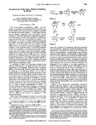
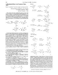
Cyclotheonamides (CyA and CyB, see 1 and 2 and Figure 1)<br />
first attracted our attention because they contained two structural<br />
elements that we had studied in very different contexts: a<br />
vinylogous tyrosine (V-Tyr) and an a-keto amide. We explored<br />
vinylogous amino acids as peptidomimetics with predictable<br />
secondary structures4 and studied a-keto amides as key structural<br />
features for complexing the immunosuppressive agents FK506<br />
and rapamycin to FKBP.5 We were curious to see how these<br />
structural elements, as well as the other features of cyclotheonamide<br />
A (CyA), were used in binding interactions with its<br />
receptor(s). CyA inhibits serine proteases such as human<br />
thrombin, bovine trypsin, and human two-chain tissue plasminogen<br />
activator with Ki values of 1.0, 0.2, and 40 nM, respectively5<br />
Inhibition by CyA was also slow, with second-order rate constants<br />
for complex formation of 4.6 X lo4, 4.8 X lo4, 2.7 X lo2 M-' s-1,<br />
respectively, for the same proteases.6 We elected to study CyA<br />
complexed to bovine /3 trypsin for two reasons: this complex has<br />
the tightest binding, and trypsin is arguably the best characterized<br />
of all the serine proteases. The three-dimensional structure of<br />
the complex was revealed by a 2.0-A resolution X-ray diffraction<br />
study.<br />
Single crystals of the complex, grown by the hanging-drop<br />
vapor-diffusion method? belong to space group P43212 with a =<br />
b = 71.73 and c = 88.82 A. X-ray data were collected on a<br />
t Cornell University.<br />
f Harvard University.<br />
I Present address: Ube Industries, Ltd., Japan.<br />
(1) Fusetani, N.; Matsunaga, S.; Matsumoto, H.; Tukebayashi, Y. J. <strong>Am</strong>.<br />
<strong>Chem</strong>. <strong>SOC</strong>. 1990, 112,7053-7054.<br />
(2) Hagihara, M.; Schreiber, S. L. J. <strong>Am</strong>. <strong>Chem</strong>. <strong>SOC</strong>. 1992, 114, 6570-<br />
6571.<br />
(3) Rosen, M. K.; Schreiber, S. L. Angew. <strong>Chem</strong>., Int. Ed. Engl. 1992,31,<br />
384-400.<br />
(4) Hagihara, M.; Anthony, N. J.; Stout, T. J.; Clardy, J.; Schreiber, S.<br />
L. J. <strong>Am</strong>. <strong>Chem</strong>. <strong>SOC</strong>. 1992, 114, 6568-6570.<br />
(5) (a) Van Duyne, G. D.; Standaert, R. F.; Karplus, P. A.; Schreiber, S.<br />
L.; Clardy, J. Science 1991,252,839-842. (b) Van Duyne, G. D.; Standaert,<br />
R. F.; Schreiber, S. L.; Clardy, J. J. <strong>Am</strong>. <strong>Chem</strong>. <strong>SOC</strong>. 1991,113,7433-7434.<br />
(c) Van Duyne, G. D.; Standaert, R. F.; Karplus, P. A.; Schreiber, S. L.;<br />
Clardy, J. J. Mol. Biol. <strong>1993</strong>, 229, 105-124.<br />
(6) Lewis, S. D.; Ng, A. S.; Baldwin, J. J.; Fusetani, N.; Naylor, A. M.;<br />
Shafer, J. A. Thromb. Res. <strong>1993</strong>, 70, 173-190.<br />
0002-7863/93/1515-<strong>12619</strong>$04.00/00 <strong>1993</strong> <strong>Am</strong>erican <strong>Chem</strong>ical <strong>Society</strong><br />
1<br />
2<br />
-OH<br />
Pro 8<br />
\ +<br />
HAYNH2<br />
R = H Cyclotheonamide A (CyA)<br />
R = Me Cyclotheonamide 8 (CyB)<br />
023<br />
Figure 1. Two different renderings of cyclothwnamide A (CyA). On<br />
the top is a conventional structural drawing, and on the bottom is a<br />
perspective drawing of the conformation bound to bovine @-trypsin. Note<br />
that the a-keto amide is part of a hemiketal with Ser 195 0, (not shown)<br />
on the right.<br />
SDMS Mark I1 area detector coupled to a Rigaku rotating anode<br />
X-ray generator.* The structure was solved using the X-PLOR9<br />
implementation of the molecular replacement technique with the<br />
coordinates of bovine &trypsin as the search model.'* Refinement<br />
used the X-PLOR slow-cooling simulated annealing and leastsquares<br />
energy refinement technique." The final model contains<br />
(7) Ten-microliter droplets containing an initial protein concentration of<br />
15 mg mL in 45 mM Tris, pH 7.0, buffer were equilibrated against 0.7 mL<br />
of 30dPEG 4000 and 0.2 M sodium citrate buffered with 0.1 M Tris at 4<br />
"C. Crystals of 0.3 X 0.4 X 0.6 mm3 appeared in 1-2 months.<br />
(8) Howard, A. D.; Nielson, C.; Zuong, N. H. In Methods in Enzymology,<br />
Volume 114; Wyckoff, H. W., Hirs, C. H. W., Timasheff, S. N., Fds.;Academic<br />
Press: SanDiego,CA, 1985;pp452-472. The&was5.0for 16 217unique<br />
reflections (92.2% completeness).<br />
3
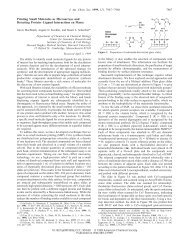
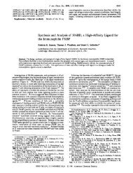
12620 J. <strong>Am</strong>. <strong>Chem</strong>. <strong>SOC</strong>., Vol. <strong>115</strong>, No. 26, <strong>1993</strong><br />
Communications to the Editor<br />
Y 39<br />
Y 39<br />
D10<br />
010<br />
v<br />
D189<br />
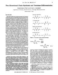
Figure 2. Stereoscopic drawing of cyclotheonamide A in the active site of bovine 8-trypsin.<br />
the protein, CyA, and 128 solvent molecules. In the final (21F,I<br />
- IFc/) map, the electron density is well defined for all atoms<br />
except Asn 11 5, Ser 116, and Arg 117. The final model has an<br />
R-factor of 17.0 for the 16 217 reflections greater than 2a in the<br />
resolution range 8.00-2.0 A. The root mean square deviations<br />
for bond lengths and angles are 0.010 A and 1.7', respectively.<br />
A stereoview of CyA bound in the active site of trypsin is<br />
shown in Figure 2. Most of CyA, the portion from Ca of<br />
diaminopropionic acid (Dpr) to the amide of D-Phe, lies along the<br />
active site groove in an extended substrate-like fashion; the rest<br />
of CyA, the upper right hand portion, is exposed to solvent.<br />
Trypsin's catalytic triad-Ser 195, His 57, and Asp 102-is on<br />
the left-hand side of Figure 2. Well-defined continuous electron<br />
density between Ser 195 0, and the ketone carbon of the a-keto<br />
amide strongly indicates the formation of a covalent complex<br />
through the formation of a tetrahedral hemiketal (0,-C10, 1.41<br />
A). The resulting oxygen accepts hydrogen bonds from the mainchain<br />
NH groups of Gly 193 and Ser 195 in the oxyanion binding<br />
pocket. There is also a strong hydrogen bond between the amide<br />
carbonyl of the a-keto amide and the imidazolium of His 57-the<br />
residue that deprotonates Ser 195 (040-H57NH, 2.68 A).<br />
Other structural features of CyA display exquisite complementarity<br />
to the active site of trypsin. The phenyl ring of D-Phe<br />
is in a hydrophobic groove composed of Tyr 39 and Phe 41 at the<br />
top of Figure 2. The face of the phenyl ring of D-Phe is oriented<br />
toward the edges of Tyr 39, Phe 41, and the phenol ring of V-Tyr.<br />
~~~~~ ~~~~<br />
(9) (a) Brhger, A. T.; Kuriyan, J.; KarplucM. Science 1987,235,458.<br />
(b) Briinger, A. T. X-PLOR Version 3.1; Yale University: Ne_w Haven, CT,<br />
1992. The coordinates for bovine @-trypsin were placed in a P1 unit cell with<br />
dimensionso = b = c = 100 A and a = 8 = y = 90'. Triclinic structure factors<br />
calculated within the 4-8-A resolution range were used for a rotation search<br />
with a 25-A Patterson radius. PC-refinement of the largest peaks in the<br />
rotation search gave a clear solution. The translation search was broken into<br />
two individual translation searches, corresponding to the alternative space<br />
groups P43212 or P412.12, of this best solution using 8-4.0-A data. A single<br />
unambiguous solution for space group P43212 was obtained. Forty cycles of<br />
X-PLOR rigid-body refinement dropped the R-factor from 38.0 to 33.6.<br />
(10) Protein Data Bank code ITLD as described in Bartunik, H. D.;<br />
Summers, L. J.; Bartsch, H. H. J. Mol. Biol. 1989,210,813. See also: Huber,<br />
R.; Bode, W. Acc. <strong>Chem</strong>. Res. 1978, 11, 114-122. Finer-Moore, J. S.;<br />
Kossiakoff, A. A.; Harley, J. H.; Earnest, T.; Stroud, R. M. Proteins 1992,<br />
l2(3), 203-22.<br />
(11) Model building used CHAIN. Sack, J. S. J. Mol. Graphics 1988,6,<br />
224-225. An initial 2lFd - W map at 2.5-A resolution was initially used for<br />
manual adjustment of the protein model. The inhibitor, CtA, was then added<br />
to the active site cleft to account for positive peaks in an IFd = IFd map. The<br />
resolution was gradually increased to 2.0 A. In the final stages of refinement,<br />
diffehce electron density maps were used to locate water molecules.<br />
Restrained, individual temperature factors were refined for each residue.<br />
AverageB-values were 14.63,19.24, and 29.25 Azfor trypsin, CtA, and water,<br />
respectively. Coordinates have been deposited in the Protein Data Bank.<br />
\t+ 0189<br />
Note that L-Phe would not fit into this binding pocket. The<br />
amide proton of Arg (N12) forms a hydrogen bond with the<br />
main-chain carbonyl of Ser 214, while its side chain extends into<br />
the deep, narrow SI specificity pocket at the bottom of Figure 2.<br />
The guanidinium forms hydrogen bonds to the carboxylate of<br />
Asp 189, the carbonyl of Gly 219, and the 0, of Ser 190. Finally,<br />
there is a hydrogen bond between the carbonyl of Dpr (050) and<br />
the NH of Gly 216 and a water-mediated (Owl) hydrogen bond<br />
between the same carbonyl and guanidinium N45. V-Tyr is<br />
oriented away from the active site and exposed to solvent. A<br />
strong (N22-049,2.79 A, see Figure 1) intramolecular hydrogen<br />
bond forms between Dpr amide N-H (N22) and the proline<br />
carbonyl (049). The Pro exists as the trans rotamer in bound<br />
CyA.<br />
This structural work provides important clues for the design<br />
of synthetic analogs that will elucidate the molecular basis of the<br />
observed slow and tight binding behavior.12 For example, one<br />
such analog that has been synthesized and is currently under<br />
investigation is dihydro-CyB, an analog where the ketone carbonyl<br />
(C10) has been reduced to an alcohol, and results of this study<br />
will be reported separately. An independent study has provided<br />
similar insights into the complex formed between CyA and<br />
thrombin.13 These two complementary studies should facilitate<br />
the design of new serine protease inhibitors exhibiting higher<br />
specificity. Moregenerally, they highlight the exciting possibilities<br />
offered by studying novel natural products complexed to their<br />
macromolecular receptors.<br />
Acknowledgment. This work was partially supported by NIH<br />
grants CA24487 (J.C.), GM44993 (S.L.S.), NIH Training Grant<br />
GM08267 in Molecular Biophysics to A.Y.L. and an HHMI<br />
Predoctoral Fellowship to M.W.A. M.H. thanks Ube Industries,<br />
Ltd. for support as a Visiting Scientist. We also thank A. Tulinsky<br />
and B. Maryanoff for sharing the results of ref 13 and J. Shafer<br />
for providing the results of ref 6 prior to publication.<br />
(12) Both CyA and CyB are slow-binding inhibitors of human thrombin<br />
(in 50 mM Tris, pH 7.5, 100 mM NaC1, 0.1% PEG-8000, 0.1 nM purified<br />
human thrombin, 8 pM N-p-tosyl-Gly-Pro-Arg-pNA (Sigma) at 37 "C and<br />
in 50 mM Tris, pH 7.5, 100 mM NaC1, 0.1 nM purified human thrombin,<br />
lOOpM H-D-Phe-Pip-Arg -pNA (Chromogenix) at 37 "C) and bovine trypsin<br />
(in 50 mM Tris, pH 7.4, 150 mM NaC1, 10 mM CaCI2,0.1% PEG-8000,0.4<br />
nM purified bovine trypsin, 20 pM Sar-PR-pNA (Sigma) at 25 "C) (R.K.,<br />
M.W.A., and S.L.S., unpublished results). Second-order rate constants for<br />
the slow binding of CyA to several trypsin-like serine proteases including<br />
thrombin, trypsin, plasmin, t-PA, and factor Xa have been reported by Lewis<br />
et a1.6<br />
( 13) Maryanoff, B. E.; Qiu, X.; Padmanabhan, K. P.; Tulinsky, A.; Almond,<br />
H. R.; Andrade-Gordon, P.; Greco, M. N.; Kauffman, J. A.; Nicolaou, K. C.;<br />
Liu, A,; Brungs, P. H.; Fusetani, N. Proc. Natl. Acod. Sci. U.S.A. <strong>1993</strong>,90,<br />
8048-8052.