POLYPHENOLS AND BIOLOGICAL ACTIVITIES OF Feijoa ...
POLYPHENOLS AND BIOLOGICAL ACTIVITIES OF Feijoa ...
POLYPHENOLS AND BIOLOGICAL ACTIVITIES OF Feijoa ...

You also want an ePaper? Increase the reach of your titles
YUMPU automatically turns print PDFs into web optimized ePapers that Google loves.
<strong>POLYPHENOLS</strong> <strong>AND</strong> <strong>BIOLOGICAL</strong> <strong>ACTIVITIES</strong> <strong>OF</strong> <strong>Feijoa</strong> sellowiana<br />
LEAVES <strong>AND</strong> TWIGS<br />
Siham M. El-Shenawy a , Mohamed S. Marzouk b , Rabab A. El Dib c* , Heba E. Abo Elyazed c , Nermeen M. Shaffie d , Fatma A.<br />
Moharram c<br />
(Received October 2008; Accepted December 2008)<br />
Abstract<br />
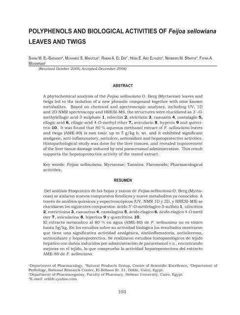
A phytochemical analysis of the <strong>Feijoa</strong> sellowiana O. Berg (Myrtaceae) leaves and<br />
twigs led to the isolation of a new phenolic compound together with nine known<br />
metabolites. Based on chemical and spectroscopic analyses, including UV, 1D<br />
and 2D NMR spectroscopy and HRESI-MS, the structures were elucidated as 3`-Omethylellagic<br />
acid 3-sulphate 1, nilocitin 2, strictinin 3, casuarin 4, castalagin 5,<br />
ellagic acid 6, ellagic acid 4-O-methyl ether 7, avicularin 8, hyperin 9 and quercitrin<br />
10. It was found that 80 % aqueous methanol extract of F. sellowiana leaves<br />
and twigs (AME-80) is non toxic up to 5 g/kg b. wt. and it exhibited significant<br />
analgesic, anti-inflammatory, antiulcer, antioxidant and hepatoprotective activities.<br />
Histopathological study was done for the liver tissues, and revealed improvement<br />
of the liver tissue damage induced by oral paracetamol administration. This result<br />
supports the hepatoprotective activity of the tested extract.<br />
Key words: <strong>Feijoa</strong> sellowiana; Myrtaceae; Tannins; Flavonoids; Pharmacological<br />
activities.<br />
RESUMEN<br />
Del análisis fitoquímico de las hojas y ramas de <strong>Feijoa</strong> sellowiana O. Berg (Myrtaceae)<br />
se aislaron nuevos compuestos fenolicos y nueve metabolitos ya conocidos. A<br />
través de análisis químicos y espectroscópicos (UV, NMR 1D y 2D, y HRESI-MS) se<br />
elucidaron los siguientes compuestos: ácido 3`-O-metilelagico 3-sulfato 1, nilocitina<br />
2, estrictinina 3, casuarina 4, castalagina 5, ácido elagico 6, ácido elagico 4-O-metil<br />
eter 7, avicularina 8, hiperina 9 y quercitrina 10.<br />
El extracto metanolico al 80 % en agua (AME-80) de F. sellowiana no es tóxico<br />
hasta 5g/kg. En los estudios sobre su actividad biológica los resultados mostraron<br />
que tiene una significativa actividad analgésica, aintiinflamatoria, antiulcerosa,<br />
antioxidante y hepatoprotectiva. Se realizaron estudios histopatológicos de tejido<br />
hepático con daños inducidos por administración de paracetamol v.o., encontrando<br />
mejoras en el tejido, lo que comprueba la actividad hepatoprotectora del extracto<br />
AME-80 de F. sellowiana.<br />
a<br />
Department of Pharmacology, b Natural Products Group, Centre of Scientific Excellence, d Department of<br />
Pathology, National Research Center, El-Behoos St. 31, Dokki, Cairo, Egypt.<br />
c<br />
Department of Pharmacognosy, Faculty of Pharmacy, Helwan University, Cairo, Egypt.<br />
*E-mail: reldib @yahoo.com<br />
103
104 S.M. El-shenawy et al.<br />
INTRODUCTION<br />
<strong>Feijoa</strong> sellowiana, O. Berg (syn Acca sellowiana)<br />
is belonging to family Myrtaceae.<br />
It is native to southern Brazil, northern<br />
Argentina, western Paraguay (Trease and<br />
Evans, 1978) being traditionally known as<br />
pineapple guava. The plant is used in many<br />
industrialized products particularly in the<br />
Australian area, in the form of jams, syrups,<br />
liquors and crystallized fruits (Di. Cesare et<br />
al., 1998).<br />
F. sellowiana leaves, fruits and<br />
stems were reported to exhibit antimicrobial,<br />
antitumor, anti-inflammatory and antioxidant<br />
effects (Isobe<br />
et al., 2003; Vuotto et al.,<br />
1999). Few studies dealt with the chemistry<br />
of F. sellowiana as a flavone (Ruberto and<br />
Tringali, 2004), volatile components (Fernandez<br />
et al., 2004; Di. Cesare et al., 2000;<br />
Shaw et al., 1990), lipids (Di. Cesare et al.,<br />
1998) and tannins (Okuda et al., 1982).<br />
This study aims at the isolation and<br />
identification of the constitutive polyphenols<br />
in the aqueous methanol extract of<br />
the leaves and twigs of F. sellowiana in<br />
addition to evaluation of the analgesic,<br />
anti-inflammatory, antiulcer, antioxidant<br />
and hepatoprotective effects of the investigated<br />
extract.<br />
MATERIALS <strong>AND</strong> METHODS<br />
Equipments<br />
The NMR spectra were recorded at 300, 400<br />
and 500 ( 1 H) and 75, 100, 125 ( 13 C) MHz,<br />
on a Varian Mercury 300, Bruker APX-400<br />
and JEOL GX-500 NMR spectrometers and<br />
δ-values are reported as ppm relative to<br />
TMS in the convenient solvent. HRESI-MS<br />
analyses were run on LTQ-FT-MS spectrometer<br />
(Thermo Electron, 400, Germany). UV<br />
analyses for pure samples were recorded as<br />
MeOH solutions and with different diagnostic<br />
UV shift reagents on a Shimadzu UV 240<br />
(P/N240–58000) and Ebeckman DU7 spectrophotometer.<br />
For column chromatography,<br />
Sephadex LH-20 (Pharmacia, Uppsala,<br />
Sweden), microcrystalline cellulose (E. Merck,<br />
Darmstadt, Germany) and polyamide 6<br />
(Fluka Chemie AG, Switzerland) were used.<br />
For paper chromatography; Whatman No.<br />
1 sheets (Whatman Ltd., Maidstone, Kent,<br />
England) were used.<br />
Plant material<br />
Leaves and twigs of F. sellowiana O. Berg<br />
were collected in February 2004 and April<br />
2005 from Zohria Botanical Garden, Cairo,<br />
Egypt. The identification of the plant was<br />
performed by Dr. Amal Abdel-Aziz, Lecturer<br />
of Taxonomy, Institute of Horticulture,<br />
Zohria Botanical Garden. A voucher<br />
sample (No.: F-1) is kept in the Herbarium,<br />
Pharmacognosy Department, Faculty of<br />
Pharmacy, Helwan University.<br />
Extraction and Isolation<br />
Air-dried ground leaves and twigs (850<br />
g) were extracted with hot 80 % aqueous<br />
methanol (5L, then 4 x 4L) under reflux<br />
(80 ˚C). After evaporation of solvent under<br />
reduced pressure, the residue (60 g) was<br />
defatted with petroleum ether (60-80 ˚C)<br />
under reflux (5 x 1.5 L, 60 ˚C). The residue<br />
(40 g), after evaporation of petroleum ether<br />
was re-dissolved in pure methanol to yield<br />
29 g as dry methanol soluble portion. It was<br />
preliminary fractionated on a polyamide<br />
column (300 g, 110 x 7 cm) using a step<br />
gradient elution with H 2<br />
O-MeOH in ratio of<br />
100:0 – 0:100 giving 28 fractions of 1L each,<br />
which were collected and monitored by<br />
Comp-PC (systems S 1<br />
and S 2<br />
) and UV-light<br />
into three major collective fractions (I-III)<br />
with other non-phenolic fractions. Fraction<br />
I (3.5 g) was subjected to repeated column<br />
chromatography (CC) on microcrystalline<br />
cellulose using n-BuOH-2-propanol-H 2<br />
O<br />
BIW (4:1:5, organic layer as an eluent) followed<br />
by repeated and separately cellulose<br />
column for each major subfraction using<br />
MeOH/BIW (50 %) to give compounds 8<br />
(13 mg), 9 (25 mg) and 10 (20 mg). Fraction<br />
II (1.7 g) was chromatographed on a<br />
Sephadex C and eluted with MeOH to give
Polyphenols and biological activities of <strong>Feijoa</strong> sellowiana Rev. Latinoamer. Quím. 36/3 (2008) 105<br />
pure sample of 3 (19 mg). Compound 5 (22<br />
mg) was obtained by precipitation of fraction<br />
III (4.1 g) from conc. MeOH solution<br />
by excess EtOAc. Methanol insoluble portion<br />
(10 g) was re-dissolved in H 2<br />
O-MeOH<br />
(1:10), filtered and dried under vacuum to<br />
gives 8.5 g dry residue. Thereafter, it was<br />
fractionated on cellulose C (150 g, 70 x 5<br />
cm) using aq. MeOH (10-70 %) to give three<br />
main fractions A, B and C. Fractionation of<br />
fraction A (250 mg) on Sephadex C (10 %<br />
aq. MeOH as eluent) led to isolate 2 (15 mg).<br />
Fraction B (2.8 g) was chromatographed on<br />
Sephadex C (10 % aq. MeOH as eluent) to<br />
give two major subfractions. The first main<br />
subfraction was then fractionated on Sephadex<br />
with BIW for elution to afford pure<br />
sample of 7 (11 mg), while the second one<br />
was separated on Sephadex C and eluted<br />
with 40 % aq. MeOH to give isolate 1 (9<br />
mg). Fractionation of fraction C (95 mg) on<br />
Sephadex C using 10 % aq. MeOH followed<br />
by repeated and separately Sephadex C for<br />
each of the two major subfractions led to<br />
compounds 4 (9 mg) and 6 (20 mg). The<br />
homogeneity of the fractions was tested<br />
on 2D- and Comp-PC using Whatman No.<br />
1 paper (systems S 1<br />
and S 2<br />
); S 1<br />
: n-BuOH-<br />
HOAc-H 2<br />
O (4:1:5, top layer) and S 2<br />
: 15%<br />
aqueous HOAc. The compounds were visualized<br />
by spraying with Naturstoff reagent<br />
for detection of flavonoids and nitrous acid<br />
or KIO 3<br />
-reagents for detection of tannins.<br />
3`-O-methyl-ellagic acid 3-sulphate (1)<br />
Brown amorphous powder; R f<br />
-values: 0.33<br />
(S 1<br />
), 0.56 (S 2<br />
) on PC; mauve fluorescent spot<br />
by long and short UV-light turned to greenish-yellow<br />
with ammonia vapours, faint indigo-red<br />
colour with nitrous acid and faint<br />
blue with FeCl 3<br />
; UV λ max<br />
(MeOH), nm: 240,<br />
270 sh, 353 sh and 380; Negative HRESI-<br />
MS/MS: m/z 394.97169 [M-H] ‾ (calcd.:<br />
394.97145), (MS 2 ) 315.05426 [M-H-SO 3<br />
] ‾ ,<br />
(MS 3 ) 300.06757 [M-H-SO 3<br />
-CH 3<br />
] ‾ = [Ellagic<br />
acid-2H] ‾ . 1 H NMR (300 MHz, DMSO-d 6<br />
): δ<br />
ppm 7.14 (2 H, br s, H-5/5`), 3.74 (3 H, s,<br />
OCH 3<br />
-3`).<br />
Nilocitin (2)<br />
Brown amorphous powder; R f<br />
-values: 0.19<br />
(S 1<br />
), 0.7 (S 2<br />
) on PC; it gives dark purple<br />
fluorescent spot by short UV-light, turned<br />
to indigo-red and deep blue colour by spraying<br />
with NaNO 2<br />
-glacial AcOH and FeCl 3<br />
,<br />
respectively. UV λ max<br />
(MeOH), nm: strong<br />
absorption band at 245 with 283 sh. Negative<br />
ESI-MS/MS: m/z 481.25 [M-H] ‾ , (MS 2 )<br />
301.22 [Ellagic acid-H] ‾ , (MS 3 ) 257.07 [Ellagic<br />
acid-H-CO 2<br />
] ‾ , 229.10 [Ellagic acid-H-<br />
CO 2<br />
-CO] ‾ . 1 H NMR (300 MHz, DMSO-d 6<br />
): δ<br />
ppm 6.43 (1 H in total, s, H-3``), 6.32 (1 H<br />
in total, s, H-3`), 5.23 (1/2 H, br s, H-1α),<br />
5.10 (1/2 H, t-like, J = 9.6 Hz, H-3α), 4.85<br />
(1/2 H, t-like, J = 9.9 Hz, H-3β), 4.81 (1/2<br />
H, d, J = 8.1 Hz, H-1β), 4.69 (1 H, br d, J =<br />
9.6 Hz, H-2α), 4.47 (1/2 H, t-like, J = 8.7 Hz,<br />
H-2β), 4.00 – 3.00 (m, hidden by H 2<br />
O-signal,<br />
remaining sugar protons). 13 C NMR (75<br />
MHz, DMSO-d 6<br />
): δ ppm 169.00 (C-7``α, β),<br />
168.57, 168.34 (C-7`α, β), 144.46 (C-6`/6``α,<br />
β), 144.30 (C-4`/4``α, β), 134.91, 134.79<br />
(C-5`/5``α, β), 125.49, 125.287 (C-2``α, β),<br />
124.849 (C-2`α, β), 113.78, 113.70 (C-1``α,<br />
β), 113.67, 113.50 (C-1`α, β), 105.30, 104.92<br />
(C-3`/3``α, β), 93.27 (C-1β), 89.76 (C-1α),<br />
79.54 (C-3α), 77.12 (C-3β), 76.91, 76.68<br />
(C-2α/2β), 74.24 (C-4β), 72.17 (C-5α), 67.51<br />
(C-4α), 67.03 (C-5β), 60.46 (C-6α, β).<br />
Strictinin (3)<br />
Brown amorphous powder; R f<br />
-values:<br />
0.27 (S 1<br />
), 0.36 (S 2<br />
) on PC; it gives dark<br />
purple fluorescent spot by short UV-light,<br />
turned to indigo-red and deep blue colour<br />
by spraying with NaNO 2<br />
-glacial AcOH and<br />
FeCl 3<br />
respectively; UV λ max<br />
(MeOH), nm:<br />
246, 288 sh. Negative HRESI-MS/MS: m/z<br />
633.07410 [M-H] ‾ (calcd. 633.07317), (MS 2 )<br />
481.04634 [M-H-galloyl] ‾ , 300.98930 [Ellagic<br />
acid-H] ‾ , (MS 3 ) 257.01764 [Ellagic-H-<br />
CO 2<br />
] ‾ , 229.02834 [Ellagic acid-H-CO 2<br />
-CO] ‾ ,<br />
213.01816 [Ellagic acid-H-OH-CO-CO 2<br />
] ‾ ,<br />
(MS 4 ) 185.03564 [Ellagic acid-H-CO-2<br />
CO 2<br />
] ‾ . 1 H NMR (500 MHz, DMSO-d 6<br />
): δ ppm<br />
6.98 (2 H, s, H-2`/6` G), 6.48 (1 H, s, H-<br />
3```HHDP), 6.29 (1 H, s, H-3`` HHDP), 5.56
106 S.M. El-shenawy et al.<br />
(1 H, d, J = 7.7 Hz, H-1), 4.94 (1 H, dd, J =<br />
13, 6 Hz, H-6 a<br />
), 4.58 (1 H, t-like, J = 10 Hz,<br />
H-4), 4.03 (1 H, br dd, J = 9.2, 6.2 Hz, H-5),<br />
3.67 (1 H, br d, J = 13 Hz, H-6 b<br />
), 3.55 (1 H,<br />
t-like, J = 9.2 Hz, H-3), 3.35 (1 H, t-like, J =<br />
8.4 Hz, H-2). 13 C NMR (75 MHz, DMSO-d 6<br />
):<br />
δ ppm 167.83, 167.09 (C-7```/7``HHDP),<br />
164.62 (C-7` G), 145.62 (C-3`⁄5` G), 144.50,<br />
144.41 (C-6``/6``` HHDP), 144.26. 144.23<br />
(C-4``/4``` HHDP), 139.17 (C-4` G), 135.33,<br />
134.96 (C-5``/5``` HHDP), 124.70, 124.42<br />
(C-2``/2```HHDP), 118.42 (C-1` G), 115.55,<br />
115.23 (1``/1```HHDP), 109.09 (C-2`/6` G),<br />
106.29, 105.51 (C-3``⁄ 3``` HHDP), 94.73<br />
(C-1), 73.82 (C-3), 73.33 (C-5), 71.61 (C-<br />
4/2), 62.74 (C-6); G = galloyl and HHDP =<br />
hexahydroxydiphenoyl moieties.<br />
Casuarin (4)<br />
Brown amorphous powder; R f<br />
-values: 0.14<br />
(S 1<br />
), 0.30 (S 2<br />
) on PC; it gives dark purple<br />
fluorescent spot by short UV-light and dull<br />
brown under long UV-light, turned to indigo-red<br />
and deep blue colour with NaNO 2<br />
-<br />
glacial AcOH and FeCl 3<br />
, respectively. UV<br />
λ max<br />
(MeOH), nm: 246, 285 sh. 1 H NMR<br />
(500 MHz, DMSO-d 6<br />
): δ ppm 6.44, 6.24,<br />
6.15 (1 H each, s, H-3````/3``/3``` HHDP),<br />
5.38 (1 H, br s, H-1), 5.18 (1 H, br s, H-3),<br />
4.8 (1 H, br s, H-4), 4.65 (1 H, br s, H-2),<br />
4.34 (1 H, br d, J = 9.2 Hz, H-6 a<br />
), 3.91 (1<br />
H, br s, H-5), 3.73 (1 H, br d, J = 10.7 Hz,<br />
H-6 b<br />
). 13 C NMR (75 MHz, DMSO-d 6<br />
): δ ppm<br />
169.27, 168.87, 167.57 (C-7``, C-7````, C-<br />
7```), 163.63 (C-7`), 145.79, 144.92, 144.64,<br />
144.60 (C-6`/6``/6```/6```` HHDP), 144.42,<br />
144.30, 144.11 (C-4``/4```/4```` HHDP),<br />
142.62 (C-4` HHDP), 137.64 (C-5`), 135.68,<br />
134.30, 133.74 (C-5``/5```/5```` HHDP),<br />
126.06 (C-2``), 125.55 (C-2````), 123.56 (C-<br />
2```), 119.35 (C-2`), 115.89 (C-1`), 115.70,<br />
115.41 (2C) (C-1``/1```/1```` HHDP), 114.32<br />
(C-3` HHDP), 106.27 (C-3````), 104.67 (C-<br />
3``), 103.03 (C-3``` HHDP), 75.78 (C-2),<br />
75.33 (C-4), 69.69 (C-3), 67.50 (C-1), 66.91<br />
(C-5), 66.14 (C-6).<br />
Castalagin (5)<br />
Brown amorphous powder; R f<br />
-values: 0.04<br />
(S 1<br />
), 0.61 (S 2<br />
) on PC. It gives dark purple<br />
fluorescent spot by short UV-light and dull<br />
brown under long UV-light, turned to indigo-red<br />
and deep blue colour with NaNO 2<br />
-<br />
glacial AcOH and FeCl 3<br />
, respectively. UV<br />
λmax (MeOH), nm: 246, 288 sh. Negative<br />
HRESI-MS/MS: m/z 933.06324 [M-H] ‾<br />
(calcd. 933.06363), (MS 2 ) 914.92332 [M-H-<br />
H 2<br />
O] ‾ , 631.06571 [M-H-ellagic acid] ‾ , (MS 3 )<br />
896.95412 [M-H-2H 2<br />
O] ‾ , (MS 4 ) 878.95503<br />
[M-H-3H 2<br />
O] ‾ , 301.01411 [Ellagic acid-H] ‾ ;<br />
1<br />
H NMR (300 MHz, DMSO-d 6<br />
): δ ppm 6.62<br />
(1 H, s, H-3``` FLG), 6.52 (1 H, s, H-3````<br />
HHDP), 6.38 (1 H, s, H-3````` HHDP), 5.47<br />
(1 H, d, J = 4.5 Hz, H-1), 5.40 (1 H, br<br />
d, J = 7.8 Hz, H-5), 4.91 (1 H, t-like, J<br />
= 7.8 Hz, H-4), 4.89 (1 H, dd, J = 12.6,<br />
3 Hz, H-6a), 4.82 (2 H, m, H-3/2), 3.96<br />
(1H, br d, J = 12.6 Hz, H-6b). 13 C NMR<br />
(100 MHz, DMSO-d 6<br />
): δ ppm 168.14,<br />
165.84 (C-7````/C-7````` HHDP), 165.80<br />
(C-7``` FLG), 165.32 (C-7`` FLG), 162.87<br />
(C-7` FLG), 146.40 (C-4``` FLG), 144.85,<br />
144.73 (C-6````/6````` HHDP), 144.46,<br />
144.12 (C-4````/4````` HHDP, 4`/4`` FLG),<br />
143.67, 142.47, 142.35 (C-6`/6``/6```<br />
FLG), 137.14 (C-5``` FLG), 135.75, 135.57<br />
(C-5````/5````` HHDP), 135.11 (C-5`` FLG),<br />
133.20 (C-5``` FLG), 125.91 (C-2`` FLG),<br />
124.80, 123.52 (C-2````/2````` HHDP),<br />
123.42 (C-2``` FLG), 121.10 (C-2` FLG),<br />
115.25, 114.47, 114.25 (each 2C), (C-<br />
1````/1````` HHDP, C-1`/1``/1``` FLG, C-3`<br />
FLG), 111.70 (C-3`` FLG), 106.84 (each 2<br />
C), 105.60 (C-3``` FLG, C-3````/3````` HHDP),<br />
72.73 (C-2), 69.86 (C-5), 67.90 (C-4), 66.68<br />
(C-1), 65.99 (C-3), 65.12 (C-6).<br />
Ellagic acid (6)<br />
Off-white amorphous powder; R f<br />
-values:<br />
0.39 (S 1<br />
), 0.19 (S 2<br />
) on PC; it gives buff fluorescent<br />
spot by long and short UV light,<br />
turned to dull yellow fluorescence on exposure<br />
to ammonia vapours, greenish yellow<br />
with Naturstoff, and faint blue colour with<br />
FeCl 3<br />
. UV λ max<br />
(MeOH), nm: 215, 256, 362.
Polyphenols and biological activities of <strong>Feijoa</strong> sellowiana Rev. Latinoamer. Quím. 36/3 (2008) 107<br />
Negative HRESI-MS, m/z 300.99789 [M-H] ‾<br />
(calcd. 300.99896).<br />
Ellagic acid 4-O-methyl ether (7)<br />
Pale yellow amorphous powder; R f<br />
-values:<br />
0.29 (S 1<br />
), 0.30 (S 2<br />
) on PC; it gives faint<br />
mauve fluorescent spot under long and<br />
short UV light turned to greenish-yellow<br />
with ammonia vapours and faint blue with<br />
FeCl 3<br />
. UV λ max<br />
(MeOH), nm: 245, 272 sh,<br />
353 sh and 385. 1 H NMR (300 MHz, DMSOd<br />
6<br />
): δ ppm 7.91 (1 H, s, H-5), 7.33 (1 H, s,<br />
H-5`), 3.86 (3 H, s, OCH 3<br />
-3).<br />
Avicularin (8)<br />
Yellow amorphous powder; R f<br />
-values: 0.38<br />
(S 1<br />
), 0.44 (S 2<br />
) on PC; it gave dark purple<br />
spot by long UV light turned to green with<br />
FeCl 3<br />
and orange with Naturstoff reagent.<br />
UV λ max<br />
(MeOH), nm: 296, 296 sh, 351;<br />
(+ NaOMe): 273, 327 sh, 403; (+ NaOAc):<br />
270, 320 sh, 361; (+ NaOAc/ H 3<br />
BO 3<br />
):<br />
270, 400; (+ AlCl 3<br />
): 273, 304 sh, 335<br />
sh, 421; (+ AlCl 3<br />
/HCl): 268, 290 sh,<br />
361 sh, 398. Negative HRESI/MS: m/z<br />
433.07730 [M-H] ‾ (calcd.: 433.07763),<br />
867.16187 [2M-H] ‾ , 300.02756 [Mpentoside]<br />
‾ = [quercetin-H] ‾ . 1 H and 13 C<br />
NMR data are listed in Table 1.<br />
Hyperin (9)<br />
Yellow amorphous powder; R f<br />
-values: 0.48<br />
(S 1<br />
), 0.61 (S 2<br />
) on PC; it gave dark purple<br />
spot by long UV light turned to green with<br />
FeCl 3<br />
and orange with Naturstoff reagent.<br />
UV λ max<br />
(MeOH), nm: 261, 359; (+ NaOMe):<br />
273, 411; (+ NaOAc): 265, 374; (+ NaOAc/<br />
H 3<br />
BO 3<br />
): 262, 374; (+ AlCl 3<br />
): 252 sh, 260,<br />
270, 408; (+ AlCl 3<br />
/HCl): 252 sh, 260, 270,<br />
364. Negative HRESI/MS: m/z 463.08789<br />
[M-H] ‾ (calcd.: 463.08820), 927.18396<br />
[2M-H] ‾ , 301.02255 [M-deoxyhexoside] ‾ =<br />
[quercetin-H] ‾ , (MS 3 ) 178.89877 (C 9<br />
H 7<br />
O 4<br />
,<br />
cinnamoyl fragment), 150.89915 (C 6<br />
H 3<br />
O 4<br />
,<br />
Table 1: 1 H and 13 C NMR spectral data of 8-10 (DMSO-d 6<br />
, 300 and 75 MHz)<br />
Carbon 8 9 10<br />
No<br />
13<br />
C 1<br />
H<br />
13<br />
C 1<br />
H<br />
13<br />
C 1<br />
H<br />
C-2 156.91 156.28 157.65<br />
C-3 133.37 133.49 134.58<br />
C-4 177.67 177.46 178.10<br />
C-5 161.18 161.17 161.66<br />
C-6 98.64 6.2 (d, 2.1) 98.68 6.2 (d,2.4) 99.05 6.2 (d,2.4)<br />
C-7 164.19 164.06 164.58<br />
C-8 93.53 6.4 (d, 2.1) 93.52 6.4 (d,2.4) 93.98 6.39 (d,2.4)<br />
C-9 156.32 156.25 156.80<br />
C-10 103.94 103.92 104.43<br />
C-1` 121.68 121.34 121.09<br />
C-2` 115.53 7.47 (d, 2.1) 115.19 7.58 (d,2.4) 115.82 7.23 (d,2.1)<br />
C-3` 145.05 144.78 145.56<br />
C- 4` 148.43 148.43 148.80<br />
C-5` 115.53 6.85 (d, 8.1) 115.59 6.83 (d, 8.7) 116.01 6.86 (d,8.7)<br />
C-6` 120.94 7.56 (dd, 8.1, 2.1) 121.10 7.66 (dd, 8.2, 2.4) 121.46 7.27 (dd, 8.7,2.1)<br />
C-1`` 107.86 5.58 (d, 1.2) 101.86 5.36 (d,7.8) 102.20 5.25 (d,1.5)<br />
C-2`` 82.09 4.15 (dd, 3.6, 1.2) 71.22 3.67-3.26 (m, 70.72 3.97dd (3.3,1.5)<br />
H-2``, H-3``,<br />
H-4``, H-5``)<br />
C-3`` 76.96 3.72 (m) 73.20 70.94 3.77-3.05 (m, H3``,<br />
H-4”, H-5”)<br />
C-4`` 85.86 3.57 (m) 67.34 71.55<br />
C-5`` 60.66 3.33 (dd, 11.7, 2.75``a) 75.81 70.41<br />
3.26 (dd, 11.7, 6.95``b)<br />
C-6`` - 60.15 17.85 0.80 (d, 5.7)
108 S.M. El-shenawy et al.<br />
benzoyl fragment). 1 H and 13 C NMR data<br />
are listed in Table 1.<br />
Quercitrin (10)<br />
Yellow amorphous powder; R f<br />
-values: 0.50<br />
(S 1<br />
), 0.64 (S 2<br />
) same as (9) on PC; it gave<br />
dark purple spot by long UV light turned<br />
to green with FeCl 3<br />
and orange with Naturstoff<br />
reagent. UV λ max<br />
(MeOH) nm: 255,<br />
299 sh, 353; (+ NaOMe): 268, 325 sh, 400;<br />
(+ NaOAc): 270, 320 sh, 370; (+ NaOAc/<br />
H 3<br />
BO 3<br />
): 260, 299 sh, 374; (+ AlCl 3<br />
): 273, 304<br />
sh, 408; (+ AlCl 3<br />
/HCl): 268, 304 sh, 353 sh,<br />
398. Negative HRESI/MS: m/z 447.09296<br />
[M-H] ‾ (calcd.: 447.09312), 301.03607<br />
[M-deoxyhexoside] ‾ = [quercetin-H] ‾ , (MS 3 )<br />
178.93030 (C 9<br />
H 7<br />
O 4<br />
, cinnamoyl fragment),<br />
150.93245 (C 6<br />
H 3<br />
O 4<br />
, benzoyl fragment). 1 H<br />
and 13 C NMR data are listed in Table 1.<br />
Pharmacological studies<br />
Animals<br />
Adult male pathogen-free Sprague-Dawley<br />
rats (120-130 g) and Swiss mice weighing<br />
(20-30 g) were purchased from the animal<br />
house of National Research Centre were<br />
used. The animals were housed in standard<br />
metal cages in an air-conditioned room at<br />
22 ± 3˚C, 55 ± 5% humidity, and 12 h light<br />
and provided with standard laboratory<br />
diet and water ad libitum. All experimental<br />
procedures were conducted in accordance<br />
with the guide for care and use of laboratory<br />
animals and in accordance with the<br />
Local Animal Care and Use Committee. The<br />
distilled water was used as a vehicle for<br />
extract and paracetamol and 5 % sodium<br />
bicarbonate for indomethacin.<br />
Determination of median lethal dose<br />
(LD 50<br />
)<br />
The LD 50<br />
of the AME-80 was determined<br />
using rats. No percentage mortality was<br />
recorded after 24 hours up to dose of 5<br />
g/kg and according to Semler (1992) who<br />
reported that if just one dose level at 5 g/kg<br />
is not lethal, regulatory agencies no longer<br />
require the determination of an LD 50<br />
value.<br />
So the experimental doses used were 1/20,<br />
1/10 and 1/5 of 5/ g/kg of AME-80 (250,<br />
500 and 1000 mg kg -1 ).<br />
Analgesic activity<br />
This activity was determined by measuring<br />
the responses of animals to both thermal<br />
and chemical stimulus.<br />
Thermal test<br />
Hot-plate test was conducted using an<br />
electronically controlled hot-plate (Ugo<br />
Basile, Italy) adjusted at 52°C ± 0.1°C<br />
and the cut-off time was 30s (Eddy and<br />
Leimback, 1953). Five groups of mice<br />
each of six were used. The time elapsed<br />
until either paw licking or jumping is<br />
recorded 60 min before, and 1 and 2 h<br />
after oral administration of AME-80 (250,<br />
500, 1000 mg kg -1 ), saline and tramadol<br />
(20 mg kg -1 orally, a reference analgesic<br />
drug, October Pharma, 6 th October, Egypt)<br />
(Sacerdote et al., 1997).<br />
Chemical test<br />
Acetic acid-induced writhing in mice was<br />
performed according to the convenient<br />
published method (Collier et al., 1968). The<br />
mice were divided into six groups each of<br />
six and received the same doses of AME-<br />
80, control as mentioned before, in thermal<br />
test. Indomethacin (25 mg kg -1 , Epico,<br />
Egypt) was used as reference drug. After<br />
60 min interval, the mice received 0.6 %<br />
acetic acid ip (0.2 ml/mice). The number<br />
of writhes in 30 min period was counted<br />
and compared.<br />
Anti-inflammatory activity<br />
Anti-inflammatory activity in acute model<br />
was carried out according to the convenient<br />
reported method (Winter et al., 1962). Rats<br />
were divided into five groups each of six and<br />
received orally saline as control, AME-80<br />
(250, 500, 1000 mg kg -1 ), and indomethacin<br />
(25 mg kg -1 orally) one hour before induction<br />
of oedema by subplanter injection of<br />
100 µL of 1 % carrageenan injection (Sigma,
Polyphenols and biological activities of <strong>Feijoa</strong> sellowiana Rev. Latinoamer. Quím. 36/3 (2008) 109<br />
USA) in saline into the pad of right paw.<br />
The difference in hind foot pad thickness<br />
was measured immediately before and 1, 2,<br />
3, 4 h after carrageenan injection with a micrometer<br />
caliber (Obukowicz et al., 1998).<br />
The oedema was expressed as a percentage<br />
of change from the control group.<br />
The percentage of oedema inhibition<br />
was calculated from the mean effect in the<br />
control and treated animals according to<br />
the following equation:<br />
% Oedema inhibition =<br />
% oedema formation of control group - % oedema formation of treated group<br />
% oedema formation of control group x 100<br />
Gastric ulcerogenic studies<br />
Gastric lesions were induced in rats by<br />
ethanol (1 ml of 50 % orally). Animals fasted<br />
for 24 h and then divided into four groups,<br />
one group received ethanol and served as<br />
control group, and the remaining groups<br />
received AME-80 (250, 500 and 1000 mg<br />
kg -1 ) one hour before the ethanol was given.<br />
Rats were killed 1 h following ethanol administration<br />
after being lightly anaesthetized<br />
with ether and the stomach was excised,<br />
opened along greater curvature, rinsed<br />
with saline, extended on plastic board and<br />
examined for mucosal lesions, using an<br />
ulcer score of 5 as described by Mozsik et<br />
al., 1982. The ulcer scores were evaluated<br />
as follows: petechial lesions = 1, lesions less<br />
than 1 mm = 2, lesions between 1 and 2<br />
mm = 3, lesions between 2 and 4 mm = 4,<br />
and lesions more than 4 mm = 5. A total<br />
lesion score for each animal is calculated<br />
as the total number of lesions multiplied<br />
by the respective severity score. Results are<br />
expressed as severity of lesions/rat.<br />
Hepatoprotective study<br />
The hepatic damage was induced in rats by<br />
oral administration of paracetamol 1 g/kg<br />
(Silva et al., 2005). Fifty four rats were divided<br />
into nine groups of six animals each<br />
as following: group 1 (normal control group)<br />
received a daily oral dose of 1ml saline;<br />
groups 2, 3, 4 received a daily oral dose<br />
of AME-80 (250, 500 and 1000 mg/kg b.<br />
wt.) alone for successive 10 days; group 5<br />
received a single oral dose of paracetamol<br />
(1 g kg -1 b. wt.); groups 6, 7, 8 received a<br />
daily oral dose of AME-80 (250, 500 and<br />
1000 mg/kg b. wt.) for 10 successive days<br />
before paracetamol administration (1g/kg<br />
b. wt.); and group 9 received a daily oral<br />
dose of silymarin (25 mg/kg b. wt.) for<br />
successive 10 days before paracetamol<br />
(reference drug for hepatoprotective studies).<br />
At the end of the experimental period<br />
(24 h after paracetamol administration),<br />
the blood was obtained from all groups of<br />
rats after being lightly anaesthetized with<br />
ether by puncturing rato-orbital plexus<br />
(Sorg and Buckner, 1964). Finally, the following<br />
biochemical tests: Alanine aminotransferase<br />
(ALT) (Bergmeyer et al.,1986),<br />
aspartate aminotransferase (AST) (Klauke<br />
et al., 1993) and serum alkaline phosphatase<br />
(ALP) (Tietz and Shuey, 1986) were<br />
performed.<br />
Antioxidant activity<br />
The activity of (1,1-Diphenyl,2-picryl hydrazyl)<br />
DPPH radical scavenging activity<br />
was investigated in vitro according to the<br />
method of Peiwu et al., 1999. A methanolic<br />
solution of DPPH (2.95 ml) was added to 50<br />
µl sample (AME-80) dissolved in methanol<br />
at different concentrations (10-100 mg/ml)<br />
in a disposable cuvette. The absorbance<br />
was measured at 517 nm at regular intervals<br />
of 15 seconds for 5 minutes. Ascorbic<br />
acid was used as a standard (0.1 M) as<br />
described by Govindarajan et al., 2003.<br />
% inhibition Abs. (DPPH solution)-Abs. (sample)<br />
(Reactive reaction rate) = × 100<br />
Abs (DPPH solution)<br />
Statistical analysis<br />
The results are expressed as mean ± S.E.<br />
and the statistical significance was evaluated<br />
by the student’s t-test (Sendocor and<br />
Cechran, 1971) and one way ANOVA
110 S.M. El-shenawy et al.<br />
(Dunnnett’s multiple comparison test).<br />
The * P values < 0.05 were considered to<br />
be significant.<br />
RESULTS <strong>AND</strong> DISCUSSION<br />
Ten polyphenolic metabolites (1-10) were<br />
isolated from the aqueous methanol extract<br />
of F. sellowiana leaves and twigs<br />
using repeated column chromatographic<br />
separations with convenient adsorbents<br />
and solvent systems (see Materials and<br />
Methods section). Compounds 2 and 3<br />
appeared as dark purple fluorescent spots<br />
under short UV-light changed into deep<br />
blue colour with FeCl 3<br />
and indigo-red colour<br />
with HNO 2<br />
spray reagent characteristic<br />
for hexahydroxydiphenoyl esters (ellagitannins),<br />
(Gupta et al., 1982). The UV-spectrum<br />
showed intrinsic broad absorption<br />
band with a shoulder at about λ max<br />
283<br />
nm characteristic for the ellagitannins. On<br />
complete acid hydrolysis 2 gave ellagic acid<br />
and glucose, whereas 3 yielded ellagic acid,<br />
gallic acid and glucose as well (Comp-PC).<br />
Negative ESI-MS of 2 exhibited a molecular<br />
ion peak at m/z 481.25 [M-H] ‾ corresponding<br />
to a mono-hexahydroxydiphenoyl-glucose,<br />
while that of 3 showed a molecular<br />
ion peak at m/z 633.07410 with 152 amu<br />
more than that of 2 for extra galloyl moiety.<br />
As well as, at high fragmentation energy<br />
MS 2 , MS 3 and MS 4 spectra exhibited the<br />
fragments, which were confirmative for<br />
the presence of a hexahydroxydiphenoyl<br />
group (HHDP) in 2 and HHDP with gallic in<br />
case of 3. The positions of the attachment<br />
of galloyl or HHDP onto glucose core and<br />
its stereochemistry were concluded from<br />
NMR analysis. 1 H NMR of 2 exhibited two<br />
singlet signals, each one proton, at δ ppm<br />
6.43 and 6.32 of one HHDP group (H-3``,<br />
3`), while 3 exhibited two singlet signals,<br />
each of one proton (δ 6.48 and 6.29) with<br />
a singlet signal of two protons (δ 6.98)<br />
characteristic for a galloyl moiety. In the<br />
aliphatic region, 2 exhibited duplication of<br />
all 1 H-resonances which was indicative to a<br />
free anomeric-OH and the presence of 2 in<br />
the form of α/β-anomeric mixture (Okuda<br />
et al., 1989). The downfield shift of H-3 and<br />
H-2 in both anomers at 5.10, 4.85 (H-3α/β)<br />
and 4.69, 4.47 (H-2α/β) indicated the attachment<br />
of the HHDP-group at C-2 and<br />
C-3 in case of 2. In case of 3, the downfield<br />
shift (∆ ≈ +1 ppm) of H-1, H-6a and H-4 at<br />
5.56, 4.94 and 4.58 was intrinsic for the<br />
attachment of galloyl ester on OH-1 and<br />
bifunctional esterification of both OH-4<br />
and OH-6 with HHDP moiety (Isaza et al.,<br />
2004). The large J-values for all sugar<br />
signals were confirmative evidence for an<br />
α/β- 4 C 1<br />
-pyranose structure of the glucose<br />
moiety in both compounds (Gupta et al.,<br />
1982; Okuda et al., 1983). The 13 C NMR<br />
spectrum of 2 showed also the duplication<br />
of all aliphatic and aromatic signals to confirm<br />
the α/β-configuration of the glucose,<br />
especially those of the anomeric carbon at<br />
89.76 and 93.27 for C-1α and C-1β. In case<br />
of 3 the downfield location of C-1 (94.73,<br />
+ 2.5 ppm) and upfield of C-2 (71.61, (≈ -2<br />
ppm) gave an evidence of galloyl moiety at<br />
OH-1 (Okuda et al., 1989). Similar effects<br />
were recorded due to esterification of OH-6<br />
and OH-4 with HHDP as a downfield shift<br />
of C-4 and C-6 at δ 71.61 and 62.74 (∆ ≈ +<br />
2.5 ppm) and upfield shift of C-3 and C-5<br />
at 73.82 and 73.33, (∆ ≈ - 2 and 3 ppm).<br />
The remaining 13 C-signals were assigned<br />
by the comparison with published data<br />
of structural related compounds (Okuda<br />
et al., 1983). Thus, 2 was confirmed as<br />
2,3-(S)-hexahydroxydiphenoyl-(α/β)-Dglucopyranose<br />
(nilocitin) and 3 as 1-Ogalloyl-4,6-(S)-hexahydroxydiphenoyl-β-Dglucopyranose<br />
(strictinin).<br />
Like previous tannins 2 and 3, isolates<br />
4 and 5 exhibited characteristic chromatographic<br />
behavior and UV spectral data for<br />
ellagitannins. In addition the intensifying of<br />
UV-absorption and detection of these compounds<br />
as dull brown spot under long UVlight,<br />
referring to probability of more than<br />
one HHDP chromophore in the structure
Polyphenols and biological activities of <strong>Feijoa</strong> sellowiana Rev. Latinoamer. Quím. 36/3 (2008) 111<br />
of 4 and 5. On complete acid hydrolysis,<br />
4 and 5 gave ellagic acid together with an<br />
ellagitannin intermediate in the organic<br />
phase. The absence of the glucose in the<br />
aqueous hydrolysate and detection of the<br />
ellagitannin intermediate was strong evidence<br />
for C-glycosidic structure of 4 and 5.<br />
1<br />
H NMR spectrum of 4 showed in its aromatic<br />
region three singlets at δ 6.44, 6.24<br />
and 6.15 (each one proton) of two HHDP<br />
ester moieties with absence of one proton<br />
(H-3`) due to oxidative coupling and formation<br />
of extra C-C linkage with the anomeric<br />
carbon. Whereas, 1 H NMR of 5 exhibits<br />
three singlets, each of one proton, at δ<br />
6.62, 6.52 and 6.38 ppm for a C-glycosidic<br />
flavogallonoyl attached to C-1, C-2, C-3<br />
and C-5 and HHDP at C-4 and C-6 of an<br />
open chain glucose structure. The δ- and<br />
J- values of the glucose moiety, especially<br />
that of H-1 at ~ 5.4 as broad singlet in 4<br />
and with J = 4.5 in 5, were confirmative<br />
for full substituted open chain glucose with<br />
an anomeric axial hydroxyl group (Okuda<br />
et al., 1983; Moharram et al., 2003). Unlike<br />
2 and 3, the nature of glucose in 4<br />
and 5 as open chain instead of hemiacetal<br />
4<br />
C 1<br />
–pyranose was clearly proved due to<br />
the strong upfield location of C-1 at about<br />
67 ppm in comparison to that of pyranose<br />
at about 90 – 95 ppm. As well as the C-<br />
glycosidic nature was further evidenced<br />
from the typical down field shift of C-3`<br />
at ~114 (≈ +10 ppm) and upfield location<br />
of C-7` at ~ 163 (≈ - 6 ppm) of HHDP in<br />
4 and flavogallonoyl in 5 with respect to<br />
those of HHDP in case of 2 and 3 (Okuda<br />
et al., 1983; Moharram et al., 2003). All<br />
other 13 C resonances were assigned as it<br />
was mentioned above according to a comparison<br />
study with previously reported<br />
data of C-glycosidic tannins (Okuda<br />
et al.,<br />
1983; Moharram et al., 2003; Vivas et al.,<br />
1995). Accordingly, 4 was identified as<br />
2,3:4,6-bis-[(S)-hexahydroxydiphenoyl]-<br />
ß-D-glucopyranose (casuariin), and 5 as<br />
2,3,5-(S)-flavogallonoyl-4,6-(S)-hexahydroxydiphenoyl-D-glucose<br />
(castalagin).<br />
Isolates 1, 6 and 7 displayed more or<br />
less the same chromatographic properties<br />
(R f<br />
-values, colour under UV-light and<br />
fluorescence changes on exposure to ammonia<br />
vapours and with different spray<br />
reagents) of ellagic acid and ellagic acid<br />
derivatives (Hillis and Yazaki, 1973). Also,<br />
their UV spectral data are characteristic<br />
for ellagic acid and its derivatives (Tanaka<br />
et al., 1986). Negative HRESI-MS revealed<br />
a molecular ion peak at m/z 300.99789<br />
[M-H] ‾ (MF C 14<br />
H 5<br />
O 8<br />
) and 394.97169 [M-H] ‾<br />
(MF C 15<br />
H 7<br />
O 11<br />
S 1<br />
) for 6 and 1, consistent<br />
with ellagic acid and methoxy ellagic acid<br />
sulphate, respectively. 1 H NMR spectrum<br />
of 1 exhibited a relatively upfield broad singlet<br />
of two protons at δ 7.14 together with<br />
a singlet of three protons at δ 3.74, conforming<br />
the attachment of the methoxy and<br />
sulphate groups to C-3 and C-3`. In case<br />
of 7, the downfield shift of H-5 at δ 7.91<br />
together with other singlet for H-5 at δ 7.33<br />
and singlet of three protons at δ 3.86 were<br />
informative for the methoxylation of 4-OH.<br />
Therefore, 6 was identified as ellagic acid,<br />
7 as ellagic acid 4-O-methyl ether and 1 as<br />
ellagic acid 3`-O-methyl ether 3-sulphate.<br />
Based on their chromatographic properties<br />
and UV spectral data, compounds<br />
8-10 were expected to be quercetin 3-O-glycoside.<br />
UV-spectrum in MeOH showed the<br />
two characteristic absorption maxima at<br />
about λ max<br />
260 (band II) and 354 (band I) for<br />
a quercetin aglycone (Mabry et al., 1970).<br />
The presence of a free 4`-OH group was deduced<br />
from the bathochromic shift in band<br />
I (+ ~50 nm) that was accompanied with<br />
hyperchromic effect (Mabry et al., 1970).<br />
On addition of NaOAc, a bathochromic shift<br />
in band II was an evidence for a free 7-OH.<br />
Moreover, the remaining of the bathochromic<br />
shifts occurred in band I and II in both<br />
NaOAc and AlCl 3<br />
spectra after the addition<br />
of H 3<br />
BO 3<br />
and HCl revealed the ortho 3`,4`dihydroxy-function<br />
in ring B and the free<br />
OH-5 in ring A. Complete acid hydrolysis<br />
produced quercetin in the organic phase<br />
of the three compounds (Comp-PC), while
112 S.M. El-shenawy et al.<br />
arabinose, galactose or rhamnose were<br />
separately detected in the aqueous hydrolysate<br />
of 8, 9, and 10, respectively. Negative<br />
HRESI-MS spectrum exhibits a molecular<br />
ion peak at m/z 433.07730, corresponding<br />
to a Mwt of 434.36 and MF C 20<br />
H 18<br />
O 11<br />
for a quercetin pentoside in 8, 463.08789<br />
corresponding to the MF C 21<br />
H 20<br />
O 12<br />
of a<br />
quercetin hexoside for 9 and 447.09296<br />
corresponding to the MF C 21<br />
H 19<br />
O 11<br />
in case<br />
of 10. This evidence was further supported<br />
by the fragment ions at m/z 301.05, 179.00<br />
and 151.00 as characteristic peaks for<br />
quercetin aglycone. The 1 H NMR spectra<br />
of 8-10 showed in the aromatic region the<br />
two characteristic spin coupling systems<br />
i.e. ABX (H-2`d, H-6` dd, H-5`d) and AM<br />
(H-8 d; H-6 d) for 3`,4`-dihydroxy ring B<br />
and 5,7-dihydroxy ring A protons. In the<br />
aliphatic region, a doublet at 5.58 with J 12<br />
=<br />
1.2 Hz, was characteristic for an α-L-arabinofuranoside<br />
moiety (Servettaz et al., 1984)<br />
in case of 8. Also, a doublet at 5.36 (J 12<br />
=<br />
7.8) gave an evidence for a β-galactopyranose<br />
structure of galactoside in 9; as well<br />
as the sugar moiety in 10 was identified as<br />
α-rhamnopyranoside from the two doublet<br />
signals at 5.25 (J 12<br />
= 1.5) and 0.8 (J = 5.7)<br />
for H-1`` and CH 3<br />
-6``. 13 C NMR spectra of<br />
8-10 showed 15 carbon resonances among<br />
which the key signals at about 177 (C-4),<br />
148 (C-4`), 145 (C-3`) and 133 (C-3) for a<br />
3-O-glycosylquercetin (Agrawal and Bansal,<br />
1989). Concerning sugar moiety of 8, the<br />
presence of five highly strained and downfield<br />
shifted 13 C resonances particularly<br />
those at 107.86, 85.86 and 82.09 assignable<br />
to C-1``, C-4`` and C-2`` gave an evidence for<br />
an arabinofuranoside moiety (Agrawal and<br />
Bansal, 1989). In case of 9 the presence of<br />
galactose moiety was confirmed from typical<br />
six signals, particularly those of C-5`` and<br />
C-3``, which have a difference more than 1<br />
ppm (Agrawal and Bansal, 1989). Finally<br />
sugar moiety in 10 was identified as rhamnose<br />
from the presence of characteristic six<br />
carbon signals particularly that of CH 3<br />
-6`` at<br />
δ 17.85. Thus, 8 was identified as quercetin<br />
3-O-α-L-arabinofuranoside -L-arabinofuranoside (avicularin), 9<br />
as quercetin 3-O-β-D- 4 C 1<br />
-galactopyranoside<br />
(hyperin) and 10 as quercetin 3-O-α-L- 1 C 4<br />
-<br />
rhamnopyranoside (quercitrin).<br />
The determination of the LD 50<br />
in rats revealed<br />
that oral administration of AME-80<br />
was non toxic up to 5 g/kg. In the thermal<br />
test, it produced a significant prolongation<br />
in the reaction time to the thermal stimulus<br />
in mice by 26.4, 34.9 and 40.1 (after<br />
1 h) and 31.8, 39.2 and 44.6 % (after 2 h)<br />
for the three doses used as compared with<br />
control pre-drug value. Moreover, tramadol<br />
exhibited a significant prolongation in the<br />
reaction time by 52.8 and 87.2 % at 1 and 2<br />
h, respectively (Fig. 1a). On the other hand,<br />
in the chemical test the oral administration<br />
of AME-80 showed a significant decrease of<br />
the number of writhes in mice after acetic<br />
acid injection in dose-dependant manner<br />
(27.2, 45.8 and 67.5 %, at 250, 500 and<br />
1000 mg kg -1 , respectively) as compared<br />
with saline control group. Indomethacin<br />
showed a significant decrease of number of<br />
writhes by 82.29 % (Fig. 1b). In addition,<br />
AME-80 exhibited a significant anti-inflammatory<br />
effect at 250 and 500 mg kg -1 after<br />
1 and 2 h post carrageenan injection, while<br />
the dose of 1000 mg kg -1 exhibited a significant<br />
inhibition of oedema after 1, 2, 3 and<br />
4 hours as compared with saline control<br />
(Fig. 2). The administration of AME-80,<br />
1 h before the induction of gastric lesions<br />
by oral administration of ethanol, induced<br />
the reduction of the number and severity<br />
of gastric mucosal lesions at 500 and 1000<br />
mg kg -1 by 31.8, 34.3 and 45.9, 42.5 %,<br />
respectively compared to ethanol control<br />
group (Fig. 3).<br />
Concerning the hepatoprotective effect,<br />
AME-80 given to rats at 250, 500 and 1000<br />
mg kg -1 showed significant reduction in<br />
elevated serum levels of ALT and AST by<br />
44.2, 51.1, 50.2 % and 20.7, 21.9, 30.4 %,<br />
respectively in dose dependant manner as<br />
compared with paracetamol treated group<br />
(Fig. 4a & b). Also, a significant decrease in<br />
ALP serum level was recorded on treatment
Polyphenols and biological activities of <strong>Feijoa</strong> sellowiana Rev. Latinoamer. Quím. 36/3 (2008) 113<br />
with the same above mentioned doses by<br />
20.1, 23.3 and 36.9 %, respectively. Additionally,<br />
silymarin (25 mg/kg) exhibited significant<br />
reduction in serum ALT, AST and<br />
ALP levels as compared with paracetamol<br />
treated group (Isaza et al., 2004; Moharram<br />
et al., 2003; Vivas et al., 1995).<br />
The hepatoprotective study of the AME-<br />
80 was supported by histopathological<br />
study of the liver tissues, which showed<br />
a significant gradual improvement in liver<br />
architecture. The high dose level (1000<br />
mg kg -1 ) led to a noticeable improvement<br />
in the portal area oedema, congestion and<br />
fibrosis. Blood sinusoids were returned to<br />
approximately normal size. Normal-shaped<br />
and sized hepatocytes were also (Fig. 5a,<br />
b, c). Also normal shape hepatocytes are observed.<br />
Finally, the AME-80 exhibited an in<br />
vitro marked significant scavenging activity<br />
for DPPH radicals at different concentrations<br />
(10-100 mg/ml). The maximum reactive reaction<br />
rate after 5 minutes was 88.8, 87.1,<br />
87.7, 86.6, 88.1, 84.9, 88.1, 85.8, 85.4 and<br />
85.6 %, respectively as compared with 96.1<br />
% solution of L-ascorbic acid (antioxidant<br />
reference drug) (Fig. 6a & b).<br />
Figure 1a. Analgesic effect of oral administration of AME-80 (250, 500 and 1000 mg/kg) and tramadol (20<br />
mg/kg) on thermal pain induced by using the hot plate test in mice. Data represent the percentage change<br />
from the basal (zero time), 1h and 2h values for each group (saline, tramadol and tested extract). Statistical<br />
comparisons between basal (pre-drug values) and post-drug values. Data were analyzed by using (Student´s<br />
t test). * = P < 0.05
114 S.M. El-shenawy et al.<br />
Figure 1b. Analgesic effect of oral administration of AME-80 (250, 500 and 1000 mg/kg) and indomethacin<br />
(25 mg/kg) on visceral pain [total number of abdominal stretches (contractions)] by using writhing test in<br />
mice. Data represent the percentage inhibition of number of writhes/30 min. Statistical comparison of the<br />
difference between saline control group and treated groups is done by (Student´s t test), * = p < 0.001<br />
Figure 2. The anti-oedema effect of AME-80 and indomethacin in rats. Result are expressed as a percentage<br />
change from control (pre-extract) values, each point represents mean ± S.E of rats per group. Data were<br />
analyzed using one way ANOVA and Duncan’s multiple comparison test * P < 0.05. Asterisks indicate<br />
significant change from control value at respective time points.
Polyphenols and biological activities of <strong>Feijoa</strong> sellowiana Rev. Latinoamer. Quím. 36/3 (2008) 115<br />
Figure 3. The effect of AME-80 (250, 500 and 1000 mg/kg) on the number and severity of gastric mucosal<br />
lesion in the ethanol treated rats (1ml of 50 % ethanol orally). Data represent the mean value ± SE of six<br />
rat per group. Asterisks indicate significant change from ethanol group. Data were analyzed using one way<br />
ANOVA and Duncan’s multiple comparison test * P < 0.05.<br />
Figure 4a: The effect of rats´ oral administration of<br />
AME-80 on AST, ALT and ALP serum activity<br />
Figure 4b: Effect of oral rats´ oral administration<br />
of AME-80 on AST, ALT and ALP serum activity in<br />
paracetamol induced hepatotoxicity
116 S.M. El-shenawy et al.<br />
Figure 5a. Photomicrograph of a liver section of:<br />
(a) a control rat<br />
(b) a paracetamol treated rat<br />
(c) a silymarin treated rat, 10 days before paracetamol administration<br />
(d) a rat treated with AME-80 in a dose of 250 mg, 10 days before paracetamol administration<br />
(Hx. & E. x 200)
Polyphenols and biological activities of <strong>Feijoa</strong> sellowiana Rev. Latinoamer. Quím. 36/3 (2008) 117<br />
Figure 5b. Photomicrograph of a liver section of:<br />
(a) a rat treated with AME-80 in a dose of 500 mg, 10 days before paracetamol administration<br />
(b) a rat treated with AME-80 in a dose of 1000 mg, 10 days before paracetamol administration (Hx. & E. x 200)<br />
(c) a control rat (M.T. x 100)<br />
(d) a rat treated with paracetamol<br />
Figure 5c. Photomicrograph of a liver section of:<br />
(a) a rat treated with silymarin, 10 days before paracetamol injection<br />
(b) a rat treated with AME-80 in a dose of 250 mg, 10 days before paracetamol administration<br />
(c) a rat treated with AME-80 in a dose of 500 mg, 10 days before paracetamol administration<br />
(d) a rat treated with AME-80 in a dose of 1000 mg, 10 days before paracetamol administration<br />
(M.T. x 200)
118 S.M. El-shenawy et al.<br />
Figure 6a & b. Antioxidant activity of AME-80 and ascorbic acid in vitro, using DPPH radical scavenging<br />
activity method.<br />
CONCLUSION<br />
The 80 % aqueous methanol extract of F.<br />
sellowiana leaves and twigs (AME-80), that<br />
is rich in tannins and flavonoids, exhibited<br />
significant analgesic, anti-inflammatory,<br />
antiulcer, antioxidant and hepatoprotective<br />
activities. These effects appeared to be<br />
dose-dependant.<br />
ACKNOWLEDGEMENTS<br />
Thanks are due to Dr. H. Hein, ISAS-Institute<br />
of Analytical Sciences, 44139 Dortmund,<br />
Germany, for the ESI-MS measurements.<br />
REFERENCES<br />
Agrawal, P.K., Bansal, M.C. (1989) Flavonoid glycosides. In Studies in organic chemistry 39,<br />
13<br />
C NMR of flavonoids. Agrawal PK (ed). Elsevier: New York, pp. 283-364.<br />
Bergmeyer, H.U., Hørder, M., Rej, R. (1986) Approved recommendation (1985) on IFCC methods<br />
for the measurement of catalytic concentration of enzymes. Part 3. IFCC method for<br />
alanine aminotransferase (L-alanine:2-oxoglutarate aminotransferase. Journal of Clinical<br />
Chemistry and Clinical Biochemistry 24: 481-495.<br />
Collier, H.D.J., Dinnin, L.C., Jonson, C.A., Schneider, C. (1968) The abdominal response and<br />
its suppression by analgesic drugs in mouse. The British Journal of Pharmacology 32:<br />
295-310.<br />
Di. Cesare, L.F., Nani, R., Citro, D., Proietti, M. (1998) Concentration of aroma compounds and<br />
Concentration of aroma compounds and<br />
soluble substances in a <strong>Feijoa</strong> juice by cold finger freezing-out. Industrie Della Bevande<br />
27: 126-130.<br />
Di. Cesare, L.F., Nani, R., Brambilla, A., Tessari, D., Fusari, E.L. (2000) Volatile compounds<br />
and soluble substances in freeze-concentrated <strong>Feijoa</strong> juices. Industrie Della Bevande 29:<br />
125-128.<br />
Eddy, N.B., Leimback, D. (1953) Synthetic analgesic. II. Dithienyl-butenyl and dithienylbutilamines.<br />
The Journal of Pharmacology and Experimental Therapeutics 107: 385-393.
Polyphenols and biological activities of <strong>Feijoa</strong> sellowiana Rev. Latinoamer. Quím. 36/3 (2008) 119<br />
Fernandez, X., Loiseau, A., Poulain, S., Lizzani-Cuvelier, L., Monnier, Y. (2004) Chemical<br />
composition of the essential oil from <strong>Feijoa</strong> (<strong>Feijoa</strong> sellowiana Berg). Journal of Essential<br />
Oil Research 16: 274-275.<br />
Govindarajan, R., Rastogi, S., Vijayakumar, M., Shirwaikar, A., Rawat, A.K.S., Mehrotra, S.,<br />
Pushpangadan, P. (2003) Studies on the Antioxidant Activities of Desmodium gangeticum.<br />
Biological & Pharmaceutical Bulletin 26: 1424-1427.<br />
Gupta, R.K., Al-Shafi, S.M., Layden, K., Haslam, E. (1982) The metabolism of gallic acid and<br />
hexahydroxydiphenic acid in plants. Part 2. Esters of (S)-hexahydroxydiphenic acid with D-<br />
glucopyranose ( 4 C 1<br />
). Journal of the Chemical Society Perkin Transactions 1. 2525-2534.<br />
Hillis, W.E., Yazaki, Y. (1973) Properties of some methylellagic acids and their glycosides.<br />
Phytochemistry 12: 2963-2968.<br />
Isaza, J.H., Ito, H., Yoshida, T. (2004) Oligomeric hydrolysable tannins from Monochaetum<br />
multiflorum. Phytochemistry 65: 359-367.<br />
Isobe, Y., Kase, Y., Narita, M., Komiya, T. (2003) Antioxidant activity of tropical fruit, <strong>Feijoa</strong><br />
sellowiana Berg. Nippon Kasei Gakkaishi 54: 945-949.<br />
Klauke, R., Schmidt, E., Lorentz, K. (1993) Recommendations for carrying out standard ECCLS<br />
procedures (1988) for the catalytic concentrations of creatine kinase, aspartate aminotransferase,<br />
alanine aminotransferase and gamma-glutamyltransferase at 37 degrees C.<br />
Standardization Committee of the German Society for Clinical Chemistry, Enzyme Working<br />
Group of the German Society for Clinical Chemistry. European Journal of Clinical Chemistry<br />
and Clinical Biochemistry 31: 901-909.<br />
Mabry, T.J., Markham, K.R., Thomas, M.B. (1970) The systematic identification of flavonoids.<br />
In: The ultraviolet spectra of flavones and flavonols, Part II, Chapter V, Berlin, Springer<br />
Verlag, pp. 41-164.<br />
Moharram, F.A., Marzouk, M.S., El-Toumy, S.A.A., Ahmed, A.A.E., Aboutabl, E.A. (2003)<br />
Polyphenols of Melaleuca quinquenervia leaves-pharmacological studies of grandinin.<br />
Phytotherapy Research 17: 767-773.<br />
Mozsik, Gy., Moron, J., Javor, T. (1982) Cellular mechanism of the development of gastric<br />
mucosal damage and of gastrocytoprotection induced by prostacyclin in rats. A pharmacological<br />
study. Prostaglandins Leukotrienes and Medicine 9: 71-84.<br />
Obukowicz, M.G., Welsch, D.J., Salsgiver, W.J., Martin-Berger, Chinn, K.S., Duffin, K.L. (1998)<br />
Novel selective 6 or 5 fatty acid desaturase inhibitors as anti-inflammatory agents in mice.<br />
Journal of Pharmacology and Experimental Therapeutics 287: 157-66.<br />
Okuda, T., Yoshida, T., Hatano, T., Yazaki, K., Ashida, M. (1982) Ellagitannins of the Casuarinaceae,<br />
Stachyuraceae and Myrtaceae. Phytochemistry 21: 2871-2874.<br />
Okuda, T., Yoshida, T., Hatano, T., Ashida, M., Yazaki, K. (1983) Tannis of Casuarina and<br />
Stachyurus species. Part 1. Structures of pendunculagin, casuarictin, strictinin, casuarinin,<br />
casuariin, and stachyurin. Journal of the Chemical Society Perkin Transactions<br />
1. 1765-1772.<br />
Okuda, T., Yoshida, T., Hatano, T. (1989) New methods of analyzing tannins. Journal of Natural<br />
Products 52: 1-31.<br />
Peiwu, L., Hopia A., S. Jari, , Yrjonen, T. and Vuorela, H. (1999) TLC method for evaluation of<br />
free radical scavenging activity of rapeseed meal by video scanning technology. In Proceedings<br />
of the 10 th International Rapeseed Congress, Canberra, Australia, 1999. Available at<br />
http://www.regional.org.au/au/gcirc/1/551.htm (11th March 2003.)<br />
Ruberto, G., Tringali, C. (2004) Secondary metabolites from the leaves of <strong>Feijoa</strong> sellowiana.<br />
Phytochemistry 65: 2947-2951.
120 S.M. El-shenawy et al.<br />
Sacerdote, P., Bianchi, M., Manfredi, B., Panerai, AE. (1997) Effects of tramadol on immune<br />
responses and nociceptive thresholds in mice. Pain 72: 325-30.<br />
Semler, D.E. (1992) The rats toxicology in Animal models in toxicology (Eds. Gad SC & Chengelis<br />
CP. Marcel Dekker, Inc. New York, Basel, Hong Kong. pp. 39.<br />
Sendocor, W.G., Cechran, G.W. (1971) Statistical Methods. University Press, Loma State, Ames.<br />
Servettaz, O, Colombo, M. L., De Bernardi, M., Uberti, E., Vidari, G., Vita-Finzi, P. (1984) Flavonol<br />
glycosides from Dryas octopetala. Journal of Natural Products 47: 809-814.<br />
Shaw, G.J., Allen, J.M., Yates, M.K., Franich, R.A. (1990) Volatile flavor constituents of feijoa<br />
(<strong>Feijoa</strong> sellowiana) Journal of the Science of Food and Agriculture 50: 357-61.<br />
Silva, V.M., Thibodeau, M.S., Chen, C., Manautou, J.E. (2005) Transport deficient (TR − ) hyperbilirubinemic<br />
rats are resistant to acetaminophen hepatotoxicity. Biochemical Pharmacology<br />
70: 1832-1839.<br />
Sorg, D.A., Buckner, B. (1964) A simple method of obtaining venous blood from small animals.<br />
Proceedings of the Society for Experimental Biology and Medicine 115: 1131-1132.<br />
Tanaka, T., Nonaka, G., Nishioka, I. (1986) Tannins and related compounds XLII. Isolation<br />
and characterization of four new hydrolysable tannins, terflavins A and B, tergallagin and<br />
tercatain from the leaves of Terminalia catappa L. Chemical & Pharmaceutical Bulletin 34:<br />
1039-1049.<br />
Tietz, N.W. and Shuey, D.F. (1986) Reference intervals for alkaline phosphatase activity determined<br />
by the IFCC and AACC reference methods. Clinical chemistry 32: 1593-1594.<br />
Trease, G.E., Evans, W.C. (1978) A Text Book of Pharmacognosy. 11 th Edition. 120 Bailliere<br />
Tindall, London pp. 84-85.<br />
Vivas, N., Laguerre, M., Glories, Y., Bourgeois, G., Vitry, C. (1995) Structure simulation of two<br />
ellagitannins from Quercus robur L. Phytochemistry 39: 1193-1199.<br />
Vuotto, M.L., Basile, A., Ielpo, M.T.L., Castaldo-Cobinachi, R., Moscatiello, V., Laghi, E., De<br />
Sole, P. (1999) Antioxidant activities of new plant extracts. Bioluminescence & Chemiluminescence,<br />
Proceedings of the international symposium. 10th. pp. 287– 290.<br />
Winter, C.A., Risley, E.A., Nuss, G.W. (1962) Carrageenan-induced oedema in hind paw of the<br />
rat as an assay for anti-inflammatory drugs. Proceedings of the Society for Experimental<br />
Biology and Medicine 111: 544.