Leica M720 OH5 - Leica Microsystems
Leica M720 OH5 - Leica Microsystems
Leica M720 OH5 - Leica Microsystems

You also want an ePaper? Increase the reach of your titles
YUMPU automatically turns print PDFs into web optimized ePapers that Google loves.
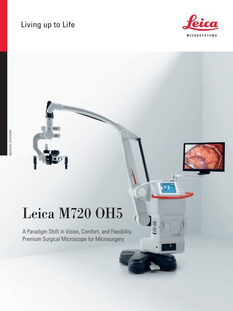
Medical Division<br />
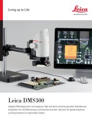
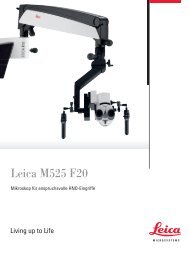
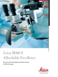
<strong>Leica</strong> <strong>M720</strong> <strong>OH5</strong><br />
A Paradigm Shift in Vision, Comfort, and Flexibility<br />
Premium Surgical Microscope for Microsurgery
A Paradigm<br />
Shift<br />
Comfort through ergonomic design<br />
Brilliant images<br />
Unsurpassed patient safety<br />
Intraoperative fluorescence<br />
Viewing for the entire OR team<br />
Positioning flexibility<br />
Superior maneuverability
<strong>Leica</strong> <strong>M720</strong> <strong>OH5</strong><br />
A Paradigm Shift in Vision,<br />
Comfort, and Flexibility<br />
For years, surgeons have needed a surgical microscope with<br />
smaller, more compact optics. Traditional microscope design<br />
has evolved over the years using large, vertical optical zoom<br />
lens systems, which have inherently limited the surgeon’s<br />
amount of working room, and the ability to work in the<br />
right ergonomic position. With the <strong>Leica</strong> <strong>M720</strong> <strong>OH5</strong>,<br />
<strong>Leica</strong> <strong>Microsystems</strong> writes a revolutionary new chapter<br />
in microscope innovation. At the heart of the innovation:<br />
Horizontal Optics Technology.<br />
The heart of the innovation:<br />
Horizontal Optics Technology<br />
reduces the size of the optical<br />
head and gives the user more<br />
space to work. At the same time<br />
it dramatically increases comfort.
More Space<br />
to Work
<strong>Leica</strong> <strong>M720</strong> <strong>OH5</strong> a paradigm shift in vision, comfort, and flexibility<br />
5<br />
<strong>Leica</strong> <strong>M720</strong> <strong>OH5</strong><br />
Comfort Through<br />
Ergonomic Design<br />
The <strong>Leica</strong> <strong>M720</strong> optical head is the most compact of all<br />
neurosurgical microscopes. Designed along a horizontal plane,<br />
the compact optics carrier helps the surgeon naturally align<br />
for a healthier working posture. Whatever the position of the<br />
patient, even sitting upright, the surgeon can see more, can<br />
work more precisely, and benefits from superior ergonomics.<br />
Compact Horizontal Optics<br />
The sub stantial gain in free working<br />
distance gives the surgeon unobstructed<br />
access to the surgical area, greater<br />
instrument maneuverability, and an<br />
optimal view.<br />
Butterfly Binoculars<br />
<strong>Leica</strong>’s butterfly binoculars accommodate<br />
all body heights, for both the surgeon<br />
and the assistant, as well as the most<br />
challenging surgical positions. The tubes<br />
have an inclination range of 115°, and the<br />
eyepieces swing to a second viewing<br />
plane quickly and easily.<br />
SpeedSpot <br />
Two laser beams act as a focusing<br />
reference to quickly provide a defined<br />
focus point for all three viewing ports<br />
(surgeon, assistant, and camera).
Light<br />
Where You<br />
Need It
<strong>Leica</strong> <strong>M720</strong> <strong>OH5</strong> a paradigm shift in vision, comfort, and flexibility<br />
7<br />
<strong>Leica</strong> <strong>M720</strong> <strong>OH5</strong><br />
Brilliant Images<br />
Enhanced 3D Images:<br />
Depth per ception is improved thanks<br />
to <strong>Leica</strong>’s large stereo base of 24 mm,<br />
creating a more true-to-life 3D effect<br />
compared to other microscopes.<br />
The <strong>Leica</strong> <strong>M720</strong> <strong>OH5</strong> is equipped with Small Angle<br />
Illumination (SAI) to distribute more light to the bottom of<br />
deep cavities. SAI provides a concentrated light beam,<br />
closely aligned to the optical axis.<br />
Combined with outstanding <strong>Leica</strong> APO OptiChrome optics,<br />
the result is significantly improved depth perception and<br />
better light penetration, specifically for new surgical access<br />
techniques such as intra-tracheal, transsphenoidal or METRx .<br />
Images have outstanding contrast, brilliance, sharpness,<br />
resolution, and color fidelity.<br />
Small Angle Illumination (SAI)<br />
SAI distributes light more evenly, and<br />
reduces shadows in the surgical field,<br />
providing:<br />
Conventional microscope illumination,<br />
WD 400 mm<br />
<strong>Leica</strong> Microscope with SAI<br />
WD 400 mm<br />
Deeper light penetration<br />
Increased detail visibility<br />
Improved depth perception<br />
e.g. transsphenoidal surgery, general illustration
1<br />
Safety<br />
Without<br />
Compromise<br />
2 3
<strong>Leica</strong> <strong>M720</strong> <strong>OH5</strong> a paradigm shift in vision, comfort, and flexibility<br />
9<br />
<strong>Leica</strong> <strong>M720</strong> <strong>OH5</strong><br />
Unsurpassed Patient Safety<br />
Dual independent light sources [1]:<br />
The <strong>Leica</strong> <strong>M720</strong> <strong>OH5</strong> features two completely<br />
independent 400 W xenon arc-lamp illumination<br />
systems, giving the surgeon confidence to know<br />
that surgery will not be jeopardized due to lamp or<br />
board failure.<br />
The <strong>Leica</strong> <strong>M720</strong> <strong>OH5</strong> offers innovative illumination solutions<br />
to improve outcomes for both the surgeon and the patient.<br />
Fast system reboot: If the power cable becomes<br />
disconnected for any reason, the system reboots in<br />
the fastest reset time available today.<br />
Illumination Setting<br />
Illumination Setting<br />
Intuitive user controls [2]: The graphical user<br />
interface and hard keys allow the user to<br />
conveniently and intuitively control all microscope<br />
functions during surgery.<br />
Max. illumination<br />
Max. illumination<br />
(BrightCare Plus<br />
inactive)<br />
Microscope with<br />
BrightCare Plus<br />
activated<br />
Conventional<br />
microscope at low<br />
magnification<br />
Conventional<br />
microscope at high<br />
magnification<br />
<strong>Leica</strong> Microscope<br />
with AutoIris <br />
Independent microscope controls: Stand, video,<br />
light, and microscope controls work independently.<br />
For example, should the video system fail, surgery<br />
can continue because the light and microscope are<br />
unaffected.<br />
Antimicrobial surface coating [3]: <strong>Leica</strong>’s<br />
AgProtect limits user exposure to surface<br />
pathogens. This nano silver coating covers the<br />
microscope’s outside surfaces and penetrates the<br />
membranes of microbes to prevent replication.<br />
Long working<br />
distance.<br />
Decreased working<br />
distance at<br />
same illumination<br />
setting (left)<br />
creates burn<br />
potential in<br />
conventional<br />
microscopes.<br />
BrightCare Plus<br />
automatically<br />
adapts light<br />
intensity to the<br />
working distance,<br />
providing safer<br />
illumination (up<br />
to 60% reduction<br />
of intensity).<br />
At low magnification,<br />
the field of<br />
illumination (yellow)<br />
fills the field<br />
of view (green)<br />
completely.<br />
Previously, as<br />
magnification<br />
increased, the<br />
field of view became<br />
smaller, but<br />
the illumination<br />
outside the field<br />
of view could<br />
potentially cause<br />
tissue burns (red).<br />
AutoIris automatically<br />
works with the<br />
zoom, decreasing the<br />
field of illumination<br />
as the field of view<br />
decreases. There is<br />
no peripheral illumination<br />
to cause<br />
tissue burns outside<br />
the field of view.<br />
BrightCare Plus – Light Intensity<br />
BrightCare Plus optimizes the light<br />
intensity relative to the working distance.<br />
As working distance decreases, the<br />
light intensity is reduced automatically,<br />
minimizing incidents of patient burns.<br />
As working distance increases, the light<br />
intensity rises again accordingly.<br />
AutoIris – Light Diameter<br />
AutoIris automatically adjusts the<br />
diaphragm so that only the visible area is<br />
illuminated. When zoomed in, the light<br />
circle adapts automatically: the higher<br />
the magnification, the smaller the light<br />
circle. This prevents the possibility of<br />
drying or burning exposed tissue, outside<br />
of the actual field of view.
Invisible<br />
Becomes<br />
Visible
<strong>Leica</strong> <strong>M720</strong> <strong>OH5</strong> a paradigm shift in vision, comfort, and flexibility<br />
11<br />
<strong>Leica</strong> <strong>M720</strong> <strong>OH5</strong><br />
Intraoperative Fluorescence<br />
Surgical fluorescence: The study of<br />
fluorescence microscopy has a long<br />
tradition at <strong>Leica</strong> <strong>Microsystems</strong>, dating<br />
back to the beginning of the 20th century.<br />
An indispensable component in biological<br />
research, fluorescence science is now an<br />
integral part of the surgical workflow to<br />
improve the patient’s quality of life.<br />
Fluorescence technologies provide intraoperative information<br />
to the surgeon and OR team, directly through the microscope<br />
eyepieces or on a monitor. The information gained allows the<br />
surgeon to make faster progress in work, increase surgical<br />
precision, and improve patient outcomes. Switching between<br />
white light and fluorescence mode requires only the push of a<br />
button on the hand grip or foot control. The <strong>Leica</strong> <strong>M720</strong> <strong>OH5</strong><br />
is well prepared for new and future types of surgical<br />
fluorescence, with a selectable third fluorescence mode.<br />
Oncological Fluorescence<br />
<strong>Leica</strong> FL400* intraoperative module is<br />
used in conjunction with 5-ALA<br />
fluorescent agent to show tumor cells,<br />
and thus enables much higher accuracy<br />
with tumor resection.<br />
Vascular Fluorescence<br />
<strong>Leica</strong> FL800* intraoperative videoangiography<br />
module is used in conjunction<br />
with ICG fluorescent agent and allows<br />
surgeons to see blood flow through<br />
vessels in real time during surgery.<br />
* Please check the status of <strong>Leica</strong> FL400<br />
and <strong>Leica</strong> FL800 regulatory approval<br />
for your country with your local<br />
<strong>Leica</strong> <strong>Microsystems</strong> representative.<br />
Malignant glioma, blue-violet light mode<br />
ICG injection after 9 seconds: venous view
12<br />
<strong>Leica</strong> HD C100 Camera<br />
The <strong>Leica</strong> high-definition medical-grade<br />
camera delivers bright, sharp pictures<br />
and videos, and features an innovative<br />
one-touch control button for easy use.<br />
<strong>Leica</strong> Video Adapters<br />
<strong>Leica</strong> HD video adapters offer<br />
intraoperative fine focus and manual<br />
or remote control, to always achieve<br />
crisp and clear image quality in<br />
documentation.<br />
Integrated HD Monitor<br />
The <strong>Leica</strong> <strong>M720</strong> <strong>OH5</strong> features a builtin,<br />
movable video monitor arm with<br />
three rotation axes and an inclination<br />
axis to easily maneuver the large<br />
video screen into the perfect position<br />
for all viewers.<br />
image acquisition & displ ay<br />
i m age p r oc e s s i ng<br />
HD Documentation Systems<br />
The Med X Change HDMD ® 1080p or<br />
720p is a user-friendly digital recording<br />
system for the surgical environment. The<br />
1080p version records videos in Full HD<br />
and detects ICG automatically. Image<br />
and video files can be transferred to a<br />
USB, external hard drive or wirelessly to<br />
an Apple ® device within seconds.<br />
3D Documentation System<br />
The TrueVision ® 3D Surgical* system<br />
combines 3D visualization and guidance<br />
software applications focused on<br />
improving accuracy, efficiency, and<br />
outcomes for both surgeons and patients.<br />
* Please check the status of TrueVision ® product availability<br />
with your local <strong>Leica</strong> <strong>Microsystems</strong> representative.
<strong>Leica</strong> <strong>M720</strong> <strong>OH5</strong> a paradigm shift in vision, comfort, and flexibility<br />
13<br />
<strong>Leica</strong> <strong>M720</strong> <strong>OH5</strong><br />
Viewing for the OR Team<br />
<strong>Leica</strong> <strong>M720</strong> <strong>OH5</strong> OpenArchitecture allows for easy upgrades<br />
of rapidly evolving imaging technology. User-friendly<br />
operation ensures easy recording and editing of videos and<br />
photos for presentations, teaching, or medical records.<br />
IGS<br />
I m age I n j ec t ion<br />
IGS Integration<br />
The <strong>Leica</strong> <strong>M720</strong> <strong>OH5</strong> includes mechanical<br />
and electronic interfaces to accept and<br />
easily integrate commonly used imageguided<br />
surgery (IGS) systems and their<br />
tool tracking capabilities.<br />
<strong>Leica</strong> DI C700<br />
The <strong>Leica</strong> DI C700 dual imaging color module<br />
allows the surgeon to inject data directly<br />
into the eyepiece, from external and internal<br />
sources, such as MRI, CT, IGS, endoscopes<br />
and <strong>Leica</strong> FL800 video sequences. The data<br />
are displayed with the highest resolution<br />
and contrast currently available.
14<br />
<strong>Leica</strong> <strong>M720</strong> <strong>OH5</strong><br />
Positioning Flexibility to<br />
Suit Surgeon and OR Staff<br />
The <strong>Leica</strong> <strong>M720</strong> <strong>OH5</strong> provides ultimate<br />
positioning flexibility with the highest<br />
overhead clearance and longest reach of any<br />
surgical microscope on the market. Superior<br />
reach and a compact footprint, give the<br />
surgeon positioning flexibility to place the<br />
microscope wherever it is most beneficial for<br />
the surgery. Alternatively, the <strong>Leica</strong> OHC5<br />
ceiling mount option optimizes performance<br />
in space-restricted ORs.<br />
<strong>Leica</strong> Design by Christophe Apothéloz
15<br />
Freedom of<br />
Positioning<br />
Efficiency in Work: The compact base<br />
of the <strong>Leica</strong> <strong>M720</strong> <strong>OH5</strong> creates a smaller<br />
footprint, yet provides superior reach<br />
and ample overhead clearance to work<br />
in comfort during any surgical case.
1 2<br />
3<br />
Precise<br />
Movement<br />
6<br />
4 5 6
<strong>Leica</strong> <strong>M720</strong> <strong>OH5</strong> a paradigm shift in vision, comfort, and flexibility<br />
17<br />
<strong>Leica</strong> <strong>M720</strong> <strong>OH5</strong><br />
Superior Maneuverability<br />
Optics Carrier Tilt [1]: The improved inclination<br />
angle combined with the most compact optical<br />
system provides the surgeon with unmatched<br />
comfort and gives much more flexibility for<br />
transsphenoidal and posterior fossa cases.<br />
Optics Carrier Lateral Movement [2]: With the<br />
longest range of lateral movement available today,<br />
the surgeon can easily achieve the most challenging<br />
lateral approaches.<br />
The <strong>Leica</strong> <strong>M720</strong> <strong>OH5</strong> offers a greatly expanded range of<br />
movement in all dimensions, with intuitive functionality and<br />
minimal vibration at all magnification levels.<br />
ErgoLock [3]: The main surgeon’s binocular tube<br />
can be easily locked in five defined positions,<br />
ensuring stability of an individual’s selected<br />
binocular position, especially when using the mouth<br />
switch.<br />
Mouth Switch (Optional) [4]: The ergonomicallydesigned<br />
mouth switch allows the surgeon to easily<br />
position the microscope while leaving both hands<br />
free for surgery.<br />
Brakes: Silent, high-precision electromagnetic<br />
<strong>Leica</strong> OH technology.<br />
Hand Grip [5]: The ergonomic design and sturdy,<br />
all-metal construction of the hand grip ensure<br />
comfort and stability when moving the microscope.<br />
Foot Control (Optional) [6]: For maximum mobility<br />
and for fast, easy adjustments, <strong>Leica</strong> <strong>Microsystems</strong><br />
offers four models of foot controls: cabled or<br />
wireless, 12-function or 16-function.<br />
AutoBalance [7]<br />
The hard key “AutoBalance“ on the stand<br />
saves valuable time. With only two<br />
pushes of one button, the system fully<br />
balances all six axes quickly and<br />
accurately.<br />
Intraoperative AutoBalance [8]<br />
A microscope may need rebalancing<br />
during surgery due to changing needs for<br />
the surgeon’s and assistant’s positioning.<br />
With one push of the AC/BC button,<br />
conveniently located above the optical<br />
head, the surgeon can rebalance in<br />
seconds, even through the sterile drape.<br />
7 8
<strong>Leica</strong> <strong>M720</strong> <strong>OH5</strong> a paradigm shift in vision, comfort, and flexibility<br />
19<br />
<strong>Leica</strong> <strong>M720</strong> <strong>OH5</strong><br />
Technical Specifications<br />
The <strong>Leica</strong> <strong>M720</strong> <strong>OH5</strong> / OHC5 surgical microscopes feature<br />
innovative Horizontal Optics Technology for more room to<br />
work, a Small Angle Illumination system for better depth<br />
perception, and an OpenArchitecture platform to integrate<br />
the newest imaging technologies such as Full HD.<br />
Electrical Data<br />
Power connection<br />
Safety class<br />
1600 VA 50/60 Hz<br />
100 V (+10 % / −15 %), 120 V (+10 % / −15 %),<br />
220 V (+10 % / −15 %), 240 V (+10 % / −15 %)<br />
Class I<br />
<strong>Leica</strong> <strong>M720</strong> Microscope<br />
Magnification APO OptiChrome -6:1 zoom, motorized<br />
Revolutionary new optical concept with horizontal zoom for<br />
maximum compactness of the microscope<br />
Focus<br />
Eyepieces<br />
Objective<br />
Illumination<br />
Main light source<br />
Emergency lamp<br />
AutoIris <br />
BrightCare Plus<br />
SpeedSpot <br />
Binocular tubes<br />
ErgoLock <br />
Motorized via multifocal lens, with manual adjustment<br />
Widefield eyepieces for eyeglass wearers, 10× for main surgeon<br />
and opposite assistant, 12.5× for lateral assistant, dioptric<br />
settings ±5 with adjustable eye cup<br />
APO OptiChrome multifocal lens<br />
200 mm to 500 mm variable working distance through motorized<br />
function, with manual override<br />
Continuously adjustable illumination field diameter with<br />
gaussian-shaped light distribution; continuously adjustable<br />
brightness at a constant color temperature<br />
High-performance 400 Watt xenon arc-lamp through fiber optic<br />
400-Watt xenon arc-lamp on a separate electrical system<br />
Built-in, automatic, zoom-synchronized illumination field diameter,<br />
with manual override and reset feature<br />
Safety feature for the working distance-synchronized light control<br />
Dual laser focusing device for fast, precise microscope positioning<br />
Binocular tubes feature flexible butterfly ergonomic height<br />
adjustment for optimal body position at the microscope; 115°<br />
variable angle: 0° to 115° range for main surgeon, –55° to +60°<br />
for opposite assistant<br />
Built-in locking device to hold main surgeon’s binocular tube fixed<br />
in five predefined angles: 10°, 35°, 65°, 90°, and 115°<br />
Compact dimensions<br />
Only 72 mm minimal height from the main surgeon’s binocular<br />
to the objective, with the microscope in a horizontal position<br />
Only 232 mm minimal length from the main surgeon’s binocular<br />
to the objective, with the microscope in posterior fossa seated<br />
patient position<br />
Surface coating Covered with antimicrobial coating (AgProtect )<br />
Optical Data<br />
Magnification range<br />
Field of view<br />
1.5× to 17.0× with 10× eyepiece<br />
12.5 mm to 143 mm with 10× eyepiece<br />
Microscope Carrier<br />
Rotation of optics 540°<br />
Lateral tilt<br />
50° to left / 50° to right<br />
Inclination tilt −30° to +150°<br />
XY speed<br />
Zoom-correlated XY speed<br />
Balancing<br />
A, B, C, and D axes are fully automatic, each can be manually<br />
balanced<br />
Intraoperative<br />
balancing<br />
Brakes<br />
Indicator<br />
AC/BC button for automatic intraoperative re-balancing of the<br />
A and C axes, and for re-balancing the B and C axes<br />
One brake for A/B axis, one brake for C axis<br />
LED for fluorescence mode status, LED for video recording status
20<br />
Accessories (optional)<br />
Second observer Stereo attachment to beam splitter for second observer<br />
Binocular tube Variable angle 30° to 150° for second observer<br />
Video adapter <strong>Leica</strong> Manual Video Adapter (MVA), 55 mm, 70 mm, 107 mm focal<br />
length, c-mount, with fine focus<br />
<strong>Leica</strong> Remote Video Adapter (RVA), 55 mm, 70 mm, 107 mm focal<br />
length, c-mount, with fine focus<br />
<strong>Leica</strong> Zoom Video Adapter (ZVA), 3:1 zoom, 35 mm to 100 mm<br />
focal length, c-mount, with fine focus<br />
<strong>Leica</strong> NIR Dual Video Adapter (DVA), 60.5 mm, 79.5 mm focal<br />
length, c-mount, with fine focus<br />
Autofocus<br />
Image injection<br />
Asepsis<br />
Laser<br />
IGS<br />
Interface /<br />
Compatibility<br />
The <strong>Leica</strong> Video-Analysis Autofocus gives the surgeon more<br />
precision and greater control by means of keeping the image<br />
crisp and clear.<br />
<strong>Leica</strong> DI C700 high-resolution, true color dual imaging module<br />
for correlated and non correlated data display, resolution<br />
1024 × 768 pixels, color depth 24 bit, gray scale 256,<br />
contrast >= 1:300, color temperature 2500° – 9000° K<br />
Sterilizable protective glass cover for the objective, sterilizable<br />
components for all drive knobs, commercially available drapes<br />
(specifically designed for the <strong>Leica</strong> <strong>M720</strong>)<br />
Laser micromanipulator available from 3rd party<br />
Open architecture for IGS systems<br />
Controls<br />
Integration of<br />
documentation<br />
Connectors<br />
Carrier for monitor<br />
Materials<br />
10-function pistol grips for zoom, focus, all-free release of six<br />
brakes. Side button to control three user-defined brakes,<br />
motorized lateral tilt and inclination (XY), and <strong>Leica</strong> DI C700<br />
functions. Except for the all-free button, all functions are freely<br />
programmable.<br />
Mouth switch for three brakes (XYZ) (optional)<br />
12-function foot pedal with controls arranged longitudinally or<br />
transversely, 16-function foot pedal with controls arranged<br />
transversely, wired or wireless (optional)<br />
Hand switch (optional)<br />
Prepared for integration of video and digital recording systems.<br />
Open architecture<br />
Numerous built-in connectors for video, IGS, and control data<br />
transfer<br />
12 Volt DC, 19 Volt DC, and AC connections<br />
700 mm long<br />
Flexible arm with 4 axes for rotation and inclination to carry<br />
optional video monitor<br />
All-solid metal construction<br />
Surface coating Covered with antimicrobial coating (AgProtect )<br />
Range cantilever<br />
Load<br />
Weight<br />
Storage dimensions<br />
Max. 1925 mm<br />
Min. 8.0 kg and max. 11.7 kg of accessories to the microscope<br />
Approx. 310 kg as a fully configured system<br />
1945 mm (height) × 1395 mm (width) × 830 mm (depth)<br />
Fluorescence* (OPtional)<br />
Vascular fluorescence Optional <strong>Leica</strong> FL800 is available in the USA, EU, and most other<br />
countries<br />
Oncological<br />
fluorescence<br />
Optional <strong>Leica</strong> FL400 is available in the EU, and some other<br />
countries<br />
* Please check the status of regulatory approval for your country with your local<br />
<strong>Leica</strong> <strong>Microsystems</strong> representative.<br />
<strong>Leica</strong> <strong>OH5</strong> Floor Stand<br />
Type<br />
Floor stand with six electromagnetic brakes<br />
Base<br />
720 mm × 720 mm with four 360° rotatable casters of 130 mm<br />
diameter each; one central single step foot brake<br />
Balancing<br />
Intraoperative<br />
re-balancing<br />
Swing arm<br />
Floor stand control<br />
unit<br />
Light source<br />
“No brake release” Auto-balance<br />
One button / two pushes for complete, automatic balancing of<br />
stand and optics<br />
AC/BC button for automatic intraoperative re-balancing of AC axis<br />
and BC axis<br />
Patented advanced movement system for perfect balance in six<br />
axes, vibration-dissipating technology<br />
New generation touch panel technology. The latest electronics<br />
control for the continuous operation of all motorized functions and<br />
illumination intensity. Data displayed via LCD. Built-in BrightCare<br />
Plus technology for working distance synchronized illumination<br />
control. ISUS Intelligent Setup System, menu selection based<br />
on unique software for user-specific configuration, with built-in<br />
electronic auto-diagnosis and user support. Software-independent<br />
hard keys for illumination and autobalancing; indicator for main /<br />
backup illumination and fluorescence mode. Open architecture for<br />
future software developments.<br />
400-Watt dual xenon arc-lamp illumination system and built-in,<br />
automatic (after notice), lamp quick changer<br />
Ambient conditions<br />
In use +10° C to +40° C (+50° F to +104° F)<br />
30 % to 95 % relative humidity<br />
500 mbar to 1060 mbar atmospheric pressure<br />
Storage −40° C to +70° C (−40° F to +158° F)<br />
10 % to 100 % relative humidity<br />
500 mbar to 1060 mbar atmospheric pressure<br />
Limitations of use<br />
The <strong>Leica</strong> <strong>M720</strong> <strong>OH5</strong> surgical microscope may be used only in closed rooms and must be<br />
placed on a solid floor. It may not be used in Ophthalmology.<br />
Conformity<br />
Council Directive 93/42/EEC on Medical Devices (MDD) and its amendments. Classification:<br />
Class I, in compliance with Annex IX, rule 1 and rule 12 of the directive.<br />
Medical Electrical Equipment, Part 1: General Requirements for Safety IEC 60601-1;<br />
EN 60601-1; UL60601-1; CAN/CSA-C22.2 NO. 601.1-M90.<br />
Electromagnetic compatibility IEC 60601-1-2; EN 60601-1-2.<br />
The Medical Division, within <strong>Leica</strong> <strong>Microsystems</strong> (Schweiz) AG, holds the management<br />
system certificates for the international standards ISO 9001, ISO 13485, and ISO 14001<br />
relating to quality management, quality assurance and environmental management.<br />
› Apple is a trademark of Apple Inc., registered in the U.S. and other countries.<br />
› HDMD and Med X Change are trademarks of Med X Change Inc., registered in the U.S.<br />
and other countries.<br />
› METRx is a trademark of Medtronic Inc., registered in the U.S. and other countries.<br />
› TrueVision is a trademark of TrueVision 3D Surgical Inc., registered in the U.S. and<br />
other countries.
R560 **<br />
<strong>Leica</strong> <strong>M720</strong> <strong>OH5</strong> a paradigm shift in vision, comfort, and flexibility<br />
21<br />
<strong>Leica</strong> <strong>M720</strong> <strong>OH5</strong><br />
2740<br />
1395<br />
1945<br />
1840<br />
720×720<br />
930<br />
R1480 *<br />
6 – Ø 18<br />
60<br />
485<br />
1235<br />
1785<br />
<strong>Leica</strong> <strong>M720</strong> OHC5<br />
Concrete ceiling<br />
∅ 630<br />
450 – 1350<br />
Suspended ceiling<br />
Ø 470<br />
Ø 530<br />
(358)<br />
Center of <strong>Leica</strong> OHC<br />
930<br />
1280<br />
1980<br />
2955<br />
Floor
A Paradigm Shift<br />
in Vision, Comfort,<br />
and Flexibility
www.leica-microsystems.com<br />
The fruitful collaboration ”with the user, for the user“ has always been the<br />
foundation of <strong>Leica</strong> <strong>Microsystems</strong>‘ innovative strength. On this basis, we<br />
have developed our five corporate values: Pioneering, High-end Quality,<br />
Team Spirit, Dedication to Science, and Continuous Improvement.<br />
MeDICaL DIvISIOn<br />
What does a surgeon expect from an outstanding surgical microscope?<br />
Sharp, clear images, and a modular system aligned with the surgeon and<br />
OR staff needs.<br />
Innovations for your practice<br />
From the first surgical microscope with widefield optics in the 1980s to the<br />
first microscopes with Horizontal Optics and with LeD illumination, <strong>Leica</strong><br />
<strong>Microsystems</strong> has been at the forefront of innovation in the development of<br />
surgical microscopes.<br />
HD video, fluorescence and retinal viewing systems also demonstrate the<br />
continued innovative nature of the <strong>Leica</strong> team. We strive to provide the<br />
surgeon with leading edge technology to enhance performance, surgeon<br />
comfort, and patient outcomes.<br />
<strong>Leica</strong> <strong>Microsystems</strong> – an international company with a strong network of<br />
worldwide customer services:<br />
Active worldwide Tel. Fax<br />
USA ∙ Buffalo Grove/Illinois +1 800 248 0123 +1 847 405 0164<br />
Canada ∙ Concord/Ontario +1 800 248 0123 +1 847 405 0164<br />
Australia ∙ North Ryde/NSW +61 2 8870 3500 +61 2 9878 1055<br />
Austria ∙ Vienna +43 1 486 80 50 0 +43 1 486 80 50 30<br />
Belgium ∙ Diegem +32 2 790 98 50 +32 2 790 98 68<br />
Denmark ∙ Ballerup +45 4454 0101 +45 4454 0111<br />
France ∙ Nanterre Cedex +33 811 000 664 +33 1 56 05 23 23<br />
Germany ∙ Wetzlar +49 64 41 29 40 00 +49 64 41 29 41 55<br />
Italy ∙ Milan +39 02 574 861 +39 02 574 03392<br />
Netherlands ∙ Rijswijk +31 70 4132 100 +31 70 4132 109<br />
Portugal ∙ Lisbon +351 21 388 9112 +351 21 385 4668<br />
Spain ∙ Barcelona +34 900 210 992 +34 93 494 95 40<br />
Sweden ∙ Kista +46 8 625 45 45 +46 8 625 45 10<br />
Switzerland ∙ Heerbrugg +41 71 726 34 34 +41 71 726 34 44<br />
United Kingdom ∙ Milton Keynes +44 800 298 2344 +44 1908 246 312<br />
China ∙ Hong Kong +852 2 564 6699 +852 2 564 4163<br />
∙ Shanghai +86 21 6387 6606 +86 21 6387 6698<br />
Japan ∙ Tokyo +81 3 5421 2800 +81 3 5421 2896<br />
Korea ∙ Seoul +82 2 514 65 43 +82 2 514 65 48<br />
Singapore +65 6779 7823 +65 6773 0628<br />
10 M1 721 0en/00 • Copyright © by <strong>Leica</strong> <strong>Microsystems</strong> (Schweiz) AG, Medical Division, CH-9435 Heerbrugg, 2013 • Printed in Switzerland – IV.2013 – galledia – Subject to modifications.<br />
LEICA and the <strong>Leica</strong> Logo are registered trademarks of <strong>Leica</strong> <strong>Microsystems</strong> IR GmbH.