Pan Arab Journal of Oncology - Arab Medical Association Against ...
Pan Arab Journal of Oncology - Arab Medical Association Against ...
Pan Arab Journal of Oncology - Arab Medical Association Against ...

You also want an ePaper? Increase the reach of your titles
YUMPU automatically turns print PDFs into web optimized ePapers that Google loves.
Table 1: Tumor characteristics <strong>of</strong> the 10 patients included in this study:<br />
Characteristics<br />
Tumor site<br />
Left breast<br />
Right breast<br />
For Tumors located at a distance < 4 cm<br />
Proximal depth tumor (cm)<br />
Mean<br />
Std dev.<br />
Distal depth tumor (cm)<br />
Mean<br />
Std dev<br />
For Tumors located at a distance > 4 cm<br />
Proximal depth tumor (cm)<br />
Mean<br />
Std dev.<br />
.No<br />
4<br />
6<br />
0.5<br />
+/- 0.5<br />
3.6<br />
+/- 0.4<br />
1.4<br />
+/- 1.1<br />
Seeds implanted around the resection cavity by the surgeon or defined by<br />
the tumor cavity. To account for treatment set-up uncertainties and breathing<br />
motion the boost planning treatment volume (PTV) was defined as a 1.0-cm<br />
expansion <strong>of</strong> the CTV. The prescribed dose was 10 Gy in five daily fractions<br />
given concomitant at the last week <strong>of</strong> treatment.<br />
All patients were panned by 2 techniques: 3D-conformal fields and electron<br />
beam with calculating the dose to the CTV, PTV and organ at risk.<br />
3D-conformal static fields<br />
Multiple static fields ( 2 to 4 fields ) were used with a similar beam arrangement<br />
as in the conformal plans with simple field conformation to the PTV. All static<br />
fields included an enhanced dynamic wedge (EDW). Plans were calculated with<br />
the pencil beam algorithm based on the work by Sturchi et al. [7, 8].<br />
Distal depth tumor (cm)<br />
Mean<br />
Std dev<br />
For Tumors located at a distance < 4 cm<br />
CTV (cc)<br />
Mean<br />
Std dev<br />
For Tumors located at a distance > 4 cm<br />
CTV (cc)<br />
Mean<br />
Std dev<br />
For Tumors located at a distance < 4 cm<br />
6.4<br />
+/- 1.5<br />
19.8<br />
+/- 9.1<br />
64.96<br />
+/- 41.8<br />
Electron beams<br />
The boost was planned with a single conformal portal. The beam energy was<br />
selected in order to comply with the dosimetric goal mentioned above. The entry<br />
angle was selected so that the entrance surface was approximately perpendicular<br />
to the beam central axis Seven patients were planned with 12 MeV electron<br />
beam and three with 16MeV, respectively. The dose distribution was computed<br />
with the Generalized Gaussian Pencil Beam model [9, 10].<br />
PTV (cc)<br />
Mean<br />
Std dev<br />
For Tumors located at a distance > 4 cm<br />
45.9<br />
+/- 5.3<br />
PTV (cc)<br />
Mean<br />
Std dev<br />
142.1<br />
+/- 87<br />
Std dev, standard deviation; CTV, clinical target volume; PTV, planning target<br />
volume<br />
The planning CT <strong>of</strong> the breast region in a free-breathing setting was performed<br />
postoperatively with the patient in treatment position (i.e., patient on a breast<br />
board, lying supine, and with the ipsi-lateral arm above the head), radiopaque<br />
contrast material was placed at the superior, inferior, medial and lateral limits <strong>of</strong><br />
the clinically palpable breast tissue, cuts were taken using a Semins Tomoscan<br />
AV Helical CT scanner. CT images were acquired in 5 mm slice intervals from<br />
the mandible through the lung bases. The anatomic information from the CT<br />
scan was used to define the target volume and normal structures at risk.<br />
The following organs at risk (OARs) were outlined: ipsi-lateral and contralateral<br />
breasts and lungs, heart, and the skin covering the ipsi-lateral breast (a<br />
5-mm thick segment on the breast surface). In all cases surgical clips were placed<br />
by the surgeon (IR) surrounding the tumor cavity at the time <strong>of</strong> lumpectomy.<br />
All patients were first treated with 6 MV photon beams to the entire breast with<br />
two tangential fields. A total dose <strong>of</strong> 50 Gy in 25 daily fractions during 5 weeks<br />
was delivered. The boost clinical target volume (CTV) was defined as the area <strong>of</strong><br />
architectural distortion inside the breast (i.e., tumor bed) surrounded by metallic<br />
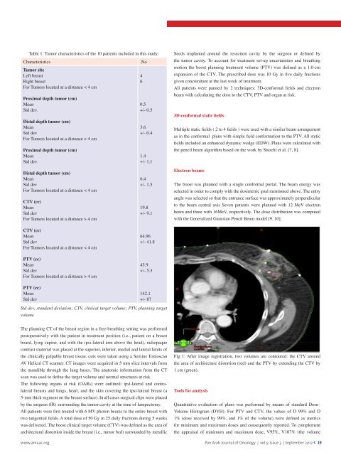
Fig 1: After image registration, two volumes are contoured: the CTV around<br />
the area <strong>of</strong> architecture distortion (red) and the PTV by extending the CTV by<br />
1 cm (green).<br />
Tools for analysis<br />
Quantitative evaluation <strong>of</strong> plans was performed by means <strong>of</strong> standard Dose–<br />
Volume Histogram (DVH). For PTV and CTV, the values <strong>of</strong> D 99% and D<br />
1% (dose received by 99%, and 1% <strong>of</strong> the volume) were defined as metrics<br />
for minimum and maximum doses and consequently reported. To complement<br />
the appraisal <strong>of</strong> minimum and maximum dose, V95%, V107% (the volume<br />
www.amaac.org <strong>Pan</strong> <strong>Arab</strong> <strong>Journal</strong> <strong>of</strong> <strong>Oncology</strong> | vol 5; issue 3 | September 2012 < 19