Pan Arab Journal of Oncology - Arab Medical Association Against ...
Pan Arab Journal of Oncology - Arab Medical Association Against ...
Pan Arab Journal of Oncology - Arab Medical Association Against ...

Create successful ePaper yourself
Turn your PDF publications into a flip-book with our unique Google optimized e-Paper software.
original article <<br />
percentage within PTV was calculated for all patients by subtracting the<br />
minimum from the maximum dose <strong>of</strong> the PTV. Sparing <strong>of</strong> OARs was assessed<br />
using the mean dose for contralateral parotid, cochlea & oral cavity & the<br />
maximum point dose <strong>of</strong> spinal cord, brain stem, lenses, and optic nerves.<br />
Statistical analysis: For all patients, these dose volume parameters were<br />
recorded and analyzed statistically using excel sheet 2003 (table 1).<br />
Summary for the dose distribution in ten cases <strong>of</strong> parotid gland cancer<br />
For planning target volume (PTV)<br />
Regarding PTV dose coverage; the average <strong>of</strong> minimum dose to PTV is 57<br />
Gy and the average <strong>of</strong> maximum dose is 66 Gy but the percentage <strong>of</strong> the dose<br />
inhomogeneity within PTV is 15%. Regarding PTV dose conformity; 95%<br />
isodose wash closely match the shape <strong>of</strong> PTV.<br />
For organs at risk (OAR)<br />
The average <strong>of</strong> the mean dose to contralateral parotid, oral cavity, ipsilateral,<br />
contralateral cochlea and both eyes is 8Gy, 36Gy, 15Gy, 4Gy and 120cGy<br />
respectively. The average <strong>of</strong> the maximum point dose to spinal cord, brain<br />
stem, both lenses and right and left optic nerve is 32Gy, 21Gy, 180cGy, 180cGy<br />
& 120cGy respectively. All values are far less than their tolerance. This is<br />
confirmed by DVH <strong>of</strong> one case shown in figure 3. As the dose to eyes, lenses and<br />
optic nerves are comparable for all patients so we did not list it in the table we<br />
just mentioned the average.<br />
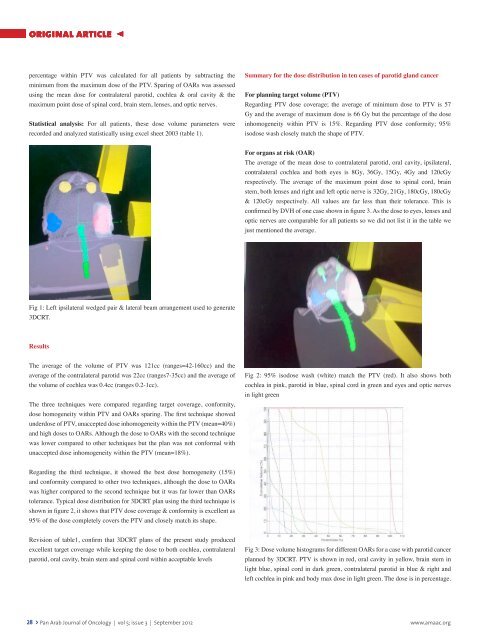
Fig 1: Left ipsilateral wedged pair & lateral beam arrangement used to generate<br />
3DCRT.<br />
Results<br />
The average <strong>of</strong> the volume <strong>of</strong> PTV was 121cc (ranges=42-160cc) and the<br />
average <strong>of</strong> the contralateral parotid was 22cc (ranges7-35cc) and the average <strong>of</strong><br />
the volume <strong>of</strong> cochlea was 0.4cc (ranges 0.2-1cc).<br />
The three techniques were compared regarding target coverage, conformity,<br />
dose homogeneity within PTV and OARs sparing. The first technique showed<br />
underdose <strong>of</strong> PTV, unaccepted dose inhomogeneity within the PTV (mean=40%)<br />
and high doses to OARs. Although the dose to OARs with the second technique<br />
was lower compared to other techniques but the plan was not conformal with<br />
unaccepted dose inhomogeneity within the PTV (mean=18%).<br />
Fig 2: 95% isodose wash (white) match the PTV (red). It also shows both<br />
cochlea in pink, parotid in blue, spinal cord in green and eyes and optic nerves<br />
in light green<br />
Regarding the third technique, it showed the best dose homogeneity (15%)<br />
and conformity compared to other two techniques, although the dose to OARs<br />
was higher compared to the second technique but it was far lower than OARs<br />
tolerance. Typical dose distribution for 3DCRT plan using the third technique is<br />
shown in figure 2, it shows that PTV dose coverage & conformity is excellent as<br />
95% <strong>of</strong> the dose completely covers the PTV and closely match its shape.<br />
Revision <strong>of</strong> table1, confirm that 3DCRT plans <strong>of</strong> the present study produced<br />
excellent target coverage while keeping the dose to both cochlea, contralateral<br />
parotid, oral cavity, brain stem and spinal cord within acceptable levels<br />
Fig 3: Dose volume histograms for different OARs for a case with parotid cancer<br />
planned by 3DCRT. PTV is shown in red, oral cavity in yellow, brain stem in<br />
light blue, spinal cord in dark green, contralateral parotid in blue & right and<br />
left cochlea in pink and body max dose in light green. The dose is in percentage.<br />
28 > <strong>Pan</strong> <strong>Arab</strong> <strong>Journal</strong> <strong>of</strong> <strong>Oncology</strong> | vol 5; issue 3 | September 2012<br />
www.amaac.org