Liability in Obstetrical and Gynecologic Ultrasound - Cmebyplaza.com
Liability in Obstetrical and Gynecologic Ultrasound - Cmebyplaza.com
Liability in Obstetrical and Gynecologic Ultrasound - Cmebyplaza.com

Create successful ePaper yourself
Turn your PDF publications into a flip-book with our unique Google optimized e-Paper software.
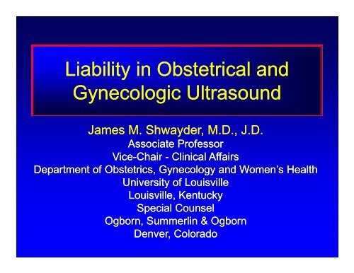
<strong>Liability</strong> <strong>in</strong> <strong>Obstetrical</strong> <strong>and</strong><br />
<strong>Gynecologic</strong> <strong>Ultrasound</strong><br />
James M. Shwayder, M.D., J.D.<br />
Associate Professor<br />
Vice-Chair<br />
i - Cl<strong>in</strong>ical i<br />
l Affairs<br />
Department of Obstetrics, Gynecology <strong>and</strong> Women’s Health<br />
University of Louisville<br />
Louisville, Kentucky<br />
Special Counsel<br />
Ogborn, Summerl<strong>in</strong> & Ogborn<br />
Denver, Colorado
<strong>Liability</strong> <strong>in</strong> <strong>Obstetrical</strong> <strong>and</strong><br />
<strong>Gynecologic</strong> <strong>Ultrasound</strong><br />
James M. Shwayder, M.D., J.D.<br />
Has no significant f<strong>in</strong>ancial <strong>in</strong>terest or other<br />
relationship p( (1) with any manufacturer or (2) with<br />
any <strong>com</strong>mercial product discussed <strong>in</strong> this<br />
presentation.
Outl<strong>in</strong>e<br />
• Def<strong>in</strong>e malpractice, as it relates to<br />
ultrasound<br />
• Legal concepts<br />
• Common types of litigation<br />
• Errors that lead to litigation<br />
• Case examples
Legal Concept<br />
Malpractice<br />
Elements of Negligence<br />
1. Duty<br />
2. Breach of that duty<br />
3. Proximate cause of <strong>in</strong>jury<br />
4. Damages
Legal Concepts<br />
• Wrongful Birth<br />
• Wrongful Life<br />
• Wrongful Death
Wrongful Birth<br />
“A claim for relief by parents who allege they<br />
would have avoided conception or would have<br />
term<strong>in</strong>ated a pregnancy but for the negligence<br />
of those charged with prenatal test<strong>in</strong>g, genetic<br />
prognosticat<strong>in</strong>g, or counsel<strong>in</strong>g parents as to the<br />
likelihood of giv<strong>in</strong>g birth to a physically or<br />
mentally impaired child.<br />
”<br />
Keel v. Banach, , 624 So. 2d 1022 (Ala. 1993)
Wrongful Life<br />
A cause of action for wrongful life<br />
arises <strong>in</strong> favor of a special needs child<br />
who claims damages because he was<br />
conceived or was not aborted due to<br />
the negligence gg<br />
of the py<br />
physician.<br />
Kimble, 55 Ala. Law 84 (1994)
Wrongful Death<br />
A cause of action for wrongful death<br />
arises when an otherwise normal<br />
pregnancy, which has reached viability,<br />
is term<strong>in</strong>ated t as a result of a<br />
misdiagnosis.<br />
– i.e. renal agenesis<br />
Lollar v. Tankersley, , 613 So. 2d 1249 (Ala. 1993)
Cases by Specialty Area<br />
180<br />
160<br />
177<br />
140 OB<br />
120<br />
Gyn<br />
100<br />
Abdom<strong>in</strong>al<br />
80 Neonatal<br />
60 63<br />
62<br />
Breast<br />
60<br />
Eye<br />
40<br />
11<br />
29<br />
Misc<br />
20<br />
1<br />
9<br />
0<br />
1983 1986 1996 2002<br />
RC S<strong>and</strong>ers. J <strong>Ultrasound</strong> Med 2003; 22: 1009-15. 15.
Types of Litigation<br />
• Missed diagnosis<br />
• Mis<strong>in</strong>terpreted sonograms<br />
• Invented lesions<br />
• Delay y[ [or failure] <strong>in</strong> <strong>com</strong>munication<br />
• Failure to perform ultrasound<br />
• Fraud cases<br />
• Procedure-related related cases<br />
• Sonographer-related related cases<br />
RC S<strong>and</strong>ers. J <strong>Ultrasound</strong> Med 2003; 22: 1009-15. 15.
Missed Diagnosis<br />
40<br />
35<br />
30<br />
25<br />
20<br />
15<br />
10<br />
5<br />
Ectopic pregnancy<br />
Fetal anomaly<br />
Multiple pregnancy<br />
IUGR<br />
Ovarian Mass<br />
0<br />
1983 1986 1996 2002<br />
RC S<strong>and</strong>ers. J <strong>Ultrasound</strong> Med 2003; 22: 1009-15. 15.
Missed Diagnosis<br />
New Jersey<br />
• Four ultrasounds performed dur<strong>in</strong>g pregnancy.<br />
• Images lacked clear anatomic l<strong>and</strong>marks, thus<br />
no accurate measurements of fetus made<br />
• Physician reviewed one ultrasound<br />
• Sonographer reported on three ultrasounds<br />
– “Structural irregularities that require further<br />
evaluation”<br />
• Physician told the patient the “ultrasounds were<br />
<strong>com</strong>pletely normal”
Missed Diagnosis<br />
New Jersey<br />
• Midl<strong>in</strong>e facial defect<br />
• Cleft palate<br />
• Club foot<br />
• Lower-limb anomalies<br />
• Limited cognitive <strong>and</strong><br />
<strong>com</strong>munication skills
Missed Diagnosis<br />
New Jersey<br />
• Suit aga<strong>in</strong>st physician<br />
i<br />
• Suit aga<strong>in</strong>st professional group he<br />
owned<br />
• Performs ultrasounds<br />
• Settlement = $1.98 million
Delay <strong>in</strong> Diagnosis<br />
North Carol<strong>in</strong>a<br />
• 46 year old patient presented with<br />
abnormal uter<strong>in</strong>e bleed<strong>in</strong>g<br />
• Physician assistant saw patient<br />
• No biopsy performed<br />
• <strong>Ultrasound</strong> = negative<br />
- Subsequently could not produce<br />
photograph taken at the time of<br />
ultrasound
Delay <strong>in</strong> Diagnosis<br />
North Carol<strong>in</strong>a<br />
• 18 months later presented with<br />
persistent bleed<strong>in</strong>g<br />
• Physician assistant aga<strong>in</strong> saw patient<br />
• No biopsy performed<br />
• <strong>Ultrasound</strong> = negative<br />
– Photograph for second ultrasound<br />
found: showed existence of tumor
Delay <strong>in</strong> Diagnosis<br />
i<br />
North Carol<strong>in</strong>a<br />
• After another 10 months, sought care<br />
from another physician<br />
• Physician performed biopsy<br />
• Endometrial carc<strong>in</strong>oma<br />
• Patient died from disease
Delay <strong>in</strong> Diagnosis<br />
i<br />
North Carol<strong>in</strong>a<br />
• Suit filed aga<strong>in</strong>st 1<br />
st physician<br />
– After defendant physician’s<br />
deposition<br />
– No expert testimony required<br />
• Settled for $800,000
Legal Concepts<br />
• Res ipsa loquitur<br />
– But for the failure to exercise due care the<br />
<strong>in</strong>jury would not have occurred<br />
• Delay <strong>in</strong> diagnosis <strong>and</strong> subsequent death<br />
• Respondeat superior<br />
– An employer is liable for the wrong of an<br />
employee if it was <strong>com</strong>mitted with<strong>in</strong> the<br />
scope of employment
Mis<strong>in</strong>terpreted Images<br />
8 Misdated fetus<br />
7<br />
6<br />
5<br />
4<br />
3<br />
2<br />
1<br />
0<br />
1983 1986 1996 2002<br />
Fetal anomaly<br />
Size underestimated<br />
Miscalled ovarian<br />
cancer<br />
Bladder called<br />
ovarian cyst<br />
Decidual cast called<br />
gestational sac<br />
RC S<strong>and</strong>ers. J <strong>Ultrasound</strong> Med 2003; 22: 1009-15. 15.
<strong>Ultrasound</strong> – Dat<strong>in</strong>g Error<br />
• Patient was 20 weeks pregnant by dates<br />
• Scan EGA = 17 week size. Dates not<br />
adjusted.<br />
• Repeat C/S done at 39 weeks (36 weeks)<br />
• Good APGAR’s. 6 ½ lbs <strong>in</strong>fant to term<br />
nursery.<br />
• Mother anxious for discharge.<br />
• Both discharged on Day # 2.<br />
Case courtesy of Steven Warsof, M.D.
<strong>Ultrasound</strong> – Dat<strong>in</strong>g Error<br />
• Infant missed f/u Bilirub<strong>in</strong> check.<br />
Developed Kernicterus<br />
• Has long term permanent neurologic<br />
disability<br />
• Case settled for $4,000,000 aga<strong>in</strong>st<br />
hospital, pediatrician i i <strong>and</strong> Obstetrician<br />
t i<br />
prior to trial<br />
Case courtesy of Steven Warsof, M.D.
US adjusted EGA<br />
New York Case<br />
• Referred for evaluation of fluid<br />
• 43 week IUP misdiagnosed as be<strong>in</strong>g 37<br />
weeks<br />
• Post-dates fetus with oxygen deprivation<br />
– O 2 deprivation = paraplegic <strong>and</strong> speechless<br />
• $27 million award
US adjusted EGA<br />
New York Case<br />
• $1.4 million for special education<br />
• $65,000 annually until patient is 73<br />
• Lump sum payments of $100,000,<br />
000<br />
$200,000, <strong>and</strong> $300,000 at ages 5, 10,<br />
15<br />
• $13 million aga<strong>in</strong>st Director of <strong>Ultrasound</strong>
<strong>Ultrasound</strong> - <strong>Liability</strong><br />
• Failure to conduct additional test<strong>in</strong>g upon<br />
<strong>in</strong>ability to visualize all four chambers of<br />
the heart dur<strong>in</strong>g a rout<strong>in</strong>e sonogram<br />
• $4,200,000<br />
• Failure to detect men<strong>in</strong>gomyelocele on<br />
ultrasound at 15 weeks. <strong>Ultrasound</strong><br />
reported as normal. (coupled with lack of<br />
AFP test<strong>in</strong>g)<br />
• $4,350,000<br />
• Failure to detect severe hydrocephalus<br />
• $5,500,000
Invented Lesions<br />
4<br />
3<br />
2<br />
1<br />
0<br />
1983 1986 1996 2002<br />
Fetal abnormality<br />
IUGR<br />
Fetal Death<br />
Normal Pregnancy<br />
called Ectopic: MTX<br />
RC S<strong>and</strong>ers. J <strong>Ultrasound</strong> Med 2003; 22: 1009-15. 15.
“Ectopic Pregnancy”<br />
• 34 y.o. G1P0 presents to ED with c/o<br />
abdom<strong>in</strong>al pa<strong>in</strong> <strong>and</strong> vag<strong>in</strong>al bleed<strong>in</strong>g.<br />
• Underwent IVF ~ 2 weeks earlier<br />
• hCG = 4,654
“Ectopic Pregnancy”<br />
<strong>Ultrasound</strong> <strong>in</strong> radiology<br />
“Uterus normal sized with a thickened<br />
decidual reaction <strong>in</strong> the uterus. No fetal<br />
pole is identified. There is a moderate<br />
amount of fluid <strong>in</strong> the cul-de-sac. There is a<br />
right adnexal mass = 2.2 x 1.9 x 2.1 cm.<br />
These f<strong>in</strong>d<strong>in</strong>gs could be <strong>com</strong>patible with the<br />
presence of an ectopic. Cl<strong>in</strong>ical i l correlation<br />
<strong>and</strong>, if <strong>in</strong>dicated, serial hCG levels <strong>and</strong><br />
follow-up ultrasound studies should be<br />
considered.”
“Ectopic Pregnancy”<br />
Patient is cl<strong>in</strong>ically stable<br />
Lab<br />
• Hct = 38.9<br />
• Blood type: O positive<br />
Treatment<br />
t<br />
• Methotrexate: 80 mg IM<br />
• Excellent MTX consent form reviewed <strong>and</strong><br />
signed by patient
“Ectopic Pregnancy”<br />
Quantitative hCG<br />
• Day 0 4,654 (MTX)<br />
• Day 4 16,069<br />
• Day 7 42,125<br />
<strong>Ultrasound</strong><br />
– Tw<strong>in</strong> IUP with two yolk sacs <strong>and</strong> possible<br />
cardiac activity.<br />
– Tw<strong>in</strong> IUP at ~ 5 weeks of gestation
“Ectopic Pregnancy”<br />
<strong>Ultrasound</strong> 2 weeks later<br />
• Tw<strong>in</strong> IUP with two yolk sacs, two<br />
fetuses, both with cardiac activity, c/w 7<br />
weeks of gestation<br />
• Patient referred for counsel<strong>in</strong>g re: risks<br />
of fetal anomalies associated with MTX
Tw<strong>in</strong> IUP + MTX<br />
Per<strong>in</strong>atal counsel<strong>in</strong>g<br />
• Risks of MTX very low<br />
• Fetal anomalies associated with MTX<br />
can be seen on ultrasound<br />
Re<strong>com</strong>mendation<br />
• Serial ultrasounds<br />
• Reassurance
Tw<strong>in</strong> IUP + MTX<br />
<strong>Ultrasound</strong> at 16 weeks<br />
• Normally grow<strong>in</strong>g tw<strong>in</strong> gestation with no<br />
abnormalities visualized<br />
• Reassured
Tw<strong>in</strong> IUP + MTX<br />
26 weeks – Per<strong>in</strong>atologist B<br />
• <strong>Ultrasound</strong><br />
– Shortened limbs<br />
– Small ch<strong>in</strong>s<br />
– One fetus: echogenic bowel<br />
– One fetus: 2 vessel cord<br />
Genetic counsel<strong>in</strong>g<br />
• Potential risk of MTX exposure<br />
• Greatest risk at 6-8 weeks after conception
Tw<strong>in</strong> IUP + MTX<br />
Delivered by C-section<br />
• Hypotonia<br />
• Micrognathia<br />
• Short limbs<br />
• Dysmorphic facies<br />
Growth <strong>and</strong> development<br />
• Feed<strong>in</strong>g difficulties<br />
• Growth delays<br />
• Developmental delays
Tw<strong>in</strong> IUP + MTX<br />
Suit filed aga<strong>in</strong>st<br />
• Radiologist<br />
– Misdiagnosis<br />
• REI Gynecologist<br />
– Misdiagnosis<br />
– Inappropriate treatment with MTX<br />
– Wrongful birth<br />
• Per<strong>in</strong>atologist A<br />
– Wrongful Birth
Tw<strong>in</strong> IUP + MTX<br />
Trial<br />
Ti Pla<strong>in</strong>tiff<br />
• With h/o IVF, tw<strong>in</strong> gestation more likely<br />
• Thus, high level of hCG without<br />
demonstrable IUP is not un<strong>com</strong>mon<br />
• Patient was stable, thus immediate<br />
<strong>in</strong>tervention was unnecessary<br />
• If follow-up hCG <strong>and</strong> ultrasounds would<br />
have been obta<strong>in</strong>ed, the correct diagnosis<br />
of a IU tw<strong>in</strong> gestation would have been<br />
made
Tw<strong>in</strong> IUP + MTX<br />
Pla<strong>in</strong>tiff<br />
Trial<br />
• MTX was the proximate cause of the<br />
observed fetal anomalies<br />
• Per<strong>in</strong>atologist A was negligent <strong>in</strong><br />
provid<strong>in</strong>g <strong>in</strong>adequate <strong>and</strong> <strong>in</strong>accurate<br />
counsel<strong>in</strong>g as to the risks of MTX.<br />
• Had the patient been appropriately<br />
counseled she would have term<strong>in</strong>ated the<br />
pregnancy
Tw<strong>in</strong> IUP + MTX<br />
Defense<br />
Trial<br />
• The orig<strong>in</strong>al ultrasound was <strong>in</strong>terpreted by<br />
the radiologist<br />
• REI-gyn<br />
– Relied upon the radiologist’s diagnosis<br />
• Radiologist<br />
– The <strong>in</strong>terpretation of the ultrasound was<br />
correct, particularly <strong>in</strong> light of the hCG levels.<br />
F/U re<strong>com</strong>mendations were appropriate.
Defense<br />
Tw<strong>in</strong> IUP + MTX<br />
Trial<br />
Ti • Use of methotrexate t t for treatment t t of<br />
suspected ectopic pregnancy is with<strong>in</strong> the<br />
SOC<br />
• The risk of fetal anomalies with MTX is<br />
low<br />
• The patient received appropriate<br />
counsel<strong>in</strong>g <strong>and</strong> signed a written consent<br />
for use of MTX
MTX <strong>and</strong> Anomalies<br />
Am<strong>in</strong>opter<strong>in</strong>/MTX Syndrome<br />
• Dose effect (threshold)<br />
• Tim<strong>in</strong>g<br />
– > 10 mg/week<br />
–2-2.52.5 weeks<br />
• Undifferentiated cells<br />
• All or none effect (SAB)<br />
–4-10 weeks (6-8 weeks)<br />
• Effect on differentiat<strong>in</strong>g cells<br />
Clayton et al. Obstet Gynecol 2006;107:598-604.<br />
604.
MTX <strong>and</strong> Anomalies<br />
Effects of Methotrexate<br />
• IUGR<br />
• Abn head shape<br />
• Larger fontanelles<br />
• Craniosynostosis<br />
• Ocular<br />
hypertelorism<br />
• Low set ears<br />
• Micrognathia<br />
• Limb abnormalities<br />
• Developmental<br />
delays<br />
Our Babies<br />
• Hypotonia<br />
• Micrognathia<br />
• Short limbs<br />
• Dysmorphic facies<br />
• Feed<strong>in</strong>g difficulties<br />
• Growth delays<br />
• Developmental<br />
delays
Tw<strong>in</strong> IUP + MTX<br />
Defense<br />
Trial<br />
• Use of methotrexate for treatment of<br />
suspected ectopic pregnancy is with<strong>in</strong> the<br />
SOC<br />
• The risk of fetal anomalies with MTX is<br />
low<br />
• The patient received appropriate<br />
counsel<strong>in</strong>g <strong>and</strong> signed a written consent<br />
for use of MTX
You cannot consent a<br />
patient to negligence<br />
Judge Harry Re<strong>in</strong>, M.D. J.D.<br />
Florida
Tw<strong>in</strong> IUP + MTX<br />
Trial<br />
Defense<br />
• <strong>Ultrasound</strong> is useful <strong>in</strong> detect<strong>in</strong>g<br />
potential fetal anomalies<br />
• The ultrasound at 16 weeks was normal<br />
• This was a highly desired pregnancy<br />
<strong>and</strong> it is likely that the patient would not<br />
have term<strong>in</strong>ated the pregnancy even if<br />
abnormalities were visualized
Tw<strong>in</strong> IUP + MTX<br />
Defense<br />
ee Trial<br />
• When abnormalities were identified<br />
at 26 weeks the patient still had the<br />
option of term<strong>in</strong>at<strong>in</strong>g pregnancy<br />
• The fetal anomalies seen can occur<br />
even without exposure to MTX
What was the verdict for the<br />
parties?
Tw<strong>in</strong> IUP + MTX<br />
Radiologist<br />
• Defense verdict<br />
Verdict
Tw<strong>in</strong> IUP + MTX<br />
REI<br />
– Pla<strong>in</strong>tiff verdict<br />
Verdict<br />
– Misdiagnosis of ectopic pregnancy/tw<strong>in</strong><br />
gestation<br />
– Negligent <strong>in</strong> the use of MTX
Tw<strong>in</strong> IUP + MTX<br />
• Per<strong>in</strong>atologist A<br />
– Pla<strong>in</strong>tiff verdict<br />
Verdict<br />
– Negligent counsel<strong>in</strong>g<br />
– Wrongful birth
Tw<strong>in</strong> IUP + MTX<br />
Verdict<br />
• Jo<strong>in</strong>t <strong>and</strong> Severally Liable<br />
– Pa<strong>in</strong> <strong>and</strong> suffer<strong>in</strong>g<br />
– Long-term support <strong>and</strong> therapy of two<br />
<strong>in</strong>fants with anticipated life-span of 72<br />
years<br />
• $73 million
Types of Errors<br />
• Perception errors<br />
• Interpretation errors<br />
• Fail<strong>in</strong>g to suggest the next appropriate<br />
procedure<br />
• Failure to <strong>com</strong>municate<br />
M.M. Rask<strong>in</strong>. <strong>Liability</strong> of Radiologists, , <strong>in</strong> Legal Medic<strong>in</strong>e. Am<br />
College of Legal Med. 6 th edition. 456-460. 460.
Perception Errors<br />
The abnormality is seen <strong>in</strong> retrospect but it is<br />
missed when <strong>in</strong>terpret<strong>in</strong>g ti the <strong>in</strong>itial iti study.<br />
• Error rate <strong>in</strong> radiology is ~ 30% 1<br />
• Question: Was it below the st<strong>and</strong>ard d of care<br />
for the physician not to have seen the<br />
abnormality. 2<br />
• Most suits are settled<br />
– 80% are lost if cases go to jury verdict<br />
1 Berl<strong>in</strong> <strong>and</strong> Hendrix. Perceptual Errors <strong>and</strong> Negligence. Am J Roentgenol<br />
1996; 170: 863-67.<br />
67.<br />
2 L. Berl<strong>in</strong>. Malpractice Issues <strong>in</strong> Radiology: Defend<strong>in</strong>g the<br />
“Missed” Radiographic Diagnosis. . Am J Roentgenol 2001; 176: 317-32. 32.
Interpretation Errors<br />
The abnormality is perceived but is <strong>in</strong>correctly<br />
described<br />
• Most often occur due to lack of knowledge<br />
or faulty judgment<br />
– Malignant lesion called benign<br />
– Normal variant is called abnormal<br />
• The best defense is an appropriate<br />
differential diagnosis, preferably <strong>in</strong>clud<strong>in</strong>g<br />
the correct diagnosis<br />
• Lawsuits <strong>in</strong>volv<strong>in</strong>g an <strong>in</strong>terpretation errors<br />
– 75% are won if cases go to jury verdict
Fail<strong>in</strong>g to Suggest the Next<br />
Appropriate Procedure<br />
The prudent radiologist/physician will suggest the next<br />
appropriate study or procedure based upon the<br />
f<strong>in</strong>d<strong>in</strong>gs <strong>and</strong> the cl<strong>in</strong>ical <strong>in</strong>formation.<br />
• The additional studies should add mean<strong>in</strong>gful<br />
<strong>in</strong>formation to clarify, confirm or rule out the <strong>in</strong>itial<br />
impression<br />
• The re<strong>com</strong>mended study should never be for<br />
enhanced referral <strong>in</strong><strong>com</strong>e<br />
• Generally, the radiologist is not expected to follow<br />
up on the re<strong>com</strong>mendation.<br />
– Exception: Beware of re<strong>in</strong>terpret<strong>in</strong>g images on multiple<br />
occasions<br />
1<br />
1 Montgomery v. South County Radiologists, Inc., , 49 S.W.2d 191 (2001).
Failure to Communicate<br />
• F<strong>in</strong>al written report is considered the<br />
def<strong>in</strong>itive means of <strong>com</strong>municat<strong>in</strong>g the<br />
results of an imag<strong>in</strong>g study or procedure<br />
• Direct or personal <strong>com</strong>munication must<br />
occur <strong>in</strong> certa<strong>in</strong> situations<br />
ti<br />
– Document <strong>com</strong>munication<br />
• Cause of action: Failure to <strong>com</strong>municate <strong>in</strong><br />
a timely <strong>and</strong> cl<strong>in</strong>ically appropriate manner<br />
1 M.M. Rask<strong>in</strong>. Why Radiologists Get Sued. Applied Radiol 2001; 30: 9-13.<br />
2 ACR St<strong>and</strong>ard for Communication
Failure to Communicate<br />
• 33 y.o. G3P2002<br />
• Quad screen at 15 weeks<br />
– Risk of Down Syndrome = 1/1100<br />
• US performed at 19w1d <strong>in</strong> radiology<br />
• Reported as “normal”<br />
• No mention of subtle f<strong>in</strong>d<strong>in</strong>gs<br />
– UPJ = 4.3 <strong>and</strong> 4.4<br />
– EIF noted
Failure to Communicate<br />
• 33 y.o. G3P2002<br />
• Down Syndrome<br />
• Claim for Wrongful Birth<br />
– Had the patient known of an <strong>in</strong>creased<br />
risk<br />
– She would have elected on<br />
amniocentesis<br />
– If abnormal, she would have term<strong>in</strong>ated
Likelihood Ratios for DS with<br />
Isolated Markers<br />
Marker AAURA Nyberg Bromley<br />
Smith-<br />
B<strong>in</strong>dman<br />
Nuchal fold 18.6 11 12 17<br />
Hyperechoic<br />
bowel<br />
5.5 6.7 NA 6.1<br />
Short humerus 2.5 5.1 5.8 7.5<br />
Short femur 22 2.2 15 1.5 12 1.2 27 2.7<br />
EIF 2.0 1.8 1.4 2.8<br />
Pyelectasis 15 1.5 15 1.5 15 1.5 19 1.9<br />
Normal 0.4 0.36 0.22 ??
Isolated Marker<br />
• EIF<br />
– LR = 1.4 – 28<br />
2.8<br />
– Adjustment<br />
• Risk of Down’s<br />
– Orig<strong>in</strong>ally 1 <strong>in</strong> 1100<br />
– Adjusted d 1 <strong>in</strong> 392-785<br />
• No amnio
Isolated Marker<br />
• UPJ = 4.3 <strong>and</strong> 4.4<br />
• Pyelectasis<br />
– LR = 1.5 – 1.9<br />
– Adjustment<br />
• Risk of Down’s<br />
– Orig<strong>in</strong>ally 1 <strong>in</strong> 1100<br />
– Adjusted 1 <strong>in</strong> 579-733733<br />
• No amnio
Prevalence of Markers <strong>and</strong><br />
Likelihood Ratios<br />
#<br />
Markers<br />
DS = 164 Nml = 656<br />
LR<br />
0 32 575 0.2<br />
1* 32 66 1.9<br />
2 20 13 62 6.2<br />
3 40 2 80<br />
* Individual LR better<br />
Benacerraf et al. Radiology 1994; 193: 135-140140
Failure to Communicate<br />
• 33 y.o. G3P2002<br />
• Quad screen at 15 weeks<br />
– Risk of Down Syndrome = 1/1100<br />
• 2 markers: LR = 6.2<br />
• Adjusted Risk for DS = 1/177
Failure to Communicate<br />
Defense<br />
• Radiologist<br />
– They round to the nearest whole number.<br />
– This patient’s UPJ’s were thus 4 <strong>and</strong> WNL<br />
– The UPJ dilation was < 5 mm, which is “normal”<br />
<strong>in</strong> their lab<br />
– EIF is a worthless marker <strong>and</strong> of no<br />
consequence<br />
– It is the obstetrician’s duty to re<strong>com</strong>mend<br />
amniocentesis to the patient
Failure to Communicate<br />
Defense<br />
• Obstetrician<br />
– The radiologist’s report was “normal” with no<br />
mention of subtle markers for DS.<br />
– I had no reason to re<strong>com</strong>mend amniocentesis<br />
– Had I known of the subtle f<strong>in</strong>d<strong>in</strong>gs I would have<br />
recalculated the patient’s risk <strong>and</strong> would have<br />
re<strong>com</strong>mended amniocentesis
Failure to Communicate<br />
Radiologist<br />
Defense<br />
– The UPJ dilation was < 5 mm, which is<br />
“normal <strong>in</strong> their lab”<br />
Pla<strong>in</strong>tiff’s cross<br />
– The defendant radiologist had provided<br />
the syllabus from a recently attended CME<br />
course provided by the parent <strong>in</strong>stitution,<br />
that <strong>in</strong>dicated that > 4 mm was abnormal<br />
for < 20 weeks EGA
Failure to Communicate<br />
Radiologist<br />
Defense<br />
– EIF is a worthless marker. We don’t even mention it.<br />
Pla<strong>in</strong>tiff’s expert<br />
– As an isolated f<strong>in</strong>d<strong>in</strong>g, EIF is a very poor marker.<br />
However, it should at least be mentioned <strong>in</strong> the report.<br />
Further, <strong>in</strong> the presence of additional markers, for<br />
example pyelectasis, EIF carries more significance.<br />
– Both subtle f<strong>in</strong>d<strong>in</strong>gs should have been noted <strong>in</strong> the<br />
report <strong>and</strong> re<strong>com</strong>mendations made to recalculate the<br />
patient’s risk for DS <strong>and</strong> amniocentesis if appropriate
Verdict<br />
Obstetrician<br />
• Defense verdict
Verdict<br />
Radiologist<br />
• Pla<strong>in</strong>tiff Verdict<br />
– Mis<strong>in</strong>terpreted the images<br />
– Duty to report the f<strong>in</strong>d<strong>in</strong>gs to the<br />
obstetrician. If he had done so, the duty for<br />
further counsel<strong>in</strong>g, evaluation, <strong>and</strong><br />
treatment would have transferred to the<br />
obstetrician.
Failure to Communicate<br />
Verdict<br />
Pla<strong>in</strong>tiff Verdict<br />
Radiologist<br />
– Fail<strong>in</strong>g to appropriately <strong>com</strong>municate the<br />
f<strong>in</strong>d<strong>in</strong>gs to the obstetrician directly<br />
resulted <strong>in</strong> the cont<strong>in</strong>uation of an<br />
abnormal pregnancy that the patient,<br />
had she known of the abnormality,<br />
would have term<strong>in</strong>ated.
Wrongful Birth<br />
Reed v. Campagnolo<br />
The court ruled that “… parents may<br />
ma<strong>in</strong>ta<strong>in</strong> an action for wrongful birth if the<br />
physician fails to disclose the availability<br />
of tests which would have detected birth<br />
defects present <strong>in</strong> the fetus <strong>and</strong> if the<br />
woman would have had an abortion had<br />
she known the fetus’s deformities”<br />
Reed v. Campagnolo, , 810 F.Supp. 167 (D.Md. 1993)
Failure to Communicate<br />
Judgment Amount<br />
Verdict<br />
• Sealed settlement while the jury was<br />
<strong>in</strong> deliberation
Legal Considerations of U/S<br />
Communication<br />
• Discuss abnormal f<strong>in</strong>d<strong>in</strong>gs with referr<strong>in</strong>g<br />
physician before send<strong>in</strong>g written report<br />
– If unavailable, leave message with staff<br />
– Document phone call or conversation<br />
– Fax report<br />
• If diagnosis is cancer, write “cancer”<br />
– Report is not subject to mis<strong>in</strong>terpretation<br />
• Track abnormal studies for follow-up
Prenatal Screen<strong>in</strong>g<br />
• Increased emphasis on prenatal<br />
screen<strong>in</strong>g <strong>and</strong> diagnosis<br />
i<br />
• Failure to properly counsel can lead to<br />
a claim of wrongful birth
“Screen<strong>in</strong>g for Fetal Chromosomal<br />
Abnormalities”<br />
• Ideally, all women should be offered<br />
aneuploidy screen<strong>in</strong>g before 20 weeks of<br />
gestation, regardless of age.<br />
• A strategy that <strong>in</strong>corporates both first- <strong>and</strong><br />
second-trimester screen<strong>in</strong>g should be<br />
offered to women who seek prenatal care<br />
<strong>in</strong> the first trimester.<br />
ACOG Cl<strong>in</strong>ical Management Guidel<strong>in</strong>es<br />
Number 77, January 2007
1st Trimester Screen<strong>in</strong>g<br />
Nuchal Translucency/Nasal Bone<br />
• Guidel<strong>in</strong>es <strong>and</strong> Quality Assessment<br />
• Nuchal Translucency Quality Review<br />
Program (NTQR)<br />
• Fetal Medic<strong>in</strong>e Foundation<br />
ACOG Practice Bullet<strong>in</strong><br />
Ultrasonography <strong>in</strong> Pregnancy<br />
Number 101, February 2009
Keepsake <strong>Ultrasound</strong>s
“Keepsake” Malpractice<br />
Any malpractice claim concern<strong>in</strong>g<br />
keepsake video production will be a<br />
case of first impression.
Enterta<strong>in</strong>ment <strong>Ultrasound</strong><br />
Case of First Impression<br />
Colorado 2009<br />
• Down’s Syndrome<br />
• Alleged missed anomaly dur<strong>in</strong>g<br />
“Keepsake <strong>Ultrasound</strong>” <strong>in</strong> the 3 rd<br />
trimester
Legal Considerations of <strong>Ultrasound</strong><br />
• Perform US when cl<strong>in</strong>ically <strong>in</strong>dicated<br />
• Must temper “over-utilization”<br />
• Adequately tra<strong>in</strong>ed personnel<br />
• Sonographers<br />
• Physicians<br />
• Perform US <strong>in</strong> accordance with current<br />
guidel<strong>in</strong>es<br />
• Supervision<br />
• Documentation
Legal Considerations of <strong>Ultrasound</strong><br />
• Proper <strong>in</strong>terpretation of the sonogram<br />
•Appropriate p tra<strong>in</strong><strong>in</strong>g <strong>and</strong> referral<br />
• Use modern equipment<br />
• Properly ma<strong>in</strong>ta<strong>in</strong>ed<br />
• Communicate f<strong>in</strong>d<strong>in</strong>gs<br />
• To referr<strong>in</strong>g physician or representative<br />
• To patient, when appropriate<br />
• Formal report should be explicit<br />
• Code properly
Barbaro<br />
2007<br />
Thank You<br />
James M. Shwayder, M.D, J.D.<br />
M<strong>in</strong>d That Bird<br />
2009