1999 QAP Report - Mycology Online
1999 QAP Report - Mycology Online
1999 QAP Report - Mycology Online

Create successful ePaper yourself
Turn your PDF publications into a flip-book with our unique Google optimized e-Paper software.
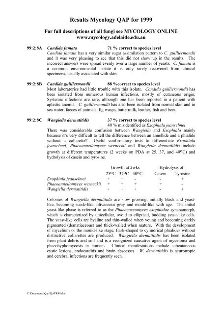
Results <strong>Mycology</strong> <strong>QAP</strong> for <strong>1999</strong><br />
For full descriptions of all fungi see MYCOLOGY ONLINE<br />
www.mycology.adelaide.edu.au<br />
99:2:8A Candida famata 71 % correct to species level<br />
Candida famata has a very similar sugar assimilation pattern to C. guilliermondii<br />
and it was very pleasing to see that this did not show up in the results. The<br />
incorrect answers were spread evenly over a large number of yeasts. C. famata is<br />
a common environmental isolate it is only rarely recovered from clinical<br />
specimens, usually associated with skin.<br />
99:2:8B Candida guilliermondii 88 %correct to species level<br />
Most laboratories had little trouble with this isolate. Candida guilliermondii has<br />
been isolated from numerous human infections, mostly of cutaneous origin.<br />
Systemic infections are rare, although one has been reported in a patient with<br />
aplastic anemia. C. guilliermondii has also been isolated from normal skin and in<br />
sea water, faeces of animals, fig wasps, buttermilk, leather, fish and beer.<br />
99:2:8C Wangiella dermatitidis 37 % correct to species level<br />
40 % misidentified as Exophiala jeanselmei<br />
There was considerable confusion between Wangiella and Exophiala mainly<br />
because it’s very difficult to tell the difference between an annellide and a phialide<br />
without a collarette? Useful confirmatory tests to differentiate Exophiala<br />
jeanselmei, Phaeoannellomyces werneckii and Wangiella dermatitidis include<br />
growth at different temperatures (2 weeks on PDA at 25, 37, and 40 o C) and<br />
hydrolysis of casein and tyrosine.<br />
Growth at 2wks Hydrolysis of<br />
25 o C 37 o C 40 o C Casein Tyrosine<br />
Exophiala jeanselmei + + - - +<br />
Phaeoannellomyces werneckii + + + + -<br />
Wangiella dermatitidis + + + - +<br />
Colonies of Wangiella dermatitidis are slow growing, initially black and yeastlike,<br />
becoming suede-like, olivaceous gray and mould-like with age. The initial<br />
yeast-like phase is referred to as the Phaeococcomyces exophialae synanamorph,<br />
which is characterized by unicellular, ovoid to elliptical, budding yeast-like cells.<br />
The yeast-like cells are hyaline and thin-walled when young and becoming darkly<br />
pigmented (dematiaceous) and thick-walled when mature. With the development<br />
of mycelium or the mould-like stage, flask-shaped to cylindrical phialides without<br />
distinctive collarettes are produced. Wangiella dermatitidis has been isolated<br />
from plant debris and soil and is a recognized causative agent of mycetoma and<br />
phaeohyphomycosis in humans. Clinical manifestations include subcutaneous<br />
cystic lesions, endocarditis and brain abscesses. W. dermatitidis is neurotropic<br />
and cerebral infections are frequently seen.<br />
C:\Documents\Qap\<strong>QAP</strong>R99.doc
99:4:8A Onychocola canadensis 41 % correct to species level<br />
This was the first time this organism was sent out in the <strong>QAP</strong> so the results were<br />
quite good. The incorrect answers were spread evenly over a large number of<br />
yeasts and moulds. Onychocola canadensis is an arthroconidial fungus and a rare<br />
cause of nail and skin infections. Colonies are slow growing, white to yellowish<br />
with a suede-like surface and brownish reverse. Hyphae form long chains of<br />
arthroconidia which are cylindrical to broadly ellipsoidal, 1-2 celled, hyaline to<br />
subhyaline, 4-16 x 2-5 ìm in size. Older cultures form broad, brown, septate,<br />
sterile hyphae with darker brown knobs.<br />
99:4:8B Trichophyton tonsurans 85 % correct to species level<br />
Trichophyton tonsurans has been sent out several times in recent years and<br />
proficiency in identification has increased from ~40% to 85%. T. tonsurans is an<br />
anthropophilic fungus with a worldwide distribution, which causes inflammatory<br />
or chronic non-inflammatory finely scaling lesions of skin, nails and scalp. It is a<br />
common cause of tinea capitis in the Australian Aborigine. Invaded hairs show an<br />
endothrix infection and do not fluoresce under Wood's ultra-violet light.<br />
99:4:8C Trichophyton terrestre 67 % correct to species level<br />
Trichophyton terrestre is a geophilic fungus of worldwide distribution, which may<br />
occur as a saprophytic contaminant on humans and animals. It is not known to<br />
invade hair in vivo, but produces hair perforations in vitro.<br />
99:6:8A Scedosporium prolificans 57 % correct to species level<br />
86 % correct to genus level<br />
30% of respondents called this S. apiospermum. Scedosporium prolificans is<br />
distinguished from other members of the genus, in particular, S. apiospermum, by<br />
having basally swollen (inflated), flask-shaped annellides, slower colony<br />
development on nutrient agar media, and by not growing on media containing<br />
cycloheximide (actidione). Disseminated disease has been reported in<br />
immunosuppressed patients especially those with prolonged neutropenia and posttransplantation<br />
therapy. However, localized invasive infections, especially septic<br />
arthritis and osteomyelitis following penetrating injuries to joints, are now an<br />
emerging clinical problem, accounting for 80% of the reported cases. Culture<br />
identification is important, because this fungus is often resistant to antifungal<br />
therapy and treatment may require surgical intervention.<br />
99:6:8B Absidia corymbifera 57 % correct to species level<br />
73 % correct to genus level<br />
20 % misidentified as Mucor sp.<br />
The genus Absidia is characterized by a differentiation of the hyphae into arched<br />
stolons bearing more or less verticillate sporangiophores at the internode, and<br />
rhizoids formed at the point of contact with the substrate (at the node). This<br />
feature separates species of Absidia from the genus Rhizopus, where the sporangia<br />
arise from the nodes and are therefore found opposite the rhizoids. The sporangia<br />
are relatively small, globose, pyriform- or pear-shaped and are supported by a<br />
characteristic funnel-shaped apophysis. This distinguishes Absidia from the<br />
genera Mucor and Rhizomucor, which have large, globose sporangia without an<br />
apophysis. Absidia currently contains 21 mostly soil-borne species. A.<br />
corymbifera is the only species of Absidia known to cause disease in man and<br />
animals. It is also the only species of Absidia to grow at 40 O C. Absidia<br />
corymbifera is a common human pathogen, causing pulmonary, rhinocerebral,<br />
C:\Documents\Qap\<strong>QAP</strong>R99.doc
disseminated, CNS or cutaneous types of infection. It is also often associated with<br />
animal disease, especially mycotic abortion. A. corymbifera has a world-wide<br />
distribution mostly in association with soil and decaying plant debris.<br />
99:6:8C Rhizomucor pusillus 47 % correct to species level<br />
24 % misidentified as Mucor sp.<br />
Temperature studies are useful here because Rhizomucor pusillus is the only<br />
Mucor look alike that will grow at 45C. The genus Rhizomucor is distinguished<br />
from Mucor by the presence of stolons and poorly developed rhizoids at the base<br />
of the sporangiophores [you will have to look in the agar to see them] and it’s<br />
growth at high temperature. R. pusillus was originally described in the genus<br />
Mucor. It is a rare human pathogen, causing pulmonary, disseminated or<br />
cutaneous types of infection. It is more often associated with animal disease. R.<br />
pusillus has a worldwide distribution and is commonly associated with compost<br />
heaps.<br />
99:8:8A Sporothrix schenckii 59 % correct to species level<br />
22 % misidentified as Acremonium sp.<br />
In Sporothrix, the conidia are formed in clusters on tiny denticles by sympodial<br />
proliferation of the conidiophore, their arrangement often suggestive of a flower.<br />
In Acremonium conidia are usually formed in slimy balls at the apex of awl<br />
shaped phialide. Sporothrix schenckii has a worldwide distribution, particularly in<br />
tropical and temperate regions. It is commonly found in soil and on decaying<br />
vegetation and is a well known pathogen of humans and animals. Sporotrichosis<br />
is primarily a chronic mycotic infection of the cutaneous or subcutaneous tissues<br />
and adjacent lymphatics characterized by nodular lesions, which may suppurate<br />
and ulcerate. Infections are caused by the traumatic implantation of the fungus<br />
into the skin, or very rarely, by inhalation into the lungs. Secondary spread to<br />
articular surfaces, bone and muscle is not infrequent, and the infection may also<br />
occasionally involve the central nervous system, lungs or genitourinary tract.<br />
99:8:8B Aspergillus nidulans 61 % correct to species level<br />
96 % correct to genus level<br />
Colonies are typically plain green in color with dark red-brown cleistothecia<br />
developing within and upon the conidial layer. Reverse may be olive to drab-gray<br />
or purple-brown. Conidial heads are short, columnar (up to 70 x 30 um in<br />
diameter) and biseriate. Conidiophores are usually short, brownish and smoothwalled.<br />
Conidia are globose (3.0-3.5 um in diameter) and rough-walled.<br />
Cleistothecium of Emericella nidulans (teleomorph) show numerous reddishbrown<br />
ascospores and are often surrounded by a mass of Hülle cells which are up<br />
to 25 um in diameter. Aspergillus nidulans is a typical soil fungus with a<br />
worldwide distribution. It has also been reported as a causative agent of<br />
aspergillosis in humans and animals.<br />
99:8:8C Scopulariopsis brevicaulis 60 % correct to species level<br />
93 % correct to genus level<br />
In Scopulariopsis, annellides may be solitary, in groups, or organized into a<br />
distinct penicillus. Conidia are globose to pyriform, usually truncate, with a<br />
rounded distal portion, smooth to rough, and hyaline to brown in color. Most<br />
members of the genus Scopulariopsis are soil fungi, however a few, in particular<br />
S. brevicaulis, have been reported as causative agents of onychomycosis however<br />
it is also a common contaminant isolated from nails.<br />
C:\Documents\Qap\<strong>QAP</strong>R99.doc

![List for QAP Fungi [2006].](https://img.yumpu.com/36263775/1/184x260/list-for-qap-fungi-2006.jpg?quality=85)