Research Groups - Max-Planck-Institut für biophysikalische Chemie
Research Groups - Max-Planck-Institut für biophysikalische Chemie
Research Groups - Max-Planck-Institut für biophysikalische Chemie

Create successful ePaper yourself
Turn your PDF publications into a flip-book with our unique Google optimized e-Paper software.
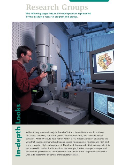
<strong>Research</strong> <strong>Groups</strong><br />
The following pages feature the wide spectrum represented<br />
by the institute’s research program and groups.<br />
In-depth Looks<br />
Without X-ray structural analysis, Francis Crick and James Watson would not have<br />
discovered that DNA, our prime genetic information carrier, has a double helical<br />
structure. And how would have Robert Koch – also a Nobel Laureate – discovered the<br />
virus that causes anthrax without having a good microscope at his disposal? High-end<br />
science requires high-end equipment. Therefore, it is no wonder that so many scientists<br />
are involved in methodical innovations. For example, it takes new spectroscopic and<br />
microscopic procedures to determine structural details at the single molecule level as<br />
well as to explore the dynamics of molecular processes.
NanoBiophotonics<br />
Dr. Stefan W. Hell<br />
Stefan Hell received his doctorate<br />
in physics from the University of<br />
Heidelberg in 1990. From 1991 to<br />
1993, he worked at the European<br />
Molecular Biology Laboratory in<br />
Heidelberg, followed by a 1993 to<br />
1996 stay as a senior researcher at the<br />
University of Turku (Finland), and as a<br />
visiting scientist at Oxford University<br />
(England). In 1996, he received his<br />
›Habilitation‹ from the University of<br />
Heidelberg, where he teaches physics.<br />
In 1997, he joined the <strong>Max</strong> <strong>Planck</strong><br />
<strong>Institut</strong>e for Biophysical Chemistry as a<br />
junior <strong>Max</strong> <strong>Planck</strong> research group<br />
leader. In 2002, he was appointed<br />
director at the <strong>Max</strong> <strong>Planck</strong> <strong>Institut</strong>e for<br />
Biophysical Chemistry, where he heads<br />
the newly established Department of<br />
NanoBiophotonics.<br />
Stefan Hell is the recipient of the Prize<br />
of the International Commission for<br />
Optics (2000), a co-recipient of the<br />
Helmholtz Prize (2001), and of<br />
the Leibinger Innovation award.<br />
In 2002, he received the<br />
Carl Zeiss Prize and the<br />
Karl Heinz Beckurts Prize.<br />
shell@gwdg.de<br />
Bewersdorf, J., R. Pick and S. W. Hell: Multifocal<br />
multiphoton microscopy. Optics Letters 23 (9),<br />
655–657 (1998).<br />
Hell, S.W. and M. Nagorni: 4Pi-confocal<br />
microscopy with alternate interference. Optics<br />
Letters 23, 1567–1569 (1998).<br />
Klar, T.A., S. Jakobs, M. Dyba, A. Egner and<br />
S.W. Hell: Fluorescence microscopy with diffraction<br />
resolution barrier broken by stimulated<br />
emission. Proc. Natl. Acad. Sci. USA 97,<br />
8206–8210 (2000).<br />
Egner, A., S. Jakobs and S.W. Hell: Fast 100-nm<br />
resolution three-dimensional microscope<br />
reveals structural plasticity of mitochondria in<br />
live yeast. Proc. Natl. Acad. Sci. USA 99,<br />
3370–3375 (2002).<br />
Dyba, M. and S.W. Hell: Focal spots of size<br />
λ/23 open up far-field fluorescence microscopy<br />
at 33 nm axial resolution. Phys. Rev. Lett. 88,<br />
163901–163901 (2002).<br />
Would it not be great to observe the cell organelles and<br />
the myriad of proteins in a living cell in 3D with<br />
nanoscale resolution and with a non-invasive<br />
method? Until recently, devising or even building such a microscope<br />
had been a fundamental scientific problem. The reason for<br />
this is that a non-invasive, high-resolution microscope needs to<br />
rely on focused visible light. Fluorescently labeled proteins, nucleic<br />
acids, and lipids can be mapped in a living cell with unmatched<br />
sensitivity. However, the established light microscopy methods suffer<br />
from a critical disadvantage, namely their diffraction-imposed<br />
resolution, which is limited at best to ~500 nm in the direction of<br />
light propagation (z-axis) and ~200 nm in the transverse (xy)<br />
direction. The diffraction barrier was discovered by Abbe in 1873<br />
and has been paradigmatic ever since.<br />
We have recently succeeded in introducing physical concepts that<br />
break Abbe’s barrier. In fact, we intend to explore all the possibilities<br />
of making the microscope sharper. Our primary scientific aim<br />
is to push fluorescence microscopy resolution down to the<br />
nanometer range and to eventually apply it to the field of biology.<br />
This challenging endeavour requires a joint effort of physicists,<br />
chemists, engineers, and biologists alike.<br />
Perhaps the most intuitive method for sharpening the spot is to<br />
selectively inhibit the fluorescence at the circumference by stimulated<br />
emission, a natural competitor to fluorescence. Technically,<br />
the Stimulated Emission Depletion (STED-) microscope relies on<br />
pairs of synchronized laser pulses. The primary excitation pulse<br />
produces a 3D spot of excited molecules of regular diffraction size,<br />
and the immediately following red-shifted STED pulse quenches<br />
the excited molecules down to the ground state. By arranging the<br />
STED pulse in a doughnut mode, only the fluorescence at the<br />
periphery of the spot is inhibited, whereas the very centre ideally<br />
remains untouched. To date, an improvement of factor 3 in the<br />
transverse direction and up to 6 along the optic axis has been<br />
demonstrated (Fig.1). In a similar manner, it is possible to deplete<br />
the ground state of the fluorophore. Both concepts rely on the nonlinear<br />
optical phenomenon of saturation and have the potential to<br />
provide resolution down to the molecular scale.<br />
A powerful way of sharpening the axial resolution of a microscope<br />
is the coherent superposition of focal wavefronts. This idea has<br />
been realized in our 4Pi-microscope for which we use two opposing<br />
objective lenses, adding up their wavefronts to form a 3- to 7-fold<br />
improvement in spatial resolution along the optic axis. The resolution<br />
is further enhanced by the application of (non-linear) image<br />
18 departments and research groups<br />
www.mpibpc.mpg.de/abteilungen/200/
estoration techniques (Fig.2). The<br />
superior 3D-resolution of a 4Piconfocal<br />
microscope is illustrated in<br />
Fig.3, which depicts a 3D-rendered<br />
image of the mitochondrial network<br />
of a live budding yeast cell. The<br />
combination of both microscopes,<br />
namely the STED-4Pi-microscope,<br />
was the first to demonstrate a spatial<br />
resolution in the double-digit nanometer<br />
range (30-40 nm) with visible<br />
light and regular objective lenses.<br />
Fig. 1. Stimulated emission depletion microscope. The diffraction-limited<br />
distribution of excited molecules generated by the green beam is depleted<br />
at the periphery through stimulated emission with the red beam (a). The<br />
resulting fluorescence spot (b) is fundamentally reduced as compared to its<br />
standard counterpart (c).<br />
Fig. 2. Optical section-image through the microtubules of a fixed mousefibroblast<br />
cell, labeled by immunofluorescence. In conjunction with image<br />
restoration, 4Pi-confocal microscopy provides a 3D-resolution in the range<br />
of ∼100 nm.<br />
Fig. 3. 3D-rendered image of the mitochondrial<br />
network of a live yeast cell. The mitochondrial<br />
matrix is labeled with green fluorescent protein,<br />
whereas the cell wall is counterstained with the dye<br />
calcofluor white and displayed by ray tracing.<br />
Live cell 4Pi-imaging with ∼100 nm 3D-resolution<br />
allowed us to study the influence of selected<br />
mitochondrial proteins on mitochondrial<br />
morphology.<br />
departments and research groups 19
Spectroscopy and<br />
Photochemical Kinetics<br />
Professor Hans-Jürgen Troe<br />
Hans-Jürgen Troe received his<br />
doctorate at the University of<br />
Göttingen in 1965, where he also<br />
completed his ›Habilitation‹ in<br />
physical chemistry in 1967. In 1971,<br />
he was appointed Full Professor for<br />
Physical Chemistry at the École<br />
Polytechnique Fédérale de Lausanne.<br />
In 1975, he returned to Göttingen as<br />
director of the <strong>Institut</strong> für Physikalische<br />
<strong>Chemie</strong> of the University. Since 1990, he<br />
has also been director of the Department<br />
of Spectroscopy and Photochemical<br />
Kinetics at the <strong>Max</strong> <strong>Planck</strong> <strong>Institut</strong>e for<br />
Biophysical Chemistry.<br />
Hans-Jürgen Troe has received honorary<br />
doctorates from the Universities of<br />
Bordeaux and Karlsruhe, and he is an<br />
Honorary Professor of the École<br />
Polytechnique Fédérale de Lausanne. In<br />
addition, he has received many awards, is<br />
a member of several scientific academies<br />
and currently serves as a member of<br />
the Senate of the Deutsche<br />
Forschungsgemeinschaft.<br />
Internal research groups:<br />
Prof. Walter Hack,<br />
Dr. Günter Käb<br />
Dr. Thomas Lenzer<br />
Prof. Klaus Luther<br />
Dr. Derek Marsh<br />
Prof. Jörg Schroeder<br />
Prof. Dirk Schwarzer<br />
Dr. Simone Techert<br />
Dr. <strong>Max</strong> Teubner<br />
Dr. Jomo Walla<br />
Dr. Klaas Zachariasse<br />
Independent research group:<br />
Prof. Michael Stuke<br />
Contact person:<br />
Dirk Schwarzer<br />
dschwar@gwdg.de<br />
Many phenomena in the animate and inanimate realms<br />
of nature can be traced back to molecular processes. In<br />
the atmosphere, molecules are continuously created by<br />
collisions of reactive particles, while absorption of light or heat<br />
energy makes them dissociate once again. Surprisingly, these<br />
atmospheric reactions resemble those occurring both in flames and<br />
in internal combustion engines. Solvents play an important role in<br />
chemical reactions in fluid environments, whereas molecular packing<br />
density is the decisive factor for solid-state reactions. For larger<br />
molecules, which include such biopolymers as proteins and<br />
nucleic acids, internal molecular dynamics make an additional<br />
contribution to the interactions between single molecules.<br />
The time dependence of chemical reactions, also known as reaction<br />
kinetics, is determined by intermolecular interactions as well<br />
as by processes occurring within the molecule itself. This ensures<br />
that the energy transfer is used to effect changes in the structure,<br />
and thus, the function of the molecule. Therefore, the visualization<br />
of molecular processes is an important prerequisite for understanding<br />
nature.<br />
Molecular spectroscopy enables us to take this close-up look at<br />
nature: thus, it is possible for us to identify different molecular<br />
species, but also those that differ merely in structure or energy<br />
An iodine molecule is split with a<br />
laser pulse; 100 picoseconds later,<br />
the diffraction pattern of an x-ray<br />
pulse reveals how the iodine<br />
atoms manage to recombine.<br />
20 departments and research groups<br />
www.mpibpc.mpg.de/abteilungen/010/
state, by their spectra. In addition, molecules can be<br />
excited photochemically by the absorption of light<br />
energy. This procedure makes it possible for us to<br />
follow the photochemical kinetics by spectroscopy:<br />
one pulse of light initiates the reaction, and a second<br />
pulse interrogates the response.<br />
A main focus is the dynamics of chemical reactions in<br />
different environments: We study this phenomenon in<br />
isolated molecules as well as in molecular aggregates,<br />
gases, supercritical fluids and liquids, and also in<br />
crystals and membranes. The experiments are conducted<br />
under a wide range of conditions – temperatures<br />
vary from a few degrees absolute up to<br />
thousands of degrees, pressures range from a few<br />
thousandths of an atmosphere up to several thousand<br />
atmospheres, and we also work with high magnetic<br />
fields and different types of solvents. This enables us<br />
to unravel the various interdependent factors that<br />
play a part in the course of a chemical reaction. Not<br />
only do the spectra make it possible to distinguish different<br />
molecules and their energy states, they also<br />
yield important information about the effect of the<br />
surroundings. Modern experimental methods permit<br />
us to achieve a time resolution that extends down to<br />
about ten femtoseconds (the one hundred-millionth<br />
part of a millionth of a second). This is the timescale<br />
on which the motions of atoms in molecules occur. In<br />
this way, the internal dynamics of a molecule can be<br />
›seen‹ directly. Numerical simulations allow the observed<br />
phenomenon to be reproduced by molecular<br />
dynamics on the computer.<br />
In molecular rearrangements, the crossing of energy<br />
barriers is of central importance: in the transition<br />
from reactants to products of a chemical reaction,<br />
often such a barrier must be overcome first, rather<br />
like crossing a mountain pass between two valleys.<br />
The height of the barrier may be modified either by<br />
specific structural changes in the molecule or by its<br />
Diiodomethane in chloroform<br />
– snapshot from a molecular<br />
dynamics computer simulation<br />
(iodine atoms: violet; chlorine<br />
atoms: orange).<br />
departments and research groups 21
environment. In this process, the solvent can act as a<br />
viscous medium and oppose the crossing of the<br />
barrier. The dynamics of such events can be followed<br />
directly with ultra short light pulses.<br />
Our group also investigates the transport of electrons,<br />
protons and hydrogen atoms within the molecule.<br />
Time-resolved absorption and emission spectroscopy<br />
allow us to study the influence of<br />
structure and<br />
energetics on<br />
fluorescence<br />
quenching,<br />
on charge<br />
separation,<br />
of spin-labeled biomolecules it is possible to study<br />
dynamic processes in membranes, lipid-protein interactions,<br />
and essential aspects of membrane assembly<br />
in considerable molecular<br />
detail.<br />
Molecular structures<br />
can also<br />
be determined<br />
by high-resolution<br />
x-ray<br />
diffraction<br />
methods.<br />
Such experiments<br />
are<br />
made with a<br />
pulsed x-ray<br />
beam that is<br />
Laser spectroscopy<br />
allows the study of<br />
energy transport:<br />
The dye azulene<br />
(red) is excited by a<br />
laser pulse. Within<br />
four picoseconds a<br />
large part of the<br />
absorbed energy is<br />
transmitted to the<br />
anthracene chromophore<br />
(blue) via<br />
the hydrocarbon<br />
chain (green).<br />
and charge recombination. To know the mechanism<br />
for fluorescence quenching in detail is one of the<br />
essential prerequisites for the use of fluorescent dyes<br />
as probes in biological systems, such as in the analysis<br />
of DNA.<br />
Biochemical reactions in cell membranes are also controlled<br />
by molecular dynamics. Electron spin resonance<br />
spectroscopy is a highly sensitive biophysical<br />
tool for the study of these processes. Through the use<br />
synchronized with the light pulse that starts the<br />
photochemical reaction. Pilot studies with simple<br />
reactions in the liquid or solid state have yielded<br />
promising results.<br />
Innovative experiments, numerical simulations and<br />
theoretical studies of the physicochemical principles<br />
of molecular processes complement each other in our<br />
aim of reaching a quantitative understanding of<br />
molecular dynamics.<br />
Marsh, D., and L.I. Horváth: Structure, dynamics and composition of the<br />
lipid-protein interface. Perspectives from spin-labelling; Biochim. Biophys.<br />
Acta 1376, 267 (1998).<br />
Heidelbach, C., V.S. Vikhrenko, D. Schwarzer, I.I. Fedchenia and J. Schroeder:<br />
Molecular dynamics simulation of vibrational energy relaxation of highly<br />
excited molecules in fluids. III. Equilibrium simulations of vibrational energy<br />
relaxation of azulene in carbon dioxide. J. Chem. Phys. 111, 8022 (1999).<br />
Harding, L.B., A.I. Maergoiz, J. Troe, and V.G. Ushakov: Statistical rate theory<br />
for the HO + O ⇔ HO 2 ⇔ H + O 2 reaction system: SACM/CT calculations<br />
between 0 and 5000 K. J. Chem. Phys. 113, 11019 (2000).<br />
Charvat, A., J. Aßmann, B. Abel, D. Schwarzer, K. Henning, K. Luther and J.<br />
Troe : Direkt observation of intramolecular vibrational energy redistribution of<br />
selectively excited CH 2 I 2 and C 3 H 5 I molecules in solution. Phys. Chem. Chem.<br />
Phys. 3, 2230 (2001).<br />
Daum, R., S. Druzhinin, D. Ernst, L. Rupp, J. Schroeder, and K.A.<br />
Zachariasse : Fluorescence excitation spectra of jet-cooled 4-(diisopropylamino)benzonitril<br />
and related compounds. Chem. Phys. Lett. 341, 272<br />
(2001).<br />
Neutze, R., R.Wouts, S.Techert, A.Kirrander, J.Davidson, M.Kocsis, F.Schotte,<br />
and M.Wulff : Visualizing photochemical dynamics in solution through<br />
picosecond x-ray scattering. Phys. Rev. Lett. 87, 195508 (2001).<br />
22 departments and research groups www.mpibpc.mpg.de/abteilungen/010/
Biochemical Kinetics<br />
Alack of perfection during the reproduction process – that<br />
is the main reason for the fascinating variety of life<br />
forms on earth. Although DNA, our genetic information<br />
carrier, is an extremely stable macromolecule, errors sometimes do<br />
occur when information is copied and duplicated. Since not all of<br />
these errors are corrected and repaired, new variants emerge all the<br />
time. These variants form the basis for the evolution of organisms.<br />
All living species are subject to these transformations.<br />
We can define evolution as a natural phenomenon, but we may<br />
also re-enact it in a controlled setting to meet our own needs. In<br />
these experiments, bacteria, viruses, or single nucleic acid molecules<br />
are encouraged to reproduce, and then selected according to<br />
specific criteria. For this purpose, we have developed special<br />
equipment that can process many thousand samples simultaneously.<br />
In this way, we can study basic mechanisms of evolution,<br />
e. g. the tricks that HIV and other cunning pathogens use to outsmart<br />
our immune system. Moreover, such »evolution machines«<br />
can help developing the molecular agents needed for novel drugs.<br />
These different applications have one crucial aspect in common: In<br />
all of these cases, tiny quantities of substances need to be analyzed.<br />
This amounts to »finding a needle in the haystack.« It also<br />
goes for diagnostic applications, such as the early diagnosis of BSE.<br />
Special spectroscopic methods enable us to detect even single molecules.<br />
This has lead to the development of evolutive biotechniques<br />
that permit the recognition of single biomolecules in a specific<br />
distribution as well as the optimization of their function.<br />
Professor Manfred Eigen<br />
Manfred Eigen received his doctorate<br />
from the University of Göttingen in 1951,<br />
where he subsequently worked at the<br />
<strong>Institut</strong>e of Physical Chemistry. After joining<br />
the <strong>Max</strong> <strong>Planck</strong> <strong>Institut</strong>e for Physical<br />
Chemistry in 1953, he became director<br />
of its Chemical Kinetics department in<br />
1964. On Professor Eigen’s suggestion,<br />
this institute became part of today’s<br />
<strong>Max</strong> <strong>Planck</strong> <strong>Institut</strong>e for Biophysical<br />
Chemistry in 1971, where he has been<br />
director of the department of<br />
Biochemical Kinetics. Even after his<br />
retirement in 1995, he has been<br />
active to the present day. In 1967,<br />
Manfred Eigen was awarded<br />
the Nobel Prize for Chemistry<br />
for his research on ultra-fast<br />
chemical reactions<br />
in solutions.<br />
Eigen, M.: Molekulare Diagnostik. In: Das Gen<br />
und der Mensch. (Ed.) G. Gottschalk.<br />
Wallstein-Verlag, Göttingen, 2000, 137–167.<br />
Eigen, M.: Natural selection: a phase transition?<br />
Biophys. Chem. 85 (2-3), 101–123 (2000).<br />
Koltermann, A., U. Kettling, J. Bieschke, T.<br />
Winkler, M. Eigen: Rapid assay processing by<br />
integration of dual-color fluorescence crosscorrelation<br />
spectroscopy: High throughput screening<br />
for enzyme activity. Proc. Natl. Acad. Sci.<br />
USA 95, 1421–1426 (1998).<br />
Eigen, M.: Steps towards life, a perspective on<br />
evolution. Oxford University Press, Oxford,<br />
1992.<br />
Brakmann, S., U. Kettling, und F. Oehlenschläger:<br />
Die Evolutive Biotechnologie und ihre<br />
Perspektiven. Biologie in unserer Zeit 6,<br />
355–366 (1995).<br />
Automated evolution and screening device of DIREVO Biosystems AG/<br />
Cologne, a biotech company that grew out of the Department of<br />
Biochemical Kinetics at the <strong>Max</strong> <strong>Planck</strong> <strong>Institut</strong>e for Biophysical Chemistry.<br />
www.mpibpc.mpg.de/abteilungen/081/<br />
departments and research groups 23