Lab 2: Endocrine Anatomy & Histology
Lab 2: Endocrine Anatomy & Histology
Lab 2: Endocrine Anatomy & Histology

You also want an ePaper? Increase the reach of your titles
YUMPU automatically turns print PDFs into web optimized ePapers that Google loves.
1<br />
<strong>Lab</strong> 2: <strong>Endocrine</strong> <strong>Anatomy</strong> & <strong>Histology</strong><br />
Reading<br />
LABPAQ: <strong>Endocrine</strong> System pages 14-34<br />
Objectives<br />
1. To identify the major endocrine glands and tissues of the body.<br />
2. To identify the histology of the major endocrine glands and relate their structure<br />
to their function. Identify the major endocrine glands and tissues of the body.<br />
3. Relate each endocrine gland to the hormone(s) it produces.<br />
4. Explain how hormones work to maintain homeostasis in the body.<br />
Introduction<br />
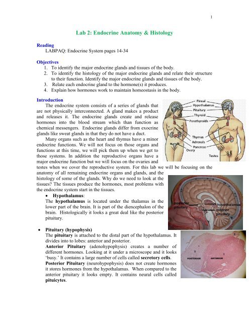
The endocrine system consists of a series of glands that<br />
are not physically interconnected. A gland makes a product<br />
and releases it. The endocrine glands create and release<br />
hormones into the blood stream which than function as<br />
chemical messengers. <strong>Endocrine</strong> glands differ from exocrine<br />
glands like sweat glands in that they do not have a duct.<br />
Many organs such as the heart and thymus have a minor<br />
endocrine functions. We will not focus on those organs and<br />
functions at this time, we will pick them up when we get to<br />
those systems. In addition the reproductive organs have a<br />
major endocrine function but we will focus on the ovaries and<br />
testes when we cover the reproductive system. For this lab we will be focusing on the<br />
anatomy of all remaining endocrine organs and glands, and the<br />
histology of some of the glands. Why do we need to look at the<br />
tissues? The tissues produce the hormones, most problems with<br />
the endocrine system start in the tissues.<br />
Hypothalamus:<br />
The hypothalamus is located under the thalamus in the<br />
lower part of the brain. It is part of the diencephalon of the<br />
brain. Histologically it looks a great deal like the posterior<br />
pituitary.<br />
<br />
Pituitary (hypophysis)<br />
The pituitary is attached to the distal part of the hypothalamus. It<br />
divides into to lobes: anterior and posterior.<br />
Anterior Pituitary (adenohypophysis) creates a number of<br />
different hormones. Looking at it under a microscope and it looks<br />
‘busy.’ It contains a large number of cells called secretory cells.<br />
Posterior Pituitary (neurohypophysis) does not create hormones<br />
it stores hormones from the hypothalamus. When compared to the<br />
anterior pituitary it looks empty. It contains neural cells called<br />
pituicytes.
Anterior Pituitary<br />
Posterior Pituitary<br />
Mark any cell as a secretory slide<br />
Mark any nuclei as a pituicyte<br />
<br />
<br />
Thyroid<br />
The thyroid is located anterior to the larynx. It consists<br />
of 2-lobes connected by an isthmus. Thyroid tissue looks<br />
like it contains bubbles. The bubble is a follicle. Each<br />
follicle is made up of follicular cells (those are the<br />
building blocks of a follicle) and it is filled with a<br />
colloid mixture. Parafollicular cells make up all of the<br />
cells outside of the follicle.<br />
Parathyroid<br />
The parathyroid is found on the posterior side of the<br />
thyroid. It is made up of 4 small glands although the number may vary. The tissue is<br />
densely packed with chief cells. Most of the cells present are chief cells and those are<br />
the cells that produce parathyroid hormone.<br />
Thyroid<br />
Parathyroid
3<br />
<br />
Adrenal Gland (also called suprarenal gland)<br />
Paired adrenal glands are located on the top of the kidneys. They are made up of a<br />
capsule, cortex (outer region) and medulla (inner region). An opening in the middle<br />
of the adrenal medulla is just the blood vessel. The cortex divides into 3 zones or<br />
zona. Each zone produces a separate steroid hormone.<br />
Adrenal Gland<br />
To the left the adrenal cortex is broken into<br />
zones. For this lab students will not be asked<br />
to label the zones. The notation for the<br />
medulla means that it depends on how the<br />
slide was cut. Sometimes the medulla is not<br />
clear. That should not be a problem in this<br />
lab.
Pancreas<br />
The pancreas is located under the stomach and close to the upper small intestine<br />
(duodenum). It looks like ‘knobby’ tissue. In addition to its endocrine function the<br />
pancreas is an exocrine organ. The exocrine function involves producing digestive<br />
enzymes released into the small intestine. We make more digestive enzymes than<br />
insulin so most of the cells are acinar cells that produce digestive enzymes. The<br />
pancreatic isles, also called isles of Langerhan, produce insulin. It is very difficult to<br />
identify islets. Many students identify ducts as islets. It is easy to tell the difference. If<br />
you focus closely and there is a hole in the middle than it is a duct. If not it is an islet.<br />
You have to use a lower power to identify them. If you are using a higher power you<br />
cannot separate them from the background acinar cells.<br />
Pancreas<br />
Pancreas under the stomach<br />
Pancreas at low scanning power<br />
Duct compared to islets<br />
The duct has a very small opening and the<br />
islets are labeled with an I.<br />
Islet at high power, it is difficult to<br />
seprate from the rest of the tissues. That<br />
is why it is best to identify them using a<br />
lower power.
5<br />
<br />
<br />
<br />
Pineal gland<br />
The pineal gland is part of the diencephalon. It is located behind<br />
the thalamus. The histology is primarily nervous and it looks very<br />
much like the posterior pituitary.<br />
Reproductive Glands<br />
The ovaries and testes are very important endocrine glands, but we<br />
will focus on them in the upcoming unit.<br />
Thymus<br />
The thymus is located anterior to the heart distal to the thyroid. It primarily<br />
functions as a lymphoid tissue although it has an important endocrine<br />
function that involves the production of thymosins. We will view the<br />
histology during the lymphatic and immunity unit.<br />
Materials<br />
Microscope<br />
Slides LABPAQ Kit<br />
o Slide - Adrenal Gland<br />
o Slide - Anterior Pituitary Gland<br />
o Slide - Thyroid Gland<br />
o Slide – Pancreas<br />
Images (supplied)<br />
o <strong>Endocrine</strong> system<br />
o Posterior pituitary<br />
o Parathyroid<br />
Digital Camera<br />
Paint program or other labeling software<br />
Procedure<br />
Although the LABPAQ kit is pretty complete it does not include some of the slides that<br />
we need nor does it include some of the images that you need to review. When that<br />
happens I will supply the images in 2 locations. The first location will be embedded in<br />
the lab assignment; the second location will be as independent files that can be added to<br />
the write-up. All images must be completely labeled with all of the structures listed in<br />
blue. Points will be lost for images that are not completely labeled.<br />
HINT: When we examined tissue types in API, students needed to use the highest power<br />
possible to focus on the small structures. In APII we will be looking at larger structures<br />
so you will want to use 4x (40x total) or 10x (100x total) first, you may need 40x (400x)<br />
but use the best image. If you use the higher power you may not be able to identify<br />
structures. I will recommend a power for the first couple of labs but it is really up to<br />
what gives you the best image. I am just giving suggestions.
Assignments will be graded based on:<br />
Images-do not ever turn in blanks.<br />
Properly labeled images and slides—are you labeling the right structure<br />
Completely labeled images and slides. Everything in blue must be labeled.<br />
Total magnification. Remember you multiply the ocular lens (10x) x the<br />
objective lens for the total magnification.<br />
‣ Image 1: <strong>Endocrine</strong> System.<br />
Use the image at the right (image 1).<br />
<strong>Lab</strong>el: ALL lines 1 to 10.<br />
‣ Photo 2: Anterior Pituitary<br />
Take a photo of the anterior pituitary<br />
slide. You may want to use 100x (total).<br />
<strong>Lab</strong>el: Secretory Cells.<br />
‣ Image 3: Posterior Pituitary:<br />
Use the image given below (image 3).<br />
Magnification 40x.<br />
<strong>Lab</strong>el:<br />
Anterior pituitary<br />
Secretory cells<br />
Posterior pituitary<br />
Pituicytes.
7<br />
‣ Photo 4: Thyroid<br />
Take a photo of the thyroid slide. You may want to use 100x to 400x.<br />
<strong>Lab</strong>el:<br />
Thyroid<br />
Follicle<br />
Follicular Cells<br />
Colloid<br />
Parafollicular cells<br />
‣ Image 5: Parathyroid<br />
Use the image given below (image 5). Magnification 40x.<br />
<strong>Lab</strong>el:<br />
Thyroid<br />
Follicle<br />
Follicular Cells<br />
Colloid<br />
Parafollicular cells<br />
Parathyroid<br />
Chief cells
‣ Photo 6: Adrenal Gland<br />
Take a photo of the adrenal gland slide. You may want to use 40x (total). You may<br />
have to take 2 slides to get all of the structures. For instance take one image that<br />
focuses on the capsule and cortex and another that focuses on the bottom cortex and<br />
medulla.<br />
<strong>Lab</strong>el:<br />
Capsule<br />
Cortex<br />
Medulla<br />
Capillary (only if an opening in the center is shown. Students will not lose<br />
points if their slide does not show the capillary.)<br />
‣ Photo 7: Pancreas<br />
Take a photo of the pancreas slide. You may want to use 40x (total).<br />
<strong>Lab</strong>el:<br />
Acinar cells<br />
Pancreatic isles (isles of Langerhan)<br />
Pancreatic duct<br />
We will cover the slides of the gonads in the upcoming labs.