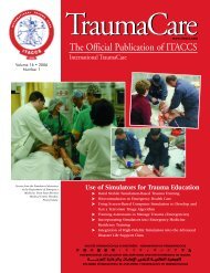
The Official Publication of ITACCS - International Trauma ...
The Official Publication of ITACCS - International Trauma ...
The Official Publication of ITACCS - International Trauma ...

Create successful ePaper yourself
Turn your PDF publications into a flip-book with our unique Google optimized e-Paper software.
<strong>ITACCS</strong> Spring 2003<br />
toxic zone (TOXALS) introduced by <strong>ITACCS</strong> in 1996 has now been widely accepted and the<br />
new approach to CBW agent management is based upon life support coupled with appropriate<br />
antidote therapy where possible.<br />
Consideration <strong>of</strong> the pathophysiological processes following toxic agent exposure provides<br />
a rational platform for prehospital and hospital clinical management. CBW agents can<br />
attack all systems <strong>of</strong> the body, including the central and peripheral nervous systems, the skin<br />
and epithelia, and particularly the respiratory system. <strong>The</strong> latter is vulnerable to attack from<br />
blockage <strong>of</strong> the airways through vesication and excess secretions, from failure <strong>of</strong> central control<br />
and peripheral neuromuscular transmission due to disruption <strong>of</strong> the cholinergic transmission<br />
system, and from the development <strong>of</strong> toxic pulmonary oedema. Certain classes <strong>of</strong><br />
agent are clearly associated with different processes: nerve agents and neurotoxins have a<br />
direct effect on synaptic and neural transmission while the classic war gases such as phosgene<br />
and industrial agents such as methyl isocyanate have been demonstrated to cause mass toxic<br />
pulmonary edema. Vesicant agents such as mustard gas are now known to have effects at all<br />
levels <strong>of</strong> the respiratory tree apart from causing vesication <strong>of</strong> the dermis.<br />
<strong>The</strong> explosion in research into the cholinergic transmission system that followed<br />
World War II led to a good understanding <strong>of</strong> the pathophysiology <strong>of</strong> acetylcholinesterase inhibition<br />
and the development <strong>of</strong> a range <strong>of</strong> antidotes that can be used in both military or civil<br />
exposure. <strong>The</strong>re are valuable lessons to be learned from the management <strong>of</strong> pesticide poisoning,<br />
and the rational use <strong>of</strong> oximes has been reappraised. <strong>The</strong> interest in cholinergic<br />
mechanisms has been matched in recent years by work on the pathogenesis <strong>of</strong> toxic pulmonary<br />
edema and new treatment strategies have resulted. Work has focused on increasing<br />
cellular glutathione (GSH) levels as a means <strong>of</strong> preventing lipid–peroxidation induced pulmonary<br />
edema. <strong>The</strong>re are suggestions that N-acetyl cysteine may provide protection by maintaining<br />
GSH levels and inhibiting production <strong>of</strong> inflammatory leukotrienes.<br />
Further application <strong>of</strong> knowledge gained from normal clinical research into disease<br />
mechanisms is likely to have a continued beneficial effect on the management <strong>of</strong> the rarer circumstances<br />
<strong>of</strong> mass toxic trauma.<br />
End Tidal CO2: From Airway to Cardiac Output<br />
Marvin A. Wayne, MD, FACEP<br />
Associate Clinical Pr<strong>of</strong>essor, University <strong>of</strong> Washington<br />
Assistant Clinical Pr<strong>of</strong>essor, Yale University<br />
Emergency Medical Services, Bellingham, Washington, USA<br />
Learning Objectives: 1) to review the physiology <strong>of</strong> CO2 production and excretion<br />
and 2) to describe the use <strong>of</strong> ETCO2 to monitor ventilation, tube placement, and indirect<br />
cardiac output.<br />
Principles. A capnometer projects filtered infrared light at a wavelength <strong>of</strong> 4.28 um<br />
across a sample chamber to a semiconductor detector. <strong>The</strong> detector generates an electrical<br />
signal inversely proportional to the light absorbed by CO2 in the sample chamber, which ultimately<br />
is translated into a capnometer value (CO2 concentration). Capnography is the realtime<br />
graphic depiction <strong>of</strong> CO2 concentration throughout the respiratory cycle.<br />
Gas sampling may be by “sidestream” or “mainstream” technique. Mainstream technique<br />
incorporates the sensor into the ventilation circuit. Sidestream technique samples<br />
exchanged gases through a small branch tube from the ventilation circuit or breathing<br />
patient. Sidestream sampling is more susceptible to clogging with water or mucus and results<br />
in a slower response time compared with mainstream capnometry.<br />
Applications. Carbon dioxide is a by-product <strong>of</strong> cellular metabolism, constantly produced<br />
in proportion to the cellular metabolic rate. Because CO2 is transported via the bloodstream<br />
for elimination by the lungs, measurement <strong>of</strong> end-tidal CO2 (ET CO2) may reflect the<br />
cellular metabolic rate, vascular system intactness, cardiac function, and ventilation.<br />
Capnometry and capnography enjoy widespread use in anesthesia and the ICU. Similar technology<br />
appears transferable to the emergency department and prehospital setting.<br />
Decreased CO2 concentrations, detected by capnometry or capnography, are indicative<br />
<strong>of</strong> endotracheal tube disconnection, extubation, obstruction, or misplacement. ETCO2<br />
was found more reliable than direct visualization, auscultation, or observation <strong>of</strong> chest wall<br />
motion. Capnometry/capnography can confirm esophageal intubation within one respiratory<br />
cycle, whereas pulse oximetry may not detect desaturation for several minutes in the preoxygenated<br />
patient. For these important reasons, capnometry is strongly advocated in the<br />
emergency department and prehospital arena, where physical diagnosis is difficult at best<br />
and patient motion increases the risk <strong>of</strong> endotracheal tube displacement.<br />
Capnometry and capnography may be useful for assessing the effects <strong>of</strong> cardiopulmonary<br />
resuscitation (CPR). During cardiopulmonary arrest, CO2 is neither efficiently transported<br />
to, nor expired from the lungs, resulting in decreased ETCO2. A rising ETCO2 may be<br />
the first sign <strong>of</strong> improved patient status during CPR. CO2 concentrations as measured by capnometry<br />
have been shown to correlate well with coronary perfusion pressure during CPR.<br />
<strong>The</strong>se data may suggest prognostic application <strong>of</strong> capnometry. Capnogram waveform analysis<br />
enhances the specificity <strong>of</strong> capnometric ETCO2 measurements. Baseline changes may indicate<br />
CO2 rebreathing, while slope changes may indicate resistance that occurs with endotracheal<br />
tube kinking or expiratory obstruction. “Staircase effects” in the capnogram descending limb<br />
may be seen in situations such as pneumothorax. <strong>The</strong>se applications <strong>of</strong> capnography and capnometry<br />
are important in the emergency department and prehospital setting.<br />
Recently a new dimension has been added with the introduction <strong>of</strong> noninvasive cardiac<br />
output. This system utilizes a CO2 Fick principle to simulate the equivalency <strong>of</strong> an O2<br />
Fick application. Initially usable only on intubated patients, this has now been expanded to<br />
those spontaneously breathing. This system combines ETCO2, flow, and differential dead<br />
space rebreathing to non-invasively accomplish this.<br />
Limitations. While highly accurate, in certain clinical situations the ability <strong>of</strong> capnometric<br />
measurement to estimate arterial CO2 tension (PaCO2) is hampered. In healthy<br />
patients with well-matched alveolar ventilation and perfusion, ET CO2 estimates PaCO2 within<br />
2 or 3 mmHg. When expired gas is from poorly or nonperfused alveoli (ventilation-perfusion<br />
ratio, or V/Q, mismatching), ETCO2 underestimates PaCO2. V/Q mismatching has multiple<br />
etiologies, including body position changes, acute and chronic pulmonary disease, and<br />
adult respiratory distress syndrome. While absolute values <strong>of</strong> CO2 concentration may be inaccurate<br />
in these circumstances, trend information may remain valuable. Also, the arterial-alveolar<br />
CO2 difference may be calculated in these patients. <strong>The</strong> arterial-alveolar difference, a-<br />
ADCO2, is the difference between arterial CO2 and end tidal CO2 (ET CO2). Normally this<br />
difference is small (2–3 mmHg) in patients with normal physiology and without cardiovascular<br />
or pulmonary disease. <strong>The</strong> a-ADCO2 difference may be elevated due to incomplete emptying,<br />
shunt perfusion, or dead space ventilation.<br />
In shunt perfusion, a particular lung unit is underventilated relative to the amount <strong>of</strong><br />
perfusion (normal 5L blood flow to 4L ventilation). Here the a-ADCO2 difference will be<br />
slightly elevated in the range <strong>of</strong> 5 to10 mmHg. This small difference is related to the efficiency<br />
<strong>of</strong> the lung and the excellent diffusability <strong>of</strong> CO2.<br />
In dead space ventilation, a particular lung unit is underperfused relative to the<br />
amount <strong>of</strong> ventilation. Dead space ventilation widens the a-ADCO2 much more than does<br />
shunt perfusion alone. This difference may be 10 to 20 mmHg or more. Further, dead space<br />
ventilation and shunt perfusion are not mutually exclusive and may both occur to some<br />
degree in the same patient.<br />
Conclusion. Capnometry/capnography are <strong>of</strong> confirmed value in the hospital setting,<br />
and their applications continue to broaden. Situations unique to the out-<strong>of</strong>-hospital environment<br />
invite their rapid extrapolation to the prehospital setting. <strong>The</strong>ir ultimate value will<br />
depend upon the training, level <strong>of</strong> understanding, and clinical acumen <strong>of</strong> emergency care<br />
providers. New application to allow noninvasive cardiac output will provide a viable alternative<br />
to invasive systems.<br />
A New Approach to Monitoring Early Hemodynamic Performance:<br />
Transesophageal Echo Doppler Ultrasound<br />
Yves Lambert, Olivier Richard, Pascal Renoux<br />
Service d’Aide Médicale Urgente (SAMU 78) & Service Mobile d’Urgence et de<br />
Réanimation<br />
Versailles Hospital, Le Chesnay, France<br />
Early resuscitation <strong>of</strong> severe trauma is first based on physical examination like heart<br />
rate and arterial blood pressure. <strong>The</strong>se parameters are not accurate enough to give a reliable<br />
circulatory status <strong>of</strong> the patient. <strong>The</strong>rapeutic adjustments based on these clinical parameters<br />
are thus difficult to implement and control, even after an initial aggressive therapy. <strong>The</strong> outcoming<br />
<strong>of</strong> the Transesophageal Echo Doppler system, which is a new noninvasive or light<br />
invasive hemodynamic monitor, could lead to an interesting approach in the rapid management<br />
<strong>of</strong> severe trauma patients. Early detection <strong>of</strong> cardiocirculatory disorders and prompt<br />
adequate treatment in order to prevent serious complication like organ failure are required<br />
during the very first hours after trauma.<br />
Transesophageal Echo Doppler monitoring is easy to use and reproducible. On the<br />
trauma field or in the emergency room, the rapid settlement <strong>of</strong> the TEE is a major asset which<br />
allows to obtain a hemodynamic pr<strong>of</strong>ile in just a few minutes. Its logical first use gives the<br />
chance, in clinical practice, to adjust the therapy based on the displayed hemodynamic<br />
parameters: aortic blood flow, stroke volume in the aorta and total systemic vascular resistance<br />
<strong>of</strong> the aortic circuit.<br />
• For severe head trauma patients with no active bleeding, in order to maintain a<br />
mean arterial blood pressure (MABP) <strong>of</strong> 90 mmHg and avoid a secondary cerebral ischemia,<br />
we highly suggest a continuous measurement <strong>of</strong> MABP. Thus, interpreting the arterial blood<br />
pressure data correlated to ABF, Sva and TSVRa, the therapy can be orientated either toward<br />
fluid loading or toward the use <strong>of</strong> catecholamines, i.e., when vascular resistances are low with<br />
a normal flow (neurogenic hypotension). In all cases, continuous monitoring and trends<br />
allow to assess a real time hemodynamic status and help to adjust treatment.<br />
• For polytrauma patients (penetrating or blunt trauma), with an unstable hemodynamic<br />
condition, the arterial blood pressure and heart rate are useless in estimating the right<br />
amount <strong>of</strong> blood loss. Keeping in mind that treatment should not delay surgery, monitoring Sva<br />
and ABF will help guiding the vascular fluid loading. Increase <strong>of</strong> ABF or Sva will reflect more<br />
accurately efficacy <strong>of</strong> initial treatment than any increase <strong>of</strong> arterial blood pressure.<br />
Another challenge provided by Transesophageal Echo Doppler is the possibility to determine<br />
factors able to predict occurrence <strong>of</strong> MOF. Continuous measurement <strong>of</strong> ABF and Sva as<br />
early as possible after the trauma could help to quantify early low flow and poor tissue perfusion<br />
states. Correlation betwin those variation and occurrence <strong>of</strong> MOF have to be determined.<br />
Minimally invasive, hemodynamic monitoring using the Transesophageal Echo<br />
Doppler approach opens very enthusiastic perspectives. Earlier diagnosis and continuous<br />
monitoring <strong>of</strong> circulatory deficiencies must be very useful for the trauma physician to start<br />
adequate resuscitation and minimize tissue ischemia.<br />
— Session B —<br />
Disaster Management and Emergency Medicine<br />
Co-Chair: Dario Gonzalez, MD, New York, New York, USA<br />
Co-Chair: Andreas Thierbach, MD, Mainz, Germany<br />
<strong>The</strong> <strong>International</strong> Chief Emergency Physician Training Course<br />
Freddy Lippert, MD<br />
<strong>Trauma</strong> Center and Major Incident Command Centre,<br />
H:S Rigshospitalet, Copenhagen University Hospital, Denmark<br />
Learning Objectives: To review the development, content, and importance <strong>of</strong> <strong>The</strong><br />
<strong>International</strong> Chief Emergency Physician Training Course.<br />
Education and training are essential in handling major incidents and disasters. It is an<br />
obligation <strong>of</strong> any medical system to meet these challenges. Guidelines for education and<br />
training in disaster medicine have been proposed. 1<br />
<strong>The</strong> <strong>International</strong> Chief Emergency Physician Training course on Command Incident<br />
Management and Mass Casualty Disasters (ICEP) is one <strong>of</strong> the international courses with a<br />
well-established curriculum.<br />
<strong>The</strong> first CEP course was set up in 1998 by <strong>The</strong> <strong>International</strong> <strong>Trauma</strong> Anesthesia and<br />
Critical Care Society (<strong>ITACCS</strong>) in cooperation with a group <strong>of</strong> leading chief emergency physicians<br />
in Mainz and based on experiences from national German CEP courses. 2<br />
<strong>The</strong> international faculty consists <strong>of</strong> experienced chief emergency physicians and<br />
experts in different fields from various parts <strong>of</strong> the world to cover all issues. Participants make<br />
up an international forum <strong>of</strong> physicians with experiences within prehospital care, trauma<br />
care, emergency medicine, and command incident management. This constellation facilitates<br />
international exchange <strong>of</strong> experiences and ideas.<br />
<strong>The</strong> CEP course has an intensive 40 hours curriculum with lectures, discussions <strong>of</strong> case<br />
reports, excursions, and in-field training. Subjects include epidemiology <strong>of</strong> mass casualty incidents<br />
and disasters, responsibilities and duties <strong>of</strong> the CEP, command and control, triage concepts,<br />
personal protection and equipment, communication devices and techniques, evacuation<br />
and long-range transport, planning for mass gatherings, VIP protection, hostage situations,<br />
counter terrorism, HazMat, bio-threats and chemical warfare, medical relief, and media<br />
contact and management.<br />
Lectures are interactive and most are accompanied by practical training sessions or<br />
tabletop discussions. <strong>The</strong>ory and practical training result in full-scale disaster exercises in<br />
cooperation with all local authorities. 3<br />
This international concept facilitates post-course exchange <strong>of</strong> knowledge and experiences<br />
based on the network established during the 5-day intensive curriculum <strong>of</strong> chief emer-<br />
43