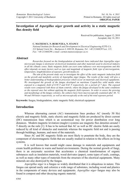
Investigation of Aspergillus niger growth and activity in ... - Rombio.eu
Investigation of Aspergillus niger growth and activity in ... - Rombio.eu
Investigation of Aspergillus niger growth and activity in ... - Rombio.eu

You also want an ePaper? Increase the reach of your titles
YUMPU automatically turns print PDFs into web optimized ePapers that Google loves.
Romanian Biotechnological Letters Vol. 16, No. 4, 2011<br />
Copyright © 2011 University <strong>of</strong> Bucharest<br />
Pr<strong>in</strong>ted <strong>in</strong> Romania. All rights reserved<br />
ORIGINAL PAPER<br />
<strong>Investigation</strong> <strong>of</strong> <strong>Aspergillus</strong> <strong>niger</strong> <strong>growth</strong> <strong>and</strong> <strong>activity</strong> <strong>in</strong> a static magnetic<br />
flux density field<br />
Abstract<br />
6364<br />
Received for publication, August 12, 2010<br />
Accepted, July 14, 2011<br />
C. MATEESCU, N. BURUNŢEA, N. STANCU<br />
National Institute for Research <strong>and</strong> Development <strong>in</strong> Electrical Eng<strong>in</strong>eer<strong>in</strong>g ICPE-CA,<br />
313 Splaiul Unirii Str.., Bucharest-3, 030138, Romania, Tel: +40 213468297/ext. 115,<br />
Fax: +40 213468299, e-mail: carmen.mateescu@icpe-ca.ro<br />
Researches focused on the biodegradation <strong>of</strong> materials have <strong>in</strong>dicated that <strong>Aspergillus</strong> <strong>niger</strong><br />
microscopic fungus is destructor <strong>of</strong> electrical <strong>in</strong>sulations <strong>and</strong> other materials used <strong>in</strong> electrical <strong>in</strong>dustry<br />
<strong>in</strong> all the climatic zones. Static magnetic fields can exert some <strong>in</strong>fluence over microorganisms <strong>and</strong> this<br />
effect depends on many factors such as type <strong>and</strong> magnitude <strong>of</strong> magnetic field, type <strong>of</strong> microorganism,<br />
temperature, length/duration <strong>of</strong> exposure, <strong>growth</strong> media etc.<br />
The aim <strong>of</strong> the present study was to <strong>in</strong>vestigate the effect <strong>of</strong> the static magnetic <strong>in</strong>duction field<br />
on the <strong>growth</strong> <strong>and</strong> metabolic <strong>activity</strong> <strong>of</strong> <strong>Aspergillus</strong> <strong>niger</strong> fungus. The results <strong>of</strong> the study will give a<br />
better underst<strong>and</strong><strong>in</strong>g <strong>of</strong> biodegradation processes which occur on materials <strong>and</strong> electrical equipments.<br />
We <strong>in</strong>vestigated the <strong>growth</strong> <strong>of</strong> the fungus developed on nutritious Czapek-Dox solid media when<br />
exposed to a magnetic field <strong>of</strong> 0.50 T <strong>and</strong> 0.62 T respectively, for a period <strong>of</strong> 7 days. The exposed<br />
results were compared with those <strong>of</strong> sham controls, where the fungus developed <strong>in</strong> the same conditions<br />
as the exposed ones but without apply<strong>in</strong>g the magnetic field exposure. In order to assess the grow<strong>in</strong>g<br />
<strong>and</strong> morphology <strong>of</strong> the fungus colonies, the cultures have been macroscopically exam<strong>in</strong>ed after 48, 72,<br />
96 <strong>and</strong> 168 hours respectively, as well as microscopically at the end <strong>of</strong> the total exposure period.<br />
Keywords: fungus, biodegradation, static magnetic field, electrical equipment<br />
Introduction<br />
Whereas alternat<strong>in</strong>g current (AC) transmission l<strong>in</strong>es produce AC (mostly 50 Hz)<br />
electric <strong>and</strong> magnetic fields, static electric <strong>and</strong> magnetic fields are produced by direct current<br />
(DC) transmission l<strong>in</strong>es which is an economical way for power distribution over long<br />
distances. Modern magnetic levitation (maglev) systems use magnetic flux densities around 1<br />
T directly on the rails [1]. It has to be noticed that AC as well as DC electric fields are easily<br />
reduced by all k<strong>in</strong>d <strong>of</strong> obstacles <strong>and</strong> materials whereas the magnetic field not <strong>and</strong> is pass<strong>in</strong>g<br />
through build<strong>in</strong>gs, humans, <strong>and</strong> most <strong>of</strong> the materials.<br />
S<strong>in</strong>ce AC <strong>and</strong> DC magnetic fields are most likely to penetrate the body, they are the<br />
component <strong>of</strong> electromagnetic fields that are usually studied <strong>in</strong> relation to the effects on liv<strong>in</strong>g<br />
systems.<br />
It is well known that mould might cause damage to materials <strong>and</strong> equipments <strong>and</strong><br />
create health problems <strong>in</strong> warm <strong>and</strong> humid environments. Dur<strong>in</strong>g the normal <strong>growth</strong> <strong>of</strong> fungi,<br />
there is an enzymatic secretion that accelerates a chemical transformation <strong>of</strong> organic<br />
substances such as <strong>in</strong>sulation materials, wood derivates (paper), some types <strong>of</strong> seals, coat<strong>in</strong>gs,<br />
as well as many other types <strong>of</strong> materials from the structure <strong>of</strong> the electrical equipments. Many<br />
m<strong>in</strong>erals are also destroyed by the fungus.<br />
<strong>Aspergillus</strong> <strong>niger</strong> is a fungus so widely distributed that it is ubiquitous <strong>in</strong> nature. This<br />
fungus can spread easily <strong>and</strong> colonize a wide range <strong>of</strong> substrates, <strong>in</strong>clud<strong>in</strong>g wood <strong>and</strong> plastics<br />
<strong>in</strong> the components <strong>of</strong> many devices <strong>and</strong> equipments. <strong>Aspergillus</strong> <strong>niger</strong> fungus may also be<br />
found <strong>in</strong> compost <strong>and</strong> other decay<strong>in</strong>g organic material.
C. MATEESCU, N. BURUNŢEA, N. STANCU<br />
This paper aimed to <strong>in</strong>vestigate the <strong>growth</strong> <strong>and</strong> <strong>activity</strong> <strong>of</strong> <strong>Aspergillus</strong> <strong>niger</strong> fungus by<br />
expos<strong>in</strong>g it under two magnetic flux densities (B-field), for a total exposure period <strong>of</strong> 168<br />
hours. S<strong>in</strong>ce the present study is quite orig<strong>in</strong>al data on a similar issue are scarce <strong>in</strong> literature.<br />
Manoliu et al (2007) [3] <strong>in</strong>vestigated the development <strong>of</strong> <strong>Aspergillus</strong> <strong>niger</strong> exposed to<br />
a static B-field vary<strong>in</strong>g from 40-80 T. The results showed a 1.5 - 2 times faster <strong>growth</strong> rate<br />
than <strong>in</strong> sham controls. Moreover, it has been showed that the B-field exposure can have an<br />
effect on the biodegradability <strong>of</strong> materials by enhanc<strong>in</strong>g the <strong>growth</strong> rate <strong>and</strong> the<br />
aggressiveness <strong>of</strong> the fungus. It has also been noticed that a B-field exposure <strong>of</strong> more than<br />
350 T may delay the <strong>growth</strong> <strong>of</strong> microscopic fungus.<br />
The <strong>in</strong>fluence <strong>of</strong> the B-field exposure on cellulases <strong>and</strong> catalase <strong>activity</strong> <strong>in</strong> cellulolytic<br />
fungi Chaetomium globosum <strong>and</strong> Trichoderma viride cultivated on media with waste from<br />
<strong>in</strong>dustry <strong>of</strong> panification has been <strong>in</strong>vestigated too. It has been shown that the <strong>activity</strong> <strong>of</strong> these<br />
enzymes was <strong>in</strong>fluenced by both studied cellulolytic species <strong>and</strong> the exposure length to the B-<br />
field [4].<br />
Besides <strong>Aspergillus</strong> <strong>niger</strong> fungus, the effect <strong>of</strong> static magnetic fields has also been<br />
studied at the molecular <strong>and</strong> cellular levels <strong>in</strong> bacteria species, yeasts <strong>and</strong> molds. Moreover,<br />
the <strong>growth</strong> rate, colony size, gas production, viability <strong>and</strong> mutation effects, enzyme <strong>activity</strong><br />
<strong>and</strong> germ<strong>in</strong>ation <strong>of</strong> spores [4] have also been <strong>in</strong>vestigated.<br />
The motivation <strong>of</strong> our study is double. Firstly, because the <strong>Aspergillus</strong> <strong>niger</strong> fungus is<br />
widely spread on electrical equipment <strong>in</strong> various environmental conditions <strong>and</strong> secondly<br />
because there is a lack <strong>of</strong> scientific data on the effects <strong>of</strong> static magnetic fields on this fungus.<br />
Materials <strong>and</strong> Methods<br />
For <strong>Aspergillus</strong> <strong>niger</strong> <strong>growth</strong> it has been used Czapek-Dox agar, a synthetic solid<br />
medium, conta<strong>in</strong><strong>in</strong>g sucrose as carbon source <strong>and</strong> nitrate as nitrogen source. On Czapek-Dox<br />
agar, colonies <strong>of</strong> <strong>Aspergillus</strong> <strong>niger</strong> consist <strong>of</strong> a compact white or yellow basal felt covered by<br />
a dense layer <strong>of</strong> dark-brown to black colonial heads.<br />
The culture medium was prepared by dissolv<strong>in</strong>g <strong>of</strong> the follow<strong>in</strong>g components <strong>in</strong>to one<br />
liter <strong>of</strong> distilled water: sucrose 30 g, sodium nitrate 3 g, potassium chloride 0.5 g, magnesium<br />
sulfate heptahydrate 0.5 g, iron (II) sulfate heptahydrate 0.01 g, di-potassium hydrogen<br />
phosphate 1 g <strong>and</strong> agar 15 g. The suspension was brought to the boil <strong>in</strong> order to dissolve<br />
completely all the <strong>in</strong>gredients <strong>and</strong> then was sterilized by autoclav<strong>in</strong>g at 121 0 C for 15 m<strong>in</strong>utes<br />
[5]. The molten medium was poured <strong>in</strong>to three 7 cm diameter Petri dishes.<br />
The plates were <strong>in</strong>oculated with <strong>Aspergillus</strong> <strong>niger</strong> us<strong>in</strong>g a sterile wire. The fungus<br />
which we used for test<strong>in</strong>g was 30 days old <strong>and</strong> was obta<strong>in</strong>ed from our own collection.<br />
The environment conditions for fungus <strong>growth</strong> were room temperature <strong>of</strong> 25 ± 2 0 C,<br />
natural light <strong>and</strong> the humidity provided by the culture medium [6].<br />
For study<strong>in</strong>g the efficiency <strong>of</strong> the B-field, one sham control <strong>and</strong> two irradiated<br />
(exposed) <strong>in</strong>oculation plates were used. Whereas the sham <strong>in</strong>oculated plate was treated <strong>in</strong> the<br />
same way as the irradiated ones but without apply<strong>in</strong>g the B-field, the two other <strong>in</strong>oculated plates<br />
were exposed to a B-field <strong>of</strong> 0.5 T <strong>and</strong> 0.62 T respectively. Though the total exposure time was<br />
168 hours, the cultures were periodically analyzed after 48, 72, 96 <strong>and</strong> 168 hours respectively.<br />
After <strong>in</strong>cubations all the cultures were microscopically analyzed as regards the<br />
<strong>Aspergillus</strong> <strong>niger</strong> sporulation <strong>growth</strong>, us<strong>in</strong>g an optical <strong>in</strong>verted microscope type Nikon<br />
Eclipse Ti-E fitted with a confocal system Eclipse C1si. The microscopic exam<strong>in</strong>ation <strong>of</strong> the<br />
fungus was performed <strong>in</strong> bright field with a magnitude <strong>of</strong> 40 X.<br />
In order to make a comparative analysis <strong>of</strong> <strong>Aspergillus</strong> <strong>niger</strong> <strong>growth</strong> under different<br />
static magnetic flux densities, two magnetic systems have been calculated <strong>and</strong> designed for<br />
Romanian Biotechnological Letters, Vol. 16, No. 4, 2011 6365
<strong>Investigation</strong> <strong>of</strong> <strong>Aspergillus</strong> <strong>niger</strong> <strong>growth</strong> <strong>and</strong> <strong>activity</strong> <strong>in</strong> a static magnetic flux density field<br />
perform<strong>in</strong>g exposures to B-field <strong>of</strong> 0.5 T <strong>and</strong> 0.62 T. These magnetic systems consisted <strong>of</strong> the<br />
follow<strong>in</strong>g components:<br />
a. Magnetic circuit made <strong>of</strong> steel type OL 35;<br />
b. Permanent magnets, made <strong>of</strong> magnetic alloy based on NdFeB, hav<strong>in</strong>g the follow<strong>in</strong>g<br />
magnetic field characteristics: Br = 1.2 T, Hcb = 970 kA/m, (BH) max = 35 MGsOe,<br />
respectively;<br />
c. Polar pieces made <strong>of</strong> steel type OL 35.<br />
The exposure strength <strong>of</strong> the B-field <strong>in</strong> air has been performed by simulation <strong>of</strong><br />
magnetic fields us<strong>in</strong>g a magnetic calculation s<strong>of</strong>t type FEM (F<strong>in</strong>it Element Method).<br />
Results <strong>and</strong> discussions<br />
For the <strong>in</strong>vestigations <strong>of</strong> the effect <strong>of</strong> a static B-field on the mycelial <strong>growth</strong> <strong>and</strong><br />
conidia formation, the plates were exam<strong>in</strong>ed after an <strong>in</strong>cubation period <strong>of</strong> 48, 72, 96 <strong>and</strong> 168<br />
hours respectively. Figures 1-4 (a,b,c) show the <strong>Aspergillus</strong> <strong>niger</strong> <strong>growth</strong> without B-field<br />
exposure (sham control), under a B-field exposure <strong>of</strong> 0.5 T <strong>and</strong> <strong>of</strong> 0.62 T, for 48, 72, 96 <strong>and</strong><br />
168 hours respectively.<br />
Dur<strong>in</strong>g the first 48 hours we observed that the non-exposed fungus has grown faster<br />
than the two other samples which were exposed to the B-field. It means that after an<br />
<strong>in</strong>cubation <strong>of</strong> 48 hours, the <strong>growth</strong> <strong>of</strong> <strong>Aspergillus</strong> <strong>niger</strong> was characterized by the development<br />
<strong>of</strong> small but compact colonies with dense sporulation, spread from the <strong>in</strong>oculated po<strong>in</strong>t to the<br />
whole surface <strong>of</strong> the culture medium (Figure 1 a). The fungus exposed to the B-field produced<br />
less but larger colonies, with strong sporulation. The colonies are not spread on the whole<br />
surface <strong>of</strong> the culture medium (Figures 1b, 1c).<br />
a b c<br />
Fig 1. (a,b,c) <strong>Aspergillus</strong> <strong>niger</strong> <strong>growth</strong> after 48 hours <strong>of</strong> <strong>in</strong>cubation (a -without magnetic field;<br />
b – magnetic field <strong>of</strong> 0.5T; c – magnetic field <strong>of</strong> 0.62T)<br />
After a B-field exposure <strong>of</strong> 72 hours, the mould followed an atypical <strong>growth</strong>,<br />
characterized by about 20 larger <strong>and</strong> bombastic colonies hav<strong>in</strong>g white-yellowish normal<br />
aspect <strong>and</strong> very few dark-brown colonial heads. A stronger B-field exposure resulted <strong>in</strong> a<br />
larger but rarer colony formation <strong>in</strong> the culture medium. Thus, for the plate exposed to a B-<br />
field <strong>of</strong> 0.5 T (Figure 2b), we observed more but smaller colonies which tend to jo<strong>in</strong> each<br />
other, compar<strong>in</strong>g to the sample exposed <strong>in</strong> B-field <strong>of</strong> 0.62 T (Figure 2c). By compar<strong>in</strong>g the<br />
0.5 T (Figure 2b) <strong>and</strong> the 0.62 T exposure results, we observe <strong>in</strong> the first case more but<br />
smaller colonies which tend to stick together than <strong>in</strong> the second case.<br />
6366 Romanian Biotechnological Letters, Vol. 16, No. 4, 2011
C. MATEESCU, N. BURUNŢEA, N. STANCU<br />
a b c<br />
Fig 2. (a,b,c) <strong>Aspergillus</strong> <strong>niger</strong> <strong>growth</strong> after 72 hours <strong>of</strong> <strong>in</strong>cubation (a -without magnetic field; b – magnetic<br />
field <strong>of</strong> 0.5; c – magnetic field <strong>of</strong> 0.62T)<br />
After 96 hours <strong>of</strong> <strong>in</strong>cubation, some notches or cuts became visible on the swollen<br />
surface <strong>of</strong> the colonies. They are more explicit for the stronger B-field <strong>of</strong> 0.62 T (Figure 3c).<br />
These notches did not arise when the B-field was not applied.<br />
a b c<br />
Fig 3. (a,b,c) <strong>Aspergillus</strong> <strong>niger</strong> <strong>growth</strong> after 96 hours <strong>of</strong> <strong>in</strong>cubation (a -without magnetic field;b – magnetic field<br />
<strong>of</strong> 0.5T; c – magnetic field <strong>of</strong> 0.62T)<br />
After an <strong>in</strong>cubation period <strong>of</strong> 168 hours (at the end <strong>of</strong> test<strong>in</strong>g), we observed a<br />
relatively homogenous <strong>growth</strong> <strong>of</strong> the fungus <strong>in</strong> the plate not exposed to the B-field (Figure<br />
4a). The colonies became completely black on the whole surface <strong>of</strong> the culture medium.<br />
The sample exposed to the strongest B-field <strong>of</strong> 0.62 T (Figure 4c) showed <strong>in</strong>tensively<br />
black colored <strong>and</strong> large colonies. In this case the black colonial heads are denser as compared<br />
to the fungus developed <strong>in</strong> cultures not exposed to a B-field . This atypical <strong>growth</strong> does not<br />
cover the entire surface <strong>of</strong> the Petri dish. The sample exposed <strong>in</strong> the B-field <strong>of</strong> 0.5 T (Figure<br />
4b) shows an <strong>in</strong>termediary aspect between the two plates.<br />
a b c<br />
Fig 4. (a,b,c) <strong>Aspergillus</strong> <strong>niger</strong> <strong>growth</strong> after 168 hours <strong>of</strong> <strong>in</strong>cubation (a -without magnetic field; b – magnetic<br />
field <strong>of</strong> 0.5T; c – magnetic field <strong>of</strong> 0.62T)<br />
Romanian Biotechnological Letters, Vol. 16, No. 4, 2011 6367
<strong>Investigation</strong> <strong>of</strong> <strong>Aspergillus</strong> <strong>niger</strong> <strong>growth</strong> <strong>and</strong> <strong>activity</strong> <strong>in</strong> a static magnetic flux density field<br />
Figure 5 shows a microscopic photograph <strong>of</strong> radiate conidial head <strong>and</strong> brown-colored<br />
round-shaped unicellular conidia <strong>of</strong> <strong>Aspergillus</strong> <strong>niger</strong>.<br />
Fig. 5. Micrograph <strong>of</strong> <strong>Aspergillus</strong> <strong>niger</strong> conidia with a full-field on the left <strong>and</strong> a detail shot on the right<br />
Conidia <strong>in</strong> cha<strong>in</strong>s or detached <strong>and</strong> dispersed can be observed <strong>in</strong> this microscopy<br />
image. S<strong>in</strong>gle or paired conidia may resemble yeast cells. The size <strong>of</strong> spores <strong>of</strong> <strong>Aspergillus</strong><br />
<strong>niger</strong> ranges from 2 to 4 microns.<br />
Conclusions<br />
The effect <strong>of</strong> a static magnetic flux density on the <strong>growth</strong> <strong>and</strong> <strong>activity</strong> <strong>of</strong> <strong>Aspergillus</strong><br />
<strong>niger</strong> have been macroscopically <strong>and</strong> microscopically <strong>in</strong>vestigated by means <strong>of</strong> two exposure<br />
strengths. It is concluded that the static magnetic field produces an atypical <strong>growth</strong> <strong>of</strong> the<br />
fungus that is characterized by less <strong>and</strong> swollen, bombastic colonies which did not spread on<br />
the entire surface <strong>of</strong> the culture medium. From the comparison between the exposed <strong>and</strong> the<br />
sham, we conclude that the magnetic field is an efficient <strong>in</strong>hibitor <strong>of</strong> the surface <strong>growth</strong> <strong>of</strong> the<br />
fungus.<br />
The validation or practical application <strong>of</strong> the present study lies <strong>in</strong> the fact that the<br />
results can be used for develop<strong>in</strong>g magnetic field sources/methods that are able to reduce or<br />
elim<strong>in</strong>ate bio-damage <strong>of</strong> components <strong>of</strong> electrical equipments or <strong>in</strong>stallations <strong>and</strong> materials<br />
which are sensitive to <strong>Aspergillus</strong> <strong>niger</strong> <strong>and</strong> perhaps to other fungi.<br />
References<br />
1. ICNIRP 13/2003. Exposure to Static <strong>and</strong> Low Frequency Electromagnetic Fields. Biological Effects <strong>and</strong><br />
Health Consequences (0-100 kHz). Publication <strong>of</strong> the International Commission on Non-Ionizng Radiation<br />
Protection. Pr<strong>in</strong>ted by Märkl-Druck, München.<br />
2. S. A. SEMENOV,KLARA Z. GUMARGALIEVA,GENNADIĬ EFREMOVICH ZAIKOV, Biodegradation <strong>and</strong><br />
durability <strong>of</strong> materials under the effect <strong>of</strong> microorganisms, VSP BV, ISBN 90-6764-388-2, pp. 190-192, (2003).<br />
3. A. MANOLIU, L. OPRICK, D. CREANGA, The <strong>in</strong>fluence <strong>of</strong> the static magnetic field (SMF) on some<br />
biochemical parameters <strong>in</strong> cellulolytic fungi Chaetomium globosum <strong>and</strong> Trichoderma viride cultivated on<br />
media supplemented with panification <strong>in</strong>dustrial wastes, Rom. Journal Biol, Vol. 51-52, 2007, pp. 25-37<br />
4. P.E KOVACS., R.L VALENTINE., P.J. ALVAREZ, The effect <strong>of</strong> static magnetic fields on biological<br />
systems: Implications for enhanced biodegradation, Crit. Rev. Environ. Sci. Technol., 27, 1997, pp. 319-382.<br />
5. www.sigmaaldrich.com/etc/medialib/docs/Fluka/usage/70185_czapek_dox_agar.Par.0001.File.tmp/70185_c<br />
zapek_dox_agar.pdf<br />
6. M. MITITIUC, C. TANASE, Micologie, Editura Univ. Al. I. Cuza, Iasi, 2000, pp. 47-52<br />
6368 Romanian Biotechnological Letters, Vol. 16, No. 4, 2011