No.42 - è¾²æ¥çç©è³æºç 究æ
No.42 - è¾²æ¥çç©è³æºç 究æ
No.42 - è¾²æ¥çç©è³æºç 究æ

You also want an ePaper? Increase the reach of your titles
YUMPU automatically turns print PDFs into web optimized ePapers that Google loves.
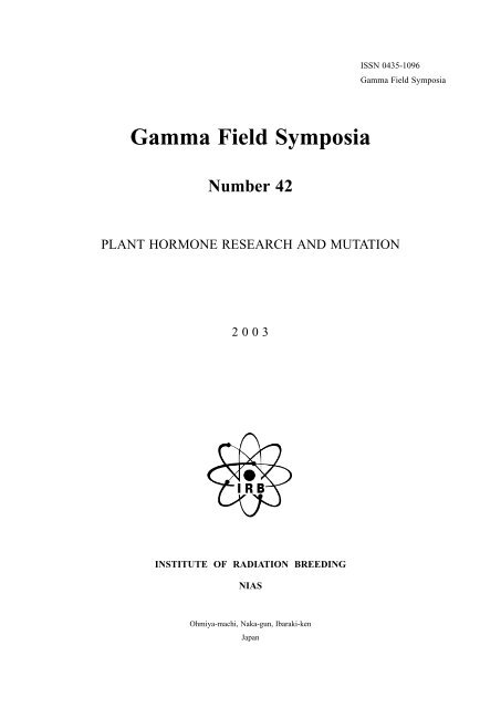
ISSN 0435-1096<br />
Gamma Field Symposia<br />
Gamma Field Symposia<br />
Number 42<br />
PLANT HORMONE RESEARCH AND MUTATION<br />
2 0 0 3<br />
INSTITUTE OF RADIATION BREEDING<br />
NIAS<br />
Ohmiya-machi, Naka-gun, Ibaraki-ken<br />
Japan
PLANTHORMONERESEARCH AND MUTATION<br />
Report of Symposium<br />
held on<br />
July 16-17, 2003<br />
Institute of Radiation Breeding<br />
NIAS<br />
Ohmiya-machi, Naka-gun, Ibaraki-ken 319-2293<br />
Japan
The lecturers and the members of the Symposium Committee<br />
General discussion
List of Participants<br />
(42nd GF Symposium)<br />
ABE, F.<br />
AJIRO, T.<br />
ARAI, T.<br />
ASAUMI, H.<br />
ASHIKARI, M.<br />
DEGI, K.<br />
EHIRA, M.<br />
ENDO, T.<br />
EZURA, H.<br />
FUJITA, M.<br />
FUJITA, Y.<br />
FUKATSU, E.<br />
FUWA, N.<br />
GONAI, T.<br />
GOTO, Y.<br />
GOTO, Y.<br />
HARA, H.<br />
HASEGAWA, H.<br />
HATASHITA, M.<br />
HATTORI, E.<br />
HATTORI, K.<br />
HATTORI, T.<br />
HAYAKAWA, Y.<br />
HAYASHI, Y.<br />
HIGO, K.<br />
HIRAI, T.<br />
HIRATA, Y.<br />
HIRAYAMA, T.<br />
HOBO, T.<br />
HOSOYA, T.<br />
INAGAKI, H.<br />
INOUE, E.<br />
ISHII, T.<br />
ISHIMARU, K.<br />
ITO, A.<br />
ITO, Y.<br />
IWABUCHI, M.<br />
IYOZUMI, H.<br />
KADOTA, N.<br />
KAMIYA, Y.<br />
National Institute of Crop Science<br />
Tokyo University of Agriculture and Technology<br />
Gifu Research Institute for Agricultural Sciences<br />
Ehime Agricultural Experiment Station<br />
Bioscience and Biotechnology Center, Nagoya University<br />
Institute of Radiation Breeding, National Institute of Agrobiological Sciences<br />
Tokyo University of Agriculture and Technology<br />
Miyagi Furukawa Agricultural Experiment Station<br />
University of Tsukuba<br />
RIKEN Tsukuba Institute<br />
Japan International Research Center for Agricultural Sciences<br />
Forest Tree Breeding Center<br />
Snow Brand Seed Co., Ltd.<br />
Ibaraki Plant Biotechnology Institute<br />
Forest Tree Breeding Center<br />
Hitachi High-Technologies<br />
Ibaraki University<br />
University of Shiga Prefecture<br />
The Wakasawan Energy Research Center<br />
Toyota Motor Corporation<br />
Graduated School of Bioagricultural Sciences, Nagoya University<br />
Bioscience and Biotechnology Center, Nagoya University<br />
Fukui Prefectural University<br />
Fukushima Agricultural Experiment Station<br />
National Institute of Agrobiological Sciences<br />
Tokyo University of Agriculture<br />
Tokyo University of Agriculture and Technology<br />
RIKEN Central Institute<br />
RIKEN Laboratory of Plant Molecular Biology<br />
High School of Agriculture, Mito Ibaraki<br />
Shizuoka Agricultural Experiment Station<br />
Ibaraki University<br />
Ibaraki Agricultural Center<br />
University of Tsukuba<br />
National Institute of Fruit Tree Science<br />
Institute of Radiation Breeding, National Institute of Agrobiological Sciences<br />
National Institute of Agrobiological Sciences<br />
Shizuoka Agricultural Experiment Station<br />
Takii & Co., LTD<br />
RIKEN Yokohama Institute
KANEKO, T.<br />
KARITA, E.<br />
KASHIMA, M.<br />
KATAOKA, H.<br />
KATO, K.<br />
KATO, M.<br />
KAWAKATSU, M.<br />
KOBAYASHI, M.<br />
KOBAYASHI, S.<br />
KUBOTA, K.<br />
KUBOYAMA, T.<br />
KUDO, S.<br />
KUJIOKA, H.<br />
KURODA, K.<br />
KUSABA, M.<br />
KUSANO, M.<br />
KUSANO, T.<br />
KUSHIRO, T.<br />
KUZUYA, M.<br />
MATSUBAYASHI, Y.<br />
MATSUI, H.<br />
MIYANO, S.<br />
MORISHITA, T.<br />
MORITA, M.<br />
MORITA, R.<br />
MORITA, R.<br />
MORITA, Y.<br />
MOTODA, J.<br />
NAGATO, Y.<br />
NAGATOMI, S.<br />
NAITO, K.<br />
NAITO, T.<br />
NAITO, Y.<br />
NAKAJIMA, I.<br />
NAKAMURA, S.<br />
NAKANO, T.<br />
NARA, Y.<br />
NARUKAWA, M.<br />
NIKI, T.<br />
NISHIMURA, M.<br />
NISHIMURA, S.<br />
NONAKA, S.<br />
NOZAWA, G.T.<br />
OBARA, N.<br />
Plant Bioengineering Research Laboratories, Sapporo Breweries<br />
Tokyo University of Science<br />
Ibaraki Agricultural Center<br />
University of Tsukuba<br />
Shizuoka Agricultural Experiment Station<br />
Takii Plant Breeding & Experiment Station<br />
Institute of Radiation Breeding, National Institute of Agrobiological Sciences<br />
RIKEN Tsukuba Institute<br />
National Institute of Fruit Tree Science<br />
Nagano Agricultural Experiment Station<br />
Ibaraki University<br />
Graduate school of Agricultural Science, Tohoku University<br />
High School of Agriculture, Mito Ibaraki<br />
National Institute of Agrobiological Sciences<br />
Institute of Radiation Breeding, National Institute of Agrobiological Sciences<br />
Kaisui Chemical Industry Co., LTD.<br />
Graduate School of Life Sciences, Tohoku University<br />
RIKEN Plant Science Center<br />
Ibaraki Agricultural Center<br />
Graduated School of Bioagricultural Sciences, Nagoya University<br />
Kyoto University<br />
Keisei Rose Nurseries, LTD.<br />
Institute of Radiation Breeding, National Institute of Agrobiological Sciences<br />
Kyoto University<br />
Institute of Radiation Breeding, National Institute of Agrobiological Sciences<br />
Tokyo University of Agriculture<br />
The University of Tokyo<br />
Graduated School of Bioagricultural Sciences, Nagoya University<br />
Graduate School of Agricultural and Life Science, The University of Tokyo<br />
Institute of Radiation Breeding, National Institute of Agrobiological Sciences<br />
Institute of Radiation Breeding, National Institute of Agrobiological Sciences<br />
School of Agriculture, Meiji University<br />
Tokyo University of Agriculture<br />
National Institute of Fruit Tree Science<br />
National Institute of Crop Science<br />
RIKEN Central Institute<br />
Tokyo University of Science<br />
Tokyo University of Science<br />
National Institute of Floricultural Science<br />
Institute of Radiation Breeding, National Institute of Agrobiological Sciences<br />
University of Tsukuba<br />
University of Tsukuba<br />
Tokyo University of Agriculture and Technology<br />
VISTA, LTD
OHMIYA, Y.<br />
OHSAWA, K.<br />
OHTA, K.<br />
OHTA, Y.<br />
OHTSUBO, N.<br />
OKA, A.<br />
OKA, S.<br />
OKAMURA, J.<br />
OKAMURA, M.<br />
OKAZAKI, K.<br />
OKUDAIRA, M.<br />
OKUMOTO, Y.<br />
ONO, Y.<br />
ONOZAKI, T.<br />
RIKIISHI, K.<br />
SAISHO, D.<br />
SAITO, K.<br />
SAITO, M.<br />
SANADA, T.<br />
SANO, Y.<br />
SATO, M.<br />
SATO, M.<br />
SATO, T.<br />
SATO, Y.<br />
SEKIGUCHI, F.<br />
SHIMOMURA, S.<br />
SOEJIMA, J.<br />
SUGIYAMA, K.<br />
SUNOHARA, H.<br />
SUZUKI, A.<br />
SUZUKI, Y.<br />
TAJI, T.<br />
TAKABATAKE, R.<br />
TAKAHASHI, H.<br />
TAKAHASHI, M.<br />
TAKAHASHI, T.<br />
TAKANO, T.<br />
TAKASAKI, T.<br />
TAKATSU, Y.<br />
TAKEUCHI, S.<br />
TAKYU, T.<br />
TANAKA, M.<br />
TANIGUCHI, T.<br />
TANISAKA, T.<br />
Forest Tree Breeding Center<br />
Nagano Agricultural Research Center<br />
Tokyo University of Agriculture and Technology<br />
Tokyo University of Agriculture and Technology<br />
Ministry of Education, Culture, Sports, Science and Technology<br />
Institute for Chemical Research, Kyoto University<br />
National Institute of Agrobiological Sciences<br />
Sakata Seed Corp.<br />
Plant Lab., Kirin Brewery Co., Ltd.<br />
Niigata University<br />
Iwate Agricultural Research Center<br />
Kyoto University<br />
Bio-oriented Technology Research Advancement Institution<br />
National Institute of Floricultural Science<br />
Okayama University<br />
Okayama University<br />
Saitama Agriculture and Forestry Research Center<br />
Fukui Agriculture Experiment Station<br />
National Institute of Fruit Tree Science<br />
Graduate School of Agriculture, Hokkaido University<br />
Oita Agricultural Research Center<br />
Japan International Research Center for Agricultural Sciences<br />
Akita Agricultural Experiment Station<br />
Toyota Motor Corporation<br />
Japan Women’s University<br />
National Institute of Agrobiological Sciences<br />
National Institute of Fruit Tree Science<br />
Shizuoka Citrus Experiment Station<br />
Nagoya University<br />
Iwate University<br />
National Institute of Crop Science<br />
RIKEN Laboratory of Plant Molecular Biology<br />
National Institute of Agrobiological Sciences<br />
Tokyo University of Science<br />
Forest Tree Breeding Center<br />
Watanabe Seed Co., LTD.<br />
Institute of Radiation Breeding, National Institute of Agrobiological Sciences<br />
Tochigi Agricultural Experiment Station<br />
Ibaraki Agricultural Center<br />
Keisei Rose Nurseries LTD.<br />
Institute of Radiation Breeding, National Institute of Agrobiological Sciences<br />
Nagasaki Fruit Tree Experiment Station<br />
Forest Tree Breeding Center<br />
Graduate School of Agriculture, Kyoto University
TANO, S.<br />
TSUCHIDA, Y.<br />
TSUTSUMI, N.<br />
UCHIDA, H.<br />
UEKI, T.<br />
WATANABE, H.<br />
WATANABE, M.<br />
YAMAGUCHI, H.<br />
YAMAGUCHI, I.<br />
YAMANOUCHI, H.<br />
YASHIRO, K.<br />
YAZAWA, H.<br />
YOKOYAMA, T.<br />
YOSHIDA, T.<br />
YOSHIKAWA, T.<br />
YOSHIOKA, T.<br />
YUHASHI, K.<br />
ZHOU, T.<br />
National Institute of Fruit Tree Science<br />
Graduate School of Agricultural and Life Science, The University of Tokyo<br />
Hamamatsu Photonics, LTD<br />
High School of Agriculture, Mito Ibaraki<br />
Japan Atomic Energy Research Institute-Takasaki<br />
Iwate University<br />
Institute of Radiation Breeding, National Institute of Agrobiological Sciences<br />
Japan Seed Trade Association<br />
Institute of Radiation Breeding, National Institute of Agrobiological Sciences<br />
Ibaraki Agricultural Center<br />
Yokohama Nursery Co,. LTD.<br />
Saitama Flower & Garden Center<br />
Takii & Co., LTD.<br />
Kyoto University<br />
National Institute of Crop Science<br />
University of Tsukuba<br />
Sapporo Breweries
FOREWORD<br />
Forty years have passed since full-scale mutation breeding research began with the founding<br />
of the Institute of Radiation Breeding (IRB). During this time, many mutation varieties bred in<br />
Japan have been accepted as familiar agricultural products. Mutation breeding is expected to play<br />
an increasingly important role in Japan for improving plants. In this sense, this symposium<br />
provides a vital opportunity for exchanging research information and engaging in much-needed<br />
discussion.<br />
Dramatic progress has been made during the last decade in the study of biosynthesis,<br />
perception, and signal transduction of plant hormones. Due to the importance of recent basic<br />
molecular knowledge of plant hormones to future research in mutation breeding, we have selected<br />
the theme “Plant hormone research and mutation” for the 42nd Gamma Field Symposium. We<br />
have invited eight lectures on this subject, including a special lecture entitled, “Biosyntheses and<br />
regulation of plant hormones” on the physiological action of plant hormones to be given by Dr.<br />
Yuji Kamiya of the Institute of Physical and Chemical Research (RIKEN).<br />
The IRB has economically evaluated mutation breeding in agricultural production and<br />
prepared pamphlets to introduce mutation varieties released in Japan. As of March 2003, 320<br />
mutant varieties have served in developing 50 kinds of crops. The cumulative cultivation of<br />
mutation varieties has occupied 5.5 million hectares, corresponding to 1.1-fold of the total area<br />
under cultivation per year in Japan. The cumulative production value of such agricultural products<br />
reached 7 trillion yen, which corresponds to total annual domestic agricultural production.<br />
Mutagens (agents of mutation induction) used for these 320 mutant varieties included gammarays<br />
at 72%, tissue culture at 11%, chemical substances at 10%, and X-rays and others at 7%.<br />
Among mutation varieties, 145 were used as is, obtained directly from mutagen processing. Of<br />
these 74 varieties used IRB facilities, accounting for 51%. The number of indirectly used varieties<br />
obtained by hybridizing mutants or the progeny of mutants was 175, demonstrating how the use of<br />
mutated genes had advanced.<br />
In closing, we thank the lecturers who have so kindly taken time out from busy schedules to<br />
prepare for this symposium and to all who have provided their unstinting support in making this<br />
symposium a success.
The Symposium Committee<br />
Shigeki NAGATOMI, Chairperson<br />
Yuji ITO<br />
Toshikazu MORISHITA<br />
Yasuo NAGATO<br />
Minoru NISHIMURA<br />
Seibi OKA<br />
Tetsuro SANADA<br />
Yoshio SANO<br />
Takatoshi TANISAKA<br />
Nobuhiro TSUTSUMI
PROGRAM<br />
Opening address : S. NAGATOMI<br />
Congratulatory address : M. IWABUCHI<br />
Special lecture<br />
Chairperson : S. TANO<br />
Biosyntheses and Regulation of Plant Hormones Y. KAMIYA<br />
Session <br />
Chairperson : S. OKA<br />
Molecular Mechanisms for Auxin Responce and Signal Transduction S. SHIMOMURA<br />
Session <br />
Chairperson : Y. NAGATO<br />
Gibberellin Responce and Signal Transduction M. ASHIKARI<br />
Session <br />
Chairperson : T. TANISAKA<br />
Cytokinin Signal Transduction and Two-Component Regulatory System A. OKA<br />
Session <br />
Chairperson : N. TSUTSUMI<br />
Molecular Mechanisms for ABA Responce and Signal Transduction T. HATTORI<br />
Session <br />
Chairperson : T. SANADA<br />
Molecular Mechanisms for Ethylene Perception and Signal Transduction T. HIRAYAMA<br />
Session <br />
Chairperson : H. HASEGAWA<br />
Mechanism of Brassinosteroid Signaling T. NAKANO<br />
Session <br />
Chairperson : K. HATTORI<br />
Peptide Plant Hormone, Phytosulfokine Y. MATSUBAYASHI<br />
Session <br />
Chairperson : Y. SANO<br />
General discussion<br />
Closing address : Y. NAGATO
CONTENTS<br />
Y. KAMIYA Biosyntheses and Regulation of Plant Hormones 1<br />
M. ASHIKARI Gibberellin Responce and Signal Transduction 13<br />
H. ITOH<br />
M. UEGUCHI<br />
M. MATSUOKA<br />
A. OKA Cytokinin Signal Transduction and Two-Component Regulatory System 25<br />
T. HIRAYAMA Molecular Mechanisms for Ethylene Perception and<br />
T. UGAJIN Signal Transduction 41<br />
T. NAKANO Mechanism of Brassinosteroid Signaling 53<br />
S. YOSHIDA<br />
T. ASAMI<br />
Y. MATSUBAYASHI Peptide Plant Hormone, Phytosulfokine 67<br />
General discussion (in Japanese 79
Gamma Field Symposia, No. 42, 2003 Institute of Radiation Breeding<br />
NIAS, Japan<br />
REGULATION OF PLANT HORMONE BIOSYNTHESES<br />
1<br />
BIOSYNTHESES AND REGULATION OF<br />
PLANT HORMONES<br />
Yuji KAMIYA<br />
RIKEN Plant Science Center<br />
1-7-22, Suehiro-cho, Tsurumi-ku, Yokohama, 230-0045<br />
Introduction<br />
Plant hormones are signal molecules, present in trace quantities. Changes in hormone<br />
concentration and tissue sensitivity mediate a whole range of developmental process in plants,<br />
many of which involve interactions with environmental factors. So far seven hormones are<br />
considered as major plant hormones, namely auxin, gibberellins, cytokinins, abscisic acid,<br />
ethylene, brassinosteroids and jasmonic acid. Each of these hormones has its own particular<br />
properties, so the pathways regulating their production and degradation are quite diverse and have<br />
been elucidated by use of chemistry, biochemistry, plant physiology, genetics and molecular<br />
genetics. Recently the genomic sequence of Arabidopsis is available for hormone study and that of<br />
the rice is also in hand. Now it is possible to study hormone biosyntheses and their regulation by<br />
reverse genetic approach. In this symposium I focused on biosyntheses of gibberellins, cytokinins<br />
and absisic acid, which we are now studying in my laboratories. Part of this paper involves some<br />
new results and data, which are now provisionally accepted in some journals. Therefore this paper<br />
is neither a review nor an original paper. However, this paper describes what I talked during the<br />
symposium. In order to give credits to the researchers in the three different topics, major<br />
researchers are listed in parentheses.<br />
Regulation of GA biosynthesis by cold temperature in Arabidopsis germinating seeds<br />
(Yukika YAMAUCHI and Shinjiro YAMAGUCHI)<br />
Gibberellins (GA)s are involved in many processes of plant development, such as seed<br />
germination, stem elongation, leaf expansion, flowering, and seed development (DAVIES 1995).<br />
GAs are synthesized from geranylgeranyl diphosphate (GGDP), which is sequentially converted to<br />
biologically active GAs by terpene cyclases, cytochrome P450 monooxygenases and 2-oxoglutaratedependent<br />
dioxygenases (Fig. 1) (HEDDEN and KAMIYA 1997). Most of the genes encoding GA<br />
biosynthesis and catabolism enzymes have now been identified (OLSZEWSKI et al. 2002).
2<br />
Yuji KAMIYA<br />
Fig. 1. Gibberellin biosynthesis in Arabidopsis.<br />
It has been known that GA promotes seed germination in many plant species. In Arabidopsis,<br />
severe GA deficient-mutants, such as ga1-3 and ga2-1, are defective in seed germination<br />
(KOORNNEEF and VAN DER VEEN 1980), and chemical inhibitors of GA biosynthesis inhibit<br />
germination (NAMBARA et al. 1991). These observations indicate that de novo GA biosynthesis is<br />
necessary for seed germination in Arabidopsis (HEDDEN and KAMIYA 1997).<br />
Light is a critical environmental factor for seed germination in some small-seeded plants such<br />
as lettuce, tomato and Arabidopsis (SHINOMURA, 1997). The effect of light on seed germination is<br />
primarily mediated by phytochromes (BORTHWICK et al. 1952; BUTLER et al. 1959). Genes<br />
encoding GA 3-oxidases, which convert inactive precursor to active GAs, are regulated by<br />
phytochromes in germinating lettuce and Arabidopsis seeds (TOYOMASU et al. 1998; YAMAGUCHI<br />
et al. 1998).<br />
Temperature is another crucial external cue that controls seed germination (BEWLEY and<br />
BLACK 1982). In many plant species, exposure of seeds to low temperature (typically 2-5)<br />
immediately after imbibition is effective to promote germination. (SHROPSHIRE et al. 1961; CONE<br />
and SPRUIT 1983). This treatment is called “stratification”. Although this method is widely used to<br />
improve the frequency and synchronization of germination, the mechanism for the thermoregulation<br />
of seed germination has been unclear.<br />
The effect of cold treatment on GA content has been reported in the 1970s, based on semiquantitative<br />
analysis of endogenous GAs using -amylase bioassay and/or gas chromatographymass<br />
spectrometry (GC-MS) (ROSS and BRADBEER 1971; SINSKA et al. 1973; WILLIAMS et al.<br />
1974). DERKX et al. (1994) analyzed the effect of pre-chilling of Arabidopsis seeds and reported<br />
that bioactive GA4 was detectable only in pre-chilled seeds, but not in dark-imbibed seeds. It has<br />
not been clear whether the effect of pre-chilling is a direct response to low temperature because pre-
REGULATION OF PLANT HORMONE BIOSYNTHESES<br />
3<br />
chilling treatment in published studies also involved a longer imbibition period.<br />
Yukika YAMAUCHI and Shinjiro YAMAGUCHI have worked intensively about the effect of cold<br />
treatment. We first carried out large-scale expression analysis during imbibition of after-ripened<br />
Arabidopsis dry seeds at 4. This investigation indicates that a number of GA-related genes are<br />
differentially expressed between dry and cold-treated seeds. Our GC-MS and reverse transcription-<br />
PCR analyses show that GA biosynthesis is activated in response to low temperature in darkimbibed<br />
seeds, and that the effect of temperature is targeted to particular GA biosynthesis genes.<br />
Using a loss-of-function mutant of the cold-inducible AtGA3ox1 gene, we show that this gene is<br />
required for cold-promoted synthesis of active GAs and seed germination. Our results suggest that<br />
germination of Arabidopsis seeds is stimulated in response to low temperature in part through<br />
modulating GA biosynthesis (Fig. 2). Furthermore, we show that cold treatment increases the<br />
number of cell types accumulating the AtGA3ox1 transcript detectable by in situ hybridization<br />
analysis, suggesting a complex regulatory mechanism by which the spatial distribution of GA<br />
biosynthesis is determined (YAMAUCHI et al. 2004).<br />
Biosynthesis of prenyl side chain of cytokinins<br />
(Hiroyuki KASAHARA, Kentaro TAKEI, Shinjiro YAMAGUCHI and Hitoshi SAKAKIBARA)<br />
Cytokinins (CKs) have many physiological roles in plants, such as promotion of cell division<br />
and shoot formation in the presence of auxin, release of lateral buds from apical dominance,<br />
stimulation of chloroplast development, and delay of senescence. The biological activity, biosynthesis<br />
and metabolism of CKs have been well studied (MOK and MOK, 2001). Recently, a CK receptor<br />
has been identified in Arabidopsis (INOUE et al. 2001; YAMADA et al. 2001). Functional analysis<br />
Fig. 2. In Arabidopsis germinating seeds, GA3ox1 gene is regulated by<br />
light, temperature and negative feed back. Red and blue and<br />
arrows indicate effects of light and cold temperature respectively.
4<br />
Yuji KAMIYA<br />
using the receptor protein expressed in Escherichia coli and yeast have indicated that free CKs, transzeatin<br />
(tZ) and isopentenyl adenine (iP), are active CK species in Arabidopsis (INOUE et al. 2001).<br />
Most of CKs identified from plants are derivatives of N 6 -prenylated adenine. Plants have two<br />
possible biosynthetic pathways for the production of the side chain of CKs, namely mevalonate<br />
(MVA) pathway in the cytosol and methylerythritol phosphate (MEP) pathway in plastids<br />
(LICHITENTHALER 1999; ROHMER 2003). Both pathways supply common precursors, isopentenyl<br />
diphosphate (IPP) and dimethylallyl diphosphate (DMAPP). Although the MVA and MEP<br />
pathways are localized in different subcellular compartments, there is some exchange of common<br />
precursor(s) between the two pathways (KASAHARA et al. 2002). Therefore, application of a<br />
precursor specific to the MEP pathway can suppress the growth inhibition caused by a block in the<br />
MVA pathway, and vice versa (HEMMERLIN et al. 2003).<br />
Hiroyuki KASAHARA and Shinjiro YAMAGUCHI in collaboration with Kentaro TAKEI and<br />
Hitoshi SAKAKIBARA worked about the prenylation of CK side chain.<br />
CK biosynthesis pathway in plants has initially been deduced on the basis of bacterial<br />
enzymes. The formation of N 6 -prenylated adenine from AMP was first demonstrated using cellfree<br />
extracts of a slime mold, Dictyostelium discoideum (TAYA et al. 1978). A CK biosynthesis<br />
gene TMR, which encode AMP:DMAPP-isopentenyltransferase (AMP:DMAPP-IPT), was isolated<br />
from Ti-plasmid of Agrobacterium tumefaciens (BARRY et al. 1984; AKIYOSHI et al. 1984).<br />
Because CK levels were elevated in transgenic plants that overproduce bacterial AMP: DMAPP-<br />
IPT, the formation of isopentenyl adenosine monophosphate (iPRMP) from AMP and DMAPP is<br />
likely to be a committed step in CK biosynthesis. Subsequent formation of iP from iPRMP<br />
requires 5’-nucleotidase and adenosine nucleosidase. The conversion of iP into tZ is catalyzed by<br />
trans-hydroxylase, which is probably a P450 monooxygenase (CHEN and LEISNER 1984).<br />
Recently, a search for Arabidopsis genes that are homologous to bacterial AMP:DMAPP-IPT has<br />
identified nine AtIPT genes (TAKEI et al. 2001; KAKIMOTO 2001). Unlike bacterial IPTs, seven<br />
AtIPTs were able to transfer DMAPP not only to AMP, but also to ADP and ATP, to give<br />
corresponding nucleotide CKs in vitro. Thus, the AMP/ADP/ATP-dependent pathway has been<br />
proposed for the biosyntheses of iP/tZ in plants.<br />
On the other hand, the tRNA-dependent pathway has been also proposed for the biosynthesis<br />
of CKs in plants because tRNAs in bacteria, yeast and plants contain a N 6 -prenylated adenine<br />
moiety, which, by hydrolysis, is capable of forming CKs (MURAI 1994). Among nine IPT-related<br />
sequences in Arabidopsis, two AtIPT genes encode (putative) tRNA-isoprenyltransferases (tRNA-<br />
IPTs). The prenylated adenine element in tRNA is usually consisted of iP and cis-zeatin (cZ), the<br />
cis-isomer of tZ. Therefore, the tRNA-dependent pathway has not been considered as the main<br />
route to tZ. However, the occurrence of cis-trans isomerase activity in Phaseolus vulgaris immature<br />
seeds suggested that the tRNA-dependent pathway might also contribute to the biosynthesis of tZ<br />
through cZ (BASSIL et al. 1993).<br />
The MVA pathway had been considered as the sole route providing DMAPP to CKs until the
REGULATION OF PLANT HORMONE BIOSYNTHESES<br />
5<br />
MEP pathway was uncovered recently. The incorporation of 14 C-labeled MVA into the iP element<br />
of tRNA in vivo has been demonstrated in tobacco pith tissue (CHEN and HALL 1969). Also, 13 C-<br />
labeled MVA was incorporated into iP and trans-zeatin riboside (tZR) in vitro in the endosperm of<br />
Sechium edule (PIAGGESI et al. 1997). In addition, there are some reports that indicate that CK<br />
levels are reduced in plants when the MVA pathway is limited (ÅSTOT et al. 2000). On the other<br />
hand, the contribution of the MEP pathway to CK biosynthesis has never been issued before. It<br />
should be noted that the incorporation of MVA does not exclude a potential role of the MEP<br />
pathway in the biosynthesis of CKs because it has often been observed that isoprene units from<br />
both MVA and MEP pathways are incorporated into a single downstream isoprenoid (KASAHARA<br />
et al. 2002; HEMMERLIN et al. 2003). Thus, a possible contribution of the MEP pathway to the<br />
biosynthesis of CKs needs to be examined to better understand how CKs are synthesized in plant.<br />
In order to selectively label metabolites from the MVA or MEP pathways with 13 C in vivo, we<br />
have previously carried out feeding of [1- 13 C] 1-deoxy-D-xylulose (DX) or [2- 13 C]mevalonolactone<br />
(MVL) to Arabidopsis seedlings. DX is converted into an MEP pathway intermediate 1-deoxy-Dxylulose<br />
5-phosphate (DXP) by phosphorylation. Therefore, exogenous DX is able to complement<br />
the albino phenotype of the cla1-1 mutant (ESTEVEZ et al. 2000), which is defective in DXP in the<br />
MEP pathway. Similarly, the growth inhibition due to a block in the MVA pathway by mevastatin<br />
(an inhibitor of HMG-CoA reductase) is rescued by exogenous application of MVL (KASAHARA<br />
et al. 2002). Efficient 13 C-labeling of metabolites from the MVA or MEP pathways was thus<br />
achieved by feeding 13 C-labeled DX and MVL to the cla1-1 mutant and mevastatin-treated plants,<br />
respectively. These 13 C-labeling systems allowed us to determine contribution of the MVA and<br />
MEP pathways to the biosynthesis of GAs by gas chromatography-mass spectrometry (KASAHARA<br />
et al. 2002).<br />
We studied the biosynthesis route for the prenyl moiety of CKs using the 13 C-labeled tracers in<br />
Arabidopsis seedlings. Our data demonstrate that the prenyl side chain of tZ- and iP-type CKs are<br />
mainly produced through the MEP pathway (Fig. 3), whereas a large fraction of cZ derivatives is<br />
synthesized through the MVA pathway. We also show the subcellular location of AtTPTs produced<br />
as GFP-fusion proteins. Based on these data, we proposed a crucial role of the plastid-localized<br />
MEP pathway in CK biosynthesis (KASAHARA et al. 2004).<br />
Cloning of P450 genes involved in abscisic acid degradation<br />
(Tetsuo KUSHIRO, Masanori OKAMOTO, Kazumi NAKABAYASHI and Eiji NAMBARA)<br />
Abscisic acid (ABA) controls numerous aspects of plant life cycle including seed dormancy,<br />
germination and adaptive responses to environmental stresses (ZEEVAART and CREELMAN, 1988).<br />
ABA-deficient mutants from several plant species show reduced seed dormancy and wilty<br />
phenotype (MCCARTY 1995). ABA content increases during seed development or when a plant is<br />
subjected to various stresses such as osmotic stress, while it rapidly decreases during subsequent
6<br />
Yuji KAMIYA<br />
Fig. 3. Origin of side chains of trans-zeatin and cis-zeatin in Arabidopsis.<br />
cZ, cis-zeatin; cZR, cis-zeatin riboside; cZRMP, cis-zeatin ribosidemonophosphate;<br />
DMAPP, dimethylallyl diphosphate; iP, isopentenyladenine; iPR, isopentenyladenine<br />
riboside; iPRMP, isopentenyladenine riboside monophosphate; MEP, methylerythritol<br />
phosphate; MVA, mevalonate<br />
germination or the recovery from stress. ABA content is determined by the balance between<br />
biosynthesis and catabolism. When endogenous ABA levels is maintained high, both ABA<br />
biosynthesis and catabolism are active (HARRISON and WALTON 1975; ZEEVAART 1980; PIERCE<br />
and RASCHKE, 1981). Constitutive expression of ABA biosynthetic gene in transgenic plants<br />
exhibits a more prominent accumulation of the catabolites compared to a moderate increase in<br />
ABA contents (QIN and ZEEVAART, 2002). Recently, most of ABA biosynthetic genes have been<br />
identified (SCHWARTZ et al. 2003; SEO and KOSHIBA 2002). However, molecular mechanisms<br />
underlying ABA catabolism remain poorly understood.<br />
ABA is catabolized into inactive forms either by oxidation or conjugation (MILBORROW<br />
1969; MILBORROW 1975; WALTON and SONDHEIMER 1972; SONDHEIMER et al. 1974; XU et al.<br />
2002; see review; CUTLER and KROCHKO 1999) (See Fig. 4). The predominant pathway for ABA<br />
catabolism is the oxidative pathway, which is triggered by hydroxylation at C-8’ to produce 8’-<br />
hydroxy ABA. The 8’-hydroxy ABA is subsequently isomerized spontaneously to form phaseic<br />
acid (PA) (MILBORROW et al. 1988). Biological activity of PA is significantly less than that of<br />
ABA, therefore, the major regulatory step in inactivation is likely to be 8’-hydroxylation of ABA<br />
(ARAI et al. 1999). This reaction is known to be catalyzed by a cytochrome P450 monooxygenase<br />
(P450) (GILLARD and WALTON, 1976; KROCHKO et al. 1998). However, the gene encoding ABA<br />
8’-hydroxylase remained elusive. It is necessary to identify this gene to understand the molecular<br />
mechanism controlling the hormonal level of ABA. Tetsuo KUSHIRO, Masanori OKAMOTO (Ph.D.<br />
student from the Tokyo Metropolitan Univ., Prof. Tomokazu KOSHIBA), Kazumi NAKABAYASHI
REGULATION OF PLANT HORMONE BIOSYNTHESES<br />
7<br />
Fig. 4. Abscisic acid catabolism. 8’-Hydroxylation is catalyzed by CYP707A1-A4 in Arabidopsis.<br />
and Eiji NAMBARA have worked intensively about the cloning and characterization of the ABA<br />
catabolic enzyme.<br />
P450s are large family of enzymes that catalyze the oxidation of various low molecular<br />
weight compounds. They have been conserved for billions of years and exist in most of the<br />
organisms on the earth ranging from bacteria to mammals. In mammals, P450s play a major role in<br />
drug metabolism, and have been a center of research in the pharmaceutical field. In plants, P450s<br />
participate in numerous aspects of plant metabolism, which include phytohormones and secondary<br />
metabolites (SCHULER 1996; CHAPPLE 1998).<br />
Completion of the Arabidopsis genome sequencing has revealed that there are at least 272<br />
P450 genes in this single organism. In the following completion of the rice genome sequencing,<br />
nearly 450 P450 genes have been identified (http://drnelson.utmem.edu/rice.html). These numbers<br />
obviously indicate how widely these genes have evolved and deeply rooted in the plant life cycle.<br />
The genome sequencing efforts were successful in accumulating sequence information, however, it<br />
is still a major challenge to identify the function of each gene. This will be a major task in the post<br />
genomic era, and will require novel approaches as well as systematic analysis of the gene. P450s<br />
are especially challenging since in most cases, the substrate of the enzyme cannot be easily<br />
predicted. Furthermore, the number of possible steps where P450 participates along the metabolic<br />
pathways is largely unknown.<br />
In order to identify the P450 gene for ABA 8’-hydroxylase, we have set out to search for the<br />
gene of our interest among hundreds of candidate genes. Once the gene for ABA 8’-hydroxylase is<br />
identified, it would be possible to fine-tune the level of ABA in plants, and thus, would expect to<br />
improve drought tolerance as well as to prevent precocious germination in crops. Therefore, the<br />
identification of this gene would have an enormous impact on the agricultural industry. Our<br />
extensive and careful prediction led to the first successful identification of the members of<br />
CYP707A family as ABA 8’-hydroxylase genes. Expression analysis and genetic analysis<br />
demonstrated that CYP707 genes play a regulatory role in vivo to define the ABA level during<br />
these processes (KUSHIRO et al. 2004).
8<br />
Yuji KAMIYA<br />
References<br />
AKIYOSHI, D.E., KLEE, H., AMASINO, R.M., NESTER, E.W. and GORDON, M.P. (1984) T-DNA of Agrobacterium<br />
tumefaciens encodes an enzyme of cytokinin biosynthesis. Proc. Natl. Acad. Sci. USA 81: 5994-5998.<br />
ARAI, S., TODOROKI, Y., IBARAKI, S., NAOE, Y., HIRAI, N. and OHIGASHI, H. (1999) Synthesis and activity of<br />
3’-chloro, -bromo, and -iodoabscisic acids, and biological activity of 3’-fluoro-8’-hydroxy abscisic acid.<br />
Phytochemistry 52: 1185-1193.<br />
ÅSTOT, C., DOLEZAL, K., NORDSTRÖM, A., WANG, Q., KUNKEL, T., MORITZ, T., CHUA, N.H. and SANDBERG,<br />
G. (2000) An alternative cytokinin biosynthesis pathway. Proc. Natl. Acad. Sci. USA 97: 14778-14783.<br />
BARRY, G.F., ROGERS, S.G., FRALEY, R.T. and BRAND, L. (1984) Identification of a cloned cytokinin<br />
biosynthetic gene. Proc. Natl. Acad. Sci. USA 81: 4776-4780.<br />
BASSIL, N.V., MOK, D. and MOK, M.C. (1993) Partial Purification of a cis-trans-Isomerase of Zeatin from<br />
Immature Seed of Phaseolus vulgaris L. Plant Physiol. 102: 867-872.<br />
BEWLEY, J.D. and BLACK, M. (1982) Physiology and biochemistry of seeds. (Volume 2). Viability, dormancy<br />
and environmental control. (Berlin, Germany: Springer-Verlag).<br />
BORTHWICK, H.A., HENDRICKS, S.B., PARKER, M.W., TOOLE, E.H. and TOOLE, V.K. (1952) A reversible<br />
photoreaction controlling seed germination. Proc. Natl. Acad. Sci. USA 38: 662-666.<br />
BUTLER, W.L., NORRIS, K.H., SIEGELMAN, H.W. and HENDRICKS, S.B. (1959) Detection, assay, and<br />
preliminary purification of the pigment controlling phtoresponsive development of plants. Proc. Natl.<br />
Acad. Sci. USA 45: 1703-1708.<br />
CHAPPLE, C. (1998) Molecular genetics analysis of plant cytochrome P450-dependent monooxygenases.<br />
Annu. Rev. Plant Physiol. Mol. Biol. 49: 311-343.<br />
CHEN, C.M. and HALL, R.H. (1969) Biosynthesis of N 6 -(d2-isopentenyl) adenosine in the transfer ribonucleic<br />
acid of cultured tabacco pith tissue. Phytochemistry 8: 1687-1696.<br />
CHEN, C.M. and LEISNER, S.M. (1984) Modification of cytokinins by cauliflower microsomal enzymes. Plant<br />
Physiol. 75: 442-446.<br />
CONE, J.W. and SPRUIT, C.J.P. (1983) Imbibition conditions and seed dormancy of Arabidopsis thaliana.<br />
Physiol. Plant. 59: 416-420.<br />
CUTLER, A.J. and KROCHKO, J.E. (1999) Formation and breakdown of ABA. Trends Plant Sci. 4: 472-478.<br />
DAVIES, P.J. (1995) Plant Hormones: Physiology, Biochemistry and Molecular Biology. (Dordrecht, The<br />
Netherlands: Kluwer Academic Publishers).<br />
DERKX, M.P.M., VERMEER, E. and KARSSEN, C.M. (1994) Gibberellins in seeds of Arabidopsis thaliana:<br />
Biological activities, identification and effects of light and chilling on endogenous levels. Plant Growth<br />
Regul. 15: 223-234.<br />
ESTEVEZ, J.M., CANTERO, A., ROMERO, C., KAWAIDE, H., JIMENEZ, L.F., KUZUYAMA, T., SETO, H., KAMIYA,<br />
Y. and LEON, P. (2000) Complementation of albino mutant cla1-1 of Arabidopsis thaliana by 1-deoxy-Dxylulose:<br />
A CLA1 gene require for the non-mevalonate pathway. Plant Physiol. 124: 95-104.<br />
GILLARD, D.F. and WALTON, D.C. (1976) Abscisic acid metabolism by a cell-free preparation from<br />
Echinocystis lobata liquid endosperm. Plant Physiol. 58: 790-795.<br />
HARRISON, M. A. and WALTON, D.C. (1975) Abscisic acid metabolism in water-stressed bean leaves. Plant<br />
Physiol. 56: 250-254.<br />
HEDDEN, P. and KAMIYA, Y. (1997) Gibberellin biosynthesis: Enzymes, genes and their regulation. Annu. Rev.<br />
Plant Phys. Plant Mol. Biol. 48: 431-460.<br />
HEMMERLIN, A., HOEFFLER, J. F., MEYER, O., TRITSCH, D., KAGAN, I.A., GROSDEMANGE-BILLIARD, C.,<br />
ROHMER, M. and BACH, T.J. (2003) Cross-talk between the cytosolic mevalonate and the plastidial<br />
methylerythritol phosphate pathways in tobacco Bright Yellow-2 cells. J. Biol. Chem. 278: 26666-26676.
REGULATION OF PLANT HORMONE BIOSYNTHESES<br />
9<br />
HOLTON, T.A. and CORNISH, E.C. (1995) Genetics and biochemistry of anthocyanin biosynthesis. Plant Cell 7:<br />
1071-1083.<br />
INOUE, T., HIGUCHI, M., HASHIMOTO, Y., SEKI, M., KOBAYASHI, M., KATO, T., TABATA, S., SHINOZAKI, K.<br />
and KAKIMOTO, T. (2001) Identification of CRE1 as a cytokinin receptor from Arabidopsis. Narure 409:<br />
1060-1063.<br />
KAKIMOTO, T. (2001) Identification of plant cytokinin biosynthetic enzymes as dimethylallyl diphosphate:<br />
ATP/ADP isopentenyltransferases. Plant Cell Physiol. 42: 677-685.<br />
KASAHARA, H., HANADA, A., KUZUYAMA, T., TAKAGI, M., KAMIYA, Y. and YAMAGUCHI, S. (2002)<br />
Contribution of the mevalonate and methylerythritol phosphate pathways to the biosynthesis of<br />
gibberellins in Arabidopsis. J. Biol. Chem. 277: 45188-94.<br />
KASAHARA, H., TAKEI, K., UEDA, N., HISHIYAMA, S., YAMAYA. T., KAMIYA, Y., YAMAGUCHI, S. and<br />
SAKAKIBARA, H. (2004) Distinct isoprenoid origins of cis-and trans-Zeatin biosyntheses. J. Biol. Chem.<br />
279: 14049-14054.<br />
KOORNNEEF, M. and VAN DER VEEN, J.H. (1980) Induction and analysis of gibberellin sensitive mutants in<br />
Arabidopsis thaliana (L.) Heynh. Theor. Appl. Genet. 58: 257-263.<br />
KROCHKO, J.E., ABRAMS, G.D., LOEWEN, M.K., ABRAMS, S.R. and CUTLER, A.J. (1998) (+)-Abscisic acid 8’-<br />
hydroxylase is a cytochrome P450 monooxygenase. Plant Physiol. 118: 849-860.<br />
KUSHIRO, T., OKAMOTO, M., NAKABAYASHI, K., KITAMURA, S., ASAMI, T., HIRAI, N., KOSHIBA, T.,<br />
KAMIYA, Y. and NAMBARA, E. (2004) The Arabidopsis cytochrome P450 CYP707A encodes ABA8’-<br />
hydoroxylases: Key enzyme in ABA catabolism. The EMBO Jornal 23: 1647-1656.<br />
LICHITENTHALER, H.K. (1999) The 1-deoxy-D-xylulose-5-phosphate pathway of isoprenoid biosynthesis in<br />
plants. Annu. Rev. Plant Physiol. Plant Mol. Biol. 50: 47-65.<br />
MCCARTY (1995) Genetic control and integration of maturation and germination pathways in seed<br />
development. Annu. Rev. Plant Physiol. Plant Mol. Biol. 46: 71-93.<br />
MILBORROW, B.V. (1969) Identification of “Metabolite C” from abscisic acid and a new structure for phaseic<br />
acid. Chem. Commun. 966-967.<br />
MILBORROW, B.V. (1975) The absolute configuration of phaseic and dihrdrophaseic acids. Phytochemistry 14:<br />
1045-1053.<br />
MILBORROW, B.V., CARRINGTON, N.J. and VAUGHAN, G.T. (1988) The cyclization of 8’-hydroxy abscisic acid<br />
to phaseic acid in vivo. Phytochemistry 27: 757-759.<br />
MOK, D.W.S. and MOK, M.C. (2001) Cytokinin metabolism and action. Annu. Rev. Plant Physiol. Plant Mol.<br />
Biol. 52: 89-118.<br />
NAMBARA, E., AKAZAWA, T. and MCCOURT, P. (1991) Effects of the gibberellin biosynthetic inhibitor<br />
uniconazol on mutants of Arabidopsis. Plant Physiol. 97: 736-738.<br />
PIAGGESI, A., PICCIARELLI, P., CECCARELLI, N. and LORENZI, R. (1997) Cytokinin biosynthesis in endosperm<br />
of Sechium edule Sw.. Plant Sci. 129: 131-140.<br />
PIERCE, M. and RASCHKE, K. (1981) Synthesis and metabolism of abscisic acid in detached leaves of<br />
Phaseolus vulgaris L. after loss and recovery of turgor. Planta 153: 156-165.<br />
QIN, X. and ZEEVAART, J.A.D. (1999) The 9-cis-epoxycarotenoid cleavage reaction is the key regulatory step<br />
of abscisic acid biosynthesis in water-stressed bean. Proc. Natl. Acad. Sci. USA 96: 15354-15361.<br />
ROHMER, M. (2003) Mevalonate-independent methylerythritol phosphate pathway for isoprenoid biosynthesis.<br />
Elucidation and distribution. Pure Appl. Chem. 75: 375-387.<br />
ROSS, J.D. and BRADBEER, J.W. (1971) Studies in seed dormancy. V. The content of endogenous gibberellins<br />
in seeds of Corylus avellana L. Planta 100: 288-302.<br />
SCHULER, M.A. (1996) Plant cytochrome P450 monooxygenases. Crit. Rev. Plant Sci. 15: 235-284.<br />
SCHWARTZ, S.H., QIN, X. and ZEEVAART, J.A.D. (2003) Elucidation of the indirect pathway of abscisic acid<br />
biosynthesis by mutants, genes, and enzymes. Plant Physiol. 131: 1591-1601.<br />
SEO, M. and KOSHIBA, T. (2002) Complex regulation of ABA biosynthesis in plants. Trends Plant Sci. 7: 41-48.
10<br />
Yuji KAMIYA<br />
SHINOMURA, T. (1997) Phytochrome regulation of seed germination. J. Plant Res. 110: 151-161.<br />
SHROPSHIRE, J.W., KLEIN, W.H. and ELSTAD, V.B. (1961) Action spectra of photomorphogenic induction and<br />
photoinactivation of germination in Arabidopsis thaliana. Plant Cell Physiol. 2: 63-69.<br />
SINSKA, I., LEWAK, S., GASKIN, P. and MACMILLAN, J. (1973) Reinvestigation of apple-seed gibberellins.<br />
Planta 114: 359-364.<br />
SONDHEIMER, E., GALSON, E.C., TINELLI, E. and WALTON D.C. (1974) The metabolism of hormones during<br />
seed germination and dormancy. Plant Physiol. 54: 803-808.<br />
TAKEI, K., SAKAKIBARA, H. and SUGIYAMA, T. (2001) Identification of genes encoding adenylate<br />
isopentenyltransferase, a cytokinin biosynthesis enzyme, in Arabidopsis thaliana. J. Biol. Chem. 276:<br />
26405-26410.<br />
TAYA, Y., TANAKA, Y. and NISHIMURA, S. (1978) 5’-AMP is a direct precursor of cytokinin in Dictyostelium<br />
discoideum. Nature 271:545-547.<br />
TOYOMASU, T., KAWAIDE, H., MITSUHASHI, W., INOUE, Y. and KAMIYA, Y. (1998) Phytochrome regulates<br />
gibberellin biosynthesis during germination of photoblastic lettuce seeds. Plant Physiol. 118: 1517-1523.<br />
WALTON, D.C., and SONDHEIMER, E. (1972) Metabolism of 2- 14 C-()-abscisic acid in excised bean axes. Plant<br />
Physiol. 49: 285-289.<br />
WILLIAMS, P.M., BRADBEER, J.W., GASKIN, P. and MACMILLAN, J. (1974) Studies in seed dormancy VIII.<br />
The identification and determination of gibberellins A1 and A9 in seed of Corylus avellana L. Planta 117:<br />
101-108.<br />
XU, Z-J., NAKAJIMA, M., SUZUKI, Y. and YAMAGUCHI, I. (2002) Cloning and characterization of the abscisic<br />
acid-specific glucosyltransferase gene from Adzuki bean seedlings. Plant Physiol. 129: 1285-1295.<br />
YAMADA, H., SUZUKI, T., TERADA, K., TAKEI, K., ISHIKAWA, K., MIWA, K., YAMASHINO, T. and MIZUNO, T.<br />
(2001) The Arabidopsis AHK4 histidine kinase is a cytokinin-binding receptor that transduces cytokinin<br />
signals across the membrane. Plant Cell Physiol. 42: 1017-1023.<br />
YAMAGUCHI, S., SMITH, M.W., BROWN, R.G., KAMIYA, Y. and SUN, T.-P. (1998) Phytochrome regulation and<br />
differential expression of gibberellin 3b-hydroxylase genes in germinating Arabidopsis seeds. Plant Cell<br />
10: 2115-2126.<br />
YAMAUCHI, Y., OGAWA, M., KUWAHARA, A., HANADA, A., KAMIYA, Y. and YAMAGUCHI, S. (2004)<br />
Activation of gibberellin biosynthesis and response pathways by low temperature during imbibition of<br />
Arabidopsis thaliana seeds. Plant Cell 16: 367-378.<br />
ZEEVAART, J.A.D. (1980) Changes in the levels of abscisic acid and its metabolites in excised leaf blades of<br />
Xanthium strumarium during and after water stress. Plant Physiol. 66: 672-678.<br />
ZEEVAART, J.A.D. and CREELMAN, R.A. (1988) Metabolism and physiology of abscisic acid. Annu. Rev. Plant<br />
Physiol. Plant Mol. Biol. 39: 439-473.
REGULATION OF PLANT HORMONE BIOSYNTHESES<br />
11<br />
<br />
<br />
<br />
230-0045 1-7-22<br />
<br />
<br />
<br />
<br />
<br />
Stratification<br />
2 GA3 <br />
AtGA3ox1 AtGA3ox2 GC-<br />
MS <br />
AtGA3ox1 GA4 <br />
<br />
AtGA3ox1 T-DNA <br />
AtGA3ox1 <br />
<br />
MVA <br />
MEP <br />
cla1 <br />
MEP <br />
<br />
MEP <br />
trans- <br />
cis- trans- MEP<br />
cis- MVA <br />
<br />
<br />
<br />
<br />
P450
12<br />
Yuji KAMIYA<br />
8’ 8’ <br />
<br />
273 P450 <br />
<br />
4 NADPH <br />
<br />
CYP707A 8’
Gamma Field Symposia, No. 42, 2003 Institute of Radiation Breeding<br />
NIAS, Japan<br />
GA SIGNAL TRANSDUCTION<br />
13<br />
GIBBERELLIN RESPONSE AND SIGNAL TRANSDUCTION<br />
Motoyuki ASHIKARI, Hironori ITOH, Miyako UEGUCHI and Makoto MATSUOKA<br />
Bioscience and Biotechnology Center, Nagoya University<br />
Furoucho, Chikusaku, Nagoya, 464-8601<br />
E-mail : ashi@agr.nagoya-u.ac.jp<br />
Introduction<br />
GAs are a large family of tetracyclic diterpenoid plant growth regulators and have been<br />
reported to be associated with a number of plant growth and development processes such as seed<br />
germination, stem elongation, flowering and fruit development (Reid 1993; Hooley 1994; Ross et<br />
al. 1997). GA-related mutants in plants show dwarf or elongated phenotypes, and these mutants are<br />
crucial for elucidating the regulatory mechanisms governing the GA biosynthetic and signal<br />
transduction pathways. Because dwarf characteristics are favored in plant breeding, the study of<br />
these characteristics has applications not only for understanding basic plant biology but also for<br />
molecular breeding. Many GA-related mutants have been isolated from numerous plant species<br />
(Reid 1993; Hooley 1994; Ross et al. 1997) and can be roughly classified into 2 categories: GAsensitive<br />
and GA-insensitive. A GA-sensitive mutant responds to exogenous GA because it cannot<br />
produce GA, or it produces insufficient GA due to a deficiency in genes encoding GA catalytic<br />
enzymes. On the other hand, a GA-insensitive mutant does not respond to exogenous GA, and gene<br />
related to GA-insensitivity may be associated with GA signal transduction (Hedden and Phillips<br />
2000; Olszewski et al. 2002). Here shows the rice GA insensitive mutants and their gene functions.<br />
slender rice 1 (slr1) mutant<br />
The slender rice 1 (slr1) mutant show a slender phenotype with an elongated stem and leaf<br />
and reduced root number and length, which is similar to that of rice plants treated with GA3 (Fig. 1)<br />
(Ikeda et al. 2001; Itoh et al. 2002). The slr1 mutant was first identified on the basis of its<br />
abnormal elongation phenotype at the seedling stage, which is similar to the appearance of wildtype<br />
rice plants infected by “Bakanae-disease”. In fact, it is difficult to distinguish between slr1 and<br />
“Bakanae- disease” plants. The slr1 phenotype seems to be the result of saturation with GAs,<br />
however, the levels of endogenous GAs (GA19, GA20 and GA1) in slr1 are actually lower than in the<br />
wild-type. Also, GA-inducible -amylase (Ramy1A) is produced in the aleurone cells in the<br />
absence of GA application. However, the GA-saturation phenotype of slr1 is not affected by
14<br />
Motoyuki ASHIKARI, Hironori ITOH, Miyako UEGUCHI and Makoto MATSUOKA<br />
Fig. 1 Gross morphology of slender1 (slr1) and domain structure of SLR1<br />
treatment with uniconazole, a GA biosynthesis inhibitor (Ikeda et al. 2001). These results indicate<br />
that slr1 is a constitutive GA response mutant and that the SLR1 protein may be associated with<br />
GA signal transduction as a negative regulator (Ikeda and et al. 2001; Itoh et al. 2002).<br />
The SLR1 gene has been isolated by linkage analyses between a rice gene homologous to<br />
Arabidopsis GAI and the slender phenotype. Some slr1 alleles contain a nucleotide substitution or<br />
deletion that disrupts the open reading frame, and therefore these are considered to be loss-of-function<br />
alleles. Actually, the introduction of the wild-type SLR1 gene complements the slender mutation<br />
(Ikeda et al. 2001). On the basis of these findings, the SLR1 gene is regarded to be homologous to<br />
Arabidopsis GAI, which encodes a putative repressor protein for the GA signaling pathway.<br />
The SLR1 protein shares high amino acid identity with Arabidopsis GAI (47.2 %), RGA<br />
(41.2 %), wheat RHT-D1a (77.2 %) and d8 (80.3 %). The SLR1 gene is located on the long arm of<br />
rice chromosome 3, a region which shows the genome synteny with the wheat Rht locus of<br />
chromosome 4 and maize D8 locus of chromosome 1, confirming that these genes of grass species<br />
are orthologous (Peng et al. 1999; Ikeda et al. 2001).<br />
The deduced SLR1 protein has 625 amino acid residues and contains the DELLA, TVHYNP<br />
domain (called regions I and II in GAI) in the N-terminal region which is conserved among<br />
Arabidopsis GAI and RGA, wheat RHT and maize d8 (Peng et al. 1999) (Fig. 2). SLR1 also contains<br />
other consensus domains at the C-terminal region, such as a leucine heptad repeat, NLS, VHIID,<br />
PFYRF and SAW, which belong to the GRAS family (Pysh et al. 1999). Since proteins in the GRAS<br />
family, including Arabidopsis SCR (Laurenzio et al. 1996), are considered to function as<br />
transcriptional factors, SLR1 may have a similar role. Biochemical analyzes of SLR1, namely nuclear<br />
localization and transcriptional activity, support this idea (Itoh et al. 2002; Ogawa et al. 2000).<br />
To investigate the function of SLR1 in plants, we have generated transgenic rice plants that
GA SIGNAL TRANSDUCTION<br />
15<br />
Fig. 2 Schematic structure of SLR1<br />
constitutively produce the SLR1-GFP protein under the control of the rice Actin1 promoter. These<br />
transgenic plants show the dwarf phenotype, supporting the idea that SLR1 functions as a negative<br />
regulator of GA signaling (Itoh et al. 2002). The GFP signal is localized in the nucleus but<br />
disappears following treatment with GA3; this effect is accompanied by leaf and stem elongation.<br />
The disappearance of SLR1 in response to GA3 treatment has been confirmed by immunoblot<br />
analysis using an anti-SLR1 antibody (Itoh et al. 2002). Based on these results, we have proposed a<br />
model for SLR1 function whereby, in the absence of a GA signal, the SLR1 protein localized in the<br />
nucleus suppresses GA activity as a transcriptional regulator, but SLR1 rapidly degrades in<br />
response to a GA signal, thereby releasing the suppression of GA action (Itoh et al. 2002). Similar<br />
findings have also been reported for SLR1 homologous proteins: the Arabidopsis RGA protein and<br />
barley SLN protein are localized in the nuclei (Dill and Sun 2001; Silverstone et al. 2001; Gubler<br />
et al. 2002) and RGA and SLN disappear following the application of GA3 (Dill and Sun 2001;<br />
Silverstone et al. 2001; Fu et al. 2002; Gubler et al. 2002). This suggests that the suppressive<br />
action of SLR1, SLN1, and RGA in rice, barley, and Arabidopsis, respectively, is similar in the<br />
regulation of GA signaling.<br />
Unlike SLR1, RGA and SLN1 proteins, the GAI and RGL1 (RGA-like1) proteins in<br />
Arabidopsis are not degraded by the GA treatment (Fleck and Harberd 2002; Wen and Chang<br />
2002). There are two classes of the SLR1 orthologous proteins in Arabidopsis, one of which<br />
(RGA) disappears from the nucleus in response to GA-treatment, the other (GAI and RGL1) does<br />
not (Fleck and Harberd 2002).<br />
Dominant alleles in the Arabidopsis gai, wheat Rht-B1/Rht-D1, and maize D8 loci confer GAinsensitive<br />
mutants with the dwarf phenotype (Koornneef et al. 1985; Peng et al. 1993; Peng et al.<br />
1997; Harberd and Freeling 1989; Winkler and Freeling 1994). Molecular cloning of Arabidopsis<br />
GAI has demonstrated that the in-frame deletion of its N-terminal domain, DELLA (region I),<br />
induces the gai mutant (Peng et al. 1997). Similarly, wheat Rht-B1/Rht-D1 and maize D8 have<br />
mutations in their N-terminal domains, DELLA (region I) and TVHYNP (region II), as in GAI<br />
(Peng et al. 1999). Transgenic plants that overproduce a SLR1 protein truncated in the DELLA<br />
domain have a dominant dwarf phenotype similar to Arabidopsis gai (Ikeda et al. 2001; Itoh et al.<br />
2002). Interestingly, all of these mutants and transgenic plants that overproduce the truncated form<br />
of SLR1 show GA-insensitive characteristics. These results suggest that the N-terminal region<br />
involving the DELLA and TVHYNP domains may function as a receptor for upstream GA signals.
16<br />
Motoyuki ASHIKARI, Hironori ITOH, Miyako UEGUCHI and Makoto MATSUOKA<br />
To examine the function of the conserved domain of SLR1, we have performed a domain<br />
analysis of SLR1 using transgenic plants that overproduce various truncated SLR1 proteins.<br />
Transformants overproducing DELLA or TVHYNP show a severe dwarf phenotype and lack<br />
GA-responsiveness. Correspondingly, the DELLA and TVHYNP proteins do not degrade<br />
following GA treatment. These results strongly suggest that these N-terminal domains are involved<br />
in the perception of GA signals. In contrast to the N-terminal proteins, the C-terminal region<br />
containing the VHIID, PFYRE, and SAW domains is involved in the suppressive function of<br />
SLR1. This is supported by the finding that the null alleles of slr1 often contain nucleotide<br />
substitutions or deletions in the C-terminal region. Domain analysis has also revealed that there are<br />
an additional two functional domains in SLR1, that is, a dimer formation domain and a regulatory<br />
domain. As its name suggests, the dimer domain is important for formation of a dimer of SLR1,<br />
and proteins lacking this domain (LZ) do not retain their repressive function. Conversely, if a<br />
truncated SLR1 protein containing the dimer domain, but not the suppressive domain (C-ter<br />
protein), is overproduced in the wild-type, the transformants show the slender phenotype,<br />
demonstrating the dominant negative function of the truncated SLR1 containing the dimer domain.<br />
The regulatory domain, which is rich in serine/threonine residues, may be involved in the<br />
regulation of SLR1 repression activity (Itoh et al. 2002). In fact, it has been proposed that the<br />
activity or stability of SLR1 is regulated by O-GluNAcylation or phosphorylation via the action of<br />
the SPINDLY protein (Thornton et al. 1999) or kinase, with the serine/threonine residues as the<br />
target site<br />
Dill et al. (2001) have also performed a domain analysis of RGA in Arabidopsis using<br />
transgenic plants overproducing truncated RGA proteins. Transgenic plants with DELLA show<br />
the GA-insensitive severe dwarf phenotype and the protein is resistant to degradation following GA<br />
treatment. This also demonstrates that the DELLA motif is essential for GA-induced RGA degradation.<br />
Why do the loss-of-function alleles of RGA or GAI show an almost normal phenotype, even<br />
though rice slr1 and barley sln1 show the GA-constitutive response phenotype? For example, gait6,<br />
the loss-of-function allele of gai has wild-type features but has slightly increased resistance to<br />
paclobutrazol (PAC), an inhibitor of GA biosynthesis. This has been explained by a functional<br />
redundancy of GAI, RGA and other orthologous proteins. Indeed, RGA has a highly similar<br />
structure to that of GAI, and also works as a negative regulator of GA signaling (Silverstone et al.<br />
1998). Consequently, the loss-of-function of RGA does not result in a typical constitutive GA<br />
response phenotype but rather a partial suppression of the dwarf phenotype conferred by the GAdeficient<br />
mutation, ga1-3 (Silverstone et al. 1997; Silverstone et al. 1998). Double mutants gai/gai,<br />
rga/rga do not show the slender phenotype, but it slightly higher than wild. It is probably due to the<br />
presence of redundant genes, RGLs (RGL1, RGL2 and RGL3) (Gill and Sun 2001). However,<br />
recently it is reported that RGL1 and RGL2 play a larger role in seed germination than does GAI or<br />
RGA which are mainly associate with stem elongation (Wen and Chang 2002; Lee et al. 2002). In<br />
contrast, the barley sln1 mutant has the slender phenotype (Foster 1977) and induces -amylase
GA SIGNAL TRANSDUCTION<br />
17<br />
expression without GA treatment, as is the case in rice (Chandler 1988; Lanahan and Ho 1988;<br />
Croker et al. 1990). The rice and barley genomes have only one gene encoding an orthologous<br />
protein to GAI/RGA (Chandler et al. 2002). Such non-redundancy of GA-related genes in rice<br />
should provide an advantage for studying the GA signal transduction pathway.<br />
gid2 mutant<br />
The gid2 mutant lines show a severe dwarf phenotype with wide leaf blades and dark green<br />
leaves (Fig. 3), which are features of GA-related mutants such as d1 and d18 (Ashikari et al. 1999;<br />
Itoh et al. 2001). gid2 does not show any GA-responsiveness when measured against the three<br />
criteria as follows, second leaf sheath elongation, -amylase induction in aleurone, and feed-back<br />
expression of GA20 oxidase. Moreover, even though the gid2 mutants have severe dwarfism, they<br />
accumulate more than 150 times the level of bioactive GA1 than that in wild-type plants. Given the<br />
GA-insensitivity of the gid2 mutant, we expect that the GID2 gene encodes a positive regulator of<br />
GA signaling.<br />
To elucidate the molecular function of GID2, the gene has been isolated by positional cloning.<br />
Genetic analysis enabled us to narrow-down the gid2 mutation to a 13kb region on rice<br />
chromosome 2. A comparison of the nucleotide sequence of this region between gid2 and the wildtype<br />
revealed that all three gid2 alleles have nucleotide substitutions or deletions in one putative<br />
gene that introduce novel stop codons, suggesting that these are null alleles. Introduction of a wild<br />
DNA fragment spanning the entire region of the candidate gene into the gid2 mutant rescues the<br />
Fig. 3 Gross morphology of gibberellin-insensitive<br />
dwarf 1 and 2 mutants (gid1 and gid2)<br />
Left: wild-type, Right: gid2
18<br />
Motoyuki ASHIKARI, Hironori ITOH, Miyako UEGUCHI and Makoto MATSUOKA<br />
gid2 phenotype to normal. The GID2 gene encodes a 636bp open reading frame, capable of<br />
producing a polypeptide of 212 amino acid residues. The deduced amino acid sequence of GID2<br />
contains an F-box domain, which is a conserved motif of F-box proteins that form a component of<br />
an E3 ubiquitin-ligase complex. The F-box sequence in GID2 is well conserved in other F-box<br />
proteins from Arabidopsis, yeast, mold, and humans. Many F-box proteins contain a proteinprotein<br />
interaction domain, such as leucine-rich repeat (LRR) or WD-40 repeat sequences outside<br />
the F-box (Dashaies 1999; Yang et al. 1999; Li and Jonston 1997; Skowyra et al. 1997; Winston et<br />
al. 1999). However, we have not found any conserved motifs outside the F-box in the GID2<br />
structure, but the structure of GID2 is similar to that of Arabidopsis SLY1 protein which is<br />
considered to be a positive regulator of GA signaling in Arabidopsis (McGinnis et al. 2003) It is<br />
very likely that the rice GID2 and Arabidopsis SLY1 are orthologous proteins.<br />
As described above, the SLR1 protein functions as a repressor of GA signaling in rice and its<br />
degradation is essential for the downstream action of GA. Since the GID2 gene encodes a F-box<br />
protein, which is a component of a SCF complex (E3 ubiquitin-ligase complex), we thought that<br />
the SLR1 protein might be targeted for degradation by the SCF complex in a GA-dependent<br />
manner. Immunoblot analysis with an anti-SLR1 antibody has revealed that the SLR1 protein<br />
accumulates at a high level in the gid2 mutant, whereas it is only present at low levels in the wildtype.<br />
The immunoreactive SLR1 protein in the wild-type is degraded following GA3 treatment, but<br />
this does not occur in the gid2 mutant. These findings indicate that the GA-dependent degradation<br />
of SLR1 is defective in gid2 and therefore SCF GID2 may directly target the SLR1 protein for<br />
degradation through ubiquitination.<br />
Interestingly, there are two immunoreactive bands with different mobilities on SDS-PAGE in<br />
the gid2 mutant whereas only one band is detected in the wild-type. (Sasaki et al. 2003). In gid2,<br />
the band with higher mobility (Form I) has the same mobility as the protein synthesized in E. coli,<br />
indicating that this band corresponds to the nascent protein of SLR1. We suspect that the band with<br />
lower mobility (Form II) may be an intermediate in the SLR1 degradation process (Sasaki et al.<br />
2003). Actually, the band with higher mobility is not detected under natural SCF GID2 functional<br />
conditions. The appearance of a band with higher mobility has also been noted in the barley sln1d<br />
mutant (Gubler and others 2002), and therefore may be a common part of the degradation process<br />
of the SLR1/ RGA/ SLN1 proteins.<br />
Treatment of a crude extract of gid2 with calf intestine alkaline phosphatase (CIP) prior to<br />
immunoblotting leads to the disappearance of Form II SLR1.This suggests that Form II is a<br />
phosphorylated form of the SLR1 protein. Phosphorylation of SLR1 has also been examined by in<br />
vivo labeling with radioactive phosphate, 32 - PO4 (Sasaki et al. 2003). When the wild-type plants<br />
were treated with 32 - PO4 , we detected one faint radioactive SLR1 band, which disappeared<br />
following GA3 treatment. In contrast, one strong radioactive band was observed when the gid2<br />
plants were treated with 32 - PO4 , and its intensity was increased by GA3 treatment. These results<br />
suggest to us that GA increases the phosphorylated form of SLR1 and leads to its degradation by
GA SIGNAL TRANSDUCTION<br />
19<br />
interacting with the SCF GID2 complex. In gid2, the phosphorylation of SLR1 also occurs following<br />
GA3 treatment but the degradation does not occur due to the loss-of-function of the GID2 protein,<br />
and consequently the SLR1 protein is accumulated (Sasaki et al. 2003). This model is consistent<br />
with previous findings in yeast, mammals and plant, that is, phosphorylation of a target protein<br />
triggers the degradation process (Deshaies 1999). A recent publication describes the inhibition of<br />
barley SLN1 protein degradation by a proteasome inhibitor (Fu et al. 2002). This supports the<br />
notion that the SLR1 protein is degraded through the proteasome.<br />
Conclusions and Prospects<br />
Based on the results described in this review, we conclude that SLR1 functions as a molecular<br />
switch in GA signaling in rice plants. Actually, whether GA activity occurs or not is readily<br />
determined by the absence or presence, respectively, of the functional SLR1 protein in the nucleus.<br />
GID2 encodes an F-box protein that may be a component of an SCF ubiquitin-ligase complex.<br />
The fact that GID2 encodes an F-box protein and SLR1 is highly accumulated in the gid2 mutant<br />
led us to speculate that GA-dependent degradation of SLR1 is mediated by the SCF GID2 complex.<br />
This is supported by the finding that a phosphorylated form of the SLR1 protein is also<br />
accumulated in gid2. So far, there are previous reports that phosphorylation of target proteins<br />
triggers SCF-mediated degradation, our results also indicate that GA-dependent phosphorylation of<br />
SLR1 triggers the ubiquitin-mediated degradation (Fig. 3), in a similar manner to the SCFmediated<br />
pathway in plant, yeast and animals. On the other hand, the mechanism by which SLR1<br />
perceives the GA signal is still unknown. It is possible that the other GA-insensitive dwarf gene,<br />
GID1, modifies the molecular structure of the SLR1 protein.<br />
Unlike other plant hormones, the GA receptor has not yet been identified. Identification of<br />
new mutants associated with GA signaling will be important for elucidating the mechanism of the<br />
GA signal transduction pathway, including identification of the GA receptor. As in the case of<br />
Fig. 4 Putative model for the GA signal transduction pathway in rice.
20<br />
Motoyuki ASHIKARI, Hironori ITOH, Miyako UEGUCHI and Makoto MATSUOKA<br />
SLR1, there is a tendency for the rice genome to have a single gene associated with GA signaling.<br />
This non-redundant relationship of GA signal-related genes in rice plants should facilitate the<br />
study of the GA signal transduction pathway.<br />
References<br />
1. Ashikari, M., Wu, J., Yano, M., Sasaki, T. and Yoshimura, A. (1999) Rice gibberellin-insensitive dwarf<br />
mutant gene Dwarf 1 encodes the alpha-subunit of GTP-binding protein. PNAS 96: 10284-10289.<br />
2. Chandler, P. M. (1988) Hormonal regulation of gene expression in the “slender” mutant of barley<br />
(Hordeum vulgare L.). Planta 175: 115-120.<br />
3. Chandler, P. M., Marion, P. A, Ellis, M. and Gubler, F. (2002) Mutants at the Slender1 locus of barley cv<br />
Himalaya. Molecular and physiological characterization. Plant Physiol 129: 181-190.<br />
4. Croker, S. J., Hedden, P., Lenton, J. R. and Stoddart, J. (1990) Comparison of gibberellin in normal and<br />
slender barley seedlings. Plant Physiol 94: 194-200.<br />
5. Dashaies, R. J. (1999) SCF and Cullin/Ring H2-based ubiquitin ligases. Annu. Rev. Cell Dev. Biol. 15:<br />
435-467.<br />
6. Dill, A., Jung, H. S., Sun, T. P. (2001) The DELLA motif is essential for gibberellin-induced degradation<br />
of RGA. PNAS 98: 14162-14167.<br />
7. Fleck, B. and Harberd, N. P. (2002) Evidence that the Arabidopsis nuclear gibberellin signaling protein<br />
GAI is not destabilized by gibberellin. Plant Journal. 32: 935-947.<br />
8. Foster, C. A. (1977) Slender: an accelerated extension growth mutant of barley. Barley Genetics<br />
Newsletter 7: 24-27.<br />
9. Fu, X., Richards, D. E., Ait-Ali, T., Hynes, L. W., Ougham, H., Peng, J. and Harberd, N. P. (2002)<br />
Gibberellin-mediated proteasome-dependent degradation of the barley DELLA protein SLN1 repressor.<br />
Plant Cell 14: 3191-3200.<br />
10. Gubler, F., Chandler, P. M., White, R. G., Llewellyn, D. J. and Jacobsen, J. V. (2002) Gibberellin<br />
signaling in barley aleurone cells. Control of SLN1 and GAMYB expression. Plant Physiol 129: 191-200.<br />
11. Harberd, N. P. and Freeling, M. (1989) Genetics of dominant gibberellin-insensitive dwarfism in maize.<br />
Genetics 121: 827-838.<br />
12. Hedden, P. and Phillips, A. L. (2000) Gibberellin metabolism: new insights revealed by the genes. Trend<br />
Plant Sci 5: 523-530.<br />
13. Hooley, R. (1994) Gibberellin: perception, transduction and responses. Plant Mol Bio 26: 1529-1555.<br />
14. Ikeda, A., Ueguchi-Tanaka, M., Sonoda, Y., Kitano, H., Koshioka, M., Futsuhara, Y., Matsuoka, M. and<br />
Yamaguchi, J. (2001) slender rice, a constitutive gibberellin response mutant, is caused by a null mutation<br />
of the SLR1 gene, an ortholog of the height-regulating gene GAI/RGA/RHT/D8. Plant Cell 13: 999-1010.<br />
15. Itoh, H., Ueguchi-Tanaka, M., Sentoku, N., Kitano, H., Matsuoka, M. and Kobayashi, M. (2001) Cloning<br />
and functional analysis of two gibberellin 3 beta -hydroxylase genes that are differently expressed during<br />
the growth of rice. PNAS 98: 8909-8914.<br />
16. Itoh, H., Ueguchi-Tanaka, M., Sato, Y., Ashikari, M. and Matsuoka, M. (2002) The gibberellin signaling<br />
pathway is regulated by the appearance and disappearance of SLENDER RICE1 in nuclei. Plant Cell 14:<br />
57-70.<br />
17. Koornneef, M., Elgersma, A., Hanhart, C. J., van Loenen-Martinet, E. P., van Rign, L., and Zeevaart, J. A.<br />
D. (1985) A gibberellin insensitive mutant of Arabidopsis thaliana. Physiol Plant 65: 33-39.<br />
18. Lanahan, M. B. and Ho, T. H. D. (1988) Slender barley: A constitutive gibberellin-response mutant.<br />
Planta 175: 107-114.<br />
19. Laurenzio, D. L., Wysocka-Diller, J., Malamy, J. E., Pysh, L., Helariutta, Y., Freshour, G., Hahn, M. G.,<br />
Feldmann, K. A. and Benfey, P. N. (1996) The SCARECROW gene regulates an asymmetric cell division
GA SIGNAL TRANSDUCTION<br />
21<br />
that is essential for generating the radial organization of the Arabidopsis root. Cell. 86:423-33.<br />
20. Lee, S., Cheng, H., King, K. E., Wang, W., He, Y., Hussain, A., Lo, J., Harverd, N. P. and Peng J. (2002)<br />
Gibberellin regulates Arabidopsis seed germination via RGL2, a GAI/RGA-like gene whose expression is<br />
up-regulated following imbibition. Gene Develop. 16 : 646-658.<br />
21. Li, F. N. and Johnston, M. (1997) Grr1 of Saccharomyces cerevisiae is connected to the ubiquitin<br />
proteolysis machinery through Skp1: coupling glucose sensing to gene expression and the cell cycle.<br />
EMBO J. 16: 5629-5638.<br />
22. McGinnis, K. M., Thomas, S. G., Soule, J. D., Strader, L. C., Zale, J. M., Sun, T. P. and Steber, C. M.<br />
(2003) The Arabidopsis SLEEPY1 gene encodes a putative F-BOX subunit of an E3 ubiquitin ligase.<br />
Plant Cell 15: 1120-1130.<br />
23. Ogawa, M., Kusano, T., Katsumi, M. and Sano, H. (2000) Rice giberellin-insensitive gene homolog, OsGAI,<br />
encodes a nuclear-localized protein capable of gene activation at transcriptional level. Gene 245: 21-29.<br />
24. Olszewski, N., Sun, T. P. and Gubler, F. (2002) Gibberellin signaling: biosynthesis, catabolism, and<br />
response pathway. Plant Cell 14: 61-80.<br />
25. Peng, J. and Harberd, N. P. (1993) Derivative alleles of the Arabidopsis gibberellin-insensitive (gai)<br />
mutation confer a wild-type phenotype. Plant Cell 5: 351-360.<br />
26. Peng, J., Carol, P., Richards, D. E., King, K. E., Cowling, R. J., Murphy, G. P. and Harberd, N. P. (1997)<br />
The Arabidopsis GAI gene defines a signaling pathway that negatively regulates gibberellin responses.<br />
Gene Develop. 11: 3194-3205.<br />
27. Peng, J., Richards, D. E., Hartley, N. M., Murphy, G. P., Devos, K. M., Flintham, J. E., Beales, J., Fish, L.<br />
J., Worland, A. J., Pelica, F., Sudhakar, D., Christou, P., Snape, J. W., Gale, M. D. and Harberd, N. P.<br />
(1999) ‘Green revolution’ genes encode mutant gibberellin response modulators. Nature 400: 256-261.<br />
28. Pysh, L. D., Wysocka-Diller, J. W., Camilleri, C., Bouchez, D. and Benfey, P. N. (1999) The GRAS gene<br />
family in Arabidopsis: sequence characterization and basic expression analysis of the SCARECROW-<br />
LIKE genes. Plant J. 18: 111-119.<br />
29. Reid, J. B. (1993) Plant hormone mutants. J Plant Growth Regul. 12: 207-226.<br />
30. Ross, J. J., Murfet, I. C. and Reid, J. B. (1997) Gibberellin mutants. Physiol. Plant 100: 550-560.<br />
31. Sasaki, A., Itoh, H., Gomi, K., Ueguchi-Tanaka, M., Ishiyama, K., Kobayashi, M., Jeong, D-H., An, G.,<br />
Kitano, H., Ashikari, M. and Matsuoka, M. (2003) Accumulation of phosphorylated repressor for<br />
gibberellin signaling in an F-box mutant. Science 299: 1896-1898.<br />
32. Silverstone, A. L., Mak, P. Y., Martinez, E. C. and Sun, T. P. (1997) The new RGA locus encodes a<br />
negative regulator of gibberellin response in Arabidopsis thaliana. Genetics. 146: 1087-1099.<br />
33. Silverstone, A. L, Ciampaglio, C. N. and Sun, T. S. (1998) The Arabidopsis RGA gene encodes a<br />
transcriptional regulator repressing the gibberellin signal transduction pathway. Plant Cell 10: 155-169.<br />
34. Silverstone, A. L, Jung, H. S., Dill, A., Kawaide, H., Kamiya, Y. and Sun, T. S. (2001) Repressing a<br />
repressor: Gibberellin-induced rapid reduction of the RGA protein in Arabidopsis. Plant Cell 13: 1555-1566.<br />
35. Skowyra, D., Craig, K. L., Tyers, M., Elledge, S. J. and Harper, J. W. (1997) F-box proteins are receptors<br />
that recruit phosphorylated substrates to the SCF ubiquitin-ligase complex. Cell 91: 209-219.<br />
36. Thornton, T. M., Swain, S. M. and Olszewski, N. E. (1999) Gibberellin signal transduction presents...the<br />
SPY who O-GlcNAc’d me. Trends Plant Sci. 4: 424-428.<br />
37. Wen, C. K. and Chang, C. (2002) Arabidopsis RGL1 encodes a negative regulator of gibberellin response.<br />
Plant Cell. 14:87-100.<br />
38. Winkler, R. G. and Freeling, M. (1994) Physiological genetics of the dominant gibberellin-nonresponsive<br />
maize dwarf, Dwarf8 and Dwarf9. Planta 193: 341-348.<br />
39. Winston, J. T., Strack, P., Beer-Romero, P., Chu, C. Y., Elledge, S. J. and Harper, J. W. (1999) The SCF-<br />
TRCP-ubiquitin ligase complex associates specifically with phosphorylated destruction motifs in IB and -<br />
catenin and stimulates IB ubiquitination in vitro. Genes Dev. 13: 270-283.<br />
40. Yang, M., Hu, Y., Lodhi, M., McCombie, W. R. and Ma, H. (1999) The Arabidopsis SKP1-LIKE1 gene is<br />
essential for male meiosis and may control homologue separation. PNAS 96: 11416-11421.
22 Motoyuki ASHIKARI, Hironori ITOH, Miyako UEGUCHI and Makoto MATSUOKA<br />
<br />
<br />
<br />
464-8601<br />
GA: gibberellin<br />
GA <br />
<br />
<br />
GA <br />
GA GA<br />
GA <br />
GA GA <br />
<br />
GA GA GA <br />
GA <br />
<br />
GA <br />
GA <br />
<br />
GA DELLA GA <br />
GA <br />
DELLA GA GA <br />
<br />
DELLA <br />
<br />
slr1 <br />
<br />
<br />
slender1
GA SIGNAL TRANSDUCTION<br />
23<br />
<br />
<br />
<br />
gid1 slr1 <br />
<br />
gid1 slender slender gid2 <br />
slender slender slender gid1 gid2 <br />
<br />
<br />
D1G <br />
<br />
D1 D1 <br />
- <br />
- <br />
D1 - <br />
100 <br />
<br />
G <br />
<br />
<br />
D1 <br />
100 <br />
D1 <br />
<br />
D1 <br />
<br />
<br />
slr1 <br />
<br />
gid2 <br />
slender <br />
gid2 slender <br />
<br />
<br />
<br />
<br />
gid1 gid2 D1 DM 2
24 Motoyuki ASHIKARI, Hironori ITOH, Miyako UEGUCHI and Makoto MATSUOKA<br />
<br />
gid2 <br />
10 <br />
D1 <br />
<br />
<br />
gid1 slender <br />
gid2 <br />
<br />
<br />
gid2 <br />
<br />
<br />
gid2gid2 <br />
5 10
Gamma Field Symposia, No. 42, 2003 Institute of Radiation Breeding<br />
NIAS, Japan<br />
CYTOKININ SIGNAL TRANSDUCTION AND TWO-COMPONENT REGULATORY SYSTEM<br />
25<br />
CYTOKININ SIGNAL TRANSDUCTION AND<br />
TWO-COMPONENT REGULATORY SYSTEM<br />
Atsuhiro OKA<br />
Institute for Chemical Research, Kyoto University<br />
Uji, Kyoto 611-0011, Japan<br />
Introduction<br />
Expression of bacterial genes is frequently modulated in response to specific environmental<br />
stimuli (e.g., the genes involved in Escherichia coli chemotaxis, Rhizobium nitrogen fixation, and<br />
Agrobacterium pathogenicity). In most such cases, several functionally related genes are<br />
collectively governed by a single regulatory system, thus constituting a regulon. Many of these<br />
regulons have a common regulatory feature, termed the two-component regulatory system (STOCK<br />
et al. 2000). This type of system consists of two signal transducers, a sensor protein histidine<br />
kinase anchored at the cell membrane and a response regulator present in the cytoplasm. The twocomponent<br />
regulatory system appears to be a powerful device for a wide variety of adaptive<br />
responses in bacterial cells, as the genome of each bacterial species encodes mostly 30-60 different<br />
pairs of sensor histidine kinases and response regulators. Although this regulatory system was<br />
initially thought to be specific to prokaryotes, many instances have since been uncovered in diverse<br />
eukaryotic species, including higher plants, yeast, fungi, and slime molds.<br />
Cytokinins are a class of plant hormones that are responsible for a variety of physiological<br />
events. The first cytokinin identified, kinetin, was purified from an autoclaved DNA sample<br />
through tracing its ability to promote cell division of tobacco parenchymal cells, and its chemical<br />
structure was identified as 6-furfurylaminopurine (for a history of cytokinin research, see OKA<br />
2003). Both natural and synthetic cytokinins are now known, and chemically they are either N 6 -<br />
substituted aminopurines (e.g., trans-zeatin, isopentenyladenine, kinetin, 6-benzylaminopurine) or<br />
diphenylurea derivatives (e.g., thidiazuron). Along with identification of these compounds, a great<br />
deal of physiological information on cytokinins has been accumulated. In addition to promoting<br />
cell division, cytokinins induce chloroplast development, seed germination, sink nutritional<br />
enhancement, release of lateral bad inhibition, inhibition of root elongation, formation of vascular<br />
bundles, increase of pollination efficiency, stomatal opening, cotyledon and leaf development,<br />
delay of leaf senescence, shoot formation from calli, etc. (MOK and MOK 2001). For more than two<br />
decades, a number of attempts were made to elucidate how cytokinins are recognized by plant cells
26<br />
Atsuhiro OKA<br />
and how their signals are transduced intracellularly. However, no accurate answers to these<br />
questions were obtained until 2001. Using both forward and reverse genetics approaches, two<br />
protein species, each resembling the bacterial histidine kinases and response regulators, were found<br />
to sense and to be activated by cytokinins, respectively (INOUE et al. 2001; SAKAI et al. 2001). This<br />
review will briefly summarize the characteristics of the prokaryotic two-component regulatory<br />
system, and then present the results of recent research on cytokinin signaling in Arabidopsis,<br />
focusing on the similarities and dissimilarities between the plant and prokaryotic two-component<br />
regulatory systems. For further information regarding the observations and experimental results<br />
presented without citations in the text to save space, the reader should refer to the appropriate<br />
reviews together with references therein (AOYAMA and OKA 2003; HABERER and KIEBER 2002;<br />
HWANG et al. 2002; OKA et al. 2002; STOCK et al. 2000; WURGLER-MURPHY and SAITO 1997).<br />
Phosphorelay signaling by bacterial two-component regulatory systems<br />
The two-component regulatory system was so named because of a set of two protein components<br />
that collectively control expression of the member genes of a regulon (Fig. 1). One component, a<br />
sensor protein histidine kinase, is generally composed of an individual N-terminal periplasmic<br />
domain with membrane-anchored regions, and a common C-terminal transmitter/kinase (HK)<br />
domain, which extends into the cytoplasm. The N-terminal domain together with the neighboring<br />
Fig. 1. Phosphorelay and the domain architecture of protein components involved in the two-component<br />
regulatory system. The arrow indicates transfer of the phosphoryl group after autophosphorylation<br />
of sensor histidine kinase. “H” within the HK (brown) and HPt (blue) domains and “D” within<br />
the RR domain (yellow) are histidine and aspartate residues that are targeted by the phosphoryl<br />
group. (a) represents the most popular phosphorelay example of the bacterial two-component<br />
regulatory system. Instances corresponding to (b), (c), and (d) are the Agrobacterium VirA-VirG<br />
pathogenicity system, the yeast Sln1-Ypd1-Ssk1 osmosis system, and the B. subtilis KinB-<br />
Spo0F-Spo0B-Spo0A sporulation system, respectively.
CYTOKININ SIGNAL TRANSDUCTION AND TWO-COMPONENT REGULATORY SYSTEM<br />
27<br />
regions are thought to be involved in monitoring environmental stimuli either directly or indirectly,<br />
whereas the HK domain phosphorylates its own specific histidine residue and then transfers the<br />
phosphoryl group to the other component, a cytoplasmic response regulator. It also consists of two<br />
domains. The common N-terminal region is called the signal receiver (RR) domain that contains,<br />
as a hallmark, two aspartates and one lysine residue (D-D-K) separated by invariant distances. The<br />
central aspartate acquires the phosphoryl group from the phospho-histidine on the HK domain. The<br />
RR domain is followed by an individual output domain, which is involved mainly in binding to DNA<br />
and activating transcription. Some sensor kinases, such as VirA (Agrobacterium phenolic compound<br />
sensor) and ArcB (E. coli anaerobic sensor), are called hybrid-type sensor kinases, in which an extra<br />
region resembling the RR domain of the cognate response regulator follows the HK domain.<br />
Bacterial two-component regulatory systems usually involve no additional component for<br />
signal transduction. However, the phosphoryl group is sometimes transferred through a bridge<br />
component that carries the histidine-containing phosphotransfer (HPt) domain (e.g., Bacillus<br />
subtilis Spo0B and E. coli ArcB), which exists either alone or as a portion of the sensor kinase.<br />
Another bridge component is the polypeptide molecule that harbors the RR domain without any<br />
obvious output domain (e.g., B. subtilis Spo0F). The RR domain on hybrid-type sensor kinases<br />
might be included in this category. Phosphotransfer always occurs as either His-to-Asp or Asp-to-<br />
His (Fig. 1). Thus, the two-component regulatory system is also called the His-Asp phosphorelay<br />
signal transduction system.<br />
A brief history of the early research on plant two-component regulatory systems<br />
By 1993, it was evident that a considerable number of bacterial two-component regulatory<br />
systems function in adaptive responses, and that the underlying molecular mechanism involves a<br />
unique His-Asp phosphorelay. At about the same time, one of the first eukaryotic sensor histidine<br />
kinases, Sln1, was discovered in the budding yeast Saccharomyces cerevisiae (OTA and<br />
VARSHAVSKY 1993). Sln1 is an osmosensor, the architecture of which closely resembles that of the<br />
bacterial hybrid-type kinase. Subsequently, two downstream components, Ypd1 of an HPt protein<br />
and Ssk1 of a response regulator, were identified. Phosphorelay in this system occurs in the order<br />
Sln1 (His)>Sln1 (Asp)>Ypd1 (His)>Ssk1 (Asp). The Sln1 RR domain and the Ypd1 HPt protein<br />
act as bridge components between Sln1 HK and Ssk1 RR. An interesting feature is that the Ssk1<br />
response regulator is not located at the end of the signal flow, further modulating the downstream<br />
Hog1 MAP kinase cascade, the underlying mechanism of which is not phosphorelay but consecutive<br />
protein-protein interactions. Exposure to high osmolarity inhibits Sln1 autophosphorylation, and<br />
the resulting accumulation of non-phosphorylated molecules of Ssk1 activates the Ssk2 MAPKKK<br />
(POSAS and SAITO 1998). Bacterial sensor kinases are generally active under conditions that are not<br />
conducive to survival, but the opposite is true in the case of Sln1. Furthermore, the genome<br />
sequence indicates that S. cerevisiae harbors no other typical two-component regulatory system.
28<br />
Atsuhiro OKA<br />
At almost the same time as the identification of Sln1, the Arabidopsis ETR1 gene encoding a<br />
hybrid-type histidine kinase was identified as the causative gene of a dominant ethylene-insensitive<br />
mutant (CHANG et al. 1993). It was later shown that the ETR1 protein actually binds to ethylene,<br />
and acts as an ethylene receptor (SCHALLER and BLEECKER 1995). Another histidine kinase gene,<br />
CKI1, was identified using an activation-tagging procedure with hypocotyl explants (KAKIMOTO<br />
1996). Overexpression of CKI1 promoted greening and shoot formation from calli in the absence<br />
of exogenous cytokinin, which is usually required for wild-type explants to generate green shoots.<br />
Therefore, the CKI1 protein was presumed to be a cytokinin sensor, although its involvement in<br />
cytokinin signaling is still not clear. The view was thus established in 1996 that the plant histidine<br />
kinases participate in the first step of some phytohormone signaling pathways, as in the case of<br />
bacterial adaptive responses. However, information regarding their expected partners, plant<br />
response regulators, was not available from studies with phytohormone-insensitive mutants.<br />
In 1996, we began in silico screening of higher plants for bacterial-like response regulators,<br />
and found that a considerable number of response regulator genes are distributed along the<br />
Arabidopsis and rice genomes. Furthermore, these loci encode proteins that can be classified into<br />
two groups. The A-type proteins are relatively small molecules consisting of only the RR domain<br />
with an extremely short stretch at the N- and/or C-termini, whereas the B-type proteins contain<br />
additional functional domains downstream of the RR domain (IMAMURA et al. 1998; SAKAI et al.<br />
1998). As the entire genome sequence of Arabidopsis is now available (The Arabidopsis Genome<br />
Initiative 2000), we know that Arabidopsis carries eleven genes for each of the A-type and B-type<br />
response regulators (ARR1 to ARR22), eleven sensor histidine kinase genes, and five HPt genes<br />
(AHP1 to AHP5). Moreover, there are five genes encoding phytochromes for light sensing (PHYA<br />
to PHYE), which show extremely weak homology to the HK domain (SCHNEIDER-POETSCH et al.<br />
1991), and seven genes for pseudo-response regulators (APRR), in which the aspartate<br />
phosphorylation target is substituted by glutamate. The architectures of all these components, with<br />
the exception of APRRs that are not relevant to this review, are illustrated schematically in Fig. 2.<br />
The CRE1 histidine kinase is a cytokinin sensor<br />
In early 2001, mutants were isolated that showed impaired generation of green calli from<br />
hypocotyl explants grown in the presence of exogenous cytokinin (INOUE et al. 2001). Another<br />
mutant was also reported, in which root elongation was less inhibited by cytokinin than in wildtype<br />
plants (UEGUCHI et al. 2001). Their causative genes were identified independently and found<br />
to be the same (CRE1/AHK4). This gene encodes a hybrid-type histidine kinase with an extra,<br />
atypical, RR segment (Fig. 2). Two additional genes, AHK2 and AHK3, resembling CRE1 were<br />
also found. These gene products actually function as cytokinin sensors, as demonstrated by<br />
functional complementation with heterologous systems (INOUE et al. 2001; SUZUKI et al. 2001;<br />
UEGUCHI et al. 2001). In S. cerevisiae cells, CRE1 functions as a substitute for the Sln1
CYTOKININ SIGNAL TRANSDUCTION AND TWO-COMPONENT REGULATORY SYSTEM<br />
29<br />
Fig. 2. Architecture of Arabidopsis proteins belonging to the family of two-component<br />
regulatory systems. The relative sizes of proteins and functional domains are<br />
drawn on an approximate scale. Vertical bars show putative membrane-spanning<br />
regions. Each functional domain is colored as in Fig. 1.<br />
osmosensor only when cytokinin is present in medium. Similar replacements are possible for the<br />
Schizosaccharomyces pombe Phk1/Phk2/Phk3 osmosensors and the E. coli RcsC regulator for<br />
extracellular polysaccharide synthesis. The signal flow in these systems appears to occur through<br />
the His-Asp phosphorelay, as (i) mutations in the putative phosphorylation sites of CRE1 (His-459<br />
and Asp-973) abolish the ability of CRE1 to complement the defect of Sln1, and (ii) the<br />
heterologous downstream components (Ypd1 and Ssk1) are absolutely required for complementation.<br />
Furthermore, CRE1 has the ability to bind to cytokinin chemicals, as shown using the membrane<br />
fractions derived from S. pombe synthesizing CRE1 (YAMADA et al. 2001). Therefore, CRE1 is<br />
able to deliver a signal to a non-cognate HPt protein, dependent on cytokinin, as a substitute for the<br />
respective sensor kinases in hetero-complementation analyses. As both N 6 -substituted aminopurines<br />
and diphenylurea derivatives are effective in this regard, CRE1 is a universal receptor for<br />
cytokinins. The wooden leg (wol) mutant, which is defective in generation of phloem and cambium<br />
cells in the root vasculature, carries a missense mutation in the CRE1 gene. This mutant protein<br />
cannot bind cytokinin. Cytokinin detected by CRE1 is thus implicated in asymmetrical cell
30<br />
Atsuhiro OKA<br />
division for xylem and phloem development during early embryogenesis (MÄHÖNEN et al. 2000).<br />
The visible wol phenotype indicates limited functional redundancy among CRE1, AHK2, and<br />
AHK3, although their expression patterns macroscopically overlap in the roots and other adult<br />
tissues. Thus, it is obvious that CRE1, AHK2, and AHK3 recognize cytokinin in a partly<br />
overlapping manner followed by initiation of His-Asp phosphorelay to HPt factors.<br />
The B-type response regulators are transcription factors activated by cytokinin<br />
The B-type response regulators have been identified utilizing sequence similarity to known<br />
RR domains (SAKAI et al. 1998). The ARR1 gene was the first to be cloned and has been examined<br />
extensively. Its translation product has an architecture similar to that of the typical bacterial<br />
response regulators. The RR domain is located at the N-terminal end, and is followed by the<br />
ARRM (or GARP) and Q domains (Fig. 2). The ARRM domain, which faintly resembles the DNA<br />
binding domain of mammalian Myb, has the ability to bind double-stranded DNA in a sequencespecific<br />
manner in vitro (5’-AGATT-3’). NMR spectroscopy indicated that it contains three -helices,<br />
of which the latter two constitute a helix-turn-helix motif, and the most C-terminal -helix<br />
together with the N-terminal flexible arm are involved in base-pair recognition (HOSODA et al.<br />
2002). The Q domain is rich in glutamine and proline residues, and is capable of activating<br />
transcription (SAKAI et al. 2000). In addition, there is a typical nuclear localization signal between<br />
RR and ARRM, together with a few additional elements along the ARR1 molecule. Therefore,<br />
ARR1 is equipped with all the functional domains essential for transcription factors. In fact, ARR1<br />
and its truncated version lacking the RR domain are localized almost constantly in nuclei, and<br />
overexpression of ARR1 leads to transcriptional activation of a reporter gene preceded by the<br />
ARR1 target sequence in its promoter region. Transactivation exerted by the truncated ARR1 is<br />
much higher than that by the full-length ARR1, indicating that the RR domain masks the ability of<br />
ARR1 to activate transcription (SAKAI et al. 2000). These structural and biochemical characteristics<br />
are common to ARR2, ARR10, and ARR11, and all of the eleven B-type ARRs may be<br />
functionally redundant.<br />
Most of the B-type ARR genes are probably expressed in all adult tissues at different levels,<br />
and at significantly higher levels in roots than in other tissues, at least in the cases of ARR1, ARR2,<br />
ARR10, and ARR11. Overexpression of ARR1 does not significantly affect plant morphology<br />
except for hypertrophic cotyledons, longer cotyledonary petioles, and shorter roots than wild-type<br />
plants. Removing a DNA region corresponding to the RR domain from this ARR1 transgene<br />
however results in severe phenotypic changes, such as occasional formation of ectopic shoots on<br />
the adaxial surface of cotyledons and growth inhibition with concomitant disordered cell<br />
proliferation around the shoot apex, depending on the expression level of the transgene. These<br />
serious phenotypes probably result from the constitutive transactivating function of the truncated<br />
ARR1, independent of a signal from an upstream component. The phenomena, such as shorter
CYTOKININ SIGNAL TRANSDUCTION AND TWO-COMPONENT REGULATORY SYSTEM<br />
31<br />
roots, ectopic shoot formation, and disordered cell division, suggest the occurrence of<br />
hypercytokinin responses. This has been verified by characterization of an ARR1 mutant and by<br />
demonstration that the cytokinin sensitivity of plants is correlated to the expression level of ARR1<br />
(SAKAI et al. 2001). Overexpression of ARR2 shows a delayed leaf senescence phenotype (HWANG<br />
and SHEEN 2001). Definitive evidence for the involvement of B-type ARRs in cytokinin signaling<br />
has been provided by using, as molecular markers, the A-type ARR genes, expression of which is<br />
induced by cytokinin without protein synthesis (see below). Higher levels of expression of ARR1<br />
lead to higher levels of induction of the A-type genes. Furthermore, artificial activation of ARR1<br />
without de novo protein synthesis elevates the levels of A-type gene transcripts (SAKAI et al. 2001).<br />
Thus, ARR1 is a transcriptional activator for the A-type genes. All of these observations are<br />
consistent with the view that ARR1 and most B-type ARRs are activated by the cytokinin signal,<br />
presumably derived through the CRE1 sensor.<br />
The A-type response regulator genes respond to cytokinin<br />
The A-type response regulators were identified both by their sequence similarity to known RR<br />
domains and by analysis of genes up-regulated upon cytokinin treatment, although no such upregulation<br />
was found for the B-type ARR genes. Subsequent studies revealed that exogenous<br />
cytokinin up-regulates the transcript levels of all the A-type genes to a greater or lesser degree<br />
depending on the gene (D’AGOSTINO et al. 2000). This up-regulation occurs without de novo<br />
protein synthesis, indicating that A-type genes are the genes primarily responsive to cytokinin.<br />
Therefore, the A-type ARRs appear to be effectors acting downstream from cytokinin signal<br />
transduction.<br />
Among the A-type genes, ARR4 has been studied most extensively. This gene product can be<br />
associated with the AtDBP1 and AtDBP2 DNA-binding proteins, and also with PhyB. The binding<br />
of phospho-ARR4 to the PhyB N-terminal portion stabilizes its active Pfr form, leading to<br />
elevation of red light sensitivity (SWEERE et al. 2001). ARR4 thus appears to modulate light<br />
signaling by PhyB, as an effector of the cytokinin signal. The expression patterns of the A-type<br />
genes significantly differ from one another, although all of these genes are expressed in all tissues.<br />
Their intracellular localization patterns are also different: some occur exclusively in the nucleus,<br />
while others have been detected in both the cytoplasm and the nucleus. These variations in the<br />
expression and intracellular localization patterns of the A-type ARRs may reflect their possible<br />
diverse functions as effectors of cytokinin signal transduction.<br />
The AHP (HPt) proteins bridge the cytokinin signal from CRE1 to ARR1<br />
The B-type ARRs are transcription factors that are activated by cytokinin in plant cells, while<br />
CRE1 together with its relatives are cytokinin sensors. Expression of the truncated ARR1 missing
32<br />
Atsuhiro OKA<br />
the RR domain in the wol mutant suppresses the wol phenotype, implying that ARR1 and probably<br />
also the other B-type members are actually located downstream of CRE1 in the cytokinin signaling<br />
cascade (Fig. 3). CRE1 is presumably anchored at the plasma membrane, whereas ARR1 is always<br />
localized in the nucleus, thereby preventing any direct association. Their functional relationship<br />
seems to be mediated by the HPt proteins encoded by the five AHP genes (SUZUKI et al. 2000), as<br />
(i) AHPs can potentially associate directly with several B-type and A-type ARRs, including ARR1,<br />
and some histidine kinases, such as ETR1 and CKI1, and (ii) hetero-complementation by CRE1 in<br />
yeast and eubacteria requires the respective HPt components. Although AHP molecules are small<br />
enough to diffuse through the nuclear pore complex, they are localized mainly in the cytoplasmic<br />
compartment. Some fractions might be translocated to the nucleus transiently upon cytokinin<br />
treatment. The five AHP genes have different expression patterns, which are not influenced by<br />
exogenous cytokinin. These gene products actually function as HPt factors in phosphorelay<br />
because (i) they act as substitutes for Ypd1, but interfere with the ability of CRE1 to complement<br />
the Sln1 defect in the yeast hetero-complementation system, and (ii) they have the potential to<br />
deliver the phosphoryl group to both B- and A-type ARRs under specific in vitro conditions. Based<br />
on this circumstantial evidence and analogies with bacterial systems, it is reasonable to assume that<br />
Fig. 3. Framework of intracellular cytokinin signal transduction pathway in Arabidopsis.<br />
The hybrid-type sensor histidine kinases, CRE1, AHK2, and AHK3, perceive<br />
cytokinin and phosphorylate their own conserved histidine residues. The phosphoryl<br />
group is transferred to HPt factors (AHPs) via the C-terminal RR domain of the<br />
sensor histidine kinases. AHPs carrying the phosphoryl group move into the nucleus<br />
and transfer it to ARR1 and other B-type response regulators. ARR1 transactivates<br />
cytokinin-responsive genes including the A-type response regulator genes.
CYTOKININ SIGNAL TRANSDUCTION AND TWO-COMPONENT REGULATORY SYSTEM<br />
33<br />
the cytokinin signal flows from CRE1 to ARR1 via AHP as the following phosphotransfer:<br />
cytokinin stimulus>CRE1 (His)>CRE1 (Asp)>AHP (His)>B-type ARR (Asp)>transactivation of A-<br />
type ARRs>cytokinin responses. It is unclear whether the A-type ARRs are directly involved in the<br />
cytokinin-dependent phosphorelay from CRE1, although they may compete with the B-type ARRs,<br />
negatively regulating the phosphorelay from CRE1 to B-type ARRs.<br />
Two-component regulatory systems for ethylene, osmosis, and light signaling<br />
Ethylene is a well-known phytohormone that modulates a wide range of physiological actions,<br />
including apoptosis of leaves and flowers, fruit ripening, germination, and defense responses.<br />
Many ethylene-insensitive mutants have been isolated by screening for alterations in these<br />
attributes. Among the causative agents, the ETR1, ETR2, and EIN4 genes, encode the hybrid-type<br />
histidine kinase, whereas their two homologs ERS1 and ERS2, which were identified based on<br />
similarities to ETR1 and EIN4, encode prototypal histidine kinases. From the Arabidopsis genome<br />
sequence, we know that there is no additional gene encoding ethylene-related histidine kinases. As<br />
ETR1 binds to ethylene, these kinases function as ethylene receptors. Although the individual<br />
abolition of each of these five genes generally results in slight phenotypic changes, simultaneous<br />
alterations in three or more of the five genes result in constitutive ethylene responses (HUA and<br />
MEYEROWITZ 1998). Therefore, these gene products overlap functionally, and negatively control<br />
the downstream pathway for ethylene responses. CTR1, which interacts directly with ETR1 and<br />
resembles Raf1 (a member of the MAPKKK family), plays a leading role in negative regulation,<br />
because CTR1 mutants show constitutive ethylene responses. CTR1 is activated by ETR1 in the<br />
absence of ethylene to block the downstream pathway for ethylene responses, whereas CTR1<br />
remains inactive in the presence of ethylene, thereby relieving the negative regulation of the<br />
ethylene responses. This regulatory process is similar to that of Sln1 in modulating Ssk2<br />
MAPKKK through Ypd1 and Ssk1. No biochemical or genetic connections have been established<br />
between the ethylene sensors and the downstream HPt proteins and response regulators. As the<br />
ethylene sensors, at least ETR1, bear the enzyme activity of histidine kinase (probably in the<br />
absence of ethylene) and thus may also deliver the phosphoryl group to HPts and then to ARRs,<br />
they seem to be able to attend cytokinin signaling through the pool of five AHPs and eleven B-type<br />
ARRs (and possibly eleven A-type ARRs), generating cross-talk between ethylene and cytokinin<br />
signaling (Fig. 4). This cross-talk appears consistent with the observation that cytokinin and<br />
ethylene have roughly opposite physiological roles. Cytokinin, as a hormone involved in vitality,<br />
stimulates cell division, whereas ethylene provokes maturation and senescence and represents a<br />
hormone for aging and apoptosis.<br />
Among the eleven sensor histidine kinases of Arabidopsis, AHK1 was initially considered an<br />
osmosensor, based on both its relatively high degree of homology to the yeast osmosensor Sln1<br />
and its potential to substitute for Sln1 in yeast cells. However, definitive evidence regarding the
34<br />
Atsuhiro OKA<br />
Fig. 4. Cross-talk between cytokinin, ethylene, and red light signal transduction systems<br />
through the pool of response regulators and HPt proteins in plant cells (green<br />
ellipse). Cytokinin signaling occurs synergistically with light signaling, but is<br />
counterbalanced by ethylene signaling. Ligand binding to AHK1, CKI1, and<br />
CKI2 might also initiate phosphorelay to AHPs and participate in the cross-talk.<br />
role of AHK1 is currently lacking. As described above, CKI1 together with CKI2 missing the<br />
membrane-spanning regions were identified by altering cytokinin-dependence through activation<br />
tagging. Although there is no direct evidence that CKI1 and CKI2 participate in cytokinin<br />
recognition, they may contribute to cytokinin sensing in certain tissues and cells, e.g., CKI1 is<br />
expressed specifically in female gametes. There is no cognate response regulator corresponding to<br />
AHK1, CKI2, and CKI2, but they seem to have the histidine kinase activity, perhaps triggering<br />
phosphorelay to the pool of AHKs and ARRs in plant cells. Therefore, these three histidine kinases<br />
might also affect cytokinin signaling similarly to the ethylene sensors.<br />
In addition to these eleven sensor histidine kinases, five phytochromes (PHYA to PHYE)<br />
belong, in a broad sense, to this category. However, the histidine kinase-like structure located in the<br />
C-terminus of the phytochromes is considerably diverged from the typical histidine kinase, and the<br />
histidine residue that corresponds to the phosphorylation site is missing. As mentioned above,<br />
ARR4 associates specifically with PhyB to preserve the active Pfr form for longer periods. Light<br />
signaling acts synergistically with cytokinin to produce multifarious physiological effects, such as<br />
chloroplast development. As the expression of ARR4, as well as the other A-type ARR genes, is<br />
induced by cytokinin through the CRE1-ARR1 phosphorelay, it is likely that cytokinins indirectly<br />
enhance red light signaling. Therefore, light signaling is another participant in the cross-talk that<br />
occurs between the cytokinin and ethylene signal transduction systems (Fig. 4).<br />
Summary<br />
This review has presented a framework for the Arabidopsis two-component regulatory<br />
systems that are involved in the intracellular transduction of signals from cytokinin, ethylene, and<br />
light. With regard to cytokinin signal transduction, cytokinin recognition by CRE1, AHK2, and<br />
AHK3 is followed by a phosphorelay signal transfer that involves AHPs and B-type ARRs (Fig. 3).<br />
The organization of this system, the structural characteristics of the component proteins, and their
CYTOKININ SIGNAL TRANSDUCTION AND TWO-COMPONENT REGULATORY SYSTEM<br />
35<br />
molecular activities resemble those of bacterial systems, suggesting that the His-Asp phosphorelay<br />
signal transduction system is a powerful device in plat cells for a variety of environmental stress<br />
responses. Meanwhile, characteristics that are not seen in prokaryotes have been elucidated for<br />
plants. Three sensor kinases, five AHPs, and multiple B-type ARRs (probably almost all of the B-<br />
type ARRs) are involved in cytokinin signal transduction and transactivate multiple A-type ARR<br />
genes. In prokaryotes, a histidine kinase generally partners with a cognate response regulator in a<br />
specific one-to-one manner, and phosphorelay swapping among multiple molecular species is rare.<br />
Furthermore, there is no equivalent of ETR1 in prokaryotes, for which a partner response regulator<br />
is absent. Although the elements of the two-component regulatory systems of plants do not differ<br />
markedly from those of bacteria, the plant systems collectively employ complex cross-talk<br />
networks to govern different adaptive responses.<br />
References<br />
AOYAMA, T. and OKA, A. (2003). Cytokinin signal transduction in plant cells. J. Plant Res. 116: 221-223.<br />
CHANG, C., KWOK, S.F., BLEECKER, A.B. and MEYEROWITZ, E.M. (1993). Arabidopsis ethylene-response<br />
gene ETR1: similarity of product to two-component regulators. Science 262: 539-544.<br />
D’AGOSTINO, I.B., DERUERE, J. and KIEBER, J.J. (2000). Characterization of the response of the Arabidopsis<br />
response regulator gene family to cytokinin. Plant Physiol. 124: 1706-1717.<br />
HABERER, G. and KIEBER, J.J. (2002). Cytokinins. New insights into a classic phytohormone. Plant Physiol.<br />
128: 354-362.<br />
HOSODA, K., IMAMURA, A., KATOH, E., HATTA, T., TACHIKI, M., YAMADA, H., MIZUNO, T. and YAMAZAKI,<br />
T. (2002). Molecular structure of the GARP family of plant Myb-related DNA binding motifs of the<br />
Arabidopsis response regulators. Plant Cell 14: 2015-2029.<br />
HUA, J. and MEYEROWITZ, E.M. (1998). Ethylene responses are negatively regulated by a receptor gene family<br />
in Arabidopsis thaliana. Cell 94: 261-271.<br />
HWANG, I., CHEN, H.C. and SHEEN, J. (2002). Two-component signal transduction pathways in Arabidopsis.<br />
Plant Physiol. 129: 500-515.<br />
HWANG, I. and SHEEN, J. (2001). Two-component circuitry in Arabidopsis cytokinin signal transduction.<br />
Nature 413: 383-389.<br />
IMAMURA, A., HANAKI, N., UMEDA, H., NAKAMURA, A., SUZUKI, T., UEGUCHI, C. and MIZUNO, T. (1998).<br />
Response regulators implicated in His-to-Asp phosphotransfer signaling in Arabidopsis. Proc. Natl. Acad.<br />
Sci. USA 95: 2691-2696.<br />
INOUE, T., HIGUCHI, M., HASHIMOTO, Y., SEKI, M., KOBAYASHI, M., KATO, T., TABATA, S., SHINOZAKI, K.<br />
and KAKIMOTO, T. (2001). Identification of CRE1 as a cytokinin receptor from Arabidopsis. Nature 409:<br />
1060-1063.<br />
KAKIMOTO, T. (1996). CKI1, a histidine kinase homolog implicated in cytokinin signal transduction. Science<br />
274: 982-985.<br />
MÄHÖNEN, A.P., BONKE, M., KAUPPINEN, L., RIIKONEN, M., BENFEY, P.N. and HELARIUTTA, Y. (2000). A<br />
novel two-component hybrid molecule regulates vascular morphogenesis of the Arabidopsis root. Genes<br />
Dev. 14: 2938-2943.<br />
MOK, D.W.S. and MOK, M.C. (2001). Cytokinin metabolism and action. Annu. Rev. Plant Physiol. Plant Mol.<br />
Biol. 52: 89-118.<br />
OKA, A. (2003). New insights into cytokinins. J. Plant Res. 116: 217-220.
36<br />
Atsuhiro OKA<br />
OKA, A., SAKAI, H. and IWAKOSHI, S. (2002). His-Asp phosphorelay signal transduction in higher plants:<br />
receptors and response regulators for cytokinin signaling in Arabidopsis thaliana. Genes Genet. Sys. 77:<br />
383-391.<br />
OTA, I.M. and VARSHAVSKY, A. (1993). A yeast protein similar to bacterial two-component regulators. Science<br />
262: 566-569.<br />
POSAS, F. and SAITO, H. (1998). Activation of the yeast SSK2 MAP kinase kinase kinase by the SSK1 twocomponent<br />
response regulator. EMBO J. 17: 1385-1394.<br />
SAKAI, H., AOYAMA, T., BONO, H. and OKA, A. (1998). Two-component response regulators from Arabidopsis<br />
thaliana contain a putative DNA-binding motif. Plant Cell Physiol. 39: 1232-1239.<br />
SAKAI, H., AOYAMA, T. and OKA, A. (2000). Arabidopsis ARR1 and ARR2 response regulators operate as<br />
transcriptional activators. Plant J. 24: 703-711.<br />
SAKAI, H., HONMA, T., AOYAMA, T., SATO, S., KATO, T., TABATA, S. and OKA, A. (2001). ARR1, a<br />
transcription factor for genes immediately responsive to cytokinins. Science 294: 1519-1521.<br />
SCHALLER, G.E. and BLEECKER, A.B. (1995). Ethylene-binding sites generated in yeast expressing the<br />
Arabidopsis ETR1 gene. Science 270: 1809-1811.<br />
SCHNEIDER-POETSCH, H.A.W. (1992). Signal transduction by phytochrome: phytochromes have a module<br />
related to the transmitter modules of bacterial sensor proteins. Photochem. Photobiol. 56: 839-846.<br />
STOCK, A.M., ROBINSON, V.L. and GOUDREAU, P.N. (2000). Two-component signal transduction. Annu. Rev.<br />
Biochem. 69: 183-215.<br />
SUZUKI, T., MIWA, K., ISHIKAWA, K., YAMADA, H., AIBA, H. and MIZUNO, T. (2001). The Arabidopsis sensor<br />
His-kinase, AHK4, can respond to cytokinins. Plant Cell Physiol. 42: 107-113.<br />
SUZUKI, T., SAKURAI, K., IMAMURA, A., NAKAMURA, A., UEGUCHI, C. and MIZUNO, T. (2000). Compilation<br />
and characterization of histidine-containing phosphotransmitters implicated in His-to-Asp phosphorelay<br />
in plants: AHP signal transducers of Arabidopsis thaliana. Biosci. Biotechnol. Biochem. 64: 2486-2489.<br />
SWEERE, U., EICHENBERG, K., LOHRMANN, J., MIRA-RODADO, V., BÄURLE, I., KUDLA, J., NAGY, F.,<br />
SCHÄFER, E. and HARTER, K. (2001). Interaction of the response regulator ARR4 with phytochrome B in<br />
modulating red light signaling. Science 294: 1108-1111.<br />
THE ARABIDOPSIS GENOME INITIATIVE (2000). Analysis of the genome sequence of the flowering<br />
plant Arabidopsis thaliana. Nature 408: 796-815.<br />
UEGUCHI, C., SATO, S., KATO, T. and TABATA, S. (2001). The AHK4 gene involved in the cytokinin-signaling<br />
pathway as a direct receptor molecule in Arabidopsis thaliana. Plant Cell Physiol. 42: 751-755.<br />
WURGLER-MURPHY, S.M. and SAITO, H. (1997). Two-component signal transducers and MAPK cascades.<br />
Trends Biochem. Sci. 22: 172-176.<br />
YAMADA, H., SUZUKI, T., TERADA, K., TAKEI, K., ISHIKAWA, K., MIWA, K., YAMASHINO, T. and MIZUNO, T.<br />
(2001). The Arabidopsis AHK4 histidine kinase is a cytokinin-binding receptor that transduces cytokinin<br />
signals across the membrane. Plant Cell Physiol. 42: 1017-1023.
CYTOKININ SIGNAL TRANSDUCTION AND TWO-COMPONENT REGULATORY SYSTEM<br />
37<br />
<br />
<br />
<br />
611-0011<br />
http://molbio.dyndns.org/<br />
22 ARR1-<br />
ARR22<br />
2 ARR1 <br />
DNA ARR1 <br />
RR ARR1 RR ARR1 RR<br />
ARR1 ARR1 ARR1 RR <br />
ARR1 ARR1 <br />
ARR1 11 <br />
ARR <br />
ARR4 11 ARR ARR <br />
<br />
HK<br />
11 CRE1AHK2 AHK3 3 <br />
iCRE1 ii Sln1<br />
HK CRE1 <br />
iiiCRE1 <br />
CRE1 ARR1 RR <br />
ARR <br />
CRE1 <br />
ARR1 <br />
5 AHP HPt ARR1 <br />
ARR HK <br />
CRE1<br />
AHK2AHK3>AHPs> ARRs<br />
ARR ARR <br />
<br />
11 ARR
38 Atsuhiro OKA<br />
<br />
ARR4 PHYB <br />
PHYBPfr ARR4 <br />
<br />
<br />
ETR1 5 HK <br />
3 HK CKI1 CKI2 <br />
AHK1 <br />
ETR1 HK <br />
CKI1CKI2AHK1 <br />
HK CRE1 5 AHP <br />
11 ARR
CYTOKININ SIGNAL TRANSDUCTION AND TWO-COMPONENT REGULATORY SYSTEM<br />
39<br />
<br />
30 <br />
<br />
30 <br />
1 1<br />
2 <br />
3 11 11 <br />
6<br />
A
Gamma Field Symposia, No. 42, 2003 Institute of Radiation Breeding<br />
NIAS, Japan<br />
ETHYLENE-SIGNALING PATHWAY<br />
41<br />
MOLECULAR MECHANISMS FOR ETHYLENE PERCEPTION<br />
AND SIGNAL TRANSDUCTION<br />
Takashi HIRAYAMA 1), 2) and Tsutomu UGAJIN 2)<br />
1)<br />
Laboratory of Plant Molecular Biology, Yokohama Institute, RIKEN<br />
2)<br />
Graduate School of Integrated Science, Yokohama City Univ.<br />
1-7-29, Suehiro, Tsurumi, Yokohama, Kanagawa, 230-0045, Japan<br />
Introduction<br />
The gaseous plant hormone ethylene is involved in a variety of growth and developmental<br />
processes including germination, cell elongation, flower and leaf senescence, sex determination<br />
and fruit ripening. To gain insight into the molecular mechanisms of ethylene action, a molecular<br />
genetic approach has been applied using the ethylene-evoked triple response phenotype of<br />
Arabidopsis seedlings. Analysis of those ethylene related mutants allowed us to draw the overall<br />
structure of the ethylene signal transduction pathway, from ethylene perception to nuclear events.<br />
Based on the similarity of the sensor proteins, the ethylene signal transduction pathway has been<br />
thought to be similar to the yeast osmosensing signaling pathway. However, recent studies have<br />
revealed that the ethylene pathway is unique and quite complicated. Although the ethylenesignaling<br />
pathway is most understood among the signal transduction pathways for plant hormones,<br />
there are still many matters to be clarified. Here, we are going to describe the recent studies on the<br />
ethylene-signaling components and discuss the possible model for the ethylene-signaling pathway.<br />
Isolation of ethylene related mutants<br />
A gaseous hormone ethylene is involved in diverse developmental and physiological<br />
processes of plants. Treatment of etiolated seedlings with ethylene evokes dramatic morphological<br />
changes referred to as the “triple response” that includes exaggerated apical hook, radial swelling<br />
of hypocotyl and inhibition of hypocotyl and root elongation in Arabidopsis. These morphological<br />
changes are highly specific for ethylene. A genetic approach that relies on the triple response<br />
phenotype as a morphological marker has allowed the identification of several classes of mutants<br />
with impaired responses to ethylene (BLEECKER et al. 1988; GUZMAN and ECKER 1990). These<br />
mutants can be classified into several groups; ethylene insensitive mutant, etr1, etr2, ein2, ein3,<br />
ein4, ein5/ain1 and ein6; constitutive ethylene response mutant, eto1, 2, 3 and ctr1; and tissue-
42<br />
Takashi HIRAYAMA and Tsutomu UGAJIN<br />
specific ethylene response mutant hls1 and eir1; enhanced ethylene sensitive mutant, eer1; finally,<br />
altered ethylene recognition mutant, ran1. Based on the results from extensive genetic studies with<br />
these mutants, a model has been drawn for the ethylene-signaling pathway, in which identified<br />
components act in a linear pathway (Figure 1) (GUZMAN and ECKER 1990). Isolation of the<br />
corresponding genes and molecular analysis of encoded proteins have greatly facilitated our<br />
understanding of the ethylene-signaling pathway at the molecular level.<br />
Ethylene receptors<br />
Based on the genomic sequence, Arabidopsis has five ethylene receptors, namely, ETR1,<br />
ERS1, ETR2, EIN4 and ERS2. Structure of ethylene receptor protein is reminiscent of membranespanning<br />
histidine kinase. The three N-terminal membrane-spanning domains are necessary and<br />
sufficient for ethylene binding. This region is highly conserved among ethylene receptors. Analysis<br />
of mutants such as etr1-1 and knock-out mutants have shown that ethylene receptors negatively<br />
regulate down stream components (HUA and MEYEROWITZ 1998). In the absent of ethylene<br />
molecule, ethylene receptors are active and inhibit the ethylene response. On the other hands, in<br />
the presence of ethylene, ethylene receptors become inactive and let downstream ethylene-<br />
Figure 1 The ethylene-signaling pathway drawn based on the results obtained from genetic<br />
analyses. Genetically identified components involved in the ethylene response are<br />
shown. The allow heads indicate only the direction of the signal. RAN1 and EER1<br />
function as modifier for ethylene receptors and CTR1, respectively (see text).
ETHYLENE-SIGNALING PATHWAY<br />
43<br />
signaling pathway turns on. However, as discussed below, the biochemical property of active<br />
ethylene receptor has not been elucidated yet.<br />
As ethylene is a tiny olefin molecule, it has been proposed that transition metals such as<br />
copper or zinc ion are required for ethylene recognition. Bleecker’s group has demonstrated that<br />
copper (I) ion is required for ethylene binding, and that Cys65 and His69 residues in the second<br />
membrane spanning domain are required for copper coordination and ethylene binding activities<br />
using recombinant ETR1 proteins expressed in the budding yeast cell (RODRIGUEZ et al. 1999).<br />
Based on these results, they proposed a model for ethylene recognition in which two Cys65<br />
residues and two His69 residues of ETR1 dimer coordinate with one copper (I) ion that is able to<br />
bind one ethylene molecule (Figure 2). These results, however, did not offer any idea on the<br />
linkage between copper requirement and ethylene receptor activity. It could be speculated at this<br />
time that ethylene receptor is active without copper ion since the conversion Cys65 to Tyr (etr1-1<br />
mutation) confers an ethylene insensitive phenotype.<br />
It is the analysis of the ran1 mutants that offered in planta functional evidence for the copper<br />
requirement for both ethylene perception and the proper conformation of ethylene receptors<br />
(HIRAYAMA et al. 1999). The ran1 mutants were isolated in a screen for mutants with altered<br />
ethylene-recognition specificities aiming to gain insight into the mechanism of ethylene<br />
Figure 2 A proposed model for ethylene recognition. Two ethylene receptor<br />
molecules constitute a functional ethylene receptor complex. Cys65<br />
and His69 on the second membrane-spanning domain coordinate<br />
with one copper (I) ion that binds one ethylene molecule.
44<br />
Takashi HIRAYAMA and Tsutomu UGAJIN<br />
perception. The ran1 mutants show the ethylene phenotype in response to treatment with transcyclooctene,<br />
a potent ethylene binding inhibitor, which normally inhibits the ethylene response.<br />
The ran1 mutants seem to have a relaxed ligand-specificity since they respond normally to<br />
ethylene. The RAN1 gene has been identified by map based cloning and shown to encode a P-type<br />
copper transporter similar to human Menkes or Wilson disease protein, or yeast Ccc2p. These<br />
proteins have been demonstrated to deliver copper ions to the post Golgi compartment where<br />
copper requiring proteins are modified and sorted subsequently to the plasma membrane or outside<br />
of the cell, etc. These finding lead us the idea that RAN1 delivers copper ions to ethylene<br />
receptors. Several RAN1 co-suppressed transgenic lines and the null-type ran1 mutant (ran1-3)<br />
exhibit strong ethylene constitutive responding phenotypes (HIRAYAMA et al. 1999; WOESTE and<br />
KIEBER 2000). In the ran1-3 mutant, ethylene receptors are expressed at the same level as wild<br />
type (ZHAO et al. 2002). These results strongly suggest that ethylene receptors cannot function<br />
without copper delivery.<br />
However, this conclusion seems inconsistent with the etr1-1 phenotype. As discussed above<br />
the conversion Cys65 to Tyr confers an ethylene insensitive phenotype, while the defect in RAN1<br />
results in the constitutive activation of the ethylene response. One possible and plausible<br />
explanation for this discrepancy is that Cys65 is required not only for copper coordination but also<br />
for the proper conformation of ethylene receptor. Presumably, ethylene receptor is locked at active<br />
state without Cys65.<br />
Since both of the ran1-1 and ran1-2 mutations are missense mutations, these mutated genes<br />
presumably express mutated copper transporters. These mutations cause the conversion of<br />
important amino acid residue for copper transporting activity to another residue, indicating the<br />
mutated copper transporters have reduced activities. Actually a recombinant ran1-1 protein has a<br />
reduced copper transporting activity in the yeast cells (HIRAYAMA et al. 1999). If one copper ion is<br />
incorporated in a functional ethylene receptor as Bleecker’s group proposed, the ran1-1 or ran1-2<br />
plant would have just two types of ethylene receptors, receptor with copper or without copper.<br />
However, having these two types of ethylene receptors cannot explain the relaxed ligand specificity<br />
of the ran1-1 and ran1-2 mutants. Determination of the fine structure of ethylene recognition<br />
domain is necessary.<br />
Although all the ethylene receptors have a His-kinase like domain, the amino acid sequences are<br />
different among them. ETR1 and ERS1 have all the motifs that are required for His-kinase activity,<br />
namely H, N, G1, F and G2 motifs. By contrast, ETR2, EIN4 and ERS2 lack some or all of them,<br />
indicating these His-kinase like domains do not have His-kinase activity. Recently, Wang et al.<br />
reported that the physiological roles of His-kinase accompanied receptors, ETR1, ERS1, are different<br />
from those of ETR2, EIN4 and ERS2, and that their His-kinase activities are not required for the<br />
ethylene response (WANG et al. 2003). Furthermore, NTHK1, a tobacco ethylene receptor, was<br />
shown to have Ser/Thr kinase activity (XIE et al. 2003). Based on these reports, it is more unlikely<br />
that the ethylene-signaling pathway belongs to His-Asp phospho-relay signaling pathway.
ETHYLENE-SIGNALING PATHWAY<br />
45<br />
The fact that the ethylene-signaling pathway does not require the His-kinase activity of<br />
ethylene receptors raises another question why ETR1 and ERS1 possess His-kinase activity. It is<br />
possible that the His-kinase activities of those ethylene receptors are required for functions other<br />
than the ethylene-signaling pathway. Arabidopsis has several His-kinases although their<br />
physiological roles are not fully elucidated. A cytokinin receptor, CRE1, is one of the histidine<br />
kinase signal transducers whose physiological functions have been assigned. Recent studies have<br />
demonstrated that the cytokinin signal was transduced from CRE1 to nucleus by phospho-relay<br />
components (SAKAI et al. 2001). Some of those components have the ability to interact with ETR1<br />
(URAO et al. 2000). It might be possible that the His-kinase activity of ETR1 or ERS1 is involved<br />
in the crosstalk between the signal transduction pathways for ethylene and other stimuli or plant<br />
hormones such as cytokinin.<br />
Since ethylene molecules can pass freely through the lipid bilayer membrane, ethylene<br />
receptors do not need to localize to the plasma membrane. Schaller’s group showed that ethylene<br />
receptors localized to the membrane of endoplasmic reticulum (CHEN et al. 2002). By contrast,<br />
Xie et al. demonstrated that a tobacco ethylene receptor, NTHK1, fused to green fluorescent<br />
protein localized to the plasma membrane (XIE et al. 2003). Further analysis is required for the<br />
determination of the subcellular localization of ethylene receptors.<br />
Signaling components from receptor to nucleus<br />
How do the ethylene receptors transduce the signal downstream? The genetically identified<br />
component is CTR1, a Raf-type Ser/Thr kinase. Loss-of-function mutations of CTR1 cause<br />
recessive ethylene constitutive response phenotypes, suggesting CTR1 functions as a negative<br />
regulator like ethylene receptors. Thus ethylene receptors presumably activate CTR1. Two-hybrid<br />
analysis using budding yeast revealed that CTR1 interacted with ETR1 and ERS1 (CLARK et al.<br />
1998). Recently, one of the mutant alleles, ctr1-8, turned out to be defective in the association with<br />
ETR1, providing in vivo evidence for the requirement of physical interaction between them for the<br />
CTR1 function (HUANG et al. 2003). Furthermore, a recent study showed that ETR1 and CTR1 colocalized<br />
to ER membrane, supporting this idea (GAO et al. 2003).<br />
Raf-1 in mammal cells functions as a MAP-kinase-kinase-kinase. It has been postulated that<br />
CTR1 is one of the components of a MAP kinase cascade that is responsible for the ethylenesignaling<br />
pathway. Ouaked et al. reported that a sort of MAP-Kinase-kinase and MAP-kinase were<br />
activated upon ethylene treatment in a CTR1-dependent manner (OUAKED et al. 2003). The<br />
authors reported overexpression of this MAPKK induced the triple response phenotype in the<br />
absence of ethylene. These kinases might constitute a MAP kinase cascade with CTR1. However<br />
these kinases are activated by ethylene while CTR1 is inactivated. Such a regulation has not been<br />
reported in other MAP kinase systems so far.<br />
Although the ctr1 mutants exhibit constitutive ethylene phenotypes, treatment with ethylene
46<br />
Takashi HIRAYAMA and Tsutomu UGAJIN<br />
enhances ethylene phenotypes. This observation suggests the existence of other CTR1 like<br />
components. There are several CTR1 like genes on the Arabidopsis genome. Although the<br />
physiological functions of those are not known yet, these kinases might function in the ethylenesignaling<br />
pathway.<br />
Recently, Larsen and Chang reported another ethylene related mutant, eer1. This mutant<br />
shows enhanced ethylene response phenotypes (LARSEN and CHANG 2001). The ctr1 eer1 double<br />
mutant exhibits stronger constitutive ethylene response phenotypes. The EER1 gene has been<br />
cloned and shown to be identical to RCN1 that encodes an A subunit of protein phosphatase 2A<br />
(PP2A) (LARSEN and CANCEL 2003). RCN1 has been reported to be involved in the responses to<br />
auxin and abscisic acid in root and in guard cell, respectively. In mammal cells, PP2A positively<br />
regulates Raf-1 and shown to interact with Raf-1 kinase directly (ABRAHAM et al. 2000). Actually,<br />
not RCN1 but a C subunit of PP2A, PP2A-1C, can interact with the N-terminal domain of CTR1 in<br />
vitro. It might be possible that RCN1/EER1 and/or other PP2As are involved in the regulation of<br />
CTR1 activity.<br />
The ein2 mutants have a semi-dominant strong ethylene insensitive phenotype, suggesting its<br />
important function in the ethylene-signaling pathway. However, little is known about EIN2. The<br />
EIN2 gene encodes a novel membrane-spanning protein (ALONSO et al. 1999). EIN2 has twelve<br />
putative membrane-spanning domains in its N-terminal half. This region has a significant<br />
similarity to Nramp divalent cation transporters. However, there is no evidence for the transporter<br />
activity of EIN2. In addition, two residues that have been shown to be required for the transporting<br />
activity of yeast Smf1p are not conserved in EIN2, suggesting that EIN2 does not have such an iontransporting<br />
activity. Although EIN2 must localize to membrane structures because of these<br />
membrane-spanning domains, the subcellular localization of EIN2 has not been determined yet.<br />
The C-terminal half seems to be a cytoplasmic domain. The function of this region is also not<br />
clarified yet since this region does not have any known motifs. However, overexpression of the C-<br />
terminal half confers constitutive ethylene response phenotypes in the absence of ethylene,<br />
suggesting that this region has a pivotal role in the activation of the down stream signal transducer.<br />
The mutation sites of dozens of the ein2 mutants have been determined. All of them except one<br />
(ein2-9) are non-sense mutations or frame-sift mutations. Given that the C-terminal domain is<br />
required for the EIN2 function, these mutant EIN2 proteins cannot activate the downstream<br />
component(s) and result in the ethylene insensitive phenotype. ein2-9 is a missense mutation that<br />
causes an amino acid conversion His1143 to Pro in the C-terminal domain. Since this residue is<br />
not conserved in the rice EIN2 homologue, it is postulated that this His residue does not have a<br />
specific function but the conversion from this His residue to Pro might disturb the functional<br />
structure of the C-terminal region.<br />
The relationship between EIN2 and Nramp seems similar to that of yeast glucose transporters<br />
(HXTs) and glucose sensors (Snf3p and Rgt2p). Snf3p and Rgt2p have a structure similar to<br />
HXTs at their N-terminal regions and a unique C-terminal cytoplasmic domain. At the beginning,
ETHYLENE-SIGNALING PATHWAY<br />
47<br />
Snf3p and Rgt2p had been thought to be a kind of glucose transporter because of the similarity in<br />
their N-terminal domain. Detailed studies of those proteins have revealed that these proteins<br />
function as not glucose transporters but glucose sensors, and that the C-terminal domains of those<br />
proteins have the ability to function as a signal transducer. Overexpression of the C-terminal<br />
domain of those proteins in yeast cells activates the glucose response. Analogy to this relationship,<br />
it can be postulated that EIN2 might be a sensor for something, for example a divalent cation. So<br />
far, no missense mutation in the N-terminal region has been reported. To address this possibility,<br />
we generated dozens of the mutated ein2 genes that have an insertion of five-amino-acid in the N-<br />
terminal region. Preliminary results indicate that the N-terminal region of EIN2 is also required<br />
for the ethylene response (Ugajin and Hirayama, unpublished data). Detailed analysis of this<br />
region will reveal the EIN2 function in the ethylene response. The identification of the EIN2<br />
function is necessary for the understanding of the ethylene-signaling pathway.<br />
Nuclear events<br />
Ethylene treatment induces the expression of a lot of genes. The inducible expression of such<br />
genes evokes the ethylene response. The known downstream component of EIN2 is EIN3. Defect<br />
in the EIN3 gene confers a weak ethylene insensitive phenotype. The EIN3 gene encodes a novel<br />
protein. Although it does not have any known motifs, the amino acid composition of EIN3 implies<br />
its nuclear function (CHAO et al. 1997). Actually, the EIN3-GUS fusion protein expressed<br />
transiently in Arabidopsis cells localized to nucleus. It has been reported that ethylene inducible<br />
genes has a cis-element called GCC-box in their promoter regions. A screen for Arabidopsis<br />
proteins that bind GCC-box sequence identified EREBP, an AP2 transcriptional factor, functioning<br />
in the gene activation by ethylene (OHME-TAKAGI and SHINSHI 1995). EIN3 does not have an AP2-<br />
like structure and the ability to bind GCC-box sequence. The level of EIN3 mRNA is not affected<br />
by ethylene treatment. Therefore, Solano et al. thought that early ethylene inducible genes could<br />
be candidates for EIN3 target, and tried to find such genes. They found that a gene encoding an<br />
EREBP-like protein, ERF1, was induced very quickly by ethylene treatment in an EIN3-dependent<br />
manner (SOLANO et al. 1998). In addition, a recombinant EIN3 protein had the ability to bind the<br />
promoter region of ERF1. Based on these results, they concluded that EIN3 activates the ERF1<br />
gene and produced ERF1 in turn activates ethylene inducible genes through GCC-box.<br />
Overexpression of ERF1 in the ein3 mutant induced the ethylene constitutive phenotypes,<br />
confirming their idea. The mechanisms for EIN3 activation have not been clarified yet.<br />
Arabidopsis has several EIN3-like genes. Among them, at least EIL1 and EIL2 have similar<br />
function since overexpression of EIL1 or EIL2 suppresses the ein3 mutant phenotype. The eil1<br />
mutant exhibits a very weak ethylene insensitive phenotype. Interestingly, the ein3 eil1 double<br />
mutant shows a strong ethylene insensitive phenotype similar to ein2, suggesting that the functions<br />
of EIN3 and EIL1 in the ethylene-signaling pathway are largely overlapping (ALONSO et al. 2003).
48<br />
Takashi HIRAYAMA and Tsutomu UGAJIN<br />
This idea is consistent with the weak ethylene insensitive phenotype of the ein3 mutants.<br />
Future perspective<br />
As described above, the over all structure of the ethylene-signaling pathway, from the<br />
perception to the gene expression, has been elucidated through the studies using Arabidopsis<br />
(Figure 3). Orthologues for Arabidopsis ethylene-signaling components have been found in other<br />
plant systems, including tomato, maize and rice. Therefore the ethylene-signaling pathway<br />
described here presumably is common in large parts in plant kingdom.<br />
Figure 3 Schematic representation of a model for the ethylene-signaling pathway. Ethylene receptor<br />
binds a copper ion presumably during protein modification in the Golgi apparatus. Copper<br />
ion is delivered by RAN1 copper transporter. Ethylene receptor seems to localize to the ER<br />
membrane although this is still controversial. The subcellular localization of EIN2 is not<br />
elucidated yet. EIN2 might sense some signals although there is no evidence. EIN3 functions<br />
in the nuclear although it is not clear if EIN3 localizes to the nuclear in the absence of<br />
ethylene or not. In the absence of ethylene, ethylene receptor and CTR1 are active and<br />
presumably inhibit EIN2. When ethylene receptor recognizes ethylene molecule, it turns off<br />
and consequently allows EIN2 to activate EIN3 somehow. Activated EIN3 induces the<br />
expressions of the ERF1 gene and some ethylene responsive genes (not illustrated). Produced<br />
ERF1, a transcriptional factor, in turn activates ethylene responsive genes.
ETHYLENE-SIGNALING PATHWAY<br />
49<br />
In spite of amount of efforts made by many researchers world wide, there are still many<br />
unsolved issues. For example, how do ethylene receptors activate down stream signaling<br />
molecules?, what is the CTR1 substrate?, what is the EIN2 function?, how is EIN3 regulated?, how<br />
is EIN5 or EIN6 involved in the ethylene-signaling pathway?, etc. Additional genetic studies, such<br />
as the isolation of suppressor mutants for existing mutants, may be required to identify new<br />
components. Furthermore, describing the biochemical properties, cellular functions and threedimensional<br />
structures of known components is necessary for understanding the ethylene-signaling<br />
pathway.<br />
Reference<br />
1. ABRAHAM, D., PODAR, K., PACHER, M., KUBICEK, M., WELZEL, N., HEMMINGS, B.A., DILWORTH, S.M.,<br />
MISCHAK, H., KOLCH, W. and BACCARINI, M. (2000). Raf-1-associated protein phosphatase 2A as a<br />
positive regulator of kinase activation. J Biol Chem. 275: 22300-22304.<br />
2. ALONSO, J.M., HIRAYAMA, T., ROMAN, G., NOURIZADEH, S. and ECKER, J.R. (1999). EIN2, a<br />
bifunctional transducer of ethylene and stress responses in Arabidopsis. Science. 284: 2148-2152.<br />
3. ALONSO, J.M., STEPANOVA, A.N., SOLANO, R., WISMAN, E., FERRARI, S., AUSUBEL, F.M. and ECKER,<br />
J.R. (2003). Five components of the ethylene-response pathway identified in a screen for weak ethyleneinsensitive<br />
mutants in Arabidopsis. Proc Natl Acad Sci U S A. 100: 2992-2297.<br />
4. BLEECKER, A.B., ESTELLE, M.A., SOMERVILLE, C. and KENDE, H. (1988). Insensitivity to ethylene<br />
conferred by a dominant mutation in Arabidopsis thaliana. Science. 241: 1086-1089.<br />
5. CHAO, Q., ROTHENBERG, M., SOLAN, R., ROMAN, G., TERZAGHI, W. and ECKER, J.R. (1997). Activation<br />
of the ethylene gas response pathway in Arabidopsis by the nuclear protein ETHYLENE-INSENSITIVE3<br />
and related proteins. Cell. 89: 1133-1144.<br />
6. CHEN, Y.F., RANDLETT, M.D., FINDELL, J.L. and SCHALLER, G.E. (2002). Localization of the ethylene<br />
receptor ETR1 to the endoplasmic reticulum of Arabidopsis. Journal of Biological Chemistry. 277: 19861-<br />
19866.<br />
7. CLARK, K.L., LARSEN, P.B., WANG, X. and CHANG, C. (1998). Association of the Arabidopsis CTR1 Raflike<br />
kinase with the ETR1 and ERS ethylene receptors. Proc Natl Acad Sci U S A. 95: 5401-5406.<br />
8. GAO, Z., CHEN, Y.F., RANDLETT, M.D., ZHAO, X.C., FINDELL, J.L., KIEBER, J.J. and SCHALLER, G.E.<br />
(2003). Localization of the Raf-like Kinase CTR1 to the Endoplasmic Reticulum of Arabidopsis through<br />
Participation in Ethylene Receptor Signaling Complexes. J Biol Chem. 278: 34725-34732.<br />
9. GUZMAN, P. and ECKER, J.R. (1990). Exploiting the triple response of Arabidopsis to identify ethylenerelated<br />
mutants. Plant Cell. 2: 513-523.<br />
10. HIRAYAMA, T., KIEBER, J.J., HIRAYAMA, N., KOGAN, M., GUZMAN, P., NOURIZADEH, S., ALONSO, J.M.,<br />
DAILEY, W.P., DANCIS, A. and ECKER, J.R. (1999). RESPONSIVE-TO-ANTAGONIST1, a Menkes/Wilson<br />
disease-related copper transporter, is required for ethylene signaling in Arabidopsis. Cell. 97: 383-393.<br />
11. HUA, J. and MEYEROWITZ, E.M. (1998). Ethylene responses are negatively regulated by a receptor gene<br />
family in Arabidopsis thaliana. Cell. 94: 261-271.<br />
12. HUANG, Y., LI, H., HUTCHISON, C.E., LASKEY, J. and KIEBER, J.J. (2003). Biochemical and functional<br />
analysis of CTR1, a protein kinase that negatively regulates ethylene signaling in Arabidopsis. Plant<br />
Journal. 33: 221-233.<br />
13. LARSEN, P.B. and CANCEL, J.D. (2003). Enhanced ethylene responsiveness in the Arabidopsis eer1<br />
mutant results from a loss-of-function mutation in the protein phosphatase 2A A regulatory subunit,<br />
RCN1. Plant Journal. 34: 709-718.
50<br />
Takashi HIRAYAMA and Tsutomu UGAJIN<br />
14. LARSEN, P.B. and CHANG, C. (2001). The Arabidopsis eer1 mutant has enhanced ethylene responses in<br />
the hypocotyl and stem. Plant Physiology. 125: 1061-1073.<br />
15. OHME-TAKAGI, M. and SHINSHI, H. (1995). Ethylene-inducible DNA-binding proteins that interact with<br />
an ethylene-responsive element. Plant Cell. 7: 173-182.<br />
16. OUAKED, F., ROZHON, W., LECOURIEUX, D. and HIRT, H. (2003). A MAPK pathway mediates ethylene<br />
signaling. The EMBO Journal. 22: 1282-1288.<br />
17. RODRIGUEZ, F.I., ESCH, J.J., HALL, A.E., BINDER, B.M., SCHALLER, G.E. and BLEECKER, A.B. (1999).<br />
A copper cofactor for the ethylene receptor ETR1 from Arabidopsis. Science. 283: 996-998.<br />
18. SAKAI, H., HONMA, T., AOYAMA, T., SATO, S., KATO, T., TABATA, S. and OKA, A. (2001). ARR1, a<br />
transcription factor for genes immediately responsive to cytokinins. Science. 294: 1519-1521.<br />
19. SOLANO, R., STEPANOVA, A., CHAO, Q.M. and ECKER, J.R. (1998). Nuclear events in ethylene signaling:<br />
a transcriptional cascade mediated by ETHYLENE-INSENSITIVE3 and ETHYLENE-RESPONSE-<br />
FACTOR1. Genes & Development. 12: 3703-3714.<br />
20. URAO, T., MIYATA, S., YAMAGUCHI-SHINOZAKI, K. and SHINOZAKI, K. (2000). Possible His to Asp<br />
phosphorelay signaling in an Arabidopsis two-component system. FEBS Letters. 478: 227-232.<br />
21. WANG, W., HALL, A.E., O’MALLEY, R. and BLEECKER, A.B. (2003). Canonical histidine kinase activity<br />
of the transmitter domain of the ETR1 ethylene receptor from Arabidopsis is not required for signal<br />
transmission. Proc Natl Acad Sci USA. 100: 352-357.<br />
22. WOESTE, K.E. and KIEBER, J.J. (2000). A strong loss-of-function mutation in RAN1 results in constitutive<br />
activation of the ethylene response pathway as well as a rosette-lethal phenotype. Plant Cell. 12: 443-455.<br />
23. XIE, C., ZHANG, J.S., ZHOU, H.L., LI, J., ZHANG, Z.G., WANG, D.W. and CHEN, S.Y. (2003).<br />
Serine/threonine kinase activity in the putative histidine kinase-like ethylene receptor NTHK1 from<br />
tobacco. Plant Journal. 33: 385-393.<br />
24. ZHAO, X.-C., QU, X., MATHEWS, D.E. and SCHALLER, G.E. (2002). Effect of ethylene pathway mutations<br />
upon expression of the ethylene receptor ETR1 from Arabidopsis. Plant Physiology. 130: 1983-1991.
ETHYLENE-SIGNALING PATHWAY<br />
51<br />
<br />
<br />
1<br />
<br />
2<br />
<br />
230-0045 1-7-29<br />
<br />
<br />
<br />
<br />
<br />
<br />
<br />
<br />
ETR1 <br />
<br />
Raf-1 <br />
CTR1 EIN2 EIN3 <br />
<br />
CTR1 <br />
EIN2 <br />
<br />
His-Asp <br />
CTR1 EIN2 <br />
<br />
<br />
<br />
CTR1 EIN2 EIN3 <br />
EIN5, EIN6<br />
<br />
<br />
ETR1 CTR1 <br />
EIN2 EIN3
52 Takashi HIRAYAMA and Tsutomu UGAJIN<br />
RAN1 <br />
<br />
<br />
<br />
<br />
<br />
<br />
<br />
2 <br />
RAN1 <br />
GFP <br />
RAN1 <br />
<br />
<br />
<br />
<br />
<br />
<br />
etr1<br />
<br />
EIN3 EIN2
Gamma Field Symposia, No. 42, 2003 Institute of Radiation Breeding<br />
NIAS, Japan<br />
MECHANISM OF BRASSINOSTEROID SIGNALING<br />
53<br />
MECHANISM OF BRASSINOSTEROID SIGNALING<br />
Takeshi NAKANO, Shigeo YOSHIDA and Tadao ASAMI<br />
Plant Functions Lab., RIKEN<br />
2-1 Hirosawa, Wako 351-0198<br />
Introduction<br />
Steroid hormones, compounds with a signature tetracyclic structure, are synthesized<br />
downstream of the isoprenoid pathway in many organisms. In animals, steroid hormones regulate<br />
embryonic and adult cell development, and also act on neurons, heart tissue, sperm, oocytes and<br />
other organs and cells. In insects, the steroid hormone ecdysone also controls metamorphosis and<br />
reproduction. Brassinolide is the most bioactive form in all plant steroids and the first purification<br />
and determination of the structure was done in using of chemicals from bee-collected rape pollen.<br />
Brassinosteroids, plant steroid hormones that are widely distributed throughout the plant kingdom,<br />
have biological effects on many plant growth processes, such as stem and pollen tube elongation,<br />
leaf development, and xylem development. Brassinosteroids can also regulate chloroplast that is<br />
closely associated with the unique plant organelle. As these steroids can regulate each organism<br />
specific organ development, steroids are interesting in the aspect of molecular evolution (Clouse,<br />
2001, Fujioka and Yokota, 2003).<br />
Brassinosteroid biosynthesis<br />
In the past decade, Arabidopsis brassinosteroid biosynthetic mutants such as det2 (Li et al.,<br />
1996), dwf4 (Choe et al., 1998) and cpd (Szekeres et al., 1996) had been identified and<br />
characterized (Fig. 1). Before the screening of these mutants, the importance of brassinosteroid for<br />
plant growth had not been confirmed yet. Brassinosteroid feeding for plant could not cause so<br />
drastic change on plant phenotype and some visible effects might be thought as similar to other<br />
known phytohormones. But, these brassinosteroid-deficient mutants have a pleiotropic dwarf<br />
phenotype with very short stem, shorten and waving leaves, and dark greened leaves. Gibberellin<br />
deficient mutants were known to similar to the dwarf phenotype, but these brassinosteroid-deficient<br />
mutants did not showed the germination inhibition and the late flowering that were observed in<br />
gibberellin deficient mutants. Then, brassinosteroids could be identified as special and unique<br />
bioactivity in comparison with another known phytohormones, and had participated in actual
54<br />
Takeshi NAKANO, Shigeo YOSHIDA and Tadao ASAMI<br />
Fig. 1 Brassinosteroid biosynthesis pathway.<br />
Arabidopsis mutants such as det2, dwf4 and cpd clarified the detail<br />
brassinosteroid biosynthetic pathway. One possible target of Brz is the<br />
cytochrome P450 enzyme encoded by DWF4.<br />
phytohormone group.<br />
Brz, Brassinosteroid biosynthesis inhibitor, revealed brassinosteroid functions in plant growth<br />
The recently-synthesized compound brassinazole (Brz or Brz220) was the first specific<br />
inhibitor of brassinosteroid biosynthesis to be discovered. One possible target of Brz220 is the<br />
cytochrome P450 enzyme encoded by DWF4 (Asami et al., 2001) (Fig. 1). Brz220 treatment of
MECHANISM OF BRASSINOSTEROID SIGNALING<br />
55<br />
plants under dark conditions causes phenotypes similar to those displayed by brassinosteroiddeficient<br />
mutants, such as shortened hypocotyl, cotyledon opening, and de-etiolation. In the light,<br />
plants treated with the Brz displayed dwarf and highly greened leaves (Asami et al., 2000, Asami<br />
and Yoshida, 1999). In the stem tissues, development of the vascular bundle cells was inhibited by<br />
Brz. (Nagata et al., 2001)<br />
The Brz also revealed the regulation of chloroplast development by brassinosteroid. In the<br />
cotyledon of dark-germinated plant, the mRNA of chloroplast genes; i.e., cab and rbcS, did not<br />
expressed. But, in the cotyledon of dark-germinated plant with Brz, these chloroplast genes<br />
expression was detected (Asami et al., 2000, Nagata et al., 2000). After 2 hour light emission to<br />
these dark-germinated plant, the Brz-treated plant harbored very quick developed chloroplast that<br />
contained more numbered thylakoid membrane layer that Brz-untreated plant. These result<br />
revealed that plastid development was regulated brassinosteroid. (Fig. 2)<br />
Brassinosteroid receper BRI1<br />
The Arabidopsis bri1 mutant was also identified by its dwarf phenotype, but brassinosteroid<br />
treatment did not recover the dwarfism to wild-type phenotype and did not inhibit the elongation of<br />
the roots of this mutant (Clouse et al., 1996, Li and Chory, 1997) (Fig. 3). Study of bri1 revealed<br />
that BRI1 is a critical component in brassinosteroid signaling, and that mutation in its BRI1 gene<br />
Fig. 2 Brz effect to the plant.<br />
Brz treatment of plants under dark conditions causes photomorphogenesis such as shortened<br />
hypocotyl, cotyledon opening. In the light, plants treated with the Brz displayed dwarf and highly<br />
greened leaves. The Brz also revealed the regulation of chloroplast development by brassinosteroid.
56<br />
Takeshi NAKANO, Shigeo YOSHIDA and Tadao ASAMI<br />
Fig. 3 Brassinosteroid receptor mutant bri1.<br />
The Arabidopsis bri1 mutant was identified by its dwarf phenotype.<br />
BRI1 is a member of the leucine-rich repeat (LRR) receptor kinase family.<br />
causes a deficiency in brassinosteroids. BRI1 is a member of the leucine-rich repeat (LRR)<br />
receptor kinase family, and brassinolide binds strongly to a plasma membrane fraction purified<br />
using an anti-BRI1 antibody (Wang et al., 2001). In animal cells, steroid hormones are perceived<br />
through nuclear-localized steroid-binding proteins, but plants can perceive steroid hormones at the<br />
cell surface by the BRI1 component (Schumacher and Chory, 2000). How this signal is transduced<br />
to regulate plant nuclear gene expression is unknown.<br />
Application of Brz (brassinosteroid biosyhthesis inhibitor)<br />
for screening of brassinosteroid signaling mutants<br />
Many research on molecular biological mechanism for plant growth has been performed using<br />
genetic methods in Arabidopsis. As initial steps in the study of the molecular genetics of a plant<br />
hormone, screens are conducted to identify phytohormone-deficient and phytohormone-insensitive<br />
mutants. These trials can identify a number of genes, but these genes are likely not all of the<br />
players in the regulation of plant growth by the phytohormone (Kende and Zeevaart, 1997). The<br />
next step to consider is a screen to identify suppressor mutants that repress phytohormone<br />
deficiency symptoms, as these mutants may be permanently activated in phytohormone signaling.<br />
In the gibberellin research field, rga was identified as a suppressor mutant of the ga1-3 gibberellin<br />
biosynthesis mutant , and the mutated gene was found to belong to the VHIID family (Silverstone<br />
et al., 2001). In addition, the spy mutant was identified on the basis of its resistance to the
MECHANISM OF BRASSINOSTEROID SIGNALING<br />
57<br />
gibberellin biosynthesis inhibitor paclobutrazol, and the gene was found to encode a homolog of N-<br />
acetylglucosamine transferase (Jacobsen et al., 1996). Research in the brassinosteroid signaling<br />
field is now proceeding to the second strategy for Arabidopsis mutant screening, using<br />
brassinosteroid-deficient mutants.<br />
In order to analyze in detail the mechanisms of brassinosteroid biosynthesis and signal<br />
transduction, we performed a screen for mutants with altered responses to Brz220 treatment in<br />
darkness in the germination stage (Fig. 4). A screen of 140,000 Arabidopsis seeds that had been<br />
subjected to EMS and fast neutron mutagenesis revealed several mutants that had significantly<br />
longer hypocotyls than the wild type when grown in the dark and treated with Brz220. These<br />
plants were designated bil mutants (Brz-insensitive-long hypocotyl).<br />
bil1, Brz-insensitive-long hypocotyl1<br />
Initially, we identified a dominant mutant, bil1-1D, from the EMS-treated lines. When grown<br />
in medium containing 3 µM Brz, wild-type plants had quite short hypocotyls, but bil1 mutants had<br />
hypocotyls as long as those of wild-type plants grown on unsupplemented medium (Fig. 5). In<br />
parallel, bzr1-1D and bes1-1D were identified as Brz-resistant and bri1-suppressor mutants,<br />
respectively (Fig. 5). Gene sequencing revealed that the bzr1-1D gene is the same gene as bil1-1D,<br />
even containing the same mutation (Wang et al., 2002). These genes are 88% identical to BES1,<br />
Fig. 4 Strategy for new brassinosteroid signaling mutants by using of Brz.<br />
Hopeful bil (Brz-insensitive-long hypocotyl) mutant can overcome the dwarf<br />
and shorten hypocotyl that are caused by brassinosteroid deficiency with Brz.
58<br />
Takeshi NAKANO, Shigeo YOSHIDA and Tadao ASAMI<br />
Fig. 5 Screened bil (Brz-insensitive-long hypocotyl) mutants of Arabidopsis.<br />
bil1 mutants with 3 M Brz had hypocotyls as long as those of wild-type plants<br />
grown on unsupplemented medium.<br />
and the bes1 mutant has the same nucleotide substitution (Yin et al., 2002). The plant-specific<br />
gene family encompassing BZR1, BES1 and BIL1 encodes novel phosphoproteins containing a<br />
putative nuclear localization signal. The BIL1/BZR1:CFP fusion protein localizes mainly to the<br />
cytoplasm and also to the nucleus at low levels, but treatment with brassinosteroids results in a<br />
significant increase of BIL1/BZR1:CFP levels in the nucleus within thirty minutes. In contrast, a<br />
BZR1:CFP protein containing the mutation localizes continuously to the nucleus (Wang et al.,<br />
2002). Then, the mutants felt that brassinosteroid signaling is on in every time, and these can<br />
showed wild-type like phenotype in brassinosteroid deficient condition. These results suggest that<br />
BIL1/BZR1 and BES1 is a key component in brassinosteroid signaling from the cell surface to the<br />
nucleus (Fig. 6).<br />
The phenotypes of the bzr1/bil1 and bes1 Brz-resistance and bri1-suppressor mutants,<br />
respectively, are very strong, with the resistant mutant just like wild-type plants in appearance,<br />
even though it is severely deficient in brassinosteroids. These mutants, the mutant alleles of which<br />
are both dominants, resulted from the substitution of just one amino acid as compared to the wild<br />
type. Overexpression of the BZR1 or BES1 wild-type genes via the CaMV 35S promoter resulted<br />
in only weak resistance against brassinosteroid deficiency (Wang et al., 2002, Yin et al., 2002).<br />
These results suggest that bzr1/bil1 and bes1 mutants would be difficult to identify from activationtagged<br />
pools of plants in the background of a mutant deficient in brassinosteroids, such as det2 or<br />
dwf4. An alternative method is to induce point mutations by chemical treatment of brassinosteroiddeficient<br />
mutants. The most widely-used method to identify point mutations is genomic walking,<br />
performed using backcrosses with another ecotype. The screening is best performed on<br />
recombinant F2 plants, to allow identification of the brassinosteroid-deficiency mutation as a
MECHANISM OF BRASSINOSTEROID SIGNALING<br />
59<br />
Fig. 6 Mechanism of brassinosteroid signaling conducted by BIL1/BZR1/BES1 protein.<br />
Treatment with brassinosteroids results in movement of BIL1/BZR1/BES1 from cytosol to<br />
nucleus. BIL1/BZR1/BES1 protein containing the mutation localizes continuously to the nucleus.<br />
homozygous det2/det2 plant, and not as det2/DET2 or DET2/DET2 plants. This is because<br />
distinguishing between putative brassinosteroid-signaling mutants with a det2/det2 homozygous<br />
background and det2/DET2 or DET2/DET2 plants with no mutation is very difficult, since they<br />
would all have a phenotype of resistance against brassinosteroid deficiency. Identifying a<br />
suppressor mutant resulting from a point mutation might also be challenging. These predictions<br />
suggest that the combination of Brz and a simple point mutation in the wild-type Colombia<br />
ecotype can allow rapid identification of crucial signaling proteins. This role for Brz will be a<br />
great contribution to plant science.<br />
bil5, Brz-insensitive-long hypocotyl5<br />
We have identified a recessive mutant, bil5, from seeds that received fast neutron treatment.<br />
The length of the hypocotyls of this mutant on medium containing Brz is less than that of bil1-D,<br />
but at least twice that of the wild type (Fig. 5). Interestingly, adult bil5 plants have pale green, thin<br />
stems, thin leaves and a shortened stem length. The dwarf-like phenotype is distinct from the<br />
brassinosteroid-deficient dwarf phenotype, and the pale-green leaves of bil5 are in contrast to the<br />
dark-green leaves of brassinosteroid-deficient mutants. A preliminary analysis has shown that<br />
chloroplast gene expression is lower in bil5 than in the wild type. At least in darkness,<br />
brassinosteroid is a negative regulator of chloroplast development. BIL5 may be a key protein in<br />
the brassinosteroid regulation of chloroplasts. In addition, the bil5 shortened stem phenotype can<br />
be rescued by humidity of 85% or above and light conditions of less than 25 µE, and its stomata<br />
have lowered responses to ABA and tend to stay opened. This observation suggests that the
60<br />
Takeshi NAKANO, Shigeo YOSHIDA and Tadao ASAMI<br />
mutant can be rescued by less stressful conditions, which may relate to the cross-talk between<br />
brassinosteroids and ABA. The bil5 mutation maps to the lower arm of chromosome I. Isolation<br />
of the bil5 mutant gene, by sequencing and complementation, is in progress.<br />
To screen for more bil mutants, we are starting from an activation-tagged line, in collaboration<br />
with the groups of Dr. Shinozaki and Dr. Matsui. The genes identified by analysis of these mutants<br />
may also be members of the brassinosteroid signaling cascade.<br />
Additional new players for brassinosteroid signaling<br />
brs1 and bak1 were identified as bri1-5 dwarf suppressor mutants by activation tagging.<br />
BRS1 encodes a carboxypeptidase, and its role in BR signalling has not been defined (Li et al.,<br />
2001) (Fig. 7). BAK1, however, encodes a leucine-rich-repeat type receptor-like kinase that could<br />
interact directly with BRI1 (Li et al., 2002, Nam and Li, 2002) (Fig. 7). On the basis of their<br />
phenotypes, bak1-1D and brs1-1D mutants could potentially be Brz-insensitive mutants. Several<br />
Fig. 7 Current key players on brassinosteroid signaling.<br />
BIL1: Brz-insensitive-long hypocotyl, BRI1: brassinosteroid insentive1, BAK1:bri1<br />
associated kinase, BIN: brassinosteorid insenstive, BRS1: bri1 suppressor. Brz:<br />
Brassinosteroid biosynthesis inhibitor.
MECHANISM OF BRASSINOSTEROID SIGNALING<br />
61<br />
brassinosteroid-insensitive dwarf mutants, bin2 and bin3/bin5, have also been identified. BIN2<br />
encodes a cytosolic GSK-kinase (He et al., 2002), whereas BIN3 and BIN5 encode proteins of the<br />
topoisomerase family (Yin et al., 2002) (Fig. 7). These proteins could be related to brassinosteroid<br />
signaling. This idea could be investigated by examining the phenotypes of transformed plants in<br />
which expression of these genes has been modified, such as by overexpression, and monitoring the<br />
plants for a Brz-insensitive phenotype. Rop2 is a type of GTPase, and transformants in which this<br />
protein is constitutively active show hypersensitivity to BRs. (Li et al., 2001) (Fig. 7). As a<br />
dominant negative Rop2 transformant does not display BR insensitivity, the actual relationship of<br />
this gene with BRs is not yet clear. The det3 mutant, with a lesion in a gene encoding a vacuolelocalized<br />
ATPase, is less sensitive to BRs (Schumacher et al., 1999). Future studies using this<br />
mutant should help reveal the currently unknown role of the vacuole in BR signaling. These two<br />
genes have possible roles in brassinosteroid signal transduction, and transformed plants with altered<br />
expression of these genes also could be Brz-insensitive. The combined analysis of the above<br />
mutants, gene-modified plants, and Brz should give further insights into brassinosteroid signaling.<br />
In another approach toward the understanding of brassinosteroid signaling, Drs. Joanne Chory<br />
and Detlef Weigel have mapped quantitative trait loci (QTL) responsible for natural variations in<br />
hormone and light responses (Borevitz et al., 2002). They first collected 141 Arabidopsis thaliana<br />
accessions from the Northern hemisphere and analyzed the lengths of their hypocotyls in different<br />
hormone and light conditions. From these accessions, an Arabidopsis recombinant inbred line<br />
(RIL) resulting from a cross of the Cape Verde Islands (Cvi) and Landsberg erecta (Ler) accessions<br />
was chosen for detailed analysis with Brz treatment. The resulting QTL map predicted at least<br />
three strong loci that confer Brz insensitivity and long hypocotyls in darkness, and five weaker loci<br />
were also identified. As these strong Brz-insensitivity loci do not map near the already confirmed<br />
or potential Brz-insensitivity genes, a more detailed QTL analysis and more genetic screening for<br />
BR signaling mutants will be needed to clarify the mysterious mechanisms of plant growth<br />
regulation by brassinosteroids.<br />
Recently, gene chip methods have been used to predict genes induced by brassinosteroids<br />
(Mussig et al., 2002, Goda et al., 2002). However, it is difficult to determine which genes are<br />
actually involved in brassinosteroid signaling in plants under normal conditions, because these<br />
methods are based on artificial situations such as chemical stimulation. Reverse genetics<br />
approaches that study the phenotypes of transgenic plants in which brassinosteroid or Brz synthesis<br />
genes are overexpressed or suppressed should be more useful in determining which genes are truly<br />
involved in brassinosteroid signaling.<br />
Acknowledgements<br />
We are grateful to Drs. J. Chory, Y. Yin, and S. Mora-Garcia, at Plant Biology Laboratory,<br />
Salk Institute, CA, USA for their kind help and coopolation in this research. We also thank to Ms.
62<br />
Takeshi NAKANO, Shigeo YOSHIDA and Tadao ASAMI<br />
T. Sato, S. Kanda, A. Yamagami, R. Kiuchi, and Mrs. T. Matsuyama, K. Sekimata, T. Suzuki,<br />
T.Komatsu, at Plant Functions Laboratory, RIKEN, for their valuable technical assistance. This<br />
research was supported in part by Grants-in-Aid from the Ministry of Education, Culture, Sports,<br />
Science and Technology of Japan (No. 15031229 to T.N.), and a grant from Bioarchitect Research<br />
Project of RIKEN (to T.N.).<br />
References<br />
1. ASAMI, T., MIN, Y.K., NAGATA, N., YAMAGISHI, K., TAKATSUTO, S., FUJIOKA, S., MUROFUSHI, N.,<br />
YAMAGUCHI, I. and YOSHIDA, S. (2000) Characterization of brassinazole, a triazole-type brassinosteroid<br />
biosynthesis inhibitor. Plant Physiology 123: 93-99.<br />
2. ASAMI, T., MIZUTANI, M., FUJIOKA, S., GODA, H., MIN, Y.K., SHIMADA, Y., NAKANO, T., TAKATSUTO,<br />
S., MATSUYAMA, T., NAGATA, N., SAKATA, K. and YOSHIDA, S. (2001) Selective interaction of triazole<br />
derivatives with DWF4, a cytochrome P450 monooxygenase of the brassinosteroid biosynthetic pathway,<br />
correlates with brassinosteroid deficiency in Planta. Journal of Biological Chemistry 276: 25687-25691.<br />
3. ASAMI, T. and YOSHIDA, S. (1999) Brassinosteroid biosynthesis inhibitors. Trends in Plant Science 4:<br />
348-353.<br />
4. BOREVITZ, J.O., MALOOF, J.N., LUTES, J., DABI, T., REDFERN, J.L., TRAINER, G.T., WERNER, J.D.,<br />
ASAMI, T., BERRY, C.C., WEIGEL, D. and CHORY, J. (2002) Quantitative trait loci controlling light and<br />
hormone response in two accessions of Arabidopsis thaliana. Genetics 160: 683-696.<br />
5. CHOE, S.W., DILKES, B.P., FUJIOKA, S., TAKATSUTO, S., SAKURAI, A. and FELDMANN, K.A. (1998) The<br />
DWF4 gene of Arabidopsis encodes a cytochrome P450 that mediates multiple 22 alpha-hydroxylation<br />
steps in brassinosteroid biosynthesis. Plant Cell 10: 231-243.<br />
6. CLOUSE, S.D. (2001) Integration of light and brassinosteroid signals in etiolated seedling growth. Trends<br />
in Plant Science 6: 443-445.<br />
7. CLOUSE, S.D., LANGFORD, M. and MCMORRIS, T.C. (1996) A brassinosteroid-insensitive mutant in<br />
Arabidopsis thaliana exhibits multiple defects in growth and development. Plant Physiology 111: 671-678.<br />
8. FUJIOKA, S. and YOKOTA, T. (2003) Biosynthesis and metabolism of brassinosteroid. Annual Review of<br />
Plant Biology 54: 137-164.<br />
9. GODA, H., SHIMADA, Y., ASAMI, T., FUJIOKA, S. and YOSHIDA, S. (2002) Microarray analysis of<br />
brassinosteroid-regulated genes in Arabidopsis. Plant Physiology 130: 1319-1334.<br />
10. HE, J.X., GENDRON, J.M., YANG, Y.L., LI, J.M. and WANG, Z.Y. (2002) The GSK3-like kinase BIN2<br />
phosphorylates and destabilizes BZR1, a positive regulator of the brassinosteroid signaling pathway in<br />
Arabidopsis. Proceedings of the National Academy of Sciences of the United States of America 99:<br />
10185-10190.<br />
11. JACOBSEN, S.E., BINKOWSKI, K.A. and OLSZEWSKI, N.E. (1996) SPINDLY, a tetratricopeptide repeat<br />
protein invlovled in gibberellin signal transduction in Arabidopsis. Proceedings of the National Academy<br />
of Sciences of the United States of America 93: 9292-9296.<br />
12. KENDE, H. and ZEEVAART, J.A.D. (1997) The five “classical” plant hormones. Plant Cell 9: 1197-1210.<br />
13. LI, H., SHEN, J.J., ZHENG, Z.L., LIN, Y. and YANG, Z. (2001) The Rop GTPase switch controls multiple<br />
developmental processes in Arabidopsis. Plant Physiology 126: 670-684.<br />
14. LI, J., LEASE, K.A., TAX, F.E. and WALKER, J.C. (2001) BRS1, a serine carboxypeptidase, regulates BRI1<br />
signaling in Arabidopsis thaliana. Proceedings of the National Academy of Sciences of the United States<br />
of America 98: 5916-5921.<br />
15. LI, J., WEN, J.Q., LEASE, K.A., DOKE, J.T., TAX, F.E. and WALKER, J.C. (2002) BAK1, an Arabidopsis
MECHANISM OF BRASSINOSTEROID SIGNALING<br />
63<br />
LRR receptor-like protein kinase, interacts with BRI1 and modulates brassinosteroid signaling. Cell 110:<br />
213-222.<br />
16. LI, J.M. and CHORY, J. (1997) A putative leucine-rich repeat receptor kinase involved in brassinosteroid<br />
signal transduction. Cell 90: 929-938.<br />
17. LI, J.M., NAGPAL, P., VITART, V., MCMORRIS, T.C. and CHORY, J. (1996) A role for brassinosteroids in<br />
light-dependent development of Arabidopsis. Science 272: 398-401.<br />
18. MUSSIG, C., FISCHER, S. and ALTMANN, T. (2002) Brassinosteroid-regulated gene expression. Plant<br />
Physiology 129: 1241-1251.<br />
19. NAGATA, N., ASAMI, T. and YOSHIDA, S. (2001) Brassinazole, an inhibitor of brassinosteroid<br />
biosynthesis, inhibits development of secondary xylem in cress plants (Lepidium sativum). Plant and Cell<br />
Physiology 42: 1006-1011.<br />
20. NAGATA, N., MIN, Y.K., NAKANO, T., ASAMI, T. and YOSHIDA, S. (2000) Treatment of dark-grown<br />
Arabidopsis thaliana with a brassinosteroid-biosynthesis inhibitor, brassinazole, induces some<br />
characteristics of light-grown plants. Planta 211: 781-790.<br />
21. NAM, K.H. and LI, J.M. (2002) BRI1/BAK1, a receptor kinase pair mediating brassinosteroid signaling.<br />
Cell 110: 203-212.<br />
22. SCHUMACHER, K. and CHORY, J. (2000) Brassinosteroid signal transduction: still casting the actors.<br />
Current Opinion in Plant Biology 3: 79-84.<br />
23. SCHUMACHER, K., VAFEADOS, D., MCCARTHY, M., SZE, H., WILKINS, T. and CHORY, J. (1999) The<br />
Arabidopsis det3 mutant reveals a central role for the vacuolar H+-ATPase in plant growth and<br />
development. Genes & Development 13: 3259-3270.<br />
24. SILVERSTONE, A.L., JUNG, H.S., DILL, A., KAWAIDE, H., KAMIYA, Y. and SUN, T.P. (2001) Repressing a<br />
repressor: Gibberellin-induced rapid reduction of the RGA protein in Arabidopsis. Plant Cell 13: 1555-<br />
1565.<br />
25. SZEKERES, M., NEMETH, K., KONCZKALMAN, Z., MATHUR, J., KAUSCHMANN, A., ALTMANN, T., REDEI,<br />
G.P., NAGY, F., SCHELL, J. and KONCZ, C. (1996) Brassinosteroids rescue the deficiency of CYP90, a<br />
cytochrome P450, controlling cell elongation and de-etiolation in Arabidopsis. Cell 85: 171-182.<br />
26. WANG, Z.Y., NAKANO, T., GENDRON, J., HE, J.X., CHEN, M., VAFEADOS, D., YANG, Y.L., FUJIOKA, S.,<br />
YOSHIDA, S., ASAMI, T. and CHORY, J. (2002) Nuclear-localized BZR1 mediates brassinosteroid-induced<br />
growth and feedback suppression of brassinosteroid biosynthesis. Developmental Cell 2: 505-513.<br />
27. WANG, Z.Y., SETO, H., FUJIOKA, S., YOSHIDA, S. and CHORY, J. (2001) BRI1 is a critical component of<br />
a plasma-membrane receptor for plant steroids. Nature 410: 380-383.<br />
28. YIN, Y.H., CHEONG, H., FRIEDRICHSEN, D., ZHAO, Y.D., HU, J.P., MORA-GARCIA, S. and CHORY, J.<br />
(2002) A crucial role for the putative Arabidopsis topoisomerase VI in plant growth and development.<br />
Proceedings of the National Academy of Sciences of the United States of America 99: 10191-10196.<br />
29. YIN, Y.H., WANG, Z.Y., MORA-GARCIA, S., LI, J.M., YOSHIDA, S., ASAMI, T. and CHORY, J. (2002)<br />
BES1 accumulates in the nucleus in response to brassinosteroids to regulate gene expression and promote<br />
stem elongation. Cell 109: 181-191.
64 Takeshi NAKANO, Shigeo YOSHIDA and Tadao ASAMI<br />
<br />
<br />
<br />
351-0198 2-1<br />
<br />
<br />
<br />
1979 <br />
<br />
40 <br />
10 <br />
<br />
<br />
<br />
<br />
<br />
bri1 <br />
LRR<br />
<br />
BRI1 GFP <br />
GFP BRI1 <br />
<br />
BRI1 <br />
<br />
1999 Brz <br />
Brz<br />
Arabidopsis <br />
Brz <br />
<br />
Brz bil1Brz-insensitive-long hypocotyl1 Arabidopsis EMS<br />
bil1 <br />
BIL1CFP
MECHANISM OF BRASSINOSTEROID SIGNALING<br />
65<br />
BIl1/BZR1 mRNA<br />
30 CFP <br />
BIL1 <br />
GSK3/Shaggy kinase BIN2 <br />
BIL1/BZR1 <br />
BIL1/BZR <br />
BIN2 kinase
Gamma Field Symposia, No. 42, 2003 Institute of Radiation Breeding<br />
NIAS, Japan<br />
PEPTIDE PLANT HORMONE, PHYTOSULFOKINE<br />
67<br />
PEPTIDE PLANT HORMONE, PHYTOSULFOKINE<br />
Yoshikatsu MATSUBAYASHI<br />
Graduate School of Bio-Agricultural Sciences, Nagoya University<br />
Chikusa, Nagoya, 464-8601, Japan<br />
e-mail: matsu@agr.nagoya-u.ac.jp<br />
Key words: phytosulfokine, peptide hormone, receptor kinase, dedifferentiation, proliferation<br />
Abstract<br />
Plant cells retain features characteristic of totipotent stem cells. That is, they have the<br />
potential to dedifferentiate, re-differentiate, and give rise to all the organs of a new plant. However,<br />
relative rates of these cellular processes are strictly dependent on initial cell density, suggesting<br />
that cell-to-cell communication is necessary for these processes. Recent biochemical purification<br />
studies have demonstrated that phytosulfokine (PSK), a small sulfated peptide, acts as an<br />
extracellular ligand involved in the initial step of cellular dedifferentiation, proliferation and redifferentiation.<br />
Furthermore, a 120-kD leucine-rich repeat receptor kinase, specifically interacting<br />
with PSK, has been purified from plasma membranes using ligand-based affinity chromatography.<br />
Lines of evidence suggest that this ligand-receptor pair confers competence for dedifferentiation<br />
and re-differentiation to individual cells, rather than directly determining cellular fate. In this<br />
article, I review what is known about PSK signaling.<br />
Introduction<br />
A high proportion of plant cells, even at a fully differentiated stage, can dedifferentiate and<br />
proliferate in vitro as totipotent stem cells, forming a structure called a callus, after treatment with<br />
plant hormones such as auxin and cytokinin. (Skoog F & Miller CO 1967) Callus cells can<br />
differentiate into various organs and give rise to a new plant, indicating that plant cells from<br />
specific adult tissues are capable of differentiating into cells of all tissues. However, relative rates<br />
of cellular dedifferentiation and growth in vitro generally depend on initial cell density. Cellular<br />
dedifferentiation and growth progress efficiently under high cell density, but are significantly<br />
suppressed under low cell density. To promote cellular growth at low cell density, several<br />
researchers have used specialized culture techniques such as nurse cultures, in which target cells
68<br />
Yoshikatsu MATSUBAYASHI<br />
are grown close to but physically separated from nurse cells (Fig. 1). (Muir WH et al. 1954; Torrey<br />
JG 1957; Raveh et al. 1973) Interestingly, growth suppression of low-density cells is also negated<br />
by addition of conditioned medium in which cells have previously been grown. (Stuart R & Street<br />
HE 1969; Somers DA et al. 1985) These observations strongly indicate that plant cells secrete an<br />
autocrine-type factor responsible for cellular dedifferentiation and growth.<br />
Discovery of phytosulfokine<br />
We tested several cell types for use in a high-throughput bioassay system for purification of<br />
the conditioning factor. Among these, the best results were obtained with a primary culture system<br />
of mechanically dispersed mesophyll cells prepared from asparagus cladodes. Dedifferentiation<br />
and proliferation of dispersed asparagus cells was completely suppressed under low cell density,<br />
but was significantly promoted by addition of conditioned medium derived from asparagus cell culture.<br />
This bioassay system clearly responds to a small amount of crude conditioned medium, and provides<br />
reliable results within one week. (Matsubayashi Y & Sakagami Y. 1996) The activity of the conditioning<br />
factor was completely lost by the protease treatments, indicating that this factor is a peptide.<br />
Using this bioassay system, in 1996, we succeeded in isolating a growth factor from<br />
conditioned medium derived from asparagus suspension culture. (Matsubayashi Y & Sakagami Y.<br />
1996) A purification of approximately 10 7 -fold was achieved, with recovery of activity of about<br />
15%. This factor is a sulfated peptide composed of only 5 amino acids (Fig. 2), and, due to the<br />
presence of sulfate esters, it was named phytosulfokine (PSK). PSK induces cellular dedifferentiation<br />
and proliferation of dispersed plant cells at concentrations as low as 1.0 nM. Sulfated tyrosines are<br />
Fig. 1. Density effect in plant cell culture. Cellular dedifferentiation and<br />
growth are significantly suppressed under the low-density. This growth<br />
suppression is negated by the use of nurse cultures in which target cells<br />
are grown close to but physically separated from nurse cells. These<br />
phenomena strongly indicate that plant cells secrete an autocrine-type<br />
growth factor responsible for cellular dedifferentiation and growth.
PEPTIDE PLANT HORMONE, PHYTOSULFOKINE<br />
69<br />
Fig. 2. Chemical structure of the purified growth factor. This<br />
factor is a sulfated peptide composed of only five amino<br />
acids, and due to the presence of sulfate esters, it was<br />
named phytosulfokine (PSK).<br />
often found in secreted peptides in animals, (Huttner WB. 1982, Niehrs C et al. 1993) but, to date,<br />
PSK is the only example of post-translational sulfation of tyrosine in plants. PSK is present, with<br />
identical structure, in conditioned medium derived from cell lines of many plants, including<br />
dicotyledons and monocotyledons, indicating that it is widely distributed among higher plants.<br />
Possible function of PSK<br />
Initial cell density is a determining factor in in vitro cellular re-differentiation such as tracheary<br />
element (TE) formation, which occur at high frequency under high cell density but are significantly<br />
suppressed below threshold densities, suggesting that intercellular signaling is involved in<br />
initiation and/or subsequent progress in cellular re-differentiation. (Fukuda H, & Komamine A.<br />
1980) Interestingly, PSK triggers TE differentiation of Zinnia mesophyll cells at nanomolar<br />
concentrations.(Matsubayashi Y. et al. 1999) This phenomenon is not a secondary effect caused by<br />
increased cell density, because a high proportion of TEs differentiate directly from dispersed<br />
mesophyll cells without intervening cell division. It has also been demonstrated that PSK signaling<br />
is involved in somatic embryogenesis in a carrot system (Kobayashi T. et al. 1999, Hanai H. et al.<br />
2000), and in adventitious bud formation on callus of Antirrhinum majus. (Yang G. et al. 1999)<br />
However, neither cellular re-differentiation nor dedifferentiation can be induced by PSK alone;<br />
they require certain ratios and concentrations of auxin and cytokinin in addition to PSK. (Matsubayashi<br />
Y. et al. 1999) Whereas PSK triggers TE differentiation of Zinnia mesophyll cells without intervening<br />
cell division in a cytokinin-rich medium, it induces cellular dedifferentiation and proliferation in an<br />
auxin-rich medium. Neither dedifferentiation nor re-differentiation occurs without PSK.<br />
In this context, it is possible that PSK first confers competence to individual cell plants, and<br />
that auxin/cytokinin then determines cell fate (Fig. 3). PSK also promotes adventitious root<br />
formation on cucumber hypocotyls (Yamakawa S. et al. 1998); it is likely that this organ<br />
development is determined by the endogenous auxin/cytokinin level.<br />
Genes for PSK precursor proteins<br />
Five paralogous genes encoding ≈ 80-amino-acid precursors of PSK have been identified in
70<br />
Yoshikatsu MATSUBAYASHI<br />
Fig. 3. Possible mode of action of PSK. PSK confers a competence for<br />
dedifferentiation and/or re-differentiation on fully differentiated cells.<br />
In this step, no morphological change is observed. After the acquisition<br />
of competence, auxin, cytokinin, and other factors determine cell fate.<br />
Arabidopsis. (The Arabidopsis Genome Initiative. 2000, Yang H. et al. 2001) Each predicted<br />
protein has a probable secretion signal at the N-terminus and a single PSK sequence close to the C-<br />
terminus. In addition, there are dibasic amino acid residues immediately upstream from the PSK<br />
domain. (Fig. 4) Peptide-hormones and other biologically active peptides are generally synthesized<br />
as inactive higher-molecular-weight precursors that must undergo a variety of post-translational<br />
processing steps to yield the active peptides. (Harris RB. 1989)<br />
PSK mRNAs are detected not only in dedifferentiated callus cells but also in leaves and roots<br />
Fig. 4. Alignment of the deduced amino acid sequences of PSK precursor proteins in Arabidopsis. Red box<br />
indicate the five-amino-acid PSK domain. Predicted amino terminal signal sequences are underlined<br />
and putative processing site immediately upstream from PSK domain are boxed by yellow. Aspartic<br />
acid residues on the amino-terminal side of the first tyrosine of PSK domain, one of the most<br />
important determinant of the sulfation of PSK precursor, are boxed by blue. Identical amino acid<br />
residues are indicated by an asterisk, and similar amino acid residues are indicated by a colon.
PEPTIDE PLANT HORMONE, PHYTOSULFOKINE<br />
71<br />
of intact plants, indicating that PSK expression is not limited to regions in which cells actively<br />
divide and differentiate. (Yang H. et al. 2001) To study the function of PSK in plants, a large<br />
number of transformation experiments have been performed using Arabidopsis. However, loss-offunction<br />
techniques have not produced visible, directly informative phenotypes, suggesting<br />
functional redundancy between these 5 PSK genes in Arabidopsis. A definite picture will only<br />
emerge when combinations of all five knockouts are available. Overexpression of PSK slightly<br />
promotes callus formation in vitro in the presence of auxin/cytokinin, (Yang H. et al. 2001) but<br />
does not affect the growth of seedlings.<br />
Structure-activity relationships of PSK<br />
Derivatization of peptide hormones with biochemical tags such as photoactivatable groups has<br />
been used in characterization and purification of hormone receptors. (Hazum E. 1983) A key factor<br />
in the use of such functional groups is the ability to derivatize peptides without loss of binding<br />
activity or biological activity.<br />
To identify the active core of PSK, we synthesized several PSK analogs by solid phase peptide<br />
synthesis and direct sulfation of the peptide-resin using dimethylformamide-sulfurtrioxide<br />
complex. (Matsubayashi Y. et al. 1996) As shown in Fig. 5, N-terminal tetrapeptide and tripeptide<br />
of PSK retained 8% and 20% of the activity of the parent pentapeptide, respectively, but N-<br />
terminal dipeptide showed no activity. Deletion of the sulfate groups of Tyr 1 and Tyr 3 resulted in<br />
compounds with 0.6% and 4% of the activity of PSK, respectively, indicating that the sulfate group<br />
of Tyr 1 is more important than that of Tyr 3 for activity. In contrast, the N-terminal-truncated analog<br />
and an unsulfated analog exhibited no activity. Thus, the N-terminal tripeptide fragment Tyr(SO3H)-<br />
Ile-Tyr(SO3H) has been identified as the active core of PSK.<br />
A popular method for covalently linking functional groups to a peptide involves the use of<br />
activated esters of the functional groups, which react with primary amines to form amide bonds.<br />
However, modification of the N-terminal amino group of PSK by addition of Gly strongly<br />
decreases its biological activity. (Matsubayashi Y. et al. 1996) Thus, functional derivatization of<br />
PSK requires incorporation of an additional primary amino group at the C-terminal region, which<br />
is less involved in PSK activity than the N-terminal. To fulfill these requirements, several Alasubstituted<br />
PSK analogs were tested for activity, and the analog [Ala 5 ]PSK and [Lys 5 ]PSK was<br />
found to possess binding activity equal to that of PSK (Fig. 5). (Matsubayashi Y. et al. 1999)<br />
Interestingly, [Lys 5 ]PSK retained significant activity after derivatization of the side chain of Lys 5 by<br />
biotin, even when a very long spacer chain was inserted between the amino group of Lys 5 and<br />
the carboxyl group of biotin. This finding provided the breakthrough in a series of experiments<br />
conducted with the aim of visualization and purification of PSK receptors.
72<br />
Yoshikatsu MATSUBAYASHI<br />
Fig. 5. Structure-activity relationships of PSK. PSK analogs were prepared by solid phase<br />
peptide synthesis and direct sulfation of the peptide-resin. Relative activity of each<br />
analog was determined by the bioassay using asparagus mesophyll cells or the<br />
competitive receptor binding assay (asterisk). ND=not determined. Among these,<br />
[ 125 I]-[N -(4-azidosalicyl)Lys 5 ]PSK was used for photoaffinity labeling experiments,<br />
and [Lys 5 ]PSK-Sepharose was used for purification of the PSK receptor.<br />
PSK receptor<br />
Because of the presence of highly hydrophilic sulfate groups in PSK molecules, it is unlikely<br />
that they pass through plasma membranes and directly interact with target molecules inside cells.<br />
To determine whether a cell surface receptor for PSK exists, radiolabeled PSK was synthesized by<br />
coupling [ 35 S]sulfuric acid with the phenolic groups of tyrosine. (Matsubayashi Y. et al. 1997)<br />
Binding of [ 35 S]PSK was detected on the surface of suspension cultured rice cells and in the<br />
plasma-membrane-enriched fractions. The binding is reversible and saturable, and only PSK<br />
analogs that possess biological activity can effectively displace the radioligand.<br />
To further characterize the PSK receptor, [ 3 H]PSK, which has higher specific radioactivity,<br />
was synthesized by catalytic reduction of a PSK analog containing tetradehydroisoleucine.<br />
(Matsubayashi Y & Sakagami Y. 1999) Ligand saturation analysis using [ 3 H]PSK revealed the<br />
existence of a high-affinity binding site in microsomal fractions derived from rice, maize,
PEPTIDE PLANT HORMONE, PHYTOSULFOKINE<br />
73<br />
asparagus and carrot. Among these, the greatest abundance of binding sites was found in the carrot<br />
membranes: approximately 150 fmol per mg microsomal proteins, with an estimated dissociation<br />
constant (Kd) of 4.2 nM.<br />
This PSK receptor protein was visualized by photoaffinity labeling of carrot plasma<br />
membrane fractions using the photoactivable 125 I-labeled PSK analog [N -(4-azidosalicyl) Lys 5 ]PSK<br />
(Fig. 5), the binding activity of which are ≈ 10% of unmodified PSK.(Matsubayashi Y & Sakagami<br />
Y. 2000) SDS-PAGE analysis of the labeled proteins indicated that a 120-kD protein and a minor<br />
150-kD protein specifically interact with PSK. Both proteins contain approximately 10 kD of N-<br />
linked oligosaccharide chains that can be cleaved by treatment with peptide N-glycosidase F.<br />
We purified these PSK-binding proteins from microsomal fractions of carrot cells by Triton X-<br />
100 solubilization and specific ligand-based affinity chromatography using a [Lys 5 ]PSK-Sepharose<br />
column containing a long spacer chain between ligand and matrix. (Matsubayashi Y. et al. 2002)<br />
SDS-PAGE of the proteins in the fractions eluted by PSK showed specific recovery of a major 120-<br />
kD protein and a minor 150-kD protein. Both proteins were absent in the fractions eluted by an<br />
inactive PSK analogs; this indicates that binding of the proteins is specific.<br />
Several independent purifications were performed, yielding 50 µg of the major 120-kD<br />
protein with 96,000-fold purification from 4,800 mg of microsomal proteins (corresponds to 24 L<br />
of suspension-cultured cells), with an overall recovery rate of 40%. (Matsubayashi Y. et al. 2002)<br />
Based on the internal sequence of this protein, a 3.5-kb cDNA clone was isolated from the carrot<br />
cDNA library. The cDNA encoded a 1021-amino-acid protein with a deduced molecular mass of<br />
112 kD, with features found in several hormone receptors in plants and animals (Fig. 5). It<br />
contained an N-terminal hydrophobic signal sequence, extracellular leucine-rich repeats (LRRs), a<br />
transmembrane domain, and a cytoplasmic kinase domain. Northern blot analysis showed that<br />
mRNA of this protein accumulated ubiquitously in leaf, apical meristem, hypocotyl and root of<br />
carrot seedlings, although its expression level was far lower than in cultured carrot cells. The major<br />
extracellular domain of this protein contained 21 tandem copies of a 24-amino-acid LRR; it has<br />
been suggested that this string of LRRs plays a key role in protein-protein interactions. (Kobe B &<br />
Deisenhofer J. 1994) In addition, a 36-amino-acid island was detected in the 18 th LRR. An island<br />
domain has also been found among the extracellular LRRs of the brassinosteroid receptor BRI1,<br />
and has been shown to be critical for its function. (Li J & Chory J. 1997)<br />
Transgenic carrot cells overexpressing the cDNA of the major 120-kD protein showed a<br />
significant increase in PSK binding sites in the membrane fractions and accelerated growth in<br />
response to PSK, compared with control cells. Photoaffinity cross-linking and immunoprecipitation<br />
analysis of the membrane proteins derived from the transformants revealed expression of the 150-<br />
kD protein in addition to the 120-kD protein, indicating that both proteins are encoded by a single<br />
gene. In contrast, expression of antisense mRNA of the major 120-kD protein significantly<br />
inhibited callus growth. These findings strongly suggest that this receptor kinase is a component of<br />
a functional PSK receptor that directly interacts with PSK.
74<br />
Yoshikatsu MATSUBAYASHI<br />
Fig. 6. Schematic of the 120-kD PSK receptor. The diagram shows the<br />
signal peptide (SP, red), extracellular leucine-rich repeats (LRRs,<br />
yellow), a 36-amino-acid island, a transmembrane domain (TM,<br />
blue), and a cytoplasmic kinase domain (green).<br />
Future perspectives<br />
Now that in vitro function of PSK and the molecular basis of ligand-receptor interaction in<br />
PSK signaling have been established, the next phase of research is characterization of the in vivo<br />
role of PSK and its downstream signaling pathway in plants. The carrot PSK receptor exhibits a<br />
high percentage of amino acid identity with several LRR receptor-like kinases found in<br />
Arabidopsis. The sequencing of the Arabidopsis genome is now complete, and large collections of<br />
gene-disruption lines are available. Once PSK-binding activities of these LRR-RLKs are<br />
confirmed, direct clues to in vivo function of PSK will be provided by phenotypes of knockout<br />
mutants.<br />
References<br />
1. Beisswanger R, Corbeil D, Vannier C, Thiele C, Dohrmann U, Kellner R, Ashman K, Niehrs C, Huttner<br />
WB. (1998) Existence of distinct tyrosylprotein sulfotransferase genes: molecular characterization of<br />
tyrosylprotein sulfotransferase-2. Proc Natl Acad Sci USA 95: 11134-11139.<br />
2. Bundgaard JR, Vuust J, Rehfeld JF. (1997) New consensus features for tyrosine O-sulfation determined<br />
by mutational analysis. J Biol Chem 272: 21700-21705.<br />
3. Fukuda H, Komamine A. (1980) Establishment of an experimental system for the tracheary element<br />
differentiation from single cells isolated from the mesophyll of Zinnia elegans. Plant Physiol 65: 57-60.<br />
4. Hanai H, Matsuno T, Yamamoto M, Matsubayashi Y, Kobayashi T, Kamada H, Sakagami Y. (2000) A<br />
secreted peptide growth factor, phytosulfokine, acting as a stimulatory factor of carrot somatic embryo<br />
formation. Plant Cell Physiol 41: 27-32.<br />
5. Hanai H, Nakayama D, Yang H, Matsubayashi Y, Hirota Y, Sakagami Y. (2000) Existence of a plant<br />
tyrosylprotein sulfotransferase: novel plant enzyme catalyzing tyrosine O-sulfation of preprophytosulfokine<br />
variants in vitro. FEBS Lett 470: 97-101.<br />
6. Harris RB. (1989) Processing of pro-hormone precursor proteins. Arch Biochem Biophys 275: 315-333.<br />
7. Hazum E. (1983) Photoaffinity labeling of peptide hormone receptors. Endocr Rev 4: 352-362.<br />
8. Huttner WB. (1982) Sulphation of tyrosine residues, a widespread modification of proteins. Nature 299:<br />
273-276.<br />
9. Huttner WB. (1988) Tyrosine sulfation and the secretory pathway. Annu Rev Physiol 50: 363-376.<br />
10. Kobayashi T, Eun C-H, Hanai H, Matsubayashi Y, Sakagami Y, Kamada H. (1999) Phytosulfokine-, a<br />
peptidyl plant growth factor, stimulates somatic embryogenesis in carrot. J Exp Botany 50:1123-1128.<br />
11. Kobe B, Deisenhofer J. (1994) The leucine-rich repeat: a versatile binding motif. Trends Biochem Sci 19:<br />
415-421.
PEPTIDE PLANT HORMONE, PHYTOSULFOKINE<br />
75<br />
12. Li J, Chory J. (1997) A putative leucine-rich repeat receptor kinase involved in brassinosteroid signal<br />
transduction. Cell 90: 929-938.<br />
13. Matsubayashi Y, Hanai H, Hara O, Sakagami Y. (1996) Active fragments and analogs of the plant growth<br />
factor, phytosulfokine: structure-activity relationships. Biochem Biophys Res Commun 225: 209-214.<br />
14. Matsubayashi Y, Sakagami Y. (1996) Phytosulfokine, sulfated peptides that induce the proliferation of<br />
single mesophyll cells of Asparagus officinalis L. Proc Natl Acad Sci USA 93: 7623-7627.<br />
15. Matsubayashi Y, Takagi L, Sakagami Y. (1997) Phytosulfokine-, a sulfated pentapeptide, stimulates the<br />
proliferation of rice cells by means of specific high- and low-affinity binding sites. Proc Natl Acad Sci<br />
USA 94: 13357-13362.<br />
16. Matsubayashi Y, Takagi L, Omura N, Morita A, Sakagami Y. (1999) The endogenous sulfated<br />
pentapeptide phytosulfokine- stimulates tracheary element differentiation of isolated mesophyll cells of<br />
Zinnia. Plant Physiol 120: 1043-1048.<br />
17. Matsubayashi Y, Morita A, Matsunaga E, Furuya A, Hanai N, Sakagami Y. (1999) Physiological<br />
relationships between auxin, cytokinin, and a peptide growth factor, phytosulfokine-, in stimulation of<br />
asparagus cell proliferation. Planta 207: 559-565.<br />
18. Matsubayashi Y, Takahata Y, Morita A, Atsumi K, Sakagami Y. (1999) Preparation and characterization<br />
of fully active biotinylated analogs of phytosulfokine-. Biosci Biotechnol Biochem 63: 1847-1849.<br />
19. Matsubayashi Y, Sakagami Y. (1999) Characterization of specific binding sites for a mitogenic sulfated<br />
peptide, phytosulfokine-, in the plasma-membrane fraction derived from Oryza sativa L. Eur J Biochem<br />
262: 666-671.<br />
20. Matsubayashi Y, Sakagami Y. (2000) 120- and 160-kDa receptors for endogenous mitogenic peptide,<br />
phytosulfokine-, in rice plasma membranes. J Biol Chem 275: 15520-15525.<br />
21. Matsubayashi Y, Ogawa M, Morita A, Sakagami Y. (2002) An LRR receptor kinase involved in<br />
perception of a peptide plant hormone, phytosulfokine. Science 296: 1470-1472.<br />
22. Muir WH, Hildebrandt AC, Riker AJ. (1954) Plant tissue cultures produced from single isolated cells.<br />
Science 119: 877-878.<br />
23. Niehrs C, Beisswanger R, Huttner WB. (1993) Protein tyrosine sulfation, an update. (1994) Chem Biol<br />
Interact 92: 257-271.<br />
24. Ouyang Y, Lane WS, Moore KL. (1998) Tyrosylprotein sulfotransferase: purification and molecular<br />
cloning of an enzyme that catalyzes tyrosine O-sulfation, a common posttranslational modification of<br />
eukaryotic proteins. Proc Natl Acad Sci USA 95: 2896-2901.<br />
25. Raveh D, Hubermann E, Galun E. (1973) In vitro culture of tobacco protoplasts: use of feeder techniques<br />
to support division of cells plated at low densities. In Vitro 9: 216-222.<br />
26. Skoog F, Miller CO. (1967) Chemical regulation of growth and organ formation in plant tissues cultured<br />
in vitro. Symp Soc Exp Biol. 11: 118-140.<br />
27. Somers DA, Birnberg PR, Petersen WL, Brenner ML. (1985) The effect of conditioned medium on<br />
colony formation from Black Mexican sweet corn protoplasts. Plant Cell Rep 4: 155-157.<br />
28. Stuart R, Street HE. (1969) Studies on the growth in culture of plant cells. IV. The initiation of division in<br />
suspensions of stationary phase cells of Acer pseudoplatanus L. J Exp Bot 20: 556-571.<br />
29. The Arabidopsis Genome Initiative. (2000) Analysis of the genome sequence of the flowering plant<br />
Arabidopsis thaliana. Nature ;408: 796-815.<br />
30. Torrey JG. (1957) Cell division in single isolated cells in vitro. Proc Natl Acad Sci USA 43: 887-891.<br />
31. Yamakawa S, Sakuta C, Matsubayashi Y, Sakagami Y, Kamada H, Satoh S. (1998) The promotive effects<br />
of a peptidyl plant growth factor, phytosulfokine-, on the formation of adventitious roots and expression<br />
of a gene for a root-specific cystatin in cucumber hypocotyls. J Plant Res 111: 453-458.<br />
32. Yang G, Shen S, Kobayashi T, Matsubayashi Y, Sakagami Y, Kamada H. (1999) Stimulatory effects of a<br />
novel peptidyl plant growth factor, phytosulfokine-, on the adventitious bud formation from callus of<br />
Antirrhinum majus. Plant Biotechnol 16: 231-234.
76<br />
Yoshikatsu MATSUBAYASHI<br />
33. Yang H, Matsubayashi Y, Hanai H, Nakamura K, Sakagami Y. (2000) Molecular cloning and<br />
characterization of OsPSK, a gene encoding a precursor for phytosulfokine-, required for rice cell proliferation.<br />
Plant Mol Biol 635: 635-647.<br />
34. Yang H, Matsubayashi Y, Nakamura K, Sakagami Y. (2001) Diversity of Arabidopsis genes encoding<br />
precursors for phytosulfokine, a peptide growth factor. Plant Physiol 127: 842-851.
PEPTIDE PLANT HORMONE, PHYTOSULFOKINE<br />
77<br />
<br />
<br />
<br />
464-8601<br />
<br />
<br />
<br />
in vitro <br />
<br />
<br />
<br />
<br />
<br />
PSK<br />
PSK <br />
<br />
<br />
PSK 120 kD LRR<br />
PSK <br />
PSK <br />
<br />
<br />
PSK3 APPSK3
78 Yoshikatsu MATSUBAYASHI<br />
GUS <br />
<br />
<br />
<br />
<br />
<br />
<br />
GUS PSK <br />
<br />
<br />
<br />
BRI BRI GUS
79
80<br />
<br />
<br />
<br />
<br />
<br />
<br />
<br />
<br />
<br />
<br />
<br />
<br />
<br />
<br />
<br />
<br />
<br />
<br />
<br />
<br />
<br />
<br />
<br />
BRI1 <br />
<br />
<br />
<br />
<br />
1 100 <br />
5 5 <br />
<br />
<br />
<br />
<br />
<br />
<br />
<br />
10
81<br />
RHT3 RHT <br />
10 <br />
<br />
<br />
<br />
<br />
<br />
<br />
<br />
<br />
<br />
<br />
<br />
<br />
<br />
<br />
<br />
10 <br />
slender rice <br />
<br />
<br />
<br />
slender<br />
rice RHT sd1<br />
6 <br />
2 <br />
sd1 gain-of-function <br />
sd1 <br />
10 <br />
<br />
<br />
<br />
sd1 <br />
<br />
BRI1
82<br />
<br />
<br />
<br />
BRI 10
83<br />
<br />
<br />
<br />
<br />
<br />
<br />
<br />
<br />
<br />
<br />
<br />
<br />
<br />
<br />
<br />
<br />
<br />
<br />
<br />
<br />
<br />
<br />
SD1 C23 <br />
<br />
D35 <br />
KO <br />
2 <br />
SD1 <br />
slender <br />
<br />
XA1 XA21
84<br />
<br />
<br />
<br />
<br />
<br />
<br />
<br />
<br />
<br />
<br />
<br />
<br />
<br />
<br />
<br />
<br />
<br />
<br />
<br />
<br />
<br />
<br />
<br />
<br />
<br />
<br />
<br />
<br />
100 <br />
<br />
1970 <br />
ent- <br />
AMO1618 ent- <br />
<br />
P450 <br />
P450
85<br />
2- <br />
3 <br />
<br />
<br />
ABA <br />
<br />
<br />
<br />
<br />
<br />
<br />
<br />
<br />
<br />
<br />
<br />
<br />
<br />
<br />
<br />
<br />
<br />
<br />
<br />
PSK <br />
<br />
<br />
PSK
86<br />
<br />
PSK <br />
<br />
<br />
<br />
<br />
<br />
<br />
<br />
23 <br />
<br />
<br />
<br />
BIL1/CFP <br />
<br />
<br />
<br />
<br />
<br />
<br />
<br />
<br />
<br />
<br />
<br />
<br />
<br />
<br />
<br />
1 <br />
<br />
<br />
<br />
<br />
<br />
<br />
<br />
2
87<br />
<br />
<br />
2 <br />
<br />
<br />
<br />
<br />
<br />
<br />
<br />
<br />
<br />
<br />
<br />
<br />
<br />
<br />
<br />
<br />
<br />
2
87<br />
<br />
<br />
2 <br />
<br />
<br />
<br />
<br />
<br />
<br />
<br />
<br />
<br />
<br />
<br />
<br />
<br />
<br />
<br />
<br />
<br />
2