A Bisdesmosidic Cholestane Glycoside from the Rhizomes of ...
A Bisdesmosidic Cholestane Glycoside from the Rhizomes of ...
A Bisdesmosidic Cholestane Glycoside from the Rhizomes of ...

Create successful ePaper yourself
Turn your PDF publications into a flip-book with our unique Google optimized e-Paper software.
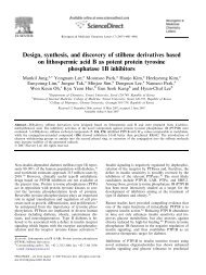
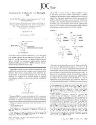
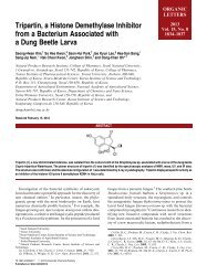
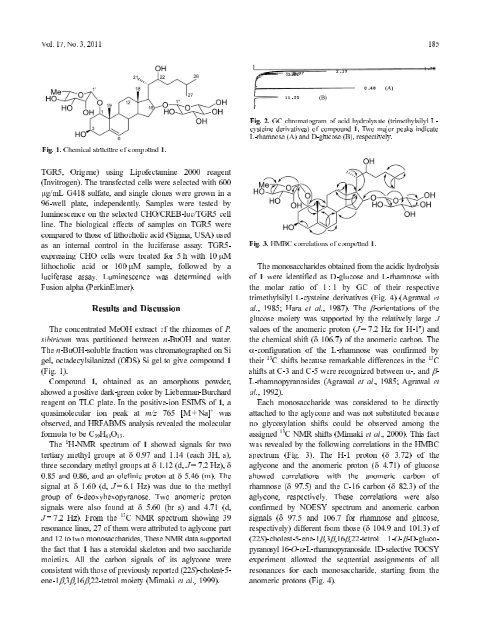
Fig. 1. Chemical structure <strong>of</strong> compound 1.<br />
Origene) using Lip<strong>of</strong>ectamine 2000 reagent<br />
TGR5,<br />
The transfected cells were selected with 600<br />
(Invitrogen).<br />
G418 sulfate, and single clones were grown in a<br />
µg/mL<br />
plate, independently. Samples were tested by<br />
96-well<br />
on <strong>the</strong> selected CHO/CREB-luc/TGR5 cell<br />
luminescence<br />
The biological effects <strong>of</strong> samples on TGR5 were<br />
line.<br />
to those <strong>of</strong> lithocholic acid (Sigma, USA) used<br />
compared<br />
an internal control in <strong>the</strong> luciferase assay. TGR5-<br />
as<br />
CHO cells were treated for 5 h with 10 µM<br />
expressing<br />
acid or 100 µM sample, followed by a<br />
lithocholic<br />
assay. Luminescence was determined with<br />
luciferase<br />
alpha (PerkinElmer).<br />
Fusion<br />
concentrated MeOH extract <strong>of</strong> <strong>the</strong> rhizomes <strong>of</strong> P.<br />
The<br />
was partitioned between n-BuOH and water.<br />
sibiricum<br />
n-BuOH-soluble fraction was chromatographed on Si<br />
The<br />
octadecylsilanized (ODS) Si gel to give compound 1<br />
gel,<br />
1). (Fig.<br />
1, obtained as an amorphous powder,<br />
Compound<br />
a positive dark-green color by Lieberman-Burchard<br />
showed<br />
on TLC plate. In <strong>the</strong> positive-ion ESIMS <strong>of</strong> 1, a<br />
reagent<br />
quasimolecular ion peak at m/z 765 [M + Na] +<br />
and HRFABMS analysis revealed <strong>the</strong> molecular<br />
observed,<br />
to be C 39 H 66 O 13 .<br />
formula<br />
1 H-NMR spectrum <strong>of</strong> 1 showed signals for two<br />
The<br />
methyl groups at δ 0.97 and 1.14 (each 3H, s),<br />
tertiary<br />
at δ 1.60 (d, J = 6.1 Hz) was due to <strong>the</strong> methyl<br />
signal<br />
<strong>of</strong> 6-deoxyhexopyranose. Two anomeric proton<br />
group<br />
were also found at δ 5.60 (br s) and 4.71 (d,<br />
signals<br />
= 7.2 Hz). From <strong>the</strong> 13 C NMR spectrum showing 39<br />
J<br />
lines, 27 <strong>of</strong> <strong>the</strong>m were attributed to aglycone part<br />
resonance<br />
12 to two monosaccharides. These NMR data supported<br />
and<br />
fact that 1 has a steroidal skeleton and two saccharide<br />
<strong>the</strong><br />
All <strong>the</strong> carbon signals <strong>of</strong> its aglycone were<br />
moieties.<br />
with those <strong>of</strong> previously reported (22S)-cholest-5-<br />
consistent<br />
moiety (Mimaki et al., 1999).<br />
ene-1β,3β,16β,22-tetrol<br />
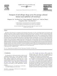
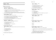
2. GC chromatogram <strong>of</strong> acid hydrolysate (trimethylsilyl L-<br />
Fig.<br />
derivatives) <strong>of</strong> compound 1. Two major peaks indicate<br />
cysteine<br />
monosaccharides obtained <strong>from</strong> <strong>the</strong> acidic hydrolysis<br />
The<br />
1 were identified as D-glucose and L-rhamnose with<br />
<strong>of</strong><br />
molar ratio <strong>of</strong> 1 : 1 by GC <strong>of</strong> <strong>the</strong>ir respective<br />
<strong>the</strong><br />
L-cysteine derivatives (Fig. 4) (Agrawal et<br />
trimethylsilyl<br />
1985; Hara et al., 1987). The β-orientations <strong>of</strong> <strong>the</strong><br />
al.,<br />
moiety was supported by <strong>the</strong> relatively large J<br />
glucose<br />
<strong>of</strong> <strong>the</strong> anomeric proton (J = 7.2 Hz for H-1'') and<br />
values<br />
chemical shift (δ 106.7) <strong>of</strong> <strong>the</strong> anomeric carbon. The<br />
<strong>the</strong><br />
<strong>of</strong> <strong>the</strong> L-rhamnose was confirmed by<br />
α-configuration 13<br />
C shifts because remarkable differences in <strong>the</strong> 13 C<br />
<strong>the</strong>ir<br />
at C-3 and C-5 were recognized between α-, and β-<br />
shifts<br />
(Agrawal et al., 1985; Agrawal et<br />
L-rhamnopyranosides<br />
1992). al.,<br />
monosaccharide was considered to be directly<br />
Each<br />
to <strong>the</strong> aglycone and was not substituted because<br />
attached<br />
glycosylation shifts could be observed among <strong>the</strong><br />
no<br />
13 C NMR shifts (Mimaki et al., 2000). This fact<br />
assigned<br />
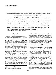
revealed by <strong>the</strong> following correlations in <strong>the</strong> HMBC<br />
was<br />
(Fig. 3). The H-1 proton (δ 3.72) <strong>of</strong> <strong>the</strong><br />
spectrum<br />
and <strong>the</strong> anomeric proton (δ 4.71) <strong>of</strong> glucose<br />
aglycone<br />
correlations with <strong>the</strong> anomeric carbon <strong>of</strong><br />
showed<br />
(δ 97.5) and <strong>the</strong> C-16 carbon (δ 82.3) <strong>of</strong> <strong>the</strong><br />
rhamnose<br />
respectively. These correlations were also<br />
aglycone,<br />
by NOESY spectrum and anomeric carbon<br />
confirmed<br />
(δ 97.5 and 106.7 for rhamnose and glucose,<br />
signals<br />
different <strong>from</strong> those (δ 104.9 and 101.3) <strong>of</strong><br />
respectively)<br />
1-O-β-D-gluco-<br />
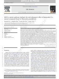
16-O-α-L-rhamnopyranoside. 1D-selective TOCSY<br />
pyranosyl<br />
allowed <strong>the</strong> sequential assignments <strong>of</strong> all<br />
experiment<br />
for each monosaccharide, starting <strong>from</strong> <strong>the</strong><br />
resonances<br />
protons (Fig. 4).<br />
anomeric<br />
Vol. 17, No. 3, 2011 185<br />
L-rhamnose (A) and D-glucose (B), respectively.<br />
Fig. 3. HMBC correlations <strong>of</strong> compound 1.<br />
Results and Discussion<br />
was<br />
secondary methyl groups at δ 1.12 (d, J = 7.2 Hz), δ<br />
three<br />
and 0.86, and an olefinic proton at δ 5.46 (m). The<br />
0.85<br />
(22S)-cholest-5-ene-1β,3β,16β,22-tetrol