Evaluation of Discolouration of Some Composite Restorative Materials
Evaluation of Discolouration of Some Composite Restorative Materials
Evaluation of Discolouration of Some Composite Restorative Materials

You also want an ePaper? Increase the reach of your titles
YUMPU automatically turns print PDFs into web optimized ePapers that Google loves.
<strong>Evaluation</strong> <strong>of</strong> Discoloration od some <strong>Composite</strong> <strong>Restorative</strong> <strong>Materials</strong><br />
EVALUATION OF DISCOLOURATION OF SOME COMPOSITE<br />
RESTORATIVE MATERIALS<br />
1<br />
NADEEM HAFEEZ KHOKHAR, DMD (Philippines), M.Sc (Malaysia)<br />
2<br />
REHAN QURESHI, BSc, BDS, MSc (Manchester)<br />
3<br />
SYED MUMTAZ ALI, BDS (Sindh), MCPS<br />
ABSTRACT<br />
This in-Vitro study was conducted to investigate the stain resistance <strong>of</strong> direct composite resin<br />
restorative materials with different polishing systems (diamond finishing and polishing burs,<br />
aluminium oxide disks and abrasive impregnated disks) in comparison to the material cured against<br />
a mylar strip. The composite resin materials investigated included Clearfil AP-X (hybrid), Filtek Z-250<br />
(hybrid), Definite (Ormocer), and Silux Plus (micr<strong>of</strong>ill).<br />
Forty disk specimens (10 mm x 2 mm) <strong>of</strong> each composite resin were prepared. Ten mylar cured<br />
specimens were assigned for each polishing system and ten were used as controls with no further<br />
treatment after curing.<br />
A spectrophotometer (Spectraflash 500) was used to determine the initial colour and the colour<br />
change after one week immersion. All specimens were stored in tea separately for one week at 39 °C.<br />
Post immersion colour measurements were undertaken to assess colour change (Δ E).<br />
One way ANOVA showed that there was statistically significant difference between the different<br />
composites and the polish groups for discoloration at p < 0.05. All the composites tested showed<br />
significant discoloration when polished with Diamond burs. The least discoloration was found when<br />
specimens were polished with s<strong>of</strong>lex except with Z-250 which discoloured less under mylar than s<strong>of</strong>lex<br />
but the difference was not significant. Among the materials tested AP-X showed the least discoloration.<br />
Key words: <strong>Composite</strong>s, discolouration<br />
INTRODUCTION<br />
Aesthetics plays a major role in the development <strong>of</strong><br />
dentistry and dental research. The trend towards the<br />
natural look has paved the way for the development <strong>of</strong><br />
tooth coloured restoratives that simulate the tooth as<br />
closely as possible. 1<br />
<strong>Composite</strong> resins are gaining popularity and drawing<br />
major attention as aesthetic anterior restoratives<br />
2<br />
and as a dominant alternative to amalgam for direct<br />
restoration <strong>of</strong> posterior teeth. 3 <strong>Composite</strong> resins being<br />
used are susceptible to discolour by taking up stains<br />
from normal diet and food dyes. 4 Discoloration is<br />
caused by distinct pigments that penetrate the composite<br />
resins. 5,6 Tannin is the component in tea, which<br />
contains tannic acid, which causes the discoloration in<br />
composite resins. 7 The extent <strong>of</strong> discoloration may be<br />
associated with individuals’ dietary habits. As suggested<br />
the acid causes the swelling <strong>of</strong> the restoration<br />
influencing the susceptibility to surface staining. The<br />
presence <strong>of</strong> tannic acid may also induce staining by the<br />
same phenomenon. 8<br />
One <strong>of</strong> the most troublesome features <strong>of</strong> these<br />
aesthetic materials is the difficulty <strong>of</strong> finishing the<br />
1<br />
Associate Pr<strong>of</strong>essor & Head, Department <strong>of</strong> Dental <strong>Materials</strong> & Basic Dental Sciences,.Hamdard University,<br />
Karachi.<br />
2<br />
Assistant Pr<strong>of</strong>essor & Head, Department <strong>of</strong> Operative Dentistry, Karachi Medical & Dental College, Karachi.<br />
3<br />
Associate Pr<strong>of</strong>essor & Head, Department <strong>of</strong> Community Dentistry, Hamdard University, Karachi.<br />
Correspondence: Dr. Rehan Qureshi, 68/2, Khayaban-e-badban, 11 th , Street, Phase 5,Defence Housing<br />
Authority, Karachi. E-mail: rehanq20@hotmail.com, Phone No: 0300-2111175.<br />
Pakistan Oral & Dental Journal Vol 29, No. 1, (June 2009)<br />
123
<strong>Evaluation</strong> <strong>of</strong> Discoloration od some <strong>Composite</strong> <strong>Restorative</strong> <strong>Materials</strong><br />
restoration surface to decrease adherence <strong>of</strong> food debris<br />
onto the restoration that may lead to discoloration<br />
especially at the margins. 9 The rougher the finished<br />
surface is, the more chance there is for bacterial<br />
accumulation and discoloration <strong>of</strong> restoration along<br />
the restoration margin with secondary caries formation.<br />
10, 11<br />
To prevent the accumulation <strong>of</strong> plaque and<br />
stain pigmentation from food, a finished composite<br />
resin surface should be highly polished and very<br />
12, 13, 14,15,16,17<br />
smooth.<br />
Previously a lot <strong>of</strong> attention has been paid in the<br />
surface effects <strong>of</strong> surface roughness but not much work<br />
has been done on discoloration and stain control in<br />
newer materials.<br />
METHODOLOGY<br />
Forty specimens <strong>of</strong> 10 mm diameter and 2 mm<br />
thickness were made from each composite resin and<br />
ormocer listed in Table 1 & shown in figure 1, to form<br />
four experimental groups. The composite resin was<br />
injected directly into a cylindrical polytetrafluoroethylene<br />
(PTFE) mould that was placed on a<br />
microscopic slide with a celluloid mylar strip over it.<br />
The open surface <strong>of</strong> the mould was covered with a<br />
mylar strip and a microscopic glass slide. Finger<br />
pressure was applied on top <strong>of</strong> the glass slide to remove<br />
excess material and to form a parallel plane surface.<br />
The glass slide was used to protect the composite resin<br />
from oxygen inhibition.<br />
The resin was cured through the upper side <strong>of</strong><br />
the glass slide for 40 seconds with a visible light<br />
activation unit (Coltolux 3 Coltené/ Whaledent<br />
Inc., USA). The plates were then reversed to cure<br />
the resin from the other side <strong>of</strong> the plate for 40 seconds.<br />
The tip <strong>of</strong> the light source was placed against<br />
the glass slide to ensure uniform distance from<br />
the light source to the composite resin. All procedures<br />
were carried out at room temperature (23°C<br />
± 2°C).<br />
Each group <strong>of</strong> specimens was randomly divided into<br />
four subgroups <strong>of</strong> 10 specimens each and was randomly<br />
assigned as either the control group or to one <strong>of</strong> the<br />
three polishing systems listed in Table 2 and shown in<br />
Figure 2.<br />
Pakistan Oral & Dental Journal Vol 29, No. 1, (June 2009)<br />
Polishing <strong>of</strong> the samples<br />
1 Polishing with aluminium oxide disks (S<strong>of</strong>lex®)<br />
The specimens in this subgroup were polished<br />
immediately after curing. S<strong>of</strong>-lex pop-on contouring<br />
and polishing disc system (3M Dental products, St Paul<br />
Minn USA) was used according to the manufacturer’s<br />
instructions. Coarse grit was used at about 10,000 rpm<br />
for gross reduction. The surface <strong>of</strong> the filling material<br />
was then rinsed and dried. The medium grit was used<br />
at 10,000 rpm for 15 seconds followed by fine grit at<br />
30,000 rpm for 15 seconds and superfine grit at 30,000<br />
rpm for 15 seconds. Any debris or powder formed on<br />
the surface <strong>of</strong> the filling materials was washed away<br />
with water.<br />
2 Diamond polishing burs (Komet® Germany)<br />
The Komet diamond polishing burs were used at<br />
high speed in a sequential manner from coarse grit,<br />
medium grit, fine grit to ultra fine grit. The procedure<br />
was performed using water spray. Each bur was used<br />
for 30 seconds.<br />
3 Abrasive impregnated polishing system (Enhance®<br />
Polishing System)<br />
The Enhance polishing system (Dentsply/Caulk<br />
USA) finishing discs were used to finish the composite<br />
resin surface. Polishing was done at low speed using<br />
light pressure in a circular motion as per manufacturer’s<br />
recommendation.<br />
The surface was then polished with the Prisma<br />
gloss paste. A small amount <strong>of</strong> Prisma gloss was<br />
applied to the surface <strong>of</strong> the finishing cup. The cup<br />
worked on the surface <strong>of</strong> restoration with moderate<br />
speed and pressure, initially for 30 seconds. To increase<br />
lustre, water was added in small amounts (drop<br />
wise) to dilute the paste using a light buffering motion<br />
for 30 seconds. The paste was rinsed <strong>of</strong>f from the<br />
restoration and cup. The other composite resin materials<br />
were further polished with Prisma gloss extrafine<br />
paste using the same procedure as that used with<br />
Prisma gloss. The cup was discarded after each use.<br />
Post polishing treatment<br />
All specimens were observed under light microscope<br />
(Kyawa Optical Co. Ltd., Japan) under 23 x 10<br />
124
<strong>Evaluation</strong> <strong>of</strong> Discoloration od some <strong>Composite</strong> <strong>Restorative</strong> <strong>Materials</strong><br />
TABLE 1: COMPOSITE RESINS AND ORMOCER RESTORATIVE MATERIALS USED IN THE STUDY<br />
<strong>Composite</strong> resin & Shade Composition Number <strong>of</strong> Manufacturer Batch<br />
ormocer material specimens number<br />
Silux-Plus U Heterogeneous 40 3M Dental products, Lot<br />
micr<strong>of</strong>illed St Paul Minn USA 19990222<br />
Ref 5702-U<br />
Clearfil AP-X A3 Midifilled Hybrid 40 Kuraray Co. Ltd Japan 0596AB<br />
Filtek Z-250 A3 Minifilled Hybrid 40 3M Dental products, Lot 9BN<br />
St Paul Minn USA Ref 1370AB<br />
Definite ormocer A3 Minifilled Hybrid 40 Degussa AG, Lot 21<br />
Geschäftsber-eich Ref 4380003<br />
Dental Hanau<br />
TABLE 2: POLISHING MATERIALS USED IN THIS STUDY<br />
Polishing system Manufacturer Composition<br />
S<strong>of</strong>-lex Pop-on contouring 3M Dental products, Aluminium oxide discs coarse,<br />
and polishing disc system St Paul Minn USA medium, fine and superfine<br />
Komet® Diamond polishing burs Komet, Germany Diamond finishing and<br />
polishing burs coarse,<br />
medium, fine and superfine<br />
Enhance® composite finishing Dentsply/ Caulk USA Abrasive impregnated silicon<br />
and polishing system<br />
discs with 2 polishing pastes<br />
Ruwa mylar strips Kem-Dent, UK Polyester foil strips for uni<br />
versal use<br />
TABLE 3: COMPARISON OF MEAN<br />
DISCOLORATION FOR SILUX PLUS<br />
TABLE 5: COMPARISON OF MEAN<br />
DISCOLORATION FOR DEFINITE<br />
Polishing material<br />
Mean discoloration<br />
(ΔE)<br />
Polishing material<br />
Mean discoloration<br />
(ΔE)<br />
S<strong>of</strong>lex 3.20<br />
Enhance 3.50<br />
Mylar 4.56<br />
Diamond 4.90<br />
S<strong>of</strong>lex 3.62<br />
Mylar 4.11<br />
Enhance 4.24<br />
Diamond 5.29<br />
TABLE 4: COMPARISON OF MEAN<br />
DISCOLORATION FOR CLEARFIL AP-X<br />
TABLE 6: COMPARISON OF MEAN<br />
DISCOLORATION FOR FILTEK Z-250<br />
Polishing material<br />
Mean discoloration<br />
(ΔE)<br />
Polishing material<br />
Mean discoloration<br />
(ΔE)<br />
S<strong>of</strong>lex 2.63<br />
Mylar 3.03<br />
Enhance 3.06<br />
Diamond 5.27<br />
S<strong>of</strong>lex 3.79<br />
Mylar 3.92<br />
Enhance 4.55<br />
Diamond 5.88<br />
Pakistan Oral & Dental Journal Vol 29, No. 1, (June 2009)<br />
125
<strong>Evaluation</strong> <strong>of</strong> Discoloration od some <strong>Composite</strong> <strong>Restorative</strong> <strong>Materials</strong><br />
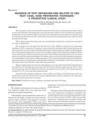
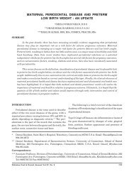
a = spectrophotometer, b= calibrating box<br />
Fig 3: Spectraflash spectrophotometer<br />
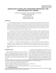
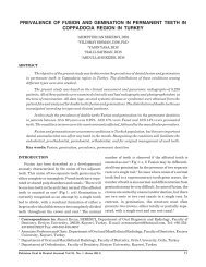
Fig 1:<br />
Left to right: Clearfil AP-X, Filtek Z-250, Silux<br />
Plus, Definite)<br />
magnification to see the surface topography and the<br />
effect <strong>of</strong> polishing on the surface. Each specimen was<br />
placed in dark container filled with 20 ml distilled water<br />
individually for 24 hours.<br />
Initial colorimetric evaluations were carried out<br />
using (Spectraflash) spectrophotometer as shown in<br />
Figure 3. The CIE (Comission Internationale de<br />
L’Eclairage) standard illuminant D 65<br />
was used in this<br />
study. Each sample was removed from the container,<br />
rinsed for 60 seconds and dried with an absorbent paper<br />
before placing it against the eyelet <strong>of</strong> the aperture <strong>of</strong> 6<br />
mm diameter. The disc came in contact with the eyelet<br />
at a radius <strong>of</strong> 3 mm from the center. Five readings were<br />
taken per specimen. The average CIElab reading was<br />
computed by the s<strong>of</strong>tware program and was recorded.<br />
The samples were then immersed in the tea (Lipton<br />
Yellow label tea bags, Lipton Pvt Ltd, UK) solution for<br />
one week. The tea was prepared by placing two tea<br />
bags in 250 ml boiling water for 5 minutes as per<br />
manufacturer’s instructions. Each specimen was stored<br />
in one container at 37ºC for seven days in an incubator<br />
to simulate the temperature <strong>of</strong> the oral cavity. Each<br />
container was separately marked.<br />
On the 8 th day, the samples were taken out <strong>of</strong> the<br />
tea solution, rinsed thoroughly and dried with an<br />
absorbent paper. Another colorimetric reading was<br />
performed.<br />
The measurements <strong>of</strong> experimental group <strong>of</strong><br />
samples were compared with regards to different surface<br />
treatments.<br />
The difference was calculated as:<br />
whereby:<br />
ΔE<br />
ΔE = [(ΔL) 2 + (Δa) 2 + (Δb) 2 ]½<br />
= is the difference in colour<br />
ΔL = is the difference in brightness values (L 2<br />
- L 1<br />
).<br />
“L 1<br />
” indicates the pre staining value, “L 2<br />
” indi-<br />
7.00<br />
DISCOLORATION EVALUATION<br />
DISCOLORATION<br />
6.00<br />
5.00<br />
4.00<br />
3.00<br />
2.00<br />
1.00<br />
S<strong>of</strong>lex<br />
Mylar<br />
Enhance<br />
Diamond<br />
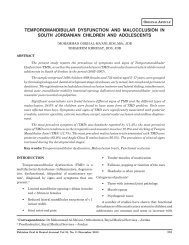
Fig 2: S<strong>of</strong>lex, Enhance, Mylar, Diamond burs<br />
Pakistan Oral & Dental Journal Vol 29, No. 1, (June 2009)<br />
0.00<br />
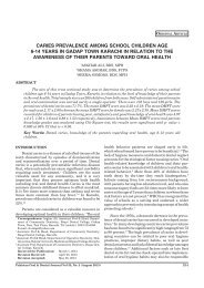
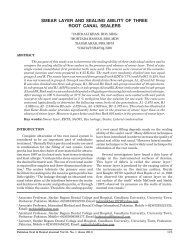
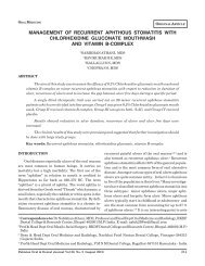
Fig 4:<br />
APX DE SP Z-250<br />
<strong>Evaluation</strong> <strong>of</strong> discoloration between composite<br />
groups and polishing groups<br />
126
<strong>Evaluation</strong> <strong>of</strong> Discoloration od some <strong>Composite</strong> <strong>Restorative</strong> <strong>Materials</strong><br />
cates the post staining value and “L” = indicates<br />
the brightness (a value <strong>of</strong> 100 corresponds to<br />
perfect white and that <strong>of</strong> zero to black)<br />
Δa = is the difference in the red green scale (a 2<br />
– a 1<br />
).<br />
a 1<br />
indicates the pre staining value, a 2<br />
indicates<br />
the post staining values and “a” = determines<br />
the amount <strong>of</strong> red (positive values) and green<br />
(negative values)<br />
Δb<br />
= determines the difference in yellow blue scale<br />
(b 2<br />
– b 1<br />
). b 1<br />
indicates the pre staining value, b 2<br />
indicates the post staining values and “b”=<br />
determines the amount <strong>of</strong> yellow (positive<br />
values) and blue (negative values).<br />
The difference between the initial colorimetric<br />
measurement and the post immersion measurement<br />
was calculated for each specimen and used for statistical<br />
analysis.<br />
RESULTS<br />
Upon visual examination, the gloss <strong>of</strong> the polished<br />
specimens were found to be less than that <strong>of</strong> the<br />
Control (mylar cured) specimens. All specimens exhibited<br />
discoloration after one week <strong>of</strong> immersion. The<br />
light microscopy examination showed porosities on the<br />
surface <strong>of</strong> the specimens cured under the mylar strip.<br />
Superficially the extrinsic discoloration was seen along<br />
the air entrapment areas.<br />
Statistical analysis was performed using One way<br />
ANOVA. Among the composite materials tested,<br />
Clearfil AP-X discoloured the least followed by Silux<br />
Plus, Definite and Filtek Z-250 except for mylar treated<br />
group where Filtek Z-250 discoloured less than Silux<br />
Plus and Definite. Post-hoc Scheffe’s test showed that<br />
there was no significant difference (p < 0.05) found<br />
between the Clearfil AP-X, Silux Plus and Definite.<br />
Among the polishing materials used the least<br />
discoloration was found when the specimens were<br />
polished with S<strong>of</strong>lex disks followed by mylar, Enhance<br />
and diamond burs except Clearfil AP-X (where the<br />
specimens polished with Enhance discoloured less than<br />
that <strong>of</strong> mylar) and Z-250 (where the least discoloration<br />
was found with mylar).<br />
DISCUSSION<br />
Pakistan Oral & Dental Journal Vol 29, No. 1, (June 2009)<br />
The methods currently available and used as a<br />
research methodology to measure discoloration include<br />
visual and instrumental techniques. Instrumental<br />
techniques, however, are most commonly used in<br />
research. The human eye is best in detecting colour<br />
difference by comparison. 18 Other detectors used in<br />
place <strong>of</strong> eye as an observer are photo-detectors like<br />
colorimeters and spectrophotometers 19 whose use make<br />
it possible to overcome basic difficulties <strong>of</strong> evaluation.<br />
20 The National Bureau <strong>of</strong> Standards (NBS) established<br />
a sophisticated classification to describe colour<br />
differences by NBS units. A human eye can perceive a<br />
colour shift at ΔE > 1. The acceptable limit for the<br />
colour shift set by NBS is ΔE < 3.3. Colour shift with<br />
corresponding E values less than 3.3 are acceptable. 21<br />
Above this value is marked colour shift. 22<br />
Instrumental colorimetry is more authentic over<br />
visual colorimetry because descriptive visual comparisons<br />
are less reliable in making quantitative comparison<br />
<strong>of</strong> materials and treatments. Instrumental colorimetry<br />
can potentially eliminate the subjective errors<br />
<strong>of</strong> colour assessments 23 like observer’s variability (physiological<br />
and psychological) and optical metamerism. 24<br />
The advantage <strong>of</strong> spectrophotometer is that it measures<br />
the amount <strong>of</strong> light reflected by the surface<br />
within a full reflectance spectrum. The perceivable<br />
values set by NBS are DE between 1 and 3.3 NBS units.<br />
In the visual method, colour difference value greater<br />
than 2 NBS units can always be correctly judged. When<br />
the measured colour difference falls within 1 to 2 NBS<br />
units, incorrect judgements become frequent and when<br />
the measured colour difference becomes less than 1 ΔE<br />
unit, the visual technique cannot distinguish the colour<br />
change. 25<br />
In this study, all ΔE values were above 1 NBS unit.<br />
Only one observation had a reading below 2 ΔE.<br />
Majority <strong>of</strong> the readings were above 3.3 NBS units<br />
except for the Clearfil AP-X specimens polished with<br />
mylar, S<strong>of</strong>lex and Enhance and the Silux Plus specimens<br />
polished with S<strong>of</strong>lex. This indicates a severe<br />
colour change in the majority <strong>of</strong> the specimens.<br />
It should also be noted that there were statistically<br />
significant differences (p < 0.05) in the initial colorimetric<br />
readings found between some <strong>of</strong> the restorative<br />
materials tested even though same shade A3 was used<br />
by different manufacturers. This suggests that there<br />
exist a significant difference between the CIElab value<br />
and the hue and chroma <strong>of</strong> these materials. This in<br />
turn suggests a need for improved shade standardisation.<br />
127
<strong>Evaluation</strong> <strong>of</strong> Discoloration od some <strong>Composite</strong> <strong>Restorative</strong> <strong>Materials</strong><br />
The different polishing systems were chosen because<br />
<strong>of</strong> their popular use in clinical practice. The<br />
mylar subgroup acted as the control as it has been<br />
generally accepted that composite resin cured under a<br />
mylar strip would produce the smoothest surface. It<br />
has been found that the surface cured directly in<br />
contact with mylar discoloured more than the polished<br />
surface. 26, 27 This was true only for Silux Plus specimens<br />
cured under the mylar strip which discoloured more<br />
than those in the S<strong>of</strong>lex and Enhance subgroups at a<br />
visually perceptible level. Filtek Z-250 specimens, on<br />
other hand cured under the mylar strip registered the<br />
lowest DE value among the subgroups. The difference<br />
in the type <strong>of</strong> composite resin may explain these<br />
findings. It should also be noted that the studies cited<br />
were made in the 1970’s and 1980’s where improvement<br />
on composite resin chemistry is not comparable<br />
to the materials that are available to us today. The<br />
strain that is caused on the surface <strong>of</strong> composite cured<br />
against the mylar strip increases the activity <strong>of</strong> the<br />
atoms on the surface and facilitates accumulation <strong>of</strong><br />
stains. It has been previously found that when the<br />
resin rich layer was removed by polishing with S<strong>of</strong>lex<br />
disks the discoloration was reduced.<br />
Every component <strong>of</strong> resin may be implicated in<br />
discoloration. The resin matrix plays major part in the<br />
colour stability <strong>of</strong> composites. The high viscosity <strong>of</strong><br />
resin matrix is reduced by proper mixture <strong>of</strong> diluent, to<br />
help incorporate more filler loading. The variation in<br />
the water sorption rate between materials using Bis-<br />
GMA matrix may be due to the different proportions <strong>of</strong><br />
diluent TEGDMA. 28 The addition <strong>of</strong> fillers reduces<br />
polymerisation shrinkage, coefficient <strong>of</strong> thermal expansion<br />
and water sorption. As for water sorption it<br />
has been shown that materials exhibiting high water<br />
sorption values are more easily stained by hydrophilic<br />
colourings in aqueous solutions, the water presumably<br />
acting as a penetrating vehicle. The hydrophilic composites<br />
allow water to penetrate the matrix or filler<br />
matrix interface. 29,30, 31,32 In instances where both adsorption<br />
and absorption are known to exist and it is not<br />
clear which process predominates, the whole process is<br />
called as sorption. The staining may result from adsorption<br />
<strong>of</strong> stain on the surface <strong>of</strong> the composite 4<br />
following the absorption <strong>of</strong> water by the resin matrix. 33<br />
Pakistan Oral & Dental Journal Vol 29, No. 1, (June 2009)<br />
The least discoloration was found upon polishing<br />
with S<strong>of</strong>lex disks followed in ascending order by Enhance,<br />
mylar and diamond burs system (Table 3).<br />
There was a greater amount <strong>of</strong> discoloration in specimens<br />
polished with diamond instruments and cured<br />
under the mylar strip as compared to specimens polished<br />
with S<strong>of</strong>lex and Enhance. The difference in the<br />
discoloration (ΔE) in NBS units between the mylar and<br />
S<strong>of</strong>lex and mylar and Enhance were 1.36 and 1.06<br />
respectively, which is discernible by the naked eye. It<br />
has been shown that the resin matrix plays a major<br />
part in the colour stability <strong>of</strong> composites and water<br />
sorption rate is <strong>of</strong> particular importance. 34,35,36 The Bis-<br />
GMA matrix is a very viscous and bulky difunctional<br />
monomer. The high viscosity <strong>of</strong> Bis-GMA is reduced by<br />
the admixture <strong>of</strong> trietylene glycol dimethacrylate<br />
(TEGDMA), which is more reactive. The lower the<br />
viscosity <strong>of</strong> the monomer mixture, the more filler may<br />
be incorporated into the mixture. An increased filler<br />
content will decrease water sorption rate. 37 As the<br />
surface area to volume ratio <strong>of</strong> the colloidal particles in<br />
Silux Plus is quite high (52% wt), it is difficult to attain<br />
higher level <strong>of</strong> loading in composite resins containing<br />
larger fillers. This reduced size <strong>of</strong> pyrolytic silica and<br />
the consequently filler matrix contact may also account<br />
for the increased water sorption and staining<br />
phenomena. 38,39<br />
Silux Plus, a micr<strong>of</strong>illed composite resin used as a<br />
control among the composites, performed as expected.<br />
It contains Bis-GMA as a matrix and TEGDMA as a<br />
diluent. It uses pyrogenic silica as fillers distributed at<br />
a concentration <strong>of</strong> 52% by weight and increased water<br />
sorption.<br />
Clearfil AP-X is a midifilled hybrid type <strong>of</strong> composite<br />
resin. It contains Bis-GMA as a matrix and TEGDMA as<br />
a diluent. It uses barium glass as fillers distributed at a<br />
concentration <strong>of</strong> 85% by weight. The mean ΔE value was<br />
found to be lowest when the surface was polished with<br />
S<strong>of</strong>lex disks followed in ascending order by the mylar<br />
strip, Enhance and diamond polishing system (Table 4).<br />
The difference in colour <strong>of</strong> the specimens in the S<strong>of</strong>lex<br />
and mylar subgroups against the diamond subgroup was<br />
visually discernible. The ΔE <strong>of</strong> the specimens <strong>of</strong> the<br />
diamond subgroup (5.27) was more severe than those in<br />
the Enhance subgroup (3.06). The discoloration in the<br />
Enhance subgroup was not visually discernible to those<br />
specimens in the S<strong>of</strong>lex and mylar subgroups. The<br />
difference in the degree <strong>of</strong> discoloration is visually<br />
discernible. Seven out <strong>of</strong> 10 specimens polished with<br />
128
<strong>Evaluation</strong> <strong>of</strong> Discoloration od some <strong>Composite</strong> <strong>Restorative</strong> <strong>Materials</strong><br />
Enhance had ΔE values below 3.3. The rest <strong>of</strong> the<br />
specimens in the Enhance subgroup had DE values<br />
ranging from 3.43 to 3.71. A possible explanation for this<br />
is the use <strong>of</strong> the polishing paste in the Enhance polishing<br />
system. A film may have formed on the surface <strong>of</strong> the<br />
composite resin that hindered the adsorption and absorption<br />
<strong>of</strong> the staining solution. This decreased amount<br />
<strong>of</strong> discoloration, however, was true only for the Clearfil<br />
AP-X Enhance subgroup. The chemistry <strong>of</strong> the composite<br />
resin may have played a role in the apparent reduced<br />
stainability <strong>of</strong> these specimens. The more severe<br />
discoloration in the diamond subgroup could be perceived<br />
visually.<br />
Definite<br />
A definitive classification <strong>of</strong> ormocer has yet to be<br />
established. Ormocer is a composite material and its<br />
chemistry <strong>of</strong> this material appears to be that <strong>of</strong> a<br />
modified composite resin. It uses barium glass as<br />
fillers distributed at a concentration <strong>of</strong> 77% by weight.<br />
The material is said to have improved polymerisation<br />
shrinkage and water sorption properties. The matrix,<br />
which consists <strong>of</strong> ceramic polysiloxane, may have led to<br />
these improvements.<br />
Specimens treated with S<strong>of</strong>lex had the least mean<br />
discoloration value followed by mylar, Enhance and<br />
diamond system (Table 5). There was, however, no<br />
significant difference (p < 0.05) between the S<strong>of</strong>lex,<br />
mylar, Enhance treated specimens. There was also no<br />
significant difference (p < 0.05) in the discoloration<br />
values between the mylar, Enhance and the diamond<br />
system.<br />
These results are in agreement with the results<br />
<strong>of</strong> studies 26 that mylar treated surface discolours<br />
more than the polished surface. Enhance disks and<br />
diamond burs produced the roughest surface on Clearfil<br />
AP-X, Filtek Z-250 and Definite. This is in agreement<br />
with the findings <strong>of</strong> Stoddard and Johnson 40 that<br />
regardless <strong>of</strong> composite resin filler type, Enhance<br />
disks and diamond burs produced the roughest surface.<br />
This was not true for Silux Plus, which had more<br />
discoloration in the mylar subgroup than for Enhance.<br />
Filtek Z-250.<br />
Filtek Z-250 specimens with the lowest mean<br />
discoloration value was in the mylar subgroup followed<br />
Pakistan Oral & Dental Journal Vol 29, No. 1, (June 2009)<br />
in ascending order by those in the S<strong>of</strong>lex subgroup,<br />
Enhance subgroup and diamond system subgroup.<br />
There was no statistically significant difference (p <<br />
0.05) between mylar, S<strong>of</strong>lex and Enhance subgroups.<br />
There was also no significant difference (p < 0.05) in the<br />
mean discoloration values between the Enhance and<br />
diamond subgroup (Table 6).<br />
Comparison between the groups<br />
Clearfil AP-X discoloured the least followed by<br />
Silux Plus, Definite and Filtek Z-250 except for mylar<br />
treated group where Filtek Z-250 discoloured less than<br />
Silux Plus and Definite (Fig 1). There was no significant<br />
difference (p < 0.05) found between the ΔE<br />
value <strong>of</strong> Clearfil AP-X, Silux Plus and Definite.<br />
There was also no significant difference (p < 0.05) in<br />
the ΔE value between Silux Plus, Definite and Filtek<br />
Z-250. These results were expected based on the<br />
water sorption values given by the manufacturers.<br />
Our results are in agreement with other studies<br />
which suggest that hydrophilic composites allow<br />
water to penetrate the matrix or filler matrix interface.<br />
29,30,31,32<br />
Overall, Clearfil AP-X showed the least discoloration<br />
among the composite materials tested and among<br />
the polishing materials tested S<strong>of</strong>lex produced the<br />
least discoloration.<br />
RECOMMENDATIONS<br />
1 The Komet diamond burs system should not be<br />
regarded as a final step in the polishing procedure<br />
as it produces a rough surface and prone to discoloration.<br />
2 Results <strong>of</strong> the study also suggest that Silux Plus<br />
need to be polished when cured under the mylar<br />
strip to reduce discoloration.<br />
CONCLUSION<br />
The ΔE (discoloration) values <strong>of</strong> the test materials<br />
cured against the mylar strip is consistently greater<br />
than those polished with S<strong>of</strong>lex disks. The difference,<br />
however, is not visually perceptible.<br />
DISCLAIMER<br />
Authors state that they have not gained any material<br />
advantage from this study.<br />
129
<strong>Evaluation</strong> <strong>of</strong> Discoloration od some <strong>Composite</strong> <strong>Restorative</strong> <strong>Materials</strong><br />
REFERENCES<br />
1 Owens BM. Replacement and initial placement <strong>of</strong> tooth<br />
colored restorations: a review and discussion. J Tenn Dent<br />
Assoc. 1998; 78: 26-29<br />
2 Devoto W, Pansecilli D. <strong>Composite</strong> restoration in the anterior<br />
region: clinical and aesthetic purposes. Pract Proced Aesthetic<br />
Dent. 2007; 19 (8):465-70.<br />
3 Lutz F, Krejci I. Resin <strong>Composite</strong>s the Post Amalgam age.<br />
Compend continu Educ Dent. 2001; 20 (12): 1138-44, 1146-48.<br />
4 Dietschi D, Campanile G, Jacques H and Meyer J. Comparison<br />
<strong>of</strong> the color stability <strong>of</strong> ten new-generation composites. An In<br />
Vitro study. Dent Mater. 1994; 10: 353-62.<br />
5 Lazetti G, Burgess J, Gardiner D and Ripps. A Color stability<br />
<strong>of</strong> fluoride-containing restorative materials. Oper Dent. 2000;<br />
25: 520–25.<br />
6 Um C and Ruyter I. Staining <strong>of</strong> resin based veneering materials<br />
with c<strong>of</strong>fee and tea. Quintessence Int. 1991; 22:<br />
377-86.<br />
7 Uchida H, Vaidyanathan J, Viswanadhan T and Vaidyanathan<br />
T. Color stability <strong>of</strong> dental composites as a function <strong>of</strong> shade.<br />
J Prosthet Dent. 1998; 79: 372-77.<br />
8 Asmussen E. S<strong>of</strong>tening <strong>of</strong> BISGMA-based polymers by ethanol<br />
and by organic acids <strong>of</strong> plaque. Scand J Dent Res. 1984; 92:<br />
257-61.<br />
9 Mc Cabe J and Caddick R. The finishing <strong>of</strong> composite restorations.<br />
Br Dent J. 1978; 145: 101-10.<br />
10 Lu H, Roeder LB, Lei L, Powers JM. Effect <strong>of</strong> surface roughness<br />
on stain resistance <strong>of</strong> dental composites. J Esthet Restor<br />
Dent. 2005;17(2):102-8.<br />
11 Fruits T, Miranda F and Coury T. Effect <strong>of</strong> equivalent abrasive<br />
grit sizes utilizing differing polishing motion on selected<br />
restorative materials. Quintessence Int. 1996; 27: 279-85.<br />
12 Setcos J, Tarim B and Suzuki S. Surface finish produced on<br />
resin composites by new polishing systems. Quintessence Int.<br />
1999; 30: 169-73.<br />
13 Kaplan B, Goldstein G and Vijayaraghavan T. The effect <strong>of</strong><br />
three polishing systems on the surface roughness <strong>of</strong> four<br />
hybrid composites: A pr<strong>of</strong>ilometric and scanning electron<br />
microscopy study. J Prosthet Dent. 1996;76: 34-38.<br />
14 Wise M and Dykema R. The plaque retaining capacity <strong>of</strong> four<br />
dental materials. J Prosthet Dent. 1975; 33: 542-45.<br />
15 Chan K, Fuller J and Hormati A. The ability <strong>of</strong> food to stain two<br />
composite resins. J Prosthet Dent. 1980; 43: 542-45.<br />
16 Attar N. The effect <strong>of</strong> finishing and polishing procedures on<br />
the surface roughness <strong>of</strong> composite resins materials. J Contemp<br />
Dent Pract. 2007; 8(1): 27-35.<br />
17 LU H, Roeder LB, Powers JM. Effect <strong>of</strong> polishing systems on<br />
the surface roughness <strong>of</strong> microhybrid composites. J Esthet<br />
Restor Dent. 2003; 15(5):297-303.<br />
18 Billmeyer F Jr and Saltzman M. Principles <strong>of</strong> color technology.<br />
Second edition. John Wiley and sons Inc. New York. 1981; pp<br />
73-09.<br />
19 O’Brien W. Dental materials. Properties and selection. Quintessence<br />
Pub. Co. Inc. Chicago, London, Berlin, São Paulo,<br />
Tokyo and Hong Kong. 1989; pp 138-55.<br />
20 Knispel G. Factors affecting the process <strong>of</strong> color matching<br />
restorative materials to natural teeth. Quintessence Int.<br />
1991; 22: 525-31.<br />
Pakistan Oral & Dental Journal Vol 29, No. 1, (June 2009)<br />
21 Ruyter I, Nilner K and Moller B. Color stability <strong>of</strong> dental<br />
composite resin materials for crown and bridge veneers. Dent<br />
Mater.1987; 3: 246-51.<br />
22 Rosentritt M, Esch J, Behr M, Leibrock A and Handel G. In vivo<br />
color stability <strong>of</strong> resin composite veneers in removable partial<br />
dentures. Quintessence Int. 1998; 29: 517-22.<br />
23 Yannikakis S, Zissis A, Polyzois G and Caroni C. Color stability<br />
<strong>of</strong> resin restorative materials. J Prosthet Dent. 1998; 80:<br />
533-39.<br />
24 Gross M and Moser J. A colorimetric study <strong>of</strong> c<strong>of</strong>fee and tea<br />
staining <strong>of</strong> four composite resins. J Oral Rehabil. 1976; 4:<br />
311-22.<br />
25 Seghi R, Johnston W, O’Brien W. Performance assessment <strong>of</strong><br />
colorimetric devices on dental porcelains. J Dent Res. 1989;<br />
68: 1755-59.<br />
26 Hachiya Y, Masaaki I, Hosoda H and Fusayama T. Relation <strong>of</strong><br />
finish to discoloration <strong>of</strong> composite resin. J Prosthet Dent.<br />
1984; 52: 811-14.<br />
27 Hayashi H, Maejima K, Kezuka K, Ogushi K and Fusayama<br />
T. In-Vitro study <strong>of</strong> discoloration <strong>of</strong> composite resins. J<br />
Prosthet Dent. 1974; 32: 66-69.<br />
28 Pearson G and Longman C.Water sorption and solubility <strong>of</strong><br />
resin-based materials following inadequate polymerization by<br />
a visible-light curing system. J Oral Rehabil. 1989; 16: 57-6.<br />
29 Braden M and Clarke R. Water absorption characteristics <strong>of</strong><br />
dental micr<strong>of</strong>ine composite filling materials. I. Proprietary<br />
materials. Biomaterials. 1984; 5: 369-72.<br />
30 Fan P, Edahl A, Leung R and Stanford J. Alternative interpretation<br />
<strong>of</strong> water sorption values <strong>of</strong> composite resins. J Dent<br />
Res. 1985; 64: 78-80.<br />
31 Øysæd H and Ruyter I. <strong>Composite</strong> for use in posterior teeth.<br />
Mechanical properties tested in dry and wet conditions. J<br />
Biomed Mater Res. 1986; 20: 261-71.<br />
32 Kalachandra S. Influence <strong>of</strong> fillers on the water sorption <strong>of</strong><br />
composites. Dent Mater. 1989; 5: 283-88.<br />
33 Söderholm K, Zigan M, Ragan M, Fischlschweiger W and<br />
Bergman M. Hydrolytic degradation <strong>of</strong> dental composites. J<br />
Dent Res. 1984; 63: 1248-54.<br />
34 de Gee A, Hagenaar E and Davidson C. Color dye identification<br />
<strong>of</strong> incompletely cured composite resins. J Prosthet Dent. 1984;<br />
52: 626-31.<br />
35 Øysæd H and Ruyter I. Water sorption and filler characteristics<br />
<strong>of</strong> composite for use in posterior teeth. J Dent Res. 1986b;<br />
65: 1315–18.<br />
36 Choi K, Ferracane J, Hilton T and Charlton D. Properties <strong>of</strong><br />
packable composites. J Esthet Dent. 2000; 12: 216–26.<br />
37 Phillips R. Skinner’s Science <strong>of</strong> dental materials. Ninth edition.<br />
WB Saunders Company Philadelphia, London, Toronto.<br />
1991; pp 157-176, 215-38.<br />
38 Raptis C, Fan P and Powers J. Properties <strong>of</strong> micr<strong>of</strong>illed and<br />
visible light-cured composite resins. J Am Dent Assoc. 1979;<br />
99: 631-33.<br />
39 Dennison J and Craig R. Physical properties and finished<br />
surface texture <strong>of</strong> composite restorative resins. J Am Dent<br />
Assoc. 1972; 85: 101-108.<br />
40 Stoddard JW, Johnson GH. An evaluation <strong>of</strong> polishing agents<br />
for composite resins. J Prosthet Dent. 1991; 65(4):491-5.<br />
130