Smear layer and sealing ability of three root canal sealers â Tahir ...
Smear layer and sealing ability of three root canal sealers â Tahir ...
Smear layer and sealing ability of three root canal sealers â Tahir ...

You also want an ePaper? Increase the reach of your titles
YUMPU automatically turns print PDFs into web optimized ePapers that Google loves.
<strong>Smear</strong> <strong>layer</strong> <strong>and</strong> <strong>sealing</strong> <strong>ability</strong> <strong>of</strong> <strong>three</strong> <strong>root</strong> <strong>canal</strong> <strong>sealers</strong>SMEAR LAYER AND SEALING ABILITY OF THREEROOT CANAL SEALERS1TAHIR ALI KHAN, BDS, MDSc2MOHTADA HASSAN, BDS, MDS3BABAR AHAD, BDS, MPH4NIGHAT SHAFIQ, BDSABSTRACTThe purpose <strong>of</strong> this study was to determine the <strong>sealing</strong> <strong>ability</strong> <strong>of</strong> <strong>three</strong> individual <strong>sealers</strong> <strong>and</strong> tocompare the <strong>sealing</strong> <strong>ability</strong> <strong>of</strong> these <strong>sealers</strong> in the presence <strong>and</strong> absence <strong>of</strong> smear <strong>layer</strong>. Total <strong>of</strong> sixtysingle <strong>root</strong>ed m<strong>and</strong>ibular first premolar teeth were used. The crowns were removed at the cementoenameljunction <strong>and</strong> <strong>root</strong>s prepared to # 35 K-file. The teeth were r<strong>and</strong>omly divided into two equalgroups A <strong>and</strong> B. The smear <strong>layer</strong> was removed from group B with EDTA (17%) <strong>and</strong> NaOCl (5.25%) butit was not removed in group A. Group A was then divided into <strong>three</strong> sub-groups, A1, A2 <strong>and</strong> A3. GroupB was also divided into <strong>three</strong> sub-groups, B1, B2 <strong>and</strong> B3. Each sub-group consisted <strong>of</strong> 10 specimens.In sub-groups A1 <strong>and</strong> B1, AH Plus sealer, in sub-groups A2 <strong>and</strong> B2, Ketac-endo sealer <strong>and</strong> in sub-groupsA3 <strong>and</strong> B3, Roth sealer was used <strong>and</strong> obturated with gutta percha using lateral condensation technique.After storage in 100 % humidity at 37°C for one week, the <strong>root</strong> surfaces were coated with nail polish(except at the apex) <strong>and</strong> suspended in 2 % methylene blue dye solution for one week. The <strong>root</strong>s weresectioned logitudinally to determine the following mean levels <strong>of</strong> dye penetration, A1 -2.675 mm, B1- 2.092mm, A2 - 2.346 mm B2 endo - 4.520 mm, A3 - 4.929 mm, B3 - 3.372 mm. This study has illustratedthat Ketac-Endo sealer provides significantly better seal in the presence <strong>of</strong> smear <strong>layer</strong> than in theabsence <strong>of</strong> smear <strong>layer</strong>. Moreover it gave better seal than Roth 801 sealer in the presence <strong>of</strong> smear <strong>layer</strong>.Key words: <strong>Smear</strong> <strong>layer</strong>, EDTA, NaOCl, Dye leakage, Sealer, Lateral condensationINTRODUCTIONComplete obturation <strong>of</strong> the <strong>root</strong> <strong>canal</strong> system isconsidered to be an important part <strong>of</strong> endodontictreatment. 1 Basically Gutta percha <strong>and</strong> sealer are usedin combination for the filling <strong>of</strong> <strong>root</strong> <strong>canal</strong>s. Guttapercha has been the choice <strong>of</strong> core material for obturationsince 1867 as it is least toxic, least tissue irritating<strong>and</strong> least allergic. 2 Gutta percha alone cannot producethe desired hermetic seal. The use <strong>of</strong> <strong>root</strong> <strong>canal</strong> sealeris required for complete <strong>and</strong> <strong>three</strong>-dimensional obturation.3 The <strong>sealers</strong> lubricate the primary cone <strong>and</strong>facilitate its seating into the <strong>canal</strong> as gutta percha haslittle rigidity. 4 The leakage through an obturated <strong>root</strong><strong>canal</strong> takes place at the interfaces between the sealer<strong>and</strong> dentine or the sealer <strong>and</strong> gutta percha, or throughvoids within the sealer. Therefore, the <strong>sealing</strong> quality<strong>of</strong> a <strong>root</strong> <strong>canal</strong> filling depends much on the <strong>sealing</strong><strong>ability</strong> <strong>of</strong> the sealer used. 5 Many different techniqueshave been introduced in order to increase the quality <strong>of</strong>apical seal <strong>of</strong> the <strong>root</strong> <strong>canal</strong>. 6 However lateral condensationtechnique is the most widely used technique forobturation <strong>of</strong> the <strong>root</strong> <strong>canal</strong>s.Several investigators have found a thin <strong>layer</strong> <strong>of</strong>sludge on the instrumented surfaces <strong>of</strong> dentine.This <strong>layer</strong> <strong>of</strong> debris is called the smear <strong>layer</strong>. 7The smear <strong>layer</strong> is produced by the rasping action <strong>of</strong>the instruments on the dentine surface. Boyde<strong>and</strong> Knight (1970) reported that Boyde et al. in 1963first observed the presence <strong>of</strong> smear <strong>layer</strong> on thecut surface <strong>of</strong> the tooth 8 while McComb <strong>and</strong> Smith(1975) observed its presence on the walls <strong>of</strong> instrumented<strong>root</strong> <strong>canal</strong>s. 71Associate Pr<strong>of</strong>essor, Sardar Begum Dental College <strong>and</strong> Hospital, G<strong>and</strong>hara University, University Town,Peshawar, Pakistan. Mobile (+92)03339132759, Email: tahiralikhan1972@yahoo.com2Associate Pr<strong>of</strong>essor, Islamabad Medical <strong>and</strong> Dental College Islamabad, Pakistan. Mobile (+92)03335221333,Email: mohtada@yahoo.com3Assistant Pr<strong>of</strong>essor, Sardar Begum Dental College <strong>and</strong> Hospital, G<strong>and</strong>hara University, University Town,Peshawar, Pakistan. Mobile (+92)03008588177, Email:baberahad@hotmail.com4Lecturer, Sardar Begum Dental College <strong>and</strong> Hospital, G<strong>and</strong>hara University, University Town, Peshawar,Pakistan. Mobile (+92)03333458282, Email: nighattahir@yahoo.comPakistan Oral & Dental Journal Vol 31, No. 1 (June 2011)178
<strong>Smear</strong> <strong>layer</strong> <strong>and</strong> <strong>sealing</strong> <strong>ability</strong> <strong>of</strong> <strong>three</strong> <strong>root</strong> <strong>canal</strong> <strong>sealers</strong>The presence <strong>of</strong> smear <strong>layer</strong> has received considerableattention in the last decade. Some researchersrecommend the removal <strong>of</strong> smear <strong>layer</strong> while othersinsist on an intact smear <strong>layer</strong>. According Shahravanet al. <strong>Smear</strong>-free obturated <strong>canal</strong>s leaked significantlyless than groups with intact smear <strong>layer</strong>. 9 HoweverGalvan et al. reported that the presence <strong>of</strong> smear <strong>layer</strong>resulted in reduced apical leakage as compared tothose without smear <strong>layer</strong>. 10, Evans <strong>and</strong> Simon concludedthat the presence or absence <strong>of</strong> smear <strong>layer</strong> hasno significant effect on the apical seal. 11Controversies still exists over the effect <strong>of</strong> smear<strong>layer</strong> on the apical <strong>sealing</strong> <strong>ability</strong> <strong>of</strong> various <strong>root</strong> <strong>canal</strong>s<strong>sealers</strong>. There is no one solution for different situations.More underst<strong>and</strong>ing is required as to the influence<strong>of</strong> smear <strong>layer</strong> when different <strong>sealing</strong> materialsare used in the presence or absence <strong>of</strong> smear <strong>layer</strong>.The objectives <strong>of</strong> this study were to study theinfluence <strong>of</strong> the presence or absence <strong>of</strong> smear <strong>layer</strong> onthe <strong>sealing</strong> <strong>ability</strong> <strong>of</strong> the <strong>three</strong> individual <strong>sealers</strong> <strong>and</strong> tocompare the <strong>sealing</strong> <strong>ability</strong> <strong>of</strong> <strong>three</strong> <strong>sealers</strong> in thepresence or absence <strong>of</strong> smear <strong>layer</strong>.METHODOLOGYSixty freshly extracted m<strong>and</strong>ibular first premolarteeth with single <strong>canal</strong> were selected for the study. Theteeth had no caries or restorations <strong>and</strong> were thoseindicated for extraction for orthodontic treatment. Theteeth were stored in 0.5% Chloramines solution.The pulp tissue from the <strong>root</strong> was extirpated witha barbed broach (xxfine, Maillefer Switzerl<strong>and</strong>). A size15 K file (Colorinox, Dentsply, Maillefer, Switzerl<strong>and</strong>)was inserted into the <strong>canal</strong> to verify the patency untilit was visible at the apical foramen. The teeth weresectioned at cemento-enamel junction, where the rubberstop was adjusted in level with the coronal cut end<strong>of</strong> the <strong>root</strong>. Measurement was taken to obtain the <strong>root</strong>length. The working length was determined by subtracting1 mm from the measurement previously obtained.The same procedure was followed for all thespecimens.Instrumentation was carried out using step-backtechnique. During instrumentation 5.25% NaOCl wasused as irrigant for all specimens.Before instrumentation <strong>and</strong> between each instrument,irrigation with 1ml <strong>of</strong> 5.25% NaOCl was doneusing a 25-gauge needle placed up to two thirds thelength <strong>of</strong> the <strong>root</strong> <strong>canal</strong>. The <strong>canal</strong>s were instrumentedup to master apical file size 35.After completion <strong>of</strong> the instrumentation, thespecimens were divided into two groups withthirty specimens in each group. The groups wereidentified by labeling them as Group A (specimens withPakistan Oral & Dental Journal Vol 31, No. 1 (June 2011)smear <strong>layer</strong>) <strong>and</strong> Group B(specimens with no smear<strong>layer</strong>).Group A specimens were irrigated with a final flush<strong>of</strong> 5.25% NaOCl solution after instrumentation to keepthe smear <strong>layer</strong> intact. While the Group B specimensirrigated with a final flush <strong>of</strong> 10ml <strong>of</strong> 17% EDTAsolution <strong>and</strong> followed by 10ml <strong>of</strong> 5.25% NaOCl solutionafter instrumentation to remove the smear <strong>layer</strong>.The specimens in Group A were divided into <strong>three</strong>sub groups as A1 (AH Plus sub-group), A2 (Ketac-endosub-group), <strong>and</strong> A3 (Roth 801 sub-group) with each subgroupconsisting <strong>of</strong> ten (10) specimens.The specimens in Group B were divided into <strong>three</strong>sub-groups as B1 (AH Plus sub-group), B2 (Ketac-endosub-group), <strong>and</strong> B3 (Roth 801 sub-group) with each subgroupconsisting <strong>of</strong>ten (10) specimens.All the specimens were kept in separate plasticcontainers along with a piece <strong>of</strong> gauze soaked indistilled water until obturation.All the specimens in sub-groups were obturatedwith lateral condensation technique. The <strong>canal</strong> <strong>of</strong> eachspecimen was dried with absorbent paper points(Dentslpy, Maillefer, Switzerl<strong>and</strong>) before obturation.The <strong>sealers</strong> were mixed according to manufacturerrecommendations. A st<strong>and</strong>ardized gutta percha cone(Color coded, Maillefer instruments SA, Switzerl<strong>and</strong>)<strong>of</strong> the same size as the master apical file was placed intothe <strong>root</strong> <strong>canal</strong> up to the working length <strong>and</strong> the tugback was verified, for each specimen. Post obturationradiographs were taken for all specimens in mesiodistal<strong>and</strong> buccolingual views to assess the quality <strong>of</strong>obturation <strong>and</strong> corrections were made where neededthrough reobturation or by addition <strong>of</strong> additional guttapercha cones. After completion <strong>of</strong> obturation, an endodonticplugger was heated <strong>and</strong> 2 to 3 mm <strong>of</strong> guttapercha from the coronal end was removed in all thespecimens, to facilitate the placement <strong>of</strong> a restorationto seal the coronal access. Cavit-w (ESPE) restorationswere done for all specimens.All the specimens were placed in separate containerswith wet gauze to maintain 100% humidity <strong>and</strong>maintained at 37°C in an incubator (Memmert GmbH+Co, Schwabach, W-Germany) for seven (7) days duringthe complete setting <strong>of</strong> the <strong>sealers</strong>.After seven days each specimen was blotted dry <strong>and</strong>coated with nail polish (Express finish, Maybelline,USA), except for the apical 2 mm which was coveredwith college wax (Stretch-toughened Modelling wax,Metrodent limited, Engl<strong>and</strong>). Three coats were givento each specimen.The specimens were then suspended upright inairtight containers containing 10 ml <strong>of</strong> 2% solution <strong>of</strong>179
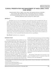
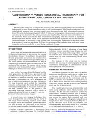
<strong>Smear</strong> <strong>layer</strong> <strong>and</strong> <strong>sealing</strong> <strong>ability</strong> <strong>of</strong> <strong>three</strong> <strong>root</strong> <strong>canal</strong> <strong>sealers</strong>TABLE 1: MINIMUM, MAXIMUM AND MEAN LEAKAGE VALUES IN TERMS OFMILLIMETER WITH STANDARD DEVIATIONSub- group Apical leakage measurement (mm) S.DMinimum Maximum MeanWith smear <strong>layer</strong> A1 0.00 4.84 2.675 1.7A2 1.00 5.66 2.346 1.4A3 1.30 8.85 4.929 3.3Without smear <strong>layer</strong> B1 0.89 3.75 2.092 0.9B2 1.1 6.91 4.520 2.5B3 0.00 8.41 3.372 2.6Methylene blue <strong>and</strong> kept in an incubator at 37°C for oneweek. The specimens were removed from the dye, <strong>and</strong>washed under running tap water. The specimens werethen dried with air syringe <strong>and</strong> the nail polish scrapped<strong>of</strong>f with a scalpel. The <strong>root</strong>s were then grooved labially<strong>and</strong> lingually with a diamond disc with intermittentcutting under water spray without involving the <strong>root</strong><strong>canal</strong>. The specimens were then split gently with achisel.The filling within the specimens were removedwith a sharp explorer. Both the <strong>root</strong> sections <strong>of</strong> eachspecimen were viewed under a stereomicroscope(Olympus, Japan) with X30 magnification using electronicdigital caliper. Linear measurements <strong>of</strong> themost coronal extent <strong>of</strong> dye penetration on the <strong>canal</strong>walls were recorded in milimeter up to 2 decimals.Measurements from all the specimens in all groupswere tabulated. Statistical analysis <strong>of</strong> the leakagevalues obtained was done using the Analysis <strong>of</strong> Variance(ANOVA), Student’s t test <strong>and</strong> Scheff e’ test at95% confidence interval. ANOVA (Analysis <strong>of</strong> Variance)was carried out to determine the significance <strong>of</strong> thevalues recorded. Where there was significant differencePost-hoc analysis (Scheff e ’ test) was carried out todetermine the difference between groups.RESULTSIn this study, measurements <strong>of</strong> maximum lineardye penetration were made to quantify the relativeleakage. By examining Table1 sub-groups A1, A2, A3,the mean leakage values ranged from 2.675, 2.346,4.929 respectively. Similarly in sub-groups B1, B2, B3,the mean leakage values were recorded as 2.092, 4.520,3.372 respectively.In group A with smear <strong>layer</strong>, the results showed thatmean values <strong>of</strong> apical leakage for sub-group A2 wassignificantly less than that for sub-group A3 (P0.05).Pakistan Oral & Dental Journal Vol 31, No. 1 (June 2011)Fig 1:Comparison <strong>of</strong> the mean leakage values <strong>of</strong> the<strong>three</strong> <strong>sealers</strong> in the presence or absence <strong>of</strong>smear <strong>layer</strong>In group B, without smear <strong>layer</strong>, statistically themean values <strong>of</strong> apical leakage for the <strong>three</strong> sub-groupsshowed no significant difference (P>0.05).Student’s t test was carried out to determine theeffect <strong>of</strong> the presence or absence <strong>of</strong> smear <strong>layer</strong> on theapical leakage <strong>of</strong> the <strong>three</strong> <strong>sealers</strong> used in this study.Ketac-endo was the only sealer that showed a statisticallysignificant difference in their mean leakage values(P0.05).DISCUSSIONIn this study, dye penetration technique with methyleneblue was used to compare the apical leakage.Linear measurement <strong>of</strong> the most coronal extent <strong>of</strong> dyepenetration on the wall was used to evaluate the180
<strong>Smear</strong> <strong>layer</strong> <strong>and</strong> <strong>sealing</strong> <strong>ability</strong> <strong>of</strong> <strong>three</strong> <strong>root</strong> <strong>canal</strong> <strong>sealers</strong><strong>sealing</strong> <strong>ability</strong> <strong>of</strong> the <strong>root</strong> <strong>canal</strong> <strong>sealers</strong>. 12,13 The in vitrotechniques to evaluate obturating materials are basedon the assessment <strong>of</strong> microleakage along the obturated<strong>root</strong> <strong>canal</strong>. 14 These include bacterial penetration 15,16 ,dye penetration 9,17,18,19 , isotope penetration 10,3 , scanningelectron microscopy 20 , electrochemical techniques 21 ,fluorometry 22 , staining technique 23 <strong>and</strong> liquid pressuretechnique. 24 Among these techniques, dye penetrationis the method most widely used (due to its simplicity) toevaluate the apical seal <strong>of</strong> <strong>root</strong> <strong>canal</strong>s.Two percent (2%) solution <strong>of</strong> methylene blue dyewas used as a leakage marker because it is readilydetectable under visible light, very soluble in water,able to diffuse easily, <strong>and</strong> is not absorbed by dentinematrix apatite crystals. 12 Kersten <strong>and</strong> Moorer havealso suggested that methylene blue dye had a comparableleakage to butyric acid (metabolic product <strong>of</strong>microorganisms). 25In 82% <strong>of</strong> leakage studies in endodontics, dye orradioisotope penetration methods have been used. 24Matl<strong>of</strong>f et al. showed that methylene blue dye penetratesfar further into the <strong>canal</strong> than isotope tracers,thus giving a better representation <strong>of</strong> apical leakage. 3According to Ahlberg et al. methylene blue dye mayserve as an adequate indicator <strong>of</strong> microleakage <strong>of</strong>microorganisms <strong>and</strong> large size endotoxins as well astoxic agents <strong>of</strong> low molecular weight. 26 However, it isstill not explained as to how dye movement compareswith tissue fluid movement <strong>and</strong> bacterial migration invivo. It seems reasonable to choose a technique thathas been demonstrated to be the most effective amongin vitro tests. 12The <strong>sealers</strong> used were epoxy amine resin-based,glass ionomer-based <strong>and</strong> zinc oxide eugenol-based <strong>sealers</strong>.The latest materials <strong>of</strong> the <strong>three</strong> types namely AHPlus, Ketac-endo <strong>and</strong> Roth 801 were selected. The <strong>root</strong><strong>canal</strong>s were obturated using lateral condensation technique<strong>and</strong> the specimens were placed in an incubatorfor one week with 100% humidity at 37 °C to ensurethat the sealer set in an environment that simulatedthe clinical situation in which they are designed to beused. 12,13The method <strong>of</strong> producing longitudinal sections wasused to split the specimens to examine the exposedfilling <strong>and</strong> any dye penetration into the material <strong>and</strong><strong>root</strong> <strong>canal</strong> wall interface. This technique would give amuch true picture <strong>of</strong> the leakage pattern as comparedto transverse sectioning method because it is possibleto examine the exposed <strong>root</strong> filling <strong>and</strong> any dye penetrationinto the material <strong>and</strong> at the <strong>canal</strong> wall-<strong>root</strong>filling interface, minimizing the risk <strong>of</strong> the dye washingaway. 26 Transverse sectioning <strong>of</strong> the <strong>root</strong>s is associatedwith the disadvantage <strong>of</strong> loss <strong>of</strong> some <strong>of</strong> the toothstructure in each cut, due to the thickness <strong>of</strong> the blade,Pakistan Oral & Dental Journal Vol 31, No. 1 (June 2011)which may affect the accuracy <strong>of</strong> the measurement <strong>of</strong>dye penetration. 12The possible routes <strong>of</strong> dye penetration throughfilled <strong>root</strong> <strong>canal</strong>s are; between the sealer <strong>and</strong> thedentine; between the core material (gutta percha) <strong>and</strong>the sealer; through the core material <strong>and</strong> through thesealer. 13 Stereomicroscope examination <strong>of</strong> the sectionedspecimens showed that leakage occurred throughapical foramen, between the sealer <strong>and</strong> the <strong>root</strong> <strong>canal</strong>wall, between the gutta percha <strong>and</strong> sealer <strong>and</strong> alsothrough the sealer. Absorption <strong>of</strong> the dye by the <strong>sealers</strong>may also represent cohesive failure in the body <strong>of</strong> thesealer, creating another pathway for leakage. Thisstudy support the findings <strong>of</strong> other investigators thatall <strong>root</strong> <strong>canal</strong>s fillings leak 12,27 except for two specimens,(one in sub-group A1<strong>and</strong> one in sub-group B3) whichshowed no leakage. This could have occurred due toinadvertent blockage <strong>of</strong> the apical foramen. Specimensthat allowed leakage indicated that all had voids throughthe sealer or along the sealer- dentine interface. Hundredpercent hermetic seal <strong>of</strong> <strong>canal</strong>s occur rarely, ifever. 3 The variations in the leakage values for theindividual specimens in all the groups could be attributedto any entrapment <strong>of</strong> air in those specimens whereleakage was minimal. 15In this study in the presence <strong>of</strong> smear <strong>layer</strong>, Roth801 sealer leaked significantly more than Ketac-Endosealer. Similar result was found by Koch et al. 28 Thequick setting <strong>of</strong> zinc oxide eugenol material (transitionfrom paste to solid mass) may be responsible for debondingfrom dentinal walls or cohesive fracture causedby stresses due to the setting shrinkage, which mayexplain the leakage. 5 The lesser leakage with the glassionomer sealer can be attributed to its basic propertiessuch as adapt<strong>ability</strong> <strong>and</strong> <strong>ability</strong> to bind to dentine 29 ,which allows less dye penetration through the interfacebetween the <strong>canal</strong> walls, cements <strong>and</strong> guttapercha. 28In the absence <strong>of</strong> smear <strong>layer</strong>, there was no significantdifference in the <strong>sealing</strong> <strong>ability</strong> <strong>of</strong> AH Plus, Ketac-Endo <strong>and</strong> Roth 801. This finding is supported by Oliver<strong>and</strong> Abbott, Timpawat <strong>and</strong> Sripanaratanakul <strong>and</strong>Tzanetakis et al. 13,30,31The mean leakage in case <strong>of</strong> Ketac-endo wasalmost double in the absence <strong>of</strong> the smear <strong>layer</strong> ascompared to the presence <strong>of</strong> smear <strong>layer</strong>. This differencewas statistically significant. When the smear<strong>layer</strong> was removed, orifices <strong>of</strong> the dentinal tubulesopened which resulted in the reduction <strong>of</strong> adhesiveproperties for Ketac-endo. 27 The opened tubules mayalso act as stress raisers, which would cause failure inthe adhesive joint. 8 It was also found that with theremoval <strong>of</strong> smear <strong>layer</strong> with conditioning <strong>of</strong> dentine invitro, bond strength <strong>of</strong> glass ionomer decreased prob-181
<strong>Smear</strong> <strong>layer</strong> <strong>and</strong> <strong>sealing</strong> <strong>ability</strong> <strong>of</strong> <strong>three</strong> <strong>root</strong> <strong>canal</strong> <strong>sealers</strong>ably due to removal <strong>of</strong> Ca ++ ions, which leads toincreased microleakage. 3 After the use <strong>of</strong> EDTA, thedentine is partially decalcified resulting in diminution<strong>of</strong> bond strength. 8 This may be the cause in the presentstudy, as EDTA was used which demineralized thedentine <strong>and</strong> there would have been lesser Ca ++ ions forreaction with carboxyl groups resulting in increasedleakage for Ketac-endo.Due to better <strong>sealing</strong> in presence <strong>of</strong> smear <strong>layer</strong> itmay be recommended that when Ketac-endo is used assealer, it is better to leave the smear <strong>layer</strong> intact. Thepropensity <strong>of</strong> methylene blue dye to bond or react to thesmear <strong>layer</strong> or any <strong>of</strong> the experimental <strong>sealers</strong> studiedcould be tested to rule out the possibility <strong>of</strong> misinterpretation<strong>of</strong> the amount <strong>of</strong> leakage.REFERENCES1 Lares C <strong>and</strong> ElDeeb. The <strong>sealing</strong> <strong>ability</strong> <strong>of</strong> the thermafilobturation technique. J Endodon 1990; 16: 474-79.2 Cohen S <strong>and</strong> Burns RC. Pathways <strong>of</strong> the pulp, 4 th ed. St. Louis,Mo, USA: CV Mosby, 1989; 183-73.3 <strong>Tahir</strong> A.K, Sapuram R <strong>and</strong> Azhar I. Evaluation <strong>of</strong> <strong>sealing</strong><strong>ability</strong> <strong>of</strong> <strong>three</strong> <strong>root</strong> <strong>canal</strong> <strong>sealers</strong>. Pakistan Oral <strong>and</strong> DentalJournal 2007; 27: 257-624 Nguyen TN Obturation <strong>of</strong> the <strong>root</strong> <strong>canal</strong> system, Pathways <strong>of</strong>the pulp, 6 th ed. Mosby, 1994; 219-71.5 Wu MK, Gee AJD <strong>and</strong> Wesselink PR. Leakage <strong>of</strong> four <strong>root</strong><strong>canal</strong> <strong>sealers</strong> at different thicknesses. Int Endodon J 1994; 27:304-08.6 Gencoglu N, Samani S <strong>and</strong> Gunday M. Evaluation <strong>of</strong> <strong>sealing</strong>properties <strong>of</strong> Thermafil <strong>and</strong> Ultrafil techniques in the absenceor presence <strong>of</strong> smear <strong>layer</strong>. J Endodon1993; 19: 599-603.7 Mader CL, Baumgartner JC <strong>and</strong> Peters DD. SEM investigation<strong>of</strong> the smeared <strong>layer</strong> on <strong>root</strong> <strong>canal</strong> walls. J Endodon 1984;10: 477-83.8 <strong>Tahir</strong> A.K. The effect <strong>of</strong> smear <strong>layer</strong> on the apical <strong>sealing</strong><strong>ability</strong> <strong>of</strong> Epoxy resin, Glass ionomer <strong>and</strong> Zinc oxide based<strong>sealers</strong>. M.D.Sc. Dissertation: University Malaya 1999; 08-25.9 Shahravan A, Haghdoost AA, Adl A, Rahimi H, Shadifar F.Effect <strong>of</strong> smear <strong>layer</strong> on <strong>sealing</strong> <strong>ability</strong> <strong>of</strong> <strong>canal</strong> obturation: asystemic review <strong>and</strong> meta analysis. J Endodon 2007; 33:96-10510 Galvan DA. Charlone AE. Pashley DH. Kulild JC. Primack PD.Simpson MD. Effect <strong>of</strong> smear <strong>layer</strong> removal on the diffusionperme<strong>ability</strong> <strong>of</strong> human <strong>root</strong>s. J Endodon 1994; 20: 83-86.11 Evans JT <strong>and</strong> Simon JHS. Evaluation <strong>of</strong> the apical sealproduced by injected thermoplasticized gutta percha in theabsence <strong>of</strong> smear <strong>layer</strong> <strong>and</strong> <strong>root</strong> <strong>canal</strong> sealer. J Endodon 1986;12: 101-07.12 Limkangwalmongkol S, Burstcher P, Abbott P, S<strong>and</strong>ler AB,Dent HD, <strong>and</strong> Bishop BM. A comparative study <strong>of</strong> the apicalleakage <strong>of</strong> four <strong>root</strong> <strong>canal</strong> <strong>sealers</strong> <strong>and</strong> laterally condensedgutta percha. J Endodon 1991; 17: 495-99.13 Dalat DM <strong>and</strong> Onal B. Apical leakage <strong>of</strong> a new glass ionomer<strong>root</strong> <strong>canal</strong> sealer. J Endodon 1998; 24: 161-63.14 Al-Ghamdi A <strong>and</strong> Wennberg A. Testing <strong>of</strong> <strong>sealing</strong> <strong>ability</strong> <strong>of</strong>endodontic filling materials. Endo don Dent Traumotol 1994; 10: 249-55.15 Goldman.M, Simmonds S, Rush R. The usefulness <strong>of</strong> dyepenetration studies re-examined. Oral Sur Oral Med OralPath 1989; 67: 327-32.16 Goldberg F, Bernat M, Spielberg C, Massone EJ <strong>and</strong> PiovanoSA. Analysis <strong>of</strong> the effect <strong>of</strong> ethylenediaminetetraacetic acidon the apical seal <strong>of</strong> <strong>root</strong> <strong>canal</strong> fillings. J Endodon 1985; 11:544-47.17 Kennedy WA, Walker WA III <strong>and</strong> Gough RW. <strong>Smear</strong> <strong>layer</strong>removal effects on apical leakage. J Endodon 1986; 12: 21-27.18 Jhamb S, Nikhil V, Singh V. An in vitro study to determinethe <strong>sealing</strong> <strong>ability</strong> <strong>of</strong> <strong>sealers</strong> with <strong>and</strong> without smear <strong>layer</strong>removal. J Conserv Dent 2009; 12: 150-53.19 Khayat A, Jahanbin A. The influence <strong>of</strong> smear <strong>layer</strong> oncoronal leakage <strong>of</strong> Roth 801 <strong>and</strong> Ah26 <strong>root</strong> <strong>canal</strong> <strong>sealers</strong>. AustEndodon 2005; 31: 66-6820 Tanzilli JP, Raphael D, <strong>and</strong> Moodnik RM. A comparison <strong>of</strong> themarginal adaptation <strong>of</strong> retrograde techniques: a scanningelectron microscopic study. Oral Surg Oral Med Oral Path1980; 50: 74-80.21 Jacobson SM <strong>and</strong> von Fraunh<strong>of</strong>er JA. The investigation <strong>of</strong>microleakage in <strong>root</strong> <strong>canal</strong> therapy. Oral Surg Oral Med OralPath 1976; 42: 817-23.22 Ainley JE. Fluorometric assay <strong>of</strong> the apical seal <strong>of</strong> <strong>root</strong> <strong>canal</strong>fillings. Oral Surg Oral Med Oral Path 1970; 29: 753-62.23 Hovl<strong>and</strong> EJ <strong>and</strong> Dumsha TC. Leakage evaluation in vitro <strong>of</strong>the <strong>root</strong> <strong>canal</strong> sealer cement Sealapex. Int Endondon J 1985;18: 179-182.24 Wu MK, Gee AJD <strong>and</strong> Moorer WR. Fluid transport model <strong>and</strong>bacterial penetration along <strong>root</strong> <strong>canal</strong> fillings. Int Endodon J1993; 26: 203-08.25 Kersten HW <strong>and</strong> Moorer WR. Particles <strong>and</strong> molecules inendodontic leakage. Int Endodon J 1989; 22: 118-24.26. Ahlberg KMF, Assavanop P <strong>and</strong> Tay WM. A comparison <strong>of</strong> theapical dye penetration patterns shown by methylene blue <strong>and</strong>india ink in <strong>root</strong> filled teeth. Int Endodon J 1995; 28: 30-34.27 Gee AJD, Wu MK <strong>and</strong> Wesselink PR. Sealing properties <strong>of</strong>Ketac-endo glass ionomer cement <strong>and</strong> AH26 <strong>root</strong> <strong>canal</strong> <strong>sealers</strong>.Int Endodon J 1994; 27: 239-44.28 Koch K, Min PS <strong>and</strong> Stewart GG. Comparison <strong>of</strong> apicalleakage between Ketac-endo sealer <strong>and</strong> Grossman sealer.Oral Surg Oral Med Oral Path 1994; 78: 784-87.29 Lee CQ, Har<strong>and</strong>i L <strong>and</strong> Cobb CM. Evaluation <strong>of</strong> glass ionomeras an endodontic sealant: An in vitro study. J Endodon 1997;23: 209-12.30 Timpawat S <strong>and</strong> Sripanaratanakul S. Apical <strong>sealing</strong> <strong>ability</strong> <strong>of</strong>glass ionomer sealer with <strong>and</strong> without smear <strong>layer</strong>. J Endodon1998; 24: 343-45.31 Tzanetakis GN, Kakavetosos VD Kontakiotis EG. Impact <strong>of</strong>smear <strong>layer</strong> on <strong>sealing</strong> property <strong>of</strong> <strong>root</strong> <strong>canal</strong> obturation using3 different techniques <strong>and</strong> <strong>sealers</strong>. Part 1.Oral surg Oral medOral Pathol Oral Radiol Endodon 2010; 109: 145-53.32 Smith DC. Composition <strong>and</strong> characteristics <strong>of</strong> glass ionomercements. JADA 1990; 120: 20-22.Pakistan Oral & Dental Journal Vol 31, No. 1 (June 2011)182