Antidiabetic Effect of : Effect on Lipid Peroxidation in - Gpb.sav.sk
Antidiabetic Effect of : Effect on Lipid Peroxidation in - Gpb.sav.sk
Antidiabetic Effect of : Effect on Lipid Peroxidation in - Gpb.sav.sk

You also want an ePaper? Increase the reach of your titles
YUMPU automatically turns print PDFs into web optimized ePapers that Google loves.
Gen. Physiol. Biophys. (2005), 24, 13—26 13<br />
<str<strong>on</strong>g>Antidiabetic</str<strong>on</strong>g> <str<strong>on</strong>g>Effect</str<strong>on</strong>g> <str<strong>on</strong>g>of</str<strong>on</strong>g> ËÓÔÖ ÙÐ×:<br />
<str<strong>on</strong>g>Effect</str<strong>on</strong>g> <strong>on</strong> <strong>Lipid</strong> Peroxidati<strong>on</strong> <strong>in</strong> Streptozotoc<strong>in</strong> Diabetes<br />
L. Pari and M. Latha<br />
Department <str<strong>on</strong>g>of</str<strong>on</strong>g> Biochemistry, Faculty <str<strong>on</strong>g>of</str<strong>on</strong>g> Science, Annamalai University,<br />
Annamalai Nagar, Tamil Nadu, India<br />
Abstract. Oxidative damage has been suggested to be a c<strong>on</strong>tributory factor <strong>in</strong> the<br />
development and complicati<strong>on</strong>s <str<strong>on</strong>g>of</str<strong>on</strong>g> diabetes. The antioxidant effect <str<strong>on</strong>g>of</str<strong>on</strong>g> an aqueous<br />
extract <str<strong>on</strong>g>of</str<strong>on</strong>g> Scoparia dulcis, an <strong>in</strong>digenous plant used <strong>in</strong> Ayurvedic medic<strong>in</strong>e <strong>in</strong> India<br />
was studied <strong>in</strong> rats with streptozotoc<strong>in</strong>-<strong>in</strong>duced diabetes. Oral adm<strong>in</strong>istrati<strong>on</strong> <str<strong>on</strong>g>of</str<strong>on</strong>g><br />
Scoparia dulcis plant extract (SPEt) (200 mg/kg body weight) for 3 weeks resulted<br />
<strong>in</strong> a significant reducti<strong>on</strong> <strong>in</strong> blood glucose and an <strong>in</strong>crease <strong>in</strong> plasma <strong>in</strong>sul<strong>in</strong>. The<br />
aqueous extract also resulted <strong>in</strong> decreased free radical formati<strong>on</strong> <strong>in</strong> tissues (liver and<br />
kidney) studied. The decrease <strong>in</strong> thiobarbituric acid reactive substances (TBARS)<br />
and hydroperoxides (HPX) and <strong>in</strong>crease <strong>in</strong> the activities <str<strong>on</strong>g>of</str<strong>on</strong>g> superoxide dismutase<br />
(SOD), catalase (CAT), glutathi<strong>on</strong>e peroxidase (GPx), reduced glutathi<strong>on</strong>e (GSH)<br />
and glutathi<strong>on</strong>e-S-transferase (GST) clearly show the antioxidant properties <str<strong>on</strong>g>of</str<strong>on</strong>g><br />
SPEt <strong>in</strong> additi<strong>on</strong> to its antidiabetic effect. The effect <str<strong>on</strong>g>of</str<strong>on</strong>g> SPEt at 200 mg/kg body<br />
weight was better than glibenclamide, a reference drug.<br />
Key words: Enzymic antioxidants — Insul<strong>in</strong> — <strong>Lipid</strong> peroxidati<strong>on</strong> — Scoparia<br />
dulcis — Streptozotoc<strong>in</strong> diabetes<br />
Introducti<strong>on</strong><br />
Scoparia dulcis (Scrophulariaceae), comm<strong>on</strong>ly known as sweet broomweed, is a<br />
perennial herb widely distributed <strong>in</strong> tropical and subtropical regi<strong>on</strong>s. In these regi<strong>on</strong>s,<br />
the fresh or dried plant <str<strong>on</strong>g>of</str<strong>on</strong>g> Scoparia dulcis has traditi<strong>on</strong>ally been used as <strong>on</strong>e<br />
<str<strong>on</strong>g>of</str<strong>on</strong>g> remedies for stomach troubles (Satyanarayana 1969), hypertensi<strong>on</strong> (Chow et al.<br />
1974), diabetes (Perry 1980), br<strong>on</strong>chitis (Freire et al 1993), and as an analgesic<br />
and antipyretic (G<strong>on</strong>zales Torres 1986). A number <str<strong>on</strong>g>of</str<strong>on</strong>g> different pr<strong>in</strong>ciples <strong>in</strong>clude<br />
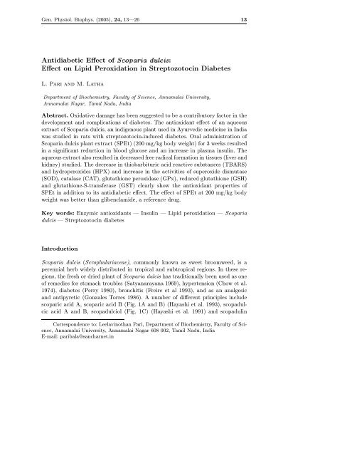
scoparic acid A, scoparic acid B (Fig. 1A and B) (Hayashi et al. 1993), scopadulcic<br />
acid A and B, scopadulciol (Fig. 1C) (Hayashi et al. 1991) and scopadul<strong>in</strong><br />
Corresp<strong>on</strong>dence to: Leelav<strong>in</strong>othan Pari, Department <str<strong>on</strong>g>of</str<strong>on</strong>g> Biochemistry, Faculty <str<strong>on</strong>g>of</str<strong>on</strong>g> Science,<br />
Annamalai University, Annamalai Nagar 608 002, Tamil Nadu, India<br />
E-mail: paribala@sancharnet.<strong>in</strong>
14 Pari and Latha<br />
A<br />
B<br />
CH 2 OH<br />
O<br />
R H O-CO-<br />
R H O-CO-<br />
Scoparic acid A: R = COOH<br />
Scopadiol: R = CH 2 OH<br />
Scoparic acid B: R = COOH<br />
C<br />
D<br />
OH<br />
O<br />
H<br />
H<br />
H<br />
R 1 R 2 Scopadul<strong>in</strong>: R = COOH<br />
O-CO-<br />
H<br />
R<br />
O-CO-<br />
Scopadulcic acid A: R 1 = COOH, R 2 = CH 2 OH<br />
Scopadulcic acid B: R 1 = CH 3 , R 2 = COOH<br />
Scopadulciol: R 1 = CH 3 , R 2 = CH 2 OH<br />
Figure 1. Biologically active compounds isolated from Scoparia dulcis.<br />
(Fig. 1D) (Hayashi et al. 1990) that have been identified as c<strong>on</strong>tributor to the<br />
observed medic<strong>in</strong>al effect <str<strong>on</strong>g>of</str<strong>on</strong>g> the plant. Am<strong>on</strong>g them, scopadulcic acid B and scopadulciol<br />
were found to be unique biomolecules with <strong>in</strong>hibitory effects <strong>on</strong> replicati<strong>on</strong><br />
<str<strong>on</strong>g>of</str<strong>on</strong>g> herpes simplex virus type 1 (Hayashi et al. 1993), gastric prot<strong>on</strong> pump<br />
and b<strong>on</strong>e resorpti<strong>on</strong> stimulated by parathyroid horm<strong>on</strong>e (Hayashi et al. 1990). In<br />
additi<strong>on</strong>, scopadulcic acid B showed antitumour promot<strong>in</strong>g activities (Hayashi et<br />
al. 1991). Because <str<strong>on</strong>g>of</str<strong>on</strong>g> their unique carb<strong>on</strong> <strong>sk</strong>elet<strong>on</strong> and many sided biological activities,<br />
they were paid much attenti<strong>on</strong> as chemical synthetic targets by organic<br />
synthetic chemists. In a previous study, Nath (1943) has studied the antidiabetic<br />
effect <str<strong>on</strong>g>of</str<strong>on</strong>g> Scoparia dulcis and obta<strong>in</strong>ed a glycoside, amell<strong>in</strong> from fresh plant and<br />
reported that it brought relief <strong>in</strong> other complicati<strong>on</strong>s accompanied with diabetes<br />
(i.e., pyorrhoea, ret<strong>in</strong>opathy, jo<strong>in</strong>t pa<strong>in</strong>, susceptibility to cold etc.) with<strong>in</strong> a very<br />
short period.
<str<strong>on</strong>g>Effect</str<strong>on</strong>g> <str<strong>on</strong>g>of</str<strong>on</strong>g> Scoparia dulcis <strong>on</strong><strong>Lipid</strong>Peroxidati<strong>on</strong><strong>in</strong>Diabetes 15<br />
The elevated levels <str<strong>on</strong>g>of</str<strong>on</strong>g> blood glucose <strong>in</strong> diabetes produce oxygen-free radicals<br />
that cause membrane damage due to peroxidati<strong>on</strong> <str<strong>on</strong>g>of</str<strong>on</strong>g> membrane lipids and prote<strong>in</strong><br />
glycati<strong>on</strong> (Baynes 1991). Glucose auto-oxidize <strong>in</strong> the presence <str<strong>on</strong>g>of</str<strong>on</strong>g> transiti<strong>on</strong><br />
metal i<strong>on</strong>s generates oxygen-free radicals, which make the membrane vulnerable<br />
to oxidative damage (Hunt et al. 1990). The oxidative stress and resultant tissue<br />
damage are important comp<strong>on</strong>ent <strong>in</strong> the pathogenesis <str<strong>on</strong>g>of</str<strong>on</strong>g> diabetic complicati<strong>on</strong>s<br />
(Baynes 1991). The free radicals react with biomembrane caus<strong>in</strong>g oxidative destructi<strong>on</strong><br />
<str<strong>on</strong>g>of</str<strong>on</strong>g> polyunsaturated fatty acids form<strong>in</strong>g cytotoxic aldehydes by a process<br />
known as lipid peroxidati<strong>on</strong> (LPO) (Wolff 1993). The extent <str<strong>on</strong>g>of</str<strong>on</strong>g> LPO was measured<br />
<strong>in</strong> terms <str<strong>on</strong>g>of</str<strong>on</strong>g> thiobarbituric acid reactive substances (TBARS) and lipid hydroperoxides<br />
(HPX), which are the end products <str<strong>on</strong>g>of</str<strong>on</strong>g> LPO. Several studies <strong>in</strong> human and<br />
animal models, us<strong>in</strong>g TBARS assay have shown <strong>in</strong>creased LPO <strong>in</strong> membranes and<br />
lipoprote<strong>in</strong>s <strong>in</strong> the diabetic state (Griesmacher et al. 1995; Krishnakumar et al.<br />
1999). HPX formed by LPO have direct toxic effects <strong>on</strong> endothelial cells and also<br />
degrade to form hydroxyl radicals (OH*) (Testafamariam 1993). The acti<strong>on</strong> <str<strong>on</strong>g>of</str<strong>on</strong>g><br />
streptozotoc<strong>in</strong> produces reactive free radicals, which have been shown to be cytotoxic<br />
to the B-cells <str<strong>on</strong>g>of</str<strong>on</strong>g> the pancreas (Ivorra et al. 1989). As the diabetogenic acti<strong>on</strong><br />
<str<strong>on</strong>g>of</str<strong>on</strong>g> streptozotoc<strong>in</strong> is preventable by superoxide dismutase (SOD), catalase (CAT)<br />
and other OH* scavengers such as ethanol and dimethyl urea, there is evidence<br />
to suggest that the acti<strong>on</strong> <str<strong>on</strong>g>of</str<strong>on</strong>g> streptozotoc<strong>in</strong> <strong>in</strong>volve a superoxide ani<strong>on</strong> and OH*<br />
(Asplund et al. 1984). Thus, streptozotoc<strong>in</strong>-<strong>in</strong>duced diabetes could elicit changes<br />
<strong>in</strong> the antioxidant defense systems <strong>in</strong> resp<strong>on</strong>se to <strong>in</strong>creased oxidative stress. The<br />
deleterious effects <str<strong>on</strong>g>of</str<strong>on</strong>g> superoxide radicals (O .−<br />
2 )andOH∗ <strong>in</strong> oxidative stress can be<br />
counteracted by antioxidant enzymes such as SOD, CAT and glutathi<strong>on</strong>e peroxidase<br />
(GPx). In additi<strong>on</strong> to these enzymes, glutathi<strong>on</strong>e-S-transferase (GST) provides<br />
glutathi<strong>on</strong>e (GSH) and help to neutralize toxic electrophiles. There is evidence to<br />
show the role <str<strong>on</strong>g>of</str<strong>on</strong>g> free radicals <strong>in</strong> diabetes and studies <strong>in</strong>dicate that tissue <strong>in</strong>jury<br />
<strong>in</strong> diabetes may be due to free radicals (Wohaieb and God<strong>in</strong> 1987; Kakkar et al.<br />
1995). Diabetes is becom<strong>in</strong>g pandemic and despite the recent surge <strong>in</strong> new drugs<br />
to treat and prevent the c<strong>on</strong>diti<strong>on</strong>, its prevalence c<strong>on</strong>t<strong>in</strong>ues to soar (Tiwari and<br />
Madhusudana Rao 2002).<br />
Thus, the present study was undertaken to assess the antiperoxidative and<br />
antioxidant effect <str<strong>on</strong>g>of</str<strong>on</strong>g> Scoparia dulcis <strong>in</strong> streptozotoc<strong>in</strong>-<strong>in</strong>duced diabetic rats.<br />
Materials and Methods<br />
Animals<br />
Male alb<strong>in</strong>o Wistar rats, body weigh<strong>in</strong>g 180–200 g bred <strong>in</strong> Central Animal House<br />
(Rajah Muthiah Medical College, Annamalai University, India) were used <strong>in</strong> this<br />
study. The animals were fed <strong>on</strong> a pellet diet (H<strong>in</strong>dustan Lever Ltd., Mumbai, India)<br />
and water ad libitum. The animals used <strong>in</strong> the present study were ma<strong>in</strong>ta<strong>in</strong>ed <strong>in</strong><br />
accordance with the guidel<strong>in</strong>es <str<strong>on</strong>g>of</str<strong>on</strong>g> the Nati<strong>on</strong>al Institute <str<strong>on</strong>g>of</str<strong>on</strong>g> Nutriti<strong>on</strong>, Indian Council
16 Pari and Latha<br />
<str<strong>on</strong>g>of</str<strong>on</strong>g> Medical Research (Hyderabad, India) and approved by the Ethical Committee<br />
(Vide No. 73, 2001) <str<strong>on</strong>g>of</str<strong>on</strong>g> Annamalai University.<br />
Drugs and chemicals<br />
All the drugs and biochemicals used <strong>in</strong> this experiment were purchased from Sigma<br />
Chemical Company Inc. (St. Louis, MO, USA). The chemicals were <str<strong>on</strong>g>of</str<strong>on</strong>g> analytical<br />
grade.<br />
Plant material<br />
Scoparia dulcis L. plants were collected from Neyveli (Cuddalore District, Tamil<br />
Nadu, India). The plant was identified and authenticated at the Herbarium <str<strong>on</strong>g>of</str<strong>on</strong>g><br />
Botany Directorate <strong>in</strong> Annamalai University. A voucher specimen (No. 3412) was<br />
deposited <strong>in</strong> the Botany Deartment <str<strong>on</strong>g>of</str<strong>on</strong>g> Annamalai University.<br />
Preparati<strong>on</strong> <str<strong>on</strong>g>of</str<strong>on</strong>g> Scoparia dulcis plant extract (SPEt)<br />
Aqueous and chlor<str<strong>on</strong>g>of</str<strong>on</strong>g>orm extracts<br />
500g<str<strong>on</strong>g>of</str<strong>on</strong>g>Scoparia dulcis fresh whole plants were extracted with 1.5 l <str<strong>on</strong>g>of</str<strong>on</strong>g> water/chlor<str<strong>on</strong>g>of</str<strong>on</strong>g>orm<br />
by the method <str<strong>on</strong>g>of</str<strong>on</strong>g> c<strong>on</strong>t<strong>in</strong>uous hot extracti<strong>on</strong>. The filtrate was evaporated to<br />
c<strong>on</strong>stant weight <strong>on</strong> a rotavapor. The residual extract was dissolved <strong>in</strong> sterile water<br />
and used <strong>in</strong> the <strong>in</strong>vestigati<strong>on</strong> (Ja<strong>in</strong> 1968).<br />
Ethanol extract<br />
500 g <str<strong>on</strong>g>of</str<strong>on</strong>g> fresh plant <str<strong>on</strong>g>of</str<strong>on</strong>g> Scoparia dulcis were chopped <strong>in</strong>to small pieces soaked<br />
overnight <strong>in</strong> 1.5 l <str<strong>on</strong>g>of</str<strong>on</strong>g> 95% ethanol. This suspensi<strong>on</strong> was filtered and the residue<br />
was resuspended <strong>in</strong> an equal volume <str<strong>on</strong>g>of</str<strong>on</strong>g> 95% ethanol for 48 h and filtered aga<strong>in</strong>.<br />
The two filtrates were pooled and the solvents were evaporated <strong>in</strong> a rotavapor at<br />
40–50 ◦ C under reduced pressure and lyophilized. A greenish-black powdered material<br />
was obta<strong>in</strong>ed (20–30 g). It was stored at 0–4 ◦ C until used. When needed, the<br />
residual extract was suspended <strong>in</strong> distilled water and used <strong>in</strong> the study (Hossa<strong>in</strong><br />
et al. 1992).<br />
Inducti<strong>on</strong> <str<strong>on</strong>g>of</str<strong>on</strong>g> experimental diabetes<br />
A freshly prepared soluti<strong>on</strong> <str<strong>on</strong>g>of</str<strong>on</strong>g> streptozotoc<strong>in</strong> (45 mg/kg) <strong>in</strong> 0.1 mol/l citrate buffer,<br />
pH 4.5 was <strong>in</strong>jected <strong>in</strong>traperit<strong>on</strong>eally <strong>in</strong> a volume <str<strong>on</strong>g>of</str<strong>on</strong>g> 1 ml/kg (Siddique et al. 1987).<br />
48 h after streptozotoc<strong>in</strong> adm<strong>in</strong>istrati<strong>on</strong>, rats with moderate diabetes hav<strong>in</strong>g glycosuria<br />
and hyperglycemia (i.e. with blood glucose <str<strong>on</strong>g>of</str<strong>on</strong>g> 200–300 mg/dl) were taken<br />
for the experiment.<br />
Experimental design<br />
In the experiment, a total <str<strong>on</strong>g>of</str<strong>on</strong>g> 72 rats (66 diabetic surviv<strong>in</strong>g rats, 6 normal rats)<br />
were used. The rats were divided <strong>in</strong>to 12 groups <str<strong>on</strong>g>of</str<strong>on</strong>g> 6 rats each. Three doses <str<strong>on</strong>g>of</str<strong>on</strong>g><br />
aqueous, ethanolic and chlor<str<strong>on</strong>g>of</str<strong>on</strong>g>orm extracts (50, 100 and 200 mg/kg body weight<br />
per day) and glibenclamide (600 µg/kg) were tested. All doses were started 48 h<br />
after streptozotoc<strong>in</strong> <strong>in</strong>jecti<strong>on</strong>. Blood samples were drawn at weekly <strong>in</strong>tervals till
<str<strong>on</strong>g>Effect</str<strong>on</strong>g> <str<strong>on</strong>g>of</str<strong>on</strong>g> Scoparia dulcis <strong>on</strong><strong>Lipid</strong>Peroxidati<strong>on</strong><strong>in</strong>Diabetes 17<br />
the end <str<strong>on</strong>g>of</str<strong>on</strong>g> study (i.e., 3 weeks). At the end <str<strong>on</strong>g>of</str<strong>on</strong>g> 3 weeks, all the rats were killed<br />
by decapitati<strong>on</strong> under pentobarbit<strong>on</strong>e sodium (60 mg/kg) anaesthesia. Blood was<br />
collected <strong>in</strong> tubes c<strong>on</strong>ta<strong>in</strong><strong>in</strong>g potassium oxalate and sodium fluoride soluti<strong>on</strong> for<br />
the estimati<strong>on</strong> <str<strong>on</strong>g>of</str<strong>on</strong>g> blood glucose and plasma was separated for assay <str<strong>on</strong>g>of</str<strong>on</strong>g> <strong>in</strong>sul<strong>in</strong>. Liver<br />
and kidney were dissected out, washed <strong>in</strong> ice cold sal<strong>in</strong>e, patted dry and weighed.<br />
Biochemical analysis<br />
Determ<strong>in</strong>ati<strong>on</strong> <str<strong>on</strong>g>of</str<strong>on</strong>g> blood glucose and plasma <strong>in</strong>sul<strong>in</strong><br />
Fast<strong>in</strong>g blood glucose was estimated by O-toluid<strong>in</strong>e method (Sasaki et al. 1972).<br />
Plasma <strong>in</strong>sul<strong>in</strong> was estimated us<strong>in</strong>g enzyme-l<strong>in</strong>ked immunosorbent assay kit (Boehr<strong>in</strong>ger<br />
Mannheim, Germany).<br />
Estimati<strong>on</strong> <str<strong>on</strong>g>of</str<strong>on</strong>g> LPO<br />
LPO <strong>in</strong> tissues were estimated colorimetrically by TBARS and HPX by the method<br />
<str<strong>on</strong>g>of</str<strong>on</strong>g> Nehius and Samuels<strong>on</strong> (1968) and Jiang et al. (1992), respectively. In brief, 0.1<br />
ml <str<strong>on</strong>g>of</str<strong>on</strong>g> tissue homogenate (Tris-HCl buffer, pH 7.5) was treated with 2 ml <str<strong>on</strong>g>of</str<strong>on</strong>g> (1 : 1 : 1<br />
ratio) TBA-TCA-HCl reagent (TBA 37%, TCA 15% and 0.25 N HCl) and placed<br />
<strong>in</strong> water bath for 15 m<strong>in</strong>, cooled and centrifuged at room temperature for 10 m<strong>in</strong><br />
at 1,000 rpm. The absorbance <str<strong>on</strong>g>of</str<strong>on</strong>g> clear supernatant was measured aga<strong>in</strong>st reference<br />
blank at 535 nm and expressed as mmol/100 g tissue.<br />
HPX were expressed as mmol/100 g tissue. 0.1 ml <str<strong>on</strong>g>of</str<strong>on</strong>g> tissue homogenate was<br />
treated with 0.9 ml <str<strong>on</strong>g>of</str<strong>on</strong>g> fox reagent (88 mg butylated hydroxytoluene, 7.6 mg xylenol<br />
orange and 9.8 mg amm<strong>on</strong>ium i<strong>on</strong> sulphate were added to 90 ml <str<strong>on</strong>g>of</str<strong>on</strong>g> methanol and<br />
10 ml 250 mmol/l sulphuric acid) and <strong>in</strong>cubated at 37 ◦ C for 30 m<strong>in</strong>. The colour<br />
developed was read colorimetrically at 560 nm.<br />
Determ<strong>in</strong>ati<strong>on</strong> <str<strong>on</strong>g>of</str<strong>on</strong>g> CAT and SOD<br />
CAT was assayed colorimetrically at 620 nm and expressed as µmoles <str<strong>on</strong>g>of</str<strong>on</strong>g> hydrogen<br />
peroxide (H 2 O 2 ) c<strong>on</strong>sumed/m<strong>in</strong>/mg prote<strong>in</strong> as described by S<strong>in</strong>ha (1972). The<br />
reacti<strong>on</strong> mixture (1.5 ml vol.) c<strong>on</strong>ta<strong>in</strong>ed 1.0 ml <str<strong>on</strong>g>of</str<strong>on</strong>g> 0.01 mol/l phosphate buffer (pH<br />
7.0), 0.1 ml <str<strong>on</strong>g>of</str<strong>on</strong>g> tissue homogenate and 0.4 ml <str<strong>on</strong>g>of</str<strong>on</strong>g> 2 mol/l H 2 O 2 . The reacti<strong>on</strong> was<br />
stopped by the additi<strong>on</strong> <str<strong>on</strong>g>of</str<strong>on</strong>g> 2.0 ml <str<strong>on</strong>g>of</str<strong>on</strong>g> dichromate-acetic acid reagent (5% potassium<br />
dichromate and glacial acetic acid were mixed <strong>in</strong> 1 : 3 ratio).<br />
SOD was assayed utiliz<strong>in</strong>g the technique <str<strong>on</strong>g>of</str<strong>on</strong>g> Kakkar et al. (1984). A s<strong>in</strong>gle<br />
unit <str<strong>on</strong>g>of</str<strong>on</strong>g> enzyme was expressed as 50% <strong>in</strong>hibiti<strong>on</strong> <str<strong>on</strong>g>of</str<strong>on</strong>g> nitroblue tetrazolium reducti<strong>on</strong>/m<strong>in</strong>/mg<br />
prote<strong>in</strong>.<br />
Determ<strong>in</strong>ati<strong>on</strong> <str<strong>on</strong>g>of</str<strong>on</strong>g> GPx and reduced GSH<br />
GPx activity was measured by the method described by Rotruck et al. (1984).<br />
Brifely, reacti<strong>on</strong> mixture c<strong>on</strong>ta<strong>in</strong>ed 0.2 ml <str<strong>on</strong>g>of</str<strong>on</strong>g> 0.4 mol/l phosphate buffer (pH 7.0),<br />
0.1 ml <str<strong>on</strong>g>of</str<strong>on</strong>g> 10 mmol/l sodium azide, 0.2 ml <str<strong>on</strong>g>of</str<strong>on</strong>g> tissue homogenate (homogenised <strong>in</strong><br />
0.4 mol/l, phosphate buffer pH 7.0), 0.2 ml GSH, 0.1 ml <str<strong>on</strong>g>of</str<strong>on</strong>g> 0.2 mmol/l H 2 O 2 .The<br />
c<strong>on</strong>tents were <strong>in</strong>cubated at 37 ◦ C for 10 m<strong>in</strong>. The reacti<strong>on</strong> was arrested by 0.4 ml<br />
<str<strong>on</strong>g>of</str<strong>on</strong>g> 10% TCA, and centrifuged. Supernatant was assayed for GSH c<strong>on</strong>tent by us<strong>in</strong>g
18 Pari and Latha<br />
Ellmans reagent (19.8 mg <str<strong>on</strong>g>of</str<strong>on</strong>g> 5,5’-dithiobisnitro benzoic acid <strong>in</strong> 100 ml <str<strong>on</strong>g>of</str<strong>on</strong>g> 0.1%<br />
sodium nitrate).<br />
GSH was determ<strong>in</strong>ed by the method <str<strong>on</strong>g>of</str<strong>on</strong>g> Ellman (1959). 1.0 ml <str<strong>on</strong>g>of</str<strong>on</strong>g> supernatant<br />
was treated with 0.5 ml <str<strong>on</strong>g>of</str<strong>on</strong>g> Ellmans reagent and 3.0 ml <str<strong>on</strong>g>of</str<strong>on</strong>g> phosphate buffer (0.2<br />
mol/l, pH 8.0). The absorbance was read at 412 nm. GPx activity was expressed<br />
as µg <str<strong>on</strong>g>of</str<strong>on</strong>g> GSH c<strong>on</strong>sumed/m<strong>in</strong>/mg prote<strong>in</strong> and reduced GSH as mg/100 g <str<strong>on</strong>g>of</str<strong>on</strong>g> tissue.<br />
Determ<strong>in</strong>ati<strong>on</strong> <str<strong>on</strong>g>of</str<strong>on</strong>g> GST<br />
The GST activity was determ<strong>in</strong>ed spectrophotometrically by the method <str<strong>on</strong>g>of</str<strong>on</strong>g> Habig<br />
et al. (1974). The reacti<strong>on</strong> mixture (3 ml) c<strong>on</strong>ta<strong>in</strong>ed 1.0 ml <str<strong>on</strong>g>of</str<strong>on</strong>g> 100 mmol/l phosphate<br />
buffer (pH 6.5), 0.1 ml <str<strong>on</strong>g>of</str<strong>on</strong>g> 30 mmol/l 1-chloro-2,4-d<strong>in</strong>itrobenzene (CDNB) and 1.7<br />
ml <str<strong>on</strong>g>of</str<strong>on</strong>g> double distilled water. After pre-<strong>in</strong>cubat<strong>in</strong>g the reacti<strong>on</strong> mixture at 37 ◦ Cfor<br />
5 m<strong>in</strong>, the reacti<strong>on</strong> was started by the additi<strong>on</strong> <str<strong>on</strong>g>of</str<strong>on</strong>g> 0.1 ml <str<strong>on</strong>g>of</str<strong>on</strong>g> tissue homogenate and<br />
0.1 ml <str<strong>on</strong>g>of</str<strong>on</strong>g> GSH as substrate. The absorbance was followed for 5 m<strong>in</strong> at 340 nm.<br />
Reacti<strong>on</strong> mixture without the enzyme was used as blank. The activity <str<strong>on</strong>g>of</str<strong>on</strong>g> GST was<br />
expressed as mmoles <str<strong>on</strong>g>of</str<strong>on</strong>g> GSH-CDNB c<strong>on</strong>jugate formed/m<strong>in</strong>/mg prote<strong>in</strong> us<strong>in</strong>g an<br />
ext<strong>in</strong>cti<strong>on</strong> coefficient <str<strong>on</strong>g>of</str<strong>on</strong>g> 9.6 (mmol/l) −1 .<br />
Estimati<strong>on</strong> <str<strong>on</strong>g>of</str<strong>on</strong>g> prote<strong>in</strong><br />
Prote<strong>in</strong> was determ<strong>in</strong>ed by the method <str<strong>on</strong>g>of</str<strong>on</strong>g> Lowry et al. (1951) us<strong>in</strong>g bov<strong>in</strong>e serum<br />
album<strong>in</strong> as standard, at 660 nm.<br />
Statistical analysis<br />
The data for various biochemical parameters were analysed us<strong>in</strong>g ANOVA and the<br />
group means were compared by Duncans multiple range test (DMRT). Values were<br />
c<strong>on</strong>sidered statistically significant when p < 0.05 (Duncan 1957).<br />
Results<br />
In all groups prior to streptozotoc<strong>in</strong> adm<strong>in</strong>istrati<strong>on</strong>, the basal levels <str<strong>on</strong>g>of</str<strong>on</strong>g> blood glucose<br />
<str<strong>on</strong>g>of</str<strong>on</strong>g> the rats were not significantly different. However, 48 h after streptozotoc<strong>in</strong><br />
adm<strong>in</strong>istrati<strong>on</strong>, blood glucose levels were significantly higher <strong>in</strong> rats selected for<br />
the study. In c<strong>on</strong>trast, n<strong>on</strong>-diabetic c<strong>on</strong>trols rema<strong>in</strong>ed persistently euglycaemic<br />
throughout the course <str<strong>on</strong>g>of</str<strong>on</strong>g> the study.<br />
Table 1 shows the effect <str<strong>on</strong>g>of</str<strong>on</strong>g> treatment with extracts <strong>on</strong> blood glucose levels.<br />
In all the SPEt-treated groups (all doses) a significant antihyperglycaemic (p <<br />
0.01) effect was evident from first week <strong>on</strong>wards the decrease <strong>in</strong> blood sugar be<strong>in</strong>g<br />
maximum <strong>on</strong> completi<strong>on</strong> <str<strong>on</strong>g>of</str<strong>on</strong>g> the third week (66.5%, p < 0.001) <strong>in</strong> the group receiv<strong>in</strong>g<br />
200 mg/kg/day <str<strong>on</strong>g>of</str<strong>on</strong>g> aqueous SPEt. On the other hand, ethanolic- and chlor<str<strong>on</strong>g>of</str<strong>on</strong>g>orm<br />
extracts-treated groups showed an antihyperglycaemic effect much later (i.e. <strong>on</strong><br />
completi<strong>on</strong> <str<strong>on</strong>g>of</str<strong>on</strong>g> the third week) <strong>in</strong> groups receiv<strong>in</strong>g 200 mg/kg per day (60.5 and<br />
56.3%, respectively). On the basis <str<strong>on</strong>g>of</str<strong>on</strong>g> these studies, doses <str<strong>on</strong>g>of</str<strong>on</strong>g> 200 mg/kg per day <str<strong>on</strong>g>of</str<strong>on</strong>g><br />
aqueous, SPEt was selected for further evaluati<strong>on</strong>.
<str<strong>on</strong>g>Effect</str<strong>on</strong>g> <str<strong>on</strong>g>of</str<strong>on</strong>g> Scoparia dulcis <strong>on</strong><strong>Lipid</strong>Peroxidati<strong>on</strong><strong>in</strong>Diabetes 19<br />
Table 1. <str<strong>on</strong>g>Effect</str<strong>on</strong>g> <str<strong>on</strong>g>of</str<strong>on</strong>g> 3-week treatment with various doses <str<strong>on</strong>g>of</str<strong>on</strong>g> aqueous, ethanolic and chlor<str<strong>on</strong>g>of</str<strong>on</strong>g>orm Scoparia dulcis plant extract (SPEt)<br />
<strong>on</strong> glucose <strong>in</strong> normal and experimental rats<br />
Groups Day 0 48 h 1 week 2 weeks 3 weeks<br />
after STZ<br />
<strong>in</strong>jecti<strong>on</strong> after treatment<br />
Blood glucose (mg/dl)<br />
Normal 80.0 ± 3.0 84.1 ± 5.1 82.1 ± 5.9 81.0 ± 6.0 81.4 ± 5.9<br />
Diabetic c<strong>on</strong>trol 82.0 ± 4.9 265.0 ± 19.4** 280.0 ± 12.9** 286.0 ± 12.9** 298.0 ± 15.7**<br />
Diabetic + SPEt-Aq-50 79. ± 3.3 255.0 ± 16.2 235.0 ± 13.8* (6.3) 209.1 ± 11.4** (16.6) 185.0 ± 9.7** (26.4)<br />
Diabetic + SPEt-Aq-100 84.0 ± 6.9 247.1 ± 13.7 211.0 ± 12.1* (14.6) 162.0 ± 13.2** (34.5) 113.0 ± 7.3** (54.3)<br />
Diabetic + SPEt-Aq-200 77.2 ± 4.0 256.3 ± 16.6 190.0 ± 12.9** (25.8) 129.0 ± 12.1** (49.7) 86.0 ± 6.5** (66.5)<br />
Diabetic + SPEt-Alc-50 79.0 ± 4.3 246.0 ± 13.2 236.3 ± 10.2* (3.7) 218.0 ± 9.7* (11.2) 190.0 ± 6.5** (22.6)<br />
Diabetic + SPEt-Alc-100 82.0 ± 4.7 253.0 ± 14.1 214.0 ± 11.4* (15.4) 173.0 ± 8.6** (31.6) 131.1 ± 5.1** (48.1)<br />
Diabetic + SPEt-Alc-200 77.3 ± 4.2 259.0 ± 18.8 199.1 ± 13.0** (23.0) 138.0 ± 6.1** (46.7) 102.1 ± 6.8** (60.5)<br />
Diabetic + SPEt-Chloro-50 80.0 ± 3.2 248.0 ± 14.0 238.0 ± 11.1* (4.0) 224.1 ± 6.7* (9.6) 206.0 ± 6.5** (17.0)<br />
Diabetic + SPEt-Chloro-100 79.0 ± 4.5 241.3 ± 14.5 218.0 ± 9.3* (10.8) 180.4 ± 9.8** (25.2) 143.0 ± 9.2** (40.7)<br />
Diabetic + SPEt-Chloro-200 82.2 ± 6.0 252.0 ± 13.0 203.0 ± 13.0** (19.4) 152.0 ± 13.3** (39.7) 110.0 ± 7.0** (56.3)<br />
Diabetic + glibenclamide 77.4 ± 4.4 246.0 ± 13.9 219.2 ± 7.0* (10.7) 192.0 ± 10.8** (21.9) 118.2 ± 4.4** (51.8)<br />
(600 µg/kg)<br />
Values are given as mean ± S.D. for 6 rats <strong>in</strong> each group. Values <strong>in</strong> parentheses <strong>in</strong>dicated the percentage lower<strong>in</strong>g <str<strong>on</strong>g>of</str<strong>on</strong>g> blood glucose<br />
<strong>in</strong> comparis<strong>on</strong> to basal read<strong>in</strong>g after streptozotoc<strong>in</strong> (STZ) adm<strong>in</strong>istrati<strong>on</strong> at 48 h. Diabetic c<strong>on</strong>trol was compared with normal.<br />
Experimental groups were compared with corresp<strong>on</strong>d<strong>in</strong>g values after STZ <strong>in</strong>jecti<strong>on</strong> (48 h). * p < 0.01, ** p < 0.001.
20 Pari and Latha<br />
Table 2. Body weight, food and fluid <strong>in</strong>take <strong>in</strong> streptozotoc<strong>in</strong> diabetic rats before and after oral treatment with Scoparia dulcis<br />
plantextract(SPEt)for3weeks<br />
Groups Body weight (g) Fluid <strong>in</strong>take (ml/rat per day) Food <strong>in</strong>take (g/rat per day)<br />
Initial F<strong>in</strong>al Before After Before After<br />
Normal 176 ± 10 179 ± 9 77 ± 4.4 74 ± 4.7 a 14 ± 1.3 13 ± 1.1 a<br />
Normal + SPEt-Aq-200 179 ± 3 182 ± 4 78 ± 2.7 75 ± 3.2 a 15 ± 2.9 14 ± 0.6 a<br />
Diabetic c<strong>on</strong>trol 181 ± 7 137 ± 7 ••• 155 ± 7.5 168 ± 6.0 b 42 ± 2.6 59 ± 2.1 b<br />
Diabetic + SPEt-Aq-200 175 ± 5 187 ± 7*** 120 ± 5.3 79 ± 5.8 c 24 ± 1.8 22 ± 9.3 c<br />
Diabetic + glibenclamide 210 ± 10 230 ± 10*** 126 ± 22.0 92 ± 1.8 d 25 ± 1.5 33 ± 2.0 d<br />
(600 µg/kg)<br />
Values are given as mean ± S.D. from six rats <strong>in</strong> each group. Values not shar<strong>in</strong>g a comm<strong>on</strong> superscript letter differ significantly at p <<br />
0.05 (Duncan’s multiple range test). Duncan procedure, ranges for the level: 2.95, 3.09, 3.20, 3.22. Diabetic c<strong>on</strong>trol was compared with<br />
normal,<br />
••• p < 0.001. Experimental groups were compared with diabetic c<strong>on</strong>trol, *** p < 0.001.
<str<strong>on</strong>g>Effect</str<strong>on</strong>g> <str<strong>on</strong>g>of</str<strong>on</strong>g> Scoparia dulcis <strong>on</strong><strong>Lipid</strong>Peroxidati<strong>on</strong><strong>in</strong>Diabetes 21<br />
Table 3. Changes <strong>in</strong> levels <str<strong>on</strong>g>of</str<strong>on</strong>g> plasma <strong>in</strong>sul<strong>in</strong>, tissue thiobarbituric acid reactive substances<br />
(TBARS) and hydroperoxides (HPX) <strong>in</strong> normal and experimental animals<br />
Groups Normal Normal + Diabetic Diabetic + Diabetic +<br />
SPEt-Aq-200 c<strong>on</strong>trol SPEt-Aq-200 glibenclamide<br />
(600 µg/kg)<br />
Plasma <strong>in</strong>sul<strong>in</strong> 10.3 ± 0.9 a 11.0 ± 0.7 a 4.2 ± 0.04 b 9.2 ± 0.4 c 8.5 ± 0.4 c<br />
(µU/ml)<br />
TBARS (mmol/100 g tissue)<br />
Liver 0.7 ± 0.04 a 0.7 ± 0.03 a 1.8 ± 0.07 b 1.1 ± 0.01 c 1.4 ± 0.05 d<br />
Kidney 1.7 ± 0.09 a,c 1.6 ± 0.1 a 3.5 ± 0.3 b 1.9 ± 0.2 c 2.3 ± 0.1 d<br />
HPX (mmol/100 g tissue)<br />
Liver 79.0 ± 3.5 a 77.0 ± 2.4 a 97.8 ± 7.5 b 82.0 ± 4.3 a,c 87.6 ± 6.1 c<br />
Kidney 54.6 ± 3.8 a 51.5 ± 4.3 a 75.4 ± 5.8 b 60.8 ± 3.6 c 68.1 ± 4.2 d<br />
Values are given as mean ± S.D. for 6 rats <strong>in</strong> each group. Values not shar<strong>in</strong>g a comm<strong>on</strong><br />
superscript letter differ significantly at p < 0.05 (DMRT). Duncan procedure, ranges for<br />
the level: 2.95, 3.09, 3.20, 3.22.<br />
The body weights <str<strong>on</strong>g>of</str<strong>on</strong>g> SPEt- and glibenclamide-treated groups were <strong>in</strong>creased<br />
significantly (p < 0.001) <strong>on</strong> 3rd week compared with diabetic c<strong>on</strong>trol rats (Table 2).<br />
The food <strong>in</strong>take was significantly lowered <strong>in</strong> the SPEt-treated groups (p < 0.001)<br />
when compared with the diabetic c<strong>on</strong>trol group. Similarly, the water <strong>in</strong>take was<br />
significantly reduced (p < 0.001) <strong>in</strong> SPEt-treated groups.<br />
TBARS and HPX (Table 3) from liver and kidney homogenate were significantly<br />
decreased and plasma <strong>in</strong>sul<strong>in</strong> was significantly <strong>in</strong>creased with SPEt treatment<br />
whereas diabetic c<strong>on</strong>trol rats showed significantly <strong>in</strong>creased levels <str<strong>on</strong>g>of</str<strong>on</strong>g> LPO<br />
products and decreased level <str<strong>on</strong>g>of</str<strong>on</strong>g> plasma <strong>in</strong>sul<strong>in</strong>.<br />
For study<strong>in</strong>g the effect <str<strong>on</strong>g>of</str<strong>on</strong>g> SPEt <strong>on</strong> antioxidant status, the activities <str<strong>on</strong>g>of</str<strong>on</strong>g> SOD,<br />
CAT, GPx, GST and GSH were measured (Table 4). They presented significant<br />
<strong>in</strong>creases <strong>in</strong> SPEt treatment when compared with diabetic c<strong>on</strong>trol rats. The extent<br />
<str<strong>on</strong>g>of</str<strong>on</strong>g> <strong>in</strong>crease was higher <strong>in</strong> groups treated with aqueous SPEt than glibenclamidetreated<br />
groups. Treatment with SPEt to normal animals did not show any significant<br />
alterati<strong>on</strong>s.<br />
Discussi<strong>on</strong><br />
The use <str<strong>on</strong>g>of</str<strong>on</strong>g> traditi<strong>on</strong>al medic<strong>in</strong>e and medic<strong>in</strong>al plants <strong>in</strong> most develop<strong>in</strong>g countries,<br />
as a normative basis for the ma<strong>in</strong>tenance <str<strong>on</strong>g>of</str<strong>on</strong>g> good health, has been widely observed<br />
(Bhattaram et al. 2002). Furthermore, an <strong>in</strong>creas<strong>in</strong>g reliance <strong>on</strong> the use <str<strong>on</strong>g>of</str<strong>on</strong>g> medic<strong>in</strong>al<br />
plants <strong>in</strong> the society has been traced to the extracti<strong>on</strong> and development <str<strong>on</strong>g>of</str<strong>on</strong>g><br />
several drugs and chemotherapeutics from theseplantsaswellasfromtraditi<strong>on</strong>ally<br />
used rural herbal remedy (Bhattaram et al. 2002). This study was therefore
22 Pari and Latha<br />
Table 4. Changes <strong>in</strong> activities <str<strong>on</strong>g>of</str<strong>on</strong>g> catalase (CAT), superoxide dismutase (SOD), glutathi<strong>on</strong>e<br />
peroxidase (GPx), glutathi<strong>on</strong>e-S-transferase (GST) and reduced glutathi<strong>on</strong>e<br />
(GSH) <strong>in</strong> liver and kidney <str<strong>on</strong>g>of</str<strong>on</strong>g> normal and experimental animals<br />
Groups Normal Normal + Diabetic Diabetic + Diabetic +<br />
SPEt-Aq-200 c<strong>on</strong>trol SPEt-Aq-200 glibenclamide<br />
(600 µg/kg)<br />
CAT (mg prote<strong>in</strong>)<br />
Liver 73.5 ± 6.3 a,b 75.3 ± 2.9 b 46.3 ± 2.5 c 69.2 ± 4.8 a 52.0 ± 2.3 d<br />
Kidney 34.9 ± 6.5 a 35.5 ± 1.9 a 17.6 ± 1.4 b 28.7 ± 2.7 c 24.3 ± 2.4 d<br />
SOD (mg prote<strong>in</strong>)<br />
Liver 6.1 ± 0.6 a 6.2 ± 0.5 a 3.6 ± 0.3 b 5.0 ± 0.6 c 4.6 ± 0.4 c<br />
Kidney 14.9 ± 1.3 a,b 15.2 ± 0.9 b 9.6 ± 0.8 c 13.7 ± 1.2 a 11.9 ± 1.7 d<br />
GPx (mg prote<strong>in</strong>)<br />
Liver 6.1 ± 0.4 a 6.4 ± 0.2 b 3.2 ± 0.1 c 5.7 ± 0.3 a 4.1 ± 0.1 d<br />
Kidney 4.2 ± 0.2 a 4.4 ± 0.2 a 2.1 ± 0.1 b 4.1 ± 0.2 a 3.8 ± 0.2 c<br />
GST (mg prote<strong>in</strong>)<br />
Liver 6.2 ± 0.5 a 6.4 ± 0.5 a 3.4 ± 0.2 b 5.9 ± 0.5 a 4.6 ± 0.3 c<br />
Kidney 5.0 ± 0.4 a 5.3 ± 0.4 a 2.1 ± 0.1 b 4.8 ± 0.3 a 3.6 ± 0.3 c<br />
GSH (mg/100 mg tissue)<br />
Liver 46.7 ± 3.9 a 48.1 ± 4.5 a 23.2 ± 1.8 b 40.0 ± 2.8 c 32.7 ± 2.1 d<br />
Kidney 34.0 ± 2.3 a 36.3 ± 1.3 b 19.0 ± 1.5 c 30.4 ± 2.6 d 23.6 ± 2.0 e<br />
Values are given as mean ± S.D. for 6 rats <strong>in</strong> each group. Values not shar<strong>in</strong>g a comm<strong>on</strong><br />
superscript letter differ significantly at p < 0.05 (DMRT). Duncan procedure, ranges for<br />
the level: 2.95, 3.09, 3.20, 3.22.<br />
undertaken to assess antioxidant effect <str<strong>on</strong>g>of</str<strong>on</strong>g> Scoparia dulcis <strong>in</strong> streptozotoc<strong>in</strong> diabetes.<br />
Streptozotoc<strong>in</strong> at a given dose preferentially destroys the pancreatic <strong>in</strong>sul<strong>in</strong><br />
secret<strong>in</strong>g β-cells, what leaves less active pancreatic cells and results <strong>in</strong> a diabetes<br />
mellitus (Gilman et al. 1990). In the present study, treatment with aqueous, ethanolic<br />
and chlor<str<strong>on</strong>g>of</str<strong>on</strong>g>orm SPEt showed significant antihyperglycaemic activity. The maximum<br />
reducti<strong>on</strong> <strong>in</strong> glucose levels was seen <strong>in</strong> groups receiv<strong>in</strong>g 200 mg/kg <str<strong>on</strong>g>of</str<strong>on</strong>g> the<br />
three extracts, respectively, and therefore the subsequent work was carried with<br />
aqueous extract at 200 mg/kg. This is probably <strong>in</strong>dicative <str<strong>on</strong>g>of</str<strong>on</strong>g> efficacy <str<strong>on</strong>g>of</str<strong>on</strong>g> the plant.<br />
Moreover, it <strong>in</strong>directly <strong>in</strong>dicates that part <str<strong>on</strong>g>of</str<strong>on</strong>g> the antihyperglycaemic activity <str<strong>on</strong>g>of</str<strong>on</strong>g> this<br />
plant is due to release <str<strong>on</strong>g>of</str<strong>on</strong>g> <strong>in</strong>sul<strong>in</strong> from the exist<strong>in</strong>g β-cells <str<strong>on</strong>g>of</str<strong>on</strong>g> pancreas. The possible<br />
mechanism <str<strong>on</strong>g>of</str<strong>on</strong>g> acti<strong>on</strong> <str<strong>on</strong>g>of</str<strong>on</strong>g> extract could be correlated with the rem<strong>in</strong>iscent effect <str<strong>on</strong>g>of</str<strong>on</strong>g><br />
the hypoglycaemic sulph<strong>on</strong>ylureas, which promote <strong>in</strong>sul<strong>in</strong>-secret<strong>in</strong>g channels, membrane<br />
depolarisati<strong>on</strong>, and stimulati<strong>on</strong> <str<strong>on</strong>g>of</str<strong>on</strong>g> Ca 2+ <strong>in</strong>flux, an <strong>in</strong>itial key step <strong>in</strong> <strong>in</strong>sul<strong>in</strong><br />
secreti<strong>on</strong>. In this c<strong>on</strong>text a number <str<strong>on</strong>g>of</str<strong>on</strong>g> other plants have also been reported to have
<str<strong>on</strong>g>Effect</str<strong>on</strong>g> <str<strong>on</strong>g>of</str<strong>on</strong>g> Scoparia dulcis <strong>on</strong><strong>Lipid</strong>Peroxidati<strong>on</strong><strong>in</strong>Diabetes 23<br />
antihyperglycaemic and <strong>in</strong>sul<strong>in</strong>-release stimulatory effects (Latha and Pari 2003;<br />
Venkateswaran et al. 2002).<br />
The <strong>in</strong>volvement <str<strong>on</strong>g>of</str<strong>on</strong>g> free radicals <strong>in</strong> diabetes and the role <str<strong>on</strong>g>of</str<strong>on</strong>g> these toxic species<br />
<strong>in</strong> LPO and the antioxidant defense system have been studied. The results show<br />
<strong>in</strong>creased LPO <strong>in</strong> the tissues <str<strong>on</strong>g>of</str<strong>on</strong>g> diabetic group. The <strong>in</strong>crease <strong>in</strong> oxygen free radicals<br />
<strong>in</strong> diabetes could be due to <strong>in</strong>crease <strong>in</strong> blood glucose levels, which up<strong>on</strong> autooxidati<strong>on</strong><br />
generate free radicals. Streptozotoc<strong>in</strong> has been shown to produce oxygen<br />
free radicals (Ivorra et al. 1989). <strong>Lipid</strong> peroxide mediated tissue damages have been<br />
observed <strong>in</strong> the development <str<strong>on</strong>g>of</str<strong>on</strong>g> type I and type II diabetes mellitus (Feillet-Coudray<br />
et al. 1999). Previous studies have reported that there was an <strong>in</strong>creased LPO <strong>in</strong><br />
liver and kidney <str<strong>on</strong>g>of</str<strong>on</strong>g> diabetic rats (Pari and Latha 2002; Venkateswaran and Pari<br />
2002).<br />
Under <strong>in</strong> vivo c<strong>on</strong>diti<strong>on</strong>s, GSH acts as an antioxidant and its decrease was<br />
reported <strong>in</strong> diabetes mellitus (Baynes and Thorpe 1999). We have observed a significant<br />
decrease <strong>in</strong> GSH levels <strong>in</strong> liver and kidney dur<strong>in</strong>g diabetes. The decrease <strong>in</strong><br />
GSH levels represents <strong>in</strong>creased utilizati<strong>on</strong> due to oxidative stress (Anuradha and<br />
Selvam 1993). The depleti<strong>on</strong> <str<strong>on</strong>g>of</str<strong>on</strong>g> GSH c<strong>on</strong>tent may also lower the GST activity as<br />
GSH is required as a substrate for GST activity (Rathore et al. 2000). Depressi<strong>on</strong> <strong>in</strong><br />
GPx activity was also observed <strong>in</strong> liver and kidney dur<strong>in</strong>g diabetes. GPx has been<br />
shown to be an important adaptive resp<strong>on</strong>se to c<strong>on</strong>diti<strong>on</strong> <str<strong>on</strong>g>of</str<strong>on</strong>g> <strong>in</strong>creased peroxidative<br />
stress (Matkovics et al. 1982). The <strong>in</strong>creased GSH c<strong>on</strong>tent <strong>in</strong> the liver and kidney<br />
<str<strong>on</strong>g>of</str<strong>on</strong>g> the rats treated with SPEt and glibenclamide may be <strong>on</strong>e factor resp<strong>on</strong>sible for<br />
<strong>in</strong>hibiti<strong>on</strong> <str<strong>on</strong>g>of</str<strong>on</strong>g> LPO.<br />
SOD and CAT are the two major scaveng<strong>in</strong>g enzymes that remove toxic free<br />
radicals <strong>in</strong> vivo. Previous studies have reported that the activity <str<strong>on</strong>g>of</str<strong>on</strong>g> SOD is low <strong>in</strong><br />
diabetes mellitus (Vucic et al. 1997). Reduced activities <str<strong>on</strong>g>of</str<strong>on</strong>g> SOD and CAT <strong>in</strong> liver<br />
and kidney have been observed dur<strong>in</strong>g diabetes and this may result <strong>in</strong> a number <str<strong>on</strong>g>of</str<strong>on</strong>g><br />
deleterious effects due to the accumulati<strong>on</strong> <str<strong>on</strong>g>of</str<strong>on</strong>g> O .−<br />
2 and H 2 O 2 (Searle and Wils<strong>on</strong><br />
1980). Adm<strong>in</strong>istrati<strong>on</strong> <str<strong>on</strong>g>of</str<strong>on</strong>g> SPEt <strong>in</strong>creased the activity <str<strong>on</strong>g>of</str<strong>on</strong>g> enzymes and may help to<br />
c<strong>on</strong>trol free radical, as Scoparia dulcis has been reported to be rich <strong>in</strong> flav<strong>on</strong>oids<br />
and diterpenoids, well-known antioxidants (Hayashi et al. 1990, 1991, 1993), which<br />
scavenge the free radicals generated dur<strong>in</strong>g diabetes. Any compound, natural or<br />
synthetic, with antioxidant properties, might c<strong>on</strong>tribute towards the partial or total<br />
alleviati<strong>on</strong> <str<strong>on</strong>g>of</str<strong>on</strong>g> this damage. Therefore, remov<strong>in</strong>g O .−<br />
2 and OH ∗ is probably <strong>on</strong>e <str<strong>on</strong>g>of</str<strong>on</strong>g><br />
the most effective defenses aga<strong>in</strong>st diseases (L<strong>in</strong> et al. 1995). The result <str<strong>on</strong>g>of</str<strong>on</strong>g> the SOD<br />
and CAT activity suggest that SPEt c<strong>on</strong>ta<strong>in</strong>s a free radical scaveng<strong>in</strong>g activity,<br />
which could exert a beneficial acti<strong>on</strong> aga<strong>in</strong>st pathological alterati<strong>on</strong>s caused by the<br />
presence <str<strong>on</strong>g>of</str<strong>on</strong>g> O .−<br />
2 ,H 2O 2 and OH ∗ . This acti<strong>on</strong> could <strong>in</strong>volve mechanisms related to<br />
scaveng<strong>in</strong>g activity.<br />
In c<strong>on</strong>clusi<strong>on</strong>, the present <strong>in</strong>vestigati<strong>on</strong> show that SPEt possesses an antidiabetic<br />
effect <strong>in</strong> additi<strong>on</strong> to antioxidant activity, which may be attributed to its<br />
protective acti<strong>on</strong> <strong>on</strong> LPO and to the enhanc<strong>in</strong>g effect <strong>on</strong> cellular antioxidant defense<br />
c<strong>on</strong>tribut<strong>in</strong>g to the protecti<strong>on</strong> aga<strong>in</strong>st oxidative damage <strong>in</strong> streptozotoc<strong>in</strong><br />
diabetes.
24 Pari and Latha<br />
Acknowledgements. The authors wish to thank the University Grants Commissi<strong>on</strong>,<br />
New Delhi, project F12-36/2001 (SR-I) for the research grant.<br />
References<br />
Anuradha C. V., Selvam R. (1993): <str<strong>on</strong>g>Effect</str<strong>on</strong>g> <str<strong>on</strong>g>of</str<strong>on</strong>g> oral methi<strong>on</strong><strong>in</strong>e <strong>on</strong> tissue lipid peroxidati<strong>on</strong><br />
and antioxidants <strong>in</strong> alloxan-<strong>in</strong>duced diabetic rats. J. Nutr. Biochem. 4, 212—217<br />
Asplund K., Grankvist K., Marklund S., Taljedal I. B. (1984): Partial protecti<strong>on</strong> aga<strong>in</strong>st<br />
streptozotoc<strong>in</strong> <strong>in</strong>duced hyperglycemia by superoxide dismutase l<strong>in</strong>ked to polyethylene<br />
glycol. Acta. Endocr<strong>in</strong>ol. 107, 390—394<br />
Baynes J. W. (1991): Perspectives <strong>in</strong> diabetes, role <str<strong>on</strong>g>of</str<strong>on</strong>g> oxidative stress <strong>on</strong> development <str<strong>on</strong>g>of</str<strong>on</strong>g><br />
complicati<strong>on</strong>s <strong>in</strong> diabetes. Diabetes 40, 405<br />
Baynes J. W., Thorpe S. R. (1999): Role <str<strong>on</strong>g>of</str<strong>on</strong>g> oxidative stress <strong>in</strong> diabetic complicati<strong>on</strong>s.<br />
Diabetes 48, 1—9<br />
Bhattaram V. A., Graefe U., Kohlert C., Veit M., Derendorf H. (2002): Pharmacok<strong>in</strong>etics<br />
and bioavailability <str<strong>on</strong>g>of</str<strong>on</strong>g> herbal medic<strong>in</strong>al products. Phytomedic<strong>in</strong>e 9, 1—36<br />
Chow S. Y., Chen S. M., Yang C. M., Hsu H. (1974): Pharmacological studies <strong>on</strong> Ch<strong>in</strong>ese<br />
herbs. 1. Hypotensive effect <str<strong>on</strong>g>of</str<strong>on</strong>g> 30 Ch<strong>in</strong>ese herbs. J. Formos. Med. Assoc. 73, 729—<br />
739<br />
Duncan B. D. (1957): Multiple range tests for correlated and heteroscedastic means.<br />
Biometrics 13, 359—364<br />
Ellman G. L. (1959): Tissue sulfhydryl groups. Arch. Biochem. Biophys. 82, 70—77<br />
Freire S. M. F., Emim A. J. S., Lapa A. J., Souccar C., Torres L. M. B. (1993): Analgesic<br />
and anti-<strong>in</strong>flammatory properties <str<strong>on</strong>g>of</str<strong>on</strong>g> Scoparia dulcis L. extract and glut<strong>in</strong>ol<br />
<strong>in</strong> rodents. Phytother. Res. 7, 408—414<br />
Feillet-Coudray C., Rock E., Coudray C., Grzelkow<strong>sk</strong>a K., Azais-Braesco V., Dardevet<br />
D., Mazur A. (1999): <strong>Lipid</strong> peroxidati<strong>on</strong> and antioxidant status <strong>in</strong> experimental<br />
diabetes. Cl<strong>in</strong>. Chim. Acta. 284, 31—43<br />
Gilman A. G., Rall T. W., Nies A. S., Tayer P. (1990): Goodman and Gilman’s the<br />
Pharmacological Basis <str<strong>on</strong>g>of</str<strong>on</strong>g> Therapeutics (8 th ed.), pp. 1317—1322, Pergam<strong>on</strong> Press,<br />
New York<br />
G<strong>on</strong>zales Torres D. M. (1986): Catalogo de Plantas Medic<strong>in</strong>ales (y Alimenticias y Utiles),<br />
pp. 394, Usada en Paraguay, Asunci<strong>on</strong>, Paraguay<br />
Griesmacher A., K<strong>in</strong>dhauser M., Andert S. E., Schre<strong>in</strong>er W., Toma C., Knoebl P., Pietschmann<br />
P., Prager R., Schnack C., Schernthaner G., et al. (1995): Enhanced serum<br />
levels <str<strong>on</strong>g>of</str<strong>on</strong>g> thiobarbituric-acid-reactive substances <strong>in</strong> diabetes mellitus. Am. J. Med.<br />
98, 469—475<br />
Habig W. R., Pbst M. J., Jakpoly W. B. (1974): Glutathi<strong>on</strong>e transferase. A first enzymatic<br />
step <strong>in</strong> mercapturic acid formati<strong>on</strong>. J. Biol. Chem.249, 7130—7139<br />
Hayashi T., Kawasaki M., Miwa Y., Taga T., Morita N. (1990): Antiviral agents <str<strong>on</strong>g>of</str<strong>on</strong>g> plant<br />
orig<strong>in</strong>. III. Scopadul<strong>in</strong>, a novel tetracyclic diterpene from Scoparia dulcis L. Chem.<br />
Pharm. Bull. (Tokyo) 38, 945—947<br />
Hayashi T., Asano S., Mizutani M., Takeguchi N., Okamura K., Morita N. (1991): Scopadulciol,<br />
an <strong>in</strong>hibitor <str<strong>on</strong>g>of</str<strong>on</strong>g> gastric H + ,K + -ATPase from Scoparia dulcis and its structure<br />
activity relati<strong>on</strong>ships. J. Nat. Prod. 54, 802—809<br />
Hayashi T., Okamura K., Tamada Y., Iida A., Fujita T., Morita N. (1993): A new chemotype<br />
<str<strong>on</strong>g>of</str<strong>on</strong>g> Scoparia dulcis. Phytochem.Anal.33, 349—352<br />
Hossa<strong>in</strong> M. Z., Shibib B. A., Rahman R. (1992): Hypoglycaemic effects <str<strong>on</strong>g>of</str<strong>on</strong>g> Cocc<strong>in</strong>ia <strong>in</strong>dica:<br />
Inhibiti<strong>on</strong> <str<strong>on</strong>g>of</str<strong>on</strong>g> key gluc<strong>on</strong>eogenic enzyme, glucose-6-phosphatase. Indian J. Exp. Biol.<br />
10, 418—420
<str<strong>on</strong>g>Effect</str<strong>on</strong>g> <str<strong>on</strong>g>of</str<strong>on</strong>g> Scoparia dulcis <strong>on</strong><strong>Lipid</strong>Peroxidati<strong>on</strong><strong>in</strong>Diabetes 25<br />
Hunt J. V., Smith C. C. T., Wolff S. P. (1990): Autooxidative glycosylati<strong>on</strong> and possible<br />
<strong>in</strong>volvement <str<strong>on</strong>g>of</str<strong>on</strong>g> peroxides and free radicals <strong>in</strong> LDL modificati<strong>on</strong> by glucose. Diabetes<br />
39, 1420—1424<br />
Ivorra M. D., Paya M., Villar A. (1989): A review <str<strong>on</strong>g>of</str<strong>on</strong>g> natural products and plants as<br />
potential antidiabetic drugs. J. Ethnopharmacol. 27, 243—275<br />
Ja<strong>in</strong> S. R. (1968): Hypoglycaemic pr<strong>in</strong>ciple <strong>in</strong> the Musa sapientum and its isolati<strong>on</strong>. Planta<br />
Med. 1, 43—47<br />
Jiang Z. Y., Hunt J. V., Wolff S. P. (1992): Ferrous i<strong>on</strong> oxidati<strong>on</strong> <strong>in</strong> the presence <str<strong>on</strong>g>of</str<strong>on</strong>g><br />
xylenol orange for detecti<strong>on</strong> <str<strong>on</strong>g>of</str<strong>on</strong>g> lipid hydroperoxide <strong>in</strong> low-density lipoprote<strong>in</strong>. Anal.<br />
Biochem. 202, 384—387<br />
Kakkar P., Das B., Viswanathan P. N. (1984): A modified spectrophotometric assay <str<strong>on</strong>g>of</str<strong>on</strong>g><br />
superoxide dismutase. Indian J. Biochem. Biophys. 21, 130—132<br />
Kakkar R., Kalra J., Mantha S. V., Prasad K. (1995): <strong>Lipid</strong> peroxidati<strong>on</strong> and activity <str<strong>on</strong>g>of</str<strong>on</strong>g><br />
antioxidant enzymes <strong>in</strong> diabetic rats. Mol. Cell. Biochem. 151, 113—119<br />
Krishnakumar K., Augusti K. T., Vijayammal P. L. (1999). Hypoglycaemic and antioxidant<br />
activities <str<strong>on</strong>g>of</str<strong>on</strong>g> Salcia obl<strong>on</strong>ga wall extract <strong>in</strong> streptozotoc<strong>in</strong> <strong>in</strong>duced diabetic<br />
rats. Indian J. Physiol. Pharmacol. 43, 510—514<br />
Latha M., Pari L. (2003): Preventive effects <str<strong>on</strong>g>of</str<strong>on</strong>g> Cassia auriculata L. flowers <strong>on</strong> bra<strong>in</strong> lipid<br />
peroxidati<strong>on</strong> <strong>in</strong> rats treated with streptozotoc<strong>in</strong>. Mol. Cell. Biochem. 243, 23—28<br />
L<strong>in</strong> J. M., L<strong>in</strong> C. C., Fengehen M., Uijiie T., Takadu A. (1995): Scaveng<strong>in</strong>g effect <str<strong>on</strong>g>of</str<strong>on</strong>g><br />
Mallotus repandus <strong>on</strong> active oxygen species. J. Ethnopharmacol. 16, 175—181<br />
Lowry O. H., Rosebroough N. J., Farr A. L., Randall R. J. (1951): Prote<strong>in</strong> measurement<br />
with Fol<strong>in</strong> phenol reagent. J. Biol. Chem. 193, 265—275<br />
Matkovics B., Varga S. I., Szabo L., Witas H. (1982): The effect <str<strong>on</strong>g>of</str<strong>on</strong>g> diabetes <strong>on</strong> the<br />
activities <str<strong>on</strong>g>of</str<strong>on</strong>g> the peroxide metabolism enzymes. Horm Metab. Res.14, 77—79<br />
Nath M. C. (1943). Investigati<strong>on</strong>s <strong>on</strong> the new antidiabetic pr<strong>in</strong>ciple (amell<strong>in</strong>) occurr<strong>in</strong>g <strong>in</strong><br />
nature. Part I. Studies <strong>on</strong> some <str<strong>on</strong>g>of</str<strong>on</strong>g> its biochemical properties. Ann. Biochem. Exp.<br />
Med. 3, 55—62<br />
Niehius W. G., Samuelss<strong>on</strong> D. (1968): Formati<strong>on</strong> <str<strong>on</strong>g>of</str<strong>on</strong>g> Malodialdehyde from phospholipid<br />
arachid<strong>on</strong>ate dur<strong>in</strong>g microsomal lipid peroxidati<strong>on</strong>. Eur. J. Biochem. 6, 126—130<br />
Pari L., Latha M. (2002): <str<strong>on</strong>g>Antidiabetic</str<strong>on</strong>g> effect <str<strong>on</strong>g>of</str<strong>on</strong>g> Cassia auriculata flowers: effect <strong>on</strong> lipid<br />
peroxidati<strong>on</strong> <strong>in</strong> streptozotoc<strong>in</strong> diabetes rats. Pharm. Biol. 40, 351—357<br />
Perry L. M. (1980): Medic<strong>in</strong>al plants <str<strong>on</strong>g>of</str<strong>on</strong>g> East and southeastasia: attributed properties and<br />
uses. pp. 385, MIT Press, Cambridge<br />
Rathore N., Kale M., John S., Bhatnagar D. (2000): <strong>Lipid</strong> peroxidati<strong>on</strong> and antioxidant<br />
enzymes <strong>in</strong> isoproterenol <strong>in</strong>duced oxidative stress <strong>in</strong> rat erythrocytes. Indian J.<br />
Physiol. Pharmacol. 44, 161—166<br />
Rotruck J. T., Pope A. L., Ganther H. E., Swans<strong>on</strong> A. B. (1984): Selenium: Biochemical<br />
roles as a comp<strong>on</strong>ent <str<strong>on</strong>g>of</str<strong>on</strong>g> glutathi<strong>on</strong>e peroxidase. Science179, 588—590<br />
Sasaki T., Masty S., S<strong>on</strong>ae A. (1972): <str<strong>on</strong>g>Effect</str<strong>on</strong>g> <str<strong>on</strong>g>of</str<strong>on</strong>g> acetic acid c<strong>on</strong>centrati<strong>on</strong> <strong>on</strong> the colour<br />
reacti<strong>on</strong> <strong>in</strong> the o-toluid<strong>in</strong>e boric acid method for blood glucose estimati<strong>on</strong>. R<strong>in</strong>sho<br />
Kagaku 1, 346—353 (<strong>in</strong> Japanese)<br />
Satyanarayana K. (1969): Chemical exam<strong>in</strong>ati<strong>on</strong> <str<strong>on</strong>g>of</str<strong>on</strong>g> Scoparia dulcis (L<strong>in</strong>n):PartI.J.<br />
Indian Chem. Soc. 46, 765—766<br />
Searle A. J., Wils<strong>on</strong> R. (1980): Glutathi<strong>on</strong>e peroxide effect <str<strong>on</strong>g>of</str<strong>on</strong>g> superoxide, hydroxyl and<br />
brom<strong>in</strong>e free radicals <strong>on</strong> enzyme activity. Int. J. Radiat. Biol. 37, 213—217<br />
Siddique O., Sun Y., L<strong>in</strong> J. C., Chien Y. W. (1987): Facilitated transdermal transport <str<strong>on</strong>g>of</str<strong>on</strong>g><br />
<strong>in</strong>sul<strong>in</strong>. J. Pharm. Sci. 76, 341—345<br />
S<strong>in</strong>ha K. A. (1972): Colorimetric assay <str<strong>on</strong>g>of</str<strong>on</strong>g> catalase. Anal. Biochem. 47, 389—394<br />
Testafamariam B. (1993): Free radicals <strong>in</strong> diabetic endothelial cell dysfuncti<strong>on</strong>. Free Radic<br />
Biol. Med. 16, 383—391
26 Pari and Latha<br />
Tiwari A. K., Madhusudana Rao J. (2002): Diabetes mellitus and multiple therapeutic<br />
approaches <str<strong>on</strong>g>of</str<strong>on</strong>g> phytochemicals: Present status and future prospects. Curr. Sci. 83,<br />
30—38<br />
Vucic M., Gavella M., Bozikov V., Ashcr<str<strong>on</strong>g>of</str<strong>on</strong>g>t S. J., Rocic B. (1997): Superoxide dismutase<br />
activity <strong>in</strong> lymphocytes and polymorph<strong>on</strong>uclear cells <str<strong>on</strong>g>of</str<strong>on</strong>g> diabetic patients. Eur. J.<br />
Cl<strong>in</strong>. Chem. Cl<strong>in</strong>. Biochem. 35, 517—521<br />
Venkateswaran S., Pari L. (2002): Antioxidant effect <str<strong>on</strong>g>of</str<strong>on</strong>g> Phaseolus vulgaris <strong>in</strong> streptozotoc<strong>in</strong>-<strong>in</strong>duced<br />
diabetic rats. Asia Pac. J. Cl<strong>in</strong>. Nutr. 11, 206—209<br />
Venkateswaran S., Pari L., Saravanan G. (2002): <str<strong>on</strong>g>Effect</str<strong>on</strong>g> <str<strong>on</strong>g>of</str<strong>on</strong>g> Phaseolus vulgaris <strong>on</strong> circulatory<br />
antioxidants and lipids <strong>in</strong> rats with streptozotoc<strong>in</strong>-<strong>in</strong>duced diabetes. J. Med. Food<br />
5, 97—103<br />
Wohaieb S. A., God<strong>in</strong> D. V. (1987): Alterati<strong>on</strong>s <strong>in</strong> free radical tissue defense mechanisms<br />
<strong>in</strong> streptozotoc<strong>in</strong> <strong>in</strong>duced diabetes <strong>in</strong> rat. <str<strong>on</strong>g>Effect</str<strong>on</strong>g>s <str<strong>on</strong>g>of</str<strong>on</strong>g> <strong>in</strong>sul<strong>in</strong> treatment. Diabetes<br />
35, 1014<br />
Wolff S. P. (1993): Diabetes mellitus and free radicals. Br. Med. Bull. 49, 642—652<br />
F<strong>in</strong>al versi<strong>on</strong> accepted: October 25, 2004