Abstracts for the 25th Annual Scientific Meeting of the International ...
Abstracts for the 25th Annual Scientific Meeting of the International ...
Abstracts for the 25th Annual Scientific Meeting of the International ...

Create successful ePaper yourself
Turn your PDF publications into a flip-book with our unique Google optimized e-Paper software.
ABSTRACTS<br />
VACCINE COMBINATIONS<br />
<strong>Abstracts</strong> <strong>for</strong> <strong>the</strong> <strong>25th</strong> <strong>Annual</strong> <strong>Scientific</strong> <strong>Meeting</strong> <strong>of</strong> <strong>the</strong><br />
<strong>International</strong> Society <strong>for</strong> Biological Therapy <strong>of</strong> Cancer<br />
(Primary Authors are Italicized)<br />
ADOPTIVE T-CELL TRANSFER:<br />
THE NEXT WAVE<br />
Development <strong>of</strong> a g-Retroviral Vector <strong>for</strong> <strong>the</strong> Expression <strong>of</strong><br />
Artificial miRNAs in Human T Lymphocytes<br />
Daniel Abate-Daga, Tristen S. Park, Douglas C. Palmer,<br />
Nicholas P. Restifo, Steven A. Rosenberg, Richard A. Morgan.<br />
Surgery Branch, National Cancer Institute. NIH, Be<strong>the</strong>sda, MD.<br />
Since its discovery, RNA interference has not only emerged as a<br />
new field <strong>of</strong> study but also provided a series <strong>of</strong> versatile tools <strong>for</strong><br />
<strong>the</strong> study <strong>of</strong> a wide range <strong>of</strong> biological processes. While chemically<br />
syn<strong>the</strong>sized short interfering RNAs (siRNA) and short hairpin<br />
RNAs (shRNA) expressed under RNA Pol III promoters have<br />
been satisfactorily utilized in basic genomic studies, <strong>the</strong>ir widespread<br />
application in <strong>the</strong>rapeutic settings is still hampered by<br />
technical issues. Although <strong>the</strong> necessity <strong>of</strong> repeated administration<br />
<strong>of</strong> siRNAs had been overcome by using vector-encoded shRNAs, it<br />
has been reported that <strong>the</strong>se may induce toxicity due to saturation<br />
<strong>of</strong> host cell processing machinery. To address this, chimeric<br />
structures that mimic endogenous miRNAs, termed artificial<br />
miRNA (amiRNA), have been developed based on mir-30 and<br />
mir-223 backbones. In this work, we sought to develop a<br />
gammaretroviral (RVV) vector to conjointly express a transgene<br />
and amiRNAs based on miRNAs naturally expressed in human<br />
lymphocytes. A set <strong>of</strong> five amiRNAs were syn<strong>the</strong>sized that contain<br />
<strong>the</strong> sequence <strong>of</strong> ei<strong>the</strong>r mir-142, mir-155, mir-223, mir-146b and mir-<br />
150 primary transcripts, in which each wild type stem region has<br />
been replaced by a control sequence with no specificity <strong>for</strong> any<br />
human gene. Each <strong>of</strong> <strong>the</strong>se chimeric contructs was cloned into<br />
BglII or BsrGI sites located in <strong>the</strong> intronic region <strong>of</strong> pMSGV2-<br />
tCD34 plasmid, which encodes <strong>for</strong> a truncated <strong>for</strong>m <strong>of</strong> CD34<br />
surface marker that was used as a reporter gene. All ten constructs<br />
were used to generate RD114-pseudotyped RVV that were tested,<br />
by flow cytometry, <strong>for</strong> <strong>the</strong>ir ability to induce tCD34 expression in<br />
transduced human PBL. This parameter was used to select <strong>the</strong> most<br />
efficient combinations <strong>of</strong> miRNA backbone and cloning site, in<br />
terms <strong>of</strong> transgene expression. Thus, mir-142-, mir-155- and mir-<br />
150-derived amiRNAs yielded <strong>the</strong> highest percentage <strong>of</strong> CD34<br />
positive cells. Of note, vectors expressing mir-223-based amiRNAs<br />
induced almost negligible expression <strong>of</strong> transgene, as opposed to<br />
previous reports using lentiviral vectors, stressing <strong>the</strong> relevance <strong>of</strong><br />
vector-specific step-wise optimization. This plat<strong>for</strong>m may allow <strong>for</strong><br />
<strong>the</strong> validation <strong>of</strong> gene silencing as an enhancer <strong>of</strong> T cell activity, as<br />
well as a delivery vehicle <strong>for</strong> tumor-specific T cell receptors (TCR)<br />
or Chimeric Antigen Receptors (CAR) genes concomitantly with<br />
silencing molecules in <strong>the</strong> context <strong>of</strong> adoptive cell <strong>the</strong>rapy.<br />
Depletion <strong>of</strong> NK Cells Enhances <strong>the</strong> Effectiveness <strong>of</strong><br />
Adoptive Cell Therapy with Naive Tumor-specific CD4+<br />
T Cells Through Surface Bound IL-15<br />
Kristina Harris*, Lukas Pfannenstielw, Malcolm Lane*, Paul A.<br />
Antony*. *Program in Molecular Microbiology and Immunology and<br />
<strong>the</strong> Department <strong>of</strong> Pathology; w Department <strong>of</strong> Head and Neck<br />
Surgery, University <strong>of</strong> Maryland School <strong>of</strong> Medicine, Baltimore, MD.<br />
CD8+ T cells have been <strong>the</strong> primary focus <strong>of</strong> immuno<strong>the</strong>rapy <strong>of</strong><br />
cancer with little focus on CD4+ T cells. Immuno<strong>the</strong>rapy<br />
involving in vitro differentiated T cells given after lymphodepleting<br />
regimens significantly augments antitumor immunity in animals<br />
and human patients with cancer. However, <strong>the</strong> mechanisms by<br />
which lymphopenia augments adoptive cell <strong>the</strong>rapy are still<br />
emerging. We demonstrate that naïve tumor/self-specific CD4+<br />
T cells naturally differentiated into T helper type 1 (Th1) cytotoxic<br />
T cells in vivo and caused <strong>the</strong> regression <strong>of</strong> established tumors and<br />
depigmentation in lymphopenic hosts. Therapy was independent <strong>of</strong><br />
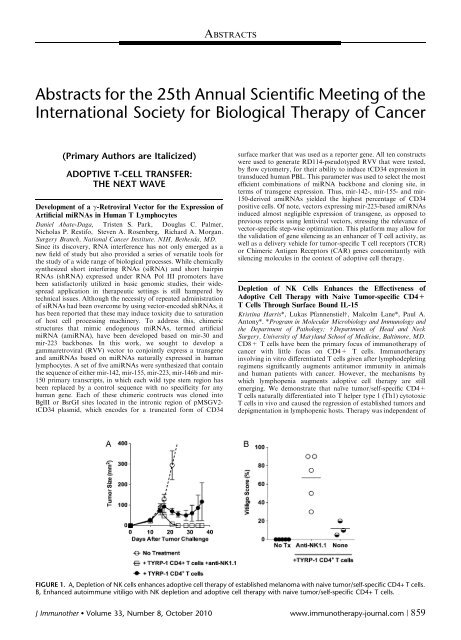
FIGURE 1. A, Depletion <strong>of</strong> NK cells enhances adoptive cell <strong>the</strong>rapy <strong>of</strong> established melanoma with naive tumor/self-specific CD4+ T cells.<br />
B, Enhanced autoimmune vitiligo with NK depletion and adoptive cell <strong>the</strong>rapy with naive tumor/self-specific CD4+ T cells.<br />
J Immuno<strong>the</strong>r Volume 33, Number 8, October 2010 www.immuno<strong>the</strong>rapy-journal.com | 859
<strong>Abstracts</strong> J Immuno<strong>the</strong>r Volume 33, Number 8, October 2010<br />
vaccination, exogenous cytokine support, CD8+-, B-, NK and NK<br />
T cells. Surprisingly NK depletion fur<strong>the</strong>r enhanced tumor <strong>the</strong>rapy<br />
by increasing T cell numbers, autoimmune vitiligo, and serum<br />
proinflammatory chemokines and cytokines. This effect may be<br />
dependent on increased levels <strong>of</strong> surface-bound IL-15 on CD11c+<br />
MHC class II+ cells which occurs after NK depletion. These data<br />
show a potential role <strong>for</strong> IL-15 in enhancing antitumor <strong>the</strong>rapies<br />
which incorporate CD4+ T cells (Fig. 1 previous page).<br />
Evaluation <strong>of</strong> CXCR3 and CCR5 Polymorphisms and Gene-<br />
Expression as Predictive Biomarkers <strong>of</strong> Clinical Response to<br />
Adoptive Therapy in Melanoma Patients<br />
Davide Bedognetti*w, Lorenzo Uccellini*, Ena Wang*, Mark E.<br />
Dudleyz, Zoltan Pos*, Maria Libera Ascierto*, Valeria De<br />
Giorgi*, Hui Liu*, Jingou Chen*, Mario Roberto Sertoliw,<br />
Francesco M. Marincola*, Steven A. Rosenbergz. *Department<br />
<strong>of</strong> Transfusion Medicine, CC, National Institutes <strong>of</strong> Health;<br />
zSurgery Branch, NCI, National Institutes <strong>of</strong> Health, Be<strong>the</strong>sda,<br />
MD; w Department <strong>of</strong> Oncology, Biology, Genetics, University <strong>of</strong><br />
Genoa, Genoa, Italy.<br />
Background: Adoptive cell <strong>the</strong>rapy induces objective responses in<br />
approximately 50% <strong>of</strong> patients with metastatic melanoma. The<br />
recruitment <strong>of</strong> T lymphocytes through CXCR3, CCR5-ligand<br />
chemokines is critical <strong>for</strong> <strong>the</strong> development <strong>of</strong> immune-mediated<br />
rejection. A common single nucleotide polymorphism <strong>of</strong> CXCR3<br />
(rs2280964) has been associated with variation in chemotactic<br />
activity. CCR5 polymorphism D32 (a deletion <strong>of</strong> 32 bases encoding<br />
a protein not expressed on cell surface), has been recently<br />
correlated with poor prognosis in metastatic melanoma patients.<br />
We postulated that polymorphisms <strong>of</strong> CXCR3, CCR5 genes may<br />
influence <strong>the</strong> migration <strong>of</strong> tumor-infiltrating lymphocytes (TIL) on<br />
tumor site and, eventually, <strong>the</strong> tumor regression.<br />
Methods: One-hundred-<strong>for</strong>ty-two TIL samples, belonging to 142<br />
melanoma patients enrolled in consecutive adoptive <strong>the</strong>rapy trials,<br />
were evaluated. Genotyping (rs2280964, D32 mutation) was<br />
per<strong>for</strong>med by sequencing. Gene-expression pr<strong>of</strong>iling <strong>of</strong> infused<br />
TIL was assessed by Affymetrix Human Gene ST 1.0 array<br />
(CXCR3, 27 probes; CCR5, 30 probes). DNA/RNA data were<br />
correlated with each o<strong>the</strong>r, with clinical response.<br />
Results: No significant correlations between genotype, gene<br />
expression were detected, nei<strong>the</strong>r between CXCR3 polymorphisms,<br />
response. Surprisingly, CCR5D32 carriers (N = 25; heterozygous,<br />
N = 24; homozygous, N = 1) had a better overall response (OR:<br />
CR, complete remission or PR, partial remission) compared to<br />
wildtype patients (OR: 68% vs. 46%, respectively, P
J Immuno<strong>the</strong>r Volume 33, Number 8, October 2010<br />
<strong>Abstracts</strong><br />
with a single peptide pool <strong>for</strong> 2 hours, and recombined <strong>for</strong> ano<strong>the</strong>r<br />
4 hours. The IFN-g Secretion Assay was used to magnetically<br />
enrich IFN-g secreting T cells to a purity <strong>of</strong> >90%. High antigen<br />
specificity and functionality <strong>of</strong> <strong>the</strong> enriched T cells was confirmed<br />
after expansion <strong>for</strong> 9 to 14 days. T cell lines contained high<br />
frequencies <strong>of</strong> pp65tetramer+ CD8+ T cells. Additionally, after<br />
restimulation with a mixture <strong>of</strong> peptide pools 21% to 53% <strong>of</strong><br />
CD4+ T cells and 53% to 87% <strong>of</strong> CD8+ T cells produced IFN-g.<br />
Moreover, comparing T cell frequencies specific <strong>for</strong> each single<br />
antigen in PBMC with that in multivirus-specific T cell line after<br />
separate reactivation with peptide pools by analysis <strong>of</strong> IFN-g<br />
production, we observed <strong>the</strong> specificity <strong>for</strong> each antigen sustained<br />
after enrichment and expansion. We found similar results in<br />
number <strong>of</strong> enriched cells, expansion rates, and antigen-specificity <strong>of</strong><br />
<strong>the</strong> T cell lines, regardless <strong>of</strong> whe<strong>the</strong>r PBMC loaded with a mixture<br />
<strong>of</strong> antigens or separately with single peptide pools to generate T cell<br />
lines. In summary, we established a protocol <strong>for</strong> rapid in vitro<br />
generation <strong>of</strong> multiantigen-specific CD4+ and CD8+ T cells using<br />
a combination <strong>of</strong> peptide pools from several antigens <strong>for</strong><br />
restimulation and subsequent magnetic selection <strong>of</strong> IFN-g secreting<br />
T cells.<br />
Hypo<strong>the</strong>ses <strong>for</strong> Improving Designer T Cells; Phase I Trials<br />
Richard Junghans*w, Ritesh Rathore*w, Barti Rathore*w,<br />
Qiangzhong Ma*w, Anthony Bais*, Erica Gomes*, Ryan<br />
Harvey*, Nithiandan Selliah*w, Shah Miah*w, Pam Davol*,<br />
Steven Cohen*, Samer Al Homsi*w. *Surgery, Roger Williams<br />
Hospital, Providence, RI; w Boston University School <strong>of</strong> Medicine,<br />
Boston, MA.<br />
In prior Phase I trials <strong>of</strong> 1st generation (1st gen) designer T cells,<br />
pro<strong>of</strong>-<strong>of</strong>-principle ‘‘biologic responses’’ were noted, but with poor<br />
in vivo persistence and transient in-tumor activity. The first<br />
problem generated <strong>the</strong> hypo<strong>the</strong>sis: If T cells were maintained<br />
systemically at high levels, a sustained T cell percolation into<br />
tumor could yield cures even if T cells survived <strong>for</strong> only a few<br />
days <strong>of</strong> killing. The second problem generated <strong>the</strong> hypo<strong>the</strong>sis: If<br />
T cells entering tumor, though few in number, were to proliferate<br />
on contact with antigen, <strong>the</strong>n tumors could be cured. In<br />
separate clinical trials, <strong>the</strong>se hypo<strong>the</strong>ses are being addressed in our<br />
group.<br />
Methods: Patient T cells are transduced and expanded ex vivo to<br />
span dose levels <strong>of</strong> 10 4 9, 10 4 10 and 10 4 11 T cells. The first study<br />
uses 1st gen designer T cells (against prostate specific membrane<br />
antigen, PSMA), but with prior non-myeloablative (NMA)<br />
conditioning to create a ‘‘hematopoietic space’’ into which designer<br />
T cells are engrafted <strong>for</strong> prolonged in vivo persistence, with coadministered<br />
continuous infusion IL2. The second study uses a<br />
simple infusion protocol that applies 2nd gen designer T cells<br />
(against CEA) with added CD28 co-stimulation with <strong>the</strong> goal <strong>of</strong> in<br />
situ amplifying <strong>the</strong> effector cells that reach tumor.<br />
Results: Each study treated five patients to date at <strong>the</strong> low 10^9 and<br />
middle 10 4 10 T cell doses. NMA conditioning led to successful<br />
T cell engraftment in <strong>the</strong> 1% to 20% range at one month, versus<br />
designer T cells being undetectable at <strong>the</strong> same time in <strong>the</strong> infusion<br />
study. No on-target T cell toxicities were noted in ei<strong>the</strong>r study. In<br />
<strong>the</strong> engraftment protocol, two patients had PSA reductions <strong>of</strong> 50<br />
and 70% in <strong>the</strong> two months following treatment. In <strong>the</strong> second<br />
study with 2nd gen anti-CEA T cells, one gastric cancer patient had<br />
shrinkage <strong>of</strong> lung and brain mets and ano<strong>the</strong>r has stable disease <strong>for</strong><br />
12+ months after treatment; future patients will have IL2 added<br />
with <strong>the</strong> next T cell dose escalation. The clinical (non-manufacturing)<br />
cost per patient was estimated at $60 to 100 K in <strong>the</strong><br />
engraftment study and $5 to 10 K in <strong>the</strong> infusion study.<br />
Conclusion: Parallel approaches are being applied to test hypo<strong>the</strong>ses<br />
<strong>of</strong> benefit <strong>of</strong> engraftment versus benefit <strong>of</strong> co-stimulation to<br />
create effective anti-tumor activity in designer T cells. Study results<br />
support <strong>the</strong> safety <strong>of</strong> <strong>the</strong> protocols with indications <strong>of</strong> efficacy but<br />
<strong>the</strong> dose escalations are at an early stage. Patient recruitments are<br />
continuing.<br />
Functional Reprogramming <strong>of</strong> <strong>the</strong> Tumor Stroma by IL-12<br />
Engineered T Cells is Required <strong>for</strong> Anti-tumor Immunity<br />
Sid P. Kerkar*, Robert Reger*, Pawel Muranski*, Zhiya Yu*,<br />
Douglas Palmer*, Dhanalakshmi Chinnasamy*, Christopher<br />
A. Kleban<strong>of</strong>f*, Yun Ji*, Luca Gattinoni*, Steven A. Rosenberg*,<br />
Giorgio Trinchieriw, Nicholas P. Restifo*. *Center <strong>for</strong> Cancer<br />
Research; w Cancer and Inflammation Program, National Cancer<br />
Institute, Be<strong>the</strong>sda, MD.<br />
Bone marrow derived stromal cells within <strong>the</strong> tumor microenvironment<br />
are capable <strong>of</strong> cross presenting antigens to cytotoxic<br />
T lymphocytes (CTL). We found that <strong>the</strong> adoptive transfer <strong>of</strong><br />
tumor-specific CD8+ T cells gene-engineered to secrete IL-12 led<br />
to <strong>the</strong> increased local infiltration <strong>of</strong> adoptively transferred T cells<br />
and caused <strong>the</strong> regression <strong>of</strong> large established B16 melanomas. The<br />
autocrine effects <strong>of</strong> IL-12 resulted in <strong>the</strong> production <strong>of</strong> large<br />
amounts <strong>of</strong> IFN-g by T cells. Surprisingly, we found that IL-12-<br />
engineered T cells that lacked <strong>the</strong> ability to receive signals from<br />
IL-12 (Il12rb2 / ), and indeed T cells that lacked <strong>the</strong> ability to<br />
produce IFN-g (Ifng / ), retained all <strong>of</strong> <strong>the</strong>ir ability to trigger<br />
tumor destruction. However, tumor treatment efficacy was<br />
abrogated when <strong>the</strong> cells in host mice lacked IL-12 receptors<br />
(Il12rb2 / ), IFN-g receptors (IfngR / ) or <strong>the</strong> ability to<br />
produce IFN-g. Thus, sensitization <strong>of</strong> host cells and not <strong>the</strong><br />
transferred T cells within <strong>the</strong> tumor microenvironment was critical<br />
<strong>for</strong> successful anti-tumor immunity. We measured increased<br />
endogenous CD8+ and host NK cells within <strong>the</strong> tumor but<br />
treatment responses remained robust in mice completely devoid <strong>of</strong><br />
T and B cells (Rag / ) and depleted <strong>of</strong> NK cells. We found that<br />
<strong>the</strong> majority <strong>of</strong> cells expressing <strong>the</strong> IL-12Rb2 receptor within <strong>the</strong><br />
tumor were CD11b+ myeloid cells. Transfer <strong>of</strong> IL-12-producing<br />
anti-tumor T cells triggered significant in situ changes in CD11b+<br />
cells including increased expression <strong>of</strong> H-2Db along with upregulation<br />
<strong>of</strong> Fas (CD95), and FADD. In addition, both <strong>the</strong><br />
numbers and percentages <strong>of</strong> CD11b+ cells dropped just prior to<br />
tumor regression. Tumor treatment was abrogated in mice deficient<br />
in MHC class I (b2M / ) but not class II (I-Ab / ),<br />
indicating <strong>the</strong> functional importance <strong>of</strong> antigen cross-presentation<br />
in vivo. These results are consistent with a model whereby IL-12<br />
triggers <strong>the</strong> functional maturation <strong>of</strong> in situ APCs capable <strong>of</strong> crosspresenting<br />
tumor antigens. Licensed recognition <strong>of</strong> <strong>the</strong>se antigens<br />
by tumor-specific T cells may in turn trigger <strong>the</strong> collapse <strong>of</strong> <strong>the</strong><br />
tumor stroma and its vasculature.<br />
Monomeric Designer T Cells Kill IL13Ra2 Expressing GBM<br />
More Efficiently<br />
Seogkyoung Kong, Richard P. Junghans, Prakash Sampath.<br />
Neurosurgery, Boston University School <strong>of</strong> Medicine, Roger<br />
Williams Medical Center, Providence, RI.<br />
GBM is a devastating primary brain tumor <strong>for</strong> which <strong>the</strong>re is no<br />
effective <strong>the</strong>rapy to specifically target tumor cells. Targeted<br />
immuno-gene <strong>the</strong>rapy has been shown effective in a number <strong>of</strong><br />
tumor models without significant toxicity. We aim to develop<br />
potent GBM-specific T cells as an innovative and unique targeted<br />
immuno-gene <strong>the</strong>rapy. I exploit IL13Ra2 as a GBM-specific tumor<br />
antigen due to its frequent overexpression on a majority <strong>of</strong> GBM<br />
but not on normal brain tissues. Targeting IL13Ra2 on GBM has<br />
a strong rationale supported by clinical development <strong>of</strong> IL13<br />
immunotoxin molecules. IL13 binds two types <strong>of</strong> receptor with<br />
different affinities: IL13 binds to GBM-associated IL13Ra2 with<br />
high affinity (Kd = 0.25 to 1.2 nM); IL13 binds to IL13Ra1 first<br />
with low affinity (Kd = 2 to 30 nM) and <strong>the</strong>n recruits IL4Ra to <strong>the</strong><br />
complex. Previous studies showed that substitutions at three sites<br />
<strong>of</strong> IL13 (Glu-11, Lys-103, or Arg-107) proved to be critical <strong>for</strong><br />
IL13 binding to IL13Ra2, and modifications <strong>of</strong> <strong>the</strong>se sites can<br />
neutralize or dramatically increase <strong>the</strong> affinity <strong>of</strong> IL13 to IL13Ra2,<br />
compared <strong>of</strong> <strong>the</strong> shared IL13Ra1, which is expressed on <strong>the</strong> normal<br />
brain cells and o<strong>the</strong>r tissues. In order to achieve both specific<br />
targeting and selectively enhanced cytotoxicity <strong>of</strong> GBM-associated<br />
r 2010 Lippincott Williams & Wilkins www.immuno<strong>the</strong>rapy-journal.com | 861
<strong>Abstracts</strong> J Immuno<strong>the</strong>r Volume 33, Number 8, October 2010<br />
IL13Ra2, I generated IL13.E11 K.R107 K (both glutamic acid at<br />
position 11 and arginine at 107 changed to lysine). I modified<br />
T cells using retroviral gene transfer to express a chimeric immune<br />
receptor (CIR) comprising an extracellular high affinity mutant<br />
IL13.E11 K.R107 K molecule linked to intracellular signaling<br />
components from CD3 zeta chain and CD28 co-stimulatory<br />
molecule. The IL13.E11 K.R107 K designer T cells show specific<br />
targeting and selectively enhanced cytotoxicity <strong>of</strong> IL13Ra2+<br />
glioblastoma cells, and inhibit tumor growth employing a glioma<br />
xenograft model established with human glioma cells in athymic<br />
nude rats. I hypo<strong>the</strong>sized that monomeric designer T cells kill<br />
IL13Ra2 expressing GBM more efficiently based on <strong>the</strong> monomeric<br />
IL13Ra2 chain is overexpressed on a majority <strong>of</strong> GBM. I generated<br />
monomeric CIR by mutating both Cys-29 <strong>of</strong> <strong>the</strong> CD8a hinge and<br />
Cys-2 <strong>of</strong> CD3 transmembrane to Ala. Monomeric designer T cells<br />
show specifically enhanced cytotoxicity/INF-g and IL-2 cytokine<br />
secretion <strong>of</strong> IL13Ra2+ glioblastoma cells versus control dimeric<br />
designer T cells. Monomeric IL13.E11 K.R107 K designer T cells<br />
show <strong>the</strong> most potent against IL13Ra2+ glioblastoma cells. We<br />
demonstrated <strong>the</strong> in vivo antiglioma efficacy <strong>of</strong> <strong>the</strong> monomeric<br />
IL13.E11 K.R107 K designer T cells employing a glioma xenograft<br />
model established with human glioblastoma cells in athymic nude<br />
rats. The monomeric IL13.E11 K.R107 K designer T cells have<br />
significant potential <strong>for</strong> <strong>the</strong> treatment <strong>of</strong> recurrent GBM.<br />
Development <strong>of</strong> a Chimeric Antigen Receptor <strong>for</strong> Prostate<br />
Cancer Stem Cell Antigen <strong>for</strong> Adoptive Cell Therapy <strong>of</strong><br />
Cancer<br />
Kiran H. Lagisetty*, William Burnsw, Zhili Zheng*, Steven<br />
A. Rosenberg*, Richard A. Morgan*. *Surgery Branch, National<br />
Cancer Institute; w Department <strong>of</strong> Surgery, Johns Hopkins Medicine,<br />
Baltimore, MD.<br />
There is currently no cure <strong>for</strong> metastatic, hormone refractory<br />
prostate cancer. Localized disease is well controlled with surgery<br />
and radiation and in many cases can be curative. However, locally<br />
advanced or widespread disease, require hormone <strong>the</strong>rapy <strong>for</strong><br />
tumor control. This will eventually trans<strong>for</strong>m into a hormone<br />
refractory state which <strong>the</strong>re are currently no good treatment<br />
options. Targeted adoptive immuno<strong>the</strong>rapy against melanoma<br />
associated antigens has shown <strong>the</strong> ability to achieve tumor<br />
regression in cases <strong>of</strong> advanced disease. Prostate stem cell antigen<br />
(PSCA) is a well described prostate cancer tumor antigen which can<br />
be present on up 48 percent <strong>of</strong> primary lesions and 64 percent <strong>of</strong><br />
metastatic lesions (Ross S, et al, Cancer Res. May 1, 2002). PSCA<br />
expression is not limited to prostate cancer as it is found in up to 60<br />
percent <strong>of</strong> primary pancreatic tumors (Argani P, et al, Cancer Res.<br />
June 1, 2001). PSCA is a member <strong>of</strong> <strong>the</strong> Thy-1/Ly-6 family <strong>of</strong> GPI<br />
anchored cell surface antigens whose function is unknown. PSCA<br />
has low expression in normal prostate tissue and its expression is<br />
increased in higher grade and stage tumors making it an ideal<br />
target <strong>for</strong> immuno<strong>the</strong>rapy. Using this model we hypo<strong>the</strong>size that<br />
transduction <strong>of</strong> T-cells containing a chimeric antigen receptor<br />
targeting PSCA can inhibit tumor growth and metastases. We<br />
constructed MSGV-1 based retroviral vectors containing six<br />
different murine derived single chain variable fragments specific<br />
<strong>for</strong> PSCA linked to T-cell signaling domains CD28 and CD3z. The<br />
PSCA-CAR’s were used to be transduce human peripheral blood<br />
lymphocytes with 80 to 90 percent transduction efficiency. Cell lines<br />
from melanoma, prostate and pancreatic cancer were screened<br />
<strong>for</strong> PSCA expression via FACS analysis. The PSCA-CAR’s were<br />
<strong>the</strong>n placed in overnight cocultures with PSCA expressing and<br />
PSCA nonexpressing cell lines. Two PSCA-CAR constructs<br />
containing single chain variable fragments, m1G8 and bm2B3,<br />
revealed a 2 to 3 fold increase in interferon gamma secretion when<br />
compared to non-PSCA expressing cell lines. We are in <strong>the</strong> process<br />
<strong>of</strong> optimizing <strong>the</strong>se constructs and fur<strong>the</strong>r testing <strong>the</strong>m against<br />
PSCA expressing prostate cancer lines. These promising results<br />
may provide a new targeted <strong>the</strong>rapy <strong>for</strong> hormone refractory<br />
prostate cancer.<br />
Large-Scale Pr<strong>of</strong>iling <strong>of</strong> Circulating Serum Markers, Single<br />
Cell Polyfunctionality and Antigen Diversity <strong>of</strong> T Cell<br />
Response Against Melanoma<br />
Chao Ma*, Ann Chueng*, Begonya Comin-Anduixw, Thinle<br />
Condonw, Antoni Ribasw, James Heath*. *NanoSystems Biology<br />
Cancer Center; Physics; Chemistry, Caltech, Pasadena; w Medicine,<br />
UCLA, Los Angeles, CA.<br />
Background: Highly multiplexed, sensitive and single cell pr<strong>of</strong>iling<br />
at multiple levels is likely to provide important in<strong>for</strong>mation to<br />
advance immuno<strong>the</strong>rapy <strong>for</strong> cancer, due to complexity <strong>of</strong> antigen<br />
specificity within cancer-targeting T cell populations and large<br />
varieties <strong>of</strong> effector molecules produced. Herein we report on<br />
immune monitoring studies using newly developed plat<strong>for</strong>ms<br />
analyzing samples from 8 patients enrolled in an ongoing clinical<br />
trial involving adoptive cell transfer (ACT) <strong>of</strong> lymphoyctes<br />
genetically modified to express a T cell receptor (TCR) specific<br />
<strong>for</strong> MART-1, which are administered to patients with metastatic<br />
melanoma toge<strong>the</strong>r with dendritic cell vaccination after nonmyeloablative<br />
lymphodepleting chemo<strong>the</strong>rapy.<br />
Methods: Miniaturized, highly multiplexed microchip based<br />
technology was adapted to follow <strong>the</strong> time course <strong>of</strong> immune<br />
responses focused on: (1) melanoma specific T cell repertoire<br />
enumeration that simultaneously detected 35 mutated, overexpressed<br />
or cancer-germline melanoma antigen specific T cell<br />
populations; (2) single cell secretome pr<strong>of</strong>iling <strong>of</strong> 20 cytolytic,<br />
inflammatory cytokines and chemokines from sorted phenotypically<br />
defined antigen-specific cytotoxic T lymphocytes and different<br />
helper T cell subclasses; (3) measurement <strong>of</strong> 35 blood melanoma<br />
tumor markers and immune proteins.<br />
Results: MART-1-specific T cells peaked at 8 to 14 days after ACT<br />
with frequencies varying from 1 to 60% <strong>of</strong> CD3+ T cells in<br />
peripheral blood. Over 5 fold expansions in frequency was<br />
demonstrated by T cells specific <strong>for</strong> <strong>the</strong> melanoma antigens<br />
tyrosinase, NY-Eso, Mage 3, Mage 10 and GnT-V (P50 different<br />
functional subtypes) and polyfunctionality, characterized by strong<br />
per<strong>for</strong>in, TNFa,IL1b and chemokine secretion. However, more than<br />
90% <strong>of</strong> MART-1 T cells at day 30 lacked TNFa or IFNg responses,<br />
suggesting a dysfunctional or partially exhausted phenotype. Tumor<br />
markers and T cell effector proteins in serum, including per<strong>for</strong>in,<br />
granzyme B and cytokines, were significantly released into peripheral<br />
blood soon after T cell reinfusion and melanoma associated markers<br />
and T cell growth factors followed similar dynamics, which indicated<br />
concurrence <strong>of</strong> tumor destruction and T cell expansion and attack.<br />
Conclusions: Our results indicate importance <strong>of</strong> epitope spreading,<br />
T cell diversity and polyfunctionality <strong>for</strong> patient’s response after<br />
<strong>the</strong> ACT <strong>of</strong> TCR transgenic lymphocytes; Tumor elimination is<br />
closely related to immune response quality. Ongoing experiments<br />
will explore quantitatively significance <strong>of</strong> each marker measured.<br />
Adoptively Transferred Anti-Tumor Th17 Cells are Long-<br />
Lived and Evolve Into a Highly Active Memory Population<br />
with a Unique Molecular Signature<br />
Pawel J. Muranski*, Zachary Borman*, Sid Kerkar*, Yun Ji*,<br />
Luca Gattinoni*, Robert Reger*, Christopher Kleban<strong>of</strong>f*, Zhiya<br />
Yu*, Gabriela Ferreyraw, Steven Kernw, Robert L. Dannerw,<br />
Nicholas P. Restifo*. *National Cancer Institute; w Critical Care<br />
Medicine Department, Clinical Center, NIH, Be<strong>the</strong>sda, MD.<br />
Tumor-specific CD4+ T cells polarized under ‘‘Th17’’ conditions<br />
efficiently eradicate advanced cancer in mouse models. Th17 cells<br />
have been recently described as a meta-stable, short-lived,<br />
population with end-effector characteristics and purportedly have<br />
a limited ability to <strong>for</strong>m memory (Pepper, Nature Immunol, 2010).<br />
Although we confirmed that Th17-polarized cells expressed low<br />
levels <strong>of</strong> CD27 compared to Th1-polarized cells, we found that<br />
<strong>the</strong>y efficiently survived and <strong>for</strong>med memory in vivo. While<br />
previous work used IL-17 to trace cells after adoptive transfer, in<br />
a congenic system we observed that most cells lost <strong>the</strong> capacity to<br />
862 | www.immuno<strong>the</strong>rapy-journal.com r 2010 Lippincott Williams & Wilkins
J Immuno<strong>the</strong>r Volume 33, Number 8, October 2010<br />
<strong>Abstracts</strong><br />
produce IL-17A during in vivo expansion. Even though inability <strong>of</strong><br />
T cells to <strong>for</strong>m memory had been previously attributed to <strong>the</strong> early<br />
loss <strong>of</strong> CD27 expression we demonstrated that in vitro generated<br />
Th17-polarized cells exhibited lower levels <strong>of</strong> multiple markers <strong>of</strong><br />
T cell senescence, including Klrg1, FasL, CD25 and PD-1. They<br />
also produced more IL-2, had high levels <strong>of</strong> <strong>the</strong> pro-survival factors<br />
bcl-2 and bcl-6 and low expression <strong>of</strong> its target, prdm1 that encodes<br />
transcriptional repressor Blimp1, a hallmark molecule <strong>of</strong> terminally<br />
differentiated effectors. Using serial global gene expression pr<strong>of</strong>iling<br />
<strong>of</strong> adoptively transferred cells, we found that Th17-polarized<br />
cells rapidly acquired some Th1-like properties including t-bet and<br />
IFN-g expression, however Th17-derived memory population was<br />
readily distinguished based on its molecular signature and had<br />
characteristics closely mimicking a pattern found in highly active<br />
CD8+ T memory stem cells described in our previous work. Th17-<br />
dervied memory cells expressed high levels <strong>of</strong> multiple self-renewal<br />
and pro-survival-associated transcription factors and regulators,<br />
had reduced expression <strong>of</strong> prdm1, as well as low levels <strong>of</strong> <strong>the</strong><br />
phenotypic markers <strong>of</strong> terminal differentiation, including pd1,<br />
klrg1, klrd1 and granzymes, in contrast to higher levels <strong>of</strong> CCR7<br />
and il-7r. In summary, we report that Th17-derived cells expressed<br />
more ‘‘stem-like’’ characteristics distinct <strong>for</strong>m Th1-derived effectors<br />
as evidenced by <strong>the</strong>ir molecular signature and dramatically enhanced<br />
ability to survive and reject tumor. This suggests that Th17-<br />
polarizing conditions trigger a distinctive developmental program<br />
representing a promising avenue <strong>for</strong> <strong>the</strong> development <strong>of</strong> potent, new<br />
T cell-based immuno<strong>the</strong>rapies <strong>of</strong> cancer and o<strong>the</strong>r diseases.<br />
Artifical MicroRNA Targeting Programmed Death Receptor-1<br />
to Enhance Adoptive Cell Transfer Therapy <strong>for</strong> Cancer<br />
Tristen S. Park, Takashi Inozume, Mojgan Ahmadzadeh, Ken-ichi<br />
Hanada, James Yang, Steven A. Rosenberg, Richard A. Morgan.<br />
Surgery Branch, National Institutes <strong>of</strong> Health, Be<strong>the</strong>sda, MD.<br />
Melanoma has <strong>the</strong> unique characteristic <strong>of</strong> inducing anti-tumor<br />
lymphocytes during tumor growth. Tumor Infiltrating Lymphocytes<br />
(TIL) demonstrate <strong>the</strong> ability to recognize and lyse tumor<br />
cells in vitro, and <strong>the</strong> infusion <strong>of</strong> large numbers <strong>of</strong> <strong>the</strong>se<br />
lymphocytes have resulted in a 49% to 72% response rate in<br />
patients with metastatic melanoma.Yet <strong>the</strong>se naturally occurring<br />
anti-tumor lymphocytes can <strong>of</strong>ten be anergized in vivo, by <strong>the</strong><br />
suppressive tumor microenvironment. Fur<strong>the</strong>r improvement to<br />
TIL <strong>the</strong>rapy may be realized by targeting negative signaling<br />
pathways in <strong>the</strong> administered TIL.<br />
Programmed death receptor-1 (PD-1) is an immunoinhibitory<br />
receptor that is expressed in CD4 and CD8 positive lymphocytes.<br />
Interaction between PD-1 and its ligands PD-L1 and PD-L2 deliver<br />
a negative signal to lymphocytes. PD-L1 is expressed in a variety <strong>of</strong><br />
human tumors including melanomas. It was recently shown that<br />
in melanoma, PD-1 expressing TIL displayed impaired effector<br />
function compared with PD-1 negative TIL. We observed that PD-<br />
1 levels were upregulated in TIL and PBL when cocultured with<br />
matched melanoma lines in vitro. This suggests that <strong>the</strong> tumor<br />
microenvironment may play a role in <strong>the</strong> induction and maintenance<br />
<strong>of</strong> PD-1 expression on tumor reactive cells and that PD-1<br />
on TIL may impair <strong>the</strong> antitumor immune response in patients.<br />
To investigate <strong>the</strong> PD-1/PD-L1 interaction in a defined in vitro<br />
setting, PBL were retrovirally engineered to express PD-1 with<br />
ei<strong>the</strong>r a Mart-1 specific TCR or a HMW (High molecular weight)<br />
specific Chimeric Antigen Receptor (CAR). In coculture assays<br />
against melanoma lines 1300, 624, 526 engineered to express PD-<br />
L1, <strong>the</strong>se Mart-1 TCR/PD-1+ PBL demonstrated impaired tumor<br />
recognition with an approximately 50 percent reduction in<br />
Interferon-g (IFN-g) secretion in <strong>the</strong> PBL <strong>of</strong> three donors. HMW<br />
CAR/PD-1+ PBL also demonstrated approximately 50 percent<br />
reduction in IFN production in a coculture assay with mel 1300,<br />
888, 526, 624 expressing PD-L1.<br />
Antibodies that block <strong>the</strong> PD-1/PD-L1 interaction are currently in<br />
Phase I/II clinical trials, but a more selective approach may be <strong>the</strong><br />
specific targeting <strong>of</strong> PD-1 in tumor specific T cells using RNA<br />
interference. We are investigating if PD-1 expression in TIL can be<br />
silenced using a microRNA-based system. Short hairpin RNA<br />
sequences targeting PD-1 were identified to downregulate <strong>the</strong> expression<br />
<strong>of</strong> PD-1 by 50% to 70% in both T cell lines and peripheral<br />
blood lymphocytes. Using <strong>the</strong>se shRNA sequences we are constructing<br />
an optimized microRNA-based retroviral vector <strong>for</strong> <strong>the</strong> engineering<br />
<strong>of</strong> tumor infiltrating lymphocytes. The downregulation <strong>of</strong> PD-1 in<br />
TIL,mayleadtoimprovedanti-tumoractivityinvitroandinvivo.<br />
Adoptive T Cell Therapy <strong>for</strong> Metastatic Melanoma: The MD<br />
Anderson Experience<br />
Laszlo G. Radvanyi, Chantale Bernatchez, Minying Zhang,<br />
Priscilla Miller, Michelle Glass, Nicholas Papadopoulos, Patrick<br />
Hwu. Melanoma Medical Oncology, MD Anderson Cancer Center,<br />
Houston, TX.<br />
Adoptive cell <strong>the</strong>rapy (ACT) using tumor-infiltrating lymphocytes<br />
(TIL) is a promising treatment <strong>for</strong> metastatic melanoma. Here, we<br />
report on <strong>the</strong> results <strong>of</strong> an ongoing Phase II clinical trial testing<br />
ACT in metastatic melanoma patients regardless <strong>of</strong> HLA subtype.<br />
Autologous TIL were expanded in large-scale using anti-CD3 and<br />
IL-2 and <strong>the</strong>n infused into patients following transient lymphodepletion.<br />
This was followed by high-dose IL-2 <strong>the</strong>rapy. The best<br />
overall response was determined and correlated with T cell phenotype<br />
as well as telomere length. The persistence <strong>of</strong> specific TCR clonotypes<br />
after infusion was also tracked. Altoge<strong>the</strong>r, 30 patients have been<br />
treated with clinical response data available from 25 patients (as <strong>of</strong><br />
June 20, 2010). Overall, 13/25 (52%) patients have had a clinical<br />
response (PR/CR), with one patient having an ongoing PR <strong>for</strong> >22<br />
months and ano<strong>the</strong>r patient having a CR. A higher percentage and<br />
number <strong>of</strong> CD8+ T cells (P
<strong>Abstracts</strong> J Immuno<strong>the</strong>r Volume 33, Number 8, October 2010<br />
FIGURE 1. Accumulation <strong>of</strong> 18 [F]-FEAU in CAR+tk+ T cells. Mice<br />
were anes<strong>the</strong>sized and CAR+tk+ T cells were injected subcutaneously<br />
in <strong>the</strong> right flank. Positron emission tomography<br />
computed tomography (PET/CT) images were acquired 2 hours<br />
after intravenous administration <strong>of</strong> 18 [F]-FEAU using Inveon<br />
micro-PET/CT scanner. Images were reconstructed by 2-dimensional<br />
ordered subsets expectation maximization algorithm. PET<br />
and CT image fusion and analysis were per<strong>for</strong>med using vendor<br />
s<strong>of</strong>tware Inveon Research Workplace.<br />
<strong>the</strong> dynamic status <strong>of</strong> infused T cells. In this study, we constructed<br />
SB DNA transposon vectors encoding a panel <strong>of</strong> transgenes<br />
expressing <strong>the</strong> wild type HSV1-tk fused to hygromycin phosphotransferase<br />
(in vitro selection) and FLAG tag (expression level).<br />
Primary T cells were co-electroporated with tkHy SB transposon<br />
and a CD19-specific chimeric antigen receptor (CAR) SB<br />
transposon and propagated on CD19-specific artificial antigen<br />
presenting cells in <strong>the</strong> presence <strong>of</strong> cytocidal concentrations <strong>of</strong><br />
hygromycin B. After 4 weeks <strong>of</strong> numeric expansion (i) 90% <strong>of</strong> <strong>the</strong><br />
T cells were CAR+tk+, (ii) accumulated high amounts <strong>of</strong> [3H]<br />
2 0 -fluoro-2 0 deoxy-1-b-D-arabion<strong>of</strong>uranosyl-5-ethyl-uracil (iii) were<br />
ablated in <strong>the</strong> presence <strong>of</strong> ganciclovir and (iv) exhibited redirected<br />
killing <strong>of</strong> CD19+ tumor targets. The CAR+tk+ T cells could be<br />
visualized by mPET imaging in mice (Fig. 1). This is <strong>the</strong> first report<br />
showing that SB transposition can generate CAR+tk+ T cells<br />
which can be imaged by mPET in vivo. We have adapted SB system<br />
<strong>for</strong> human application (IND # 14193), and thus this study has<br />
immediate translational application to infuse CD19-specific T cells<br />
co-expressing HSV1-tk <strong>for</strong> imaging.<br />
Expansion <strong>of</strong> Autologous Human Vg9Vd2 T Cells Ex Vivo:<br />
Potential <strong>for</strong> Adoptive T Cell Therapy <strong>of</strong> Prostate Cancer<br />
Andrew Roberts*, Maria Serrano*, Elisa Binda*, Pierre<br />
Vantourout*w, Hardev Pandhaz, Adrian C. Hayday*w. *Peter<br />
Gorer Department <strong>of</strong> Immunobiology, King’s College London;<br />
w London Research Institute, Cancer Research UK, London;<br />
zPostgraduate Medical School, University <strong>of</strong> Surrey, Guild<strong>for</strong>d, UK.<br />
Dysregulated cells including solid tumours express stress molecules<br />
that can be recognised by cell surface receptors on gd T cells,<br />
including <strong>the</strong> Vg9Vd2 T cell receptor (which recognises pyrophosphate<br />
molecules such as IPP that may be expressed on tumour cell<br />
surfaces) and <strong>the</strong> killer activatory receptor NKG2D. Because gd T<br />
cells exhibit marked functional pleiotropy upon activation, we<br />
employed extensive microarrays to demonstrate that <strong>the</strong> use <strong>of</strong> low<br />
molecular weight pyrophosphate antigens in combination with<br />
low-dose IL-2 could induce <strong>the</strong> expression <strong>of</strong> cytokines and<br />
cytolytic agents (eg, Granzymes, Per<strong>for</strong>in and TRAIL) that are<br />
known to be efficacious against tumours. Consequently, in a Phase<br />
I trial in which two cohorts <strong>of</strong> nine stage IV hormone-refractory<br />
prostate cancer patients received <strong>the</strong> amino-bisphosphonate<br />
zoledronate (which blocks IPP metabolism) with or without IL-2,<br />
we observed pr<strong>of</strong>ound activation <strong>of</strong> gd effector responses in vivo, as<br />
well as an indication <strong>of</strong> <strong>the</strong>rapeutic efficacy in <strong>the</strong> cohort receiving<br />
zoledronate with IL-2.<br />
However, repeated administration <strong>of</strong> gd-activating agents can lead<br />
to cell exhaustion in vivo; an indication <strong>of</strong> this is provided by <strong>the</strong><br />
failure <strong>of</strong> <strong>the</strong> bisphosphonate-induced acute phase response to<br />
recur after <strong>the</strong> initial dose. Moreover, a combination <strong>of</strong> <strong>the</strong> tumour<br />
milieu and concurrent <strong>the</strong>rapies could limit <strong>the</strong> capacity <strong>of</strong> some<br />
individuals to generate an adequate population <strong>of</strong> activated gd T<br />
cells able to target <strong>the</strong> tumour. It is <strong>the</strong>re<strong>for</strong>e logical to complement<br />
bisphosphonate treatment by <strong>the</strong> activation <strong>of</strong> autologous gd T<br />
cells ex vivo and <strong>the</strong> subsequent infusion <strong>of</strong> <strong>the</strong>m. This approach<br />
has been validated in mouse models <strong>of</strong> prostate cancer, and more<br />
recently in nude mice bearing human glioblastoma tumours that<br />
were treated by adoptive transfer <strong>of</strong> expanded human gd T cells. By<br />
extension, we are aiming to generate reproducible protocols <strong>for</strong> <strong>the</strong><br />
ex vivo expansion <strong>of</strong> human Vg9Vd2 T cells, with <strong>the</strong> objective <strong>of</strong><br />
adapting <strong>the</strong> system to GMP <strong>for</strong> intended treatment <strong>of</strong> patients<br />
with solid tumours. This approach is critically dependent upon <strong>the</strong><br />
optimisation <strong>of</strong> key factors, such as <strong>the</strong> use <strong>of</strong> cytokines that, in<br />
addition to low-dose IL-2, might serve to augment survival and<br />
effector potential <strong>of</strong> such cells in long-term cultures. Fur<strong>the</strong>rmore,<br />
ongoing work also comprises <strong>the</strong> development <strong>of</strong> appropriate<br />
functional assays, in order that we can begin to predict in vitro <strong>the</strong><br />
likely success <strong>of</strong> subsequent cell engraftment in vivo.<br />
Dissection <strong>of</strong> Therapy-Induced Melanoma-Reactive Cytotoxic<br />
T Cell Responses<br />
Ton Schumacher. Department <strong>of</strong> Immunology, Ne<strong>the</strong>rlands Cancer<br />
Institute, Amsterdam, Ne<strong>the</strong>rlands.<br />
There is strong evidence that melanoma-reactive T cell responses<br />
induced by immuno<strong>the</strong>rapeutic interventions such as anti-CTLA4<br />
treatment or TIL <strong>the</strong>rapy can exert clinically meaningful effects.<br />
However, at present we do not know which cytotoxic T cell<br />
reactivities mediate cancer regression. Fur<strong>the</strong>rmore, as <strong>the</strong> number<br />
<strong>of</strong> melanoma-associated antigens to which <strong>the</strong>se responses can be<br />
directed is very high, classical strategies to map cytotoxic T cell<br />
reactivity do not suffice. Knowledge <strong>of</strong> such reactivities would be<br />
useful to design more targeted strategies that selectively aim to<br />
induce immune reactivity against <strong>the</strong>se antigens.<br />
In <strong>the</strong> past years we have aimed to address this issue by designing<br />
MHC class I molecules occupied with UV-sensitive ‘‘conditional’’<br />
peptide ligands, <strong>the</strong>reby allowing <strong>the</strong> production <strong>of</strong> very large<br />
collections <strong>of</strong> pMHC complexes <strong>for</strong> T cell detection. Secondly, we<br />
have developed a ‘‘combinatorial coding’’ strategy that allows <strong>the</strong><br />
parallel detection <strong>of</strong> dozens <strong>of</strong> different T cell populations within a<br />
single sample. The combined use <strong>of</strong> MHC ligand exchange and<br />
combinatorial coding allows <strong>the</strong> high-throughput dissection <strong>of</strong><br />
disease- and <strong>the</strong>rapy-induced CTL immunity, and we have now<br />
used this plat<strong>for</strong>m to monitor immune reactivity against a panel<br />
<strong>of</strong> over 200 melanoma-associated epitopes. First data on <strong>the</strong><br />
composition and engraftment <strong>of</strong> TIL products used <strong>for</strong> adoptive<br />
cellular <strong>the</strong>rapy will be presented.<br />
A Data Mining Architecture <strong>for</strong> Studying Cytokine Production<br />
and T Cell Repertoire <strong>of</strong> Tumor-Specific T Cells<br />
Generated from Tumor-Infiltrating Lymphocytes<br />
Janet C. Siebert*, Sachin Puriw, Tarsem Moudgilw, William<br />
Millerw, Edwin B. Walkerw, Carlo Bifulcow, Brendan D. Curtiw,<br />
Walter J. Urbaw, Bernard A. Foxw. *CytoAnalytics, Denver, CO;<br />
w Robert W. Franz Cancer Research Center, Earle A. Chiles<br />
Research Institute, Providence Cancer Center, Portland, OR.<br />
864 | www.immuno<strong>the</strong>rapy-journal.com r 2010 Lippincott Williams & Wilkins
J Immuno<strong>the</strong>r Volume 33, Number 8, October 2010<br />
<strong>Abstracts</strong><br />
Generating T cells with specific reactivity against tumor-associated<br />
antigens is a key component <strong>of</strong> adoptive immuno<strong>the</strong>rapy. In our<br />
work, we have established lymphocyte cultures from tumorinfiltrating<br />
lymphocytes. After digestion, cells from a specific<br />
tumor are grown under different culture conditions such as IL-2,<br />
anti-OX40 mAb, NOS inhibitor+Arginase inhibitor, NOS inhibitor+Arginase<br />
inhibitor+OX40, IL-15+Low Dose IL-2, or IL-<br />
15+High Dose IL-2. We refer to <strong>the</strong> mixed cell population from<br />
each <strong>of</strong> <strong>the</strong>se tumor-culture conditions as CLOIDs. If a particular<br />
CLOID was successfully expanded over a 3 to 8 week period, that<br />
CLOID was in turn aliquoted and subjected to multiple stimulation<br />
conditions such as activation by autologous tumor, allogenic<br />
tumor, or anti-CD3. Each <strong>of</strong> <strong>the</strong>se aliquots were inspected by<br />
ELISA <strong>for</strong> <strong>the</strong> release <strong>of</strong> cytokines such as IFN-g, IL-5, and IL-17.<br />
Additionally, <strong>the</strong> T cell repertoire <strong>of</strong> each CLOID was examined by<br />
multiparameter flow cytometry <strong>for</strong> subphenotypes as defined by<br />
CD4, CD8, CD107a, CCR7, CD45RA, CD27 and CD28 mAb<br />
staining. Making sense <strong>of</strong> this complex data <strong>for</strong> multiple tumors,<br />
multiple culture conditions and multiple stimulation conditions is<br />
<strong>the</strong> challenge addressed here. We created a Rich Analytical<br />
Environment by integrating both <strong>the</strong> ELISA data and <strong>the</strong> flow<br />
cytometry data in a relational database. We also included relevant<br />
descriptive data such as source tumor and culture conditions. This<br />
approach gave us fast and reliable access to all data from <strong>the</strong> study.<br />
Then, we addressed relevant questions using a combination <strong>of</strong><br />
visual and statistical techniques. We were able to show that:<br />
For 8 <strong>of</strong> 9 tumors, <strong>the</strong>re were no statistically significant<br />
differences between <strong>the</strong> number <strong>of</strong> cells produced by tumor<br />
reactive CLOIDs (n = 156 total) digested with enzyme and<br />
those digested mechanically.<br />
For some tumors, CLOIDs digested with enzyme produced<br />
significantly more IFN-g than those digested mechanically.<br />
In tumors digested with enzyme, <strong>the</strong> autologous tumor-specific<br />
central memory (CCR7+CD45RA CD27+CD28+) and<br />
central memory-like (CCR7+CD27+CD28+) CD4 and<br />
CD8 T cell populations were significantly higher when cultured<br />
with IL-15 and low dose IL-2 than with high dose IL-2 alone<br />
(0.001< P
<strong>Abstracts</strong> J Immuno<strong>the</strong>r Volume 33, Number 8, October 2010<br />
Fratricide <strong>of</strong> Recipient Lymphocytes Expressing Surviving-<br />
Specific Transgenic T Cell Receptors<br />
Stefani Spranger*, Matthias Leisegangw, Susanne Wilde*,<br />
Slavoljub Milosevic*, Bernhard Frankenberger*, Wolfgang<br />
Uckertwz, Dolores J. Schendel*y. *Institute <strong>of</strong> Molecular Immunology,<br />
Helmholtz Zentrum München, German Research Center <strong>for</strong><br />
Environmental Health; yClinical Cooperation Group ‘Immune<br />
Monitoring’, Helmholtz Zentrum München, German Research<br />
Center <strong>for</strong> Environmental Health, Munich; w Max-Delbrück-Center<br />
<strong>for</strong> Molecular Medicine; zInstitute <strong>of</strong> Biology, Humboldt University<br />
Berlin, Berlin, Germany.<br />
Expression <strong>of</strong> T cell receptors (TCR) as transgenic proteins in<br />
peripheral blood lymphocytes (PBL) enables T cells with defined<br />
tumor specificity to be generated in high numbers <strong>for</strong> patientindividualized<br />
<strong>the</strong>rapy (TCR gene <strong>the</strong>rapy). Hereby, <strong>the</strong> selection<br />
<strong>of</strong> tumor-associated antigens (TAA) that can be effectively targeted<br />
on tumor cells is <strong>of</strong> central importance. Survivin, a wellcharacterized<br />
inhibitor <strong>of</strong> apoptosis, has been suggested as a<br />
canditate TAA because <strong>of</strong> its potential role in oncogenicity and its<br />
broad expression in most tumors but absence from most normal<br />
adult tissues. To explore use <strong>of</strong> survivin as a target antigen <strong>for</strong> TCR<br />
gene <strong>the</strong>rapy, we generated HLA-A2 allo-restricted survivinspecific<br />
T cells with high functional avidity.<br />
We isolated several high affinity TCR recognizing peptides derived<br />
from survivin presented by HLA-A2 molecules. Following transfer<br />
<strong>of</strong> <strong>the</strong>se TCR into HLA-A2-recipient lymphocytes, we found that<br />
<strong>the</strong> effector cells displayed excellent specific killing <strong>of</strong> HLA-<br />
A2+survivin+ tumor cells, indicating <strong>the</strong>ir <strong>the</strong>rapeutic potential.<br />
Surprisingly, when <strong>the</strong>se TCR were expressed as transgenic<br />
proteins in HLA-A2+ recipient lymphocytes, as <strong>the</strong>y would be<br />
applied in <strong>the</strong> clinical setting, we discovered that <strong>the</strong> recipient<br />
lymphocytes underwent extensive apoptosis over time. This demise<br />
was caused by HLA-A2-restricted fratricide that occurred due to<br />
survivin expression in recipient lymphocytes, which created ligands<br />
<strong>for</strong> transgenic TCR recognition. Fur<strong>the</strong>rmore, we could demonstrate<br />
that cytotoxic T cell clones <strong>of</strong> various specificities derived<br />
from HLA-A2+ donors were directly killed by survivin-specific<br />
TCR-modified PBL.<br />
There<strong>for</strong>e, survivin-specific TCR gene <strong>the</strong>rapy would be limited to<br />
application in HLA-A2-mismatched stem cell transplantation.<br />
However, <strong>the</strong>se results raise a general question regarding cancer<br />
vaccines targeting proteins that are also expressed in activated<br />
lymphocytes, since induction <strong>of</strong> high avidity T cells that expand in<br />
lymph nodes following vaccination or later accumulate at tumor<br />
sites might limit <strong>the</strong>mselves by self-MHC-restricted fratricide and<br />
also eliminate neighboring T cells <strong>of</strong> o<strong>the</strong>r specificities.<br />
Therapeutic Cell Engineering Using Surface-Conjugated<br />
Syn<strong>the</strong>tic Nanoparticles<br />
Matthias T. Stephan*wz, James J. Moon*wz, Soong Ho Um*,<br />
Anna Bershteyn*wz, Darrell J. Irvine*zy. *Department <strong>of</strong> Material<br />
Science and Engineering; w Department <strong>of</strong> Biological Engineering;<br />
zKoch Institute <strong>for</strong> Integrative Cancer Research; yHoward Hughes<br />
Medical Institute, Massachusetts Institute <strong>of</strong> Technology, Cambridge,<br />
MA.<br />
Adoptive cell <strong>the</strong>rapy (ACT) with tumor-specific T cells is a<br />
promising approach <strong>for</strong> cancer <strong>the</strong>rapy, but strategies to enhance<br />
<strong>the</strong> persistence and functionality <strong>of</strong> ACT T cells are still sought.<br />
Meanwhile, <strong>the</strong> use <strong>of</strong> syn<strong>the</strong>tic nanoparticles as carriers to deliver<br />
drugs to tumor environments has become <strong>of</strong> increasing interest,<br />
with <strong>the</strong> goal <strong>of</strong> targeting drugs to tumor sites. We will describe a<br />
strategy combining <strong>the</strong>se two approaches, based on <strong>the</strong> chemical<br />
conjugation <strong>of</strong> adjuvant drug loaded nanoparticles (NPs) to<br />
lymphocytes <strong>for</strong> ACT. Using a simple ex vivo conjugation process,<br />
drug-loaded particles are attached to T cells without interfering<br />
with intrinsic cell functions, including tumor/lymphoid tissue<br />
homing. We demonstrate how ACT T cells carrying cytokineloaded<br />
NPs (to permit pseudo-autocrine self-stimulation following<br />
transfer into tumor-bearing hosts) are capable <strong>of</strong> massive in vivo<br />
expansion and robust anti-tumor responses, enabled by minimal<br />
doses <strong>of</strong> cytokines that by comparison have no <strong>the</strong>rapeutic effect<br />
when given in a soluble <strong>for</strong>m systemically. This approach is a facile<br />
and generalizable strategy to augment cytoreagents while minimizing<br />
systemic side effects <strong>of</strong> adjuvant drugs. Based on <strong>the</strong> wealth <strong>of</strong><br />
available NP-<strong>for</strong>mulations tailored to deliver small molecule drugs,<br />
proteins, or siRNA, <strong>the</strong> range <strong>of</strong> <strong>the</strong>rapeutic or diagnostic cargos<br />
that can be attached to <strong>the</strong>rapeutic cells extends far beyond <strong>the</strong><br />
small molecules and recombinant proteins tested in our studies. In<br />
addition, our results suggest <strong>the</strong>rapeutic cells are promising vectors<br />
<strong>for</strong> actively targeted drug delivery.<br />
Development <strong>of</strong> a Novel Chimeric Antigen Receptor Targeting<br />
<strong>the</strong> Melanoma Antigen GP75<br />
Hea<strong>the</strong>r Van Seggelen*, Galina Denisova*, Carole Evelegh*,<br />
Brian Rabinovichw, Jonathan Bramson*. *Pathology and Molecular<br />
Medicine, McMaster University, Hamilton, ON, Canada; w MD<br />
Anderson Cancer Center, Houston, TX.<br />
Adoptive transfer <strong>of</strong> tumor-specific T cells is an effective method<br />
<strong>for</strong> treating established tumors. Un<strong>for</strong>tunately, tumor-specific<br />
T cells in cancer patients are typically low in abundance and<br />
require extensive culture ex vivo to produce cell numbers that<br />
are suitable <strong>for</strong> infusion. This limitation can be overcome by<br />
engineering T cells to express chimeric antigen receptors (CARs)<br />
where <strong>the</strong> intracellular signaling components <strong>of</strong> <strong>the</strong> T cell receptor<br />
are fused to an extracellular binding domain with specificity <strong>for</strong> a<br />
target on <strong>the</strong> tumor cell; a single chain antibody is typically used<br />
to target <strong>the</strong> CAR. These CARs can be delivered into large<br />
populations <strong>of</strong> naïve T cells, facilitating both <strong>the</strong> production <strong>of</strong><br />
tumor-specific T cells and polarization towards specific phenotypes.<br />
We have developed a novel CAR directed against <strong>the</strong> melanoma<br />
antigen GP75 by linking a single-chain antibody derived from <strong>the</strong><br />
TA99 hybridoma to a syn<strong>the</strong>tic receptor scaffold comprised <strong>of</strong> <strong>the</strong><br />
CD8 hinge (or IgG1 hinge <strong>for</strong> greater flexibility), <strong>the</strong> CD28<br />
transmembrane and signaling domains, and <strong>the</strong> signaling domain<br />
from CD3z. Recombinant MSCV expressing <strong>the</strong> GP75-CARs were<br />
prepared and used to transduce T cells. The retroviruses transduced<br />
murine T cells with high efficiency (about 60%) and we are<br />
currently testing <strong>the</strong>ir functionality in vitro and in vivo. Although<br />
GP75 is present on <strong>the</strong> tumor cell membrane in vivo, surface<br />
expression is weak in vitro. There<strong>for</strong>e, we have also generated<br />
target lines that express a truncated GP75 protein which is localized<br />
to <strong>the</strong> surface <strong>of</strong> <strong>the</strong> tumor cell <strong>for</strong> in vitro studies. The results <strong>of</strong><br />
our optimization studies and in vivo experimentation will be<br />
discussed. These CARs represent important tools <strong>for</strong> developing<br />
new methods employing CAR-engineered T cells in an immune<br />
competent mouse model.<br />
Generating Potent Anti-Tumor CD62Lhigh CD8+ T Cells<br />
<strong>for</strong> Adoptive Immuno<strong>the</strong>rapy<br />
Shicheng Yang, Ling Zhang, Zhili Zheng, Tristen Park,<br />
Steven A. Rosenberg, Richard A. Morgan. Surgery Branch,<br />
National Cancer Institute, Be<strong>the</strong>sda, MD.<br />
The T lymphocyte pool can be sub-divided into a naïve population<br />
(Tn) <strong>of</strong> antigen inexperienced cells, effector memory (Tem), and<br />
central memory (Tcm) T cells. The memory T cells mount a fast<br />
and potent recall immune response following antigen recognition<br />
and Tcm cells may contain long-term self-renewal T memory cells.<br />
Animal studies suggest that Tcm cells not only survive longer after<br />
adoptive cell <strong>the</strong>rapy, but also confer superior anti-tumor reactivity<br />
compared to Tem cells, including eradication <strong>of</strong> large established<br />
tumors. Based on <strong>the</strong> differentiation markers L-selectin/CD62L<br />
and CD45RO, and <strong>the</strong> release <strong>of</strong> <strong>the</strong> cytokine IL-2, Tcm can be<br />
distinguished from Tem cells. Antigen-specific CD8+ T cells can<br />
be used to treat cancer and prevent infections in humans. The<br />
genetic modification <strong>of</strong> CD8+ T cells using anti-tumor T cell<br />
receptors is a promising approach <strong>for</strong> <strong>the</strong> adoptive cell <strong>the</strong>rapy <strong>of</strong><br />
patients with cancer. We developed a simplified method <strong>for</strong> <strong>the</strong><br />
866 | www.immuno<strong>the</strong>rapy-journal.com r 2010 Lippincott Williams & Wilkins
J Immuno<strong>the</strong>r Volume 33, Number 8, October 2010<br />
<strong>Abstracts</strong><br />
clinical-scale generation <strong>of</strong> Tcm CD8+ T cells following transduction<br />
with lentivirus encoding anti-tumor T cell receptors. Briefly, <strong>the</strong><br />
isolated CD8+ T cells were activated using plate-bound OKT3<br />
overnight followed by lentiviral vector mediated transduction. Six<br />
hours later, <strong>the</strong> irradiated feeder cells from a pool <strong>of</strong> three allogeneic<br />
PBMC were added in a ratio <strong>of</strong> feeders to CD8+ T cells (10:1) in <strong>the</strong><br />
presence <strong>of</strong> 30 ng/mL OKT3 and maintained <strong>for</strong> 12 days in vitro<br />
culture. The TCR engineered CD8+ T cells conferred specific antitumor<br />
activity and <strong>the</strong> majority displayed a central memory-like<br />
phenotype. In this study, we optimized <strong>the</strong> culture conditions <strong>of</strong> in<br />
vitro generated anti-tumor CD8+ T cells towards <strong>the</strong> CD62Lhigh<br />
phenotype. We compared different cytokines or combinations <strong>of</strong> IL-<br />
2, IL-7, IL-12, IL-15 and IL-21 to expand genetically engineered<br />
CD8+ T cells. IL-7, IL-21 or <strong>the</strong>ir combination could increase <strong>the</strong><br />
percentage <strong>of</strong> CD62L+ cells compared to IL-2. IL-12 alone yielded<br />
<strong>the</strong> highest percentage <strong>of</strong> CD62L+ cells (up to 97%), when<br />
CD62LhighCD8+ T cells polarized by IL-12 were co-cultured with<br />
tumor lines, this population secreted <strong>the</strong> high levels <strong>of</strong> IFNg and<br />
IL-2. The methodology that we developed <strong>for</strong> generating anti-tumor<br />
CD62Lhigh CD8+ T cells ex vivo may be ideal <strong>for</strong> <strong>the</strong> adoptive<br />
immuno<strong>the</strong>rapy <strong>of</strong> cancer in humans.<br />
Improving Adoptive Cell Therapy by Blocking <strong>the</strong> TGFb<br />
Pathway at <strong>the</strong> Tumor Environment<br />
Ling Zhang, Daniel Abate-Daga, Jeremy L. Davis, Nachimuthu<br />
Chinnasamy, Steven A. Rosenberg, Richard A. Morgan. Surgery<br />
Branch, Center <strong>for</strong> Cancer Research, NCI, NIH, Be<strong>the</strong>sda, MD.<br />
The role <strong>of</strong> TGFb in cancer biology is complex and may involve<br />
tumor promotion or tumor suppression, depending on when and<br />
where this cytokine is expressed. It has been reported that multiple<br />
types <strong>of</strong> tumors can secrete TGFb at <strong>the</strong> tumor site and may,<br />
through induction <strong>of</strong> T regulatory cell (Treg) generation and<br />
inhibition <strong>of</strong> CTL and NK cell cytolytic activity, evade immune<br />
surveillance. In our lab, we have demonstrated that exogenous<br />
TGFb1 attenuated <strong>the</strong> effector function <strong>of</strong> antigen specific T cells.<br />
In an ef<strong>for</strong>t to modulate TGFb pathway in <strong>the</strong> tumor environment,<br />
we engineered antigen specific T-cells with a g-retroviral vector<br />
encoding soluble <strong>for</strong>ms <strong>of</strong> TGFb receptor II. The genetically<br />
modified T cells would traffic and secret soluble receptor at <strong>the</strong><br />
tumor site. We constructed two vectors expressing different <strong>for</strong>ms<br />
<strong>of</strong> soluble receptor, one soluble TGFb receptor II (sTGFbRII) and<br />
<strong>the</strong> o<strong>the</strong>r soluble receptor fused with human Fc IgG (sTGFbRII<br />
Fc). In vitro data indicated that transduced PBLs secreted soluble<br />
receptors, which could be detected by western blot. The PBLs<br />
secreting soluble receptors also displayed reducing sensitivity to<br />
exogenous TGFb1 treatment compared with control cells, as<br />
measured by decreased phosphorylated smad2 protein detected by<br />
western blot. Currently, we are investigating <strong>the</strong> <strong>the</strong>rapeutic<br />
efficacy <strong>of</strong> this strategy in vitro and eventually in vivo.<br />
Protein L: A Novel Reagent <strong>for</strong> Universal Detection <strong>of</strong> Chimeric<br />
Antigen Receptor (CAR) Expression by Flow Cytometry<br />
Zhili Zheng, Richard A. Morgan. Surgery Branch, National<br />
Institutes <strong>of</strong> Health, Be<strong>the</strong>sda, MD.<br />
There has been significant progress in <strong>the</strong> last two decades on <strong>the</strong><br />
design <strong>of</strong> chimeric antigen receptors (CAR) <strong>for</strong> adoptive tumor<br />
immuno<strong>the</strong>rapy targeting a variety <strong>of</strong> tumor-associated antigens.<br />
CARs consists <strong>of</strong> a single chain antibody fragment directed against<br />
a tumor-associated antigen fused to an extracellular spacer,<br />
transmembrane domain, and T cell cytoplasmic signaling moieties<br />
such as CD3 zeta, CD28, or 4-1BB. Currently several clinical trials<br />
are underway using gene modified peripheral blood lymphocytes<br />
(PBL) with CARs directed against a variety <strong>of</strong> tumor antigens (eg,<br />
ERBB2, CEA, CD19, CD20, and PSMA). Despite <strong>the</strong> improvements<br />
in <strong>the</strong> design <strong>of</strong> CARs and expansion <strong>of</strong> <strong>the</strong> number <strong>of</strong> target<br />
antigens, <strong>the</strong>re is no universal flow cytometric method available to<br />
detect <strong>the</strong> expression <strong>of</strong> CARs on <strong>the</strong> surface <strong>of</strong> lymphocytes. To<br />
determine <strong>the</strong> level <strong>of</strong> expression <strong>of</strong> CARs on gene-modified<br />
lymphocytes by flow cytometry, most investigators use anti-Fab<br />
conjugates but <strong>the</strong>se reagents are generally polyclonal antibody<br />
fragments and are species specific. To address this issue <strong>for</strong> <strong>the</strong><br />
detection <strong>of</strong> any antibody-based CAR, we employed protein L as a<br />
universal reagent to determine <strong>the</strong> expression <strong>of</strong> various types <strong>of</strong><br />
CARs on transduced lymphocytes. Protein L is an immunoglobulin-binding<br />
protein that binds to <strong>the</strong> variable light chains (kappa<br />
chain) <strong>of</strong> immunoglobulins without interfering with antigen<br />
binding site. Protein L binds to all classes <strong>of</strong> Ig (IgG, IgM, IgA,<br />
IgE and IgD), and also binds to single-chain antibody fragments<br />
(scFv) and Fab fragments. To evaluate <strong>the</strong> suitability <strong>of</strong> protein L<br />
as a reagent to determine <strong>the</strong> cell surface expression <strong>of</strong> CARs;<br />
activated human PBLs were retrovirally transduced with five<br />
different CARs expression vectors. CARs used were based on<br />
both human antibodies (anti-EGFRvIII, anti-ERBB2, and anti-<br />
VEGFR2), and murine derived antibodies (anti-HMW-MAA, and<br />
anti-CD19). Transduced cells were stained with Biotin labeled<br />
Protein L (GeneScript, cat. number M00097) at 100 ng per 1 10e6<br />
cells followed by incubation with PE-conjugated streptavidin and<br />
analysis by flow cytometry. For comparison, cells were stained in<br />
parallel with biotin conjugated goat-anti-mouse or anti-human Fab<br />
antibodies. Using Protein L, all CAR transduced lymphocytes<br />
exhibited specific staining pattern (compared to untransduced cells)<br />
and staining was more uni<strong>for</strong>m than using <strong>the</strong> anti-Fab antibodies.<br />
Our data demonstrate <strong>the</strong> feasibility <strong>of</strong> employing Protein L as<br />
a universal reagent <strong>for</strong> <strong>the</strong> detection <strong>of</strong> CAR expression on<br />
transduced cells by flow cytometry.<br />
CLINICAL TRIAL ENDPOINTS<br />
The Impact <strong>of</strong> Storage Temperature on PBMC Isolation,<br />
Viability and Immunologic Function<br />
Walter Olson*w, Mark E. Smolkinz, Craig L. Slingluff*w. *Surgery;<br />
w Human Immune Therapy Center; zDepartment <strong>of</strong> Public Health<br />
Sciences, University <strong>of</strong> Virginia, Charlottesville, VA.<br />
Correlative studies <strong>of</strong> immune function in multicenter trials <strong>of</strong><br />
immune <strong>the</strong>rapy depend on preservation <strong>of</strong> lymphocyte viability<br />
and function. Identifying factors that compromise specimen<br />
integrity during shipment will lead to measures to eliminate those<br />
variables. The effect <strong>of</strong> temperature during <strong>the</strong> storage <strong>of</strong><br />
peripheral blood and <strong>the</strong> subsequent processing, recovery and<br />
function <strong>of</strong> <strong>the</strong> lymphocytes was studied in two ways. First, whole<br />
blood specimens from patients on two vaccine-based clinical trials<br />
were monitored over a 9-month period <strong>for</strong> high and low ambient<br />
temperatures encountered during overnight shipment from <strong>of</strong>f-site<br />
trial centers prior to Ficoll gradient centrifugation to isolate<br />
peripheral blood mononuclear cells (PBMC) and cryopreservation.<br />
Effects <strong>of</strong> ambient temperature during shipment on cell yield,<br />
recovery and viability, were evaluated. In addition, heparinized<br />
blood from healthy donors was collected and stored at temperatures<br />
<strong>of</strong> 151C, 221C, 301C and 401C <strong>for</strong> varying lengths <strong>of</strong> time<br />
be<strong>for</strong>e isolation <strong>of</strong> PBMC in order to simulate blood collection and<br />
shipping practices <strong>of</strong> whole blood in various ambient temperatures/<br />
wea<strong>the</strong>r conditions. Our hypo<strong>the</strong>sis was that extreme temperatures<br />
encountered during storage and transport <strong>of</strong> blood specimens<br />
diminishes lymphocyte viability, recovery and function. Measures<br />
<strong>for</strong> specimen integrity included cell yield, recovery from liquid<br />
nitrogen storage, viability and immunologic function <strong>of</strong> <strong>the</strong><br />
isolated lymphocytes. During shipment, warmer ambient temperatures<br />
favored greater cell yields <strong>of</strong> PBMC following isolation and<br />
recovery from cryopreservation. In <strong>the</strong> simulated storage conditions,<br />
exposure <strong>of</strong> whole blood to temperatures below room<br />
temperature (221C) resulted in a suboptimal yield <strong>of</strong> PBMC after<br />
Ficoll separation (P = 0.003 <strong>for</strong> 12 h at 151C). When compared to<br />
blood stored overnight at room temperature, reduced cell recovery<br />
following cryo-preservation as well as decreases in cell viability and<br />
immune function were observed in specimens exposed to 151C<br />
(P = 0.031) or 401C (P = 0.026) <strong>for</strong> greater than 8 hours.<br />
r 2010 Lippincott Williams & Wilkins www.immuno<strong>the</strong>rapy-journal.com | 867
<strong>Abstracts</strong> J Immuno<strong>the</strong>r Volume 33, Number 8, October 2010<br />
Improvement <strong>of</strong> <strong>the</strong>se parameters was seen in blood specimens<br />
stored at 301C prior to processing <strong>for</strong> all time points tested.<br />
Excellent viability and function were obtained even after 24 hours<br />
at 22 to 301C. There<strong>for</strong>e, considerations in <strong>the</strong> design <strong>of</strong> shipping<br />
containers should be to protect against extreme ambient temperatures<br />
and to maintain specimen temperature near 301C.<br />
Decoding <strong>the</strong> Tower <strong>of</strong> Babel<br />
Vernon K. Sondak. H. Lee M<strong>of</strong>fitt Cancer Center, Tampa, FL.<br />
The past two decades have seen a dramatic increase in <strong>the</strong> number<br />
<strong>of</strong> phase II and III trials evaluating biologic <strong>the</strong>rapy <strong>for</strong> <strong>the</strong><br />
treatment <strong>of</strong> solid tumors, especially melanoma, yet very few new<br />
biologic agents have been approved <strong>for</strong> widespread use. In fact, in<br />
melanoma, no new biologic agents have been approved by <strong>the</strong> US<br />
FDA since high-dose interferon alfa in 1995 and high-dose<br />
interleukin-2 in 1998. Even those approvals have not resulted in<br />
<strong>the</strong> uni<strong>for</strong>m adoption <strong>of</strong> <strong>the</strong> approved drugs-only a minority <strong>of</strong><br />
melanoma patients meeting <strong>the</strong> label indications <strong>for</strong> <strong>the</strong>se two<br />
drugs ever receive <strong>the</strong>m. Worldwide, <strong>the</strong>se two drugs are used even<br />
more infrequently than in <strong>the</strong> US.<br />
Although <strong>the</strong> reasons behind <strong>the</strong> limited penetration <strong>of</strong> biologic<br />
<strong>the</strong>rapy into <strong>the</strong> routine management <strong>of</strong> melanoma and o<strong>the</strong>r solid<br />
tumors are manifold, a recurring <strong>the</strong>me has been <strong>the</strong> inability to<br />
agree on fundamental principles relating to clinical trial design and<br />
interpretation, which in turn complicates acceptance <strong>of</strong> clinical trial<br />
results and makes it more difficult to identify <strong>the</strong> best agents and<br />
regimens <strong>for</strong> testing in large-scale studies. Examples <strong>of</strong> <strong>the</strong><br />
contentious issues that have faced melanoma clinical trialists<br />
include choosing <strong>the</strong> optimal choice <strong>of</strong> ‘‘control’’ regimens against<br />
which investigational drugs should be compared, whe<strong>the</strong>r placebos<br />
should be utilized, and identifying <strong>the</strong> ideal patient population to<br />
study (examples include allowing previously treated vs. treatment<br />
naı¨ ve patients; restricting eligible populations based on stage,<br />
serologic markers <strong>of</strong> disease burden, HLA haplotype, or <strong>the</strong><br />
presence <strong>of</strong> a mutation or expression <strong>of</strong> defined antigens on a<br />
tumor biopsy specimen; and allowing or excluding patients with<br />
central nervous system metastasis). But perhaps no area has been<br />
more controversial than <strong>the</strong> optimum choice <strong>of</strong> endpoints <strong>for</strong><br />
melanoma clinical trials. Improvement in overall survival is widely<br />
accepted as <strong>the</strong> ultimate pro<strong>of</strong> that a <strong>the</strong>rapy has provided clinical<br />
benefit, but is it always required <strong>for</strong> a biologic <strong>the</strong>rapy to be<br />
adopted? A number <strong>of</strong> factors can limit <strong>the</strong> ability <strong>of</strong> a clinical trial<br />
to detect an overall survival benefit with <strong>the</strong> use <strong>of</strong> an efficacious<br />
<strong>the</strong>rapy, such as crossover (ei<strong>the</strong>r explicitly as part <strong>of</strong> <strong>the</strong> study<br />
design or more insidiously in cases <strong>of</strong> patients crossing over to <strong>the</strong><br />
same or a closely related drug once <strong>of</strong>f-study), salvage <strong>the</strong>rapy with<br />
a different class <strong>of</strong> effective agent, and early termination <strong>of</strong> a trial<br />
due to an evident progression-free survival or objective response<br />
advantage at an interim analysis. In <strong>the</strong> past, <strong>the</strong> likelihood <strong>of</strong><br />
salvage <strong>the</strong>rapy resulting in improved survival <strong>for</strong> patients failing<br />
protocol <strong>the</strong>rapy was considered remote, but that can no longer be<br />
assumed to be <strong>the</strong> case. Objective response rates, widely used in<br />
o<strong>the</strong>r solid tumors particularly to identify which investigational<br />
<strong>the</strong>rapies merit testing in phase III trials, have proven to be poor<br />
predictors <strong>of</strong> success <strong>for</strong> phase III trials <strong>of</strong> biologic <strong>the</strong>rapy in<br />
melanoma. Whe<strong>the</strong>r modified criteria tailored to detect ‘‘atypical’’<br />
response patterns associated with some modern <strong>for</strong>ms <strong>of</strong> biologic<br />
<strong>the</strong>rapy remains to be seen, and ano<strong>the</strong>r relatively unrecognized<br />
problem with modified criteria is that we do not have any validated<br />
database <strong>for</strong> how <strong>of</strong>ten non-biologic agents, like single or multiagent<br />
chemo<strong>the</strong>rapy, show atypical response patterns in melanoma.<br />
It is not safe to assume that atypical response patterns never<br />
or rarely occur with melanoma chemo<strong>the</strong>rapy. Regardless,<br />
objective response as an endpoint is not useful <strong>for</strong> agents that act<br />
by delaying tumor progression without leading to substantive<br />
shrinkage <strong>of</strong> individual tumors. It is not at all unreasonable to<br />
expect many biologic agents to exert a major antiproliferative effect<br />
even if <strong>the</strong>y do not cause tumor regression, and <strong>for</strong> <strong>the</strong>se agents<br />
objective response would be a poor endpoint <strong>for</strong> clinical trials.<br />
Progression-free survival is an endpoint that encompasses both<br />
tumor regression and stabilization without regression, and so is an<br />
attractive alternative to objective response rate. However, assessments<br />
<strong>of</strong> progression can be subjective-and hence subject to<br />
observer bias in unblinded studies. Moreover, some patients with<br />
slowly progressing tumors meet criteria <strong>for</strong> ‘‘stable disease,’’<br />
overstating <strong>the</strong> effect <strong>of</strong> treatment. But <strong>the</strong> biggest criticism <strong>of</strong><br />
progression-free survival as an endpoint is that minor delays in<br />
time to progression do not constitute clear evidence <strong>of</strong> ‘‘clinical<br />
benefit’’ <strong>of</strong> <strong>the</strong>rapy. For all <strong>the</strong>se reasons, progression-free survival<br />
benchmarks-most commonly <strong>the</strong> percentage <strong>of</strong> patients who are<br />
alive and progression-free at 6 months after protocol entry-have<br />
become increasingly popular as a relatively objective endpoint with<br />
readily apparent clinical benefit. The six-month progression-free<br />
survival percentage is <strong>of</strong>ten paired with <strong>the</strong> twelve-month overall<br />
survival percentage.<br />
A recent meta-analysis tabulated <strong>the</strong> six-month progression-free and<br />
twelve-month overall survival percentages <strong>for</strong> 70 phase II trials<br />
involving 2100 stage IV melanoma patients and calculated 95%<br />
confidence intervals around <strong>the</strong> median. These confidence intervals,<br />
which take into account <strong>the</strong> size <strong>of</strong> <strong>the</strong> trial, serve as ano<strong>the</strong>r<br />
barometer <strong>of</strong> whe<strong>the</strong>r <strong>the</strong> results seen with a new agent in early phase<br />
trials are sufficiently interesting to merit testing in a randomized phase<br />
III trial. Like all trial endpoints, <strong>the</strong>y have <strong>the</strong>ir limitations, and<br />
ongoing clinical trial experience is beginning to bring some <strong>of</strong> <strong>the</strong>se<br />
limitations to light. It remains unknown which <strong>of</strong> <strong>the</strong>se two<br />
benchmarks is most predictive <strong>of</strong> efficacy, and it is also unclear how<br />
<strong>the</strong>se benchmarks will translate to study populations o<strong>the</strong>r than <strong>the</strong><br />
relatively unfavorable cooperative group melanoma patients that<br />
comprised <strong>the</strong> meta-analysis. None<strong>the</strong>less, using <strong>the</strong>se benchmarks<br />
and understanding <strong>the</strong> meta-analysis results can clearly help us decode<br />
<strong>the</strong> Tower <strong>of</strong> Babel, and help us recognize and develop active biologic<br />
agents in melanoma more quickly and effectively than in <strong>the</strong> past.<br />
COUNTERING NEGATIVE REGULATION<br />
Immune Per<strong>for</strong>mance Pr<strong>of</strong>ile in Pancreatic Adenocarcinoma<br />
Patients<br />
Silvia Coin*, Giuseppe Malleow, Martina Farronato*, Nadia<br />
Brutti*, Francesca Paolinelli*, Claudio Bassiw, Paolo Pederzoliw,<br />
Vladia Monsurrò*. *Pathology and Diagnostic; w Surgery, University<br />
<strong>of</strong> Verona Medical School, Verona, Italy.<br />
Pancreatic cancer presents an enormous <strong>the</strong>rapeutical challenge as<br />
naturally resists to standard <strong>the</strong>rapies. Among <strong>the</strong> alternative<br />
approaches, immuno<strong>the</strong>rapy seems to be one <strong>of</strong> <strong>the</strong> most<br />
promising. However, a limitation can be <strong>the</strong> severe immune<br />
dysfunction due to <strong>the</strong> combinatory effects <strong>of</strong> several mechanisms<br />
presents in pancreatic cancer patients.<br />
Some tumor escape mechanisms are directly associated to <strong>the</strong><br />
antigen recognition, o<strong>the</strong>rs instead act on <strong>the</strong> immune status<br />
activation stimulating some suppressive population as T regulatory<br />
cells (Treg) or Myeloid Derived Suppressor Cells (MDSC). O<strong>the</strong>rs<br />
involved an immune dysfunction at <strong>the</strong> immune mediators<br />
receptors signaling pathways level.<br />
The overall effect is a generalized immune suppression, however we<br />
want to prove with <strong>the</strong> present study that <strong>the</strong>se mechanisms are<br />
responsible at a different extent in each single cancer patient.<br />
To test <strong>the</strong> patient specific ‘‘immune per<strong>for</strong>mance pr<strong>of</strong>ile’’ we recruited<br />
30 patients diagnosed with locally advanced or metastatic pancreatic<br />
ductal adenocarcinoma and 17 healthy donors. We per<strong>for</strong>med a<br />
phenotypical analysis to test <strong>the</strong> presence <strong>of</strong> Treg cells (CD4 CD25<br />
Foxp3 positive cells) and MDSC (CD14 and IL4Ra positive cells) in<br />
PBMCand<strong>of</strong>MDSCinPMN(CD15andIL4Ra positive cells).<br />
We noticed that this phenotypic pr<strong>of</strong>ile, divided in several<br />
subgroups <strong>the</strong> patients tested. Some have a pr<strong>of</strong>ile similar to <strong>the</strong><br />
majority <strong>of</strong> healthy donors while o<strong>the</strong>rs are characterized by a<br />
different relative frequency <strong>of</strong> <strong>the</strong> analyzed immunosuppressive<br />
subpopulations. Interestingly we noticed that metastatic patients<br />
have a more homogeneous pr<strong>of</strong>ile according to <strong>the</strong> presence<br />
<strong>of</strong> immune suppression and those pr<strong>of</strong>iles are similar to <strong>the</strong> one<br />
characteristic <strong>for</strong> healthy donors.<br />
868 | www.immuno<strong>the</strong>rapy-journal.com r 2010 Lippincott Williams & Wilkins
J Immuno<strong>the</strong>r Volume 33, Number 8, October 2010<br />
<strong>Abstracts</strong><br />
We fur<strong>the</strong>r demonstrated that CD14 positive cells sorted from<br />
PBMC <strong>of</strong> patients that expressed higher level <strong>of</strong> IL4Ra suppress in<br />
a stronger way both antigen specific and antigen aspecific<br />
lymphocytes proliferation.<br />
We added as part <strong>of</strong> <strong>the</strong> ‘‘immune per<strong>for</strong>mance pr<strong>of</strong>ile’’: a proliferation<br />
assay to assess <strong>the</strong> responsiveness <strong>of</strong> lymphocytes to OKT3 plus IL2<br />
stimulation and <strong>the</strong> speed <strong>of</strong> phosphorylation <strong>of</strong> STAT1 after IFNa<br />
stimulus. Using <strong>the</strong>se new parameters we were again able to<br />
discriminate different subgroups <strong>of</strong> patients characterised by a different<br />
behaviour. In conclusion, our study demonstrated that each patient is<br />
characterized by a unique immune per<strong>for</strong>mance pr<strong>of</strong>ile. The understanding<br />
<strong>of</strong> its uniqueness can represent a crucial help <strong>for</strong> <strong>the</strong> choice <strong>of</strong><br />
<strong>the</strong> suitable customised <strong>the</strong>rapy, in <strong>the</strong> context <strong>of</strong> combinatory<br />
<strong>the</strong>rapies, <strong>of</strong>ten aimed to rein<strong>for</strong>ce <strong>the</strong> patient immune system be<strong>for</strong>e<br />
immuno<strong>the</strong>rapeutic intervention.<br />
The Multikinase Inhibitor Sorafenib Reverses <strong>the</strong> Suppression<br />
<strong>of</strong> IL-12 and Enhancement <strong>of</strong> IL-10 by PGE2 in Murine<br />
Macrophages<br />
Justin P. Edwards, Leisha Emens. Department <strong>of</strong> Oncology, Johns<br />
Hopkins School <strong>of</strong> Medicine, Baltimore, MD.<br />
Classical activating stimuli like LPS drive macrophages to secrete a<br />
battery <strong>of</strong> inflammatory cytokines, including interleukin (IL)-12/<br />
23, through toll-like receptor (TLR) signaling. TLR activation in<br />
<strong>the</strong> presence <strong>of</strong> some factors, including prostaglandin E2 (PGE2),<br />
promotes an anti-inflammatory cytokine pr<strong>of</strong>ile, with production<br />
<strong>of</strong> IL-10 and suppression <strong>of</strong> IL-12/23 secretion. Extracellular signal<br />
regulated kinase (ERK) is a key regulator <strong>of</strong> macrophage IL-10<br />
production. Since it inhibits ERK, we investigated <strong>the</strong> impact <strong>of</strong><br />
Sorafenib on <strong>the</strong> cytokine pr<strong>of</strong>ile <strong>of</strong> macrophages. In <strong>the</strong> presence<br />
<strong>of</strong> PGE2, Sorafenib restored <strong>the</strong> secretion <strong>of</strong> IL-12 and suppressed<br />
IL-10 production. Moreover, IL-12 secretion was enhanced by<br />
Sorafenib under conditions <strong>of</strong> TLR ligation alone. Fur<strong>the</strong>rmore, <strong>the</strong><br />
impact <strong>of</strong> tumor culture supernatants, cholera toxin, and cAMP<br />
analogs (which suppress IL-12 secretion), was reversed by Sorafenib.<br />
Sorafenib inhibited <strong>the</strong> activation <strong>of</strong> <strong>the</strong> MAP-kinase p38 and its<br />
downstream target mitogen and stress activated protein kinase<br />
(MSK), and partially inhibited protein kinase B (AKT) and its<br />
subsequent inactivation <strong>of</strong> <strong>the</strong> downstream target glycogen synthase<br />
kinase 3-b (GSK-3b). Interference with <strong>the</strong>se pathways, which are<br />
pivotal in determining <strong>the</strong> balance <strong>of</strong> inflammatory versus antiinflammatory<br />
cytokines, provides a potential mechanism by which<br />
Sorafenib can modulate <strong>the</strong> macrophage cytokine phenotype. These<br />
data raise <strong>the</strong> possibility that <strong>the</strong> use <strong>of</strong> Sorafenib as cancer <strong>the</strong>rapy<br />
could potentially reverse <strong>the</strong> immunosuppressive cytokine pr<strong>of</strong>ile <strong>of</strong><br />
tumor-associated macrophages, rendering <strong>the</strong> tumor microenvironment<br />
more conducive to an anti-tumor immune response.<br />
Reversal <strong>of</strong> Immune Supression in Cancer by Manipulation<br />
<strong>of</strong> Tumor Iron Metabolism<br />
Robert L. Elliott, Xianpeng Jiang, Jonathan F. Head. Elliott-<br />
Elliott-Head Breast Cancer Research and Treatment Center, Baton<br />
Rouge, LA.<br />
Introduction: Tumor immune escape mechanisms are numerous,<br />
extremely complex, and contribute to cancer growth and progression.<br />
We investigated <strong>the</strong> role <strong>of</strong> iron on natural killer cell (NK)<br />
cytotoxicity and <strong>the</strong> effect <strong>of</strong> downregulating <strong>the</strong> iron storage<br />
protein heavy chain ferritin (Fer-H) on MCF-7 cell growth.<br />
Methods: The role <strong>of</strong> nitric oxide (NO) mediated NK cell cytolysis<br />
<strong>of</strong> MCF-7 breast cancer cells was evaluated in <strong>the</strong> presence <strong>of</strong><br />
increasing concentrations <strong>of</strong> iron, as well as in <strong>the</strong> presence <strong>of</strong> <strong>the</strong><br />
iron chelator deferoxamine (DFOM). The effect <strong>of</strong> antisense<br />
oligonucleotides targeted to Fer-H in MCF-7 cells was also<br />
investigated.<br />
Results: Iron inhibited <strong>the</strong> NO associated cytolysis <strong>of</strong> tumor cells<br />
by NK cells and protects tumor cells from NK cell mediated<br />
immune responses. DFOM increased NK cell cytolysis <strong>of</strong> MCF-7<br />
breast cancer cells after four hours incubation, but <strong>the</strong> concentration<br />
was critical. Downregulation <strong>of</strong> Fer-H with a targeted<br />
antisense oligonucleotide synergistically increased <strong>the</strong> antitumor<br />
activity <strong>of</strong> rTNFa against <strong>the</strong> MCF-7 human breast cancer cell line.<br />
The Fer-H inhibited TNFa induced apoptosis by suppressing<br />
reactive oxygen species (ROS).<br />
Conclusions: Increased iron in tumor cells and <strong>the</strong>ir microenvironment<br />
protects <strong>the</strong> tumor from T cell cytotoxicity. There<strong>for</strong>e,<br />
manipulation <strong>of</strong> iron metabolism in tumor cells and <strong>the</strong>ir microenvironment<br />
may help reverse immune suppression.<br />
Induction <strong>of</strong> Different CD4+ T Cell Subsets by HLA-DR-<br />
Myeloid Derived Suppressor Cells and CD14+ HLA-DR+<br />
Monocytes<br />
Tim F. Greten*w, Bastian Hoechst*, Fei Zhao*w, Jaba<br />
Gamrekelashvili*w, Michael Manns*, Firouzeh Korangy*w. *Gastroenterology,<br />
Hepatology and Endocrinology, Hannover Medical<br />
School, Hannover, Germany; w Medical Oncology Brnach, NCI/<br />
NIH, Be<strong>the</strong>sda, MD.<br />
The observation that tumors progress in patients with cancer<br />
despite <strong>the</strong> presence <strong>of</strong> tumor-specific immune responses suggests<br />
that tumor development leads to a number <strong>of</strong> immune suppressor<br />
mechanisms, which are important to consider when designing<br />
immuno<strong>the</strong>rapy protocols. These mechanisms include, but are not<br />
restricted to production <strong>of</strong> immunosuppressive cytokines and<br />
prostaglandins, impaired antigen-presenting cells, generation <strong>of</strong><br />
inhibitory macrophages, increase in regulatory T cells and<br />
induction <strong>of</strong> myeloid derived suppressor cells (MDSCs). We have<br />
previously identified human CD14+ HLA-DR- MDSC in patients<br />
with cancer. These cells suppress directly <strong>the</strong> function <strong>of</strong> CD4+<br />
and CD8+ T cells as well as indirectly through <strong>the</strong> generation <strong>of</strong><br />
CD4+ regulatory T cells (Hoechst et al Gastroenterology. 2008 and<br />
Hoechst et al Hepatology. 2009). We have now extended our study<br />
to investigate what type <strong>of</strong> CD4+ T cells are induced by <strong>the</strong><br />
remaining CD14+ cell population in vitro. Methods: Human<br />
CD14+ HLA-DR- MDSCs, and CD14+ HLA-DR+ monocytes<br />
were isolated from peripheral blood <strong>of</strong> healthy volunteers and<br />
co-incubated with CD3/CD28/CD2 stimulated naı¨ ve CD4+ T cells.<br />
Naı¨ ve CD4+ T cells were analyzed after 4 and 7 days <strong>of</strong> in vitro<br />
culture and analyzed <strong>for</strong> phenotype and function. Results: In this<br />
study, we show that myeloid cells <strong>of</strong> different origin can impact<br />
differentiation <strong>of</strong> naïve CD4+ T cells in human peripheral blood.<br />
We have found that human CD14+HLA-DR- MDSCs induce<br />
Foxp3+ regulatory T cells, whereas CD14+HLA-DR+ monocytes<br />
lead to generation <strong>of</strong> IL-17 secreting, RORc+ Th17 cells<br />
when co-cultured with naı¨ ve autologous CD4+ T cells. Discussion:<br />
Based on our data, we propose that <strong>the</strong> balance between <strong>the</strong>se two<br />
subsets <strong>of</strong> myeloid cells plays a crucial role in immune regulation<br />
and can define <strong>the</strong> outcome <strong>of</strong> immune response.<br />
Loss <strong>of</strong> HLA-DR Expression on CD14+ Cells; A Common<br />
Marker <strong>of</strong> Immunosuppression in Cancer Patients<br />
Michael P. Gustafson*, Yi Linw, Stacy C. League*, Peggy A.<br />
Bulurw, Roshini S. Abraham*, Stanimir Vuk-Pavlovicw, Dennis A.<br />
Gastineau*, Allan B. Dietz*. *Laboratory Medicine and Patholgy;<br />
w College <strong>of</strong> Medicine, Mayo Clinic, Rochester, MN.<br />
Cancer-associated suppression <strong>of</strong> immunity must be countered<br />
if immuno<strong>the</strong>rapy is to be effective. To study <strong>the</strong> nature <strong>of</strong> this<br />
immunosuppression, we have systematically assessed patient<br />
immunity by phenotyping leukocytes directly from peripheral<br />
blood. We have used this approach to qualitatively and quantitatively<br />
measure 40 phenotypes in more than 145 patients representing<br />
several diseases including glioblastoma, lymphoma, and<br />
prostate cancer. Cancer patients (prior to treatment, or more than<br />
2 mo after treatment) are characteristically lymphopenic, with <strong>the</strong><br />
deficit primarily due to loss <strong>of</strong> CD4 T cells, and heterogeneous<br />
levels <strong>of</strong> regulatory T cells. However, <strong>the</strong> most pr<strong>of</strong>ound change is<br />
r 2010 Lippincott Williams & Wilkins www.immuno<strong>the</strong>rapy-journal.com | 869
<strong>Abstracts</strong> J Immuno<strong>the</strong>r Volume 33, Number 8, October 2010<br />
<strong>the</strong> loss <strong>of</strong> MHC class II expression (measured via HLA-DR)<br />
on CD14+ monocytes. In all malignancies measured to date,<br />
CD14+HLA-DRlo/neg monocytes are significantly elevated in<br />
peripheral blood <strong>of</strong> patients including glioblastoma patients (about<br />
24 ± 20.0% <strong>of</strong> CD14+ monocytes were HLA-DRlo/neg; n = 24),<br />
lymphoma (27.8 ± 3.1%; n = 40), and prostate cancer (30.7 ±<br />
15%; n = 22) compared to healthy donors (8.5 ± 8.0; n = 15).<br />
These percentages were converted into absolute cell counts and, in<br />
healthy donors, <strong>the</strong> absolute cell count <strong>of</strong> CD14+HLA-DRlo/neg<br />
monocytes ranged between 8 to 70 cells/mL with a median <strong>of</strong><br />
35 cells/mL compared to 134 cells/mL (range <strong>of</strong> 1 to 794 cells/mL) in<br />
GBM patients, 155 cells/mL (range <strong>of</strong> 25 to 591) and <strong>for</strong> lymphoma<br />
patients. These values have a large dynamic range in cancer<br />
patients, suggesting clinical and pathological heterogeneity within<br />
disease categories. CD14+HLA-DRlo/neg monocytes are functionally<br />
immunosuppressive as <strong>the</strong>y inhibited T cell proliferation in<br />
an antigen independent manner. Most significantly, CD14+HLA-<br />
DRlo/neg monocytes were refractory in <strong>the</strong>ir ability to differentiate<br />
into mature dendritic cells that was independent <strong>of</strong> <strong>the</strong> maturation<br />
signal used. These findings have significant implications on <strong>the</strong><br />
generation (both in vitro and in vivo) <strong>of</strong> DC <strong>for</strong> clinical use.<br />
Independently, o<strong>the</strong>rs have shown CD14+HLA-DRlo/neg monocytes<br />
to be prognostic in <strong>the</strong> outcome <strong>of</strong> o<strong>the</strong>r immunosuppressive<br />
settings like sepsis. Our immunophenotyping analysis <strong>of</strong> a cohort<br />
<strong>of</strong> patients with/or at risk <strong>of</strong> acute lung injury with or without<br />
infection revealed that <strong>the</strong>se patients had a median <strong>of</strong> 264 cells/mL<br />
(range <strong>of</strong> 31 to 1443; n = 29). Our data suggests that CD14+HLA-<br />
DRlo/neg cells may be a useful marker <strong>of</strong> immunosuppression in<br />
cancer patients as well as <strong>for</strong> patients at risk <strong>of</strong> infection. As such,<br />
<strong>the</strong> immunosuppressive functions <strong>of</strong> CD14+HLA-DRlo/neg<br />
monocytes should be considered during <strong>the</strong> development <strong>of</strong> novel<br />
biological <strong>the</strong>rapies <strong>of</strong> cancer.<br />
Blockade <strong>of</strong> <strong>the</strong> PD-1 Pathway Improves Immuno<strong>the</strong>rapy <strong>for</strong><br />
Multiple Myeloma<br />
William H. Hallett*, William R. Drobyskiw, Bryon D. Johnson*.<br />
*Pediatrics; w Medicine, Medical Colllege <strong>of</strong> Wisconsin, Milwaukee,<br />
WI.<br />
Multiple Myeloma is an incurable plasma cell malignancy. Patients<br />
who fail conventional <strong>the</strong>rapy are treated with high-dose <strong>the</strong>rapy<br />
and hematopoietic stem cell transplantation (HSCT). This treatment<br />
typically reduces <strong>the</strong> tumor burden and can induce remission,<br />
but <strong>the</strong>se patients frequently relapse from sites <strong>of</strong> chemo<strong>the</strong>rapyresistant<br />
disease. We developed a combined immuno<strong>the</strong>rapy<br />
approach that incorporates HSCT, adoptive transfer <strong>of</strong> T<br />
lymphocytes, and administration <strong>of</strong> a cell-based vaccine. This<br />
approach has been effective in a murine neuroblastoma model, but<br />
it confers no significant improvement in survival in mice with<br />
established myeloma (5T33 tumor line). There<strong>for</strong>e, we sought<br />
to identify <strong>the</strong> mechanisms <strong>of</strong> immune evasion utilized by this<br />
myeloma. We observed high expression <strong>of</strong> B7-H1, <strong>the</strong> ligand <strong>for</strong><br />
<strong>the</strong> inhibitory receptor PD-1, on 5T33 tumor cells. We also<br />
observed increased expression <strong>of</strong> PD-1 on both CD4 and CD8<br />
T cells in tumor-bearing mice, and increased frequencies <strong>of</strong> PD-<br />
1-expressing T cells were found only in lymphoid tissues containing<br />
tumor cells. Interestingly, we observed a similar pattern <strong>of</strong><br />
expression when we evaluated clinical samples from multiple<br />
myeloma patients. We found increased B7-H1 expression on<br />
plasma cells in marrow samples from myeloma patients. Fur<strong>the</strong>rmore,<br />
we found that patient T cells displayed increased levels <strong>of</strong><br />
PD-1. In our murine model, <strong>the</strong> increased expression <strong>of</strong> PD-1 on<br />
T cells correlated with tumor burden and was restricted to areas <strong>of</strong><br />
local tumor accumulation. When PD-1 or B7-H1 blockade was<br />
used toge<strong>the</strong>r with combined immuno<strong>the</strong>rapy (HSCT, adoptive<br />
T cell transfer and vaccination) in tumor-bearing mice, we observed<br />
a significant improvement in survival (40% survival in treated mice<br />
vs. 0% survival in control mice). These data demonstrate an<br />
important role <strong>for</strong> <strong>the</strong> PD-1 pathway in suppressing immune<br />
responses to myeloma and suggest that blockade <strong>of</strong> this pathway<br />
may enhance immuno<strong>the</strong>rapy <strong>for</strong> this disease.<br />
TLR Ligands Induce SOCS2 Gene Expression in Human<br />
Monocyte-Derived Dendritic cell via NF-kB<br />
Jin Hu*, DaoHua Lou*, Gunnar Norstedtw, Ann-<br />
Charlotte Wikströmz, Ola Winqvist*. *Department <strong>of</strong> Medicine;<br />
w Department <strong>of</strong> Molecular Medicine and Surgery; zDepartment <strong>of</strong><br />
Biosciences and Nutrition, Karolinska Institute, Stockholm, Sweden.<br />
The suppressor <strong>of</strong> cytokine signaling 2 (SOCS2) negatively<br />
regulates cytokine signaling, and STAT5b has been demonstrated<br />
as its regulatory transcription factor upon GH signaling. We<br />
previously reported that SOCS2 is induced by LPS stimulation in<br />
human DCs. Here we fur<strong>the</strong>r investigate <strong>the</strong> transcriptional<br />
regulation <strong>of</strong> SOCS2 expression.<br />
Methods: Human DCs Generation: iDCs was generated by<br />
culturing monocytes with 50 ng/mL GM-CSF and 20 ng/mL IL-4<br />
<strong>for</strong> 6 days. RT-PCR: Total RNA was extracted using TRIzol and<br />
reverse-transcribed <strong>for</strong> real-time PCR analysis. Bioin<strong>for</strong>matics:<br />
Genomic sequence <strong>of</strong> human SOCS2: UCSC. Prediction <strong>of</strong><br />
transcription factor binding site (http://www.genomatix.de). Western<br />
blot: The cell lysates were submitted to SDS-PAGE,<br />
transferred to PVDF membranes <strong>for</strong> Western Blot analysis.<br />
Result: TLR ligands induce SOCS2 expression. iDCs were treated<br />
with various TLR ligands including Pam3CSK4(TLR1/2, 10<br />
mg/mL), LPS(TLR4, 200 ng/mL), flagellin(TLR5, 1 mg/mL), poly-<br />
I:C(TLR3, 30 mg/mL), imiquimod(TLR7, 5 mg/mL), ssRNA40<br />
(TLR8, 5 mg/mL) and CPG-ODN2336(TLR9, 5 mg/mL) <strong>for</strong> mRNA<br />
analysis. Real-time PCR result revealed that SOCS2 mRNA was<br />
induced by TLR 1/2, TLR 4, TLR 5, TLR 8 and TLR 9 signaling<br />
from 8 hours till 24 hours. No SOCS2 increase on TLR 3 and 7<br />
stimulations. TLR 1/2 and 4 stimulation induced <strong>the</strong> strongest<br />
SOCS2 induction. The effect was moderate on TLR5, TLR8 and 9.<br />
NF-kB was predicted as transcription factors <strong>for</strong> human SOCS2<br />
gene. STAT 3, 5 and 6, NF-kB, IRF 1, 3, 4 and 7 were found as<br />
putative SOCS2 transcriptional factors in human SOCS2 promoter<br />
region <strong>for</strong> TLR4 signaling.<br />
NF-kB is a transcription factor <strong>for</strong> human SOCS2 expression in<br />
TLR4 signaling.<br />
After inhibiting NF-kB pathway by pre-incubating iDCs 1 h with<br />
BAY11-7082 (10 mM), a IkBa phosphorylation inhibitor; BMS-<br />
345541 (20 mM), a allosteric site-binding IKK-2 inhibitor, proteasome<br />
inhibitor MG-132 (20 mM) and protein syn<strong>the</strong>sis inhibitor<br />
cyclohexamide (20 mg/mL), we stimulated <strong>the</strong> cells with LPS and<br />
extract whole protein <strong>for</strong> western blot analysis. The SOCS2 protein<br />
became significant after 8 hours and strong after 24 hours<br />
stimulation. The various NF-kB inhibitors inhibited SOCS2<br />
expression demonstrating that NF-kB was necessary <strong>for</strong> <strong>the</strong><br />
induction <strong>of</strong> SOCS2 transcription on TLR4 signaling.<br />
Discussion: SOCS2 expression is different after various TLR<br />
ligands stimulation in human DCs. Thus, SOCS2 expression is<br />
specific and restricted to certain TLRs. Inhibition <strong>of</strong> NF-kB<br />
translocation eliminated SOCS2 protein expression after TLR4<br />
signaling in DCs. Thus, we here identify <strong>the</strong> transcriptional factor<br />
NF-kB as <strong>the</strong> major SOCS2 inducer after TLR signaling.<br />
Re-Programming <strong>of</strong> Tumor Derived T Regulatory Cells<br />
Mehmet O. Kilinc, Jamie L. Harden, Rachael B. Rowswell-Turner,<br />
Lauren P. Virtuoso. Microbiology and Immunology, State University<br />
<strong>of</strong> New York at Buffalo, Buffalo, NY.<br />
Sustained intratumoral IL-12 can restore cytotoxic function to<br />
tumor-associated CD8+ T effector/memory cells and induce <strong>the</strong><br />
apoptotic death <strong>of</strong> Treg resulting in tumor regression. However,<br />
reversal <strong>of</strong> tumor immune suppression is transient and <strong>the</strong> tumors<br />
are rapidly re-infiltrated by Treg short-circuiting cytotoxic T cell<br />
activity. We predict that abrogation/delay <strong>of</strong> <strong>the</strong> Treg rebound will<br />
be critical to extending <strong>the</strong> antitumor effector window and<br />
achieving durable tumor regression. Based on recent reports<br />
demonstrating functional plasticity in CD4+ T cell subsets, we<br />
are testing strategies designed to re-program post-<strong>the</strong>rapy Treg<br />
to T helper cells. One specific approach involves <strong>the</strong> local and<br />
870 | www.immuno<strong>the</strong>rapy-journal.com r 2010 Lippincott Williams & Wilkins
J Immuno<strong>the</strong>r Volume 33, Number 8, October 2010<br />
<strong>Abstracts</strong><br />
sustained delivery <strong>of</strong> siRNA and cytokines from controlled-release<br />
adjuvants. To determine whe<strong>the</strong>r Treg isolated from tumor<br />
draining lymph nodes (TDLN) could convert to Th17 cells, and<br />
establish <strong>the</strong> minimal cytokine requirement necessary <strong>for</strong> <strong>the</strong><br />
phenotype switch, we per<strong>for</strong>med in vitro culture assays. Foxp3<br />
expression in tumor Treg cultured under Th17-polarizing conditions<br />
(TGF-B +IL-6) declined 1.5 fold. More importantly, <strong>the</strong><br />
decrease <strong>of</strong> Foxp3 was paralleled with a similar fold increase in IL-<br />
17 expressing cells (B1.8 fold) indicating that both changes may be<br />
occurring within <strong>the</strong> same subset. These findings support <strong>the</strong> idea<br />
<strong>of</strong> plasticity <strong>of</strong> mature Treg isolated from TDLN and provide a<br />
basis <strong>for</strong> <strong>the</strong> in vivo application <strong>of</strong> polarizing conditioning as part<br />
<strong>of</strong> an immuno<strong>the</strong>rapeutic approach. Separately, sustained high<br />
level Foxp3 expression is required <strong>for</strong> maintenance <strong>of</strong> Treg<br />
phenotype and function. Since <strong>the</strong> Foxp3 stability is implicated<br />
in CD4 T cell plasticity, <strong>the</strong> transient knock down <strong>of</strong> Foxp3<br />
expression in tumor Treg by siRNA may play a pivotal role in<br />
driving <strong>the</strong> conversion <strong>of</strong> tumor associated Treg to Th17. Our<br />
preliminary results in splenocytes confirmed <strong>the</strong> successful use <strong>of</strong><br />
<strong>the</strong> gold nanoparticles (GNP) <strong>for</strong> siRNA delivery with no apparent<br />
toxicity and efficient suppression <strong>of</strong> gene activity <strong>for</strong> up to 8 days.<br />
We are currently testing <strong>the</strong>se <strong>for</strong>mulations in combination to<br />
determine whe<strong>the</strong>r <strong>the</strong> phenotype <strong>of</strong> post-<strong>the</strong>rapy Treg can be<br />
modulated in vivo.<br />
A State <strong>of</strong> Dominant Tolerance is Rapidly Induced with<br />
Intravenous Dissemination but not with Subcutaneous<br />
Implantation in a Murine Leukemia Model<br />
Justin Kline*, Long Zhang*, Thomas F. Gajewski*w. *Medicine;<br />
w Pathology, University <strong>of</strong> Chicago, Chicago, IL.<br />
While significant progress has been made in <strong>the</strong> identification <strong>of</strong><br />
immune evasion mechanisms utilized by solid cancers, negative<br />
regulatory pathways employed by hematological malignancies have<br />
been under-explored. C1498 is an aggressive acute myelogenous<br />
leukemia (AML) cell line from C57BL/6 mice which we have<br />
transduced to express <strong>the</strong> model SIY peptide antigen. This model<br />
enables monitoring <strong>of</strong> endogenous CD8 + T cell responses by<br />
specific tetramer and ELISPOT analyses, and also allows use <strong>of</strong><br />
adoptively transferred 2C TCR Tg T cells to track specific<br />
responses. When implanted subcutaneously (SC) into syngeneic<br />
mice, C1498.SIY cells induced a robust functional CD8 + T cell<br />
response as measured by SIY/K b tetramer analysis and SIY-specific<br />
IFN-g ELISPOT. In contrast, when injected intravenously (IV), a<br />
minimal SIY-specific immune response was detected. Interestingly,<br />
when C1498.SIY cells were injected IV 5 days prior to a subsequent<br />
SC C1498.SIY challenge, <strong>the</strong> SIY-specific T cell response was<br />
severely blunted, suggesting that IV tumor dissemination induces a<br />
state <strong>of</strong> dominant tolerance. This observation was unique to <strong>the</strong> IV<br />
setting, as a prior SC C1498.SIY challenge did not blunt but ra<strong>the</strong>r<br />
augmented <strong>the</strong> immune response to a subsequent SC C1498.SIY<br />
challenge. Tolerance induced by IV C1498.SIY appeared to be<br />
largely antigen-specific, as it did not significantly inhibit immune<br />
responses generated against OVA-expressing tumors. In order to<br />
dissect <strong>the</strong> mechanism(s) <strong>of</strong> tolerance induced by IV C1498.SIY, we<br />
employed adoptive transfer <strong>of</strong> CFSE-labeled 2C TCR Tg T cells.<br />
While similar numbers <strong>of</strong> 2C T cells had seen antigen as measured<br />
by expression <strong>of</strong> CD44, 2C T cells with IV tumor underwent<br />
significantly fewer cell divisions and were less functional upon<br />
restimulation compared with 2C T cells with SC tumor.<br />
Interestingly, when C1498.SIY cells were administered IV first,<br />
prior to SC tumor implantation, CFSE dilution <strong>of</strong> transferred 2C T<br />
cells was severely blunted and associated with minimal accumulation<br />
<strong>of</strong> divided cells, consistent with cell death. Collectively, <strong>the</strong>se<br />
results suggest that IV C1498.SIY may induce anergy and/or<br />
deletion <strong>of</strong> tumor-specific CD8 + T cells, <strong>the</strong> final mechanism <strong>of</strong><br />
which is currently being elucidated. Our results have implications<br />
<strong>for</strong> <strong>the</strong> future development <strong>of</strong> immuno<strong>the</strong>rapy <strong>for</strong> patients with<br />
established disseminated leukemia in whom a dense state <strong>of</strong> tumor<br />
antigen-specific T cell tolerance may exist.<br />
MHC Class II-Dependent Strain-Specific Differences in<br />
Phenotype and Function <strong>of</strong> Tumor-Infiltrating Myeloid Cells<br />
are Associated with Differential Outcome <strong>of</strong> TRAMP-PSA<br />
Tumor Growth in HLA-DR2b Transgenic Mouse Model<br />
Elena N. Klyushnenkova*w, Surmeet Singhw, Richard B.<br />
Alexander*w. *VA Maryland Health Care System; w Surgery,<br />
University <strong>of</strong> Maryland, Baltimore, MD.<br />
We have recently demonstrated that MHC class II alleles can<br />
differentially affect anti-tumor immune responses and <strong>the</strong> outcome<br />
<strong>of</strong> tumor growth in HLA-DR2b transgenic mice (J Immunol.<br />
2009;182:1242–46). The presence <strong>of</strong> a ‘‘permissive’’ HLA-DR2b<br />
allele in our model was associated with <strong>the</strong> development <strong>of</strong> strong<br />
antigen-specific antibody responses, suppression <strong>of</strong> CD8 T cell<br />
responses and enhanced growth <strong>of</strong> TRAMP tumor cells engineered<br />
to express prostate-specific antigen (PSA). In contrast, mice<br />
bearing a ‘‘non-permissive’’ I-A b allele that did not support CD4<br />
T cell responses to PSA, developed strong CD8 T cell responses in<br />
<strong>the</strong> absence <strong>of</strong> antibody responses, and rejected PSA-expressing<br />
tumors. We also analyzed a composition <strong>of</strong> <strong>the</strong> tumor-infiltrating<br />
leukocytes in DR2b+ and DR2b- mice bearing TRAMP-PSA<br />
tumors by flow cytometry. In DR2b+ mice, CD4 T cells, including<br />
CD25+foxp3+ regulatory T cells, were accumulated in larger<br />
number compared to DR2b-mice. The analysis <strong>of</strong> tumor-infiltrating<br />
myeloid cells also revealed significant stain-specific differences,<br />
particularly in <strong>the</strong> phenotype and activation status <strong>of</strong><br />
CD11b+Ly6C low cells. Ly6C low cells accumulated in <strong>the</strong> tumors<br />
<strong>of</strong> DR2b+ mice had a different phenotype compared to DR2bmice,<br />
and expressed higher levels <strong>of</strong> activation markers, most<br />
notably CD40. Although <strong>the</strong> function <strong>of</strong> CD11b+Ly6C low cells is<br />
poorly characterized, <strong>the</strong>y are regarded as ‘‘stationary’’ monocytes<br />
with a potential to differentiate into CD8a-dendritic cells and<br />
cross-tolerize T cells through <strong>the</strong> programmed death ligand (PDL)-<br />
1. Due to <strong>the</strong>ir differential ability to process antigen, <strong>the</strong>se cells<br />
were also reported to favor CD4 T cell activation. Up-regulation <strong>of</strong><br />
CD40 on <strong>the</strong>se cells observed in our experiments may serve as an<br />
indicator <strong>of</strong> <strong>the</strong>ir cross-talk with CD4 T cells at <strong>the</strong> tumor site,<br />
which is consistent with a ‘‘permissive’’ status <strong>of</strong> <strong>the</strong> HLA-DR2b<br />
allele. In contrast, tumor-infiltrating Ly6C low cells from DR2bmice<br />
expressed low levels <strong>of</strong> CD40, which could indicate lack <strong>of</strong> a<br />
cross-talk between CD4 T cell and APC consistent with a ‘‘nonpermissive’’<br />
status <strong>of</strong> I-A b allele. Based on our observations, we<br />
propose that a failure to reject TRAMP-PSA tumors in DR2b+<br />
mice can be partially due to <strong>the</strong> significant presence <strong>of</strong> ‘‘stationary’’<br />
tolerogenic Ly6C low myeloid cells that could preferentially interact<br />
with tumor-infiltrating CD4 T cells in an MHC class II-restricted<br />
Ag-specific manner, thus preventing somehow <strong>the</strong> effective terminal<br />
CTL differentiation.<br />
Supported by a grant from <strong>the</strong> US Department <strong>of</strong> Veterans Affairs and<br />
a pilot grant from <strong>the</strong> Baltimore Research and Education Foundation.<br />
Human Myeloid Suppressor Cell Induction and Functional<br />
Analysis<br />
Melissa G. Lechner, Alan L. Epstein. USC Keck School <strong>of</strong><br />
Medicine, Los Angeles, CA.<br />
Tumor immune tolerance can derive from <strong>the</strong> recruitment <strong>of</strong><br />
suppressor cell populations, including myeloid suppressor cells<br />
(MSC). MSC suppress anti-tumor T-effector responses through<br />
arginine depletion, reactive oxygen and nitrogen species production,<br />
and regulatory T cell expansion. In cancer patients and<br />
experimental tumor models, increased MSC correlates with more<br />
aggressive disease and poor prognosis. This study examined <strong>the</strong><br />
ability <strong>of</strong> 100 human solid tumor cell lines to induce MSC using a<br />
novel in vitro tumor co-culture method. Newly induced MSC were<br />
characterized <strong>for</strong> suppression, morphology, phenotype, and gene<br />
expression. A group <strong>of</strong> 17 MSC-inducing cancer cell lines, as well<br />
as 6 non-MSC control cancer cell lines, was <strong>the</strong>n evaluated <strong>for</strong><br />
<strong>the</strong> production <strong>of</strong> putative MSC-promoting factors (TGFb, IL-1b,<br />
IL-4, IL-6, IL-10, GM-CSF, M-CSF, IDO, FLT3L, c-kit L, TNFa,<br />
COX2, and VEGF) by ELISA and quantitative RT-PCR<br />
r 2010 Lippincott Williams & Wilkins www.immuno<strong>the</strong>rapy-journal.com | 871
<strong>Abstracts</strong> J Immuno<strong>the</strong>r Volume 33, Number 8, October 2010<br />
techniques. To clarify <strong>the</strong> role <strong>of</strong> specific immune modulatory<br />
factors in <strong>the</strong> induction <strong>of</strong> suppressive function, 7 cytokines were<br />
tested <strong>for</strong> <strong>the</strong>ir ability to induce MSC from normal donor PBMC.<br />
Of 100 solid tumor cell lines examined (breast, cervical, ovarian,<br />
pancreatic, lung, head and neck, renal cell, liver, colorectal,<br />
prostate, thyroid, gastric, bladder, sarcomas, and glioblastomas),<br />
46 generated CD33 + cells with suppressive capacity sufficient to<br />
inhibit autologous CD8 + T cell proliferation by at least 20% at a<br />
1:4 ratio. Tumor cell line-induced MSC were CD33 + CD11b +<br />
CD66b + HLA-DR var IL-13Ra2 int large mononuclear cells with<br />
abundant basophilic cytoplasm and had strongly upregulated<br />
expression <strong>of</strong> iNOS, TGFb, NOX2, VEGF, and ARG-1, consistent<br />
with human MSC derived from cancer patients. MSC were<br />
generated with high frequency by cervical (4/5) and ovarian (8/9)<br />
cancer cell lines, but 0/9 breast cancer cell lines examined.<br />
Comparison <strong>of</strong> tumor derived factors expressed by <strong>the</strong>se cell lines<br />
supported multiple pathways <strong>for</strong> MSC generation, including IL-1b,<br />
IL-6, and COX2. Subsequent in vitro cytokine induction studies<br />
demonstrated that IL-1b, IL-6, (COX2-derived) PGE2, TNFa, and<br />
VEGF with GM-CSF, but not TGFb, are sufficient <strong>for</strong> <strong>the</strong><br />
induction <strong>of</strong> suppressive CD33 + MSC from normal donor PBMC.<br />
Human MSC had increased expression <strong>of</strong> iNOS, TGFb, NOX2,<br />
VEGF, and ARG-1 relative to non-suppressive myeloid cells,<br />
though gene expression varied with <strong>the</strong> inducing cytokine(s).<br />
Expression <strong>of</strong> inhibitory ligands PDL1, PDL2, or B7H4 by human<br />
myeloid cells did not correlate with suppressive function, nor did<br />
expression <strong>of</strong> transcription factor CCAAT/enhancer binding<br />
protein b, which has been suggested as a murine MSC marker.<br />
These data have implications <strong>for</strong> <strong>the</strong> development <strong>of</strong> MSC-targeted<br />
<strong>the</strong>rapies <strong>for</strong> <strong>the</strong> immuno<strong>the</strong>rapy <strong>of</strong> cancer and provide a novel<br />
method <strong>of</strong> human MSC generation by tumor co-culture.<br />
relationship between CD15 expression on MDSC and cytokines<br />
elevated in GI patients. A correlation was evident between IL 6 and<br />
CD15+ MDSC (P = 0.0107), but not CD15 MDSC (P =0.163)or<br />
CD14+ MDSC (P = 0.218). In contrast, IL-10 correlated with <strong>the</strong><br />
CD15 MDSC (P = 0.003) but not <strong>the</strong> CD15+ MDSC (P = 0.320)<br />
or CD14+ MDSC (P = 0.587). Plasma IL-1b did not correlate with<br />
any MDSC subset. These data suggest a unique relationship between<br />
circulating MDSC and cytokines in patients with GI malignancy. The<br />
relationship between overall survival (OS) and MDSC subsets was<br />
next analyzed. The hazard <strong>of</strong> death was unrelated to MDSC subset in<br />
a univariable Cox proportional hazards model (CD14+ P =0.179;<br />
CD15+ P =0.347; CD15 P = 0.318). However, in a multivariable<br />
model including CD15 and CD15 + MDSC, <strong>the</strong> hazard ratio (HR)<br />
increased with percentage <strong>of</strong> CD15- MDSC but decreased with<br />
increasing percentage <strong>of</strong> CD15+ MDSC (HR <strong>for</strong> 2-fold increase in<br />
CD15 =1.42,P = 0.038; HR <strong>for</strong> 2-fold increase in CD15+ = 0.71,<br />
p = 0.038). Because prior studies have highlighted <strong>the</strong> importance <strong>of</strong><br />
<strong>the</strong> CD15 marker in defining human MDSC subsets, we considered if<br />
<strong>the</strong> proportion <strong>of</strong> CD15 to CD15+ MDSC might reflect <strong>the</strong> clinical<br />
pr<strong>of</strong>ile <strong>of</strong> patients. The relationship between OS and <strong>the</strong> log <strong>of</strong> <strong>the</strong><br />
ratio <strong>of</strong> CD15 and CD15+ MDSC was evaluated. The HR<br />
significantly increased with an increased ratio <strong>of</strong> CD15 and CD15+<br />
MDSC (HR = 1.42 <strong>for</strong> a 2-fold increase in CD15 relative to<br />
CD15+; P = 0.027). Interestingly, adding stage <strong>of</strong> disease or number<br />
<strong>of</strong> systemic <strong>the</strong>rapies into this model did not alter <strong>the</strong> HR <strong>of</strong> <strong>the</strong><br />
CD15 /CD15+ ratio, indicating <strong>the</strong>se factors did not confound <strong>the</strong><br />
relationship between <strong>the</strong> ratio and survival. Finally, greater CD15 as<br />
compared to CD15+ MDSC was more likely in patients with stage III<br />
or IV cancer. These data reveal <strong>the</strong> presence <strong>of</strong> a unique relationship<br />
between systemic pro-inflammatory cytokines, MDSC phenotype and<br />
potentially disease stage or OS in patients with GI malignancy.<br />
IL-6 and IL-10 are Correlated with Distinct MDSC Pr<strong>of</strong>iles<br />
in Patients with Gastrointestinal Malignancy<br />
Gregory B. Lesinski, Bethany L. Mundy, Gregory S. Young,<br />
Todd Bauer, Elaine Binkley, Mark Bloomston, William E. Carson,<br />
Tanios Bekaii-Saab. Internal Medicine, The Ohio State University,<br />
Columbus, OH.<br />
IL-6, IL-10 and IL-1b are mediators <strong>of</strong> immunosuppression in a<br />
tumor-bearing host. These cytokines can regulate <strong>the</strong> survival and<br />
function <strong>of</strong> myeloid-derived suppressor cells (MDSC). We studied<br />
<strong>the</strong> relationship between circulating cytokines and MDSC in a<br />
cohort <strong>of</strong> n = 40 patients with gastrointestinal (GI) malignancy.<br />
Most patients (77.5%) had stage III or IV disease, Eleven (27.5%) had<br />
prior <strong>the</strong>rapy , and 13 (32.5%) had prior surgery. Plasma IL-6 and IL-<br />
10 were elevated in patients versus normal donors. CD33+HLADR-<br />
CD11b+CD15+ and CD33+HLADR-/lowCD14+ MDSC were<br />
elevated in patients versus normal donors (P
J Immuno<strong>the</strong>r Volume 33, Number 8, October 2010<br />
<strong>Abstracts</strong><br />
<strong>the</strong> peptides incorporated in <strong>the</strong> vaccine as measured by ELISpot,<br />
and by MHC/multimer reagents. Myeloid populations were identified<br />
by flow cytometry, as a proportion <strong>of</strong> CD45+, lineage (CD3,<br />
CD19, CD56)-negative cells. Mature myeloid suppressor cells (MSC)<br />
were fur<strong>the</strong>r defined as CD14 + HLA-DR, and myeloidderived<br />
suppressor cells (MDSC) <strong>of</strong> two phenotypes were defined<br />
as CD11b + CD33 + DRneg or CD33 + CD15 + . Regulatory T cell<br />
populations also were defined as CD25bright FoxP3+ CD4+ by<br />
multi-parameter flow cytometry. Twenty patients were evaluated,<br />
half in each group, pre and after 4 vaccines. After four weeks <strong>of</strong><br />
weekly immunizations, <strong>the</strong> level <strong>of</strong> MSC did not change significantly<br />
(median 1.7% after vaccine in both groups, Table 1 previous page).<br />
In addition, o<strong>the</strong>r myeloid populations did not change significantly<br />
after immunization+GMCSF (P = 0.7, 0.9, Table 1 previous page)<br />
and were not different between groups (Table 1 previous page). Also,<br />
<strong>the</strong>re was no overall change in regulatory T cells in patients receiving<br />
GM-CSF (P = 0.49, not shown). The suppressed immune responses<br />
associated with GM-CSF in this trial do not appear to be<br />
attributable to any <strong>of</strong> <strong>the</strong>se defined regulatory cell population in<br />
lymphoid or myeloid compartments <strong>of</strong> peripheral blood, but may<br />
operate by o<strong>the</strong>r mechanisms that impact <strong>the</strong> immune response to<br />
tumor-derived peptides.<br />
Blockade <strong>of</strong> TGF-b Receptor Type I Signaling Reduces<br />
<strong>the</strong> Ability Of FoxP3+ Cells to Inhibit Proliferation and<br />
IFN-Gamma Production in Effectors Cells<br />
Magdalena J. Polanczyk, Daniel Haley, Edwin Walker,<br />
Emmanuel T. Akporiaye. Providence Medical Center, Earle A.<br />
Chiles Institute, Portland, OR.<br />
Trans<strong>for</strong>ming growth factor-beta (TGF-b) plays a crucial role in<br />
tumor growth, metastasis and invasiveness. We already demonstrated<br />
that <strong>the</strong> beneficial effect <strong>of</strong> systemic blockade <strong>of</strong> TGF-b<br />
receptor type 1 by a small molecule inhibitor (SM16) in a murine<br />
breast cancer model is T cell dependent. As TGF-b is capable<br />
<strong>of</strong> redirecting peripheral CD4+ T cells toward a FoxP3+ Treg<br />
phenotype, which contribute to immune suppression and tumor<br />
evasion, <strong>the</strong> current studies were designed to evaluate how <strong>the</strong><br />
disruption <strong>of</strong> TGF-b signaling affects Treg function and contributes<br />
to <strong>the</strong> antitumor effects <strong>of</strong> SM16. We demonstrate that<br />
systemic blockade <strong>of</strong> TGF-b receptor type I by SM16 is insufficient<br />
to prevent expansion <strong>of</strong> <strong>the</strong> Treg compartment. Instead, SM16<br />
induced expansion <strong>of</strong> both Treg (CD4+CD25+FoxP3+ and<br />
CD4+CD25-FoxP3+) and effector cell compartments in naı¨ ve<br />
and tumor bearing mice (TBM). Strikingly, <strong>the</strong> SM16-induced<br />
expansion <strong>of</strong> FoxP3+ cells was associated with reduced ability to<br />
inhibit proliferation <strong>of</strong> effectors in in vitro suppression assays.<br />
Finally, SM16 impaired <strong>the</strong> ability <strong>of</strong> FoxP3+ cells to suppress<br />
IFN-g production by CD4+CD25-FoxP3- effector T cells. In<br />
summary, our data suggest that SM16 may promote tumor<br />
immunity by dampening <strong>the</strong> ability <strong>of</strong> FoxP3+ cells to inhibit<br />
proliferation and IFN-g production in effectors cells.<br />
Chronic Chemoimmuno<strong>the</strong>rapy Maintains Long-Term Anti-<br />
Tumor CD8+ T Effector Cell Activity and Results in <strong>the</strong><br />
Cure <strong>of</strong> Advanced Spontaneous Tumors<br />
Rachael B. Rowswell-Turner, Lauren P. Virtuoso, Tao Gu,<br />
Mehmet O. Kilinc, Nejat K. Egilmez. Microbiology and Immunology,<br />
University at Buffalo, School <strong>of</strong> Medicine and Biomedical<br />
Sciences, Buffalo, NY.<br />
Like o<strong>the</strong>r <strong>for</strong>ms <strong>of</strong> cancer immuno<strong>the</strong>rapy, intra-tumoral delivery<br />
<strong>of</strong> microsphere encapsulated IL-12 and GM-CSF leads to only<br />
short-lived tumor regression in <strong>the</strong> Her-2/neu spontaneous<br />
mammary tumor model. IL-12/GM-CSF treatment promotes an<br />
initial loss <strong>of</strong> T regulatory cells from tumors which corresponds<br />
with tumor regression. However, T regulatory cells inevitably reinfiltrate<br />
<strong>the</strong> tumor and tumor growth resumes as CD8+ T effector<br />
cells are rendered nonfunctional. With multiple intratumoral IL-<br />
12/GM-CSF treatments, <strong>the</strong> T-regulatory cell rebound is progressively<br />
exacerbated and tumor specific CD8+ T cells are lost. To<br />
circumvent this counter-regulatory response we have specifically<br />
targeted <strong>the</strong> T-regulatory cell re-infiltration <strong>of</strong> <strong>the</strong> tumor and<br />
draining lymph nodes. Low-dose chemo<strong>the</strong>rapy has been associated<br />
with depletion <strong>of</strong> T-regulatory cells and has been shown to<br />
enhance tumor immuno<strong>the</strong>rapy. In our studies repeated administration<br />
<strong>of</strong> cyclophosphamide and IL-12/GM-CSF microspheres<br />
over a period <strong>of</strong> nine weeks led to complete and durable regression<br />
<strong>of</strong> spontaneous tumors, while ei<strong>the</strong>r treatment alone was ineffective<br />
in achieving long-term cure. Importantly, whereas contribution <strong>of</strong><br />
CD8+ T-cells to tumor regression in <strong>the</strong> immuno<strong>the</strong>rapy alone<br />
group was minimal, tumor kill was strictly dependent on CD8+<br />
T-cells in <strong>the</strong> chemoimmuno<strong>the</strong>rapy group. Fur<strong>the</strong>r analysis<br />
revealed that chemoimmuno<strong>the</strong>rapy enhanced both <strong>the</strong> proliferation<br />
(2-fold) and <strong>the</strong> cytotoxic activity <strong>of</strong> CD8+ T cells (up to 4.7<br />
fold) over that <strong>of</strong> immune <strong>the</strong>rapy alone as assessed by BrdU<br />
uptake and granzyme B, per<strong>for</strong>in, and IFN-g expression. Ultimately,<br />
persistent suppression <strong>of</strong> <strong>the</strong> post-<strong>the</strong>rapy T regulatory<br />
cell rebound led to an elevated activity index (Proliferating CD8+<br />
T cells: T regulatory cells) in <strong>the</strong> tumor, which correlated with<br />
tumor regression. These studies demonstrate that homeostatic<br />
counter-regulation, a major challenge <strong>of</strong> immune-based cancer<br />
<strong>the</strong>rapies, can be overcome by chronic chemoimmuno<strong>the</strong>rapy.<br />
CD4+Foxp3+ Regulatory T Cells are Dependent on PI3K<br />
Pathway Allowing <strong>for</strong> Their Selective Inhibition<br />
Raed N. Samara, Yujun Dong, Maher Abdalla, Mikayel<br />
Mkrtichyan, Laurent Ozbun, Yufei Jiang, Jiahua Qian,<br />
Samir Khleif. Vaccine Branch, NCI, Be<strong>the</strong>sda, MD.<br />
Naturally-occurring Regulatory T cells (Tregs) mediate tumor<br />
immune-evasion. Their depletion or inactivation increases antitumor<br />
responses and decreases tumor burden. In this study, we<br />
identified <strong>the</strong> differences in <strong>the</strong> role <strong>of</strong> PI3K/Akt pathway in T cell<br />
receptor signal transduction in Tregs and conventional T cells<br />
(Tconv), and exploited such differences to selectively deplete Tregs<br />
in vivo, while keeping o<strong>the</strong>r T cell subsets unaffected. We found<br />
that unlike Tconv, Tregs are mainly dependent on <strong>the</strong> PI3K-Akt<br />
pathway <strong>for</strong> polyclonal signaling downstream <strong>of</strong> <strong>the</strong> T cells<br />
receptor (TCR) in vitro. Inhibiting <strong>the</strong> PI3K-Akt pathway by<br />
small molecule inhibitors in Tregs decreased S6 phosphorylation,<br />
and completely abrogated <strong>the</strong>ir proliferation in response to TCR/<br />
CD28/IL-2 stimulation. In contrast, <strong>the</strong>se inhibitors had minimal<br />
effects on S6 phosphorylation and proliferation in Tconv cells. In<br />
vivo, administering PI3K or Akt inhibitors preferentially reduced<br />
<strong>the</strong> number <strong>of</strong> Tregs and Foxp3 mRNA, increased specific CD8<br />
T cell immune responses to vaccination, and resulted in decreased<br />
Treg-dependant tumor growth, which was reversed after reinfusing<br />
Tregs, indicating that inhibitors <strong>of</strong> PI3K and Akt directly<br />
target Tregs in vivo. Accordingly, we conclude that TCR/CD28/IL-<br />
2 signaling is mainly dependent on PI3K pathway in Tregs, but not<br />
in Tconv cells, and inhibiting PI3K and Akt directly target Tregs<br />
both in vitro and in vivo. This <strong>for</strong>ms <strong>the</strong> basis <strong>for</strong> a clinicallyrelevant<br />
selective Treg cell inhibition strategy in vivo using small<br />
molecule inhibitors.<br />
Assessing and Countering Negative Immune Regulation in<br />
Renal Cell Cancer Patients-Results <strong>of</strong> a Randomized Phase<br />
II Trial with IMA901<br />
Steffen Walter*, Norbert Hilf*, Regina Mendrzyk*,<br />
Dominik Maurer*, Toni Weinschenk*, Alexandra Kirner*,<br />
Vincenzo Brontew, Susanna Mandruzzatow, Graham Pawelecz,<br />
Evelyna Derhovanessianz, Arnulf Stenzlz, Carsten Reinhardt*,<br />
Harpreet Singh*. *Immatics Biotechnologies; zUniversity <strong>of</strong> Tuebingen,<br />
Tuebingen, Germany; w University <strong>of</strong> Padova, Padova, Italy.<br />
Background: IMA901 is a <strong>the</strong>rapeutic cancer vaccine <strong>for</strong> <strong>the</strong><br />
treatment <strong>of</strong> renal cell cancer patients based on <strong>the</strong> selection <strong>of</strong><br />
naturally presented tumor-associated peptides (9 HLA-class I- and<br />
1 HLA class II-binding peptides). A previous phase I study<br />
r 2010 Lippincott Williams & Wilkins www.immuno<strong>the</strong>rapy-journal.com | 873
<strong>Abstracts</strong> J Immuno<strong>the</strong>r Volume 33, Number 8, October 2010<br />
(N = 28 patients) showed significant correlations <strong>of</strong> clinical benefit<br />
with T-cell responses to multiple IMA901 peptides and between<br />
pre-vaccine levels <strong>of</strong> Foxp3+ regulatory T cells (Tregs) and<br />
immune responses. Based on this experience, a randomized phase II<br />
study was designed to explore <strong>the</strong> biological and clinical efficacy <strong>of</strong><br />
IMA901 with or without CY as an immunomodulator. Different<br />
regulatory cell populations in patients prior to vaccination were<br />
also assessed.<br />
Phase II study IMA901-202: A total <strong>of</strong> 68 HLA-A*02-positive<br />
RCC patients with documented progression during or after firstline<br />
<strong>the</strong>rapy were randomized to receive or not a single infusion <strong>of</strong><br />
low-dose cyclophosphamide (CY; 300 mg/m 2 ) three days prior to<br />
<strong>the</strong> start <strong>of</strong> repeated i.d. vaccinations with IMA901 with 75 mgGM-<br />
CSF i.d. 64 patients were eligible according to <strong>the</strong> pre-specified perprotocol<br />
analysis. Patients receiving a single infusion <strong>of</strong> CY showed<br />
a strong trend <strong>for</strong> improved overall survival (OS) as compared to<br />
<strong>the</strong> non-CY group (OS not reached after 25 mo <strong>of</strong> follow-up vs.<br />
16 mo, respectively; P = 0.086). CY-pretreatment resulted in a<br />
significant decrease <strong>of</strong> Tregs after 3 days, most prominently <strong>of</strong><br />
proliferating Ki67+ Tregs (P = 0.006). Marginally significantly<br />
better OS rates were seen in immune responders compared to nonresponders<br />
(P = 0.048), but highly significantly so in patients<br />
randomized to CY (P = 0.006).<br />
Several cellular biomarkers associated with immune regulation were<br />
assessed prior to <strong>the</strong> start <strong>of</strong> vaccination: four populations <strong>of</strong><br />
myeloid-derived suppressor cells (MDSC) described in <strong>the</strong> literature,<br />
IL-10- and IL-17-secreting CD4 T cells and dysfunctional T cells<br />
(measured by decreased TCR zeta expression) were all highly<br />
significantly (P
J Immuno<strong>the</strong>r Volume 33, Number 8, October 2010<br />
<strong>Abstracts</strong><br />
its presentation to DC is critical in overcoming self antigen<br />
tolerance and avoiding autoimmune response. We are developing a<br />
DC based vaccine <strong>the</strong>rapy protocol targeting HAAH with a novel<br />
approach <strong>of</strong> using nano-particles to introduce HAAH to <strong>the</strong><br />
pr<strong>of</strong>essional antigen presenting cell. As <strong>the</strong> target, HAAH is a<br />
tumor specific antigen which is specifically expressed on <strong>the</strong> surface<br />
<strong>of</strong> malignant cells. It is responsible <strong>for</strong> proliferation, motility and<br />
invasive phenotype <strong>of</strong> cancer cells. HAAH internalizes when bound<br />
to <strong>the</strong> antibodies. Neutralizing or inhibiting expression <strong>of</strong> <strong>the</strong> cellsurface<br />
HAAH, reverses cancer cell to normal phenotype. Anti-<br />
HAAH antibodies have proven efficiency in inhibiting tumor<br />
growth in animal studies (passive immuno<strong>the</strong>rapy). Aside from <strong>the</strong><br />
target, <strong>the</strong> loading <strong>of</strong> DC is accomplished by utilizing nanoparticles<br />
with HAAH fragments on <strong>the</strong>ir surface. The technique is<br />
novel and consists <strong>of</strong> engineering lambda-bacteriophage to express<br />
HAAH-constructs on <strong>the</strong>ir surfaces. We have been successful in<br />
purifying this baceriophage and neutralizing <strong>the</strong>m to become nonreplicating<br />
particles. These particles have 200 to 300 copies <strong>of</strong><br />
HAAH fragments on <strong>the</strong>ir surface and <strong>the</strong>ir CpG motifs in DNA<br />
sequence act as an excellent adjuvant and stimulant. Delivering<br />
HAAH antigens on lambda head will help to induce co-stimulatory<br />
activity in antigen presenting cell such as dendritic cells, during<br />
processing <strong>of</strong> <strong>for</strong>eign antigens on those cells. Thus in this case such<br />
added lambda protein and its CpG moiety act as an adjuvant. Also<br />
due to particulate nature, phage-displayed HAAH peptides can<br />
access maturation through both <strong>the</strong> major histocompatibility<br />
complex (MHC) I and MHC II pathway, and activates both<br />
T cell and B cell mediated immunity against HAAH specific cancer<br />
antigens. The added cellular response mediated by CD8+ T cells<br />
helps to eliminate cancer cells by active lysis process. The nontypical<br />
presentation <strong>of</strong> <strong>the</strong> antigen and its specificity to cancer cells<br />
results in overcoming <strong>the</strong> self antigen tolerance an un-wanted autoimmune<br />
response. The first indication is lung cancer <strong>for</strong> which we<br />
have an HAAH-based companion diagnostic test <strong>for</strong> enrolment<br />
and monitoring.<br />
Global Gene Expression Analysis <strong>of</strong> 15 kD Granulysin<br />
Stimulated Monocytes Compared to GM-CSF<br />
Luciano Castiello*, Michael W. Finnw, Francesco M. Marincola*,<br />
Alan M. Krenskyw, Carol Claybergerw, David F. Stroncek*,<br />
Marianna Sabatino*. *Department <strong>of</strong> Transfusion Medicine;<br />
w Laboratory <strong>of</strong> Cellular and Molecular Biology, NIH/NCI,<br />
Washington, DC.<br />
Granulysin is an immunoregulatory protein existing in several<br />
iso<strong>for</strong>ms. The 9 kD iso<strong>for</strong>m is proinflammatory, chemotactic and<br />
cytotoxic against microbes and tumors. The 15 kD iso<strong>for</strong>m is not<br />
well characterized functionally although its expression plays a key<br />
role in some excessive immune reactions. Unpublished data from<br />
our laboratory indicates that <strong>the</strong> 15 kD <strong>for</strong>m induces <strong>the</strong><br />
differentiation <strong>of</strong> monocytes into immature dendritic cells (DCs).<br />
The aim <strong>of</strong> this study was to characterize at <strong>the</strong> transcriptome level<br />
<strong>the</strong> effects <strong>of</strong> 15 kD granulysin on monocytes. RNA <strong>of</strong> elutriated<br />
monocytes from three donors cultured <strong>for</strong> 4, 12, 24 and 48 hours in<br />
presence <strong>of</strong> GM-CSF or 15 kD granulysin was amplified, labeled<br />
and hybridized on Agilent Oligo Microarrays. Gene expression<br />
analysis revealed a common cytokine-induced signature at each<br />
time point, with a more specific signature <strong>for</strong> each cytokine at later<br />
time point. GM-CSF specifically downregulated genes related to<br />
cell cycle arrest and <strong>the</strong> immune response. In particular, cytokine<br />
production, lymphocyte mediated immunity and humoral immune<br />
responses were <strong>the</strong> Gene Ontology families overrepresented among<br />
<strong>the</strong> downregulated genes at late time points. On <strong>the</strong> o<strong>the</strong>r hand,<br />
15 kD granulysin induced immune response, chemotaxis and cell<br />
adhesion genes. Moreover, only 15 kD granulsyin induced <strong>the</strong><br />
activation <strong>of</strong> pathways related to fundamental DC functions, such<br />
as co-stimulation <strong>of</strong> T cell activation and Th1 development. This<br />
study provides important insights on <strong>the</strong> effects <strong>of</strong> a novel agent,<br />
15 kD granulysin, that will likely be useful <strong>for</strong> future immuno<strong>the</strong>rapies<br />
aimed at activation <strong>of</strong> <strong>the</strong> immune response (Fig. 1).<br />
FIGURE 1. A, Heat map <strong>of</strong> 6103 genes statistically differentially<br />
expressed (P
<strong>Abstracts</strong> J Immuno<strong>the</strong>r Volume 33, Number 8, October 2010<br />
a decade. Induction <strong>of</strong> apoptosis and <strong>the</strong> exposure <strong>of</strong> immune<br />
stimulating proteins are thought to be beneficial <strong>for</strong> use in<br />
immuno<strong>the</strong>rapy protocols but conclusive evidence in <strong>the</strong> clinical<br />
setting has been lacking. Incubation <strong>of</strong> commercially available<br />
melanoma cell lines (A375, SK-MEL-5, SK-MEL-28) with interferon-gamma<br />
(IFN-g) increased phosphatidylserine (a measure <strong>of</strong><br />
early apoptosis) and calreticulin exposure but not in <strong>the</strong> interferongamma<br />
resistant cell line (Lu-1205). Short term autologous<br />
melanoma cell lines used <strong>for</strong> loading dendritic cells <strong>for</strong> immuno<strong>the</strong>rapy<br />
showed differential response to <strong>the</strong> pro-apoptotic effects <strong>of</strong><br />
IFN-g.<br />
These IFN-g treated tumor cells were irradiated and used <strong>for</strong><br />
loading antigen <strong>for</strong> dendritic cell <strong>the</strong>rapy. A log-rank comparison<br />
<strong>of</strong> survival <strong>for</strong> patients whose tumor cells were found to be ei<strong>the</strong>r<br />
sensitive (up-regulated phosphatidylserine and calreticulin) or<br />
insensitive to IFN-g, revealed a strongly significant correlation to<br />
progression-free (P = 0.003) and overall survival (P = 0.002)<br />
favorably in those patients whose cell lines were resistant to <strong>the</strong><br />
proapoptotic effect <strong>of</strong> IFN-g. A remarkable 10/23 patients in <strong>the</strong><br />
insensitive cohort were still live after 60 months <strong>of</strong> follow up with 6<br />
<strong>of</strong> those still remaining disease free. These results suggest that <strong>the</strong><br />
use <strong>of</strong> IFN-g in anti-melanoma dendritic cell-based immuno<strong>the</strong>rapy<br />
may only be beneficial if <strong>the</strong> cells do not undergo apoptosis in<br />
response to IFN-g and support <strong>the</strong> contention that <strong>the</strong> use <strong>of</strong> some<br />
apoptotic cells in vaccines may be detrimental.<br />
Features Associated with Survival in Metastatic Melanoma<br />
Patients Treated with Patient-Specific Vaccines Consisting<br />
<strong>of</strong> Proliferating Autologous Tumor Cells and Autologous<br />
Dendritic Cells<br />
Robert O. Dillman*, Gary B. Fogelw, Andrew N. Corn<strong>for</strong>th*,<br />
Carol DePriest*. *Hoag Cancer Center, Newport Beach; w Natural<br />
Selection, Inc, San Diego, CA.<br />
Background: We previously reported a 54% survival rate at a<br />
median follow up <strong>of</strong> 4.5 years <strong>for</strong> 54 metastatic melanoma patients<br />
enrolled in an open-label phase II trial in which <strong>the</strong>y were treated<br />
with patient-specific vaccines consisting <strong>of</strong> autologous dendritic<br />
cells loaded with antigens from short-term autologous tumor cell<br />
lines (Cancer Bio<strong>the</strong>r Radiopharm 24:311 to 319, 2009).<br />
Methods: The dependent variable <strong>of</strong> interest was survival after<br />
initiating vaccine <strong>the</strong>rapy. Multiple univariate analyses identified<br />
factors associated with significant differences in log rank tests<br />
<strong>of</strong> survival curves (P 2
J Immuno<strong>the</strong>r Volume 33, Number 8, October 2010<br />
<strong>Abstracts</strong><br />
Results: 17 <strong>of</strong> 28 patients (60%) achieved SD after 6 vaccinations<br />
and 8 <strong>of</strong> <strong>the</strong>se (29%) maintained SD after 10 vaccines (4 mo). One<br />
patient has ongoing SD <strong>for</strong> 20 vaccines (13 mo). No objective<br />
responses were seen. Two patients were excluded prior to <strong>the</strong> first<br />
evaluation due to progression and 1 was excluded after 4 vaccines<br />
due to anemia and refusal <strong>of</strong> blood transfusion. One patient did not<br />
receive any vaccines due to rapid progression and death and is not<br />
included in <strong>the</strong> results. The treatment was associated with a marked<br />
increase in <strong>the</strong> proportion <strong>of</strong> Tregs from baseline to <strong>the</strong> 4th vaccine<br />
followed by a decrease from <strong>the</strong> 4th to <strong>the</strong> 6th vaccine although not<br />
to <strong>the</strong> baseline values.<br />
Conclusion: The combination <strong>of</strong> DC vaccination and Cy was well<br />
tolerated. The induction <strong>of</strong> Tregs was not decreased by adding Cy,<br />
on <strong>the</strong> contrary a higher Treg level was maintained. However, <strong>the</strong><br />
fraction <strong>of</strong> patients who achieved SD was more than doubled as<br />
compared to a comparable cohort <strong>of</strong> patients treated with DC<br />
vaccination without Cy. The fact that <strong>the</strong>re is no concordance<br />
between <strong>the</strong> increase in Tregs and <strong>the</strong> beneficial outcome, outline<br />
<strong>the</strong> complexity <strong>of</strong> <strong>the</strong> in vivo immunology. Evaluations <strong>of</strong> vaccine<br />
specific immune responses are ongoing.<br />
Dendritic Cell Vaccination in Cancer: Achievements,<br />
Obstacles and Future Perspectives<br />
Carl G. Figdor, Jolanda de Vries, Karlijn Bol, Erik Aarntzen,<br />
Joost Lesterhuis, Gosse Adema, Kees Punt. Tumor Immunology,<br />
Radboud University Medical Centre, Nijmegen, Ne<strong>the</strong>rlands.<br />
We exploit dendritic cells (DCs) to vaccinate melanoma patients. We<br />
recently demonstrated a statistical significant correlation between<br />
favorable clinical outcome and <strong>the</strong> presence <strong>of</strong> vaccine-related tumor<br />
antigen specific T cells in delayed type hypersensitivity (DTH) skin<br />
biopsies. While we find immunological responses in 30% to 50% <strong>of</strong><br />
<strong>the</strong> patients, favorable clinical outcome is only observed in a minority<br />
<strong>of</strong> <strong>the</strong> treated patients. There<strong>for</strong>e, it is obvious that current DC-based<br />
protocols need to be improved to increase clinical efficacy. For this<br />
reason, we study in small pro<strong>of</strong> <strong>of</strong> principle trials <strong>the</strong> fate,<br />
interactions and effectiveness <strong>of</strong> <strong>the</strong> injected DCs.<br />
We compared DC loaded with tumor antigen specific MHC class I<br />
binding peptides alone, in combination with MHC class II binding<br />
peptides or with defined tumor antigen mRNA (gp100 and<br />
tyrosinase). The results show that <strong>the</strong> presence <strong>of</strong> supplementary<br />
tumor antigen-specific MHC class II epitopes result in an T helper<br />
response that might be beneficial <strong>for</strong> <strong>the</strong> clinical outcome in <strong>the</strong>se<br />
patients. Fur<strong>the</strong>rmore, comparing different routes <strong>of</strong> administration<br />
we observed that intranodal injection is not always successful<br />
(MRI) and that only a small proportion <strong>of</strong> <strong>the</strong> intradermally<br />
administered DCs reach <strong>the</strong> lymph nodes (scintigraphy). Our data<br />
clearly indicate that <strong>the</strong> cells that reach <strong>the</strong> lymph nodes are fully<br />
mature DCs that are able to induce an immune response in vivo.<br />
We have begun to explore <strong>the</strong> potency <strong>of</strong> DC subsets in <strong>the</strong><br />
peripheral blood. Recently we have completed a phase I trial with<br />
plasmacytoid DCs. Results will be discussed as well as <strong>the</strong> potency <strong>of</strong><br />
o<strong>the</strong>r DC subsets.<br />
This work was supported by grants 1999 to 1950, 2000 to 2301, 2003 to<br />
2893, 2003 to 2917 and 2004 to 3127 from <strong>the</strong> Dutch Cancer Society,<br />
and EU projects DC-THERA, and CANCER IMMUNOTHER-<br />
APY, NWO- Spinoza, <strong>the</strong> TIL-foundation and <strong>the</strong> NOTK.<br />
Combined Intraprostatic Autologous Dendritic Cell Injection with<br />
Radiation Therapy in Localized, High Risk Prostate Cancer:<br />
Serial Assessment <strong>of</strong> Apoptosis and Lymphocyte Infiltrates<br />
Steven E. Finkelstein, Dmitry Gabrilovich, Francisco Rodriguez,<br />
Mary-Jane Farmello, Renee Smilee, William Janssen, Tian Chuang,<br />
Loveleen Kang, Javier Torres-Roca, Randy Heysek, Ravi Shankar,<br />
Mat<strong>the</strong>w Biagioli, John Seigne, Julio Pow-Sang, Scott Antonia,<br />
Mayer Fishman. M<strong>of</strong>fitt Cancer Center, Tampa, FL.<br />
Previous work in murine models suggested combining local<br />
radio<strong>the</strong>rapy (XRT) with intratumoral syngeneic dendritic cell<br />
(DC) injection could result in apoptosis/cell death mediated<br />
effective induction <strong>of</strong> cytotoxic T lymphocytes (CTL) based antitumor<br />
immunity. However, <strong>the</strong> presence, timing, and effectiveness<br />
<strong>of</strong> effector cell infiltrates prior, during, and following combined<br />
XRT/intratumoral DC injection in humans is unknown. Herein, we<br />
report <strong>the</strong> feasibility and tolerability <strong>of</strong> intraprostatic autologous<br />
dendritic cell injection; five HLA-A2+, high risk localized prostate<br />
cancer patients were treated on an investigational protocol using<br />
androgen suppression <strong>the</strong>rapy, external beam radiation <strong>the</strong>rapy<br />
(EBRT, 45 Gy) followed by brachy<strong>the</strong>rapy permanent interstitial<br />
implant with addition <strong>of</strong> experimental autologous intraprostatic<br />
DC injections during EBRT, after fractions 5, 15, and 25 <strong>of</strong> 25.<br />
Multiple serial prostate biopsies were collected be<strong>for</strong>e initiation<br />
<strong>of</strong> treatment, during EBRT and at 3, 12, 24 & 36 months after<br />
completion <strong>of</strong> treatment. Biopsies were stained <strong>for</strong> hematoxylin<br />
and eosin, CD4, CD8, and cleaved caspase 3, and evaluated in a<br />
blinded manner. The specific anti-tumor immunity was assessed via<br />
ELISpot interferon gamma production by lymphocytes stimulated<br />
by HLA-A2 peptides derived from sequences <strong>of</strong> proteins associated<br />
with prostate cancer. Apheresis, intraprostatic DC injections, and<br />
biopsies were well tolerated. The pattern <strong>of</strong> distribution <strong>of</strong> CD8+<br />
cells was consistent with prostate cancer antigen targeting, ra<strong>the</strong>r<br />
than non-specific organ infiltration. There was no immediate<br />
obvious intraprostratic infiltrate by effector cells after DC injection<br />
in vivo, in humans. Measurable, induced increases in ELISpot<br />
titers in peripheral blood CTL were observed <strong>for</strong> some subjects, <strong>for</strong><br />
some antigens, but non-specific immunity also fluctuated. This<br />
initial translational experience demonstrates safety <strong>of</strong> intraprostatic<br />
injection <strong>of</strong> autologous DC injection coordinated combined<br />
modality hormone suppression and radiation. Design <strong>of</strong> future<br />
trials employing combination XRT and intratumoral DC injection<br />
should consider appropriate timing concerns to match <strong>the</strong>rapyinduced<br />
apoptosis with <strong>the</strong> timing <strong>of</strong> dendritic cell injections.<br />
Dendritic Cell and Myeloid Derived Suppressor Changes<br />
with Interleukin-2 Therapeutic Administration<br />
Steven E. Finkelstein, Dmitry Gabrilovich, Timothy Carey, Ingo<br />
Fricke, Daohai Yu, Mary Dunn, Adil Daud, Ronald DeConti,<br />
Scott Antonia, Mayer Fishman. M<strong>of</strong>fitt Cancer Center, Tampa, FL.<br />
High-dose intravenous interleukin-2 (IL-2) <strong>the</strong>rapy (14 doses/course,<br />
2 courses/cycle) <strong>for</strong> metastatic melanoma or kidney cancer (RCC)<br />
can induce major responses; subcutaneous IL-2 has been also used in<br />
RCC <strong>the</strong>rapy. In three concurrent clinical trials, we evaluated<br />
(N = 45) <strong>the</strong> effect <strong>of</strong> IL-2 on dendritic cell (DC) and myeloid derived<br />
suppressor cells (MDSC). The first intervention employed a new<br />
schedule (dose <strong>of</strong> 600,000 IU/kg, 8 h between doses, 5 doses/course, 4<br />
courses at weekly intervals per cycle) <strong>of</strong> high-dose IL-2; <strong>the</strong> second<br />
used all trans retinoic acid (ATRA) followed by subcutaneous IL-2 in<br />
RCC; and <strong>the</strong> third used intravenous bevacizumab followed by subcutaneous<br />
IL-2 in RCC. Herein, we report a hypo<strong>the</strong>sis-generating<br />
observation that <strong>the</strong> patients treated with high dose IL-2, who had<br />
most favorable outcomes had high (88.8, median) pre-treatment DCto-MDSC<br />
ratios, similar to <strong>the</strong> pattern in healthy individuals.<br />
However, <strong>the</strong> DC-to-MDSC ratio, even in those <strong>for</strong> whom it was<br />
favorable at <strong>the</strong> outset declined (20.1, median). A general decline <strong>of</strong><br />
DC-to-MDSC ratio also was observed on low dose IL2 combination<br />
regimens (N = 29). Thus, changes induced by IL-2 in <strong>the</strong> MDSC<br />
number and <strong>the</strong> DC-to-MDSC ratio merit fur<strong>the</strong>r detailed clinical<br />
and laboratory evaluation; pretreatment assessment <strong>of</strong> DC phenotypic<br />
status may be a starting point <strong>for</strong> patient selection in high-dose<br />
IL-2 immuno<strong>the</strong>rapy.<br />
IFN-Gamma is Central to Both Immunogenic and Tolerogenic<br />
Properties <strong>of</strong> Dendritic Cells After IL-12 and GM-CSF<br />
Microsphere Treatment<br />
Jamie L. Harden, Tao Gu, Mehmet O. Kilinc, Lauren P. Virtuoso,<br />
Nejat K. Egilmez. Microbiology and Immunology, University at<br />
Buffalo, Buffalo, NY.<br />
A single intra-tumoral injection <strong>of</strong> IL-12 and GM-CSF microspheres<br />
results in tumor regression and initiation <strong>of</strong> an anti-tumor<br />
immune response. Activation <strong>of</strong> NK cells and CD8+ T-cells along<br />
r 2010 Lippincott Williams & Wilkins www.immuno<strong>the</strong>rapy-journal.com | 877
<strong>Abstracts</strong> J Immuno<strong>the</strong>r Volume 33, Number 8, October 2010<br />
with a decrease in T regulatory cells, after treatment have been<br />
observed. The anti-tumor effects <strong>of</strong> GM-CSF and IL-12 microsphere<br />
treatment have been shown to be dependent on IFN-g.<br />
However, fur<strong>the</strong>r studies demonstrated that <strong>the</strong> immune response<br />
was transient and that <strong>the</strong> T regulatory cells rebounded rapidly.<br />
Recent data from our laboratory suggested that indoleamine 2, 3-<br />
dioxygenase (IDO), an IFN-g-inducible immune-suppressive enzyme,<br />
may play a role in <strong>the</strong> post-<strong>the</strong>rapy T regulatory cell<br />
resurgence. Since dendritic cells (DCs) are central to both induction<br />
<strong>of</strong> an immune response, and have been shown to be significant<br />
producers <strong>of</strong> IDO, <strong>the</strong> effect <strong>of</strong> IL-12 and GM-CSF microsphere<br />
treatment on this potent antigen presenting cell was explored. We<br />
found that intra-tumoral injection <strong>of</strong> IL-12 and GM-CSF microsphere<br />
resulted in rapid recruitment <strong>of</strong> DC to tumors with<br />
subsequent migration to tumor-draining lymph nodes (TDLN).<br />
Post-treatment DCs displayed increased CD86 expression, a<br />
pro-inflammatory cytokine pr<strong>of</strong>ile and effective CD8 + and<br />
CD4 + T cell priming in vitro (immunogenic). By Day 7 posttreatment<br />
however, <strong>the</strong> priming ability <strong>of</strong> <strong>the</strong>se DCs was<br />
completely lost (tolerogenic). Fur<strong>the</strong>r analysis revealed that day 7<br />
DCs expressed high levels <strong>of</strong> IDO, inhibition <strong>of</strong> which resulted in<br />
<strong>the</strong> rescue <strong>of</strong> <strong>the</strong> ability to prime T cells. GM-CSF and IL-12<br />
mediated induction <strong>of</strong> immunogenic DCs was completely abrogated<br />
in IFN-g knockout mice, establishing <strong>the</strong> critical role <strong>of</strong> this<br />
cytokine in post-<strong>the</strong>rapy immune activation. Importantly, TDLN<br />
DCs failed to upregulate IDO and IDO-inhibition did not restore<br />
priming function to day 7 DCs in IFN-g knockout mice, revealing<br />
that IFN-g was also responsible <strong>for</strong> <strong>the</strong> induction <strong>of</strong> tolerogenic<br />
function in DCs. These results establish <strong>the</strong> dichotomous role <strong>of</strong><br />
IFN-g in <strong>the</strong> regulation <strong>of</strong> IL-12-mediated antitumor immunity<br />
and identify DC as <strong>the</strong> primary conduit that mediate <strong>the</strong>se effects.<br />
Fur<strong>the</strong>rmore, <strong>the</strong>se data support <strong>the</strong> hypo<strong>the</strong>sis that blocking<br />
IDO in <strong>the</strong>rapeutic regimen designed to induce TH1 responses<br />
may prove useful by extending <strong>the</strong> window <strong>of</strong> T cell priming and<br />
activation.<br />
F19 Labeling <strong>of</strong> Human a-Type-1-Polarized Dendritic Cells<br />
and Standard Dendritic Cells <strong>for</strong> In Vivo Trafficking by<br />
Fluorine MRI<br />
Brooke M. Helfer*, Anthony Balducci*, Robbie Mailliard*w,<br />
Eric Ahrenszy, Pawel KalinskiJ, Amy Wesa*. *Research and<br />
Development, Celsense Inc; w Infectious Deiseases and Microbiology,<br />
University <strong>of</strong> Pittsburgh; zBiological Sciences; yPittsburgh NMR<br />
Center <strong>for</strong> Biomedical Sciences; JDepartment <strong>of</strong> Surgery, Carnegie<br />
Mellon Univesrsity, Pittsburgh, PA.<br />
Human dendritic cells (DC) have great clinical promise as an<br />
immuno<strong>the</strong>rapeutic intervention against cancer. A question remains<br />
to <strong>the</strong> dispersion and persistence <strong>of</strong> DC following administration<br />
in assessing <strong>the</strong>rapeutic potency. Here we utilize a<br />
perfluoropolye<strong>the</strong>r (PFPE) magnetic resonance imaging (MRI)<br />
tracer agent that aims to address <strong>the</strong>se questions in <strong>the</strong> assessment<br />
<strong>of</strong> a-type-1-polarized dendritic cells (aDC1). aDC1 producing high<br />
levels <strong>of</strong> interleukin-12p70 with putative strong lymph node<br />
homing capabilities, are currently being evaluated clinically <strong>for</strong><br />
anti-cancer activity. 19F MRI tracking is an attractive approach to<br />
use <strong>for</strong> clinical DC tracking because administration <strong>of</strong> a fluorine<br />
label, presents a non-invasive, highly specific image and paired with<br />
proton MRI provides clear and quantitative cell localization within<br />
anatomic context. Previously, standard DC were evaluated as a<br />
‘‘pro<strong>of</strong> <strong>of</strong> principle’’ assay in a murine xenograft model to track <strong>the</strong><br />
migration <strong>of</strong> human sDC in-vivo by 19F MRI. Mature DC but not<br />
immature DC were observed to migrate into <strong>the</strong> region <strong>of</strong> draining<br />
lymph nodes 18 hours post injection by MRI. Single cell lymph<br />
node preparations fur<strong>the</strong>r determined <strong>the</strong> presence <strong>of</strong> DC by flow<br />
cytometry. Standard DC were also compared to aDC1 in both<br />
function and phenotype, following cellular labeling with PFPE.<br />
Standard DC and aDC1 had similar labeling efficiencies with<br />
PFPE, which had no effect on <strong>the</strong> DC’s ability to stimulate T cell<br />
expansion, produce IL-12p70, process antigen or express costimulatory<br />
and polarizing factors. These data support <strong>the</strong> clinical<br />
translation <strong>of</strong> PFPE to safely track and quantify <strong>the</strong> administration<br />
and migration <strong>of</strong> human aDC1 by non-invasive 19F MRI <strong>for</strong><br />
cancer patients.<br />
Polarized Dendritic Cells in <strong>the</strong> Immuno<strong>the</strong>rapy <strong>of</strong> Established<br />
Cancer: Roles <strong>of</strong> Signal 3 and Signal 4<br />
Pawel Kalinski. Surgery, University <strong>of</strong> Pittsburgh, Pittsburgh, PA.<br />
Cancer vaccines have been shown capable <strong>of</strong> prolonging survival <strong>of</strong><br />
cancer patients, but <strong>the</strong>ir ability to induce regression <strong>of</strong> established<br />
tumors remains low. The use <strong>of</strong> ex-vivo-generated dendritic cells<br />
(DCs) helps to sidestep <strong>the</strong> dysfunction <strong>of</strong> endogenous DCs in<br />
patients with advanced cancer and to deliver <strong>the</strong> key signals needed<br />
<strong>for</strong> effective anti-tumor responses. Recently, we and o<strong>the</strong>rs have<br />
shown that different DC populations can deliver specialized ‘‘signal<br />
3’’ regulating <strong>the</strong> acquisition <strong>of</strong> desirable effector functions by T<br />
cells, and an additional ‘‘signal 4’’ that regulates T cell homing<br />
properties. Moreover, ex-vivo instruction <strong>of</strong> DCs can be used to<br />
preferentially activate CTLs, Th1- and NK cells, while limiting <strong>the</strong><br />
undesirable activation <strong>of</strong> Treg cells. Type-1-polarized DCs (DC1)<br />
are characterized by strongly-enhanced, ra<strong>the</strong>r than ‘‘exhausted’’,<br />
ability to produce IL-12p70 and o<strong>the</strong>r CTL-, Th1-, and NK cellactivating<br />
factors. A single round <strong>of</strong> in vitro sensitization with<br />
DC1s loaded with tumor-related antigens induces 40 to 70-fold<br />
higher numbers <strong>of</strong> functional CTLs against multiple tumor-related<br />
antigens and multiple types <strong>of</strong> cancer cells (melanoma, glioma,<br />
CLL, CTCL, prostate, colorectal, endometrial and ovarian<br />
cancers), when compared with immature DCs and nonpolarized<br />
mature DCs. DC1s are particularly effective in inducing effector<br />
functions in CD8 + T cells, NK cells, and Th1-type CD4 + Th cells<br />
(delivery <strong>of</strong> ‘‘signal 3’’). They also induce a switch in <strong>the</strong> expression<br />
<strong>of</strong> chemokine receptors on naïve T cells (delivery <strong>of</strong> ‘‘signal 4’’),<br />
promoting T cell responsiveness to tumor-produced chemokines.<br />
DC1-based vaccines are being currently evaluated in phase<br />
I/II clinical trials <strong>for</strong> patients with different <strong>for</strong>ms <strong>of</strong> advanced<br />
cancer.<br />
Autologous Whole-Tumor Antigen Combinatorial Immuno<strong>the</strong>rapy<br />
<strong>for</strong> Recurrent Ovarian Cancer<br />
Lana E. Kandalaft*, Daniel J. Powell*, Lori Smith*, Sarah<br />
Adams*, John Liao*, Andrea Hageman*, Janos Tanyi*, Qunrui<br />
Ye*, Andrew Best*, Drew Torigianw, Christina S. Chu*, Stephen<br />
C. Rubin*, Marnix Boschz, George Coukos*. *Ovarian Cancer<br />
Research Center, School <strong>of</strong> Medicine; w Radiology, University <strong>of</strong><br />
Pennsylvania, Philadelphia, PA; zNorthwest Bio<strong>the</strong>rapuetics,<br />
Be<strong>the</strong>sda, MD.<br />
Background: Despite surgical and chemo<strong>the</strong>rapeutic advances, <strong>the</strong><br />
death rate from ovarian cancer has not changed over <strong>the</strong> past two<br />
decades, warranting <strong>the</strong> investigation <strong>of</strong> novel <strong>the</strong>rapeutic strategies.<br />
We report <strong>the</strong> pilot application <strong>of</strong> combinatorial immuno<strong>the</strong>rapy<br />
comprising dendritic cell (DC)-based autologous whole tumor<br />
antigen vaccination in combination with antiangiogenesis <strong>the</strong>rapy<br />
in patients with recurrent ovarian cancer.<br />
Method: Six patients with recurrent progressive stage III and IV<br />
ovarian cancer with available tumor lysate from secondary<br />
debulking surgery underwent priming with intravenous bevacizumab<br />
and oral metronomic cyclophosphamide (bev/cy 2 doses),<br />
followed by vaccination with DCVax-L, an autologous DC<br />
preparation pulsed with autologous tumor lysate (5 to 10 10e6<br />
DC per dose, 3 doses) plus bevacizumab (2 doses). Feasibility,<br />
safety, and biological and clinical efficacy were evaluated.<br />
Results: Therapy was feasible and well tolerated in all six subjects.<br />
Following bevacizumab and oral metronomic cyclophosphamide,<br />
vaccination with DCVax-L produced few grade 1 toxicities and<br />
elicited tumor-specific T cell responses in four <strong>of</strong> <strong>the</strong> six patients.<br />
Two <strong>of</strong> <strong>the</strong>se four patients experienced partial objective clinical<br />
878 | www.immuno<strong>the</strong>rapy-journal.com r 2010 Lippincott Williams & Wilkins
J Immuno<strong>the</strong>r Volume 33, Number 8, October 2010<br />
<strong>Abstracts</strong><br />
response after completion <strong>of</strong> vaccine while <strong>the</strong> remaining two<br />
demonstrated stable disease. Among <strong>the</strong> <strong>for</strong>mer, one subject<br />
progressed through bev/cy, but had objective response to<br />
DCVax-L. Humoral responses were elicited in <strong>the</strong> 2 patients that<br />
experienced objective response, and an HLA-A0201-restricted,<br />
Her2-specific T cell response was documented following vaccination<br />
in one patient. The over all clinical benefit achieved in this<br />
study is 60% (2PR, 2SD and 2PD).<br />
Interpretation: Our results suggest <strong>the</strong> use <strong>of</strong> combinatorial cellular<br />
immuno<strong>the</strong>rapy comprising bev/cy with DC vaccination with<br />
whole tumor antigen <strong>for</strong> <strong>the</strong> treatment <strong>of</strong> patients with recurrent<br />
ovarian cancer was well tolerated and warrants fur<strong>the</strong>r investigation.<br />
Targeting Soluble Tumor Associated Antigens CEA and<br />
HER2 to Exosomes Enhances Antigen Specific Immune<br />
Reponses<br />
H.K. Lyerly*, Jean Bernard Le Pecqw, Zach Hartman*, Tim Clay*,<br />
Alain Delcayrew, Yanal Murad*. *Comprehensive Cancer Center,<br />
Duke University, Durham, NC; w Exo<strong>the</strong>ra, Menlo, CA.<br />
Patients with cancer may have detectable levels <strong>of</strong> tumor associated<br />
antigens (TAAs) in <strong>the</strong>ir circulation, but usually have minimal<br />
adaptive immune responses to those antigens. The immunogenicity<br />
<strong>of</strong> soluble protein differs from protein associated with membrane<br />
vesicles (exosomes), and this difference may explain <strong>the</strong> minimal<br />
immune response directed against circulating self or TAAs.<br />
Adaptive immunity to model TAAs has been enhanced in<br />
experimental animal models by directing antigen expression into<br />
exosomes. We sought to enhance <strong>the</strong> immunogenicity <strong>of</strong> common<br />
circulating TAAs by generating recombinant adenoviral vectors<br />
expressing <strong>the</strong> TAA coupled to <strong>the</strong> factor VIII-like C1C2 domain<br />
<strong>of</strong> milk fat globule epidermal growth factor-factor VIII (MFG-E8)/<br />
lactadherin which targets <strong>the</strong>m to exosomes. We created novel<br />
adenoviral vectors expressing one <strong>of</strong> two non-mutated TAAs,<br />
including two <strong>of</strong>ten found in <strong>the</strong> circulation <strong>of</strong> cancer patients,<br />
carcinoembyronic antigen (CEA), and <strong>the</strong> extracellular domain<br />
(ECD) <strong>of</strong> HER2. We compared <strong>the</strong>se novel vectors to vectors<br />
expressing <strong>the</strong> nondirected antigens in animal models to determine<br />
if exosomal targeting enhanced immunogenicity. We saw robust<br />
improvement in antigen specific immune responses to each <strong>of</strong> <strong>the</strong>se<br />
antigens and conclude that <strong>the</strong> mode <strong>of</strong> secretion can influence <strong>the</strong><br />
immunogenicity <strong>of</strong> TAAs expressed by viral vectors. This finding<br />
may explain <strong>the</strong>ir lack <strong>of</strong> immunogenicity <strong>of</strong> circulating TAAs, and<br />
may be used to enhance future antitumor vaccination protocols.<br />
Comparative Analysis <strong>of</strong> Cytotoxic T Lymphocyte Cell<br />
Response <strong>of</strong> Dendritic Cells Loaded with Hepatocellular<br />
Carcinoma -Derived RNA or Cell Lysate<br />
Ke Pan, Hui Wang, Jing-jing Zhao, Jian-chuan Xia. State Key<br />
Laboratory <strong>of</strong> Oncology in Sou<strong>the</strong>rn China, Sun Yat-sen University<br />
Cancer Center, Guangzhou, China.<br />
The choice <strong>of</strong> <strong>the</strong> tumor antigen preparation used <strong>for</strong> dendritic cells<br />
(DCs) loading is important <strong>for</strong> optimizing DC vaccines. In <strong>the</strong><br />
present study, we compared DCs pulsed with hepatocellular<br />
carcinoma (HCC) total RNA or cell lysates <strong>for</strong> <strong>the</strong>ir capacity to<br />
activate T cells. We showed here that HCC total RNA pulsed-DCs<br />
induced effector T lymphocyte responses which showed higher<br />
killing HCC cell lines ability, as well as higher frequency <strong>of</strong> IFN-g<br />
production <strong>of</strong> CD4+ and CD8+ T cells when compared with<br />
lysate pulsed-DCs. Both <strong>of</strong> RNA and lysate loading did not<br />
influence <strong>the</strong> changes <strong>of</strong> mature DC phenotype and <strong>the</strong> capacity <strong>of</strong><br />
inducing T cells proliferation. However, HCC lysate loading<br />
significantly inhibited <strong>the</strong> production <strong>of</strong> inflammatory cytokines<br />
IL-12p70, IFN-g and enhanced <strong>the</strong> secretion <strong>of</strong> anti-inflammatory<br />
cytokines IL-10 <strong>of</strong> mature DCs. Our results indicated that DCs<br />
loaded with HCC RNA are superior to that loaded with lysate in<br />
priming anti-HCC CTL response, suggesting that total RNA may<br />
be a better choice <strong>for</strong> DCs-based HCC immuno<strong>the</strong>rapy.<br />
A Peptide Derived from EBV-gH Glycoprotein That<br />
Reproduces Several Inhibitory Effects <strong>of</strong> EBV on Monocyte<br />
Derived Dendritic Cells<br />
Carlos A. Parra-Lo´pez*, Darnel A. Marchenaw, Mauricio<br />
Urquiza Martínezz, Johanna Melo Cárdenasz, Magnolia<br />
Vanegasz, Manuel E. Patarroyo Murilloz. *Microbiology and<br />
Immunology, Universidad Nacional de Colombia; w Immunology and<br />
Oncology Research Group, Fundacio´n Salud de los Andes; zVirology<br />
Group, Fundacio´n Instituto de Inmunologı´a de Colombia (FIDIC),<br />
Bogota´, Colombia.<br />
By interacting with B cells, epi<strong>the</strong>lial cells and monocytes, Epstein-<br />
Barr virus (EBV) has been implicated in developing several<br />
malignant tumors such as nasopharyngeal carcinoma, Burkitt’s<br />
lymphoma and <strong>the</strong> X-associated lymphoproliferative disease. EBV<br />
interact with monocyte-derived dendritic cells (MoDCs) mainly<br />
through EBV proteins gp350/220 and gp42/gH/gp25 (gp42/gH/gL)<br />
complex which binds to CD21 (CR2), MHC Class II and <strong>the</strong><br />
integrins avb6 and avb8. EBV produces in MoDCs different effects<br />
including inhibition <strong>of</strong> development, apoptosis and induction <strong>of</strong><br />
cytokines that both stimulate and inhibit activation <strong>of</strong> Th1 CD4+<br />
T cells (IL-12 and IL-10/IL-6). The glycoprotein gp85 also named<br />
EBV-gH protein has a role in <strong>the</strong> binding <strong>of</strong> EBV to MoDCs<br />
through peptides 11435 (181TYKRVTEKGDEHVLSLVFGK200)<br />
and 11438 (241YFVPNLKDMFSRAVTMTAAS260). Here we<br />
describe that an analogue peptide from 11438 with a stable alphahelix<br />
(herein named P33210) has similar effects on MoDCs as EBV<br />
does. Cells cultured with this peptide remarkably displayed a delay<br />
in <strong>the</strong> MoDCs development evidenced by induction <strong>of</strong> apoptosis,<br />
sustained CD14 expression and impaired expression <strong>of</strong> DC-SIGN,<br />
CD83 and HLA-DR. Interestingly, in MoDCs from several normal<br />
donors, P33210 induced high levels <strong>of</strong> IL-12p70 syn<strong>the</strong>sis in<br />
complete absence <strong>of</strong> IL-10 that persisted longer than that elicited<br />
by EBV. Altoge<strong>the</strong>r, <strong>the</strong>se results suggest that peptide P33210<br />
may be a tool to dissect activation pathways leading to apoptosis/<br />
IL-12 production and/or inhibition <strong>of</strong> MoDCs maturation induced<br />
by EBV.<br />
Induction <strong>of</strong> Systemic and Therapeutic Antitumor Immunity<br />
Using Intratumoral Injection <strong>of</strong> Bone-Marrow Derived<br />
Dendritic Cells Genetically Modified to Express IL-23<br />
Marimo Sato-Matsushita*, Kimiyasu Yoneyama*w, Takafumi<br />
Nakamura*, Yuko Kitagawaw, Hideaki Tahara*. *Institute <strong>of</strong><br />
Medical Science, The University <strong>of</strong> Tokyo; w Department <strong>of</strong> Surgery,<br />
Keio University School <strong>of</strong> Medicine, Tokyo, Japan.<br />
Purpose: We have reported that <strong>the</strong> systemic administration <strong>of</strong><br />
IL-23 induces potent antitumor immunity primarily mediated<br />
when <strong>the</strong> Th1-type response is fully promoted in <strong>the</strong> presence <strong>of</strong><br />
endogenously expressed IL-12. In this study, we investigated<br />
whe<strong>the</strong>r bone marrow- derived dendritic cells (BM-DCs) adenovirally<br />
transduced with genes encoding murine IL-23 have <strong>the</strong>rapeutic<br />
benefits <strong>for</strong> antitumor immuno<strong>the</strong>rapy.<br />
Experiment Design and Results: We made RDG fiber-mutant<br />
adenovirus (Ad) vectors encoding IL-23 or EGFP. The MCA205<br />
fibrosarcoma was intradermally inoculated to C57BL/6, and <strong>the</strong><br />
mice were intratumorally injected with BM-DCs transduced with<br />
Ax3CAmIL23/RGD (Ad-IL-23-DCs) on day 8. The tumors <strong>of</strong><br />
mice treated with AD-IL-23-DCs resulted in significant growth<br />
suppression when compared to that with BM-DCs transduced Ad-<br />
EGFP-F/RGD (Ad-EGFP-DCs). Ad-IL-23-DCs treatment induced<br />
MCA-205-specific and potent CTL responses in draining<br />
lymph node. Fur<strong>the</strong>rmore, The NK activity also increased in<br />
splenocytes at levels greater than those <strong>of</strong> Ad-EGFP-DCs. In<br />
addition, The significant induction <strong>of</strong> IFN-g and IL-17 and<br />
decrease <strong>of</strong> Foxp3+CD4+Tregs in TIL were strongly suggested<br />
in <strong>the</strong> mice injected with Ad-IL-23-DCs when compared with those<br />
<strong>of</strong> Ad-EGFP-DCs.<br />
Conclusion: This strategy designed to deliver genetically modified<br />
DCs to tumor sites is associated with systemic and <strong>the</strong>rapeutic<br />
antitumor immunity and could be an alternative approach to those<br />
r 2010 Lippincott Williams & Wilkins www.immuno<strong>the</strong>rapy-journal.com | 879
<strong>Abstracts</strong> J Immuno<strong>the</strong>r Volume 33, Number 8, October 2010<br />
using delivery <strong>of</strong> DCs loaded with defined tumor antigens. These<br />
results support <strong>the</strong> clinical development <strong>of</strong> IL-23 gene-modified<br />
DCs in patients with cancer.<br />
High-Throughput Analysis <strong>of</strong> Plasmacytoid Dendritic Cells<br />
Reveals Differential Responses to Discrete Viral Entities<br />
Jaime M. Thomas*, Zoltan Pos*, Richard Y. Wang*, Ena Wang*,<br />
Paolo Lussow, Harvey J. Alter*, Francesco M. Marincola*.<br />
*Department <strong>of</strong> Transfusion Medicine, Infectious Diseases and<br />
Immunogenetics Section, Clinical Center-NIH; w Laboratory <strong>of</strong><br />
Immunoregulation, Immunopathogenesis Section, National Institute<br />
<strong>of</strong> Allergy and Infectious Diseases-NIH, Be<strong>the</strong>sda, MD.<br />
Plasmacytoid dendritic cells (pDC) are key components <strong>of</strong> <strong>the</strong><br />
innate immune system capable <strong>of</strong> syn<strong>the</strong>sizing and rapidly releasing<br />
vast amounts <strong>of</strong> type 1 interferons (IFNs), particularly IFNa.<br />
Interaction between activated pDC and T cells in both <strong>the</strong> lymph<br />
nodes and <strong>the</strong> periphery, as well as <strong>the</strong> involvement <strong>of</strong> IFNa in <strong>the</strong><br />
T-cell mediated control and/or <strong>the</strong>rapy-induced rejection <strong>of</strong> various<br />
viral infections and cancers, makes pDC an attractive but still<br />
elusive target <strong>for</strong> immuno<strong>the</strong>rapy.<br />
Here we investigated whe<strong>the</strong>r pDC, <strong>of</strong>ten regarded as mere IFN<br />
sources <strong>for</strong> <strong>the</strong> immune system, are able to discriminate between<br />
various functionally discrete stimuli, and to what extent this reflects<br />
differences in pDC responses o<strong>the</strong>r than IFNa release. To examine<br />
<strong>the</strong> ability and scope <strong>of</strong> pDC to differentially respond to various<br />
doses <strong>of</strong> intact and infectious HIV, HCV, and H1N1 viruses, whole<br />
genome gene expression analysis, ELISA, and flow cytometry, were<br />
applied to interrogate pDC responses at <strong>the</strong> transcriptional and<br />
protein level.<br />
Our data demonstrates that pDC differentially respond to various<br />
viral stimuli with significant changes in gene expression including<br />
those involved in pDC activation, migration, viral endocytosis,<br />
survival or apoptosis, CC and CXC chemokine secretion. In some<br />
cases, <strong>the</strong>se genes were induced even at levels comparable to that <strong>of</strong><br />
IFNa. Interestingly, we also found that depending on <strong>the</strong> viral<br />
entity and viral dose, induction <strong>of</strong> IFNa gene expression and <strong>the</strong><br />
actual release <strong>of</strong> IFNa are not necessarily connected. In addition,<br />
our data suggests that under certain circumstances, <strong>the</strong> same virus<br />
can support pDC survival or induce apoptosis.<br />
Conclusion: A differential pDC response to different viral stimuli<br />
points to <strong>the</strong> possibility that live viruses or viral components could<br />
be exploited to modulate pDC behavior, especially in regards to<br />
IFNa and chemokine release in support <strong>of</strong> biological <strong>the</strong>rapies to<br />
treat various cancers. In addition, <strong>the</strong> distinct pDC responses<br />
observed may provide insight into <strong>the</strong> varying clinical outcomes,<br />
including propensity <strong>for</strong> oncogenesis, resulting from exposure to<br />
<strong>the</strong> three different pathogenic viruses studied here.<br />
Clinically-Compatible 19F NMR Tracking <strong>of</strong> DC Vaccines<br />
In Vivo<br />
Julie Urban*, Brooke Helferw, Steven Thorne*, Eric Ahrensz,<br />
Amy Wesaw, Pawel Kalinski*. *Surgery, University <strong>of</strong> Pittsburgh<br />
School <strong>of</strong> Medicine; w Research and Development, Celsense, Inc.;<br />
zBiological Sciences, Carnegie Mellon University, Pittsburgh, PA.<br />
Dendritic cell (DC) vaccines are a valuable tool used in cancer<br />
immuno<strong>the</strong>rapies to reset <strong>the</strong> patient’s immune system <strong>for</strong><br />
enhanced anti-tumor activity. However, <strong>the</strong> effectiveness <strong>of</strong><br />
<strong>the</strong>rapeutic vaccines remains limited. The elimination <strong>of</strong> <strong>the</strong><br />
antigen-carrying DC vaccines by pre-existing tumor-specific CTLs<br />
have been shown to limit <strong>the</strong> effectiveness <strong>of</strong> cancer vaccines in <strong>the</strong><br />
<strong>the</strong>rapeutic settings. While luminescence-based imaging methods<br />
can be used to non-invasively and semi-quantitatively evaluate <strong>the</strong><br />
targeted delivery, migration and persistence <strong>of</strong> DC vaccines in mice<br />
and o<strong>the</strong>r small animals, to date <strong>the</strong>re are few robust ways to<br />
per<strong>for</strong>m similar studies in humans. Cell tracking <strong>of</strong> perfluorocarbon-labeled<br />
cells by 19F magnetic resonance imaging (MRI)<br />
provides a highly-specific signal to quantitatively assess in vivo<br />
migration and homing which may be used in clinical studies.<br />
CS-1000 is a self-delivering, perfluoropolye<strong>the</strong>r (PFPE) emulsion<br />
designed <strong>for</strong> optimal MRI detection. Here, we tested <strong>for</strong> <strong>the</strong> ability<br />
to safely and effectively label a novel murine DC vaccine, while not<br />
affecting <strong>the</strong> DCs’ maturation status, IL12 production, lymph<br />
node-migratory capacity, and <strong>the</strong>ir ability to generate type-1<br />
immune responses. In order to determine where <strong>the</strong> vaccinecarrying<br />
DCs are killed by <strong>the</strong> CTLs, we monitored <strong>the</strong> migration<br />
<strong>of</strong> DCs from <strong>the</strong> site <strong>of</strong> injection to <strong>the</strong> draining lymph nodes<br />
(dLN) using a semi-quantitative bioluminescence-based imaging<br />
and <strong>the</strong> more clinically-relevant and quantitative 19F MRI system.<br />
We have determined that PFPE labeling has no adverse side effects<br />
on murine DC function. Additionally, <strong>the</strong>se two imaging techniques<br />
allowed us to measure <strong>the</strong> number <strong>of</strong> DCs at <strong>the</strong> site <strong>of</strong><br />
injection (footpad) and dLN which enabled us to determine that<br />
vaccine-carrying DCs, when injected into mice with vaccine-specific<br />
CTLs, are eliminated at <strong>the</strong> site <strong>of</strong> injection without reaching <strong>the</strong><br />
dLN. These studies verify that <strong>the</strong> novel CS-1000 can be used to<br />
safely monitor vaccine-carrying DCs in vivo.<br />
Identification <strong>of</strong> Four Conventional Dendritic Cell Subsets in<br />
Human Lymph Nodes: Phenotypic and Functional Analysis<br />
Rieneke van de Ven*, Mari van den Houtw, Jelle Lindenberg*,<br />
Sinead Lougheed*, Sybren Meijerz, Alfons van den Eertwegh*,<br />
Petrousjka van den Tolz, Rik Scheperw, Tanja de Gruijl*.<br />
Departments <strong>of</strong> *Medical Oncology; w Pathology; zSurgical Oncology,<br />
VU University Medical Center, Amsterdam, Ne<strong>the</strong>rlands.<br />
The extensive knowledge which has been ga<strong>the</strong>red over <strong>the</strong> last<br />
decades on <strong>the</strong> phenotype, localization and specialized functions<br />
<strong>of</strong> Dendritic cell (DC) subsets in secondary lymphoid organs <strong>of</strong><br />
mice, has un<strong>for</strong>tunately not led to effective in vivo DC-targeted<br />
immuno<strong>the</strong>rapy strategies in humans. In order to take DC-targeted<br />
immuno<strong>the</strong>rapy to <strong>the</strong> desired next level, more insight needs to be<br />
gained in <strong>the</strong> phenotype and functionality <strong>of</strong> DC subsets in human<br />
lymphoid tissue. Here, we report on <strong>the</strong> identification <strong>of</strong> four<br />
human conventional DC (cDC) subsets in skin-draining human<br />
lymph node (LN) material from early-stage melanoma patients.<br />
Two CD1a+ subsets were discerned, which were ei<strong>the</strong>r CD11c_int<br />
DC-SIGN- Langerin+ or CD1a+ CD11c_hi DC-SIGN+ Langerin<br />
± (based on intracellular Langerin and DC-SIGN staining).<br />
Phenotypically <strong>the</strong>se subsets closely resembled skin-emigrated<br />
Langerhans cells and dermal DC, respectively. The o<strong>the</strong>r two<br />
CD1a-CD11c_hi subsets, which we consider to be more likely<br />
blood-derived LN-resident DC, were characterized as CD14-<br />
BDCA3+ CD103- and CD14 + BDCA3_int CD103+. The four<br />
subsets displayed a differential activation status based on <strong>the</strong><br />
expression <strong>of</strong> various co-stimulatory/inhibitory molecules (CD40,<br />
CD80, CD86, CD83, B7H1, B7H4). Side-by-side analysis <strong>of</strong> <strong>the</strong><br />
FACS-sorted subsets fur<strong>the</strong>r revealed differences in allogeneic<br />
T cell stimulatory capacity, cytokine release pr<strong>of</strong>iles, and Thelperskewing<br />
ability. This report is <strong>the</strong> first to present data from<br />
extensive flowcytometric and functional analyses <strong>of</strong> cDC subsets in<br />
human LN, and provides a deeper understanding <strong>of</strong> DC in human<br />
LN as well as new leads <strong>for</strong> <strong>the</strong> development <strong>of</strong> human DCtargeted<br />
immuno<strong>the</strong>rapies.<br />
Therapeutic Vaccination With Autologous mRNA Electroporated<br />
Dendritic Cells (DC) in Patients With Advanced<br />
Melanoma<br />
S<strong>of</strong>ie Wilgenh<strong>of</strong>*w, An M.T. Van Nuffelw, Daphné Benteynw,<br />
Jurgen Corthalsw, Carlo Heirmanw, Lauranne Pierretz, Arlette De<br />
Coninckz, Aude Bonehillw, Kris Thielemansw, Bart Neyns*.<br />
*Medical Oncology; zDermatology, UZ Brussel; w Laboratory <strong>of</strong><br />
Molecular and Cellular Therapy & DC-bank, Vrije Universiteit<br />
Brussel, Brussels, Belgium.<br />
Background: mRNA electroporated autologous DC are under<br />
evaluation as <strong>the</strong>rapeutic cancer vaccines. Potential advantages are<br />
<strong>the</strong> presentation <strong>of</strong> all tumor antigen confined epitopes by <strong>the</strong><br />
patient’s own HLA-molecules and <strong>the</strong> improved immunostimulatory<br />
880 | www.immuno<strong>the</strong>rapy-journal.com r 2010 Lippincott Williams & Wilkins
J Immuno<strong>the</strong>r Volume 33, Number 8, October 2010<br />
<strong>Abstracts</strong><br />
capacity <strong>of</strong> <strong>the</strong> DC-<strong>for</strong>mula. Combination <strong>of</strong> a <strong>the</strong>rapeutic vaccine<br />
with cytokine <strong>the</strong>rapy (IFN-a2b or IL-2) results in a synergistic<br />
anti-tumor effect.<br />
Methods: Immature DC (derived from peripheral blood monocytes<br />
obtained by leucapheresis and cultured <strong>for</strong> 6 days in IL-4/GM-CSF<br />
supplemented medium) were electroporated with syn<strong>the</strong>tic mRNA<br />
encoding a fusion protein between MAGE-A1, -A3, -C2, Tyrosinase,<br />
MelanA/MART-1 or gp100, and DC-LAMP, and poly-<br />
I:poly-C12U or mRNA encoding TLR-4, CD70 and CD40L<br />
(TriMix). DC (12.5 10E6 DC/antigen) were administered by 4 to 6<br />
ID-injections q2w, and q8w <strong>the</strong>reafter. IFN-a2b (5 MIU TIW) was<br />
initiated at progression, concomitant or following <strong>the</strong> 4th vaccine,<br />
respectively in cohort 1, 2, 3, and 4. Immune monitoring was<br />
per<strong>for</strong>med by skin biopsy <strong>of</strong> delayed type IV hypersensitivity<br />
(DTH) reactions.<br />
Results: 70 melanoma pts were recruited between 06/ 0 05 and 06/ 0 09:<br />
44 Male/26 Female; med age: 46 years (27 to 75); AJCC stage III:<br />
30, IV-M1a: 8, -M1b: 6, -M1c: 26; WHO-PS 0: 46, 1: 19, 2: 5.<br />
A total <strong>of</strong> 466 DC-vaccines were administered (median/patient:<br />
6, range 2 to 18). Vaccine related AE’s included: gr2 injection site<br />
reactions: all patients; gr2 fever/lethargy: 3 patients. Vaccinalantigen<br />
specific DTH infiltrating lymphocytes: 0/6 patients tested at<br />
vaccine initiation and in 12/21 (57.1%) pts after <strong>the</strong> 4th vaccine.<br />
After a mFU <strong>of</strong> 30 months, <strong>the</strong> mRFS <strong>for</strong> pts without evaluable<br />
disease (n = 30) is 23 months (95% CI: 11-34); 3 patients have died,<br />
mOS has not been reached. The tumor response among 40 patients<br />
with ED at baseline: 1 PR+14 SD [disease control rate (DCR):<br />
38%] according to RECIST; 2 CR+2 PR, and 14 SD (DCR: 46%)<br />
according to immune-related response criteria (irRC). The 6-mth<br />
PFS (32%) and 1-year OS rate (57%) in patients with ED at<br />
baseline compares favorably with historical controls. Baseline<br />
WHO-PS was identified as an independent covariable <strong>for</strong> PFS and<br />
OS, -CRP <strong>for</strong> PFS, and -LDH <strong>for</strong> OS. In a landmark survivalanalysis<br />
from week 8, DCR by irRC was <strong>the</strong> strongest independent<br />
covariable <strong>for</strong> superior PFS and OS (P
<strong>Abstracts</strong> J Immuno<strong>the</strong>r Volume 33, Number 8, October 2010<br />
per mm 2 (median 142). Based on median numbers, CD3 T cells<br />
accounted <strong>for</strong> 78%, including CD8 (64%) and CD4 T cells (14%).<br />
Macrophage lineage cells (CD163) accounted <strong>for</strong> 17%, with fewer<br />
B cells (3%) and plasma cells (CD138, 2%). Rarely detected were<br />
NK cells (CD56), and mature dendritic cells (DC-LAMP). Nuclear<br />
staining <strong>for</strong> FoxP3 was detected on 6% <strong>of</strong> immune cells. PD1 was<br />
detected on less than 2% <strong>of</strong> cells (median). Numbers <strong>of</strong> CD8 T cells<br />
per mm 2 correlated with survival (P = 0.0001), as did numbers <strong>of</strong> B<br />
cells (CD20+, P
J Immuno<strong>the</strong>r Volume 33, Number 8, October 2010<br />
<strong>Abstracts</strong><br />
that block high avidity T cell trafficking and activation in neuexpressing<br />
tumors. The results <strong>of</strong> <strong>the</strong>se studies will be discussed. In<br />
addition, data will be presented evaluating <strong>the</strong>se findings in a neoadjuvant<br />
and adjuvant vaccine study in patients with pancreatic<br />
cancer.<br />
References:<br />
1. Thomas AM, Santarsiero LM, Armstrong TD, et al. Meso<strong>the</strong>lin<br />
specific CD8+ T cell responses provide human evidence <strong>of</strong> in<br />
vivo cross-priming by antigen presenting cells in vaccinated<br />
pancreatic cancer patients. J Exp Med. 2004;200:297–306.<br />
2. Ercolini AM, Ladle BH, Manning EA, et al. Recruitment <strong>of</strong><br />
latent pools <strong>of</strong> high avidity CD8+ T cells to <strong>the</strong> antitumor<br />
immune response. Journal <strong>of</strong> Experimental Medicine. 2005;201:<br />
1591–1602.<br />
3. Laheru D, Lutz E, Burke J, et al. Allogeneic granulocyte macrophage<br />
colony-stimulating factor-secreting tumor immuno<strong>the</strong>rapy<br />
alone or in sequence with cyclophosphamide <strong>for</strong><br />
metastatic pancreatic cancer: a pilot study <strong>of</strong> safety, feasibility,<br />
and immune activation. Cancer Therapy: Clin Can Res. 2008;<br />
14:1455–1463. PMC2879140<br />
4. Emens LA, Asquith JM, Lea<strong>the</strong>rman JM, et al. Timed<br />
sequential treatment with cyclophosphamide, doxorubicin, and<br />
an allogeneic granulocyte-macrophage colony-stimulating factor-secreting<br />
breast tumor vaccine: a chemo<strong>the</strong>rapy doseranging<br />
factorial study <strong>of</strong> safety and immune activation. J Clin<br />
Oncol. 2009;27:5911–5918. PMC2793039<br />
5. Lutz E, Yeo CJ, Lillemoe KD, et al. A lethally irradiated<br />
allogeneic granulocyte-macrophage colony stimulating factorsecreting<br />
tumor vaccine in pancreatic adenocarcinoma: a phase<br />
II trial <strong>of</strong> safety, efficacy, and immune activation. Annals <strong>of</strong><br />
Surgery. In press.<br />
Tumor Elimination By Depovaxt Cancer Vaccine Plat<strong>for</strong>m<br />
is Accompanied by Reduced Regulatory/Suppressor Cell<br />
Infiltration<br />
Mohan Karkada*w, Tara Quinton*, Genevieve Weir*,<br />
Drew Debayz, Sarah LeBlancz, Chris Bowenz, Marc Mansour*.<br />
*Cellular Immunology, Immunovaccine Inc.; w Microbiology and<br />
Immunology, Dalhousie University; zBiodiagnostics, National<br />
Research Council, Halifax, NS, Canada.<br />
A successful cancer vaccine needs to overcome tumor-induced<br />
immune suppression while enhancing protective Type1-biased<br />
tumor-specific immune responses. We have developed a novel<br />
liposome-based vaccine plat<strong>for</strong>m called DepoVaxt (DPX) which<br />
increases <strong>the</strong> potency <strong>of</strong> peptide-based cancer vaccines and elicits a<br />
strong cytotoxic T cell response. DPX-0907 is a human DPX based<br />
vaccine containing 7 HLA-A2 restricted peptides indicated <strong>for</strong><br />
breast, ovarian and prostate cancer which has recently entered<br />
Phase I clinical trials. DPX-0907 has shown effective immune<br />
induction in HLA-A2 transgenic mice, even in <strong>the</strong> presence<br />
<strong>of</strong> tumor-induced suppressor cells and cytokines. To investigate<br />
<strong>the</strong> effect DPX vaccines have on <strong>the</strong> regulatory cell population,<br />
we examined <strong>the</strong> induction and distribution <strong>of</strong> CD4+<br />
CD25+Foxp3+ (Treg) lymphocytes, immune-suppressive cytokine-secreting<br />
Tr1 cells and myeloid derived suppressor cells<br />
(MDSC) using a HPV16 tumor model. C57BL/6 mice bearing<br />
established C3 tumors were immunized with DPX- or emulsionbased<br />
vaccines and regulatory/suppressor cells were examined in<br />
<strong>the</strong> blood, spleen and tumor tissue using flow cytometry and<br />
fluorescent microscopy. While emulsion-vaccinated mice showed<br />
an increase in Treg cells in both spleen and blood, particularly after<br />
repeat immunizations, mice treated with DPX vaccine showed no<br />
such increase and <strong>the</strong> levels remained similar to tumor free naı¨ ve<br />
mice. Similarly, MDSC levels were significantly lower in DPXtreated<br />
mice compared to control mice in spleen, blood and also<br />
within <strong>the</strong> tumor tissue. Production <strong>of</strong> Tr1-type cytokines IL-10<br />
and TGF-b by CD4+ and CD8+ T cells was significantly<br />
increased in untreated and mice treated with emulsion vaccine<br />
compared to DPX-treated mice. Analysis <strong>of</strong> tumor infiltrating<br />
T cells revealed DPX vaccinated mice had lower levels <strong>of</strong> Tregs and<br />
higher levels <strong>of</strong> CD8+ T cells. Fur<strong>the</strong>rmore, C3 tumor bearing<br />
mice vaccinated with a single dose <strong>of</strong> DPX-based vaccine containing<br />
<strong>the</strong> H-2Db-restricted HPV16E7 (49 to 57) peptide conjugated<br />
to <strong>the</strong> universal T helper peptide exhibited compete tumor<br />
regression two weeks later, which was confirmed by MRI scanning.<br />
These findings provide insight into <strong>the</strong> mechanisms <strong>of</strong> DepoVaxtbased<br />
cancer vaccines which may prove advantageous <strong>for</strong><br />
immuno<strong>the</strong>rapy <strong>of</strong> immune-suppressed cancer patients.<br />
Proportion <strong>of</strong> Tregs and Th17 Cells in Peripheral Blood<br />
<strong>of</strong> Patients with Gynecologic Cancer<br />
Yong-Man Kim, Shin Wha Lee, Dae-Yeon Kim, Jong-Hyeok Kim,<br />
Joo-Hyun Nam, Young-Tak Kim. Department <strong>of</strong> Obstetrics and<br />
Gynecology, University <strong>of</strong> Ulsan, Asan Medical Center, Seoul,<br />
Republic <strong>of</strong> Korea.<br />
Objectives: Regulatory T lymphocytes (Tregs) and T helper type 17<br />
(Th17) cells were known to have a reciprocal function in tumor<br />
microenvironment. However, <strong>the</strong> proportions <strong>of</strong> Tregs and Th17<br />
cells have not been identified in gynecologic cancer. The aim <strong>of</strong> this<br />
study is to investigate and compare <strong>the</strong> proportions <strong>of</strong> Tregs and<br />
Th17 cells.<br />
Materials and Methods: Peripheral blood samples were obtained<br />
from 50 patients treated with gynecologic cancer and 16 healthy<br />
donors in Asan Medical Center from July 2009 to October 2009.<br />
Blood samples were stimulated <strong>for</strong> 4 hours with PMA/ionomycin<br />
according to <strong>the</strong> manufacturer’s directions (BD) be<strong>for</strong>e intracellular<br />
cytokine staining. Cell surfaces were stained with anti CD4<br />
Abs and anti CD25Abs. And <strong>the</strong>n, cells were fixed with fix/perm kit<br />
and intracellularly stained with anti Foxp3 Abs and anti IL-17a<br />
Abs. The phenotype <strong>of</strong> lymphocytes was analyzed through <strong>the</strong> flow<br />
cytometry.<br />
Results: Mean age was 50.5 years in cancer patients and 34.8 years<br />
in healthy individuals. Cancer patients were composed with 16<br />
cervical cancer, 15 ovarian cancer, 16 uterine cancer and 3 ano<strong>the</strong>r<br />
cancer, and <strong>the</strong>y were all at diagnostic states. We observed <strong>the</strong><br />
proportion <strong>of</strong> Tregs and Th17 cells in total CD4+ T lymphocytes.<br />
Proportion <strong>of</strong> CD4+CD25high+FoxP3+ Tregs was 0.64 ±<br />
0.11% in cancer patients and 0.20 ± 0.06% in healthy individuals(P
<strong>Abstracts</strong> J Immuno<strong>the</strong>r Volume 33, Number 8, October 2010<br />
Materials and Methods: Unique anti GD3 ganglioside single chain<br />
Fv antibody fragments were developed and used <strong>for</strong> labeling.<br />
A panel <strong>of</strong> cancerous and normal cells as well as tissues were tested<br />
in immun<strong>of</strong>luorescene assay and immunohistochemistry. Various<br />
breast cancer (MCF-7, SKBR.3, MDA-MB231), melanoma (SK-<br />
Mel 28, SK Mel 24, HT-168, HT-199 ), lung carcinoma (NCI<br />
H661) and colorectal carcinoma (LS174T, HT29) cell types were<br />
cultured with anti ganglioside antibodies. MTT cell proliferation<br />
assay and various apoptosis assays (Annexin V Apoptosis<br />
Detection kit, ssDNA ELISA kit, caspase-9 Colorimetric Assay<br />
Kit) were per<strong>for</strong>med.<br />
Results: Strong immunohistochemistry and immun<strong>of</strong>luorescence<br />
activity could be measured in 90 precent <strong>of</strong> <strong>the</strong> investigated tumor<br />
types. However <strong>the</strong> expression level was different and varied<br />
parallel to <strong>the</strong> proliferation stage. The majority <strong>of</strong> <strong>the</strong> normal cells<br />
and tissues were negative, or weak positive, with <strong>the</strong> small number<br />
<strong>of</strong> exceptions <strong>of</strong> certain tissue types. Anti GD3 ganglioside<br />
antibodies could induce apoptosis events at different levels (from<br />
20%-55%) depending on certain cell types.<br />
Conclusions: Immunglobulin variable region gene fragments<br />
obtained from B cells in <strong>the</strong> tumor tissue reveal key tumor-related<br />
antigens, such as specific GD3 gangliosides, that have <strong>the</strong> potential<br />
to influence tumor cell progression. An early detection possibility<br />
<strong>of</strong> <strong>the</strong>se unique ganglioside structures are <strong>of</strong> high diagnostic<br />
interest. The special functional characteristics <strong>of</strong> <strong>the</strong>se key<br />
molecules make a <strong>the</strong>rapeutic usage feasible.<br />
Acknowledgments: The work was supported by grants to B. Kotlan<br />
(OTKA T048933, Fulbright grant: No120610 and an Award by<br />
HJLCT Melanoma Foundation).<br />
Spatial and Temporal Regulation <strong>of</strong> CXCR3 Chemokine<br />
Production and CD8 T Cell Infiltration in <strong>the</strong> Metastatic<br />
Melanoma Microenvironment<br />
David W. Mullins, Sarah J. Conine, Eleanor Clancy-Thompson.<br />
Cancer Center, University <strong>of</strong> Virginia, Charlottesville, VA.<br />
Effective immune <strong>the</strong>rapy <strong>of</strong> cancer requires infiltration <strong>of</strong> <strong>the</strong><br />
tumor microenvironment (TME) by tumor antigen (TA)-specific<br />
CD8 + T cells (T CD8 ), a concept demonstrated in murine models<br />
and underscored by <strong>the</strong> prognostic significance <strong>of</strong> infiltrated T CD8<br />
in human tumors. Infiltration <strong>of</strong> T CD8 into <strong>the</strong> TME is regulated, in<br />
part, by chemokines, yet <strong>the</strong> spatial and temporal regulation <strong>of</strong><br />
chemokine production in <strong>the</strong> TME remains poorly characterized.<br />
We reported that <strong>the</strong> presence <strong>of</strong> circulating TA-specific T CD8<br />
expressing <strong>the</strong> chemokine receptor CXCR3 correlated with a<br />
survival advantage in patients with resected stage III metastatic<br />
melanoma; fur<strong>the</strong>r, we have observed that T CD8 cells infiltrating<br />
human metastatic melanoma are predominantly CXCR3 + . However,<br />
<strong>the</strong> induction <strong>of</strong> CXCR3 + TA-specific T CD8 has no<br />
prognostic significance in patients with established disease. These<br />
data suggest that CXCR3 may facilitate T CD8 -mediated immune<br />
surveillance and eradication <strong>of</strong> early metastatic lesions, but that<br />
CXCR3 is insufficient to mediate infiltration <strong>of</strong> late-stage tumors.<br />
We hypo<strong>the</strong>sized that <strong>the</strong> differential capacity <strong>of</strong> CXCR3 + T CD8<br />
to mediate anti-tumor efficacy may reflect <strong>the</strong> chemokine status <strong>of</strong><br />
<strong>the</strong> TME. There<strong>for</strong>e, we characterized <strong>the</strong> spatial and temporal<br />
regulation <strong>of</strong> CXCR3-cognate chemokines in <strong>the</strong> metastatic<br />
melanoma microenvironment. In a murine model <strong>of</strong> metastatic<br />
melanoma growing in <strong>the</strong> lungs, production <strong>of</strong> CXCR3-cognate<br />
chemokines (CXCL9, CXCL10, and CXCL11) was induced in<br />
vascular endo<strong>the</strong>lium adjacent to tumor deposits from day 6 to day<br />
13 <strong>of</strong> tumor growth. Chemokine production was interferon-gamma<br />
(IFN-g)- and natural killer (NK) cell-dependent, and <strong>the</strong> presence<br />
<strong>of</strong> chemokine correlated with <strong>the</strong> capacity <strong>of</strong> TCR transgenic TAspecific<br />
T CD8 to infiltrate <strong>the</strong> tumor-bearing tissues in a CXCR3-<br />
dependent manner. In late tumors (>day 13), endogenous IFN-g<br />
and CXCR3-cognate chemokines were not detected, and tumors<br />
were refractory to infiltration by TA-specific T CD8 , regardless <strong>of</strong><br />
CXCR3 expression. Exogenous IFN-g induced CXCL9, CXCL10,<br />
and CXCL11 production in <strong>the</strong> late stage TME and restored<br />
CXCR3-dependent T CD8 infiltration. Dysregulation <strong>of</strong> CXCR3-<br />
cognate chemokine production in <strong>the</strong> late-stage tumor was<br />
mediated by adenosine; specific blockade <strong>of</strong> adenosine signaling<br />
restored IFN-g and chemokine production and infiltration <strong>of</strong><br />
CXCR3 + T CD8 in <strong>the</strong> TME. Thus, early-stage and late-stage<br />
tumors are differentially susceptible to immune-mediated infiltration<br />
and elimination by effector T CD8 as a consequence <strong>of</strong> <strong>the</strong><br />
temporal dysregulation <strong>of</strong> IFN-g and IFN-g-induced chemokine<br />
production.<br />
Tumor Selective Modulation <strong>of</strong> Chemokine Expression in<br />
Colon Cancer Microenvironment: Strategy to Enhance Teff<br />
Homing into Tumors<br />
Ravikumar Muthuswamy*, Beth F. Juneckow, Erik Berk*,<br />
The Minh Luongz, James J. Schlesselmanz, Herbert J. Zeh*,<br />
Todd A. Reinhartw, David L. Bartlett*, Pawel Kalinski*. *Surgery;<br />
w Infectious Diseases and Microbiology; zStatistics, University <strong>of</strong><br />
Pittsburgh, Pittsburgh, PA.<br />
Background: Local infiltration with CD8+ effector T cells (CTLs)<br />
can predict long-term survival <strong>of</strong> patients with colorectal cancer<br />
(CRC), indicating <strong>the</strong> key role <strong>for</strong> <strong>the</strong> ability <strong>of</strong> CTLs to enter<br />
tumor tissues in <strong>the</strong> effectiveness <strong>of</strong> immuno-surveillance <strong>of</strong> cancer.<br />
Current study tries to address <strong>the</strong> role <strong>of</strong> chemokines in CTL entry<br />
into tumors and fur<strong>the</strong>r analysis whe<strong>the</strong>r T cell homing and entry<br />
into tumors with no or less CD8+ TILs can be enhanced by<br />
modulation <strong>of</strong> chemokine expression in tumor Micro-environment.<br />
Methods: Expressions <strong>of</strong> chemokines in both non-cultured or<br />
ex vivo cultured tumor biopsies were analyzed by Taqman, ELISA<br />
and In-situ hybridisation. NF-kb was analyzed by confocal<br />
microscopy. In vitro Chemotaxis assays were done in 24 well<br />
Trans-well plates.<br />
Results: Analysis revealed biased expression in ratio <strong>of</strong> Treg<br />
attracting chemokines versus Teff attracting chemokines in CRC<br />
tissues as a result <strong>of</strong> <strong>the</strong> prostanoids-mediated over-production <strong>of</strong><br />
Treg-attracting CCL22/MDC and suppression <strong>of</strong> Teff-attracting<br />
CXCL10/IP10 and CCL5/RANTES. Our data indicate that <strong>the</strong><br />
combination <strong>of</strong> prostaglandin syn<strong>the</strong>sis inhibitors with IFNa (in<br />
selected patients requiring fur<strong>the</strong>r supplementation with TLR3-<br />
ligand) results in a correction <strong>of</strong> <strong>the</strong> tumor-associated chemokine<br />
pr<strong>of</strong>iles. This treatment induced CXCL10, a Teff attracting<br />
chemokine to be selectively prominent in <strong>the</strong> tumor- tissues ra<strong>the</strong>r<br />
than marginal healthy tissues. Analysis revealed that <strong>the</strong> increased<br />
propensity <strong>of</strong> tumors to activate NF-kb on treatment is responsible<br />
<strong>for</strong> this selective tumoral expression <strong>of</strong> CXCL10. In accordance<br />
with <strong>the</strong> differential ability <strong>of</strong> Teff and Treg cells to respond to<br />
different sets <strong>of</strong> chemokines, we observed that <strong>the</strong> COX-inhibitor/<br />
IFNa-treatment increases <strong>the</strong> ability <strong>of</strong> tumors to attract effectortype<br />
CD8+ T cells with a concomitant reduction <strong>of</strong> Tregattracting<br />
function.<br />
Conclusion: Our data suggests that modulation <strong>of</strong> tumor environment<br />
helps to correct <strong>the</strong> high Treg/Teff bias, default in colorectal<br />
tumors and helps to enhance Teff entry into tumors.<br />
Labeling <strong>of</strong> T Cells and NK Cells with a Clinically-<br />
Applicable Perfluorocarbon <strong>for</strong> Quantitative 19F MRI<br />
Tracking in Cancer Patients<br />
Brooke Helfer*, Anthony Balducci*, Robbie Mailliard*w, Amy<br />
Wesa*. *Research and Development, Celsense, Inc,; w Department <strong>of</strong><br />
Infectious Diseases and Microbiology, University <strong>of</strong> Pittsburgh,<br />
Pittsburgh, PA.<br />
The effectiveness <strong>of</strong> adoptively transferred lymphocytes in <strong>the</strong><br />
treatment <strong>of</strong> cancer is contingent upon <strong>the</strong>ir ability to effectively<br />
infiltrate tumors. Tracking <strong>the</strong>rapeutic cells to <strong>the</strong> tumor microenvironment<br />
may provide a non-invasive biomarker <strong>of</strong> effectiveness,<br />
and enable optimization <strong>of</strong> adoptive <strong>the</strong>rapy <strong>for</strong> cancer.<br />
However, <strong>the</strong> clinical evaluation <strong>of</strong> <strong>the</strong> tumor homing capacity <strong>of</strong><br />
adoptive cellular <strong>the</strong>rapies has been limited by (1) <strong>the</strong> paucity <strong>of</strong><br />
effective methods to specifically and non-invasively image cells in<br />
884 | www.immuno<strong>the</strong>rapy-journal.com r 2010 Lippincott Williams & Wilkins
J Immuno<strong>the</strong>r Volume 33, Number 8, October 2010<br />
<strong>Abstracts</strong><br />
human cancer patients, and (2) an inability to safely and effectively<br />
label human T cells and NK cells.<br />
Cell tracking <strong>of</strong> perfluorocarbon-labeled cells by 19F magnetic<br />
resonance imaging (MRI) provides a highly-specific signal to<br />
quantitatively assess in vivo migration and persistence. Previously,<br />
19F MRI has been used to observe and measure <strong>the</strong> accumulation<br />
<strong>of</strong> antigen-specific murine T cells to anatomic sites in vivo. A novel,<br />
self-delivering, perfluoropolye<strong>the</strong>r (PFPE) emulsion designed <strong>for</strong><br />
optimal MRI detection was tested <strong>for</strong> <strong>the</strong> ability to safely and<br />
effectively label human primary, in vitro-expanded T and NK<br />
lymphocytes.<br />
Activated human T lymphocytes and NK cells and were effectively<br />
labeled with PFPE, as measured by NMR spectroscopy, with no<br />
evidence <strong>of</strong> apoptosis or cell death. A dual-mode fluorescent PFPE<br />
probe revealed dose-dependent labeling by flow cytometry, and<br />
localization <strong>of</strong> <strong>the</strong> tracer within vesicles in <strong>the</strong> cytoplasm using<br />
confocal microscopy. The ability <strong>of</strong> labeled T cells to produce IFNg<br />
and proliferate in response to TCR stimulation indicated no<br />
impairment in <strong>the</strong>se effector functions. Similarly, labeled NK cells<br />
exhibited equal IFN-g secretion and cytotoxicity against target cells<br />
as <strong>the</strong>ir unlabeled counterparts.<br />
These results indicate that both T cells and NK cells can be labeled<br />
with PFPE tracer agents <strong>for</strong> MRI without alterations in functional<br />
properties. The clinical translation <strong>of</strong> this PFPE tracer <strong>for</strong> 19F MRI<br />
may enable <strong>the</strong> quantification <strong>of</strong> effective homing and persistence in<br />
<strong>the</strong> tumor microenvironment in cancer patients, significantly<br />
advancing <strong>the</strong> effectiveness <strong>of</strong> adoptive immuno<strong>the</strong>rapies.<br />
INNATE/ADAPTIVE IMMUNE<br />
INTERPLAY IN CANCER<br />
Monocytes Enhance Natural Killer Cell Cytokine Production<br />
in Response to Antibody Coated Tumor Cells in <strong>the</strong> Presence<br />
<strong>of</strong> IL-12<br />
Neela S. Bhave*, Robin Parihar*w, Adrian Lewis*, Volodymyr<br />
Karpa*, Cassandra Skinner*, William E. Carson*. *The Ohio State<br />
University, Columbus; w Cleveland Clinic Children’s Hospital, Cleveland,<br />
OH.<br />
Our group has shown in vitro, in murine tumor models and in<br />
phase I clinical trials that co-stimulation <strong>of</strong> NK cells via <strong>the</strong><br />
interleukin-12 receptor (IL-12R) and <strong>the</strong> FcgRIIIa activates <strong>the</strong><br />
extracellular signal-regulated kinase (ERK) signaling pathway,<br />
which in turn promotes <strong>the</strong> secretion <strong>of</strong> interferon-gamma (IFN-g)<br />
<strong>the</strong>reby promoting potent anti-tumor effects. We hypo<strong>the</strong>sized that<br />
NK cell cytokine secretion would be significantly enhanced<br />
following simultaneous stimulation <strong>of</strong> <strong>the</strong> NKG2D receptor and<br />
that monocytes could serve as a source <strong>of</strong> NKG2D ligands. Costimulation<br />
<strong>of</strong> purified NK cells with trastuzumab coated HER2+<br />
SKBR3 breast cancer cells and IL-12 (10 ng/mL) resulted in<br />
synergistic production <strong>of</strong> IFN-g (>20,000 pg/mL) as compared to<br />
<strong>the</strong> single conditions (4 fold) as compared to un-supplemented NK cells<br />
exposed to Ab and IL-12. A dose-response effect was observed with<br />
increasing numbers <strong>of</strong> added monocytes. Pre-treatment <strong>of</strong> monocytes<br />
with LPS and/or IFN-g led to increased expression <strong>of</strong><br />
NKG2D ligands (MICA and MICB) and increased <strong>the</strong> ability <strong>of</strong><br />
monocytes to act as co-stimulators <strong>of</strong> NK cell cytokine secretion.<br />
The stimulatory effects <strong>of</strong> MICA/B+ monocytes could be<br />
duplicated by <strong>the</strong> use <strong>of</strong> a MICA over-expressing cell line (C1R-<br />
MICA) but not <strong>the</strong> parental MICA-negative cell line (2-3.2 fold<br />
increase). Incubation <strong>of</strong> C1R MICA cells with a MICA neutralizing<br />
antibody prior to co-culture with NK cells led to a significant<br />
reduction in IFN-g secretion. The stimulatory effects <strong>of</strong> monocytes<br />
were also observed in whole peripheral blood mononuclear cells<br />
(PBMC) in that depletion <strong>of</strong> monocytes from PBMC markedly<br />
inhibited <strong>the</strong> production <strong>of</strong> IFN-g by <strong>the</strong> NK cell compartment<br />
whereas supplementation <strong>of</strong> PBMC with additional monocytes led<br />
to dose-dependent increases in cytokine production. These data<br />
suggest that stimulation <strong>of</strong> NK2D by monocyte ligands can<br />
enhance <strong>the</strong> NK cell cytokine response to Ab-coated targets.<br />
Enhancement <strong>of</strong> NK monocyte interactions could increase <strong>the</strong><br />
efficacy <strong>of</strong> Ab-based anti-cancer <strong>the</strong>rapies.<br />
Peripheral Blood Lymphocytes Induce Survival and Autophagy<br />
in Human Renal and Bladder Carcinoma Cell Lines<br />
William Buchser, Thomas Laskow, Tara Loux, Pawel Kalinski,<br />
Per Basse, Herbert Zeh, Michael Lotze. Surgical Oncology,<br />
University <strong>of</strong> Pittsburgh, Pittsburgh, PA and UPCI, University <strong>of</strong><br />
Pittsburgh, Pittsburgh, PA.<br />
Objectives/Background: Macroautophagy is an important physiologic<br />
process in stressed cells as well as being critical <strong>for</strong> antigen<br />
processing and cross-presentation within Class II MHC molecules.<br />
Many tumors expressing stress receptors such as MICA/MICB are<br />
susceptible to natural killer (NK) cell induced apoptosis, interacting<br />
with NKG2D on <strong>the</strong> cell surface.<br />
Methodology: We first confirmed that human primary NK cells<br />
could kill epi<strong>the</strong>lial cancers cell lines-renal RCC4, T-24 bladder<br />
carcinomas and o<strong>the</strong>rs. NK cells co-cultured with RCC4 renal<br />
cancer cells killed <strong>the</strong>ir targets at high ‘‘effector to target ratios’’<br />
(E:T, 100:1) following 16 hours in <strong>the</strong> presence <strong>of</strong> 500 IU/mL IL-2.<br />
Results: NK cells not only kill targets, but may enhance<br />
programmed autophagy/cell survival in epi<strong>the</strong>lial tumors. Interestingly,<br />
we have observed enhanced autophagy in <strong>the</strong> spared target<br />
cells in ‘‘high NK-kill’’ conditions. Both <strong>the</strong> fraction and <strong>the</strong><br />
number <strong>of</strong> autophagic cells increased (10% to 64%, 4 to 12 cells per<br />
field), comparing no PBLs to 100:1 E:T. The effect was robust<br />
(ANOVA P
<strong>Abstracts</strong> J Immuno<strong>the</strong>r Volume 33, Number 8, October 2010<br />
respective expression <strong>of</strong>: (a) IRF-1; (b) IRF-5; (c) IRF-4 or (d) no<br />
expression <strong>of</strong> any IRF (IRF-null). The IRF-1 and <strong>the</strong> IRF-5<br />
expressing clusters included also <strong>the</strong> co-expression <strong>of</strong> IRF-2, 4, 8, 9<br />
and IRF-3, 7, 1, 8 respectively.<br />
Class comparison identified genes differentially expressed (Student’s<br />
T test P 2
J Immuno<strong>the</strong>r Volume 33, Number 8, October 2010<br />
<strong>Abstracts</strong><br />
Our observations are consistent with <strong>the</strong> notion that Treg have an<br />
anti-inflammatory and protective role in CRC, however this role is<br />
compromised by <strong>the</strong>ir interaction with MC in <strong>the</strong> course <strong>of</strong><br />
progressive disease. Thus, <strong>the</strong> cross talk between MC and Treg<br />
determines <strong>the</strong> level <strong>of</strong> inflammation in CRC. The shift <strong>of</strong> Treg to a<br />
pro-inflammatory phenotype is a turning point in <strong>the</strong> escalation <strong>of</strong><br />
cancer-associated inflammation 1,2 (see commentary 3 ).Basedon<strong>the</strong>se<br />
observations we propose that MC and <strong>the</strong>ir interaction with Treg are<br />
suitable targets <strong>for</strong> effective <strong>the</strong>rapeutic intervention in CRC.<br />
References:<br />
1. Blatner NR, Bonertz A, Beckhove P, et al. In colorectal cancer<br />
mast cells contribute to systemic regulatory T-cell dysfunction.<br />
Proc Natl Acad Sci USA. 2010.<br />
2. Gounaris E, Blatner NR, Dennis K, et al. T-regulatory cells shift<br />
from a protective anti-inflammatory to a cancer-promoting proinflammatory<br />
phenotype in polyposis. Cancer Res. 2009;69:5490–5497.<br />
3. Colombo MP, Piconese S. Polyps wrap mast cells and treg<br />
within tumorigenic tentacles. Cancer Res. 2009.<br />
Roles <strong>of</strong> Interleukin-4 Receptor a-chain on Glioma-Infiltrating<br />
Monocytes<br />
Gary Kohanbash*, Arlan H. Mintz*, Kayla McKaveney*, Hea<strong>the</strong>r<br />
A. McDonald*, John R. Ohlfestw, Hideho Okada*, Mitsugu<br />
Fujita*. *University <strong>of</strong> Pittsburgh, Pittsburgh, PA; w University <strong>of</strong><br />
Minnesota, Minneapolis, MN.<br />
Several epidemiological studies have indicated association <strong>of</strong> single<br />
nucleotide polymorphisms and haplotypes in <strong>the</strong> IL-4Ra gene with<br />
altered glioma risk and prognosis. IL-4Ra is expressed on<br />
immunosuppressive cells <strong>of</strong> monocyte lineage and mediates <strong>the</strong>ir<br />
production <strong>of</strong> trans<strong>for</strong>ming growth factor (TGF)-b in response to IL-<br />
4 or IL-13. In this regard, our evaluation <strong>of</strong> tumor-infiltrating<br />
leukocytes in human malignant gliomas (n = 7) revealed that gliomainfiltrating<br />
CD14 + monocytes express high levels <strong>of</strong> IL-4Ra. By<br />
contrast, CD14 + monocytes in peripheral blood expressed barely<br />
detectable levels <strong>of</strong> IL-4Ra, suggesting <strong>the</strong> unique up-regulation <strong>of</strong><br />
IL-4Ra in <strong>the</strong> glioma microenvironment. Based on <strong>the</strong>se observations,<br />
we sought to address <strong>the</strong> functional significance <strong>of</strong> IL-4Ra in a<br />
recently developed murine de novo glioma model. We induced de<br />
novo gliomas in BALB/c-background mice by intracerebroventricular<br />
transfection <strong>of</strong> oncogenes using <strong>the</strong> Sleeping Beauty transposon<br />
system. IL-4Ra-deficient (Il4ra / ) mice exhibited significantly<br />
prolonged symptom-free survival compared with WT mice; median<br />
survival was 89 and 55.5 days, respectively. Consistently, gliomas<br />
induced in <strong>the</strong> WT mice were infiltrated with significantly higher<br />
numbers <strong>of</strong> CD11b + Gr1+ immunosuppressive monocytes than<br />
those in <strong>the</strong> Il4ra / mice. We subsequently isolated gliomainfiltrating<br />
CD11b + Gr1+ monocytes to address <strong>the</strong>ir functions. RT-<br />
PCR and ELISA revealed that <strong>the</strong> monocytes derived from WT mice<br />
express significantly higher levels <strong>of</strong> TGF-b. SinceTGF-b is known<br />
to inhibit <strong>the</strong> function <strong>of</strong> effector T cells, we addressed <strong>the</strong><br />
significance <strong>of</strong> T cells in glioma development by inducing gliomas<br />
in Rag1-deficient (Rag1 / ) mice, which lack adaptive immune<br />
cells including T cells. Rag1 / mice exhibited significantly<br />
shortened survival compared with WT mice. Taken toge<strong>the</strong>r, <strong>the</strong>se<br />
data suggest that IL-4Ra expression on glioma-infiltrating monocytes<br />
may promote <strong>the</strong> immunosuppressive microenvironment <strong>of</strong> gliomas<br />
through a variety <strong>of</strong> mechanisms including TGF-b production and T<br />
cell inhibition, <strong>the</strong>reby facilitating glioma growth.<br />
Myeloid-derived Suppressor Cells and Decreased Interferon<br />
Responsiveness in Tumor-Bearing Mice<br />
Bethany L. Mundy*, Gregory Lesinskiw, Kristen Benningerz,<br />
Mahmood Khanz, Periannan Kuppusamyz, Kristan Guenterbergw,<br />
Sri Vidya Kondadasulaz, Abhik Ray Chaudhuryz, Melanie Kreinerz,<br />
Gregory Youngz, DenisGuttridgez, William E. Carsony. *Integrated<br />
Biomedical Sciences; w Internal Medicine; ySurgery, The Ohio State<br />
University; zThe Ohio State University, Columbus, OH.<br />
Our group and o<strong>the</strong>rs have determined that immune effector cells<br />
from patients with advanced cancers exhibit reduced activation <strong>of</strong><br />
IFN induced signaling pathways although <strong>the</strong> mechanisms underlying<br />
this observation have not been delineated. We hypo<strong>the</strong>sized<br />
that increases in myeloid-derived suppressor cells (MDSC), which<br />
are known to be elevated in <strong>the</strong> setting <strong>of</strong> advanced cancers, could<br />
interfere with <strong>the</strong> host immune response to tumors by inhibiting<br />
immune cell responsiveness to interferons. The C26 murine<br />
adenocarcinoma model was employed to study immune function<br />
in advanced malignancy. This model can mimic advanced disease<br />
in humans by <strong>the</strong> development <strong>of</strong> cancer cachexia which is<br />
associated with weight loss, aggressive tumor growth, and elevated<br />
levels <strong>of</strong> IL-6. Splenocytes from tumor-bearing mice exhibited<br />
reduced phosphorylation <strong>of</strong> STAT1 (P-STAT1) on Tyr 701<br />
(P
<strong>Abstracts</strong> J Immuno<strong>the</strong>r Volume 33, Number 8, October 2010<br />
Immunoregulation by Invariant Natural Killer T Cells in<br />
a Mouse Model <strong>of</strong> Metastatic Breast Cancer<br />
Karsten A. Pilones*, Noriko Kawashima*, James Babbw,<br />
Sandra Demaria*. *Pathology; w Radiology, NYU School <strong>of</strong><br />
Medicine, New York, NY.<br />
Immunoregulatory and suppressive mechanisms are major obstacles<br />
to <strong>the</strong> success <strong>of</strong> immuno<strong>the</strong>rapy in cancer patients.<br />
Invariant natural killer T (iNKT) cells have been shown to<br />
down-regulate <strong>the</strong> immune response in autoimmune diseases, but<br />
to up-regulate anti-tumor immunity when activated by a-galactosylceramide<br />
(a-GC). We have previously shown that <strong>the</strong> combination<br />
<strong>of</strong> radio<strong>the</strong>rapy (RT) to <strong>the</strong> primary tumor and CTLA-4<br />
blockade induces a CD8-mediated anti-tumor response inhibiting<br />
metastases and extending <strong>the</strong> survival <strong>of</strong> mice bearing <strong>the</strong> poorly<br />
immunogenic and highly metastatic 4T1 mammary carcinoma.<br />
Surprisingly, <strong>the</strong> response to treatment was markedly enhanced in<br />
<strong>the</strong> absence <strong>of</strong> iNKT cells, with 50% <strong>of</strong> iNKT cells-deficient<br />
(iNKT / ) mice versus 0% <strong>of</strong> WT mice showing complete tumor<br />
regression, long-term survival, and resistance to a challenge with<br />
4T1 cells. Activation <strong>of</strong> iNKT cells by a-GC administration to wild<br />
type (WT) mice with established 4T1 tumors did not enhance <strong>the</strong><br />
response to treatment. In <strong>the</strong> absence <strong>of</strong> treatment, tumors grew<br />
similarly in WT and iNKT / mice. Repeated vaccination with<br />
irradiated tumor cells did not induce protective immunity in ei<strong>the</strong>r<br />
WT or iNKT / mice, indicating that <strong>the</strong> tumor is not<br />
differentially immunogenic in iNKT / mice. However, spontaneous<br />
lung metastases were reduced in iNKT / as compared to<br />
WT mice, and this difference was eliminated by depletion <strong>of</strong> CD8<br />
T cells, suggesting that development <strong>of</strong> anti-tumor CD8 T cell<br />
responses is enhanced in <strong>the</strong> absence <strong>of</strong> iNKT cells. Interestingly,<br />
whereas WT and iNKT / mice showed a similar systemic and<br />
intratumoral increase in myeloid-derived suppressor cells (MDSC),<br />
<strong>the</strong> numbers <strong>of</strong> intratumoral CD11c+ dendritic cells (DCs) was<br />
significantly higher in iNKT / mice (P
J Immuno<strong>the</strong>r Volume 33, Number 8, October 2010<br />
<strong>Abstracts</strong><br />
Blockade <strong>of</strong> CXCL8/IL-8 and CXCL10/IP-10 resulted in dramatic<br />
and moderate inhibition <strong>of</strong> NK cell migration, respectively.<br />
Additionally, recombinant CXCL8/IL-8 induced specific chemotaxis<br />
<strong>of</strong> CD56lo CD16+, while CXCL10/IP-10 attracted primarily<br />
CD56hi CD16- NK cells. Overall, <strong>the</strong>se data show that Ad.DC<br />
effectively recruit both major subsets <strong>of</strong> NK cells, which, coupled<br />
with <strong>the</strong>ir ability to activate NK cells, makes <strong>the</strong>m potent immune<br />
activators <strong>of</strong> not only adaptive, but also innate immune responses.<br />
The study also newly defines CXCL8/IL-8 as one <strong>of</strong> <strong>the</strong> crucial<br />
mechanisms by which DC recruit NK cells.<br />
Functional Reconstitution <strong>of</strong> NKT Cells in Cancer and<br />
Chronic Viral Diseases<br />
Prakash Thapa*, Pramod Nehetew, Hong Hez, Bharti Nehetey,<br />
Stephanie BuchlJ, Lara M. Bully, Jianfei QianJ, Qing YiJ,<br />
Hongzhou Luz, Haoxiang Zhu#, Jiming Zhang#, Peng G.<br />
Wang**, Roberto C. Arduinoww, Jagan Sastrywzzz, Dapeng<br />
Zhou*zz. *Melanoma Medical Oncology; w Veterinary Sciences;<br />
zImmunology; JLymphoma and Myeloma, University <strong>of</strong> Texas MD<br />
Anderson Cancer Center; yClinical Epidemiology and Outcomes<br />
Division, Baylor College <strong>of</strong> Medicine; wwSection <strong>of</strong> Infectious<br />
Diseases, University <strong>of</strong> Texas Health Science Center at Houston<br />
School <strong>of</strong> Medicine; zzGraduate School <strong>of</strong> Biological Sciences,<br />
University <strong>of</strong> Texas Health Science Center at Houston, Houston,<br />
TX; zShanghai Public Health Clinical Center; #Laboratory <strong>of</strong> Viral<br />
Hepatitis, Huashan Hospital, Shanghai Medical Collge, Shanghai,<br />
China; **Chemistry and Biochemistry, Ohio State Univeristy,<br />
Columbus, OH.<br />
Natural killer T cells are an innate type <strong>of</strong> immune cells responsible<br />
<strong>for</strong> first line <strong>of</strong> anti-cancer and anti-viral defense, through crosstalk<br />
with downstream antigen presenting cells, NK cells, and adaptive<br />
immune cells. Previous studies indicate that activation <strong>of</strong> NKT cells<br />
in mouse models <strong>of</strong> human diseases may elicit tumor or virusspecific<br />
adaptive immune responses. Alpha-galactosylceramide, a<br />
marine sponge-derived agonist ligand <strong>for</strong> NKT cells, has been<br />
evaluated <strong>for</strong> its efficacy in treating cancer and hepatitis in several<br />
clinical trials. In this study, we evaluated <strong>the</strong> effect <strong>of</strong> repeated<br />
treatment with alpha-galactosylceramide-loaded dendritic cells<br />
(aGalCer/DC) on adaptive immunity, in rhesus monkeys infected<br />
with SHIV. We found that NKT cells could be expanded in vivo,<br />
and were capable <strong>of</strong> producing anti-viral cytokines. Downstream<br />
activation <strong>of</strong> SHIV-specific CD8 T cells was observed in 4 out <strong>of</strong> 7<br />
SHIV-infected rhesus monkeys treated with aGalCer/DC. aGal-<br />
Cer/DC treatment was safe, and did not cause viral replication in<br />
infected monkeys. Although previous studies suggested that <strong>the</strong><br />
function <strong>of</strong> NKT cells is severely impaired in HIV-1 infected<br />
human individuals, we found that NKT cells could be expanded<br />
from HIV-1 patients with low and medium viral load, but not from<br />
advanced AIDS patients. Our results indicate that NKT cells in<br />
cancer and chronic viral infections are interesting targets <strong>for</strong><br />
<strong>the</strong>rapeutic intervention. Activation <strong>of</strong> NKT cells may induce anticancer<br />
and anti-viral cytokine release and activation <strong>of</strong> adaptive<br />
immune cells. Our data also encourage development <strong>of</strong> NKT<br />
activating pharmaceuticals targeted at dendritic cells, such as alphagalactosylceramide<br />
conjugated to biodegradable nanoparticles.<br />
In Vivo 6-thioguanine-resistant T Cells from Melanoma<br />
Patients Contain T Cells with Melanoma Specificity as well<br />
as T Cells with Suppressive Function<br />
Cindy L. Zuleger*, Michael D. Macklin*, Michael A. Newton*w,<br />
Mark R. Albertini*zy. *University <strong>of</strong> Wisconsin Carbone Comprehensive<br />
Cancer Center, University <strong>of</strong> Wisconsin School <strong>of</strong> Medicine<br />
and Public Health; w Statistics; zMedicine, University <strong>of</strong> Wisconsin<br />
School <strong>of</strong> Medicine and Public Health; yWilliam S. Middleton<br />
Memorial Veteran’s Hopital, Madison, WI.<br />
In vivo hypoxanthine-guanine phosphoribosyltransferase (HPRT)-<br />
deficient T cells (MT) from melanoma patients are enriched <strong>for</strong><br />
T cells with in vivo clonal amplifications and can traffic between<br />
blood and tumor tissues. The purpose <strong>of</strong> this study was to<br />
determine if in vivo MT from metastatic melanoma patients<br />
contained T cells with melanoma specificity as well as T cells with<br />
suppressive function (Tregs). MT were obtained by 6-thioguanine<br />
(TG) selection <strong>of</strong> lymphocytes from peripheral blood and from<br />
sites <strong>of</strong> melanoma tumor, and wild-type T cells (WT) were obtained<br />
analogously without TG selection. Using a pool <strong>of</strong> 5 peptide-MHC<br />
Class I pentamers <strong>for</strong> melanoma-associated antigens, a significant<br />
enrichment <strong>of</strong> melanoma-specific T cells in MT compared to WT<br />
was demonstrated in 3 <strong>of</strong> 5 tumor mass cultures (P
<strong>Abstracts</strong> J Immuno<strong>the</strong>r Volume 33, Number 8, October 2010<br />
4/6 mice at day 35. Depletion experiments confirmed that <strong>the</strong> antitumor<br />
response <strong>of</strong> IMQ was CD8 T cell mediated. There<strong>for</strong>e, mice<br />
were injected in both flanks with TSA cells, to assess systemic<br />
tumor control. Local treatment with IMQ to one tumor resulted in<br />
significant inhibition <strong>of</strong> a second placebo-treated tumor (P2) compared to placebo at cumulative radiation doses<br />
1; P = 0.085) and prevention <strong>of</strong> ulcerative OM in 24% <strong>of</strong><br />
subjects up to 50 Gy, while all subjects on placebo had ulcerative<br />
OM by 35 Gy. Cox regression analysis <strong>of</strong> time to initial occurrence<br />
<strong>of</strong> ulcerative OM resulted in a 52% decrease in risk <strong>for</strong> subjects on<br />
high dose SCV-07. This group also had 75% fewer unplanned<br />
<strong>of</strong>fice- and emergency room visits, fewer RT interruptions due to<br />
OM, and 14% fewer days when gastrostomy tubes were used vs.<br />
placebo.<br />
Conclusions: SCV-07 may be an effective intervention <strong>for</strong> <strong>the</strong><br />
development <strong>of</strong> OM in patients being treated <strong>for</strong> HNC. Additional<br />
dose optimization studies are planned.<br />
Checkpoint Blockade in Tumor Immuno<strong>the</strong>rapy: New Insights<br />
and Opportunities<br />
James P. Allison*w. *Ludwig Center <strong>for</strong> Cancer Immuno<strong>the</strong>rapy,<br />
Memorial Sloan-Kettering Cancer Center; w Howard Hughes Medical<br />
Institute, New York, NY.<br />
Over <strong>the</strong> past several years it has become apparent that <strong>the</strong><br />
effectiveness <strong>of</strong> active immunologic strategies <strong>for</strong> cancer <strong>the</strong>rapy<br />
have been limited by cell intrinsic and extrinsic regulatory<br />
pathways that act in concert to minimize harm to normal tissues.<br />
The prototype <strong>of</strong> cell intrinsic ‘‘checkpoints’’ whose blockade<br />
enhances anti-tumor responses is CTLA-4, which has been<br />
extensively studied in a large number <strong>of</strong> animal models and shown<br />
to be quite effective in achieving, ei<strong>the</strong>r as a single agent or in<br />
combination with o<strong>the</strong>r agents, complete tumor eradication and<br />
long lasting tumor immunity.<br />
Over 4000 patients have been treated with an antibody to human<br />
CTLA-4 (Ipilimumab, Medarex and Bristol-Myers Squibb). Significant<br />
responses, including complete remissions, have been<br />
observed in about 15% <strong>of</strong> metastatic melanoma patients, with<br />
about 40% <strong>of</strong> patients showing survival benefit. In a recent Phase<br />
III trial, Ipilimumab was shown to prolong survival <strong>of</strong> stage IV<br />
metastatic melanoma patients, with 25% alive and ongoing at 4<br />
years. This is <strong>the</strong> first drug ever to show a survival benefit in<br />
metastatic melanoma in a randomized trial. This has led to<br />
considerable ef<strong>for</strong>t to identify biomarkers that would be useful in<br />
determining <strong>the</strong> impact <strong>of</strong> CTLA-4 on human immune responses in<br />
order to identify changes that might correlate with clinical<br />
responses, as well as to address combinatorial strategies that might<br />
enhance <strong>the</strong> effectiveness/frequency <strong>of</strong> clinical responses.<br />
In both mouse and man, clinical benefit seems to correlate with an<br />
increase in <strong>the</strong> ratio <strong>of</strong> Teff/Treg cells and with an increase in <strong>the</strong><br />
proportion <strong>of</strong> IFNg producing Teff cells that express high levels <strong>of</strong><br />
<strong>the</strong> CD28/CTLA-4 homolog ICOS. In human prostate and<br />
melanoma patients clinical responses appear to correlate with<br />
pre-existing or induced high titer antibody and polyfunctional CD4<br />
T cells to <strong>the</strong> cancer testes antigen NY-ESO-1. In a presurgical<br />
bladder cancer trial it has been shown that anti-CTLA-4 treatment<br />
results in an increase in <strong>the</strong> ratio <strong>of</strong> IFNg producing effector cells<br />
that express high levels <strong>of</strong> <strong>the</strong> CD28/CTLA-4 homolog ICOS. We<br />
have confirmed this in ICOS high CD4 T cells in metastatic<br />
melanoma and hormone refractory metastatic prostate cancer. In<br />
melanoma, our data suggest that <strong>the</strong> duration <strong>of</strong> elevation <strong>of</strong><br />
ICOS high T cell numbers correlates with favorable clinical outcome.<br />
We have begun to explore combinations <strong>of</strong> anti-CTLA-4 with<br />
o<strong>the</strong>r agents.<br />
We have found that with proper consideration <strong>of</strong> dosing and<br />
timing, anti-CLTA-4 can synergize with standard chemo<strong>the</strong>rapy,<br />
cryoablation, and targeted <strong>the</strong>rapies.<br />
Increased Myeloid Derived Suppressor Cells in Advanced<br />
Prostate Cancer<br />
Nicola E. Annels, Mick Denyer, Hardev Pandha. Postgraduate<br />
Medical School, University <strong>of</strong> Surrey, Guild<strong>for</strong>d, United Kingdom.<br />
Prostate cancer is one <strong>of</strong> <strong>the</strong> leading causes <strong>of</strong> death in adult men.<br />
Whilst prostatectomy and radio<strong>the</strong>rapy are potentially curative <strong>for</strong><br />
organ-confined diseases, <strong>the</strong>re is currently no effective treatment <strong>for</strong><br />
locally advanced and/or metastatic cancer. As prostate cancer<br />
expresses several unique tumor-associated antigens, immuno<strong>the</strong>rapy<br />
af<strong>for</strong>ds a promising alternative approach to treat this disease.<br />
However, although vaccination strategies tested in phase I trials<br />
and a randomized phase III vaccine trial in prostate cancer showed<br />
encouraging clinical results, <strong>the</strong>y were still far from optimal. One<br />
major explanation <strong>for</strong> this is <strong>the</strong> fact that many patients with<br />
advanced cancer are immune suppressed and are unable to activate<br />
tumor-reactive T cells. Recent studies have shown that myeloidderived<br />
suppressor cells are major obstacles to effective cancer<br />
immuno<strong>the</strong>rapy as <strong>the</strong>y are potent immune suppressive cells that<br />
accumulate to high levels in response to tumor-secreted and proinflammatory<br />
factors. Whilst <strong>the</strong>re is now considerable evidence<br />
890 | www.immuno<strong>the</strong>rapy-journal.com r 2010 Lippincott Williams & Wilkins
J Immuno<strong>the</strong>r Volume 33, Number 8, October 2010<br />
<strong>Abstracts</strong><br />
showing <strong>the</strong> increased levels and immune suppressive ability <strong>of</strong><br />
<strong>the</strong>se cells in patients with different types <strong>of</strong> cancer, <strong>the</strong>re is at<br />
present no published data investigating MDSCs specifically in<br />
prostate cancer. Fur<strong>the</strong>rmore, prostate cancer is a particularly<br />
good model with which to study MDSCs as <strong>the</strong> disease has various<br />
stages including patients under active surveillance, those with<br />
localised disease and those with advanced stage disease. By<br />
investigating at what point MDSCs become prevalent in <strong>the</strong><br />
disease progression allows us to understand when immune<br />
suppression becomes important. We have collected peripheral<br />
blood samples from prostate cancer patients at each <strong>of</strong> <strong>the</strong>se stages<br />
<strong>of</strong> <strong>the</strong> disease and are currently determining <strong>the</strong> frequencies <strong>of</strong><br />
MDSCs present. We have already shown a significant increase<br />
(P
<strong>Abstracts</strong> J Immuno<strong>the</strong>r Volume 33, Number 8, October 2010<br />
GFP+ cells and <strong>the</strong> geometric mean <strong>of</strong> fluorescence intensity in <strong>the</strong><br />
respective population. GFP expression at <strong>the</strong> protein level was also<br />
correlated with <strong>the</strong> transcriptional activation <strong>of</strong> GFP mRNA and<br />
two VACV transcripts. Finally, cell viability was assessed by<br />
Caspase-3 activation assays by FACS analysis.<br />
Results: There was good correlation between GFP expression at<br />
protein and mRNA level suggesting that <strong>the</strong> assay was representative<br />
<strong>of</strong> viral activation. There was also good correlation in <strong>the</strong> expression<br />
<strong>of</strong> VACV and VSV suggesting that <strong>the</strong> permissivity <strong>of</strong> <strong>the</strong> individual<br />
cell lines was not specific to ei<strong>the</strong>r virus. Surprisingly, blockage <strong>of</strong><br />
NF-kB activity by <strong>the</strong> NBD peptide was associated with dramatically<br />
decreased viral replication. Analysis on caspase3 activation demonstrated<br />
that decreased virally-driven GFP expression was not due to<br />
apoptotic phenomena induced by VACV or VSV infection.<br />
Conclusions: VACV and less clearly VSV replication is modulated<br />
in cancer cell lines by <strong>the</strong> NF-kB complex; surprisingly, blockage <strong>of</strong><br />
NF-kB produced an unexpected reduction in viral replication.<br />
Present work is addressing <strong>the</strong> mechanism leading to this reduction<br />
<strong>of</strong> viral replication by assessing <strong>the</strong> alterations in <strong>the</strong> transcriptional<br />
pattern <strong>of</strong> cancer cell lines treated with NBD peptide.<br />
High Functional Competence <strong>of</strong> Vaccination-Induced Human<br />
Self/Tumor-Specific CD8 T Cells<br />
Petra Baumgaertner*, Camilla Jandus*, Jean-Paul Rivalsw,<br />
Tanja Loevgren*, Lukas Baitsch*, Maurice Matterw, Nathalie<br />
Rufer*, Olivier Michielinw, Daniel E. Speiser*. *Clinical Tumor<br />
Immune-Biology Unit, Ludwig Institute <strong>for</strong> Cancer Research Ltd;<br />
w University Hospital Center, University <strong>of</strong> Lausanne (CHUV),<br />
Lausanne, Switzerland.<br />
T cells specific <strong>for</strong> <strong>for</strong>eign (eg, viral) antigens can give rise to strong<br />
protective immune responses, whereas self/tumor antigen-specific<br />
T cells are thought to be less powerful. In deed, most data on<br />
‘‘tumor-specific’’ immune responses are actually from T cells<br />
specific <strong>for</strong> non-self antigens. However, <strong>the</strong> syn<strong>the</strong>tic T cell vaccine<br />
composed <strong>of</strong> antigenic peptides, CpG (PF-3512676) and IFA<br />
(Incomplete Freund’s Adjuvant) can induce high frequencies <strong>of</strong><br />
bona fide self-specific circulating CD8 T cells in melanoma patients.<br />
Here we analyzed <strong>the</strong> functionality <strong>of</strong> <strong>the</strong>se T cells directly ex vivo,<br />
by multiparameter flow cytometry. The production <strong>of</strong> multiple<br />
cytokines (IFNg, TNFa and IL-2) and upregulation <strong>of</strong> LAMP-1<br />
(CD107a) was efficient and similar to virus-specific T cells.<br />
Enhanced CD107a upregulation correlated with strong lytic<br />
activity ex vivo. Phosphorylation <strong>of</strong> STATs was also similar to<br />
virus-specific T cells, demonstrating competent activatory signaling<br />
pathways. Interestingly, high frequencies <strong>of</strong> functionally competent<br />
T cells were induced irrespective <strong>of</strong> patient age and gender. These<br />
data reveal <strong>for</strong> <strong>the</strong> first time that cancer antigen-specific human<br />
CD8 T cells can be mobilized by active vaccination to <strong>the</strong> extent<br />
that <strong>the</strong>y become multifunctional in vivo. This is largely different to<br />
T cell responses that arise spontaneously or due to vaccination<br />
without CpG, which show low T cell frequencies, insufficient<br />
cytokine production and cytotoxicity, and deficient signaling. Our<br />
findings <strong>of</strong> robust T cell responses support fur<strong>the</strong>r development in<br />
phase III trials assessing <strong>the</strong> clinical efficacy <strong>of</strong> state-<strong>of</strong>-<strong>the</strong>-art<br />
syn<strong>the</strong>tic T cell vaccines adjuvanted with CpGs.<br />
Investigating <strong>the</strong> Impact <strong>of</strong> Autophagy Modulation on<br />
Dendritic Cell Cancer Vaccines<br />
Dannie Bernard*, Julian J. Lumw, Yonghong Wan*, Jonathan<br />
L. Bramson*. *Pathology and Molecular Medicine, McMaster<br />
University, Hamilton, ON; w Deeley Research Center, BC Cancer<br />
Agency, Vancouver Island, BC, Canada.<br />
Autophagy has been linked to extended survival when cells are<br />
faced with cellular stress. Ex-vivo derived dendritic cells (DCs)<br />
undergo substantial stress upon antigen loading and in-vivo<br />
delivery and <strong>the</strong> importance <strong>of</strong> autophagy in protecting DCs from<br />
<strong>the</strong>se stresses is poorly understood. We have employed 2 strategies<br />
to investigate <strong>the</strong> impact <strong>of</strong> autophagy on DC vaccines. In <strong>the</strong> first<br />
case, we employed rapamycin as an inducer <strong>of</strong> autophagy in an<br />
ef<strong>for</strong>t to precondition <strong>the</strong> DCs prior to infection with recombinant<br />
adenovirus (Ad) and recombinant vesicular stomatitis virus (VSV).<br />
Kinetic analyses showed that rapamycin was effective at inhibiting<br />
its target mTOR within 5 hours <strong>of</strong> treatment and <strong>the</strong> inhibition was<br />
maintained <strong>for</strong> at least 24 hours post-transduction. Preconditioning<br />
<strong>of</strong> DCs with rapamycin did not affect infectivity, as measured by<br />
GFP expression, nor did it affect maturation, defined by MHC II,<br />
CD40, CD86, IL-12 and TNF-a expression. However, we noted a<br />
10-fold reduction in type I interferon secretion following VSV<br />
transduction, but not Ad transduction. We are currently conducting<br />
in-vivo experiments with rapamycin-preconditioned DC<br />
vaccines and results will be discussed at <strong>the</strong> meeting. In <strong>the</strong> second<br />
case, we have investigated <strong>the</strong> role <strong>of</strong> basal autophagy in <strong>the</strong><br />
context <strong>of</strong> DC vaccination. We have crossed ATG5fl/fl mice with<br />
CD11c-Cre mice to generate conditional knockout mice in which<br />
DCs are autophagy-deficient. Results obtained thus far from invitro<br />
phenotyping experiments <strong>of</strong> virally transduced DCs indicate<br />
that autophagy deficiency does not significantly impact maturation<br />
<strong>of</strong> <strong>the</strong> cells. The importance <strong>of</strong> autophagy following delivery <strong>of</strong> DC<br />
vaccines still remains to be determined. This work was supported<br />
by grants from CIHR, OCRN and TFF.<br />
The Role <strong>of</strong> p53G245D Mutant and ZBP-89 in Hepatocellular<br />
Carcinoma Cell Death Induced by Histone Deacetylase<br />
Inhibitors<br />
George G. Chen, Chris Z. Zhang, Paul B. Lai. The Chinese<br />
University <strong>of</strong> Hong Kong, Shatin, NT, Hong Kong.<br />
Histone deacetylase inhibitors (HDACi) have emerged as a new<br />
class <strong>of</strong> anti-tumor agents via enhancing <strong>the</strong> suppressive function <strong>of</strong><br />
regulatory T cells, and inducing growth arrest and apoptosis. ZBP-<br />
89, a zinc finger transcription factor, is involved in HDACimediated<br />
growth arrest and apoptosis in a number <strong>of</strong> human<br />
cancer cells. ZBP-89 is also known to interact with p53 in<br />
hepatocellular carcinoma (HCC). However, <strong>the</strong> role <strong>of</strong> p53<br />
mutants in ZBP-89- and HDACi-mediated cell death has not been<br />
studied. In this study, we aimed to analyze how efficient was<br />
HDACi in induction <strong>of</strong> cells death in HCC cells with p53 mutants.<br />
We first identified and cloned <strong>the</strong> common p53 mutants in our<br />
HCC samples and <strong>the</strong>n focused on <strong>the</strong> role <strong>of</strong> one common p53<br />
mutant, p53G245D, in HDACi-mediated cell death/apoptosis in<br />
HCC cells. The function <strong>of</strong> ZBP-89 in p53G245D and HDACimediated<br />
pathway was examined. Our data first demonstrated that<br />
ZBP-89 was essential in HDACi-mediated p21Waf1 up-regulation.<br />
We fur<strong>the</strong>r showed that p53G245D abrogated HDACi-induced<br />
p21Waf1 activation, G1 phase arrest and cell death/apoptosis in<br />
HCC cells. The inhibitory function <strong>of</strong> p53G245D was attributed to<br />
its ability to directly bind to ZBP-89 and transport it to cytoplasm.<br />
By screening relevant cell death pathways, we found that <strong>the</strong><br />
reduction <strong>of</strong> HDACi-mediated cell death by p53G245D was<br />
associated with <strong>the</strong> down-regulation <strong>of</strong> several pro-apoptotic<br />
proteins such as caspase 8, Bid and PARP. In conclusion, <strong>the</strong>se<br />
data indicate a negative role <strong>of</strong> p53G245D mutant in regulation <strong>of</strong><br />
HDACi-mediated HCC cell death/apoptosis via interacting with<br />
ZBP-89 and p21Waf1. The finding should provide some guidance<br />
in management <strong>of</strong> HCC patients who have p53G245D mutant.<br />
(This work was supported by <strong>the</strong> Research Grants Council <strong>of</strong> <strong>the</strong><br />
Hong Kong SAR. No: CUHK4551/05M and CUHK 462009).<br />
Evaluation <strong>of</strong> <strong>the</strong> Efficacy <strong>of</strong> Nimotuzumab Combined with<br />
Radio<strong>the</strong>rapy in Patients with Advanced Nasopharyngeal<br />
Carcinoma<br />
Chuanben Chen*, Lisha Chen*, Zhaodong Fei*, Jianji Pan*,<br />
Xuezhe Chuw, Jinyan Yuanw, Gang Wangw, Junqi Zhengw. *Fujian<br />
Tumor Hospital, Fuzhou; w Biotech Pharmaceutical Co Ltd, Beijing,<br />
China.<br />
Objective: To evaluate <strong>the</strong> efficacy <strong>of</strong> nimotuzumab combined with<br />
radio<strong>the</strong>rapy in treatment <strong>of</strong> advanced nasopharyngeal carcinoma.<br />
892 | www.immuno<strong>the</strong>rapy-journal.com r 2010 Lippincott Williams & Wilkins
J Immuno<strong>the</strong>r Volume 33, Number 8, October 2010<br />
<strong>Abstracts</strong><br />
Methods: Twenty-six untreated patients who have pathologically<br />
documented advanced nasopharyngeal carcinoma were randomly<br />
divided into combined <strong>the</strong>rapy group and <strong>the</strong> radio<strong>the</strong>rapy alone<br />
group. All patients in both groups received radical conventionally<br />
fractionated radio<strong>the</strong>rapy to <strong>the</strong> total dose <strong>of</strong> DT70-76GY.<br />
Nimotuzumab was added <strong>for</strong> <strong>the</strong> combined <strong>the</strong>rapy group.<br />
Results: The primary lesion complete remission (CR) rates at <strong>the</strong><br />
end <strong>of</strong> treatment <strong>for</strong> <strong>the</strong> combined <strong>the</strong>rapy group and radio<strong>the</strong>rapy<br />
alone group were 66.7% and 42.9%, respectively. The relative risk<br />
was 1.55 with 95% confidence interval between 1.19 and 2.03. The<br />
lymph node complete response (CR) rates at <strong>the</strong> end <strong>of</strong> treatment<br />
<strong>for</strong> <strong>the</strong> combined <strong>the</strong>rapy group and radio<strong>the</strong>rapy alone group<br />
were 80% and 66.7%, respectively. The relative risk was 1.19 with<br />
95% confidence interval (CI) between 1.01 and 1.42. Both data<br />
were analyzed by Mantl-Haenszel method and shown significant<br />
statistical difference which P value was 0.0007 and 0.03,<br />
respectively. The odds ratio <strong>of</strong> <strong>the</strong> combined <strong>the</strong>rapy group and<br />
radio<strong>the</strong>rapy alone group <strong>for</strong> primary lesion and lymph node<br />
CR rates were 2.67 (95%CI = 1.50B4.73) and 2.0 (95%CI =<br />
1.05B3.8), respectively. Nimotuzumab could increase <strong>the</strong> 5-year<br />
overall survival (OS) rate <strong>of</strong> <strong>the</strong> advanced nasopharyngeal<br />
carcinoma patients from 50% to 75%. The odds ratio <strong>of</strong> <strong>the</strong><br />
combined <strong>the</strong>rapy group and radio<strong>the</strong>rapy alone group was 3.5<br />
with 95% CI between 1.67 and 7.33. The mean overall survival <strong>of</strong><br />
<strong>the</strong> combined <strong>the</strong>rapy group and radio<strong>the</strong>rapy alone group was<br />
78.42 months and 58.64 months, respectively. The relative risk was<br />
1.34 with 95% CI between 1.13 and 1.59. During <strong>the</strong> treatment, no<br />
patient developed serious adverse events in combined <strong>the</strong>rapy<br />
group. No significant differences in radio<strong>the</strong>rapy-related acute<br />
radiation injury between <strong>the</strong> 2 groups were observed.<br />
Conclusion: Nimotuzumab could improve <strong>the</strong> CR rate and 5-year<br />
OS rate in <strong>the</strong> combined <strong>the</strong>rapy group. Biological safety <strong>of</strong><br />
nimotuzumab is well confirmed. Fur<strong>the</strong>r study should be developed<br />
to confirm <strong>the</strong> clinical value <strong>of</strong> targeted <strong>the</strong>rapy and nimotuzumab<br />
combined with radio<strong>the</strong>rapy in treatment <strong>of</strong> advanced nasopharyngeal<br />
carcinoma.<br />
Generation and Functional Characterization <strong>of</strong> Breast<br />
Cancer Antigenic Epitope Specific Anti-Tumor T Cell<br />
Response<br />
Arvind Chhabra. Medicine, University <strong>of</strong> Connecticut Health Center,<br />
Farmington, CT.<br />
Engaging T cell immunity is critical <strong>for</strong> developing an effective<br />
immune based cancer <strong>the</strong>rapy. However, development <strong>of</strong> an<br />
effective anti-tumor T cell response faces several inherent constraints.<br />
Among <strong>the</strong>se includes, <strong>the</strong> lack <strong>of</strong> sufficient tumor antigen<br />
specific T cell precursors in most cancer patients, immune<br />
regulatory/ suppression mechanisms, and <strong>the</strong> potential premature<br />
elimination <strong>of</strong> a significant fraction <strong>of</strong> tumor reactive T cells<br />
through epitope specific activation induced cell death (AICD). We<br />
have recently developed several methodologies to generate robust<br />
and long lasting natural anti-tumor T cell responses by expanding<br />
<strong>the</strong> host inherent natural anti-tumor T cell precursors, in human<br />
melanoma model (Chhabra et al. J Clin Immunol. 2008; Chhabra<br />
et al. EJI. 2004). We have also developed a TCR engineering based<br />
approach to create customized MHC class I TCR driven antitumor<br />
CD4 and CD8 T cells (Chhabra et al. JI. 2008, Ray et al.<br />
J Clin Immunol. 2010). Fur<strong>the</strong>rmore, we have also shown that <strong>the</strong><br />
activation induced cell death (AICD) in CD8+ human anti-tumor<br />
cytolytic T lymphocytes (CTL) is a caspase-independent, DRindependent,<br />
JNK driven, intrinsic process, and blocking JNK<br />
interferes with this AICD process (Chhabra et al. EJI. 2006;<br />
Mehrotra et al. JI. 2004).<br />
We here present our work on generation and functional<br />
characterization <strong>of</strong> breast cancer antigenic epitope specific T cell<br />
response. Our data show that we can effectively generate Her-2/neu<br />
Her-2/neu369-377, and mucinase-1 (MUC-112-20) epitope specific<br />
CTL response, and <strong>the</strong>se CTL exhibit antigen specific effector<br />
function. This will now allow us to develop strategies to create long<br />
lasting, AICD resistant breast cancer specific CTL, and also to<br />
isolate breast cancer epitope specific TCR genes to create<br />
customized breast cancer reactive anti-tumor T cells.<br />
Human Melanoma Antigen Specific MHC Class I TCR<br />
Engineered CD4 T Cells Undergo Epitope Specific AICD<br />
Through an Intrinsic Mitochondria-Centric Process that<br />
Involves JNK and p53<br />
Arvind Chhabra. Medicine, University <strong>of</strong> Connecticut Health Center,<br />
Farmington, CT.<br />
The lack <strong>of</strong> sufficient tumor antigen specific T cell precursors in most<br />
cancer patients, our inability to engage CD4 T cells in cancer<br />
immuno<strong>the</strong>rapy protocols, especially in an antigen specific manner,<br />
and <strong>the</strong> potential premature elimination <strong>of</strong> a significant fraction <strong>of</strong><br />
tumor reactive T cells through epitope specific activation induced cell<br />
death (AICD) represent key limiting factors hindering <strong>the</strong> development<br />
<strong>of</strong> an effective T cell based cancer immuno<strong>the</strong>rapy. Interestingly,<br />
utilizing a melanoma associated antigen specific transgenic<br />
TCR isolated from a melanoma patient derived CD8+ cytolytic T<br />
lymphocyte (CTL) line, we have recently shown that with TCR<br />
engineering approach, we can not only generate sufficiently large<br />
numbers <strong>of</strong> customized tumor reactive CD8+ CTL, but also can<br />
program human CD4 T cells to function as MHC class I directed<br />
simultaneous ‘‘helper as well as lytic effectors’’ (Chhabra et al, JI,<br />
2008, Ray S. Chhabra et al, J Clin Immunol. 2010). We here show<br />
that <strong>the</strong>se MHC class I TCR driven CD4 T cells are also susceptible<br />
to undergo epitope specific AICD, just like CD8+ CTL.<br />
We also show that <strong>the</strong> AICD in <strong>the</strong>se TCR engineered CD4+ T<br />
cells is a death receptor-independent, caspase-independent, intrinsic<br />
process that involves <strong>the</strong> activation <strong>of</strong> JNK and blocking JNK<br />
could rescue a substantial fraction <strong>of</strong> <strong>the</strong>se CTL from undergoing<br />
AICD, similar to our findings with <strong>the</strong> natural CD8+ CTL<br />
(Chhabra et al. EJI. 2006 and Mehrotra et al. JI. 2004).<br />
Fur<strong>the</strong>rmore, we also show that p53 plays a critical role in <strong>the</strong><br />
MHC class I TCR driven, mitochondria-centric AICD <strong>of</strong> TCR<br />
engineered CD4 T cells. These findings have implications <strong>for</strong> T cell<br />
based cancer immuno<strong>the</strong>rapy protocols.<br />
Immunobiologic Activity <strong>of</strong> <strong>the</strong> Demethylating Dinucleotide<br />
SGI-110<br />
Sandra Coral*, Luca Sigalotti*, Ester Fonsattiw, Giulia Parisi*,<br />
Francesca Colizzi*, Hugues J. Nicolay*w, Pietro Tavernaz,<br />
Michele Maio*w. *Cancer Bioimmuno<strong>the</strong>rapy Unit, Centro di<br />
Riferimento Oncologico di Aviano, Aviano; w Division <strong>of</strong> Medical<br />
Oncology and Immuno<strong>the</strong>rapy, University Hospital <strong>of</strong> Siena, Siena,<br />
Italy; zSuperGen, Dublin, CA.<br />
Epigenetic alterations play a major role in human malignancies<br />
by affecting crucial cellular pathways in cancer initiation and<br />
progression (eg, cell cycle control, apoptosis, invasive and<br />
metastatic potential, angiogenesis). In this context, we have<br />
demonstrated a key role <strong>of</strong> aberrant DNA hypermethylation in<br />
favoring tumor escape from host’s immune recognition, through<br />
<strong>the</strong> down-regulation <strong>of</strong> different components <strong>of</strong> <strong>the</strong> ‘‘tumor<br />
recognition complex’’ (ie, HLA class I antigens, tumor-associated<br />
antigens belonging to <strong>the</strong> cancer/testis antigens (CTA) class and<br />
accessory/co-stimulatory molecules) in neoplastic cells <strong>of</strong> different<br />
histotypes. These findings contribute to explain at least in part <strong>the</strong><br />
reduced clinical efficacy <strong>of</strong> immuno<strong>the</strong>rapeutic approaches <strong>for</strong><br />
cancer treatment. The present study was designed to evaluate <strong>the</strong><br />
immunomodulatory activity <strong>of</strong> <strong>the</strong> new DNA hypomethylating<br />
agent SGI-110, a dinucleotide <strong>of</strong> 5-aza-2 0 -deoxycytidine and<br />
guanosine, in different solid malignancies. Five cutaneous melanoma,<br />
2 meso<strong>the</strong>lioma, 2 renal cell carcinoma and 2 sarcoma cell lines<br />
were treated in vitro with 1 mM SGI-110, added every 12 hours <strong>for</strong><br />
2 days (4 pulses), or treated <strong>for</strong> 6 days with addition <strong>of</strong> new drug at<br />
day 3. RT-PCR analyses showed that treatment with SGI-110<br />
induced/up-regulated <strong>the</strong> expression <strong>of</strong> a large panel <strong>of</strong> CTA<br />
analyzed (ie, MAGE-A1, -A2, -A3, -A4, -A10, GAGE 1-2, GAGE<br />
1-6, NY-ESO-1, SSX 1-5) in all investigated cell lines. Consistently,<br />
quantitative real-time RT-PCR analyses <strong>of</strong> <strong>the</strong> CTA MAGE-A3<br />
and NY-ESO-1, which are currently utilized as <strong>the</strong>rapeutic targets<br />
in clinical trials <strong>of</strong> CTA-based cancer vaccination, demonstrated<br />
r 2010 Lippincott Williams & Wilkins www.immuno<strong>the</strong>rapy-journal.com | 893
<strong>Abstracts</strong> J Immuno<strong>the</strong>r Volume 33, Number 8, October 2010<br />
that SGI-110 strongly up-regulated <strong>the</strong>ir constitutive levels <strong>of</strong><br />
expression in neoplastic cells <strong>of</strong> all investigated histotypes. This<br />
latter observation was confirmed at protein level by flow cytometric<br />
analysis <strong>of</strong> <strong>the</strong> intracytoplasmic levels <strong>of</strong> CTA, in melanoma cells.<br />
Concomitantly, flow cytometric analyses revealed that treatment<br />
with SGI-110 up-regulated <strong>the</strong> expression <strong>of</strong> HLA class I antigens,<br />
HLA-A2 allospecificity and <strong>the</strong> co-stimulatory molecule ICAM-1.<br />
Altoge<strong>the</strong>r, <strong>the</strong>se preliminary in vitro experimental evidences<br />
strongly suggest that SGI-110 may represent an attractive<br />
<strong>the</strong>rapeutic agent to comprehensively increase immunogenicity<br />
and immune recognition <strong>of</strong> neoplastic cells from solid tumors, and<br />
provide <strong>the</strong> scientific rationale <strong>for</strong> its clinical development to design<br />
new and more effective combined chemo-immuno<strong>the</strong>rapeutic<br />
approaches in patients with solid malignancies.<br />
4-1BB Activation Induces <strong>the</strong> Master-regulator Eomes and A<br />
Broad-spectrum TH1 Phenotype Which Synergizes with<br />
CTLA-4 Blockade to Reject B16 Melanoma<br />
Michael A. Curran*, Myoung-joo Kim*, Aymen Al-Shamkhaniw,<br />
James P. Allison*. *Howard Hughes Medical Institute, Department<br />
<strong>of</strong> Immunology, Memorial Sloan-Kettering Cancer Center, New<br />
York, NY; w Tenovus Research Laboratory, Southampton General<br />
Hospital, Southampton, United Kingdom.<br />
Antibodies which block <strong>the</strong> co-inhibitory receptor CTLA-4 or<br />
which activate <strong>the</strong> co-stimulatory receptor 4-1BB can promote<br />
<strong>the</strong> rejection <strong>of</strong> some murine tumors, but fail to cure poorly<br />
immunogenic tumors like B16 melanoma. We find that combining<br />
<strong>the</strong>se two antibodies in <strong>the</strong> context <strong>of</strong> a Flt3-ligand, but not a GM-<br />
CSF, based B16 melanoma vaccine promoted synergistic levels <strong>of</strong><br />
tumor rejection. 4-1BB activation elicited strong infiltration <strong>of</strong> CD8+<br />
T cells into <strong>the</strong> tumor and drove <strong>the</strong> proliferation <strong>of</strong> <strong>the</strong>se cells, while<br />
CTLA-4 blockade did <strong>the</strong> same <strong>for</strong> CD4+ effector T cells. Anti-4-<br />
1BB depressed regulatory T cell infiltration <strong>of</strong> tumors and this<br />
effect was dominant over <strong>the</strong> tendency <strong>of</strong> aCTLA-4 to expand<br />
<strong>the</strong>m. 4-1BB activation strongly stimulated TH1-type inflammatory<br />
cytokine production in <strong>the</strong> vaccine and tumor draining lymph<br />
nodes as well as in <strong>the</strong> tumor itself. The addition <strong>of</strong> CTLA-4<br />
blockade fur<strong>the</strong>r increased IFN-g production from CD4+ effector<br />
T cells in <strong>the</strong> vaccine draining node and <strong>the</strong> tumor.<br />
A hallmark <strong>of</strong> 4-1BB agonist antibody treatment is <strong>the</strong> upregulation<br />
<strong>of</strong> <strong>the</strong> lectin KLRG1 on tumor infiltrating CD8+ and CD4+<br />
T cells. We find that <strong>the</strong>se KLRG1+ T cells in <strong>the</strong> tumor express<br />
high levels <strong>of</strong> multiple killing-associated genes and may be<br />
responsible <strong>for</strong> <strong>the</strong> increased anti-tumor cytotoxicity which has<br />
been attributed to a4-1BB treatment. Fur<strong>the</strong>r investigation<br />
revealed that <strong>for</strong>mation <strong>of</strong> <strong>the</strong>se cells is driven by high-level<br />
expression <strong>of</strong> <strong>the</strong> master-regulatory transcription factor Eomesodermin<br />
(Eomes) in both <strong>the</strong> CD8+ and CD4+ T cell compartments.<br />
To determine <strong>the</strong> pathway leading from 4-1BB agonist<br />
antibody to induction <strong>of</strong> Eomes expression, we have characterized<br />
<strong>the</strong> direct effects <strong>of</strong> a4-1BB on cytokine production from antigen<br />
presenting cells. Fur<strong>the</strong>r, we have used a series <strong>of</strong> gene-specific<br />
knockout mice to validate candidate cytokines and transcription<br />
factors involved in this pathway. These findings reveal a previously<br />
unappreciated role <strong>for</strong> Eomes in generating extremely potent<br />
tumor-specific effector T cells which may be critically important <strong>for</strong><br />
understanding and augmenting <strong>the</strong> effects <strong>of</strong> TNF-receptor family<br />
agonist antibodies as well as <strong>for</strong> T cell adoptive transfer <strong>the</strong>rapies.<br />
Clinical Efficacy <strong>of</strong> <strong>the</strong> Anti-Cytotoxic T Lymphocyte<br />
Antigen-4 (CTLA-4) Monoclonal Antibody Ipilimumab in<br />
Pretreated Metastatic Uveal Melanoma Patients<br />
Riccardo Danielli*, Paola Queirolow, Alessandro Testoriz,<br />
Ruth Plummery, Vanna Chiarion SileniJ, Maresa Altomonte*,<br />
Luana Calabro´ *, Anna Maria Di Giacomo*, Ruggero Ridolfiz,<br />
Michele Maio*. *Medical Oncology and Immuno<strong>the</strong>rapy, University<br />
Hospital-ITT, Siena; w Medical Oncology A, IST, Genoa;<br />
zMelanoma and Sarcoma, IEO, Milan; JMedical Oncology, IOV,<br />
Padua; zImmuno<strong>the</strong>rapy and Somatic Cell Therapy, IRST, Meldola,<br />
Italy; yNor<strong>the</strong>rn Institute <strong>for</strong> Cancer Research, Newcastle University,<br />
Newcastle upon Tyne, United Kingdom.<br />
Background: The fully human anti-CTLA-4 monoclonal antibody<br />
ipilimumab potentiates anti-tumor T cell responses. Ipilimumab<br />
has improved overall survival (OS) <strong>of</strong> advanced cutaneous<br />
melanoma patients (pts) in a phase III trial; however, no data are<br />
available <strong>of</strong> its clinical effectiveness in uveal melanoma where no<br />
effective treatment is available. We report <strong>the</strong> European experience<br />
(6 Institutions) with ipilimumab in metastatic uveal melanoma pts.<br />
Methods: Thirteen stage IV pts (8 male, 5 female), median age 57<br />
(30 to 76) years, ECOG per<strong>for</strong>mance status 0 to 1, with uveal<br />
melanoma progressing to 2 median (1 to 4) previous <strong>the</strong>rapies<br />
<strong>for</strong> metastatic disease received ipilimumab within an Expanded<br />
Access Program. All pts had history (1) or evidence (12) <strong>of</strong> liver<br />
metastases, 1 <strong>of</strong> brain metastases and 3 <strong>of</strong> elevated (>1 upper<br />
limit <strong>of</strong> normal) LDH. In <strong>the</strong> induction phase (IF) pts received<br />
ipilimumab (10 mg/Kg i.v.) q3 weeks (wk 4 cycles; after a 12<br />
weeks rest, treatment was repeated q12 weeks in <strong>the</strong> maintainance<br />
phase (MF) starting from week (wk) 24. Tumor assessment (TA)<br />
per modified WHO criteria was evaluated at baseline, at weeks 12<br />
and 24, <strong>the</strong>n q12 weeks. Adverse Events (AE) and immune related<br />
AE (irAE) were collected according to Common Terminology<br />
Criteria <strong>for</strong> Adverse events version 3.0.<br />
Results: All pts received at least one ipilimumab dose, 9/13<br />
completed all IF cycles while <strong>the</strong> remaining 4 pts were prematurely<br />
withdrawn <strong>for</strong> disease progression; 5 pts entered <strong>the</strong> MF. No<br />
objective tumor responses were observed; however, TA at wks 12,<br />
24 and 36 showed stable disease (SD) in 2/9, 3/6 and 1/4 pts,<br />
respectively. As reported <strong>for</strong> metastatic cutaneous melanoma pts,<br />
slow, steady tumor volume decline and appearance <strong>of</strong> new lesions<br />
that subsequently shrunk was observed. No grade 3/4 AEs were<br />
reported. Three pts (23%) had grade 3 IrAEs (1 trombocytopenia,<br />
1 diarrhea, 1 ALT/AST elevation) that resolved after steroid<br />
<strong>the</strong>rapy. Median OS as <strong>of</strong> March 1, 2010 was 36 weeks (range 2 to<br />
102 wk). One patient, maintaining SD at >2 years from initial<br />
ipilimumab administration, is still on treatment.<br />
Conclusions: Ipilimumab treatment <strong>of</strong> metastatic uveal melanoma<br />
pts is feasible and safe. A sizeable proportion <strong>of</strong> pts experienced<br />
prolonged SD and extended survival. These evidences, strongly<br />
identify uveal melanoma as a promising indication <strong>for</strong> ipilimumab<br />
treatment to be investigated in phase II trials.<br />
Monoclonal IgE Targeting Prostate Specific Antigen (PSA)<br />
or HER2/NEU (HER2) as a Tumor Specific Immuno<strong>the</strong>rapeutic<br />
Strategy<br />
T. Daniels*, G. Helgueraw, R. Quintero*, E. Ortiz-Sánchez*,<br />
J. Rodríguez*, B. Schultesz, M. Penichet*, C. Nicodemusz.<br />
*UCLA, Los Angeles, CA; w CONICET & FFyB, University <strong>of</strong><br />
Buenos Aires, Buenos Aires, Argentina; zAdvanced Immune<br />
Therapeutics, Charlestown, MA.<br />
Biological treatments <strong>of</strong> both breast cancer (BCa) and prostate<br />
cancer (PCa) have proven efficacy, however <strong>the</strong>se malignancies<br />
continue to be leading causes <strong>of</strong> cancer deaths worldwide. To<br />
improve <strong>the</strong> treatment <strong>of</strong> BCa and PCa, we have developed two<br />
monoclonal IgE antibodies specific <strong>for</strong> HER2 and PSA respectively.<br />
HER2 is cell surface antigen over-expressed in 30 percent <strong>of</strong><br />
BCa and PSA is a secreted antigen that accumulates locally in<br />
prostate tissue and PCa. Both antigens have been used <strong>for</strong> tumor<br />
targeting and both are associated with circulating <strong>for</strong>ms (secreted<br />
PSA & shed extracellular domain <strong>of</strong> HER2 (ECDHER2)). We<br />
hypo<strong>the</strong>sized that an IgE mediated local acute inflammatory<br />
response in <strong>the</strong> tumor microenvironment would result in tumor<br />
destruction and that a subsequent adaptive anti-tumor immune<br />
response that would lead to fur<strong>the</strong>r elimination <strong>of</strong> disease. We also<br />
hypo<strong>the</strong>sized that IgE through interaction with Fce bearing antigen<br />
presenting cells could facilitate antigen processing and cross<br />
presentation <strong>of</strong> PSA or HER2 to enhance <strong>the</strong> adaptive cellular<br />
immune response. To target HER2 we used <strong>the</strong> variable regions <strong>of</strong><br />
894 | www.immuno<strong>the</strong>rapy-journal.com r 2010 Lippincott Williams & Wilkins
J Immuno<strong>the</strong>r Volume 33, Number 8, October 2010<br />
<strong>Abstracts</strong><br />
<strong>the</strong> scFv C6MH3-B1 obtained from a human phage library and to<br />
target PSA we used <strong>the</strong> variable regions <strong>of</strong> a murine anti-human<br />
PSA antibody AR47.47. In both cases, <strong>the</strong> DNA encoding <strong>the</strong><br />
variable regions were cloned into ei<strong>the</strong>r <strong>the</strong> human k light chain or<br />
human e heavy chain expression vectors to yield <strong>the</strong> fully human<br />
anti-HER2 IgE and <strong>the</strong> mouse/human chimeric anti-PSA IgE.<br />
Both IgE antibodies, expressed in murine myeloma cell lines, are<br />
properly assembled and secreted, bind antigen (determined by<br />
ELISA, immunoprecipitation, or flow cytometry), and bind cellsurface<br />
FceR (detected by flow cytometry). Anti-HER2 IgE<br />
significantly blocked <strong>the</strong> proliferation <strong>of</strong> SK-BR-3 human BCa<br />
cells in vitro. In addition, antigen presentation assays using human<br />
dendritic cells loaded with anti-HER2 IgE complexed with<br />
ECDHER2 and with anti-PSA IgE complexed with PSA showed<br />
enhancement in <strong>the</strong> percentage <strong>of</strong> interferon-g producing CD4 and<br />
CD8 T cells as compared to priming with antigen alone. Both<br />
antibodies have initial evidence <strong>of</strong> anti-tumor activity. A pilot<br />
intravenous infusion <strong>of</strong> anti-HER2 IgE in Cynomolgus monkeys<br />
who have FceR that interact with human Fce was well tolerated.<br />
Consistent with expected tissue redistribution, <strong>the</strong> infused antibody<br />
was absent from <strong>the</strong> circulation within one week. Both antibodies<br />
are capable <strong>of</strong> enlisting a potent range <strong>of</strong> human effector cells and<br />
show promise <strong>for</strong> <strong>the</strong> immuno<strong>the</strong>rapy <strong>of</strong> solid malignancies.<br />
The Role <strong>of</strong> Interleukin-12 in Modulating <strong>the</strong> Production <strong>of</strong><br />
Interleukin-18 and Interferon-Gamma in Patients with Breast<br />
Cancer: Correlation to Clinicopathological Data<br />
Soheir R. Demian*, Mona El-Sayed*, Enas Aliw, Bassma Mersal*.<br />
*Immunology, Medical Research Institute; w Medical Research<br />
Institute, Alexandria University, Alexandria, Egypt.<br />
Breast cancer is an important public health problem. It is <strong>the</strong><br />
commonest <strong>for</strong>m <strong>of</strong> cancer in women throughout <strong>the</strong> world.<br />
Previous reports have indicated that out-come <strong>of</strong> malignant<br />
neoplasia in humans is <strong>of</strong>ten accompanied by defective cellular<br />
immunity. To induce more effective antitumor immune response,<br />
increasing <strong>the</strong> producton <strong>of</strong> Th1-cytokines are needed. The aim <strong>of</strong><br />
this work was to investigate <strong>the</strong> regulatory effects <strong>of</strong> recombinant<br />
interleukin-12 (rIL-12) on interleukin-18 (IL-18) and interferongamma<br />
(IFN-g) production in patients with breast cancer as<br />
correlated with clinicopathological data.<br />
Peripheral blood mononuclear cells were isolated from all patients<br />
and cultured without and with rIL-12 supplementation. The levels<br />
<strong>of</strong> IL-18 and IFN-g in culture supernatants were detected be<strong>for</strong>e<br />
and after IL-12 addition using enzyme- linked immunosorbant<br />
assay. Statistical analysis <strong>of</strong> data revealed that, <strong>the</strong> supplementation<br />
<strong>of</strong> cultures with IL-12 significantly increases <strong>the</strong> mean values<br />
<strong>of</strong> both IL-18 and IFN-g in different clinicopathological parameters.<br />
In univariate analysis by age, tumor size, grade, number <strong>of</strong><br />
lymph nodes, ER-BR, HER2 neu and IL-18 level, It was found that<br />
patients with IL-18 level higher than median at <strong>the</strong> time <strong>of</strong><br />
diagnosis had higher survival results than those with lower value.<br />
In multivariate analysis <strong>the</strong> tumor size and HER2 neu were found<br />
to have independent prognostic value (P
<strong>Abstracts</strong> J Immuno<strong>the</strong>r Volume 33, Number 8, October 2010<br />
common. CD4+ and CD8+ NY-ESO-1-specific T cell responses<br />
and high titer anti-NY-ESO-1 IgG responses (some >1:200,000)<br />
develop frequently following treatment with CDX-1401. Enhancement<br />
<strong>of</strong> pre-existing anti-NY-ESO-1 titers in patients with tumoral<br />
expression <strong>of</strong> NY-ESO-1 has been observed. 6 patients with stable<br />
disease (range: 5.3+ to 8.5+ mo) have been retreated, including 3<br />
patients that went on to a third cycle. Of <strong>the</strong> patients with stable<br />
disease, <strong>the</strong> majority developed NY-ESO-1-specific immune<br />
responses and 2/5 with tested tumor samples were found to express<br />
NY-ESO-1.<br />
Conclusions: CDX-1401 is well tolerated and induces robust NY-<br />
ESO-1 immunity in advanced cancer patients. Additional cohorts<br />
examining combination treatment with <strong>the</strong> MDA-5/TLR3 agonist,<br />
poly-ICLC, are planned.<br />
Unraveling <strong>the</strong> Paradoxes <strong>of</strong> ATM Resensitized Dynamics in<br />
LNCaP Cell Line via Epigallocatechin-3-Gallate (EGCG)<br />
Ammad A. Farooqi, Shahzad Bhatti. Molecular Oncology, IMBB,<br />
Lahore, Pakistan.<br />
Epigallocatechin-3-gallate (EGCG) is a major ingredient <strong>of</strong> green<br />
tea (GT) presumably holds a potential to prevent pathogenomics.<br />
Prostate cancer aggressiveness is triggered by fusion transcripts<br />
<strong>for</strong>med because <strong>of</strong> genomic instability induced by juxtapositioning<br />
<strong>of</strong> two genes. An abolished Ataxia Telangiectasia mutated kinase<br />
(ATM) dynamics is incapable <strong>of</strong> safeguarding integrity <strong>of</strong> DNA. In<br />
agreement with this assumption ATM and DNA dependant<br />
Protein Kinase (DNA-PK) were impaired in LNCaP cell line to<br />
confirm a tight interaction <strong>of</strong> ATM and DNA-PK with <strong>the</strong><br />
expression pr<strong>of</strong>ile <strong>of</strong> Transmembrane Protease Serine 2 and ETS<br />
related gene (TMPRSS2-ERG). Abolished ATM proved instrumental<br />
to expression <strong>of</strong> <strong>the</strong> fusion transcript. Similarly blunting <strong>of</strong><br />
DNA-PK down regulated <strong>the</strong> expression <strong>of</strong> <strong>the</strong> fusion transcript<br />
giving a notion that it is involved in <strong>the</strong> chromosomal translocation.<br />
LNCaP cell lines were analyzed <strong>for</strong> <strong>the</strong> effect <strong>of</strong> EGCG on <strong>the</strong><br />
expression pr<strong>of</strong>ile <strong>of</strong> TMPRSS2-ERG. In this particular unprecedented<br />
study treatment <strong>of</strong> <strong>the</strong> LNCaP cell line with EGCG<br />
recapitulated ATM expression and activity and downregulated <strong>the</strong><br />
fusion transcript generation. These results underscore <strong>the</strong> <strong>the</strong>rapeutic<br />
effect <strong>of</strong> EGCG in attenuating <strong>the</strong> exacerbation <strong>of</strong> <strong>the</strong><br />
disease.<br />
Immune Response Against DKK1 in Common Cancers<br />
Marie-Andree Forget*, Simon Turcottew, Re´ jean Lapointe*.<br />
*Oncology, Centre Hospitalier de l’Universite´ de Montre´al<br />
(CRCHUM), and Institut du Cancer de Montre´al, Montreal, QC,<br />
Canada; w Surgery Branch, National Cancer Institute/NIH, Be<strong>the</strong>sda,<br />
MD.<br />
Cancer immuno<strong>the</strong>rapy has demonstrated promising results <strong>for</strong><br />
advanced melanoma. Fur<strong>the</strong>rmore, adoptive transfer <strong>of</strong> tumorspecific<br />
T lymphocytes combined with new cytokines and peptide<br />
immunization from well defined tumor antigens (TA) may lead to<br />
even greater success in <strong>the</strong> next generation <strong>of</strong> clinical trials.<br />
Currently, such approaches are, however, difficultly transposable to<br />
common malignancies such as lung and breast cancers due to<br />
differences in <strong>the</strong> immunological response generated against <strong>the</strong>se<br />
tumors. Fur<strong>the</strong>rmore, suppression mechanisms must be better<br />
defined and controlled, expansion <strong>of</strong> tumor-specific T lymphocytes<br />
must be optimized and new TA from common cancers must be<br />
identified. We have previously reported Dickkopt-1 (DKK1)<br />
mRNA overexpression in 50% <strong>of</strong> lung cancer specimens and in<br />
29% <strong>of</strong> breast cancer samples, half <strong>of</strong> <strong>the</strong>m being estrogen and<br />
progesterone receptor-negative (Forget et al. Br J Can. 2007). We<br />
fur<strong>the</strong>r confirmed DKK1 expression in <strong>the</strong> placenta with weak or<br />
negative expression detected in an extensive panel <strong>of</strong> normal<br />
tissues. This expression pr<strong>of</strong>ile allowed us to hypo<strong>the</strong>size that<br />
DKK1 could constitute a good TA candidate <strong>for</strong> common cancer<br />
immuno<strong>the</strong>rapy.<br />
To confirm our hypo<strong>the</strong>sis, we carried out in vitro stimulations <strong>of</strong><br />
peripheral blood mononuclear cells (PBMC) from lung and breast<br />
cancer patients. DKK1 syn<strong>the</strong>tic peptides predicted <strong>for</strong> <strong>the</strong>ir<br />
capacity to be presented (Parker & SYFPEITHI Algorithms) by<br />
major histocompatibility complex (MHC)-A*0201 were syn<strong>the</strong>sized<br />
and MHC class I stabilization assays were per<strong>for</strong>med to<br />
confirm peptide binding capacity. Multiple rounds <strong>of</strong> stimulation<br />
were completed using DKK1 syn<strong>the</strong>tic peptides and specificity<br />
<strong>of</strong> subsequently expanded T lymphocytes was confirmed by<br />
enzyme-linked immunosorbent spot (ELISPOT). This method has<br />
enabled us to identify, isolate and characterize DKK1-specific<br />
T lymphocytes. Cytokine secretion pr<strong>of</strong>ile <strong>of</strong> anti-DKK1<br />
T lymphocytes was established by cytokine mutiplex assay (Bio-<br />
Rad). Production <strong>of</strong> cytokines such as IP-10 (CXCL10), MIP-1b<br />
and GM-CSF confirmed expansion <strong>of</strong> polyfunctional DKK1-<br />
specific T lymphocytes. Based on <strong>the</strong>se results, we believe that anti-<br />
DKK1 T lymphocytes could be exploited in common cancer<br />
immuno<strong>the</strong>rapy.<br />
Development <strong>of</strong> a New Formulation to Enable Sustained IL-2<br />
Release In Vivo<br />
David M. Foureau*w, Chase P. Jones*, Ka<strong>the</strong>rine Weaverz,<br />
Regina M. Vrikkis*z, Douglas R. MacFarlaney, Iain H.<br />
Mckillop*w, Jonathan C. Salow, Gloria D. Elliottz. *General<br />
Surgery Research; w Blumenthal Cancer Center, Carolinas Medical<br />
Center; zDepartment <strong>of</strong> Mechanical Engineering, UNC Charlotte,<br />
Charlotte, NC; ySchool <strong>of</strong> Chemistry, Monash University, Monash,<br />
VIC, Australia.<br />
Introduction: High-dose Interleukin-2 (IL-2) is an FDA approved<br />
<strong>the</strong>rapy <strong>for</strong> <strong>the</strong> treatment <strong>of</strong> malignant melanoma and renal<br />
cancers. IL-2 is administered systemically (i.v.) and leads to disease<br />
regression in 15% to 20% <strong>of</strong> stage IV metastatic melanoma<br />
patients. However, IL-2 is associated with grade 3 and 4 side effects<br />
and this significantly impacts its <strong>the</strong>rapeutic use. The development<br />
<strong>of</strong> alternative administration strategies is limited by IL-2 instability<br />
in solution. In recent years a family <strong>of</strong> low-melting point salts,<br />
termed ‘‘ionic liquids’’, have gained attention <strong>for</strong> <strong>the</strong>ir ability to<br />
increase protein stabilization in solution.<br />
Aims: The aim <strong>of</strong> <strong>the</strong> current study was to determine if choline<br />
dihydrogen phosphate (C-DHP) could function as a stabilizing<br />
excipient <strong>for</strong> IL-2 in liquid <strong>for</strong>mulation.<br />
Methods: The effect <strong>of</strong> C-DHP on IL-2 <strong>the</strong>rmal stability was<br />
measured between 151C and 751C by circular dichroism at 222 nm.<br />
The cytotoxicity <strong>of</strong> increasing concentrations <strong>of</strong> C-DHP was<br />
measured in mouse splenocytes and B16-F10 melanoma cells by<br />
trypan blue exclusion and resazurin reduction assay. The biological<br />
activity <strong>of</strong> IL-2 and C-DHP-IL2 was determined by flow cytometry<br />
using HT-2 T cells, a cell line that requires IL-2 to survive and<br />
proliferate.<br />
Results: C-DHP at final culture medium concentrations <strong>of</strong> 1 to<br />
30 mM was not cytotoxic to splenocytes or B16-F10 melanoma<br />
cells. However at C-DHP concentrations <strong>of</strong> >35 mM, C-DHP<br />
caused significant culture medium acidification and cell cytotoxicity,<br />
effects that were abrogated by <strong>the</strong> addition <strong>of</strong> sodium<br />
bicarbonate. Under pH-buffered conditions (final culture medium<br />
pH7.2-4), 30 mM C-DHP did not significantly modify <strong>the</strong> IL-2<br />
melting (unfolding) temperature (without C-DHP Tm = 53.91C;<br />
with CDHP Tm = 52.31C) or IL-2-dependant HT-2 cell proliferation.<br />
However, when HT-2 cells were stimulated to undergo<br />
proliferation (fetal bovine serum; 5% or 10% v/v), 30 mM C-DHP-<br />
IL-2 significantly improved HT-2 cell viability as compared to cells<br />
treated with IL-2 alone.<br />
Conclusions: IL-2 is readily soluble in a C-DHP ionic liquid. pH<br />
buffering is required when (C-DHP) >30 mM and, when used in<br />
a buffered solution, C-DHP significantly improves IL-2 activity<br />
in vitro. Fur<strong>the</strong>r studies will determine whe<strong>the</strong>r this approach can<br />
be used to improve IL-2 activity, and decrease systemic side effects,<br />
in vivo.<br />
896 | www.immuno<strong>the</strong>rapy-journal.com r 2010 Lippincott Williams & Wilkins
J Immuno<strong>the</strong>r Volume 33, Number 8, October 2010<br />
<strong>Abstracts</strong><br />
Histone Modification Upregulates Expression <strong>of</strong> IL-13Ra2<br />
in Human Pancreatic Cancer Cells and Enhances <strong>the</strong><br />
Effectiveness <strong>of</strong> IL-13 Receptor Targeted Immunotoxin<br />
IL-13-PE in Animal Model <strong>of</strong> Human Pancreatic Cancer<br />
Toshio Fujisawa, Bharat H. Joshi, Raj K. Puri. Division <strong>of</strong> Cellular<br />
Gene Therapy, Center <strong>for</strong> Biologics Evaluation & Research/FDA,<br />
Be<strong>the</strong>sda, MD.<br />
Interleukin 13 Receptor a2 (IL-13Ra2) is a cancer-associated<br />
antigen and a target <strong>for</strong> receptor directed anti-cancer <strong>the</strong>rapy. We<br />
have reported that a recombinant immunotoxin, consisting <strong>of</strong><br />
IL-13 and truncated Pseudomonas exotoxin (IL-13-PE) is highly<br />
cytotoxic to tumors that express high density IL-13Ra2 in vitro and<br />
in vivo. However, IL-13-PE is not very effective in tumors that<br />
express low level <strong>of</strong> IL-13Ra2. Herein, we have examined whe<strong>the</strong>r<br />
IL-13Ra2 can be modulated by epigenetic histone modification in<br />
human pancreatic cancer cells. When eleven pancreatic cancer and<br />
three normal (two epi<strong>the</strong>lial and one fibroblast) cell lines were<br />
treated by three different histone deacetylase (HDAC) inhibitors<br />
(trichostatin A, sodium butyrate and Suberoylanilide hydroxamic<br />
acid), we found that HDAC inhibitors significantly increased<br />
mRNA and protein levels <strong>of</strong> IL-13Ra2 in low-IL-13Ra2 expressing<br />
pancreatic cancer cells, compared to no or marginal upregulation<br />
in high-IL-13Ra2 expressing pancreatic cancer and normal cells.<br />
HDAC inhibitors enhanced anti-cancer effect <strong>of</strong> IL-13-PE in vitro<br />
in low-IL-13Ra2 expressing pancreatic cancer cell lines by lowering<br />
IC50 (concentration <strong>of</strong> IL-13-PE that kills 50% <strong>of</strong> <strong>the</strong> cells in<br />
protein syn<strong>the</strong>sis inhibition assay) from >1000 ng/mL to 40 to<br />
50 ng/mL. Similar effects did not occur in high-IL-13Ra2 expressing<br />
tumors or in normal human cell lines. In xenograft mouse<br />
models developed by implanting low-IL-13Ra2 expressing cell lines<br />
(Panc-1 and ASPC-1), IL-13-PE alone showed no anti-cancer<br />
effect, but in combination with HDAC inhibitors (trichostatin A<br />
and Suberoylanilide hydroxamic acid), IL-13-PE significantly<br />
decreased tumor burden and prolonged survival <strong>of</strong> animals.<br />
Trichostatin A increased IL-13Ra2 expression in tumors approximately<br />
500 times compared to vehicle treated mice with Panc-1<br />
tumors. A combination <strong>the</strong>rapy with HDAC inhibitors and IL-13-<br />
PE had marginal or no detectable histological changes in any vital<br />
organs <strong>of</strong> mice, which indicate that this effect is tumor specific<br />
restricted within tumor compartment. Taken toge<strong>the</strong>r, we have<br />
identified a new approach to target pancreatic cancer by combining<br />
HDAC inhibitors with a potent immunotoxin IL-13-PE. Similar<br />
approach may be useful <strong>for</strong> o<strong>the</strong>r malignancies as well.<br />
COX2 Blockade Suppresses Gliomagenesis by Inhibiting<br />
CCL2-Mediated Accumulation <strong>of</strong> Myeloid-derived Suppressor<br />
Cells in Glioma Sites<br />
Mitsugu Fujita*, Gary Kohanbash*, Hea<strong>the</strong>r A. McDonald*,<br />
Louis Delamarre*, Stacy A. Deckerw, John R. Ohlfestw, Hideho<br />
Okada*. *Neurological Surgery, University <strong>of</strong> Pittsburgh, Pittsburgh,<br />
PA; w Pediatrics, University <strong>of</strong> Minnesota, Minneapolis, MN.<br />
A line <strong>of</strong> epidemiologcal studies has suggested that <strong>the</strong> regular use<br />
<strong>of</strong> nonsteroidal anti-inflammatory drugs (NSAIDs) is associated<br />
with reduced glioma occurrence in humans. NSAIDs mediate <strong>the</strong>ir<br />
biological effects at least partially by suppressing cyclooxygenase<br />
(COX) 2 and its product prostaglandin (PG) E 2 , which induces<br />
immunoregulatory cells including myeloid-derived suppressor cells<br />
(MDSCs). PGE 2 also impacts chemokines, such as down-regulation<br />
<strong>of</strong> CXCL10 production by dendritic cells. Based on <strong>the</strong>se<br />
findings, we hypo<strong>the</strong>sized that COX2 blockade by NSAIDs would<br />
suppress gliomagenesis by inhibiting MDSC accumulation in<br />
gliomas. We chose acetylsalicylic acid (ASA) <strong>for</strong> immunological<br />
evaluation because it did not inhibit <strong>the</strong> growth <strong>of</strong> cultured glioma<br />
cells in vitro at pharmacological concentrations in contrast to<br />
selective COX2 inhibitors. To address <strong>the</strong> impact <strong>of</strong> COX2<br />
blockade on gliomagenesis, we induced de novo gliomas in mice<br />
by trasposon-mediated intracerebroventricular transfection <strong>of</strong><br />
oncogenes. Daily ASA treatment inhibited systemic PGE 2 production<br />
in wild type (WT) mice with developing gliomas. The<br />
treatment prolonged <strong>the</strong> survival <strong>of</strong> <strong>the</strong>se mice only when started<br />
simultaneously with glioma induction. Glioma tissues <strong>of</strong> <strong>the</strong> ASAtreated<br />
mice exhibited a decrease in mRNA <strong>for</strong> <strong>the</strong> MDSCattracting<br />
chemokine Ccl2 but an increase <strong>of</strong> <strong>the</strong> CTL-attracting<br />
chemokine Cxcl10. Consistently, analyses <strong>of</strong> brain tumor-infiltrating<br />
leukocytes in <strong>the</strong> ASA-treated mice revealed a decrease <strong>of</strong><br />
CD11b + Ly6G + cells but an increase <strong>of</strong> CD8 + CD107a + cells<br />
compared with those in non-treated mice. Cox2-deficient mice with<br />
developing gliomas corroborated <strong>the</strong>se phenomena. Ccl2-deficient<br />
mice with developing gliomas exhibited a prolongation <strong>of</strong> survival<br />
with a decrease in intratumoral MDSC accumulation. In addition,<br />
<strong>the</strong>se mice exhibited an increase in Cxcl10 and accumulation <strong>of</strong><br />
CD8+ cells in glioma sites. Antibody-mediated MDSC depletion<br />
<strong>for</strong> WT mice with developing gliomas exhibited an increase in<br />
Cxcl10 and prolonged survival, suggesting an impact <strong>of</strong> MDSCs on<br />
glioma development. Cxcl10-deficient mice exhibited shortened<br />
survival with a decrease in CD8+ cell accumulation, suggesting <strong>the</strong><br />
significance <strong>of</strong> CXCL10 in intratumoral T cell accumulation.<br />
Collectively, <strong>the</strong>se findings indicate important roles <strong>of</strong> COX2<br />
pathway in gliomagenesis through CCL2-mediated MDSC accumulation<br />
and reduction <strong>of</strong> CXCL10-mediated CTL accumulation<br />
in glioma sites. These data also support development <strong>of</strong> NSAIDincluding<br />
strategies <strong>for</strong> glioma immuno<strong>the</strong>rapy.<br />
Elevated MDSCs in Pancreatic and Esophago-gastric<br />
Cancer Patients Correlates with Poor Prognosis<br />
Nicola E. Annels*, Rachel F. Gabitass*w, Hardev Pandha*, Gary<br />
W. Middletonw. *Postgraduate Medical School, University <strong>of</strong><br />
Surrey; w St Luke’s Cancer Centre, Royal Surrey County Hospital,<br />
Guild<strong>for</strong>d, United Kingdom.<br />
Background: Myeloid derived suppressor cell (MDSC) and<br />
regulatory T cell (Treg) accumulation in cancer patients is<br />
proposed as an important mechanism <strong>of</strong> tumor immune evasion.<br />
We present <strong>for</strong> <strong>the</strong> first-time analysis <strong>of</strong> both MDSCs and Tregs in<br />
pancreatic and esophago-gastric cancer.<br />
Methods: Peripheral blood samples were collected from 35<br />
pancreatic, 46 esophageal and 19 gastric cancer patients and 31<br />
normal healthy volunteers. PBMC was harvested and stored in<br />
liquid N2 with subsequent flow cytometric analysis <strong>of</strong> MDSC<br />
(HLADR- Lin1- CD33 + CD11b + ) and Treg (CD4+ CD25+<br />
CD127low/- FoxP3+). Unpaired t tests were applied to analyse<br />
MDSC and Treg levels between groups. Kaplan-Meier survival<br />
analysis was per<strong>for</strong>med to compare patients with normal MDSC<br />
levels (as defined by <strong>the</strong> controls range, 0% to 2% total PBMCs)<br />
and high MDSCs (>2%).<br />
Results: The mean percentage <strong>of</strong> MDSCs in each cancer group was<br />
significantly higher than controls; pancreatic 2.455% (P = 0.0005),<br />
esophageal 1.623% (P = 0.0260) and gastric 2.029% (P = 0.0326),<br />
with controls 1.104%. Pancreatic and esophago-gastric cancer<br />
patients had significantly higher mean Treg percentages than<br />
controls, P = 0.0036 and P = 0.0189 respectively. There was a<br />
significant positive correlation between upper GI cancer patients’<br />
MDSC and Treg scores (Spearman’s rho +0.285; P = 0.008).<br />
Patients with normal MDSC levels survived longer than those with<br />
high levels (log rank: P = 0.011), median survival 231 versus 145<br />
days. The influence <strong>of</strong> MDSC score was fur<strong>the</strong>r quantified by Cox<br />
regression analysis with MDSC score as a covariate: P
<strong>Abstracts</strong> J Immuno<strong>the</strong>r Volume 33, Number 8, October 2010<br />
archived tissues. There<strong>for</strong>e, we sought to identify a molecular<br />
tissue-pr<strong>of</strong>ile that reproduces <strong>the</strong> flow cytometry-based quantification<br />
<strong>of</strong> NK-content <strong>of</strong> TIL. Using qRT-PCR, transcript levels <strong>of</strong><br />
lymphocyte markers, chemokines and cytokines were quantified<br />
in RCC tissues with predetermined NK cell-content <strong>of</strong> TIL.<br />
Transcript levels <strong>of</strong> NKp46, per<strong>for</strong>in, CX3CL1 and its receptor<br />
CX3CR1 were found to correlate significantly with <strong>the</strong> flow<br />
cytometry-based classification. Patients with tumors showing high<br />
CX3CR1 transcript level had significantly better survival than<br />
patients with low CX3CR1 transcript levels. The new gene pr<strong>of</strong>ile,<br />
being easily amenable to clinical translation, <strong>of</strong>fers <strong>the</strong> possibility to<br />
investigate <strong>the</strong> relevance <strong>of</strong> NK cells in <strong>the</strong> prediction <strong>of</strong> cancer<br />
progression or <strong>the</strong> response to immuno<strong>the</strong>rapy.<br />
FIGURE 1. Five-year survival data <strong>of</strong> 66 glioblastoma patients<br />
treated with pritumumab. CR indicates complete response; MR,<br />
moderate response; PG, progressive disease; PR, partial response;<br />
ST, stable disease.<br />
antibody with human tissues showed <strong>the</strong> antigen, altered tumorassociated<br />
vimentin, to be highly restricted to various cancers and<br />
not normal cells and tissues. In various clinical trials in Japan 249<br />
patients with brain cancer were treated with pritumumab. The<br />
overall response rate was between 25-30% with several survivors<br />
beyond 5-years post-treatment. In one Phase II study <strong>of</strong> 66 patients<br />
<strong>the</strong> survival rate <strong>of</strong> complete and partial responders was over 70%.<br />
The patients were on a low dose regimen <strong>of</strong> 1 mg given twice a week<br />
<strong>for</strong> a course <strong>of</strong> 24 weeks <strong>for</strong> a total dose <strong>of</strong> 48 mgs per course.<br />
Pritumumab appears to be a safe and effective <strong>the</strong>rapy in patients<br />
with malignant gliomas (Fig. 1).<br />
Gene Expression Signature Allows Prediction <strong>of</strong> NK cell<br />
Content in Tissue and Survival <strong>of</strong> Renal Cell Carcinoma<br />
Patients<br />
Judith Hosse*, Alexander Buchnerwz, Rainer Riesenbergw, Petra<br />
U. Prinz*, Sabine I. Siegerty, Robert KammererJ, Peter J. Nelsonz,<br />
Elfriede Noessner*. *Helmholtz Zentrum München-German Research<br />
Center <strong>for</strong> Environmental Health, Institute <strong>of</strong> Molecular<br />
Immunology; w Ludwig Maximilians University, Tumor Immunology<br />
Laboratory, LIFE Center; zLudwig Maximilians University, Department<br />
<strong>of</strong> Urology, University Clinic Grosshadern; yLudwig<br />
Maximilians University, Institute <strong>of</strong> Pathology; zLudwig Maximilians<br />
University, Clinical Biochemistry, Medical Policlinic, Munich;<br />
JFriedrich-Loeffler-Institut, Institute <strong>of</strong> Immunology, Tübingen,<br />
Germany.<br />
Renal cell carcinoma (RCC) is considered immunogenic based on<br />
<strong>the</strong> observation <strong>of</strong> spontaneous regression and response to<br />
immuno<strong>the</strong>rapy. However, only subgroups <strong>of</strong> patients are responders<br />
and <strong>the</strong> reason why <strong>the</strong>se patients respond whereas o<strong>the</strong>rs<br />
exhibit tumor progression under identical cytokine <strong>the</strong>rapies is<br />
largely unknown. RCC tissues are densely infiltrated with immune<br />
effector cells but how <strong>the</strong> infiltrate relates to tumor growth-control<br />
or patient’s responsiveness to <strong>the</strong>rapy is unknown. We previously<br />
showed that RCC tumors can be separated into two groups<br />
according to <strong>the</strong>ir percentage <strong>of</strong> NK cells within tumor-infiltrating<br />
lymphocytes (TIL): one group has a high (NKhigh), <strong>the</strong> o<strong>the</strong>r a low<br />
(NKlow) percentage <strong>of</strong> NK cells (cut-<strong>of</strong>f: 20% NK within TIL).<br />
Additionally, <strong>the</strong> functional quality <strong>of</strong> NK cells differed, with NK<br />
cells from NKhigh tumors gaining cytolytic activity by short-term<br />
incubation with low-dose IL-2. Based on this quick responsiveness<br />
to IL-2, we considered that <strong>the</strong> amount <strong>of</strong> NK cells among TIL<br />
may be a predictive marker <strong>for</strong> clinical outcome and could<br />
correlate with survival and response to immuno<strong>the</strong>rapy. Testing<br />
this hypo<strong>the</strong>sis requires <strong>the</strong> analysis <strong>of</strong> a large patient cohort,<br />
including patients with relevant follow-up data after IL-2<br />
immuno<strong>the</strong>rapy. The current procedure <strong>for</strong> NKhigh/NKlow<br />
discrimination utilizes flow cytometry <strong>of</strong> TIL isolated from fresh<br />
post-surgery tissue and, thus, is not compatible with <strong>the</strong> analysis <strong>of</strong><br />
Enhanced Anti-tumor Effect <strong>of</strong> Combination Therapy With<br />
Anti-CD40 Antibody and <strong>the</strong> mTOR Kinase Inhibitor<br />
AZD8055<br />
Qun Jiang*, Jonathan M. Weiss*, John R. Ortaldo*, Timothy<br />
Back*, Sylvie Guichardw, Anjan Thakurtaw, Robert H. Wiltrout*.<br />
*NCI-Frederick, Frederick; w AstraZeneca, Rockville, MD.<br />
mTOR signaling, which plays a central role in controlling cell<br />
proliferation, survival, mobility and angiogenesis, is considered an<br />
important target <strong>for</strong> new anticancer drugs development. The<br />
involvement <strong>of</strong> <strong>the</strong> CD40/CD154 signaling pathway has also been<br />
reported to play an important role in regulating anti-tumor<br />
immune responses. In this study, using a first-in-class orally<br />
bioavailable mTOR kinase inhibitor, AZD8055, which, unlike<br />
rapamycin, can inhibit both mTORC1 and mTORC2, we evaluated<br />
<strong>the</strong> anti-tumor efficacy <strong>of</strong> mTOR kinase inhibitor treatment alone<br />
in a murine liver metastatic renal cell carcinoma (Renca) model,<br />
and compared biological responses achieved by administering<br />
AZD8055 as a single agent to those obtained in combination with<br />
agonistic anti-CD40 antibody. In vitro survival assays showed that<br />
Renca cell apoptosis could be induced by AZD8055. In vivo,<br />
although AZD8055 alone modestly inhibited Renca tumor development<br />
in <strong>the</strong> liver, its use in combination with anti-CD40 induced<br />
significantly greater anti-tumor responses. The combination treatment<br />
also resulted in an increase and activation <strong>of</strong> macrophages,<br />
dendritic cells, NK cells and CD8 T cells in liver. AZD8055/anti-<br />
CD40 treatment also increased <strong>the</strong> level <strong>of</strong> systemic Th1 cytokines,<br />
including IL-12, IFN-g, TNFa. IFN-g production by CD8+<br />
T cells and TNFa production by macrophages were also found to<br />
be elevated in <strong>the</strong> AZD8055/anti-CD40 combination treatedgroup,<br />
compared with any single treatment group. Using IFN-g<br />
KO mice, we demonstrated that <strong>the</strong> AZD8055/anti-CD40- induced<br />
anti-tumor response was dependent on IFN-g. Fur<strong>the</strong>rmore, <strong>the</strong><br />
expression <strong>of</strong> Th1-associated chemokines, RANTES, MIG and IP-<br />
10, were significantly induced in <strong>the</strong> combination treated-group.<br />
These data indicate that <strong>the</strong> combination <strong>of</strong> mTOR kinase<br />
targeting agents with immuno<strong>the</strong>rapy results in synergistic immune<br />
responses and suggest that this combination approach could be an<br />
area <strong>of</strong> fur<strong>the</strong>r development in renal cancer <strong>the</strong>rapy.<br />
Developing a Neoadjuvant Murine Surgical Model <strong>of</strong> Cancer<br />
Using Vesicular Stomatitis Virus<br />
Agnieszka Kus*w, Lisa Mackenzie*, Chantal Lemay*w,<br />
Kelley Parato*, Harold Atkins*, John Bell*w, Rebecca Auer*.<br />
*Ottawa Hospital Research Institute; w University <strong>of</strong> Ottawa,<br />
Ottawa, ON, Canada.<br />
Introduction: Surgery is <strong>the</strong> primary treatment modality <strong>for</strong> most<br />
solid tumours. Despite complete resection, <strong>the</strong> development <strong>of</strong><br />
metastatic disease limits its curative potential and provides <strong>the</strong><br />
rationale <strong>for</strong> neoadjuvant (preoperative) <strong>the</strong>rapies. Oncolytic<br />
Viruses (OVs) are replicating <strong>the</strong>rapeutics that are selected or<br />
engineered to grow in malignant cell types and are capable <strong>of</strong><br />
killing <strong>the</strong> infected target, while leaving normal, adjacent cells<br />
unharmed. OV infection creates localized inflammation and<br />
898 | www.immuno<strong>the</strong>rapy-journal.com r 2010 Lippincott Williams & Wilkins
J Immuno<strong>the</strong>r Volume 33, Number 8, October 2010<br />
<strong>Abstracts</strong><br />
additionally <strong>the</strong> presentation <strong>of</strong> <strong>for</strong>eign viral proteins toge<strong>the</strong>r with<br />
tumour antigens, promoting adaptive anti-tumour immune responses.<br />
We have shown induction <strong>of</strong> an immune response by using<br />
Vesicular Stomatitis Virus (VSV) as an adjuvant in a tumour lysate<br />
vaccine setting. We hypo<strong>the</strong>size that <strong>the</strong> treatment <strong>of</strong> tumours prior<br />
to surgical resection will generate an in situ anti-tumour immune<br />
response which may protect immune competent hosts from<br />
recurrence or metastasis.<br />
Methods: Donor mice are injected with 1 10 4 6 CT26lacZ tumour<br />
cells subcutaneously into both flanks on day -14. Mice are<br />
euthanized on day 0 at which point <strong>the</strong> tumours are removed.<br />
Recipient mice receive 1 1 mm CT26lacZ tumour implants<br />
subcutaneous in <strong>the</strong> flank. Mice are treated with PBS or<br />
5 10 4 8pfu VSV51ÄGMCSF intravenous prior to complete<br />
surgical resection <strong>of</strong> <strong>the</strong> flank tumour. Mice are <strong>the</strong>n challenged<br />
with 1 10 4 6 CT26lacZ cells subcutaneous on <strong>the</strong> opposite flank,<br />
and tumour growth and rate is recorded.<br />
Results: We developed a CT26lacZ surgical model <strong>of</strong> cancer, where<br />
mice that received a 1 1 mm tumour implant from donor mice<br />
grew <strong>the</strong> challenge tumour more consistently compared to mice<br />
that received a subcuteneous injection <strong>of</strong> CT26lacZ cells (no treatment).<br />
We found that mice treated with PBS or VSVD51GMCSF<br />
prior to surgical resection were not protected against <strong>the</strong>ir<br />
challenge tumour. Mice that were treated with VSVD51GMCSF<br />
without surgical removal <strong>of</strong> <strong>the</strong> primary tumour were protected<br />
against <strong>the</strong> challenge tumour. On <strong>the</strong> contrary, mice that were<br />
treated with VSVD51GMCSF and received a mock surgery or<br />
complete surgical resection were not protected against <strong>the</strong>ir<br />
challenge tumour.<br />
Conclusions and Discussion: Our data demonstrates that surgical<br />
stress abrogates protection against <strong>the</strong> challenge tumour and<br />
<strong>the</strong>re<strong>for</strong>e <strong>the</strong> anti-tumour immune response generated by<br />
VSVD51GMCSF treatment. Surgical stress can induce immunosuppression<br />
or tumour growth facilitation. We are currently<br />
characterizing this period <strong>of</strong> surgical stress, and we are also aiming<br />
to overcome this stressed state by altering viral treatment regimens<br />
and route <strong>of</strong> administration.<br />
The Immune Enhancing Effects <strong>of</strong> IL-7 on Human T Cells<br />
are Due to Activation <strong>of</strong> Stat Signaling but Not Repression <strong>of</strong><br />
Cbl-b<br />
Guen<strong>the</strong>r Lametschwandtner*, Thomas Gruberw, Gottfried Baierw,<br />
Dominik Wolfz, Isabella Haslinger*, Manfred Schuster*,<br />
Hans Loibner*. *Apeiron Biologics AG, Vienna; w Experimental<br />
Cell Genetics Unit; zTyrolean Cancer Research Institute, Innsbruck<br />
Medical University, Innsbruck, Austria.<br />
The E3 ubiquitin ligase Cbl-b plays a key role <strong>for</strong> anti-tumor<br />
immune responses, as cbl-b deficient mice are protected against<br />
different tumors and cbl-b deficient CD8 T cells efficiently reject<br />
tumors. We have recently shown that transient silencing <strong>of</strong> cbl-b in<br />
human T cells was able to reproduce <strong>the</strong> phenotype <strong>of</strong> cbl-b<br />
deficient murine T cells, validating cbl-b as target <strong>for</strong> cancer<br />
immuno<strong>the</strong>rapy. Very recently, IL-7 has been described to control<br />
cbl-b expression and adjuvant IL-7 improved antitumor responses<br />
in murine models. We have <strong>the</strong>re<strong>for</strong>e investigated whe<strong>the</strong>r IL-7<br />
treatment <strong>of</strong> human T cells induces comparable biological effects as<br />
described <strong>for</strong> murine T cells. Cbl-b silencing or IL-7 treatment<br />
enhanced proliferation <strong>of</strong> TCR-stimulated CD4 and CD8 cells and<br />
induced increased production <strong>of</strong> IFN-g, TNF-a and IL-2.<br />
However, IL-7 treatment <strong>of</strong> cbl-b silenced T cells induced similar<br />
effects, suggesting mechanistic independence from cbl-b. Accordingly,<br />
IL-7 treatment <strong>of</strong> human CD4 and CD8 T cells did not<br />
modulate cbl-b expression. We next tested <strong>the</strong> role <strong>of</strong> STAT<br />
factors, as IL-7 is a member <strong>of</strong> <strong>the</strong> common cytokine receptor g-<br />
chain family (gc), which are known to signal via STAT transcription<br />
factors. Indeed, IL-7 induced STAT5 phosphorylation in<br />
human CD4 and CD8 T cells although with kinetic differences: in<br />
CD8 T cells STAT5 phosphorylation peaked earlier and declined<br />
more rapidly as compared to CD4 T cells. In agreement, <strong>the</strong><br />
enhancement <strong>of</strong> cytokine production showed similar differences.<br />
IL-2, ano<strong>the</strong>r member <strong>of</strong> <strong>the</strong> gc cytokine family, is clinically used as<br />
immune stimulant in cancer <strong>the</strong>rapy. Thus, we have directly<br />
compared <strong>the</strong> effects <strong>of</strong> IL-2 and IL-7 on human immune cells. Like<br />
IL-7, IL-2 did not modulate cbl-b expression, but both cytokines<br />
induced STAT5 phosphorylation and enhanced proliferation and<br />
cytokine production. However, IL-7 failed to support <strong>the</strong> growth <strong>of</strong><br />
human CD8 cells as efficiently as IL-2 did. Moreover, IL-2 but not<br />
IL-7 was able to activate human NK cells, thus suggesting that IL-7<br />
will not have superior effects over IL-2 in clinically applied immune<br />
<strong>the</strong>rapies against tumors. In conclusion, we show here that IL-7<br />
does not modulate cbl-b expression in human T cells and <strong>the</strong>re<strong>for</strong>e<br />
findings in <strong>the</strong> murine system using IL-7 might not be directly<br />
translatable into <strong>the</strong> clinic. In contrast, interventions targeting cblb<br />
have identical effects in murine and human T cells and thus seem<br />
to be a more promising strategy to enhance immune cell-mediated<br />
reactivity targeting tumors in vivo.<br />
Ovarian Cancer Cells Ubiquitously Express HER-2 and can<br />
be Distinguished from Normal Ovary by Genetically<br />
Redirected T Cells<br />
Evripidis Lanitis*w, Ian Hagemannz, Degang Song*, Lin Zhang*,<br />
Richard Carrolly, Raphael Sandaltzopoulosw, George Coukos*,<br />
Daniel J. Powell Jr*. *OCRC, UPENN; zPathology & Lab<br />
Medicine, UPENN; yAFCRI, UPENN, Philadelphia, PA; w MBG,<br />
Democritus University <strong>of</strong> Thrace, Alexandroupolis, Greece.<br />
Background: HER-2-specific T cells can be induced by vaccination<br />
or generated de novo by genetic engineering, however it remains<br />
uncertain to what extent T cell-based HER-2-directed immuno<strong>the</strong>rapy<br />
can be utilized <strong>for</strong> <strong>the</strong> treatment <strong>of</strong> advanced ovarian<br />
cancer.<br />
Objective: To validate HER-2 as a well-suited tumor antigen <strong>for</strong><br />
widespread T cell-based adoptive immuno<strong>the</strong>rapy <strong>of</strong> ovarian cancer.<br />
Methods: HER-2 expression was first evaluated using immunohistochemical<br />
analysis (IHC) in 50 high-grade ovarian serous<br />
carcinomas. To determine <strong>the</strong> relative expression <strong>of</strong> HER-2 in<br />
ovarian cancer cell lines, patient tumor samples and normal<br />
ovarian surface epi<strong>the</strong>lial cells (OSE), Q-PCR, FACS and western<br />
blot was per<strong>for</strong>med. Human T cells were genetically engineered to<br />
express <strong>the</strong> C6.5 HER-2-specific chimeric immune receptor (CIR).<br />
HER-2-redirected T cells were tested <strong>for</strong> <strong>the</strong>ir capacity to recognize<br />
and kill HER-2 expressing tumors and OSE cells.<br />
Results: IHC analysis showed HER-2 expression in 52% <strong>of</strong> primary<br />
OvCas; 26 cases had HER-2 expressed at one or more tumor sites<br />
while HER-2 was undetectable in 24 samples. However, Q-PCR,<br />
FACS and western blot analysis demonstrated HER-2 expression<br />
in all established ovarian tumors (13/13) and short-term cultured<br />
tumors (7/7). Consistent with <strong>the</strong>se results, all tumor cells derived<br />
from primary ascites (24/24) and solid tumor (12/12) expressed<br />
HER-2, albeit at variable levels. Compared to tumor, all (n = 4)<br />
normal OSE expressed lower but detectable HER-2 levels.<br />
Genetically redirected T cells recognized and reacted against all<br />
ovarian cancer cell lines (14/14), primary ascites (5/5) and solid<br />
tumor (5/5) tested, however little or no reactivity was observed<br />
against normal OSE (1/4).<br />
Conclusions: Our results show that IHC under represents <strong>the</strong><br />
frequency <strong>of</strong> OvCas that express/overexpress HER-2 which may<br />
exclude patients with low HER-2 expressing tumors from receiving<br />
HER-2 targeted <strong>the</strong>rapy. Utilizing more sensitive detection<br />
methods, we found that OvCas ubiquitously express HER-2, and<br />
generally at higher levels than normal ovary tissue. Importantly, all<br />
HER-2 expressing tumors are recognized by HER-2-redirected T<br />
cells and <strong>the</strong> latter are sensitive to even low levels <strong>of</strong> HER-2<br />
expressed by OvCas. Importantly <strong>the</strong> CIR is able to distinguish<br />
recognition <strong>of</strong> ovarian cancer from normal targets, despite <strong>the</strong> fact<br />
that <strong>the</strong> normal cells do express HER-2 and <strong>the</strong>re<strong>for</strong>e may<br />
minimize <strong>the</strong> potential <strong>for</strong> ‘‘<strong>of</strong>f target’’ reactivity. These findings<br />
provide <strong>the</strong> rationale <strong>for</strong> <strong>the</strong> development <strong>of</strong> HER-2-redirected T<br />
cell-based immuno<strong>the</strong>rapeutic approaches in women with ovarian<br />
carcinoma.<br />
r 2010 Lippincott Williams & Wilkins www.immuno<strong>the</strong>rapy-journal.com | 899
<strong>Abstracts</strong> J Immuno<strong>the</strong>r Volume 33, Number 8, October 2010<br />
The Expression <strong>of</strong> Foxp3 in Tumor Tissues and Cancer Cell<br />
Lines <strong>of</strong> Epi<strong>the</strong>lial Ovarian Cancer<br />
Shin Wha Lee, Yong-Man Kim, Dae-Yeon Kim, Jong-Hyeok Kim,<br />
Joo-Hyun Nam, Young-Tak Kim. Departments <strong>of</strong> Obstetrics and<br />
Gynecology, University <strong>of</strong> Ulsan, Asan Medical Center, Seoul,<br />
Republic <strong>of</strong> Korea.<br />
Objectives: The <strong>for</strong>khead box protein 3 (Foxp3) transcription<br />
factor is a master control gene <strong>of</strong> <strong>the</strong> function <strong>of</strong> regulatory T cells.<br />
Recently, some publications have revealed that Foxp3 in cancer<br />
cells was expressed with high level and Foxp3 might be associated<br />
with carcinogenesis and cancer progression. On <strong>the</strong> contrary, some<br />
researchers have reported that Foxp3 was a tumor suppressor gene.<br />
The purpose <strong>of</strong> this study was to investigate <strong>the</strong> expression <strong>of</strong><br />
Foxp3 in tumor tissues and cancer cell lines <strong>of</strong> epi<strong>the</strong>lial ovarian<br />
cancer.<br />
Materials and Methods: We obtained 21 tumor tissues from<br />
patients with epi<strong>the</strong>lial ovarian cancer during initial operation<br />
and 4 different ovarian cancer cell lines, OV-CAR-3, SK-OV-3,<br />
SNU-8 and SNU-251, were selected. We used <strong>the</strong> fresh tumor<br />
tissues and <strong>the</strong> cell lines cultured <strong>for</strong> a period <strong>of</strong> 2 weeks. We<br />
isolated total RNAs using Micro-to-Midi total RNA purification<br />
system. Foxp3 mRNA was detected using real-time RT-PCR; PCR<br />
primer set <strong>for</strong> Foxp3 (SuperArray, USA), <strong>for</strong>ward 5 0 -GGC<br />
ATCGTGATGGACTCCG-3 0 and reverse 5 0 -GCTGGAAGGTG<br />
GACAGCGA-3 0 . Foxp3 expression is presented as a semiquantitative<br />
measurement obtained by calculating <strong>the</strong> intensity quotient<br />
<strong>for</strong> <strong>the</strong> gene and b-actin.<br />
Results: The expression <strong>of</strong> Foxp3 mRNA was revealed in all tumor<br />
tissues and cancer cell lines. In 21 tumor tissues, median level <strong>of</strong><br />
dCt was 6.92 (4.71 to 10.25). Comparing <strong>the</strong> result in normal<br />
ovarian tissues, relative expression level was high (median<br />
2 4 ddCt: 4.69). This expression also increased in tumor tissues<br />
<strong>of</strong> epi<strong>the</strong>lial ovarian cancer comparing that in tissues <strong>of</strong> benign<br />
ovarian tumors. In 4 cancer cell lines, <strong>the</strong> expression <strong>of</strong> Foxp3<br />
mRNA increased, especially in OV-CAR-3 cell lines.<br />
Conclusion: Foxp3 mRNA was detected in epi<strong>the</strong>lial ovarian cancer<br />
and <strong>the</strong> expression increased in tumor tissues and cancer cell lines<br />
comparing that <strong>of</strong> control. There<strong>for</strong>e, Foxp3 could have a role in<br />
carcinogenesis or cancer progression. This result warrants fur<strong>the</strong>r<br />
studies to understand <strong>the</strong> function <strong>of</strong> Foxp3 related to cancer<br />
immunity and carcinogenesis.<br />
Extended Immunological Analysis <strong>of</strong> Two Phase I Clinical<br />
Trials <strong>of</strong> MVA-BN s -HER2 in HER-2 Overexpressing<br />
Metastatic Breast Cancer Patients<br />
Fatema A. Legrand*, Rachel Owen*, Michael Delacher*, Anh-<br />
Tuan Nguyen*, Ulf Reimerw, Holger Wensuchtw, Reiner Laus*,<br />
Wayne Godfrey*, Alain Delcayre*. *BN ImmunoTherapeutics,<br />
Mountain View, CA; w JPT Peptide Technologies GmbH, Berlin,<br />
Germany.<br />
MVA-BN s -HER2 is a poxviral vector that encodes <strong>the</strong> extracellular<br />
domain <strong>of</strong> human HER-2 as well as two universal tetanus<br />
toxin T cell epitopes. Preclinical data have demonstrated MVA-<br />
BN s -HER2 to be immunogenic, inducing strong antitumor<br />
activity against HER-2 expressing tumors (Mandl et al. Abstract<br />
ISBTC 2010). Previous immunological evaluation <strong>of</strong> MVA-BN s -<br />
HER2 treated patient samples revealed that treatment was able to<br />
break tolerance against HER-2 in a metastatic setting, inducing a<br />
humoral and/or T cell response in greater than 66% <strong>of</strong> <strong>the</strong> patients.<br />
Specifically, anti-HER-2 antibodies were detected in 52% <strong>of</strong><br />
patients tested and T cell responses were boosted in 63%<br />
<strong>of</strong> patients. Here we report on extended immunological analysis<br />
<strong>of</strong> cryopreserved PBMCs and sera from patients receiving MVA-<br />
BN s -HER2.<br />
The MVA-BN s viral vector activated innate immune responses,<br />
potentially propagating antitumor responses. This was noted by an<br />
increase in natural killer cytolytic activity in 50% <strong>of</strong> evaluated<br />
patients as well as an expansion <strong>of</strong> gamma-delta T cells, a<br />
population having direct antitumor cytotoxic functions. Adaptive<br />
cellular immune responses were also augmented post treatment.<br />
MVA-BN s -HER2 vaccination elevated CD8 effector T cell<br />
levels, resulting in an increased CD8 effector to Treg ratio. In<br />
contrast, high levels <strong>of</strong> CD4/CD8 double positive T cell levels, a<br />
possible regulatory population, were detected in low responding<br />
patients.<br />
Humoral immune responses were fur<strong>the</strong>r analyzed in two new<br />
assays: (1) a flow cytometry based titer assay to characterize anti-<br />
HER-2 antibody binding to HER-2 expressing cells, and (2) a<br />
peptide array comprised <strong>of</strong> 7590 peptides derived from 46 breast<br />
cancer tumor associated antigens (TAA)s including HER-2. In<br />
patients treated with MVA-BN s -HER2, qualitatively different<br />
anti-HER-2 antibody responses were measured by <strong>the</strong>se assays as<br />
compared to previous ELISAs. In addition, <strong>the</strong> peptide array assay<br />
revealed that repeated treatment was accompanied by a broadening<br />
<strong>of</strong> <strong>the</strong> anti-HER-2 humoral response as well as epitope spreading to<br />
o<strong>the</strong>r TAAs.<br />
Taken toge<strong>the</strong>r, <strong>the</strong>se data support that MVA-BN s -HER2<br />
treatment is a potent activator <strong>of</strong> both <strong>the</strong> innate and adaptive<br />
arms <strong>of</strong> <strong>the</strong> immune response. The broadening <strong>of</strong> immune<br />
responses to non-HER-2 TAAs suggests that <strong>the</strong> MVA-BN s -<br />
HER2-mediated immune activation resulted in anti-tumor activity.<br />
Clinical and Immunological Analysis <strong>of</strong> a Phase I Trial<br />
Evaluating MVA-BN s -PRO in patients with Non-metastatic<br />
Castration Resistant Prostate Cancer (CRPC)<br />
Fatema A. Legrand*, David McLeodw, David Bilhartzz, Marc<br />
Gittlemany, George AdamsJ, James Cochranz, Richard Reiling#,<br />
Henry Ruiz**, Gary Karlinww, Rachel Owen*, Anh-Tuan<br />
Nguyen*, Olivia Hwang*, Gayatri Paranjpe*, Leilani<br />
Fernandez*, Leslie Brand*, Reiner Laus*, Alain Delcayre*,<br />
Wayne Godfrey*. *BN ImmunoTherapeutics, Mountain View, CA;<br />
w Walter Reed Army Medical Center, Washington, DC; zUrology<br />
Associates P.C., Nashville, TN; ySouth Florida Medical Research,<br />
Aventura, FL; JUrology Centers <strong>of</strong> Alabama P.C., Birmingham, AL;<br />
zUrology Clinics <strong>of</strong> North Texas, Dallas; **Urology Associates <strong>of</strong><br />
South Texas; #Presbyterian Hospital Center <strong>for</strong> Cancer Research,<br />
Charlotte, NC; wwLawrenceville Urology, Lawrenceville, NJ.<br />
MVA-BN s -PRO is a candidate prostate cancer immuno<strong>the</strong>rapy<br />
product comprised <strong>of</strong> a highly attenuated non-replicating vaccinia<br />
virus, MVA-BN s , engineered to encode prostate specific antigen<br />
(PSA) and prostate acid phosphatase (PAP) proteins. Pre-clinical<br />
studies in mouse tumor models have demonstrated vaccinemediated<br />
induction <strong>of</strong> anti-PSA and PAP specific immune<br />
responses as well as anti-tumor activity (Rountree et al. Abstract<br />
ISBTC 2010). A Phase I trial was designed as an open-label multicenter<br />
dosing evaluation <strong>of</strong> MVA-BN s -PRO in patients with nonmetastatic<br />
castration resistant prostate cancer (CRPC). Patients<br />
were immunized subcutaneously with 1, 2 or 4 doses <strong>of</strong> <strong>the</strong> study<br />
drug three times at monthly intervals, followed by re-treatment<br />
with an additional 3 vaccinations at <strong>the</strong> same dose. Patients with<br />
stable disease were <strong>of</strong>fered a 1-year extended treatment phase. To<br />
date, <strong>the</strong>re have been no dose-limiting toxicities (DLT) identified<br />
and no drug-related severity <strong>of</strong> adverse reactions (SAE) noted in<br />
<strong>the</strong> 24 patients enrolled. Of <strong>the</strong> 21 patients having successfully<br />
completed treatment, 7 patients demonstrated stable serum PSA<br />
levels and continued treatment, receiving up to 12 additional<br />
administrations at <strong>the</strong> same dose and schedule.<br />
Preliminary immune evaluation <strong>of</strong> <strong>the</strong> 21 patients completing<br />
treatment with MVA-BN s -PRO revealed that 15 patients developed<br />
vaccine-induced humoral and/or cellular immune activity to<br />
PSA and/or PAP. Specifically, PSA and/or PAP specific antibodies<br />
were detected in 7 out <strong>of</strong> 21 patients completing treatment. And<br />
although modest, T cell responses were boosted post-treatment in<br />
12 out <strong>of</strong> 21 patients. Moreover, continued treatment (up to 18<br />
total vaccinations) did not adversely impact immune responses as<br />
measured by T cell proliferation and IFN-g secretion by ELISPOT.<br />
900 | www.immuno<strong>the</strong>rapy-journal.com r 2010 Lippincott Williams & Wilkins
J Immuno<strong>the</strong>r Volume 33, Number 8, October 2010<br />
<strong>Abstracts</strong><br />
In 5 out <strong>of</strong> 6 evaluable patients continuing treatment, <strong>the</strong> response<br />
to MVA was boosted, and in 4 out <strong>of</strong> 6 evaluable patients,<br />
transgene responses increased ei<strong>the</strong>r in magnitude and or in<br />
breadth. Fur<strong>the</strong>rmore, continued treatment did not impact<br />
regulatory T cell (Treg) levels, which predominantly remained<br />
stable with a few patients showing reduced Treg levels upon<br />
continued vaccination.<br />
Overall, this analysis indicates that MVA-BN s -PRO can break<br />
tolerance against PSA and/or PAP in cancer patients. It also<br />
suggests that transgene-specific immunity can be achieved in<br />
patients despite <strong>the</strong> pre-existence or co-induction <strong>of</strong> strong anti-<br />
MVA responses. Based on this interim analysis and contingent<br />
upon complete and confirmatory results, it appears that MVA-<br />
BN s -PRO is a biologically active treatment <strong>for</strong> patients with nonmetastatic<br />
CRPC.<br />
In Vivo Modeling and Detection <strong>of</strong> Ovarian Cancer Vascular<br />
Marker TEM1<br />
Chunsheng Li*, Kosei Hasegawa*, Ann-Marie Chackow, Vladimir<br />
Muzykantovw, Chaitanya Divgiz, Phyllis A. Gimottyy, Luigi<br />
GrassoJ, George Coukos*. *Ovarian Cancer Research Center;<br />
w Translational Medicine and Therapeutics, and Environmental<br />
Medicine; Departments <strong>of</strong> zRadiology; yBiostatistics and Epidemiology,<br />
Univ <strong>of</strong> Pennsylvania, Philadelphia; JMorphotek, Inc, Exton,<br />
PA.<br />
Rationale: Epi<strong>the</strong>lial ovarian cancer (EOC) remains <strong>the</strong> most<br />
deadly cancer without efficient detection and eradication methods.<br />
TEM1 is overexpressed specifically in <strong>the</strong> vasculature <strong>of</strong> various<br />
tumors and has been implicated in promoting adhesion, invasion<br />
and metastasis. Since EOC responds well to vascular-targeted<br />
<strong>the</strong>rapy, we hypo<strong>the</strong>size that TEM1 is a tumor vascular marker<br />
with diagnostic and <strong>the</strong>rapeutic potential.<br />
Specific Aims: (1) To characterize TEM1 expression in normal<br />
tissues and EOC samples; (2) to establish huTEM1 murine tumor<br />
vasculature model; (3) to develop a TEM1-specific PET imaging<br />
strategy.<br />
Methods: qPCR and IHC are used to characterize huTEM1<br />
expression in normal and EOC samples. Luciferase positive<br />
huTEM1 expressing murine endo<strong>the</strong>lial cell (huTEM1+ EC) lines<br />
were established and injected alone or with tumor cells onto nude<br />
mice and monitored by bioluminescent imaging.<br />
An antiTEM1 Ab, MORAb004, was radiolabeled with 124I and<br />
injected venously. PET images were acquired at various time points<br />
to visualize hTEM1+ECs in vivo and corresponding biodistribution<br />
studies were per<strong>for</strong>med.<br />
Results: (1) High TEM1 mRNA level correlates with poor outcome<br />
in 2 cohorts <strong>of</strong> EOC patients. (2) Positive TEM1 staining was<br />
observed in most EOC tissues studied, while no positive staining<br />
was seen in controls. (3) TEM1 expression in huTEM1+ ECs was<br />
confirmed by qPCR, western and FACS analysis. (4) huTEM1+<br />
and control ECs can be detected in nude mice 5 weeks p.i.5)<br />
[124I]MORAb004 has been successfully labeled in high yield and<br />
without loss <strong>of</strong> immunoreactivity. (6) PET images revealed specific<br />
retention <strong>of</strong> [124I]MORAb004 in huTEM1+ xenografts and no<br />
discernible uptake in control <strong>of</strong> <strong>the</strong> same animal. The radioactivity<br />
in TEM1+ tumor lasted >6 days p.i. (7) Ex vivo biodistribution<br />
study at 48 hours p.i. revealed hTEM1+ xenograft to nontarget<br />
ratios (T/NT) upwards <strong>of</strong> 16, 84, and 9 <strong>for</strong> blood, muscle, and<br />
control xenograft, respectively.<br />
Conclusions: (1) Our data suggest that TEM1 is a rational<br />
diagnostic and <strong>the</strong>rapeutic target <strong>for</strong> EOC. (2) We developed<br />
PET imaging strategy to visualize huTEM1+ cells in mouse model.<br />
(3) Our murine vascular model allows quantitative and specific<br />
monitoring <strong>of</strong> ECs by optical and PET imaging, <strong>the</strong>re<strong>for</strong>e serves as<br />
an unprecedented plat<strong>for</strong>m <strong>for</strong> studying <strong>the</strong> function <strong>of</strong> tumor<br />
vascular markers, as well as testing new diagnostics and <strong>the</strong>rapeutic<br />
agents against tumor vasculature in vivo. (4) Fur<strong>the</strong>r studies will<br />
evaluate TEM1 as early detection marker and prognostic factor <strong>for</strong><br />
EOC.<br />
Analysis <strong>of</strong> Intra-Tumor Immunological Changes Induced by<br />
Immunization with Two-Component Messenger RNA-Based<br />
Anti-Cancer Vaccines<br />
Christina Lorenz, Mariola Fotin-Mleczek, Katharina M. Duchardt,<br />
Karl-Josef Kallen. CureVac GmbH, Tübingen, Germany.<br />
Two-component tumor mRNA-based vaccines exhibit two principle<br />
activities: antigen expression and concomitant immune<br />
stimulation. Thus, innate as well as adaptive immune responses<br />
are induced. Intradermal vaccination <strong>of</strong> tumor-bearing mice with<br />
<strong>the</strong> two-component mRNA vaccine mediates a strong anti-tumor<br />
response under <strong>the</strong>rapeutic conditions. Depletion experiments<br />
demonstrate <strong>the</strong> requirement <strong>of</strong> CD8 T cells <strong>for</strong> <strong>the</strong> anti-tumor<br />
effect and <strong>the</strong> necessity <strong>of</strong> CD4 T cell help <strong>for</strong> <strong>the</strong> induction <strong>of</strong><br />
antigen-specific CTLs. FACS analysis <strong>of</strong> tumor tissue revealed<br />
increased infiltration <strong>of</strong> activated CD8 T cells and <strong>the</strong>ir prolonged<br />
persistence at <strong>the</strong> tumor site in response to vaccination. Moreover,<br />
<strong>the</strong>rapeutic vaccination inhibited a tumor-induced increase <strong>of</strong><br />
myeloid derived suppressor cells (MDSCs) in <strong>the</strong> spleen and at <strong>the</strong><br />
tumor site.<br />
To fur<strong>the</strong>r elucidate <strong>the</strong> mechanism <strong>of</strong> our mRNA-based anticancer<br />
vaccine, tumor-bearing mice were vaccinated repeatedly and<br />
tumors were removed at different time points. Microarray analysis<br />
<strong>of</strong> total mRNA extracted from removed tumors revealed clear<br />
differences between vaccinated and control mice. Already after two<br />
vaccinations, be<strong>for</strong>e <strong>the</strong> effect on tumor size became visible, a wide<br />
variety <strong>of</strong> immune response related genes was upregulated in<br />
vaccinated mice. A large proportion <strong>of</strong> <strong>the</strong>se genes is associated<br />
with activation and cytotoxicity <strong>of</strong> NK and T cells, Th1<br />
polarization or chemotaxis.<br />
Our findings conclusively demonstrate <strong>the</strong> comprehensive nature <strong>of</strong><br />
<strong>the</strong> immune response induced by our mRNA based vaccines and<br />
<strong>the</strong> variety <strong>of</strong> pathways involved in <strong>the</strong> anti-tumor effect. Better<br />
understanding <strong>of</strong> <strong>the</strong> mode <strong>of</strong> action allows fur<strong>the</strong>r improvement <strong>of</strong><br />
our vaccine approach and <strong>the</strong> selection <strong>of</strong> potential targets <strong>for</strong><br />
combination <strong>the</strong>rapies. Additionally, <strong>the</strong> approach opens new<br />
possibilities <strong>for</strong> targeted monitoring <strong>of</strong> induced immune responses.<br />
Targeting <strong>of</strong> Mortalin-Mutant p53 Interactions by Mortalin<br />
shRNA Leads to Selective Apoptotic Death <strong>of</strong> Human<br />
Hepatocellular Carcinoma<br />
Wen Jing Lu*, Nikki P. Lee*, Ronnie T.P. Poon*, Sunil C. Kaulw,<br />
Renu Wadhwaw, John M. Lukz. *Surgery, The University <strong>of</strong><br />
Hong Kong, Hong Kong; w National Institute <strong>of</strong> Advanced Industrial<br />
Science & Technology (AIST), Tsukuba, Japan; zNational University<br />
<strong>of</strong> Singapore, Singapore, Singapore.<br />
Restoration <strong>of</strong> deregulated apoptotic pathway is one <strong>of</strong> <strong>the</strong> main<br />
strategies <strong>for</strong> cancer <strong>the</strong>rapeutics. Mortalin/mitochondria heat<br />
shock protein 70 (mtHSP70) is a stress protein that is overexpressed<br />
in cancer cells and tissues, which has been proposed to have a role<br />
in human carcinogenesis. It was previously shown that mortalin<br />
interacts with wild type tumor suppressor protein p53 resulting<br />
in abrogation <strong>of</strong> its transcriptional activation and control <strong>of</strong><br />
centrosome duplication functions, both tightly related to cancer<br />
development. Normal human fibroblasts were shown to lack<br />
mortalin-wild type p53 interactions. It was also identified as a<br />
marker <strong>for</strong> hepatocellular carcinoma (HCC) metastasis and<br />
recurrence by proteomics analysis <strong>of</strong> matched tumor and nontumor<br />
tissues. In this study, we examined mortalin expression in<br />
100 HCC patients and found that its upregulation has strong<br />
correlation with tumor stage and microsatellite <strong>for</strong>mation. In order<br />
to validate <strong>the</strong> critical role <strong>of</strong> mortalin in HCC development and<br />
progression, we recruited its shRNA in eight HCC-derived cell lines<br />
varying in p53 status (loss/mutant p53/wild type). MortalinshRNA<br />
caused apoptosis in most, but not all, HCC cell lines. By<br />
examination <strong>of</strong> mortalin-p53 interactions and molecular pathway<br />
<strong>for</strong> apoptosis, we found that induction <strong>of</strong> apoptosis by mortalinshRNA<br />
depended on occurrence <strong>of</strong> mortalin-mutant p53 interactions;<br />
<strong>the</strong> cell lines that lacked <strong>the</strong>se interactions escaped apoptosis.<br />
Most importantly, mortalin-p53 interactions were induced by<br />
r 2010 Lippincott Williams & Wilkins www.immuno<strong>the</strong>rapy-journal.com | 901
<strong>Abstracts</strong> J Immuno<strong>the</strong>r Volume 33, Number 8, October 2010<br />
cellular stress by chemo<strong>the</strong>rapeutic drug (cisplatin) following which<br />
<strong>the</strong> cells were sensitized to mortalin shRNA induced apoptosis.<br />
Based on <strong>the</strong> data, a model <strong>for</strong> mortalin and p53 interactions in<br />
cancer (physiologically a stressed condition) cells and use <strong>of</strong><br />
mortalin-shRNA <strong>for</strong> selective killing <strong>of</strong> cancer cells is documented.<br />
Since p53 mutations are found in more than 80% <strong>of</strong> <strong>the</strong> tumors,<br />
mortalin shRNA is proposed as an effective cancer cell specific<br />
<strong>the</strong>rapeutic tool.<br />
Immunomodulation <strong>of</strong> T Cell-Mediated Activity by Cancer<br />
Stem Cells Isolated from Human Glioblastoma<br />
Cristina Maccalli*, Tiziano Di Tomaso*, Ena Wangw,<br />
Stefania Mazzoleniz, Gloria Sovena*, Rossella Galliz, Francesco<br />
M. Marincolaw, Giorgio Parmiani*. *Unit <strong>of</strong> Immuno-bio<strong>the</strong>rapy <strong>of</strong><br />
Solid Tumors; zNeural Stem Cell Unit, San Raffaele Foundation<br />
<strong>Scientific</strong> Institute, Milan, Italy; w Department <strong>of</strong> Transfusion<br />
Medicine, National Institutes <strong>of</strong> Health, Be<strong>the</strong>sda, MD.<br />
The main objectives <strong>of</strong> our study was to assess whe<strong>the</strong>r cancer stem<br />
cells (CSCs) isolated from glioblastoma multi<strong>for</strong>me (GBM) can<br />
represent a target <strong>for</strong> immuno<strong>the</strong>rapeutic treatment <strong>for</strong> GBM<br />
patients. We have previously reported that GBM CSC have a low<br />
immunogenic pr<strong>of</strong>ile, eliciting mostly TH2-mediated responses in<br />
autologous settings and inhibit allogeneic T cell proliferation<br />
compared to <strong>the</strong>ir FBS-cultured non-CSC (FBS tumor cells) pairs<br />
(Di Tomaso et al, 2010). This immune-inhibitory activity mediated<br />
by GBM CSCs was independent on <strong>the</strong> secretion <strong>of</strong> TGFb-1 or 2,<br />
that were preferentially down-modulated in CSC lines, nor on IL-10<br />
and IL-13 that were undetectable in <strong>the</strong> supernatant <strong>of</strong> <strong>the</strong>se cell<br />
lines. We found that both CSCs and FBS tumor cells expressed<br />
immune response inhibitory molecules, such as CTLA-4, PD-1 and<br />
PDL-1. Fur<strong>the</strong>rmore, a differential gene signature that was<br />
confirmed at <strong>the</strong> protein level <strong>for</strong> some immunological-related<br />
molecules was also found between CSC and FBS lines. This latter<br />
analysis provided in<strong>for</strong>mation <strong>of</strong> some candidate molecules<br />
associated with CSCs that we are functionally investigating. A<br />
candidate negative immunoregulatory molecule is represented by<br />
<strong>the</strong> indoleamine 2,3-dioxygenase (IDO), a molecule implicated in<br />
<strong>the</strong> generation <strong>of</strong> immune tolerance. By RT-PCR we detected <strong>the</strong><br />
expression <strong>of</strong> <strong>the</strong> mRNA <strong>of</strong> this molecule in both CSCs and FBS<br />
tumor cell lines with high increase following IFN-g treatment <strong>of</strong> <strong>the</strong><br />
cells. We assessed <strong>the</strong> functional activity <strong>of</strong> IDO determining by a<br />
colorimetric assay IDO-mediated tryptophan catabolism in culture<br />
supernatants, and, notably, in some cases a higher activity was<br />
associated with IFN-g treated CSCs but not with <strong>the</strong>ir FBS tumor<br />
counterparts. Interestingly, IDO-mediated activity was inhibited by<br />
treatment <strong>of</strong> <strong>the</strong>se cells with <strong>the</strong> specific inhibitor 1-Methyl<br />
Tryptophane (1-MT). Thus, <strong>the</strong>se results indicate that IDO can<br />
represent a candidate molecule mediating immunosuppressive<br />
functions <strong>of</strong> GBM CSCs. We are currently carrying out experiments<br />
to demonstrate whe<strong>the</strong>r <strong>the</strong> blocking <strong>of</strong> IDO in GBM cells<br />
can increase <strong>the</strong> efficiency in raising CSC-specific T cell responses.<br />
Altoge<strong>the</strong>r, <strong>the</strong>se results indicate a lower immunogenicity and higher<br />
suppressive activity <strong>of</strong> GBM CSC compared to autologous FBS lines<br />
and <strong>the</strong> need to identify immunomodulatory agents that can efficiently<br />
restore <strong>the</strong> expression <strong>of</strong> immunogenic molecules on CSCs.<br />
Anti-Tumor Efficacy <strong>of</strong> MVA-BN s -HER2 is Mediated by<br />
HER-2-specific Th1-immunity and <strong>the</strong> Control <strong>of</strong> Tumor-<br />
Induced Immunosuppression<br />
Stefanie J. Mandl, Ryan Rountree, Katie Dalpozzo, Lisa Do,<br />
John Lombardo, Peter Schoonmaker, Thierry Giffon, Reiner Laus,<br />
Alain Delcayre. BN-ImmunoTherapeutics, Mountain View, CA.<br />
MVA-BN s -HER2 is a candidate immuno<strong>the</strong>rapy product <strong>for</strong> <strong>the</strong><br />
treatment <strong>of</strong> HER-2-positive breast cancer that has shown<br />
biological activity in Phase I clinical trials (Legrand et al. Abstract<br />
iSBTc 2010). MVA-BN s -HER2 is derived from <strong>the</strong> highly<br />
attenuated vector MVA-BN s and encodes a modified <strong>for</strong>m <strong>of</strong><br />
<strong>the</strong> HER-2 protein, referred to as HER2. HER2 comprises <strong>of</strong> <strong>the</strong><br />
extracellular domains <strong>of</strong> HER-2 and has been modified to encode<br />
two universal T-helper cell epitopes from tetanus toxin. We have<br />
previously demonstrated in pre-clinical models that both adaptive<br />
and innate mechanisms contribute to <strong>the</strong> anti-tumor activity <strong>of</strong><br />
MVA-BN s -HER2. Here we fur<strong>the</strong>r explore <strong>the</strong> adaptive arm <strong>of</strong><br />
immune responses elicited by treatment with MVA-BN s -HER2<br />
and present insight into <strong>the</strong> mechanism <strong>of</strong> action <strong>of</strong> MVA-BN s -<br />
HER2-induced anti-tumor activity in a murine model <strong>of</strong> experimental<br />
pulmonary metastasis (CT26-HER-2). Treatment with<br />
MVA-BN s -HER2 induced potent Th1-dominated anti-HER-2<br />
antibody and T cell responses in mice. More importantly,<br />
immuno<strong>the</strong>rapy consisting <strong>of</strong> a single dose <strong>of</strong> MVA-BN s -HER2<br />
exerted significant anti-tumor efficacy which was accompanied by a<br />
high density <strong>of</strong> CD8+ T cells in <strong>the</strong> tumor. Phenotypic analysis <strong>of</strong><br />
<strong>the</strong> tumor infiltrating lymphocytes (TIL) revealed a population <strong>of</strong><br />
highly activated, HER-2-specific, CD8+CD11c+ T cells. Notably,<br />
MVA-BN s -HER2 treatment was able to override <strong>the</strong> immunosuppressive<br />
responses generated by <strong>the</strong> growing tumor by reducing<br />
<strong>the</strong> frequency <strong>of</strong> Treg cells, thus resulting in a significant<br />
improvement in <strong>the</strong> ratio <strong>of</strong> CD4 and CD8 T effector cells to Treg<br />
cells in <strong>the</strong> tumor. In contrast, treatment <strong>of</strong> tumor-bearing mice<br />
with HER2 protein <strong>for</strong>mulated in Complete Freud’s Adjuvant<br />
(CFA) induced a strong Th2-biased immune response to HER-2.<br />
However, this did not lead to significant infiltration <strong>of</strong> <strong>the</strong> tumor by<br />
CD8+ T cells, <strong>the</strong> reduction <strong>of</strong> <strong>the</strong> frequency <strong>of</strong> Treg cells, or antitumor<br />
efficacy. The respective roles <strong>of</strong> CD8, CD4, or CD25 positive<br />
T cells in <strong>the</strong> anti-tumor activity <strong>of</strong> MVA-BN s -HER2 and in this<br />
tumor model per se were fur<strong>the</strong>r investigated by in vivo depletion<br />
<strong>of</strong> specific T cell populations. Depletion <strong>of</strong> CD8+ cells strongly<br />
reduced <strong>the</strong> anti-tumor activity <strong>of</strong> MVA-BN s -HER2, confirming<br />
that CD8 T cells are required <strong>for</strong> protection. Depletion <strong>of</strong> CD4+<br />
or CD25+ cells confirmed that tumor-induced Treg cells promote<br />
tumor growth. Taken toge<strong>the</strong>r, our data demonstrate that MVA-<br />
BN s -HER2 controls tumor growth through a combination <strong>of</strong> <strong>the</strong><br />
induction <strong>of</strong> Th1-biased HER-2-specific immune responses and <strong>the</strong><br />
control <strong>of</strong> tumor-induced Treg cells.<br />
Allogeneic Transplantation and Tumor-Specific Vaccination<br />
Promote Infiltration and Eradication <strong>of</strong> Advanced Prostate<br />
Cancer<br />
Teresa Manzo*wz, Rodrigo Hess Michelini*w, Massimo Freschiy,<br />
Maurilio Ponzoniy, Elizabeth SimpsonJ, Matteo Bellone*z,<br />
Anna Mondino*w. *Program in Immunology and Bio-Immuno-gene<br />
<strong>the</strong>rapy <strong>of</strong> Cancer; w Lymphocyte Activation Unit; zVita-Salute San<br />
Raffaele University; yDepartment <strong>of</strong> Pathology; zCellular Immunology<br />
Unit, San Raffaele <strong>Scientific</strong> Institute, Milano, Italy; JDepartment<br />
<strong>of</strong> Immunology, Hammersmith Hospital, Imperial College,<br />
London, United Kingdom.<br />
Central and peripheral mechanisms <strong>of</strong> tolerance hinder protective<br />
immunity against tumor. We recently reported that non myeloablative<br />
allogeneic transplantation and tumor-specific vaccination<br />
overcome tumor-specific tolerance allowing acute rejection <strong>of</strong><br />
spontaneous prostate cancer and prolonging disease-free survival<br />
(Hess Michelini, Cancer Res. 2010). This was distinctively due to<br />
<strong>the</strong> concomitance <strong>of</strong> high affinity tumor- and minor histocompatibility<br />
(H) antigen-specific T cells. To envisage clinical translation<br />
<strong>of</strong> this strategy we evaluated its efficacy in <strong>the</strong> context <strong>of</strong> inevitable<br />
multiple minor H antigen disparities generally observed between<br />
donor/recipient pairs. In spite <strong>of</strong> donor T cells infiltrating normal<br />
tissues and <strong>of</strong> clinical signs <strong>of</strong> graft versus host responses, prostate<br />
cancer rejection was also found in this setting. Ra<strong>the</strong>r than<br />
providing reciprocal T cell help at <strong>the</strong> time <strong>of</strong> priming in peripheral<br />
lymphoid organs, concomitant minor H antigen-and tumor specific<br />
T cell responses allowed potent tumor infiltration. Confocal images<br />
showed that in recipients <strong>of</strong> minor H antigen mismatched<br />
lymphocytes CD3+ and CD8+ T cells were found juxtaposed to<br />
prostate epi<strong>the</strong>lial cells with higher expression <strong>of</strong> MHC class I<br />
molecules and <strong>of</strong> minor H antigens. This was paralleled by <strong>the</strong><br />
enrichment <strong>of</strong> IFN-g, per<strong>for</strong>in and granzyme B mRNA, and<br />
902 | www.immuno<strong>the</strong>rapy-journal.com r 2010 Lippincott Williams & Wilkins
J Immuno<strong>the</strong>r Volume 33, Number 8, October 2010<br />
<strong>Abstracts</strong><br />
caspase 3+ cells, which reflected tumor rejection. Toge<strong>the</strong>r our<br />
data indicate that non-myeloablative allotransplantation followed<br />
by tumor-specific vaccination promotes potent graft versus tumor<br />
responses by enabling massive tumor infiltration and leading to <strong>the</strong><br />
rejection <strong>of</strong> advanced prostate cancer. Thus, this combined strategy<br />
should be attempted <strong>for</strong> <strong>the</strong> cure <strong>of</strong> patients with solid tumors.<br />
Ipilimumab Mono<strong>the</strong>rapy in Melanoma Patients with Brain<br />
Metastases-Update <strong>of</strong> a Phase II Study<br />
K. Margolin*, O. Hamidw, D. McDermottz, I. Puzanovy,<br />
M. SznolJ, J.I. Clarkz, J. Wolchok#, F.S. Hodi**, K. Hellerww,<br />
D. Lawrencezz. *University <strong>of</strong> Washington, Seattle, WA; w The<br />
Angeles Clinic and Research Institute, Santa Monica, CA; zBeth<br />
Israel Deaconess Medical Center; **Dana-Farber Cancer Institute;<br />
zzMassachusetts General Hospital, Boston, MA; yVanderbilt-<br />
Ingram Cancer Center, Nashville, TN; JYale Cancer Center, New<br />
Haven, CT; zLoyola University Medical Center, Maywood, IL;<br />
#Memorial Sloan-Kettering Cancer Center, New York, NY;<br />
wwBristol-Myers Squibb Company, Plainsboro, NJ.<br />
We present results from a Phase II trial (CA184-042) to assess <strong>the</strong><br />
anti-CTLA-4 antibody ipilimumab (IPI) safety and activity in<br />
advanced melanoma patients (pts) with brain metastases (mets). Pts<br />
were corticosteroid-free (Arm A; n = 51) or required a stable dose<br />
<strong>of</strong> corticosteroid <strong>for</strong> symptoms or edema (Arm B; n = 21). All pts<br />
had measurable brain mets with at least one lesion 0.5 to 3 cm or<br />
Z2 lesions 0.3 to 3 cm in diameter. Prior treatment <strong>of</strong> non-index<br />
lesions with whole brain (WBRT) and/or stereotactic radio<strong>the</strong>rapy<br />
(SRT) was allowed. IPI was given IV, 10 mg/kg, every 3wks <strong>for</strong> 4<br />
doses; responding or stable pts were to receive maintenance IPI<br />
10 mg/kg, every 12 weeks. Tumors were assessed (modified WHO<br />
criteria) every 6 weeks; objective response (OR) and stable disease<br />
(SD) as best response required confirmation at 4 weeks. In Arm A,<br />
4 had prior SRT and 17 prior WBRT (no pt had both); in Arm B, 5<br />
had prior WBRT. Considering all lesions (global response <strong>of</strong> brain<br />
and extracranial tumor) at Wk12, 5/51 pts in Arm A had a partial<br />
response (PR) and 4/51 pts had SD [global disease control rate<br />
(DCR) 17.6% (95% CI 8.4-30.9)]. For brain alone, 8 pts in Arm A<br />
had PR and 4 SD [DCR 23.5% (95% CI 12.8-37.5)]. Starting from<br />
<strong>the</strong> Wk12 landmark, response durations ranged from 3 to<br />
17+ months and duration <strong>of</strong> SD from 1 to 11+ months. Median<br />
progression-free survival (PFS) was 1.4 months (95% CI 1.2-2.5).<br />
In Arm B, pts achieved global PR (n = 1), PR in brain (n = 1), SD<br />
in brain (n = 1) and PR (n = 1) in non-CNS tumor with a median<br />
PFS <strong>of</strong> 1.2 months (95% CI 1.2-1.3). CNS AEs were similar<br />
regardless <strong>of</strong> steroid use in pts in Arm B: CNS AEs <strong>of</strong> any grade<br />
occurred in 39/51 pts (76.5%) (16 Grade 3 to 4; 31.4%) in Arm A,<br />
and in 14/21 pts (66.7%) (7 Grade 3 to 4; 33.3%) in Arm B. The<br />
most common events (>10% pts) were headache, dizziness, and<br />
seizures; all attributed to tumor. There was no association between<br />
brain edema or cerebral hemorrhage and OR. The frequency and<br />
distribution <strong>of</strong> autoimmune AEs outside <strong>of</strong> <strong>the</strong> CNS were similar<br />
to all o<strong>the</strong>r experience with this dose and schedule <strong>of</strong> IPI. In<br />
conclusion, IPI treatment appears to be similarly active against<br />
advanced melanoma brain mets and non-CNS mets. IPI was well<br />
tolerated with no unique toxicities in melanoma pts with brain<br />
mets; pts on low doses <strong>of</strong> corticosteroids may also benefit from<br />
treatment with IPI.<br />
Clinical and Immunological Response in Castration-Resistant<br />
Prostate Cancer Patients Treated with an Epidermal Growth<br />
Factor (EGF)-Based Vaccine<br />
Zaima Mazorra*, Xitlally Popa*, Beatriz García*, Camilo<br />
Rodríguez*, Jorge Garcíaw, Antonio Bouzów, Joaquín Gonzálezz,<br />
Karla Fuentes*, Zuyen González*, Carmen Viada*, Tania<br />
Crombet*. *Clinical Research Direction, Center <strong>of</strong> Molecular<br />
Immunology; w National Institute <strong>of</strong> Radiobiology and Oncology;<br />
zHermanos Ameijeiras Hospital, Havana, Cuba.<br />
EGF is a potent growth factor that is believed to enhance <strong>the</strong><br />
proliferation <strong>of</strong> cancer cells. Recently, it was reported that 100% <strong>of</strong><br />
castration-refractory prostate tumors overexpress <strong>the</strong> Epidermal<br />
Growth Factor Receptor (EGFR). We have developed a new active<br />
specific immuno<strong>the</strong>rapy based on EGF deprivation. In this study<br />
we characterize <strong>the</strong> humoral and clinical response in advanced<br />
prostate cancer patients included in an ongoing Phase II clinical<br />
trial. Castration-resistant metastatic prostate cancer patients were<br />
randomized to receive <strong>the</strong> EGF vaccine or best supportive care.<br />
At least two vaccinations were given be<strong>for</strong>e <strong>the</strong> first line <strong>of</strong><br />
chemo<strong>the</strong>rapy treatment (mitoxantrone), with subsequent monthly<br />
vaccination after concluding chemo<strong>the</strong>rapy. The primary end<br />
points were immunogenicity and safety.<br />
A subset <strong>of</strong> patients was studied <strong>for</strong> immunological response. With<br />
this schedule <strong>of</strong> treatment, anti-EGF IgG antibody titers were more<br />
than 20 times higher than those previously obtained, without any<br />
increase in adverse events. Ninety-six percent <strong>of</strong> vaccinated patients<br />
developed a good antibody response (GAR), while none <strong>of</strong> <strong>the</strong><br />
controls did. Notably, 50% <strong>of</strong> patients were classified as very good<br />
antibody responders (SGAR). The global immune response was<br />
not affected with chemo<strong>the</strong>rapy treatment. On <strong>the</strong> o<strong>the</strong>r hand, 41%<br />
<strong>of</strong> evaluated patients showed an immunodominant antibody<br />
response against <strong>the</strong> central region on <strong>the</strong> EGF molecule (loop<br />
B). A trend to better survival was found <strong>for</strong> vaccinated patients that<br />
showed an immunodominance by <strong>the</strong> loop B. Concerning to clinical<br />
response, 104 patients were evaluated. There was a trend to an<br />
increased survival <strong>for</strong> vaccinated patients (median = 17.1 mo)<br />
compared with controls (median = 10.13 mo), without a significant<br />
change in PSA levels. The survival advantage <strong>for</strong> vaccinated patients<br />
compared with controls was statistically significant in <strong>the</strong> subgroup<br />
<strong>of</strong> patients with more undifferentiated tumors. In summary, this<br />
study has shown that combination <strong>of</strong> EGF vaccination at high dose,<br />
with chemo<strong>the</strong>rapy is feasible and well tolerated in castrationresistant<br />
prostate cancer patients. Most patients developed high anti-<br />
EGF antibody titers and immunodominance toward loop B seems to<br />
be associated with clinical benefit. In poor prognostic patients,<br />
vaccination was associated with increased survival.<br />
The High-Dose Aldesleukin (HD IL-2) ‘‘Select’’ Trial in<br />
Patients with Metastatic Renal Cell Carcinoma (mRCC)<br />
D. McDermott*, M. Ghebremichaelw, S. Signorettiw,<br />
K. Margolinz, J. Clarky, J. SosmanJ, J. Dutcherz, T. Logan#,<br />
L. Appleman**, M. Atkins*. *Beth Israel Deaconess Medical<br />
Center; w Dana Farber Cancer Institute, Boston, MA; zUniversity <strong>of</strong><br />
Washington, Seattle, WA; yLoyola University Medical Center,<br />
Chicago, IL; JVanderbilt University Medical Center, Nashville, TN;<br />
zMontefiore Medical Center, New York, NY; #Indiana University,<br />
Indianapolis, IN; **University <strong>of</strong> Pittsburgh Medical Center,<br />
Pittsburgh, PA.<br />
Background: HD IL-2 received FDA approval <strong>for</strong> mRCC in 1992,<br />
producing a 14% major response (CR+PR) rate and durable<br />
remissions in Phase II trials. Retrospective studies suggested that<br />
tumor features could predict <strong>for</strong> benefit (Atkins et al, Clin Cancer<br />
Res 2003). The Cytokine Working Group conducted this prospective<br />
trial to identify patients (pts) likely to respond to HD IL-2.<br />
Methods: In this multicenter, prospective study pts with histologically<br />
confirmed mRCC, ECOG PS 0-1 and adequate organ<br />
function received HD IL-2. The primary endpoint <strong>of</strong> <strong>the</strong> study<br />
was to determine if <strong>the</strong> major response rate (RR) <strong>of</strong> pts with<br />
‘‘good’’ predictive features was significantly higher than a<br />
historical, unselected population. All pts were consented to provide<br />
archived tumor tissue that would be used <strong>for</strong> pathology risk<br />
classification.<br />
Results: One hundred twenty eligible pts enrolled between<br />
November 2007 and July 2009. Seventy-two percent had ECOG<br />
PS 0, 71% were MSKCC intermediate risk, 96% had clear cell (cc)<br />
RCC and 99% had prior nephrectomy. There were 2 treatmentrelated<br />
deaths. At <strong>the</strong> time <strong>of</strong> this analysis <strong>the</strong> investigator assessed<br />
RR was 28% (34/120) (7 CR, 27 PR) and was significantly<br />
greater than <strong>the</strong> historical RR (95% CI = 21%-37%, P = 0.0016).<br />
r 2010 Lippincott Williams & Wilkins www.immuno<strong>the</strong>rapy-journal.com | 903
<strong>Abstracts</strong> J Immuno<strong>the</strong>r Volume 33, Number 8, October 2010<br />
TABLE 1. Response by Pathology Characteristics<br />
Histology Risk Group (RR%) (95% CI) P<br />
Good (n = 11) 36 (14%-34%) 0.61<br />
Intermediate (n = 84) 26 (17%-37%)<br />
Poor (n = 24) 33 (16%-55%)<br />
CA-9 score<br />
High (>85% n = 77) 23 (14%-34%) 0.13<br />
Low (85% n = 39) 38 (23%-55%)<br />
Combined score<br />
Good (n = 74) 24 (15%-36%) 0.67<br />
Poor (n = 42) 36 (22%-52%)<br />
The median PFS was 4.2 months (mo) and 17 responses are<br />
ongoing (range 6 to 41+ mo). The RR <strong>for</strong> pts with ccRCC was<br />
30% (35/115) (95% CI = 21%-39%, P = 0.0004). Response to IL-<br />
2 was seen in pts in all MSKCC risk classifications, but was not<br />
seen in pts with non-cc histology (5 pts) or high UCLA SANI<br />
(survival after nephrectomy and immuno<strong>the</strong>rapy) score (8 pts).<br />
Clear cell histologic classification and high CA-9 staining failed to<br />
fur<strong>the</strong>r enrich <strong>for</strong> response to IL-2 (Table 1).<br />
Conclusions: The RR to HD IL-2 in this trial was significantly<br />
better than <strong>the</strong> historical experience. Analysis <strong>of</strong> tumor based<br />
predictive markers through central pathology review was unable to<br />
improve <strong>the</strong> selection criteria <strong>for</strong> HD IL-2. Ef<strong>for</strong>ts to validate o<strong>the</strong>r<br />
proposed biomarkers (eg, CA-9 SNPs, B7H1 expression and serum<br />
VEGF) predictive <strong>of</strong> response to IL-2 are ongoing and will be<br />
presented at <strong>the</strong> meeting.<br />
A Paradigm Shift in Ovarian Cancer Immuno<strong>the</strong>rapy:<br />
Breaking Immune Tolerance Redirecting T-Helper Differentiation<br />
with Cancer/Testis Antigen Vaccination<br />
Leonardo Mirandola*w, Yuefei Yu*, Marjorie R. Jenkinswz,<br />
Everardo Cobos*w, Martin J. Cannony, Caroline ChapmanJ,<br />
W.Martin Kastz, Maurizio Chiriva-Internati*w. *Division <strong>of</strong><br />
Hematology & Oncology, Texas Tech University Health Sciences<br />
Center and Southwest Cancer Treatment and Research Center,<br />
Lubbock; w The Laura W. Bush Institute <strong>for</strong> Women’s Health and<br />
Center <strong>for</strong> Women’s Health and Gender-Based Medicine;<br />
zDepartments <strong>of</strong> Internal Medicine and Obstetrics & Gynecology,<br />
Texas Tech University Health Sciences Center, Amarillo, TX;<br />
yDepartment <strong>of</strong> Microbiology and Immunology, University <strong>of</strong><br />
Arkansas <strong>for</strong> Medical Sciences, Little Rock, AR; zDepartments <strong>of</strong><br />
Molecular Microbiology & Immunology and Obstetrics & Gynecology,<br />
Norris Comprehensive Cancer Center, University <strong>of</strong> Sou<strong>the</strong>rn<br />
Cali<strong>for</strong>nia, Los Angeles, CA; JDivision <strong>of</strong> Breast Surgery, The<br />
University <strong>of</strong> Nottingham, Nottingham, United Kingdom.<br />
Ovarian cancer (OC) is a fatal malignancy that and accounts <strong>for</strong> <strong>the</strong><br />
majority <strong>of</strong> gynecologic tumor-related deaths. Cancer vaccines are<br />
considered less toxic and more specific than standard treatments. OC<br />
expresses a number <strong>of</strong> potential antigens, but immune responses<br />
to OC are hampered by immune evasion mechanisms, as <strong>the</strong> recruitment<br />
<strong>of</strong> suppressive T cells (T-reg) and <strong>the</strong> inhibition <strong>of</strong> effector<br />
IL-17 producing T cells (Th-17). There<strong>for</strong>e, a strategy to overcome<br />
tumor-induced immune suppression could save patients’ lives. We<br />
have shown that CpG -adjuvated SP17 vaccination af<strong>for</strong>ds longterm<br />
protection <strong>for</strong>m tumor growth and spread in <strong>the</strong> C57BL/6-ID8<br />
murine model <strong>of</strong> OC. Here we elucidate <strong>the</strong> mechanism <strong>of</strong> action<br />
<strong>of</strong> our vaccine, by a complete phenotypic characterization <strong>of</strong> <strong>the</strong><br />
T-lymphocyte subsets in tumor challenged mice undergoing different<br />
vaccinations, per<strong>for</strong>med by multiple techniques, namely flowcytometry,<br />
multiplex ELISA and RT-PCR.<br />
We show that <strong>the</strong> contemporary administration <strong>of</strong> SP17 protein<br />
and CpG produced a dramatic skewing in T-lymphocytes<br />
subpopulation frequencies. Specifically, we detected a decrease in<br />
immune-suppressor T-reg and Th-2 type cells and a contemporary<br />
increase in activator Th-17 and Th-1 type cells frequency in<br />
SP17+CpG vaccinated animals only. Importantly, <strong>the</strong> striking<br />
stimulation <strong>of</strong> Th-17/Th-1 response correlated with <strong>the</strong> activation<br />
<strong>of</strong> INF-g producing CD8+ cytotoxic lymphocytes, but did not<br />
result in any toxic effect, such as autoimmune diseases.<br />
The ability <strong>of</strong> our vaccine to reprogram T cells differentiation from<br />
T-reg to Th-17 is highly relevant <strong>for</strong> translation in <strong>the</strong> clinical<br />
practice, since in OC lesions have been shown to recruit T-reg cells,<br />
while Th-17 activation is associated with better prognosis.<br />
In conclusion, or vaccine is likely to be successfully used toge<strong>the</strong>r<br />
with standard treatments <strong>for</strong> <strong>the</strong> cure <strong>of</strong> primary as well as relapsed<br />
OC, preventing tumor growth and dissemination in vivo.<br />
Characterizing Host Responsiveness to Interferon by Ex Vivo<br />
Induction <strong>of</strong> Interferon-Responsive mRNAs<br />
Masato Mitsuhashi*, Taolin Yiw, Emese Hollovaryw, Barbara<br />
Jacobsw, Ernest C. Bordenw. *Hitachi Chemical Research Center,<br />
Inc., Irvine, CA; w Cleveland Clinic Taussig Cancer Institute, The<br />
Cleveland Clinic, Cleveland, OH.<br />
Interferons (IFN) can be effective clinically <strong>for</strong> patients with<br />
infectious diseases (hepatitis B and C), autoimmune disorders<br />
(multiple sclerosis) and malignant neoplasms (leukemia, lymphoma,<br />
melanoma). However, <strong>the</strong>re remains a need <strong>for</strong> assessing <strong>the</strong><br />
likelihood that a given individual will respond to IFN administration<br />
and to assess pharmacodynamics <strong>of</strong> response. In <strong>the</strong> present<br />
study, triplicate aliquots <strong>of</strong> 0.06 mL each <strong>of</strong> heparinized whole<br />
blood were stimulated with IFNs (INF alpha-2b, INF beta-1a, INF<br />
gamma) or solvent control <strong>for</strong> only 2 hours, <strong>the</strong>n various IFNresponsive<br />
mRNAs were quantified by our high throughput assay.<br />
(Mitsuhashi et al. Clin Chem. 200652:634–642). Significant induction<br />
was identified <strong>for</strong> CCL chemokine-8, CXCL chemokine-9, 10,<br />
11, tumor necrosis factor superfamily (TNFSF)-10, guanylate<br />
binding protein 1 (GBP1), XIAP associated factor 1 (XAF1),<br />
suppressor <strong>of</strong> cytokine signaling 1 (SOCS1), ISG15 ubiquitin-like<br />
modifier (ISG15, G1P2), bone marrow stromal cell antigen 2<br />
(BST2), and interferon regulatory factor 7 (IRF7), whereas <strong>the</strong><br />
levels <strong>of</strong> TNFSF5, interleukin (IL)-8, and IL23 were decreased. The<br />
increase or decrease <strong>of</strong> <strong>the</strong>se mRNAs happened rapidly, and<br />
reached a plateau after 2 to 4 hours. The reaction was dose<br />
dependent from 10,000 to 100,000 units/mL <strong>of</strong> IFN alpha-2b. To<br />
confirm <strong>the</strong> ex vivo results, blood was drawn into PAXgene tubes<br />
be<strong>for</strong>e and after IFN-alpha 2b under an IRB-approved protocol.<br />
Total RNA was extracted from nucleated cells and used to measure<br />
mRNAs using <strong>the</strong> same method as <strong>the</strong> ex vivo assay. As a result,<br />
<strong>the</strong> increase and decrease <strong>of</strong> above mentioned mRNAs were<br />
confirmed in this in vivo assay. Within 4 hours after IFN<br />
administration, more than 32 folds induction <strong>of</strong> TNFSF10,<br />
SOCS1, CXCL10, CXCL11, G1P2, and GBP1 mRNA were<br />
identified. Moreover, <strong>the</strong> levels <strong>of</strong> <strong>the</strong>se mRNA markers changed<br />
during clinical course, suggesting that <strong>the</strong> assay can be used to<br />
monitor host responsiveness to IFN and/or o<strong>the</strong>r <strong>the</strong>rapies and<br />
immunomodulators. The throughput seamless process <strong>of</strong> <strong>the</strong> assay<br />
allows us to manipulate many samples simultaneously, and small<br />
variation among triplicate samples provides sensitivity to detect<br />
50% change with statistical significance <strong>for</strong> each mRNA.<br />
Pegylated Arginase I: A Potential Therapeutic Approach<br />
in T-ALL<br />
Kevin Morrow*, Claudia Hernandez*, Jovanny Zabaleta*, Rosa<br />
Sierra*, John Colew, Paulo Rodriguez*. *LSU Health Sciences<br />
Center; w Ochsner Clinic Foundation, New Orleans, LA.<br />
Adult patients with acute lymphoblastic T cell leukemia (T-ALL)<br />
have a very poor prognosis and few effective <strong>the</strong>rapeutic options.<br />
There<strong>for</strong>e, novel <strong>the</strong>rapies that increase <strong>the</strong> efficacy <strong>of</strong> <strong>the</strong><br />
treatments and that prolong T-ALL patient survival are needed.<br />
Malignant T cells require high concentrations <strong>of</strong> nutrients to<br />
sustain <strong>the</strong>ir increased rate <strong>of</strong> proliferation. In this study, we<br />
determined whe<strong>the</strong>r L-Arginine depletion by <strong>the</strong> pegylated <strong>for</strong>m <strong>of</strong><br />
<strong>the</strong> L-Arginine-metabolizing-enzyme arginase I (peg-Arg I) impairs<br />
904 | www.immuno<strong>the</strong>rapy-journal.com r 2010 Lippincott Williams & Wilkins
J Immuno<strong>the</strong>r Volume 33, Number 8, October 2010<br />
<strong>Abstracts</strong><br />
<strong>the</strong> proliferation <strong>of</strong> malignant T cells. Our results show that peg-<br />
Arg I depleted L-Arginine levels in vitro and in vivo. In addition,<br />
treatment <strong>of</strong> malignant T cell lines with peg-Arg I significantly<br />
impaired <strong>the</strong>ir proliferation, which correlated with a decreased<br />
progression into <strong>the</strong> cell cycle, followed by <strong>the</strong> induction <strong>of</strong> apoptosis.<br />
Fur<strong>the</strong>rmore, peg-Arg I impaired <strong>the</strong> expression <strong>of</strong> cyclin D3, a<br />
fundamental protein in T-ALL proliferation, through a global arrest<br />
in protein syn<strong>the</strong>sis. Injection <strong>of</strong> peg-Arg I plus chemo<strong>the</strong>rapy agent<br />
Cytarabine prolonged survival in mice-bearing T-ALL tumors. This<br />
anti-tumoral effect correlated with an inhibition <strong>of</strong> T-ALL proliferation<br />
in vivo, a decreased expression <strong>of</strong> cyclin D3, and T-ALL<br />
apoptosis. The results suggest <strong>the</strong> potential benefit <strong>of</strong> L-Arginine<br />
depletion by peg-Arg I in <strong>the</strong> treatment <strong>of</strong> T cell malignancies.<br />
Comparison <strong>of</strong> GMP and Non-GMP Enzymes <strong>for</strong> <strong>the</strong><br />
Isolation <strong>of</strong> Tumor Cells and Tumor-Infiltrating Lymphocytes<br />
<strong>for</strong> <strong>the</strong> Immuno<strong>the</strong>rapy <strong>of</strong> Melanoma<br />
Tarsem L. Moudgil*, Todd C<strong>of</strong>fey*, Sachin Puri*, Christopher<br />
Fountain*, William Wood*, John Vettow, Carlo Bifulco*,<br />
Brendan Curti*, Walter J. Urba*, Bernard Fox*z. *Robert W.<br />
Franz Cancer Research Center, Earle A. Chiles Research Institute,<br />
Providence Cancer Center; w Department <strong>of</strong> Surgery, OHSU;<br />
zDepartment <strong>of</strong> Molecular Microbiology and Immunology, OHSU,<br />
Portland, OR.<br />
Historically tumors were processed using non-GMP reagents to<br />
isolate tumor cells <strong>for</strong> vaccines or tumor-infiltrating lymphocytes<br />
(TIL) cultures. Encouraged by <strong>the</strong> FDA, we obtained GMP enzymes<br />
and evaluated our recovery <strong>of</strong> viable tumor cells from tumor<br />
specimens processed using <strong>the</strong>se enzymes and compared this to our<br />
previous experience using non-GMP enzymes. We processed 33<br />
freshly resected consecutive melanoma specimens using GMP<br />
enzymes. We <strong>the</strong>n compared cell yields against those obtained using<br />
non-GMP enzymes from 28 consecutive melanoma specimens<br />
received prior to January 8th, 2009. Statistical Methods: After<br />
log10-trans<strong>for</strong>mation <strong>of</strong> both variables, cell yield visually appeared as<br />
a linear function <strong>of</strong> tumor weight <strong>for</strong> both types <strong>of</strong> enzyme. The<br />
log10 cell yield was linearly regressed against log10 tumor weight.<br />
The regression model included separate intercepts and slopes <strong>for</strong> both<br />
types <strong>of</strong> enzyme. 95% confidence intervals were calculated <strong>for</strong> each<br />
model parameter. To compare <strong>the</strong> intercepts and slopes <strong>for</strong> each type<br />
<strong>of</strong> enzyme, P-values were calculated using contrasts and 90%<br />
confidence intervals were calculated to assess equivalence. To assess<br />
<strong>the</strong> suitability <strong>of</strong> <strong>the</strong> linear model, two methods were used. First, a<br />
quadratic term was added to <strong>the</strong> linear regression model. Second,<br />
local regression (loess) was fit using a local linear approximation. The<br />
smoothing parameter was chosen by visually assessing smoothness<br />
and evaluating a plot <strong>of</strong> bias-corrected AIC vs. smoothing parameter.<br />
R-squared was calculated <strong>for</strong> <strong>the</strong> model after verifying that <strong>the</strong> linear<br />
model was adequate. Results: Log10-trans<strong>for</strong>mation <strong>of</strong> both cell<br />
yield and tumor weight resulted in <strong>the</strong> linear relationship with <strong>the</strong><br />
intercepts and slopes <strong>for</strong> each type <strong>of</strong> enzyme being similar.<br />
Additionally, <strong>the</strong> generation <strong>of</strong> melanoma cell lines was similar <strong>for</strong><br />
preparations isolated using non-GMP (16/28 57.1%) or GMP<br />
enzymes (17/33 51.5%). While not directly compared, outgrowth <strong>of</strong><br />
TIL (13/19 68.4%) was comparable to our previous experience.<br />
Toge<strong>the</strong>r, <strong>the</strong>se data confirm that GMP enzymes can be used to<br />
generate tumor and TIL <strong>for</strong> cancer immuno<strong>the</strong>rapy.<br />
Supported by R01 CA119123.<br />
Effect <strong>of</strong> IgE on Cancer<br />
M. Muthumariapan. BioSciences Research Institute, Chennai, India.<br />
IgE -mediated activation <strong>of</strong> basphils and mast cells is central to <strong>the</strong><br />
allergic response. The cytosolic free calcium elevation(Ca++) that<br />
follows <strong>the</strong> activation through high-affinity IgE receptor is<br />
generally considered a key element among <strong>the</strong> signaling events<br />
responsible <strong>for</strong> secretion from <strong>the</strong>se cells. IgE binding to mast cells<br />
via high affinity receptor is simply a passive sensitization step prior<br />
to activation by receptor aggregation. Recent studies have shown<br />
that even monomeric IgE can induce its own receptor up-regulation<br />
on mast cell. Recent studies provided evidence that Fc ERI<br />
dependent activation <strong>of</strong> mast cells is regulated by alternative<br />
signaling pathway, it activates (PLC8) lending to an increase in<br />
cytosolic calcium. IL-2 production requires an increase in Ca++.<br />
This production appears to be <strong>the</strong> critical step requiring<br />
transmembrane calcium flux. Some results indicate that IL-2<br />
production required Ca++ in <strong>the</strong> early stage. The induction <strong>of</strong> IL-2<br />
receptor expression and IL-2. Production may involve different<br />
activation signals. An increase in cytosolic free calcium concentration<br />
is one <strong>of</strong> <strong>the</strong> signals involved in cellular activation nitrogen<br />
stimulation leads to rises in mean intracellular calcium level. Hata<br />
D et al, proposed that cross-linking <strong>of</strong> <strong>the</strong> high affinity IgE receptor<br />
(F cepsilon RI) on mast cells induce secretion <strong>of</strong> cytokines IL-2<br />
through transcriptional activation <strong>of</strong> cytokine genes. The majority<br />
<strong>of</strong> preclinical data ga<strong>the</strong>red lin <strong>the</strong>rapeutic model <strong>of</strong> Il-2 against<br />
several syngeneic animal tumors.<br />
Designing a New Chimeric Antigen Receptor <strong>for</strong> <strong>the</strong> Tumor<br />
Antigen ALK<br />
Rimas J. Orentas*, Anton Wellsteinw, Richard A. Morganz,<br />
Crystal L. Mackall*. *Pediatric Oncology Branch; zSurgery<br />
Branch, NCI, NIH, Be<strong>the</strong>sda, MD; w Pharmacology and Oncology,<br />
Lombardi Comprehensive Cancer Center, Georgetown University,<br />
Washington, DC.<br />
The protein ALK (CD246) is receptor tyrosine kinase expressed<br />
during embryonic development, primarily in <strong>the</strong> nervous system.<br />
ALK was originally described as an intracellular fusion protein<br />
with nucleophosmin in analpastic large cell lymphoma (ALCL). In<br />
neuroblastoma, ALK mutations have been linked to familial<br />
inheritance <strong>of</strong> <strong>the</strong> disease. Unlike hematological malignancies, fulllength<br />
ALK is re-expressed on <strong>the</strong> cell surface <strong>of</strong> pediatric solid<br />
tumors and contains activating mutations. Work in <strong>the</strong> Wellstein<br />
lab has identified <strong>the</strong> ALK ligand binding domain using a phage<br />
display methodology to block <strong>the</strong> binding <strong>of</strong> pleiotropin to ALK.<br />
We used <strong>the</strong> single chain scFV that was generated against this<br />
region <strong>of</strong> ALK as <strong>the</strong> starting point <strong>for</strong> <strong>the</strong> construction <strong>of</strong> a<br />
chimeric antigen receptor that featured <strong>the</strong> single chain Fv<br />
fragment linked to CAR transmembrane and signaling domains,<br />
or which also included <strong>the</strong> CH2CH3 regions <strong>of</strong> IgG, as a means to<br />
extend <strong>the</strong> scFV-derived sequences fur<strong>the</strong>r away from <strong>the</strong> surface<br />
<strong>of</strong> transduced T cells. Preliminary data demonstrates that <strong>the</strong><br />
extended CH2CH3 con<strong>for</strong>mation is active against two neuroblastoma<br />
cell lines, while <strong>the</strong> single Fv-expressing construct has greatly<br />
diminished activity. In fur<strong>the</strong>r work we propose to generate higher<br />
affinity CAR constructs and to develop <strong>the</strong>se as potential agents <strong>for</strong><br />
<strong>the</strong> adoptive immuno<strong>the</strong>rapy <strong>of</strong> tumors expressing surface ALK.<br />
Ipilimumab Improves Overall Survival in Previously Treated,<br />
Advanced Melanoma Patients with Good and Poor Prognostic<br />
Factors<br />
Steven J. O’Day*, David F. McDermottw, Walter J. Urbaz, Jedd<br />
D. Wolchoky, Caroline RobertJ, John B. Haanenz, Reinhard<br />
Dummer#, Marianne Messina**, Axel Hoos**, F.Stephen Hodiww.<br />
*The Angeles Clinic and Research Institute, Los Angeles, CA; w Beth<br />
Israel Deaconess Medical Center; wwDana-Farber Cancer Institute,<br />
Boston, MA; zThe Earle A. Chiles Research Institute, Portland, OR;<br />
yMemorial Sloan-Kettering Cancer Center, New York, NY; JInstitut<br />
Gustave Roussy, Villejuif, France; zNe<strong>the</strong>rlands Cancer Institute,<br />
Amsterdam, Ne<strong>the</strong>rlands; #University Hospital, Zürich, Switzerland;<br />
**Bristol-Myers Squibb, Walling<strong>for</strong>d, CT.<br />
Ipilimumab is a fully human, monoclonal antibody that blocks<br />
cytotoxic T-lymphocyte antigen-4 to potentiate an antitumor T cell<br />
response. In a phase III, randomized controlled trial (MDX010-20),<br />
ipilimumab has demonstrated a statistically significant improvement<br />
in overall survival (OS) in previously treated, advanced melanoma<br />
patients. In this trial, randomized patients included those with M1c<br />
disease and elevated baseline levels <strong>of</strong> serum lactate dehydrogenase<br />
r 2010 Lippincott Williams & Wilkins www.immuno<strong>the</strong>rapy-journal.com | 905
<strong>Abstracts</strong> J Immuno<strong>the</strong>r Volume 33, Number 8, October 2010<br />
TABLE 1.<br />
Overall Survival (OS) Ipilimumab+gp100 (n = 403) Ipilimumab Alone (n = 137) gp100 Alone (n = 136)<br />
All patients<br />
Median OS (mo) (95% CI) 10.0 (8.5-11.5) 10.1 (8.0-13.8) 6.4 (5.5-8.7)<br />
Hazard ratio vs. gp100 0.68 (0.55-0.85) 0.66 (0.51-0.87) —<br />
Log-rank P value vs. gp100 0.0004 0.0026 —<br />
M0, M1a, M1b<br />
Median OS (mo) (95% CI) 15.0 (12.7-20.2) 19.9 (10.1-NR) 9.8 (6.3-11.1)<br />
Hazard ratio vs. gp100 0.57 (0.38-0.87) 0.47 (0.27-0.82) —<br />
Log-rank P value vs. gp100 0.0079 0.0069 —<br />
M1c<br />
Median OS (mo) (95% CI) 7.8 (6.5-9.6) 8.5 (5.8-11.2) 5.9 (4.7-7.5)<br />
Hazard ratio vs. gp100 0.74 (0.58-0.95) 0.72 (0.53-0.97) —<br />
Log-rank P value vs. gp100 0.0187 0.0325 —<br />
LDH rULN<br />
Median OS (mo) (95% CI) 14.2 (11.8-16.3) 16.5 (12.3-21.6) 9.7 (8.3-10.8)<br />
Hazard ratio vs. gp100 0.70 (0.53-0.93) 0.56 (0.39-0.81) —<br />
Log-rank P value vs. gp100 0.0127 0.0016 —<br />
LDH >ULN<br />
Median OS (mo) (95% CI) 4.8 (3.5-5.6) 4.9 (3.2-6.0) 4.0 (3.0-5.0)<br />
Hazard ratio vs. gp100 0.71 (0.51-0.98) 0.76 (0.51-1.1) —<br />
Log-rank P value vs. gp100 0.0388 0.1667 —<br />
CI indicates confidence interval; NR, not reached; ULN, upper limit <strong>of</strong> normal.<br />
(LDH), which are associated with a very poor survival outcome in<br />
advanced melanoma.<br />
In MDX010-20, 676 HLA-A*0201-positive patients with previously<br />
treated, unresectable stage III or IV melanoma were randomized<br />
3:1:1 to ipilimumab plus a gp100 peptide vaccine (n = 403),<br />
ipilimumab alone (n = 137), or gp100 alone (n = 136). Ipilimumab<br />
at 3 mg/kg and/or gp100 were administered every 3 weeks <strong>for</strong> up to<br />
four treatments (induction). The primary endpoint was a comparison<br />
<strong>of</strong> OS between <strong>the</strong> combination and gp100 mono<strong>the</strong>rapy groups.<br />
Pre-specified categories <strong>for</strong> subgroup comparisons <strong>of</strong> OS included<br />
M-stage (M0, M1a or M1b versus M1c) and baseline LDH levels<br />
[rupper limit <strong>of</strong> normal (ULN) versus >ULN].<br />
Ipilimumab, with or without gp100, resulted in a statistically<br />
significant improvement in OS compared with gp100 alone (Table).<br />
There was no difference in OS between <strong>the</strong> two ipilimumab groups<br />
(hazard ratio = 1.04; P = 0.7575). Subgroup analyses suggest that<br />
ipilimumab confers a survival advantage to previously treated<br />
patients with advanced melanoma who have good and poor<br />
prognostic factors (Table 1), although smaller patient numbers in<br />
certain subgroups, that is, ipilimumab mono<strong>the</strong>rapy in subgroup<br />
LDH>ULN, may have limited <strong>the</strong> analyses.<br />
Subcloning and Antiviral Phenotype Segregation in Pancreatic<br />
Cancer Cell Line Subpopulations<br />
Francesca Paolinelli*w, Daniela Marzioniw, Silvia Coin*, Martina<br />
Farronato*, Teresa Lorenziw, Nadia Brutti*, Giulio Innamorati*,<br />
Mario Castellucciw, Vladia Monsurrò*. *Pathology and Diagnostic,<br />
University <strong>of</strong> Verona, Verona; w Pathology and Innovative Therapy,<br />
Universita` Politecnica delle Marche, Ancona, Italy.<br />
Pancreatic adenocarcinoma (PDAC) remains a leading cause <strong>of</strong><br />
cancer mortality <strong>for</strong> which novel gene <strong>the</strong>rapy approaches relying<br />
on tumor-tropic adenoviruses are being tested.We published that<br />
HPAFI cell line is characterised by having an endogenous<br />
expression <strong>of</strong> interferon stimulated genes (ISGs) testified by <strong>the</strong><br />
expression <strong>of</strong> <strong>the</strong> marker Myxovirus resistence A (MxA). This<br />
phenotype confers to this cell line a viral resistance that is able to<br />
block <strong>the</strong> oncolysis mediated by a wild type Adenovirus5 (Ad5) as<br />
well as <strong>the</strong> GFP expression Ad5 mediated (Monsurro` V, et al.<br />
J Transl Med. 2010;8:10).However, by fur<strong>the</strong>r analysis, we are now<br />
showing that <strong>the</strong> parental cell line presents, by looking in<br />
immunohistochemistry <strong>the</strong> MxA distribution, two different subgroups,<br />
one positive and one negative <strong>for</strong> <strong>the</strong> selected marker.<br />
In human prostate carcinoma cell line PC3 was demonstrated <strong>the</strong><br />
existence <strong>of</strong> different subpopulations, that once cloned, showed a<br />
different self-renewing Tumor-Initiating ability (Hangwen L, et al.<br />
Cancer Res. 2008;68).In order to analyse whe<strong>the</strong>r <strong>the</strong>re is an<br />
association <strong>of</strong> <strong>the</strong> ISG phenotype and <strong>the</strong> viral resistance and in<br />
order to assess whe<strong>the</strong>r ISGs phenotype is constantly maintained<br />
or is regenerated or lost during <strong>the</strong> tumor expansion, we subcloned<br />
<strong>the</strong> HPAFI pancreatic cancer cell line and isolate and analysed <strong>the</strong><br />
different subtypes <strong>of</strong> cells.<br />
We obtained 13% <strong>of</strong> meroclones, 43% holoclones and 43%<br />
paraclones. The holoclones kept a positive phenotype <strong>for</strong> MxA<br />
during <strong>the</strong> in vitro expansion. The paraclones showed lower MxA<br />
positivity during <strong>the</strong> early passages, however <strong>the</strong> MxA expression<br />
increased during <strong>the</strong> in vitro culture. The Meroclones looked different<br />
according to <strong>the</strong> staining <strong>for</strong> MxA. In fact, among <strong>the</strong> 3 clones that<br />
we selected <strong>for</strong> fur<strong>the</strong>r analysis, two were positive <strong>for</strong> MxA, while <strong>the</strong><br />
third one lost <strong>the</strong> MxA marker in <strong>the</strong> late passages.Interestingly all<br />
<strong>the</strong> meroclones obtained survived till <strong>the</strong> late passages while 50% <strong>of</strong><br />
holoclones and paraclones aborted during <strong>the</strong> early passages.<br />
We analysed <strong>the</strong> ablity <strong>of</strong> <strong>the</strong> subclones to resist to viral Ad5 infection<br />
and GFP production. We found that only in one out <strong>of</strong> 7 clones tested<br />
<strong>the</strong> resistance seems to be reverted, in fact, <strong>the</strong> holoclone III12D is<br />
susceptible to <strong>the</strong> infection based on GFP expression analysis.<br />
In conclusion we were able to subclone different populations <strong>of</strong><br />
human pancreatic cancer cells starting from <strong>the</strong> parental cell line<br />
HPAFI and we were able to segregate <strong>the</strong> resistance phenotype that<br />
seems to be characteristic only <strong>of</strong> a percentage <strong>of</strong> <strong>the</strong> cells<br />
composing <strong>the</strong> parental cell line.<br />
Evaluation <strong>of</strong> HER2/NEU and Folate Binding Protein (FBP)<br />
Peptides in Immuno<strong>the</strong>rapy <strong>for</strong> Endometrial Cancer<br />
Ritesh Patil*, Guy T. Cliftonwz, Cathy Cianoz, A<strong>the</strong>na Zachariaz,<br />
Mohamed Mursalz, Yusuf Jamaz, Thomas Krivaky, G. Larry<br />
MaxwellJ, George Peopleswz, Sathibalan Ponniahz. *Joyce Murtha<br />
Breast Care Center, Windber Medical Center, Windber; yUniversity<br />
<strong>of</strong> Pittsburgh, Pittsburgh, PA; w Department <strong>of</strong> Surgery, Brooke<br />
Army Medical Center, Fort Sam Houston, TX; zCancer Vaccine<br />
Development Program, Department <strong>of</strong> Surgery, Uni<strong>for</strong>med Services<br />
University <strong>of</strong> <strong>the</strong> Health Sciences, United States Military Cancer<br />
Institute, Be<strong>the</strong>sda, MD; JGynecologic Cancer Translation Research<br />
Center, Department <strong>of</strong> Obstetrics and Gynecology, Walter Reed<br />
Army Medical Center, Washington, DC.<br />
906 | www.immuno<strong>the</strong>rapy-journal.com r 2010 Lippincott Williams & Wilkins
J Immuno<strong>the</strong>r Volume 33, Number 8, October 2010<br />
<strong>Abstracts</strong><br />
TABLE 1.<br />
E75<br />
E39<br />
¾1 Stim ¾2 Stim ¾1 Stim ¾2 Stim<br />
IFN-g<br />
KLE 170 ± 55 165 ± 80 204 ± 58 126 ± 100<br />
MEF 224 ± 82 153 ± 99 348 ± 87 235 ± 125<br />
Granzyme<br />
KLE 338 ± 228 220 ± 92 367 ± 181 146 ± 90<br />
MEF 447 ± 250 142 ± 74 502 ± 218 202 ± 100<br />
All results are spots/1 10 6 PBMCs.<br />
IFN indicates interferon; PBMC, peripheral blood mononuclear cell.<br />
Introduction: We conducted a pre-clinical study to assess <strong>the</strong> ability<br />
<strong>of</strong> two HLA-A2 (A2) restricted peptides, E75 (Her2: aa369-377)<br />
and E39 (FBP: aa191-199), to generate anti-tumor immune<br />
responses in endometrial cancer (EnCa) patients. We measured<br />
ex-vivo peptide-induced cytokine secretion and cytotoxic activity in<br />
peripheral blood mononuclear cells (PBMC) from EnCa patients.<br />
In addition, PMBC stimulated with peptide-loaded dendritic cells<br />
(DC) were tested <strong>for</strong> <strong>the</strong>ir ability to elicit inflammatory and lytic<br />
activity against EnCa cell lines expressing Her2/neu and FBP.<br />
Methods: PBMC samples obtained from 16 EnCa patients were<br />
tested <strong>for</strong> A2 expression. A2 positive samples were analyzed <strong>for</strong><br />
peptide-specific stimulation <strong>of</strong> IFN-g and granzyme secretion ex<br />
vivo via <strong>the</strong> ELISPOT assay with subtracted negative controls. DC<br />
(monocyte-derived using GM-CSF and IL4) pulsed with E75 or<br />
E39 were used to stimulate PBMC cultures that were not (1 stim)<br />
or were (2 stim) previously stimulated with E75 or E39 peptides.<br />
These cultures were studied <strong>for</strong> IFN-g and granzyme secretion<br />
against HLA-A2+ EnCa cell lines (KLE and MEF280) using <strong>the</strong><br />
ELISPOT assay with subtracted negative controls. Results are<br />
reported at spots/1 106 PBMCs.<br />
Results: Twelve <strong>of</strong> <strong>the</strong> sixteen (75%) EnCa patients were found to<br />
be A2 positive. In ex-vivo ELISPOT assay <strong>the</strong> mean specific<br />
response against E75 was 95 ± 42 spots <strong>for</strong> IFN and 43 ± 22 spots<br />
<strong>for</strong> granzyme and against E39 was 28 ± 17 spots <strong>for</strong> IFN and 2 ± 2<br />
spots <strong>for</strong> granzyme. PBMC cultures stimulated once or twice with<br />
peptide-pulsed DC showed peptide-specific cytotoxic activity<br />
against HLA-A2+ EnCa cells by IFN-g and granzyme (Table 1).<br />
Conclusion: The peptides E75 and E39 are capable <strong>of</strong> inducing antitumor<br />
specific immune responses in <strong>the</strong> PBMC <strong>of</strong> EnCa patients.<br />
These peptides are being evaluated <strong>for</strong> use in vaccines in Phase I<br />
clinical trials <strong>for</strong> endometrial cancer.<br />
Artificial Neural Network (ANN) and Immune Monitoring<br />
Analysis <strong>for</strong> Patients with Progressive Castration-Resistant<br />
Prostate Cancer Undergoing Therapeutic Tumor Cell<br />
Vaccination<br />
Heike Pohla*w, Thomas H. Brillz, Alexander Buchner*, Hubert<br />
R. Ku¨ blery, Birgit Stadlbauer*, Michael Osth<strong>of</strong>f*, Udo BaronJ,<br />
Sven OlekJ, Tibor Schusterz, Bernd Eisele#, Falko Fend**,<br />
Rudolf Hartungy, Dolores J. Schendelw, Bernd Gansbacherz.<br />
*Laboratory <strong>of</strong> Tumor Immunology, LIFE Center, Ludwig-Maximilians<br />
University; w Institute <strong>of</strong> Molecular Immunology and<br />
Clinical Cooperation Group Immune Monitoring, Helmholtz Zentrum<br />
München, German Research Center <strong>for</strong> Environmental Health;<br />
zInstitute <strong>for</strong> Experimental Oncology and Therapeutic Research,<br />
Technical University; yDepartment <strong>of</strong> Urology, Technical University;<br />
zInstitute <strong>of</strong> Medical Statistics and Epidemiology; **Department <strong>of</strong><br />
Pathology, Technical University, Munich; JEpiontis GmbH, Berlin;<br />
#VPM GmbH, Hannover, Germany.<br />
Background: Prostate cancer has been historically regarded as a<br />
non-immunogenic cancer but several clinical trials using different<br />
vaccine strategies have induced anti-tumor immunity and moderate<br />
response rates. Here, a clinical phase I trial was established to<br />
determine <strong>the</strong> safety and efficacy <strong>of</strong> <strong>the</strong> allogeneic, gene-modified<br />
LNCaP cell line, expressing recombinant IL-2 and IFN-g.<br />
Methods: In a dose-escalating study patients (n = 30) were<br />
scheduled to receive four intradermal injections bi-weekly and a<br />
boost at day 92, and in <strong>the</strong> absence <strong>of</strong> disease progression every<br />
3 months. Primary objectives were safety and toxicity, <strong>the</strong><br />
determination <strong>of</strong> <strong>the</strong> PSA doubling-time (PSA-DT), and secondary<br />
objective was <strong>the</strong> immune monitoring toge<strong>the</strong>r with ANN analysis.<br />
Results: No dose-limiting or autoimmune toxicity was seen and<br />
during vaccination <strong>the</strong>re was a significant prolongation <strong>of</strong> <strong>the</strong> PSA-<br />
DT (63 to 114 d, P50% decrease<br />
in PSA versus baseline. By ELISPOT analysis an increase in<br />
antigen-specific T cells <strong>of</strong> at least 2-fold was observed. As no<br />
correlation could be drawn by simple comparison between PSA<br />
course and immune monitoring data, ANN analysis in addition to<br />
conventional statistical methods was per<strong>for</strong>med. Patients were<br />
devided into two groups, with group 1 (n = 12) showing a PSA<br />
plateau <strong>for</strong> at least 12 weeks toge<strong>the</strong>r with stable metastatic state<br />
and group 2 (n = 18) showing a shorter or no PSA plateau. For<br />
<strong>the</strong>se two groups a significant difference in <strong>the</strong>ir reactivity against<br />
survivin (P = 0.007) was observed after <strong>the</strong> third vaccination. In all<br />
patients <strong>the</strong> frequency <strong>of</strong> natural regulatory T cells (Tregs) was <strong>the</strong><br />
same as in normal healthy persons.<br />
Conclusion: In summary, our vaccination approach with an<br />
allogeneic tumor vaccine can prolong <strong>the</strong> PSA-DT. The impact<br />
<strong>of</strong> this type <strong>of</strong> vaccination on <strong>the</strong> survival <strong>of</strong> <strong>the</strong> patients must be<br />
fur<strong>the</strong>r studied.<br />
Frequency <strong>of</strong> Circulating Regulatory T Cells and Tumor-<br />
Specific Effector T Cells in Patients with Metastatic Renal<br />
Cell Carcinoma Undergoing Therapeutic Tumor Cell Vaccination<br />
Heike Pohla*w, Alexander Buchner*z, Bernhard Frankenbergerw,<br />
Birgit Stadlbauer*, Udo Barony, Sven Oleky, Ronald FrankJ,<br />
Alfons H<strong>of</strong>stetter*, Ralph Obernederz, Antonio Pezzutto#,<br />
Thomas Blankenstein**, Dolores J. Schendelw. *Laboratory <strong>of</strong><br />
Tumor Immunology, LIFE Center, Ludwig-Maximilians University;<br />
w Institute <strong>of</strong> Molecular Immunology and Clinical Cooperation Group<br />
Immune Monitoring, Helmholtz Zentrum München, German Research<br />
Center <strong>for</strong> Environmental Health; zDepartment <strong>of</strong> Urology, Ludwig-<br />
Maximilians University, Munich; yEpiontis GmbH; #Department <strong>of</strong><br />
Hematology, Oncology, and Tumor Immunology, Charite´-University<br />
Medicine, Campus Virchow, Berlin; JDepartment <strong>of</strong> Chemical<br />
Biology, Helmholtz Center <strong>for</strong> Infection Research, Braunschweig;<br />
zUrological Clinic, Munich-Planegg; **Institute <strong>of</strong> Immunology,<br />
Charite´-University Medicine, Berlin, Germany.<br />
Background: New classes <strong>of</strong> angiogenesis inhibitors were applied<br />
<strong>for</strong> patients with metastatic renal cell carcinoma (mRCC). Some<br />
tumors show regression in imaging, but most patients develop<br />
resistance over time. There<strong>for</strong>e, <strong>the</strong>re is still an important need <strong>for</strong><br />
<strong>the</strong>rapeutic generic vaccines that could be applied to many patients.<br />
For vaccine development we used a well-characterized allogeneic<br />
gene-modified RCC-26 tumor cell line that showed strong<br />
immunogenic potential in extensive preclinical studies through<br />
expression <strong>of</strong> CD80 and IL-2.<br />
Methods: The vaccine RCC-26/CD80/IL-2 was tested in a clinical<br />
phase I trial to evaluate safety and feasibility with stage IV mRCC<br />
patients matched <strong>for</strong> HLA-A*0201 allotype. Fifteen patients were<br />
enrolled in this study and <strong>the</strong> vaccine was applied s.c./i.d. in<br />
increasing doses <strong>of</strong> up to 40 mio. cells over 22 weeks.<br />
Results: 50% <strong>of</strong> <strong>the</strong> progressing patients showed a stable disease <strong>for</strong><br />
time periods ranging from 23 to 187 weeks. Median time to<br />
r 2010 Lippincott Williams & Wilkins www.immuno<strong>the</strong>rapy-journal.com | 907
<strong>Abstracts</strong> J Immuno<strong>the</strong>r Volume 33, Number 8, October 2010<br />
progression was 5.3 months and median tumor-specific survival<br />
time was 15.6 months. Interestingly, studies <strong>of</strong> much larger groups<br />
<strong>of</strong> patients showed same PFS times with sorafenib or IFN-alpha,<br />
however with side-effects that are much greater. Immune monitoring<br />
revealed that vaccine-induced T cell responses could be detected<br />
in a majority <strong>of</strong> <strong>the</strong> patients with high responses to vaccine lysates<br />
which increased in 9 patients during vaccination. The majority <strong>of</strong><br />
patients acquired increased effector T cell reactivity to several<br />
surrogate peptides that were derived from shared tumor-associated<br />
antigens. Surprisingly, a statistical significant decrease <strong>of</strong> natural<br />
Tregs was also observed in <strong>the</strong>se patients during vaccination.<br />
Conclusion: The feasibility <strong>of</strong> this vaccine study at low cost,<br />
saving time and logistics-alongside low toxicity and immunestimulatory<br />
potential supports fur<strong>the</strong>r evaluation in combination<br />
<strong>the</strong>rapies and as adjuvant <strong>the</strong>rapy in patients with minimal residual<br />
disease.<br />
Antibody Response Following Immuno<strong>the</strong>rapy Identifies<br />
Novel Antigens that are Expressed by Circulating Tumor<br />
Cells in Men with Hormone-Refractory Prostate Cancer<br />
Sachin Puri*, James A. Thompson*, Janet C. Siebertw, Tarsem<br />
L. Moudgil*, Daniel Haley*, Christian H. Poehlein*, Edwin<br />
Walker*, Nathalie Sacksz, Kristen Hegez, Hong Ming Hu*y,<br />
Carlo Bifulco*, Walter J. Urba*, Brendan Curti*, Bernard A.<br />
Fox*J. *Robert W. Franz Cancer Research Center, Earle A. Chiles<br />
Research Institute, Providence Cancer Center; Departments <strong>of</strong><br />
yRadiation Oncology; JMolecular Microbiology and Immunology,<br />
OHSU, Portland, OR; w CytoAnalytics, Denver, CO; zCell Genesys<br />
Inc., South San Fransisco, CA.<br />
A major obstacle to monitoring <strong>the</strong> immune response to<br />
immuno<strong>the</strong>rapy is <strong>the</strong> wide spectrum <strong>of</strong> potential targets <strong>the</strong><br />
immune response may recognize. Even vaccination with peptide<br />
may lead to epitope spreading against an unrelated or unknown<br />
tumor antigen. Given <strong>the</strong>se possibilities, how is it possible to study<br />
<strong>the</strong> immune response? We hypo<strong>the</strong>size that development <strong>of</strong> a<br />
strong T cell response will lead to <strong>the</strong> generation <strong>of</strong> a B cell<br />
response against <strong>the</strong> same antigen. There<strong>for</strong>e, identification <strong>of</strong> a<br />
new antibody response following immuno<strong>the</strong>rapy may provide a<br />
surrogate <strong>for</strong> generation <strong>of</strong> an anti-tumor T cell response. To begin<br />
to address this hypo<strong>the</strong>sis a protein array (8217 human proteins<br />
spotted in duplicate, Invitrogen) was used to assess <strong>the</strong> spectrum <strong>of</strong><br />
antibodies generated by immuno<strong>the</strong>rapy. The Phase I/II clinical<br />
trial randomized patients to GVAX immuno<strong>the</strong>rapy (two prostate<br />
cancer cell lines that secrete GM-CSF, Cell Genesys Inc.) alone or<br />
in combination with nonmyeloablative chemo<strong>the</strong>rapy and adoptive<br />
transfer <strong>of</strong> PBMC. Pre and week 11 aphereses were obtained to<br />
elutriate monocytes and cryopreserve PBMC <strong>for</strong> immune monitoring.<br />
Comparing pre and week 11 sera generated a top 50 ‘‘hit’’ list<br />
<strong>of</strong> proteins recognized by a strong post-treatment antibody<br />
response in 2 or more patients. Next we sought to determine<br />
whe<strong>the</strong>r <strong>the</strong>se genes were expressed by <strong>the</strong> patient’s tumor. Since no<br />
tumor biopsies were available, a method was developed to<br />
enumerate circulating tumor cells (CTC) in cryopreserved blood<br />
samples (Cell Search, Vedidex). We subsequently FACS sorted<br />
CTC, isolated and amplified RNA and per<strong>for</strong>med RT-PCR <strong>for</strong><br />
three genes, not previously described as tumor-associated antigens,<br />
that were identified by a strong post-treatment antibody response.<br />
Expression <strong>of</strong> <strong>the</strong>se three genes was confirmed in pretreatment<br />
CTC, documenting that <strong>the</strong> immune response was targeting<br />
a potentially relevant target. Current experiments are evaluating<br />
whe<strong>the</strong>r a T cell response developed against <strong>the</strong> specified<br />
antigens. In conclusion, this strategy combines protein array and<br />
CTC analysis to provide an innovative approach to identify an<br />
immune response against previously undescribed targets. We<br />
predict this approach will help explain <strong>the</strong> mechanism <strong>of</strong> action<br />
<strong>of</strong> successful immuno<strong>the</strong>rapy. Supported by The Prostate Cancer<br />
Foundation, DAMD PC02009, Robert W. Franz and <strong>the</strong> Chiles<br />
Foundation.<br />
Immunological Findings in a Phase II Immuno<strong>the</strong>rapy Study<br />
Using Allogeneic Lung Cancer Cells Modified to Express<br />
Alpha(1,3)Gal Epitopes in Patients with Advanced Non-<br />
Small Cell Lung Cancer<br />
Gabriela R. Rossi*, Nicholas N. Vahanian*, John C. Morrisw,<br />
Anatoli Malyguinez, Susan Stroblz, Lucinda Tennant*, Jay<br />
Ramsey*, Charles J. Link*. *Tumor Immunology, NewLink<br />
Genetics, Ames, IA; w Metabolism Branch, NCI, Be<strong>the</strong>sda, MD;<br />
zCMI, SAIC, Frederick, MD.<br />
Alpha(1,3)-galactose (aGal) epitopes are highly immunogenic in<br />
man. In animal models, vaccination with aGal expressing<br />
allogeneic tumor cells induced tumor rejection and improved<br />
survival. In a phase I/II trial immuno<strong>the</strong>rapy study, we evaluated<br />
<strong>the</strong> safety and immunogenicity <strong>of</strong> vaccination with allogeneic<br />
human lung cancer cells genetically modified to express aGal<br />
epitopes (HyperAcute s -HAL) vaccine. The immunological monitoring<br />
consisted <strong>of</strong> testing <strong>for</strong> anti-aGal Ab and anti-CEA Ab by<br />
ELISA. HAL vaccines express cell surface CEA. The cellular<br />
immunological response was evaluated by ELISPOT (IL-5 and<br />
IFN-g). Serum samples were collected be<strong>for</strong>e and after immunization<br />
at several time points. PBMC were collected be<strong>for</strong>e<br />
immunization, after patients receive at least 4 and 8 vaccines.<br />
PBMC were co-cultured with autologous DC pulsed with<br />
apoptotic/necrotic tumor cells from 4 NSCLC cell lines, three <strong>of</strong><br />
which were <strong>the</strong> parental cell lines used to generate HAL vaccine.<br />
The fourth cell line that was not part <strong>of</strong> <strong>the</strong> HAL vaccine. As<br />
expected all patients had anti-aGal Ab be<strong>for</strong>e immunization.<br />
After immunization all patients responded increasing <strong>the</strong> anti-aGal<br />
Ab 2-to 100 folds. Preliminary analysis suggests that maintaining<br />
<strong>the</strong> anti-aGal Ab levels after immunization might be a correlative<br />
biological marker with survival. On <strong>the</strong> o<strong>the</strong>r hand data suggests<br />
that <strong>the</strong> fold increased in <strong>the</strong> anti-aGAL Ab had no apparent<br />
correlation with better survival. Thirty one patients were tested <strong>for</strong><br />
<strong>the</strong> presence <strong>of</strong> anti-CEA Ab and 19 patients responded with<br />
significant increased anti-CEA Ab values after immunization.<br />
Analysis <strong>of</strong> <strong>the</strong> overall survival <strong>of</strong> patients analyzed suggests no<br />
correlation in patients responding increasing <strong>the</strong> anti-CEA Ab<br />
compared to patients with no change in <strong>the</strong> anti-CEA Ab levels.<br />
We detected vaccine induced IFN-g production after immunization<br />
in 13 patients out <strong>of</strong> 18 patients evaluated. Data indicates that<br />
IFN-g response correlates with favorable overall survival. The<br />
majority <strong>of</strong> <strong>the</strong> patients tested have detectable IL-5 reactivity<br />
induced after vaccination. Patients responding with higher levels <strong>of</strong><br />
IFN-g and IL-5 also showed reactivity to H522, a lung cancer cell<br />
line non-component <strong>of</strong> HAL, suggesting that reactivity to shared<br />
tumor antigens might be induced after vaccination with Hyper-<br />
Acute vaccines to patient’s own tumor cells. In conclusion data<br />
suggest that cytokines induction after vaccination and <strong>the</strong><br />
persistence but not <strong>the</strong> fold increased in <strong>the</strong> anti-aGal values<br />
correlated with better survival in tested patients.<br />
Exosome-targeting <strong>of</strong> Tumor Antigens Expressed by<br />
MVA-BN s -PRO Improves Antigen Immunogenicity and<br />
Enhances Anti-Tumor Efficacy<br />
Ryan Rountree, Stefanie Mandl, Katie Dalpozzo, John Lombardo,<br />
Lisa Do, Peter Schoonmaker, Reiner Laus, Alain Delcayre.<br />
BN-ImmunoTherapeutics, Mountain View, CA.<br />
Exosomes are small, 50 to 100 nm diameter vesicles secreted by<br />
most cell types. They have become <strong>the</strong> subject <strong>of</strong> increasing interest<br />
due to studies demonstrating activating effects <strong>of</strong> exosomes on<br />
immune cells through several different mechanisms. Fur<strong>the</strong>rmore,<br />
<strong>the</strong> immunogenicity <strong>of</strong> antigens can be improved when targeted to<br />
localize to exosomes. 1 This targeting can be achieved by engineering<br />
in-frame fusions <strong>of</strong> antigens with <strong>the</strong> C1C2 domain <strong>of</strong> a protein<br />
naturally localized to exosomes called Lactadherin.<br />
MVA-BN s -PRO is a candidate immuno<strong>the</strong>rapy product <strong>for</strong> <strong>the</strong><br />
treatment <strong>of</strong> prostate cancer that has shown biological activity in<br />
Phase I clinical trials (McLeod et al. Abstract iSBTc 2010). MVA-<br />
BN s -PRO encodes two transgenic antigens, prostatic acid<br />
908 | www.immuno<strong>the</strong>rapy-journal.com r 2010 Lippincott Williams & Wilkins
J Immuno<strong>the</strong>r Volume 33, Number 8, October 2010<br />
<strong>Abstracts</strong><br />
phosphatase (PAP) and prostate-specific antigen (PSA), and is<br />
derived from a clonal isolate <strong>of</strong> <strong>the</strong> highly attenuated Modified<br />
Vaccinia Ankara virus stock known as MVA-BN s . To test if<br />
exosome-targeting would improve <strong>the</strong> immunogenicity <strong>of</strong> PAP and<br />
PSA, two additional versions <strong>of</strong> <strong>the</strong> vector were produced,<br />
targeting ei<strong>the</strong>r PAP (MVA-BN s -PAP-C1C2) or PSA (MVA-<br />
BN s -PSA-C1C2) to exosomes, while leaving <strong>the</strong> second transgene<br />
untargeted. Treatment <strong>of</strong> mice with MVA-BN s -PAP-C1C2 led to<br />
a striking increase in <strong>the</strong> immune response against PAP. Anti-PAP<br />
antibody titers developed more rapidly and reached levels that were<br />
10 to 100-fold higher than mice treated with MVA-BN s -PRO.<br />
Fur<strong>the</strong>rmore, treatment with MVA-BN s -PAP-C1C2 increased <strong>the</strong><br />
frequency <strong>of</strong> anti-PAP T cells 5-fold compared to treatment with<br />
MVA-BN s -PRO. These improvements translated into a greater<br />
frequency <strong>of</strong> tumor rejection in a PAP-expressing solid tumor<br />
model. Likewise, treatment with MVA-BN s -PSA-C1C2 increased<br />
<strong>the</strong> antigenicity <strong>of</strong> PSA as compared to treatment with MVA-<br />
BN s -PRO, but to a lesser extent than that <strong>of</strong> targeting PAP to<br />
exosomes. Treatment with MVA-BN s -PSA-C1C2 resulted in<br />
significant anti-tumor efficacy in a PSA-expressing tumor model,<br />
with a trend <strong>of</strong> improved efficacy compared to MVA-BN s -PRO.<br />
These experiments confirm that targeting antigen localization to<br />
exosomes is a viable approach <strong>for</strong> improving <strong>the</strong> <strong>the</strong>rapeutic<br />
potential <strong>of</strong> MVA-BN s -PRO in humans.<br />
Reference<br />
1. Delcayre A, et al. Blood Cells Mol Dis. 2005;35:158.<br />
Introduction: The field <strong>of</strong> cancer vaccines has dramatically moved<br />
<strong>for</strong>ward. However, <strong>the</strong> identification <strong>of</strong> biomarkers <strong>for</strong> selecting<br />
patients adequate <strong>for</strong> cancer vaccination would be pivotal <strong>for</strong><br />
fur<strong>the</strong>r development <strong>of</strong> this field. To identify reliable biomarkers,<br />
we characterized gene expression pr<strong>of</strong>iles (transcriptome) in<br />
patients with advanced prostate cancer undergoing <strong>the</strong> personalized<br />
peptide vaccination.<br />
Methods: Gene expression pr<strong>of</strong>iles were characterized by <strong>the</strong> DNA<br />
microarray technology (Illumina; total 30,000 probes) in PBMC<br />
from patients that showed better or worse responses to <strong>the</strong> peptide<br />
vaccination (survival time > 900 or
<strong>Abstracts</strong> J Immuno<strong>the</strong>r Volume 33, Number 8, October 2010<br />
did not change phosphorylation <strong>of</strong> MAPK, AKT, STAT3, STAT1<br />
or mTOR. Importantly, HGFab inhibited proliferation <strong>of</strong> PBMC<br />
to a similar degree as melanoma cells. These data suggest that<br />
HGFab has promise as anti-melanoma <strong>the</strong>rapy, but has potentially<br />
negative effects on immune cells. Our results indicate this may be<br />
mediated by different pathways than in melanoma cells. Differences<br />
in <strong>the</strong>se signaling pathways may be exploited by targeted <strong>the</strong>rapies<br />
and need to be defined be<strong>for</strong>e combining immune <strong>the</strong>rapy and anti-<br />
HGF <strong>the</strong>rapy.<br />
Molecular Basis <strong>of</strong> Aberrant Expression <strong>of</strong> Alkaline<br />
Phosphatase in Renal Brush Border Membrane from Renal<br />
Cell Carcinoma Patients<br />
Ujjawal Sharma*, Sarwan K. Singhw, Rajendra Prasad*. *Biochemistry;<br />
w Urology, PGIMER, Chandigarh, India.<br />
Background: The incidence <strong>of</strong> renal cell carcinoma (RCC) has been<br />
increasing worldwide. It continues to present a diagnostic and<br />
<strong>the</strong>rapeutic challenge. It accounts <strong>for</strong> approximately 3% <strong>of</strong> adult<br />
malignancies and 90% to 95% <strong>of</strong> neoplasm arising from kidney.<br />
RCC originates from proximal tubular epi<strong>the</strong>lium <strong>of</strong> adult kidney.<br />
Alkaline phosphatase is abundantly expressed on <strong>the</strong> BBM and<br />
serves as an excellent marker <strong>for</strong> its integrity. Earlier reports have<br />
demonstrated that <strong>the</strong> activities <strong>of</strong> brush border membrane (BBM)<br />
enzymes are altered in cancer cells. The present study was<br />
conducted to find out <strong>the</strong> molecular basis <strong>of</strong> altered expression/<br />
activity <strong>of</strong> ALP in BBM from RCC in comparison to normal<br />
renal BBM.<br />
Methods: Total 30 histopathologically confirmed cases <strong>of</strong> RCC<br />
were included in <strong>the</strong> present study. The activity <strong>of</strong> alkaline<br />
phosphatase was determined by enzymatic assay. The expression<br />
<strong>of</strong> alkaline phosphatase at protein level was determined by western<br />
blot and at expression level by RT PCR study.<br />
Results: The specific activity <strong>of</strong> ALP was drastically reduced in<br />
homogenate as well as in BBM from RCC as compared to BBM<br />
from adjacent normal kidney parenchyma.<br />
SDS-PAGE study showed that <strong>the</strong> BBM proteins <strong>of</strong> higher<br />
molecular weights were poorly expressed in BBM from RCC as<br />
compare to BBM from adjacent normal kidney parenchyma.<br />
Incubation <strong>of</strong> gel with BCIP/NBT dye showed that <strong>the</strong> expression<br />
<strong>of</strong> ALP in BBM from RCC was reduced as compared to adjacent<br />
normal kidney BBM. Western blot analysis using anti ALP<br />
antibody also confirmed <strong>the</strong> reduced expression <strong>of</strong> ALP in RCC<br />
BBM. Fur<strong>the</strong>r, to check whe<strong>the</strong>r this reduced expression <strong>of</strong> ALP<br />
occurs at mRNA level, a semi quantitative RT-PCR was done,<br />
which showed markedly reduced expression <strong>of</strong> ALP mRNA in<br />
RCC as compared to adjacent normal kidney.<br />
Conclusion: The reduced expression/activity <strong>of</strong> alkaline phosphatase<br />
in renal brush border membrane was due to reduced<br />
expression <strong>of</strong> alkaline phosphatase at transcriptional level.<br />
Overcoming BRAF Inhibitor Resistance in Melanoma<br />
Keiran Smalley*wz, Inna Fedorenkoz, Kim H. Paraisoz, Edward<br />
Flachw, Alexander R. Andersonw. *Cutaneous Oncology; w Integrated<br />
Ma<strong>the</strong>matical Oncology; zDepartment <strong>of</strong> Molecular Oncology,<br />
H. Lee M<strong>of</strong>fitt Cancer Center & Research Institute, Tampa, FL.<br />
The discovery that B50% <strong>of</strong> human melanomas harbor activating<br />
V600E mutations in <strong>the</strong> serine/threonine kinase BRAF has raised<br />
<strong>the</strong> possibility that <strong>the</strong>se tumors may be amenable to targeted<br />
<strong>the</strong>rapy. The importance <strong>of</strong> mutated BRAF <strong>for</strong> <strong>the</strong> growth and<br />
survival <strong>of</strong> melanoma cells has since been validated in a large<br />
number <strong>of</strong> pre-clinical studies and clinical trials <strong>of</strong> <strong>the</strong> novel BRAF<br />
kinase inhibitor PLX4032 are now underway. Initial results from<br />
<strong>the</strong> phase II extension trial <strong>of</strong> PLX4032 in melanoma patients<br />
selected <strong>for</strong> <strong>the</strong> BRAF V600E mutation are highly encouraging,<br />
with responses reported in an unprecedented 81% <strong>of</strong> those treated.<br />
Although <strong>the</strong> clinical development <strong>of</strong> BRAF inhibitors is at an<br />
early stage, it is already clear that <strong>the</strong> impressive levels <strong>of</strong> response<br />
seen initially do not necessarily persist <strong>for</strong> extended periods <strong>of</strong> time.<br />
Recent data from our group and o<strong>the</strong>rs suggests that melanoma<br />
cells re-wire <strong>the</strong>ir intracellular signaling when BRAF is inhibited<br />
and use parallel signal transduction pathways <strong>for</strong> <strong>the</strong>ir growth and<br />
progression. This presentation will discuss some <strong>of</strong> <strong>the</strong> putative<br />
mechanisms by which BRAF-mutated melanoma cells escape from<br />
BRAF inhibitor <strong>the</strong>rapy and will outline how <strong>the</strong>se contribute to<br />
both intrinsic and acquired resistance. It is expected that an<br />
enhanced understanding <strong>of</strong> <strong>the</strong> signaling cross-talk mechanisms<br />
present in melanoma cells will allow combination <strong>the</strong>rapy strategies<br />
to be designed which will ei<strong>the</strong>r delay or abrogate <strong>the</strong> onset <strong>of</strong><br />
resistance when BRAF is inhibited.<br />
Superior Killing Characteristics <strong>of</strong> a Tumor-Targeted,<br />
Genetically Encoded Trail Trimer (TR3)<br />
Dirk Spitzer*, Jesse Gibbs*, William G. Hawkins*w. *Department<br />
<strong>of</strong> Surgery, Washington University School <strong>of</strong> Medicine; w Siteman<br />
Cancer Center, Barnes Jewish Hospital and Washington University<br />
School <strong>of</strong> Medicine, St. Louis, MO.<br />
Background: Cancer cells <strong>of</strong>ten develop resistance to one or more<br />
apoptotic pathways. Recently it has been shown that even within a<br />
population <strong>of</strong> homogenous cancer cells, stochastic processes render<br />
some cells refractory to mono<strong>the</strong>rapy. As a result, it is believed that<br />
effective cancer <strong>the</strong>rapeutics cannot be mono<strong>the</strong>rapies but must be<br />
combined to overcome both evolved and stochastic resistance.<br />
TRAIL is an endogenous TNF-superfamily member that causes<br />
apoptosis via <strong>the</strong> extrinsic death receptor pathway. Un<strong>for</strong>tunately,<br />
TRAIL-mediated cell death is frequently incomplete even in <strong>the</strong><br />
presence <strong>of</strong> an increased cell surface density <strong>of</strong> <strong>the</strong>se receptors.<br />
Fortunately, cancer cells <strong>of</strong>ten express o<strong>the</strong>r, secondary tumor<br />
antigens on <strong>the</strong>ir surface, which are absent or much reduced on<br />
normal host cells and could be utilized to deliver TRAIL more<br />
specifically to <strong>the</strong> tumor microenvironment.<br />
Methods: We recently developed a novel TRAIL <strong>for</strong>m, designated<br />
TR3, which represents a fusion protein <strong>of</strong> three consecutive TRAIL<br />
ectodomains that is generically extensible with stoichiometric<br />
control. Besides its improved stability over non-covalently associated<br />
TRAIL trimers, we provide here an example <strong>of</strong> adding a<br />
single chain antibody (scFv) to <strong>the</strong> N-terminus <strong>of</strong> TR3 that<br />
specifically recognizes <strong>the</strong> tumor marker meso<strong>the</strong>lin, an antigen<br />
that is highly expressed in a number <strong>of</strong> human malignancies<br />
including pancreatic cancer. We generated two meso<strong>the</strong>lin targeted<br />
TR3 fusion proteins: one spacer containing (scFv-S-TR3) and one<br />
spacer-deficient (scFv-TR3) and assessed apoptosis induction<br />
following transient expression <strong>of</strong> meso<strong>the</strong>lin in cancer cell lines.<br />
Results: Non-targeted TR3 showed no bias toward meso<strong>the</strong>linexpressing<br />
target cells. In contrast, scFv-S-TR3 killed B25% more<br />
meso<strong>the</strong>lin-positive cells compared to non-transfected controls. As<br />
predicted, <strong>the</strong> spacer played a pivotal role in mediating <strong>the</strong><br />
improved killing capacity <strong>of</strong> scFv-S-TR3.<br />
Conclusions: We have generated a cancer-specific TRAIL <strong>for</strong>m that<br />
associates with surface-expressed meso<strong>the</strong>lin and, at <strong>the</strong> same time,<br />
facilitates engagement with surface-exposed death receptors. The<br />
spacer, introduced between <strong>the</strong> scFv and TR3 was absolutely<br />
required <strong>for</strong> this augmented killing effect. The ability to simultaneously<br />
bind a cancer drug selectively to a tumor antigen and<br />
induce stronger (or more sustained) signaling events that lead to<br />
superior target cell death raises <strong>the</strong> possibility <strong>of</strong> using tumortargeted<br />
TR3 as a mono<strong>the</strong>rapy.This work was supported by NIH<br />
grants 5P30CA9184208 and 1R21CA150945.<br />
Spontaneous Cytotoxic T cell Reactivity Against IDO Exists<br />
in Cancer Patients and in Healthy Donors<br />
Rikke B. Sørensen, Per T. Straten, Mads H. Andersen. Center <strong>for</strong><br />
Cancer Immune Therapy (CCIT), University Hospital Herlev,<br />
Herlev, Denmark.<br />
Indoleamine 2,3-dioxygenase (IDO) is an immunoregulatory<br />
enzyme that is implicated in suppressing T cell immunity in normal<br />
and pathological settings, including cancer. IDO regulates immune<br />
910 | www.immuno<strong>the</strong>rapy-journal.com r 2010 Lippincott Williams & Wilkins
J Immuno<strong>the</strong>r Volume 33, Number 8, October 2010<br />
<strong>Abstracts</strong><br />
responses through <strong>the</strong> capacity to degrade <strong>the</strong> essential amino-acid<br />
tryptophan into kynurenine and o<strong>the</strong>r downstream metabolites<br />
that suppress effector T cell function and favour <strong>the</strong> differentiation<br />
<strong>of</strong> regulatory T cells. IDO can be expressed by a variety <strong>of</strong> cell<br />
types, including dendritic cells (DC), tumor cells and stromal cells.<br />
IDO is widely dysregulated in tumors and tumor-draining lymph<br />
nodes, where it can mediate immune tolerance to tumor antigens<br />
and facilitate immune escape. Recently, we showed that IDO itself<br />
is subject to cellular immune responses. Hence, spontaneous<br />
cytotoxic T cell reactivity against IDO is present in peripheral<br />
blood as well as in <strong>the</strong> tumor microenvironment <strong>of</strong> different cancer<br />
patients. We demonstrated that <strong>the</strong>se IDO reactive T cells are<br />
indeed peptide specific, cytotoxic effector cells. Hence, IDO reactive<br />
T cells are able to recognize and kill tumor cells including directly<br />
isolated AML blasts as well as IDO-expressing dendritic cells, that<br />
is, one <strong>of</strong> <strong>the</strong> major immune suppressive cell populations.<br />
Consequently, IDO may serve as an important and widely<br />
applicable target <strong>for</strong> anti-cancer immuno<strong>the</strong>rapeutic strategies.<br />
Here, we describe that spontaneous cytotoxic T cell reactivity<br />
against IDO also exists in healthy individuals. We show that such<br />
IDO-specific T cells boost immunity against viral antigens by<br />
eliminating IDO+ suppressive cells. This has pr<strong>of</strong>ound effects on<br />
<strong>the</strong> balance between IL-17-producing CD4+ T cells and regulatory<br />
T cells. Fur<strong>the</strong>rmore, IDO-specific T cells can be induced in healthy<br />
individuals by non-specific inflammatory stimuli such as IFN-g and<br />
IL-2. In <strong>the</strong> clinical setting, IDO may serve as an important and<br />
widely applicable target <strong>for</strong> immuno<strong>the</strong>rapeutic strategies where<br />
IDO constitute a significant counter-regulatory mechanism induced<br />
by pro-inflammatory signals. Regulatory T cells have so far been<br />
defined as a specialized subpopulation <strong>of</strong> T cells that act as<br />
suppressor T cells. However, here we describe effector T cells with a<br />
general regulatory function that may play a vital role <strong>for</strong> <strong>the</strong><br />
mounting or keeping <strong>of</strong> an effective adaptive immune response.<br />
Pentostatin Plus Cyclophosphamide to Eliminate Immunogenicity<br />
<strong>of</strong> Anti-Meso<strong>the</strong>lin Immunotoxin (SS1P)<br />
Justin Taylor, Miriam E. Mossoba, Paul R. Massey, Masanori<br />
Onda, Ira Pastan, Daniel H. Fowler. National Cancer Institute,<br />
Be<strong>the</strong>sda, MD.<br />
The immunotoxin (IT) SS1P is composed <strong>of</strong> an anti-meso<strong>the</strong>lin<br />
Fv fused to a 38 kDa fragment <strong>of</strong> Pseudomonas exotoxin A and is<br />
being evaluated in meso<strong>the</strong>lioma and lung cancer. But in<br />
meso<strong>the</strong>lioma patients, neutralizing antibodies develop after 1<br />
cycle <strong>of</strong> <strong>the</strong>rapy, <strong>the</strong>reby preventing additional treatment cycles. IT<br />
<strong>the</strong>rapy has been shown to be effective in hairy cell leukemia, where<br />
multiple cycles can be given without neutralizing antibody<br />
<strong>for</strong>mation. This lack <strong>of</strong> immunogenicity may result from immune<br />
dysfunction, ei<strong>the</strong>r from <strong>the</strong> disorder itself or from previous<br />
immune depleting <strong>the</strong>rapy. In this study, our aim was to determine<br />
if using chemo<strong>the</strong>rapy to dysregulate <strong>the</strong> immune system,<br />
specifically comparing immune depletion to immune suppression,<br />
might abrogate immunogenicity. BALB/c mice were injected with<br />
10 mg SS1P weekly <strong>for</strong> 3 weeks and serum was evaluated <strong>for</strong><br />
neutralizing antibodies at <strong>the</strong> end <strong>of</strong> each week. An immune<br />
depletion group was treated <strong>for</strong> six days prior to <strong>the</strong> first IT<br />
immunization with daily cyclophosphamide (50 mg/kg/d) and<br />
pentostatin (1 mg/kg/d) every o<strong>the</strong>r day, followed by weekly<br />
maintenance doses <strong>of</strong> P/C one day prior to immunization. An<br />
immune suppression group was treated on an every o<strong>the</strong>r day<br />
schedule <strong>of</strong> rapamycin (3 mg/kg/d) <strong>for</strong> <strong>the</strong> entire post-immunization<br />
period. Three weeks after <strong>the</strong> first immunization spleen cells<br />
were isolated, counted and analyzed by flow cytometry to<br />
determine <strong>the</strong> levels <strong>of</strong> depletion <strong>of</strong> CD4+ and CD8+ T cells,<br />
B cells (B220+), and myeloid cells (Gr-1+). Immunogenicity was<br />
determined by presence <strong>of</strong> antibodies to SS1P by ELISA (DC) or<br />
con<strong>for</strong>mational ELISA (ICC) assays. Both <strong>the</strong> regimens <strong>of</strong> P/C and<br />
R were safe and well tolerated in <strong>the</strong> mice in terms <strong>of</strong> weight loss<br />
and survival. The P/C regimen markedly depleted both T and<br />
B cells with relative sparing <strong>of</strong> myeloid cells. Readouts <strong>of</strong><br />
immunogenicity by <strong>the</strong> ICC assay showed 5/10 mice developed<br />
antibodies in <strong>the</strong> IT only group, while no mice (0/10) developed<br />
antibodies in ei<strong>the</strong>r <strong>the</strong> P/C and R treated groups. However, <strong>the</strong> DC<br />
assay revealed 3/10 mice treated with R indeed had anti-SS1P<br />
antibodies present but 0/10 P/C-treated mice showed immunogenicity.<br />
Although immune suppression by treatment with rapamycin<br />
did cause reduced immunogenicity, antibodies to <strong>the</strong> anti-meso<strong>the</strong>lin<br />
IT were detectable at low levels. On <strong>the</strong> o<strong>the</strong>r hand, immune<br />
depletion with pentostatin plus cyclophosphamide prevented <strong>for</strong>mation<br />
<strong>of</strong> any detectable IT-targeted antibodies. These findings will<br />
allow <strong>for</strong> <strong>the</strong> investigation <strong>of</strong> immune depletion with <strong>the</strong>se drugs as a<br />
mechanism to overcome immunogenicity in treatment <strong>of</strong> meso<strong>the</strong>lioma<br />
with <strong>the</strong> immunotoxin SSIP. Since meso<strong>the</strong>lin is also<br />
expressed on lung, ovarian and pancreatic cancer, results could be<br />
applicable to treatment <strong>of</strong> <strong>the</strong>se more common cancers as well.<br />
Humanization <strong>of</strong> a High Affinity, Neutralizing Antibody<br />
Against Hepatitis B Surface Antigen<br />
Ashutosh Tiwari*, Navin Khannaw, Subrata Sinha*. *Biochemistry,<br />
All India Institute <strong>of</strong> Medical Sciences; w Recombinant Gene Product<br />
Lab, <strong>International</strong> Center <strong>for</strong> Genetic Engineering and Biotechnology,<br />
New Delhi, India.<br />
Hepatitis B virus chronically infect more than 500 million people<br />
worldwide and account <strong>for</strong> about two-thirds <strong>of</strong> all hepatocellular<br />
carcinoma, <strong>the</strong> third most common cause <strong>of</strong> cancer-related death.<br />
Universal vaccination using Hepatitis B surface antigen (HBsAg) is<br />
a possible way to achieve eradication <strong>of</strong> <strong>the</strong> disease. However,<br />
alternative strategies to combat hepatitis B are needed because <strong>of</strong><br />
<strong>the</strong> existence <strong>of</strong> vaccine non-responders, emergence <strong>of</strong> viral escape<br />
mutants and <strong>the</strong> persistence <strong>of</strong> a subpopulation <strong>of</strong> viral carriers in<br />
developing countries. We previously generated a potentially<br />
neutralizing mouse monoclonal antibody (5S) against HBsAg.<br />
This antibody showed very high affinity and stable binding in<br />
presence <strong>of</strong> different agents that usually destabilize antigenantibody<br />
interactions. In this report we describe <strong>the</strong> humanization<br />
<strong>of</strong> 5S antibody by grafting its antigen binding site onto framework<br />
<strong>of</strong> <strong>the</strong> human consensus sequence <strong>of</strong> highest similarity. We have<br />
used molecular modeling to alter not only <strong>the</strong> clearly permissible<br />
residues but also several minimal positional template and VH/VL<br />
interface residues. The humanized antibody retained a high binding<br />
affinity (KD = 1.27 nmol/L) to HBsAg and bound to <strong>the</strong> same<br />
epitope <strong>of</strong> HBsAg as <strong>the</strong> parent molecule. This high affinity<br />
humanized antibody provides a basis <strong>for</strong> <strong>the</strong> development <strong>of</strong><br />
<strong>the</strong>rapeutic molecules that can be safely utilized <strong>for</strong> <strong>the</strong> prophylaxis<br />
and treatment <strong>for</strong> Hepatitis B infection.<br />
Development <strong>of</strong> Cancer Stem Cell Vaccine<br />
Toshihiko Torigoe, Yoshihiko Hirohashi, Akari Takahashi,<br />
Satoshi Nishizawa, Rena Morita, Satoko Inoda, Noriyuki Sato.<br />
Department <strong>of</strong> Pathology, Sapporo Medical University, Sapporo,<br />
Japan.<br />
Cancer stem cells (CSCs), a small population <strong>of</strong> cancer cells with<br />
stem cell-like phenotype and tumor-initiating capacity, are<br />
important targets <strong>for</strong> cancer <strong>the</strong>rapy because <strong>the</strong>y may be<br />
responsible <strong>for</strong> recurrence after chemo<strong>the</strong>rapy. We are exploring<br />
<strong>the</strong> possibility <strong>for</strong> development <strong>of</strong> cancer vaccine targeting CSCs.<br />
In order to clarify immunopathological properties <strong>of</strong> CSCs, CSCs<br />
with high tumor-initiating capacity were collected from various<br />
cancer cell lines by sorting a side population (SP), which has a<br />
capacity to pump out Hoechst 33342 dye through ABC transporters.<br />
By comparative gene expression analysis <strong>of</strong> SP and main<br />
population (MP) cells derived from lung, breast and colon cancer<br />
cell lines, several CSC genes were identified, including SOX2,<br />
SMCP, DNAJB8 and OR7C1. These genes were expressed preferentially<br />
in SP cells, but barely detected in normal adult tissues<br />
except <strong>for</strong> testis. Overexpression <strong>of</strong> <strong>the</strong> genes increased tumorinitiating<br />
capacity <strong>of</strong> cancer cells in vivo, whereas RNAi-mediated<br />
r 2010 Lippincott Williams & Wilkins www.immuno<strong>the</strong>rapy-journal.com | 911
<strong>Abstracts</strong> J Immuno<strong>the</strong>r Volume 33, Number 8, October 2010<br />
knockdown <strong>of</strong> <strong>the</strong> genes dramatically decreased <strong>the</strong> tumorigenic<br />
capacity, indicating that <strong>the</strong> genes could be associated with tumorinitiating<br />
capacity <strong>of</strong> CSCs. We could induce cytotoxic T cells<br />
(CTLs) from peripheral blood lymphocytes <strong>of</strong> cancer patients after<br />
in vitro stimulation with SOX2-derived peptides. SP cells were<br />
susceptible to cytotoxicity <strong>of</strong> CTLs, whereas <strong>the</strong>y were resistant to<br />
chemo<strong>the</strong>rapeutic drugs. In addition, DNA vaccine encoding <strong>the</strong><br />
CSC gene showed higher tumor suppressive capacity in vivo in a<br />
mouse tumor model. Our studies indicate that cancer vaccine<br />
targeting CSCs might serve as potent immuno<strong>the</strong>rapy <strong>for</strong> cancer.<br />
Evaluation <strong>of</strong> FOXP3 Microsatellite Polymorphisms in<br />
High-Risk Melanoma Patients Receiving Adjuvant Interferon<br />
Ena Wang*, Helen Gogasw, Alessandro Monaco*, Lorenzo<br />
Uccellini*, Charalampos S. Floudasw, Yanis Metaxasw, Urania<br />
Dafniw, George Fountzilasw, Maria Spyropoulou-Vlachouw,<br />
Francesco M. Marincola*. *Department <strong>of</strong> Transfusion Medicine,<br />
National Institutes <strong>of</strong> Health, Be<strong>the</strong>sda, MD; w Hellenic Cooperative<br />
Oncology Group, A<strong>the</strong>ns, Greece.<br />
Background: Attempts to identify patients who benefit from<br />
adjuvant treatment with interferon alfa-2b (IFN) have been<br />
disappointing. The development <strong>of</strong> autoimmunity during adjuvant<br />
<strong>the</strong>rapy with IFN cannot assist selection <strong>of</strong> patients <strong>for</strong> <strong>the</strong>rapy at<br />
baseline. The FOXP3 is located at Xp11.23 within an area <strong>of</strong><br />
autoimmune disease linkage and <strong>the</strong>re<strong>for</strong>e is an excellent positional<br />
candidate gene <strong>for</strong> autoimmunity at this locus. Consequently we<br />
per<strong>for</strong>med a microsatellite analysis <strong>of</strong> FOXP3 gene in <strong>the</strong> exon 0<br />
region in high-risk melanoma patients enrolled in a study <strong>of</strong> two<br />
regimens <strong>of</strong> high-dose IFN.<br />
Methods: A fragment analysis was per<strong>for</strong>med in <strong>the</strong> DNA <strong>of</strong> 259<br />
stage IIb, IIc and III melanoma patients. After amplification and<br />
electrophoresis run, data from each sample were analyzed using<br />
Genemapper s<strong>of</strong>tware (Applied Biosystems) which objectively calls<br />
<strong>the</strong> fragments size.<br />
Results: Thirteen alleles were detected (size: 284, 286, 288, 290, 292,<br />
298, 302, 304, 306, 310, 312, 314, 316). The tabular distribution <strong>of</strong><br />
<strong>the</strong>se alleles in melanoma patients is presented below (Table 1). At<br />
a median follow up <strong>of</strong> 71 months (range 7.1 to 138.7 mo), 144<br />
patients have recurred and 95 have died. There were no statistically<br />
significant differences in <strong>the</strong> incidence <strong>of</strong> FOXP3 alleles between<br />
melanoma patients who recurred and those with no evidence <strong>of</strong><br />
recurrence. RFS did not differ significantly between patients with<br />
recurrence and those without <strong>for</strong> each <strong>of</strong> <strong>the</strong> alleles detected.<br />
Conclusions: No allele defined by <strong>the</strong> microsatellite analysis <strong>of</strong> <strong>the</strong><br />
FOXP3 gene in exon 0 was correlated with improved RFS in this<br />
high-risk group <strong>of</strong> melanoma patients.<br />
Reshaping CD4 and CD8 Memory T Cell Proliferation by<br />
Treating Cancer Patients with an OX40 Agonist: Immunologic<br />
Assessment <strong>of</strong> a Phase I Clinical Trial<br />
Magdalena Kovacsovics-Bankowski, Edwin Walker, Lana<br />
Chisholm, Kevin Floyd, Walter Urba, Brendan Curti, Andrew<br />
Weinberg. Robert W. Franz Cancer Research Center, Earle A.<br />
Chiles Research Institute, Portland, OR.<br />
OX40, a member <strong>of</strong> <strong>the</strong> TNF superfamily, is a potent costimulatory<br />
molecule expressed upon activation at <strong>the</strong> surface <strong>of</strong><br />
CD4 and CD8 T cells. Its engagement improves T cell effector<br />
function and survival. Preclinical studies have shown that OX40<br />
agonists injected into tumor-bearing mice increase anti-tumor<br />
immunity leading tumor-free survival in several mouse models.<br />
These results prompted <strong>the</strong> initiation <strong>of</strong> phase I clinical trial using a<br />
mouse anti-human OX40 agonist antibody in patients diagnosed<br />
with a variety <strong>of</strong> solid malignancies. In this dose-escalation study,<br />
<strong>the</strong> agonist OX40 antibody was administered on Day 1, 3 and 5 at<br />
0.1, 0.4 and 2 mg/kg, respectively. Immune monitoring analysis,<br />
over a 2 month period, was per<strong>for</strong>med on PBL by flow cytometry<br />
directly ex vivo, using a 10-color antibody panel detecting CD3,<br />
CD4, CD8, CD95, CD28, CD25, CD127, CCR7, FoxP3 and<br />
Ki-67. On Day 8-15, we have observed a 2-3-fold increase in <strong>the</strong><br />
proliferating CD4+ CD95+ T cells as detected by increased Ki-67<br />
staining, mostly in <strong>the</strong> FoxP3- population with no significant<br />
increase in CD4+ FoxP3+ T cells proliferation (Treg). The<br />
proportion <strong>of</strong> cycling CD8+ CD95+ T cells peaked later 15-29<br />
days after <strong>the</strong> administration <strong>of</strong> <strong>the</strong> antibody with a 2-4.5-fold<br />
increase <strong>of</strong> cycling cells compared to a group <strong>of</strong> nine controls.<br />
Interestingly, <strong>the</strong> middle dose (0.4 mg/kg) showed <strong>the</strong> greatest<br />
sustained increase in proliferation <strong>for</strong> both <strong>the</strong> CD4 (non-Treg) and<br />
CD8 memory T cell population. Administration <strong>of</strong> anti-OX40<br />
antibody, not only triggered T cell proliferation, but also increased<br />
<strong>the</strong> activation status <strong>of</strong> <strong>the</strong> CD8+ T cells as measured by <strong>the</strong><br />
co-expression <strong>of</strong> CD38 and HLA-DR on <strong>the</strong> cycling cells. Using<br />
<strong>the</strong>se two surface markers, cycling CD8+ T cells were sorted and a<br />
gene array analysis showed increases in mRNA <strong>for</strong> CD86, CTLA-<br />
4, and 4-1BB when compared to <strong>the</strong> non-cycling CD8+ T cells.<br />
Flow cytometry data confirmed that Ki-67+ CD8+ T cells were<br />
enriched <strong>for</strong> <strong>the</strong>se markers. Finally, in 2 out <strong>of</strong> 3 patients, where<br />
autologous tumor was obtained <strong>the</strong> infusion <strong>of</strong> anti-OX40<br />
antibody appeared to increase <strong>the</strong> proportion <strong>of</strong> tumor specific T<br />
cells. PBMC collected pre and post anti-OX40 administration were<br />
co-cultured with melanoma cell lines. CD8 T cells, from PBMC<br />
obtained after anti-OX40 administration showed an increase in<br />
INF-g secretion toward autologous or HLA-matched melanoma<br />
cell lines. These results suggest that administration <strong>of</strong> an anti-OX40<br />
antibody in cancer patients increases tumor-specific CD4+ and<br />
CD8+ T cells by enhancing <strong>the</strong>ir proliferation and <strong>the</strong> production<br />
<strong>of</strong> type I cytokines.<br />
TABLE 1.<br />
Genes<br />
No Relapse,<br />
N = 115 (%)<br />
Relapse,<br />
N = 144 (%)<br />
Allele_284 2 (1.74) 2 (1.39) 1<br />
Allele_286 2 (1.74) 1 (0.69) 0.586<br />
Allele_288 79 (68.70) 90 (62.50) 0.358<br />
Allele_290 4 (3.48) 2 (1.39) 0.411<br />
Allele_292 0 (0.00) 1 (0.69) 1<br />
Allele_298 1 (0.87) 5 (3.47) 0.231<br />
Allele_302 1 (0.87) 3 (2.08) 0.632<br />
Allele_304 1 (0.87) 2 (1.39) 1<br />
Allele_306 6 (5.22) 11 (7.64) 0.463<br />
Allele_310 16 (13.91) 18 (12.50) 0.853<br />
Allele_312 1 (0.87) 5 (3.47) 0.231<br />
Allele_314 2 (1.74) 2 (1.39) 1<br />
Allele_316 0 (0.00) 2 (1.39) 0.504<br />
P<br />
Characterization <strong>of</strong> Antigen Specific T Cell Activation and<br />
Cytokine Expression Induced by Sipuleucel-T<br />
Johnna D. Wesley, Eric Chadwick, Ling-Yu Kuan, Corazon dela<br />
Rosa, Mark Frohlich, David Urdal, Nadeem Sheikh, Nicole M.<br />
Provost. Dendreon, Seattle, WA.<br />
Sipuleucel-T (PROVENGE s ) is an autologous cellular immuno<strong>the</strong>rapy<br />
designed to stimulate an immune response to prostate<br />
cancer. PA2024 [a recombinant human antigen consisting <strong>of</strong><br />
prostatic acid phosphatase (PAP) and granulocyte macrophagecolony<br />
stimulating factor (GM-CSF)] is cultured ex vivo <strong>for</strong> 2 days<br />
with peripheral blood mononuclear cells (PBMCs) isolated from a<br />
standard leukapheresis procedure. Sipuleucel-T obtained at Wks 0,<br />
2, and 4, after which cells are infused back into subjects. This study<br />
examined <strong>the</strong> T cell activation pr<strong>of</strong>ile and cytokine production in<br />
sipuleucel-T from men with asymptomatic or minimally symptomatic<br />
metastatic castrate resistant prostate cancer enrolled in a Phase<br />
3 study (D9902B, IMPACT). PBMCs from each leukapheresis<br />
were incubated with ei<strong>the</strong>r PA2024 or GM-CSF (sargramostim),<br />
912 | www.immuno<strong>the</strong>rapy-journal.com r 2010 Lippincott Williams & Wilkins
J Immuno<strong>the</strong>r Volume 33, Number 8, October 2010<br />
<strong>Abstracts</strong><br />
and evaluated <strong>for</strong> <strong>the</strong> production <strong>of</strong> cytokines. The PA2024 culture<br />
supernatants at each treatment week displayed a substantial<br />
increase in both APC activation-associated cytokines (IL-1-alpha,<br />
IL-10, IL-12p70, IFN-gamma and TNF-alpha) and T cell<br />
activation-associated cytokines (IL-2, IL-4, IL-5, IL-6, IL-10,<br />
IL-13, IFN-gamma and TNF-alpha), which increased at <strong>the</strong> second<br />
and third infusions relative to <strong>the</strong> first. In contrast, <strong>the</strong> GM-CSF<br />
culture supernatants had comparatively diminished levels <strong>of</strong> both sets<br />
<strong>of</strong> cytokines at all treatments. In additional experiments, both CD4+<br />
and CD8+ T cells generated by culture with ei<strong>the</strong>r PA2024 or GM-<br />
CSF were assessed by flow cytometry <strong>for</strong> <strong>the</strong> expression <strong>of</strong> CD134<br />
(OX40), CD137 (4-1BB), CD278 (ICOS) and CD279 (PD-1). Cells<br />
from <strong>the</strong> culture with PA2024, but not with GM-CSF, displayed a<br />
generalized pattern <strong>of</strong> enhanced expression <strong>of</strong> <strong>the</strong> T cell activation<br />
markers at <strong>the</strong> second infusion compared to <strong>the</strong> first and third.<br />
These data indicate that T cell activation and enhanced cytokine<br />
expression are a consequence <strong>of</strong> priming after <strong>the</strong> first infusion.<br />
These effects are not driven by GM-CSF, as T cell activation and<br />
enhanced cytokine production were only observed in PA2024<br />
cultures.<br />
patients: 187 (41.3%) were HLA-A2+ and 266 (58.7%) were<br />
HLA-A2-. A similar median OS was observed <strong>for</strong> HLA-A2+<br />
and HLA-A2- patients treated with ipilimumab across studies<br />
(Table 1). 1<br />
The adverse events associated with ipilimumab treatment are<br />
primarily immune-related, which reflect its immune-based mechanism<br />
<strong>of</strong> action. 1 Ipilimumab-associated irAEs can be severe and<br />
life-threatening, but most are reversible using product-specific<br />
treatment guidelines. 1 Safety analyses on data from all treated<br />
patients in <strong>the</strong> phase II trials and in patients treated with<br />
ipilimumab alone in <strong>the</strong> phase III trial showed that irAEs occur<br />
at similar frequencies in HLA-A2+ and HLA-A2 patients<br />
(Table 1).<br />
These retrospective analyses show that HLA-A2 and HLA-A2+<br />
patients with advanced melanoma have a similar survival benefit<br />
with a similar toxicity pr<strong>of</strong>ile, from ipilimumab <strong>the</strong>rapy. The data<br />
suggest that HLA-A2 subtype does not impact survival outcomes<br />
with ipilimumab in advanced melanoma patients.<br />
Reference:<br />
1. Hodi FS, et al. N Engl J Med. 2010. [Epub ahead <strong>of</strong> print].<br />
Impact <strong>of</strong> HLA-A2 Status on Ipilimumab Efficacy and<br />
Safety: Pooled Data from Four Phase II Trials in Advanced<br />
Melanoma<br />
Jedd D. Wolchok*, Jeffrey S. Weberw, Omid Hamidz, Celeste<br />
Lebbe´ y, Michele MaioJ, Dirk Schadendorf z, Veerle de Pril#,<br />
Ramy Ibrahim**, Axel Hoos**, Steven J. O’Dayz. *Memorial<br />
Sloan-Kettering Cancer Center, New York, NY; w H. Lee M<strong>of</strong>fitt<br />
Cancer Center, Tampa, FL; zThe Angeles Clinic and Research<br />
Institute, Santa Monica, CA; yInstitut Gustave Roussy, Villejuif,<br />
France; JUniversity Hospital <strong>of</strong> Siena, Siena, Italy; zUniversity<br />
Hospital Essen, Essen, Germany; #Bristol-Myers Squibb, Brainel’Alleud,<br />
Belgium; **Bristol-Myers Squibb, Walling<strong>for</strong>d, CT.<br />
Ipilimumab, which blocks cytotoxic T-lymphocyte antigen-4 to<br />
potentiate an antitumor T cell response, has demonstrated an<br />
improvement in overall survival (OS) in a phase III, randomized<br />
controlled trial in previously treated patients with advanced<br />
melanoma. 1 In this trial, ipilimumab at 3 mg/kg, with or without<br />
an HLA-A2-restricted gp100 peptide vaccine, was compared to<br />
gp100 alone. The mechanism <strong>of</strong> action <strong>of</strong> this gp100 vaccine<br />
required that all patients be HLA-A2+. However, <strong>the</strong> mechanism<br />
<strong>of</strong> action <strong>of</strong> ipilimumab suggests that HLA status has no impact on<br />
its clinical activity. We analyzed data from phase II trials to<br />
determine if HLA-A2 status impacts ipilimumab efficacy and<br />
safety.<br />
Data were pooled from 4 completed phase II trials (CA184-004, -<br />
007, -008, and -022) <strong>for</strong> previously treated patients with advanced<br />
melanoma who received ipilimumab at 0.3, 3 or 10 mg/kg, and were<br />
analyzed comparing HLA-A2+ and HLA-A2 patients. In phase<br />
II studies, HLA-A2 typing was per<strong>for</strong>med <strong>for</strong> biomarker analyses<br />
but was not used to restrict eligibility. Using a PCR-based assay,<br />
on-study HLA-A2 typing was per<strong>for</strong>med on 453 previously treated<br />
NY-ESO-1-Specific CD8 T Cell Response in NY-ESO-1<br />
Seropositive Metastatic Melanoma Patients Treated with<br />
Ipilimumab Correlates with Clinical Benefit<br />
Jianda Yuan*, Sacha Gnjaticw, Mat<strong>the</strong>w Adamow*, Brian<br />
Ginsberg*, Teresa S. Rasalan*, Erika Ritterw, Humilidad F.<br />
Gallardo*, Yinyan Xu*, Evelina Pogorilerz, Ka<strong>the</strong>rine S.<br />
Panageasz, Arvin Yang*z, Stephanie Terzulli*z, Gerd Ritterw,<br />
Lloyd J. Oldw, James P. Allison*, Jedd D. Wolchok*z. *Ludwig<br />
Center <strong>for</strong> Cancer Immuno<strong>the</strong>rapy; w Ludwig Institute <strong>of</strong> Cancer<br />
Research, NY Branch; zMedicine, MSKCC, New York, NY.<br />
Background: Ipilimumab, a monoclonal antibody against cytotoxic<br />
T lymphocyte-associated antigen 4 (CTLA-4), has been shown to<br />
elicit durable immunologic and clinical responses in patients with<br />
metastatic melanoma. Our lab has demonstrated that ipilimumab<br />
enhances B and T cell immunity to NY-ESO-1, a prototypical<br />
cancer-testes antigen expressed in melanoma.<br />
Methods: In order to better characterize <strong>the</strong> association between<br />
immune response and clinical outcome, sera from 100 advanced<br />
melanoma patients treated with ipilimumab at Memorial Sloan-<br />
Kettering Cancer Center were analyzed <strong>for</strong> NY-ESO-1 seropositivity.<br />
In addition, pre- and/or post-<strong>the</strong>rapy peripheral blood<br />
mononuclear cells from all NY-ESO-1 seropositive patients without<br />
<strong>the</strong> purification <strong>of</strong> CD4+ and CD8+ T cells were assayed<br />
<strong>for</strong> NY-ESO-1-specific CD4+ and CD8+ T cell responses by<br />
intracellular cytokine staining following a 10-day in vitro stimulation<br />
with NY-ESO-1 overlapping peptides.<br />
Results: 20 out <strong>of</strong> <strong>the</strong> 100 patients were found to be seropositive at<br />
any time-point (6 seroconverted), with a trend toward <strong>the</strong>se<br />
patients experiencing more frequent clinical benefit (11/20; 55%)<br />
than seronegative patients (25/80; 31%), P = 0.067. Within <strong>the</strong><br />
seropositive subgroup with <strong>the</strong> availability <strong>of</strong> suitable specimens,<br />
TABLE 1.<br />
Phase III<br />
Pooled Phase II Data<br />
Survival HLA-A2 + (N = 137) HLA-A2 + (N = 187) HLA-A2 (N = 266)<br />
Median OS,<br />
10.1 (8.0-13.8) 9.3 (7.4-11.5) 11.4 (9.3-15.1)<br />
months (95% CI)<br />
irAEs, no. (%) HLA-A2 + (N = 131) HLA-A2 + (N = 218)* HLA-A2 (N = 311)*<br />
Skin 57 (43.5) 97 (44.5) 153 (49.2)<br />
Gastrointestinal 38 (29.0) 65 (29.8) 119 (38.3)<br />
Hepatic 5 (3.8) 9 (4.1) 23 (7.4)<br />
O<strong>the</strong>r 6 (4.6) 5 (2.3) 18 (5.8)<br />
CI indicates confidence interval; HLA, human leukocyte antigen.<br />
*Includes previously treated and untreated patients who received ipilimumab.<br />
r 2010 Lippincott Williams & Wilkins www.immuno<strong>the</strong>rapy-journal.com | 913
<strong>Abstracts</strong> J Immuno<strong>the</strong>r Volume 33, Number 8, October 2010<br />
17/19 (89%) and 13/19 (68%) generated an NY-ESO-1-specific<br />
CD4+ and CD8+ T cell response, respectively. NY-ESO-1<br />
seropositive patients who generated NY-ESO-1-specific interferon-gamma<br />
(IFNg)+ CD8+ T cells experienced significantly more<br />
frequent clinical benefit (10/13; 77%) than those who did not<br />
mount this immune response (1/7; 14%), P = 0.017. No association<br />
was found between an NY-ESO-1-specific IFNg+ CD4+ T cell<br />
responses and clinical outcome (P = 0.16). Fur<strong>the</strong>rmore, all<br />
detectable CD4+ or CD8+ IFNg+ T cell responses showed<br />
polyfunctionality <strong>for</strong> TNFa, MIP-1b and/or CD107a. Finally,<br />
Being NY-ESO-1 seropositive with a CD8+ T cell response<br />
demonstrated a significant survival advantage compared to <strong>the</strong><br />
general population (median survival not reached vs. 8 mo,<br />
P = 0.0158).<br />
Conclusion: NY-ESO-1 seropositive patients may or may not<br />
develop NY-ESO-1 specific CD8+ T cell responses. Those who<br />
develop both antibody and CD8+ T cell responses may be more<br />
likely to experience clinical benefit. Fur<strong>the</strong>r understanding <strong>the</strong><br />
significance <strong>of</strong> this association could have prognostic or predictive<br />
value and may support future studies in patients previously<br />
immune to NY-ESO-1 or o<strong>the</strong>r relevant antigens.<br />
Immuno<strong>the</strong>rapy <strong>of</strong> Adenocarcinomas with Gc Protein-Derived<br />
Macrophage Activating Factor, GcMAF<br />
Nobuto Yamamoto, Masumi Ueda, Kazuya Hashinaka. Socrates<br />
Institute <strong>for</strong> Theapeutic Immunology, Philadelphia, PA.<br />
Intratumor BCG administration eradicates local as well as<br />
metastasized tumors. Administration <strong>of</strong> BCG into noncancerous<br />
tissues, however, results in no effect on <strong>the</strong> tumors. Inflammation<br />
induced by BCG in normal tissues releases lysophospholipids that<br />
activate macrophages. Because cancerous tissues contain alkylphospholipids,<br />
BCG-induced inflammation <strong>of</strong> cancerous tissues<br />
produces alkyl-lysophospholipids and alkylglycerols that activate<br />
macrophages approximately 400 times more effective than lysophospholipids,<br />
implying that highly activated macrophages are<br />
tumoricidal. Inflammation-primed macrophage activation is <strong>the</strong><br />
principal macrophage activation process that requires hydrolysis <strong>of</strong><br />
serum Gc protein (known as vitamin D-binding protein) with an<br />
inducible b-galactosidase <strong>of</strong> inflammatory B cells and <strong>the</strong> Neu-1<br />
sialidase <strong>of</strong> T cells to yield <strong>the</strong> macrophage activating factor<br />
(MAF). Thus, Gc protein is <strong>the</strong> precursor <strong>for</strong> MAF. However, <strong>the</strong><br />
MAF precursor activity <strong>of</strong> serum Gc protein <strong>of</strong> cancer patients was<br />
lost or reduced because Gc protein is deglycosylated by serum<br />
a-N-acetylgalactosaminidase (Nagalase) secreted from cancerous<br />
cells but not from healthy cells. Thus, serum Nagalase activity is<br />
proportional to tumor burden and serves as an excellent prognostic<br />
index. Deglycosylated serum Gc protein can not be converted to<br />
MAF, leading to immunosuppression. Stepwise treatment <strong>of</strong><br />
purified Gc protein with immobilized b-galactosidase and sialidase<br />
generates <strong>the</strong> most potent MAF (GcMAF) that produces no side<br />
effect in humans. In vitro treatment <strong>of</strong> macrophages/monocytes<br />
with 10 pg GcMAF activates macrophages at maximal level with<br />
200-fold increased ingestion index and 30-fold increased superoxide<br />
generating capacity in 3 hours. Both in vivo and in vitro GcMAF<br />
activated macrophages developed an enormous variation <strong>of</strong><br />
receptors that recognize cell surface abnormality <strong>of</strong> malignant<br />
cells and become tumoricidal to a variety <strong>of</strong> cancers indiscriminately.<br />
GcMAF also has a potent mitogenic activity on myeloid<br />
progenitor cells that generate systemically 40-fold increase in <strong>the</strong><br />
activated macrophages in 4 days. When adenocarcinoma (metastatic<br />
breast, prostate and colorectal cancers) patients were<br />
intramuscularly administered with 100 ng GcMAF/wk, <strong>the</strong>ir<br />
tumors were eradicated in 16 to 25 weeks. These patients were<br />
tumor free <strong>for</strong> more than six years after GcMAF <strong>the</strong>rapy. Since<br />
intravenous administration <strong>of</strong> GcMAF allows rapid interaction <strong>of</strong><br />
GcMAF with myeloid progenitor cells in bone marrow, <strong>the</strong><br />
systemic cell counts <strong>of</strong> <strong>the</strong> activated macrophages increased to<br />
more than 200-fold in 2 days. Weekly intravenous administration<br />
<strong>of</strong> 100 ng GcMAF to adenocarcinoma patients eradicates tumors in<br />
11 to 16 weeks.<br />
Anti-Tumor Action and Tolerance <strong>of</strong> Oral SHP-1 Inhibitor<br />
TPI-1a4 in Mouse Models<br />
Taolin Yi, Mingli Cao, Keke Fan, Dan Lindner, Ralph Tuthill,<br />
Ernest Borden. Cleveland Clinic, Cleveland, OH.<br />
Cancer <strong>the</strong>rapeutic efficacy via targeting negative immune regulators<br />
has been demonstrated by ipilimumab, which extends overall<br />
survival in advanced melanoma. Protein tyrosine phosphatase<br />
SHP-1 is a key negative regulator in anti-tumor immune cells and<br />
might be targeted as a novel cancer <strong>the</strong>rapeutic strategy. Indeed,<br />
tyrosine phosphatase inhibitor-1a4 (TPI-1a4) was identified recently<br />
as a novel small molecule inhibitor <strong>for</strong> SHP-1 and exhibited<br />
pre-clinical anti-tumors in mice. Its translational potential has been<br />
fur<strong>the</strong>r investigated in <strong>the</strong> current study.<br />
TPI-1a4 as a single oral agent inhibited <strong>the</strong> growth <strong>of</strong> murine<br />
K1735 melanoma tumors in a dose-dependent manner. The growth<br />
<strong>of</strong> 4-day established K1735 tumors (s.c.) in syngeneic C3H/HeJ<br />
mice was inhibited 44% (P60 mg/kg<br />
in CD-1 mice during a 50-day period (5 d/wk, po). Moreover, oral<br />
TPI-1a4 at 3 mg/kg was tolerated by Balb/c mice <strong>for</strong> 4 months with<br />
no apparent side effects. Consistent with a low toxicity and high<br />
efficiency in activating immune cells, TPI-1a4 at 1 to 30 mg/mL<br />
showed escalating activities in inducing mouse splenic IFNg+ cells<br />
in vitro whereas its parental compound was less effective. Novel<br />
analogs <strong>of</strong> TPI-1a4 were syn<strong>the</strong>sized and evaluated, leading to <strong>the</strong><br />
identification <strong>of</strong> a new anti-tumor agent.<br />
These data provide fur<strong>the</strong>r evidences designate TPI-1a4 as an<br />
attractive plat<strong>for</strong>m <strong>for</strong> developing novel immuno<strong>the</strong>rapeutic agents<br />
and support SHP-1 as a cancer <strong>the</strong>rapeutic target.<br />
A Flow Cytometry Method to Quantitate Internalized<br />
Immunotoxins In Vivo Explains Taxol and Immunotoxin<br />
Synergy<br />
Yujian Zhang, Johanna K. Hansen, Laiman Xiang, Seiji Kawa,<br />
Masanori Onda, Mitchell Ho, Raffit Hassan, Ira Pastan. Laboratory<br />
<strong>of</strong> Molecular Biology, National Cancer Institute/NIH, Be<strong>the</strong>sda,<br />
MD.<br />
Cancer cells within solid tumors live in a unique microenvironment<br />
containing barriers impairing <strong>the</strong> penetration <strong>of</strong> antibodies,<br />
immunoconjugates and immunotoxins. SS1P is an immunotoxin<br />
composed <strong>of</strong> <strong>the</strong> Fv portion <strong>of</strong> a meso<strong>the</strong>lin specific antibody fused<br />
to a bacterial toxin and is now undergoing Phase II testing in<br />
meso<strong>the</strong>lioma. We describe a new approach to study <strong>the</strong> targeting<br />
process <strong>of</strong> SS1P in tumors. A flow cytometry-based method (FC<br />
method) was developed to quantify <strong>the</strong> uptake <strong>of</strong> SS1P by<br />
individual tumor cells, and a gel filtration assay was developed to<br />
study shed meso<strong>the</strong>lin (sMSLN), a barrier <strong>for</strong> SS1P <strong>the</strong>rapy. The<br />
cellular uptake <strong>of</strong> SS1P in tumor cells peaked several hours after<br />
blood SS1P was cleared, reflecting <strong>the</strong> underlining intra-tumor<br />
distribution process <strong>of</strong> SS1P independent <strong>of</strong> its blood supply. With<br />
this approach, we demonstrated Taxol improved <strong>the</strong> penetration <strong>of</strong><br />
SS1P in <strong>the</strong> tumor, associated with a reduced sMSLN barrier in<br />
tumor extracellular fluid. Our study provides a mechanistic basis<br />
<strong>for</strong> <strong>the</strong> combined use <strong>of</strong> SS1P with cytotoxic drugs and helps<br />
explain <strong>the</strong> increased antitumor activity when chemo<strong>the</strong>rapy and<br />
antibody-based <strong>the</strong>rapies are combined. We also showed that <strong>the</strong><br />
FC method can quantify <strong>the</strong> tumor cell uptake <strong>of</strong> Herceptin and an<br />
immunotoxin targeting HER2/neu. This method has <strong>the</strong> advantage<br />
in quantifying drug penetration process and <strong>the</strong> ability to study<br />
drug distribution among cellular components <strong>of</strong> solid tumor. This<br />
study provided an alternative approach to study drug penetration<br />
in solid tumor.<br />
914 | www.immuno<strong>the</strong>rapy-journal.com r 2010 Lippincott Williams & Wilkins
J Immuno<strong>the</strong>r Volume 33, Number 8, October 2010<br />
<strong>Abstracts</strong><br />
L-PG-CpG, a Nanopoymer Formulated Immunomodulator<br />
with Enhanced Anticancer Activity<br />
Dapeng Zhou*, Xiaoxia Wenw, Prakash Thapa*, Chengwen Liu*,<br />
Patrick Hwu*, Chun Liw. *Melanoma Medical Oncology; w Diagnostic<br />
Imaging, University <strong>of</strong> Texas MD Anderson Cancer Center,<br />
Housotn, TX.<br />
Melanoma is <strong>the</strong> deadliest <strong>of</strong> <strong>the</strong> skin cancers due to its propensity<br />
to widely metastasize throughout <strong>the</strong> body. Conventional <strong>the</strong>rapies<br />
currently have limited efficacy against metastatic melanoma. There<br />
is now strong evidence that <strong>the</strong> immune system can play a<br />
significant role in inducing long-term benefits <strong>for</strong> some patients<br />
with metastatic melanoma. One approach towards <strong>the</strong> development<br />
<strong>of</strong> a strong immune response involves activation <strong>of</strong> innate<br />
immune cells such as plasmacytoid dendritic cells (pDC) by<br />
engaging specific toll like receptors (TLRs). TLR9 is <strong>the</strong> most<br />
specific <strong>of</strong> <strong>the</strong> human TLRs expression in pDCs and B cells that<br />
respond directly to stimulation by CpG oligodexoxynucleotide.<br />
Un<strong>for</strong>tunately, systemic injection <strong>of</strong> CpG causes activation <strong>of</strong> pDC<br />
cells in major immune organs, and exhausts <strong>the</strong> pool <strong>of</strong> this<br />
important type <strong>of</strong> anti-tumor cells outside <strong>of</strong> <strong>the</strong> tumor.<br />
Recently, we have developed a macrophage-tropic polymer<br />
technology, 1 which interact with tumor infiltrating macrophages<br />
and accumulate in tumor sites. We hypo<strong>the</strong>size that targeted<br />
delivery <strong>of</strong> CpG to melanoma in vivo through biodegradable L-PG<br />
would effectively generate protective immunity and enhance<br />
antitumor activity while reducing or even abolishing <strong>the</strong> systemic<br />
activation <strong>of</strong> pDC. In this pre-clinical study, we have applied <strong>the</strong><br />
macrophage-tropic polymer technology to deliver CpG ODN2216<br />
to tumor sites in a mouse model <strong>of</strong> melanoma. We syn<strong>the</strong>sized<br />
poly(L-glutamic acid)-CpG conjugate (L-PG-CpG), and examined<br />
its anticancer effect as compared to non-conjugated CpG<br />
ODN2216 when administered intratumorally to B16-OVA melanoma<br />
subcutaneous transplant. We found nanopolymer conjugated<br />
CpG ODN2216 reduced tumor growth, more than nonconjugated<br />
CpG; Fur<strong>the</strong>rmore, nanopolymer conjugated CpG<br />
ODN2216 triggered a stronger systemic CD8 T cell responses<br />
toward tumor antigen (OVA). Ongoing studies are being focused<br />
on determining <strong>the</strong> optimal physicochemical characteristics <strong>of</strong><br />
nanopolyer-CpG to <strong>the</strong>ir immunostimulatory activities anticancer<br />
effect following intratumoral injection; developing PG-CpG<br />
nanoconstructs actively targeted to melanoma cells through both<br />
receptor-mediated uptake and tyrosinase-mediated CpG release;<br />
and enhancing antitumor immunity by rational combination <strong>of</strong><br />
PG-CpG nanoconstructs with agonists <strong>of</strong> positive costimulatory<br />
signals and inhibitors <strong>of</strong> negative immune regulatory signals.<br />
Reference:<br />
1. Melancon MP, Lu W, Huang Q, et al. Targeted imaging <strong>of</strong> tumorassociated<br />
M2 macrophages using a macromolecular contrast<br />
agent PG-Gd-NIR813. Biomaterials. 2010. [Epub ahead <strong>of</strong> print].<br />
VACCINE COMBINATIONS<br />
Autologous Vaccine, Active Immuno<strong>the</strong>rapy <strong>of</strong> Cancer Patients<br />
AccordingtoTNF-a, IFN-g, sIL-2R, sIL-6R Monitoring<br />
Hannes Anthopoulos. K-BIO Institute <strong>for</strong> Cellbiotechnology and<br />
Immunology GmbH, Groebenzell, Germany.<br />
Autologous Vaccine, active immuno<strong>the</strong>rapy AHICE, after biochemical<br />
de-masking <strong>of</strong> <strong>the</strong> tumor-cells, is <strong>the</strong> <strong>the</strong>rapeutic<br />
treatment in <strong>the</strong> optional adjuvant approaches <strong>of</strong> cancer patients<br />
management.<br />
In <strong>the</strong> present study we report <strong>the</strong> results <strong>of</strong> lymphocytes<br />
phenotyping plus TNF-a, IFN-g, sIL-2R, sIL-6R monitoring in<br />
relation to outcomes <strong>of</strong> AHICE immuno<strong>the</strong>rapy at a series <strong>of</strong> five<br />
(5) colon, three (3) pancreas cancer patients.<br />
Surveillance <strong>of</strong> AHICE treatment was done at every two weeks<br />
following by a differential blood count, a lymphocyte immunophenotyping<br />
as wells as examining <strong>the</strong> cytokine-concentration <strong>of</strong><br />
<strong>the</strong> TNF-a, IFN-g, sIL-2R, sIL-6R in respect <strong>of</strong> those be<strong>for</strong>e<br />
AHICE-immune-stimulation. Generally, we have found:-Confluence<br />
with at first primary level <strong>of</strong> minimum >1700 lymphocytes/mL<br />
peripheral blood and rising T4 in relation to T8-lymphocytes, an<br />
index <strong>of</strong> T4 to T8 better than 1.5, <strong>the</strong> TNF-a, IFN-g, sIL-2R, sIL-<br />
6R cytokines were rising at a level <strong>of</strong> over 200% ( TNF-a sIL-2R,<br />
sIL-6R) and over 110% (IFN-g) concerning <strong>the</strong> <strong>the</strong>rapeutical<br />
outcome-one colon-ca. patient overcomes <strong>the</strong> five years after<br />
surgery, without neoplasies/metastases until now, one o<strong>the</strong>r patient<br />
after surgery <strong>of</strong> liver-metastases, stage IV without fur<strong>the</strong>r<br />
neoplasies until now, two (2) patients with liver-metas lived <strong>for</strong> 4<br />
and 2 years (<strong>the</strong>y undergone a combined chemo<strong>the</strong>rapy/embolization).<br />
One patient with undefined Ca. (colon, ovarian or and<br />
peritoneal-ca.?, stage IV) have a remission <strong>of</strong> all neoplasies. One<br />
pancreas Ca. lived over nine years with best life quality and <strong>the</strong><br />
shortest rest-over-life-time determined was 20 months.<br />
In conclusion we can refer that after a previous de-masking <strong>of</strong> <strong>the</strong><br />
tumor-cells, <strong>the</strong> so activated immune-system <strong>for</strong> autologous<br />
immuno<strong>the</strong>rapy <strong>of</strong> tumors is <strong>the</strong> significant point <strong>of</strong> reference <strong>for</strong><br />
successful-cancer treatment.<br />
Chemokine Expression from Oncolytic Vaccinia Virus<br />
Enhances Vaccine Therapies <strong>of</strong> Cancer<br />
Jun Li*, Mark E. O’Malley*, Julie Urban*, Z. Sheng Guo*, Pawel<br />
Kalinski*w, Steven H. Thorne*w, David L. Bartlett*. *Surgery;<br />
w Immunology, University <strong>of</strong> Pittsburgh, Pittsburgh, PA.<br />
Tumor vaccines are capable <strong>of</strong> inducing robust immune responses<br />
targeting tumor antigens in <strong>the</strong> clinic, but anti-tumor effects have<br />
been disappointing. One reason <strong>for</strong> this is ineffective tumor<br />
infiltration <strong>of</strong> <strong>the</strong> cytotoxic T lymphocytes (CTLs) produced.<br />
Oncolytic viruses are capable <strong>of</strong> selectively replicating within tumor<br />
tissue and also inducing a strong immune response. We <strong>the</strong>re<strong>for</strong>e<br />
sought to determine whe<strong>the</strong>r <strong>the</strong>se <strong>the</strong>rapies could be rationally<br />
combined such that modulation <strong>of</strong> <strong>the</strong> tumor microenvironment by<br />
<strong>the</strong> viral <strong>the</strong>rapy could help direct beneficial CTLs induced by <strong>the</strong><br />
vaccine into <strong>the</strong> tumor target. As such we examined <strong>the</strong> effects <strong>of</strong><br />
expressing chemokines from oncolytic vaccinia virus, including<br />
CCL5 (RANTES), whose receptors are expressed on CTLs induced<br />
by vaccination with type-1-polarized DCs (DC1). We constructed<br />
and tested vvCCL5, an oncolytic vaccinia virus expressing CCL5.<br />
vvCCL5 induced chemotaxis <strong>of</strong> lymphocyte populations in vitro,<br />
and displayed improved safety in vivo. Interestingly, enhanced<br />
<strong>the</strong>rapeutic benefits with vvCCL5 in vivo correlated with increased<br />
persistence <strong>of</strong> <strong>the</strong> viral agent exclusively within <strong>the</strong> tumor, <strong>the</strong> first<br />
time such an effect <strong>of</strong> expressing an immunomodulatory transgene<br />
from an oncolytic virus has been reported. However, vvCCL5 was<br />
also capable <strong>of</strong> attracting activated T cells into <strong>the</strong> tumor. When<br />
tumor-bearing mice were vaccinated with DC1 and <strong>the</strong>n treated<br />
with vvCCL5 a fur<strong>the</strong>r significant enhancement in tumor response<br />
was achieved which correlated with increased levels <strong>of</strong> certain<br />
tumor infiltrating lymphocytes. This approach represents a<br />
promising and novel means <strong>of</strong> applying combinations <strong>of</strong> biological<br />
<strong>the</strong>rapies <strong>for</strong> <strong>the</strong> treatment <strong>of</strong> cancer.<br />
Immunization <strong>of</strong> Disease-Free Melanoma Patients with<br />
Different HLA-A2 Peptides: Results <strong>of</strong> a Randomized Phase<br />
IB/II Trial Comparing Peptides Alone, Peptides in Montanide,<br />
Peptides+IMP321 (LAG-3Ig, An APC Activator),<br />
Peptides in Montanide+IMP321<br />
Jean-Francois Baurain*, Pierre Couliew, Frédéric Triebelz. *Saint<br />
Luc Hospital; w Catholic University Louvain, Brussels, Belgium;<br />
zImmutep S.A., Orsay, France.<br />
Design: We conduct a small open-label monocenter study. Twenty<br />
disease-free melanoma patients at high risk <strong>of</strong> relapse were<br />
randomized in 2006 to 2007. The first group <strong>of</strong> patients received<br />
300 mg <strong>of</strong> each <strong>of</strong> <strong>the</strong> MAGE-1.A2, MAGE-3.A2, MAGE-4.A2,<br />
MAGE-10.A2, MAGE-C2.A2, NA17.A2, Tyrosinase.A2 and<br />
NY-ESO-1.A2 peptides, mixed toge<strong>the</strong>r, without adjuvant. The<br />
r 2010 Lippincott Williams & Wilkins www.immuno<strong>the</strong>rapy-journal.com | 915
<strong>Abstracts</strong> J Immuno<strong>the</strong>r Volume 33, Number 8, October 2010<br />
second group received <strong>the</strong> same 8 peptides emulsified in 1 mL <strong>of</strong><br />
Montanide ISA51. The third group received <strong>the</strong> 8 peptides mixed<br />
with 200 mg <strong>of</strong> IMP321 (LAG-3Ig). The last group received <strong>the</strong> 8<br />
peptides emulsified with Montanide and IMP321. These vaccines<br />
were administered every 3 weeks on 5 occasions by intradermal and<br />
subcutaneous injections. The disease status was assessed at study<br />
entry and <strong>the</strong>reafter every 3 months.<br />
Results: Few side effects were reported with only grade 1-2 adverse<br />
events at injection sites. Immune responses were only seen in<br />
patients receiving a vaccine associating peptides and adjuvant. The<br />
median disease-free survival follow-up is now 33 months. Interestingly,<br />
all patients randomized in <strong>the</strong> peptides alone arm relapsed<br />
during <strong>the</strong> first year. In contrast, a majority <strong>of</strong> patients in <strong>the</strong> three<br />
o<strong>the</strong>r groups are still disease-free. Updated clinical and immunomonitoring<br />
data will be presented.<br />
Conclusion: As expected, <strong>the</strong>se vaccines were well tolerated. Antivaccine<br />
immune responses were observed only when an adjuvant<br />
was mixed with <strong>the</strong> peptides. Preliminary data suggest a clinical<br />
benefit <strong>for</strong> patients receiving a mix <strong>of</strong> peptides associated with an<br />
immunological adjuvant.<br />
Two-Component Messenger RNA-Based Vaccines Provide<br />
Strong Anti-Tumoral Effect Especially in Combination with<br />
Radiation Therapy<br />
Mariola Fotin-Mleczek*, Christina Lorenz*, Katharina M.<br />
Duchardt*, Stephan Huberw, Karl-Josef Kallen*. *CureVac<br />
GmbH; w Radiooncology, Universita¨tsklinikum Tübingen, Tübingen,<br />
Germany.<br />
Complexation <strong>of</strong> mRNA with <strong>the</strong> cationic protein protamine<br />
generates two-component tumor vaccines with two principle<br />
activities: antigen expression and immune stimulation.<br />
Compared to <strong>the</strong>ir single components, two-component mRNA<br />
vaccines induce superior innate as well as balanced adaptive<br />
immune responses: <strong>the</strong>se comprise humoral as well as T cell<br />
mediated immunity and include induction <strong>of</strong> memory T cells.<br />
Immunization <strong>of</strong> mice bearing ovalbumin (Ova) positive E.G7<br />
tumors with a two-component anti-Ova mRNA vaccine mediates a<br />
strong anti-tumor response also under <strong>the</strong>rapeutic conditions.<br />
Anti-tumor efficacy depends on <strong>the</strong> size <strong>of</strong> established tumors at <strong>the</strong><br />
beginning <strong>of</strong> treatment.<br />
To test whe<strong>the</strong>r a combination <strong>of</strong> our vaccine with radio<strong>the</strong>rapy<br />
could achieve a <strong>the</strong>rapeutic effect against large, clinical size tumors,<br />
mice were inoculated with E.G7 tumor cells and left untreated until<br />
<strong>the</strong> tumors reached a volume <strong>of</strong> around 200 to 250 mm 3 . Mice were<br />
treated ei<strong>the</strong>r with immuno<strong>the</strong>rapy alone, radiation alone or<br />
combined radioimmuno<strong>the</strong>rapy. Immuno<strong>the</strong>rapy alone was only<br />
marginally effective against <strong>the</strong>se large tumors, whereas radiation<br />
<strong>of</strong> <strong>the</strong> tumors induced transient growth stagnation <strong>for</strong> about<br />
7 days. However, combined radioimmuno<strong>the</strong>rapy dramatically<br />
improved anti-tumor efficacy. All mice treated this way showed<br />
pronounced tumor regression, causing complete and sustained<br />
eradication <strong>of</strong> <strong>the</strong> tumor in 3/7 mice. Median survival in <strong>the</strong><br />
combination group was 45 days after start <strong>of</strong> treatment, compared<br />
to 9 days <strong>for</strong> untreated mice, 11 days <strong>for</strong> mice receiving<br />
immuno<strong>the</strong>rapy and 17.5 days <strong>for</strong> mice in <strong>the</strong> radiation group.<br />
These findings highlight that integration <strong>of</strong> immuno<strong>the</strong>rapies with<br />
standard cancer <strong>the</strong>rapies such as radiation creates highly<br />
synergistic anti-tumor effects, that may have <strong>the</strong> potential to<br />
enable long-term survival in cancer patients and ultimately to open<br />
a <strong>the</strong>rapeutic avenue to cancer cure.<br />
Peptide/IFA Emulsion Vaccines can Form a Sink and<br />
Graveyard <strong>for</strong> Tumor-Specific CD8+ T Cells<br />
Yared Hailemichael, Zhimin Dai, Nina Jaffarzad, Brian A.<br />
Rabinovich, Yang Ye, Patrick Hwu, Willem W. Overwijk. Melanoma<br />
Medical Oncology, University <strong>of</strong> Texas MD Anderson Cancer<br />
Center and <strong>the</strong> Center <strong>for</strong> Cancer Immunology Research (CCIR),<br />
Houston, TX.<br />
FIGURE 1.<br />
Many current clinical cancer vaccine trials are based on minimal<br />
determinant peptides administered in vaccine vehicles that <strong>for</strong>m<br />
stable depots, such as water-in-oil emulsions with Incomplete<br />
Freund’s Adjuvant (IFA). Here we show that bioluminescent<br />
tumor antigen-specific CD8+ T cells preferentially localized to<br />
antigen-containing peptide/IFA vaccine depots ra<strong>the</strong>r than<br />
to antigen-positive tumors. Fur<strong>the</strong>rmore, T cells initially responded<br />
to peptide/IFA vaccination by proliferating but <strong>the</strong>n rapidly<br />
disappeared without apparent memory <strong>for</strong>mation. Peptide/IFA<br />
vaccination strongly prevented virus-induced T cell memory<br />
<strong>for</strong>mation and destroyed pre-established T cell memory. This<br />
apparent tolerizing capacity <strong>of</strong> <strong>the</strong> peptide/IFA vaccine persisted<br />
<strong>for</strong> more than 30 days in vivo and correlated with chronic peptide<br />
antigen presentation in <strong>the</strong> vaccine-draining lymph node. Addition<br />
<strong>of</strong> CD40, TLR agonists, IL-2 or IL-23, while all boosting initial<br />
T cell response to peptide/IFA vaccination, did not prevent<br />
subsequent T cell tolerance. Peptide vaccination in saline, without<br />
IFA, failed to induce any T cell priming or tolerization, but peptide<br />
in saline with aCD40, TLR triggering and IL-2 support induced<br />
strong primary and secondary responses. Importantly, while IFAbased<br />
vaccines induced tumor-specific T cell localization at <strong>the</strong><br />
vaccine site, water-based vaccines did not and instead resulted in<br />
T cell localization to <strong>the</strong> tumor site and superior anti-tumor<br />
activity. We propose that long-lived IFA vaccine depots such as<br />
currently used to vaccinate cancer patients can function as a sink<br />
and possibly graveyard <strong>for</strong> tumor-specific T cells, <strong>the</strong>reby limiting<br />
<strong>the</strong>ir <strong>the</strong>rapeutic efficacy. Reducing vaccine depot half-life in vivo<br />
may result in more effective cancer vaccines (Fig. 1).<br />
Host Immunity and Disease Free Survival are Improved<br />
in Breast Cancer Patients Receiving an Autologous Vaccine<br />
Jonathan F. Head, Robert L. Elliott. EEH Breast Cancer Research<br />
and Treatment Center, Baton Rouge, LA.<br />
Introduction: To evaluate host lymphocyte immunity and disease<br />
free survival <strong>of</strong> breast cancer patients who were vaccinated with an<br />
autologous whole cell vaccine in <strong>the</strong> adjuvant setting.<br />
Methods: We began preparing whole cell preparations <strong>for</strong> vaccine<br />
evaluation in 1995. Stage I and II breast cancer patients had host<br />
lymphocyte reactivity against tumor-associated antigens evaluated<br />
be<strong>for</strong>e and after treatment. Those patients with depressed<br />
immunity determined by a lymphocyte blastogenesis assay (LBA)<br />
916 | www.immuno<strong>the</strong>rapy-journal.com r 2010 Lippincott Williams & Wilkins
J Immuno<strong>the</strong>r Volume 33, Number 8, October 2010<br />
<strong>Abstracts</strong><br />
after standard adjuvant treatment were <strong>of</strong>fered <strong>the</strong> autologous<br />
vaccine. Patients had six intradermal injections (three weekly and<br />
three monthly). Ten weeks after <strong>the</strong> last vaccine <strong>the</strong> LBA was<br />
repeated.<br />
Results: Thirty-seven patients had autologous vaccines prepared and<br />
were vaccinated in <strong>the</strong> adjuvant setting. There were no severe<br />
toxicities and <strong>the</strong> vaccine was well tolerated with only slight pain and<br />
minimal swelling at <strong>the</strong> injection site. Patients have been followed <strong>for</strong><br />
up to 15 years. Survival data <strong>of</strong> vaccinated patients with depressed<br />
immunity was compared to historic controls <strong>of</strong> unvaccinated patients<br />
with depressed and normal immunity to <strong>the</strong>ir tumor-associated<br />
antigens. The data suggests an improvement in overall survival <strong>of</strong> <strong>the</strong><br />
vaccinated patients with depressed immunity.<br />
Conclusion: The importance <strong>of</strong> maintaining good host lymphocyte<br />
immunity with cancer immuno<strong>the</strong>rapy, after completion <strong>of</strong><br />
conventional <strong>the</strong>rapy, was confirmed by this study.<br />
Immuno<strong>the</strong>rapy <strong>of</strong> High Risk HPV Infections<br />
Cornelis J. Melief. Leiden University Medical Center, Leiden,<br />
Ne<strong>the</strong>rlands.<br />
Therapeutic vaccination <strong>of</strong> persistent virus infections and associated<br />
diseases including (pre-) cancer so far has largely evaded<br />
success, mainly due to <strong>the</strong> fact that insufficiently consistent and<br />
robust effector T cell responses were induced by <strong>the</strong> commonly<br />
used vaccine constructs and <strong>for</strong>mulations such as recombinant<br />
viruses and bacteria, recombinant proteins, DNA constructs,<br />
Dendritic Cell (DC) vaccines and exact HLA class I binding<br />
peptides (short peptides). Problems have been severe antigenic<br />
competion from vector sequences by recombinant viruses and<br />
bacteria, insufficiently powerful T cell generation by DNA<br />
constructs, insufficient homing to lymph nodes injected DC and<br />
antigen presentation <strong>of</strong> short peptides by non-pr<strong>of</strong>essional antigen<br />
presenting cells in vivo, causing tolerance instead <strong>of</strong> immunity.<br />
Much more robust and consistent T cell responses can be obtained<br />
by vaccination with long (28-35 amino acid long) syn<strong>the</strong>tic<br />
peptides. Such immunogens are more efficiently processed and<br />
presented than intact proteins by DC and only DC can efficiently<br />
per<strong>for</strong>m this task. Moreover only concentrated antigen <strong>of</strong> choice is<br />
<strong>of</strong>fered and antigenic competition <strong>the</strong>re<strong>for</strong>e plays no role.<br />
In earlier work we showed that <strong>the</strong>rapeutic vaccination with a<br />
syn<strong>the</strong>tic long peptide (SLPs) vaccine mediated <strong>the</strong> eradication <strong>of</strong><br />
established human papilloma virus type 16 (HPV16)-positive<br />
tumors in mice and controlled wart growth and latent virus<br />
infection in rabbits persistently infected with cottontail rabbit<br />
papilloma virus. Subsequent phase I/II studies with an HPV16<br />
SLPs vaccine, consisting <strong>of</strong> 13 long peptides covering <strong>the</strong> HPV16<br />
E6 and E7 antigens, in patients with advanced HPV16-positive<br />
cervical cancer, revealed that this vaccine was safe and highly<br />
immunogenic. We <strong>the</strong>n tested <strong>the</strong> clinical efficacy <strong>of</strong> this HPV16<br />
SLPs vaccine in HPV16-induced high grade vulvar intraepi<strong>the</strong>lial<br />
neoplasia (VIN3), a premalignant epi<strong>the</strong>lial disorder, spontaneous<br />
regression <strong>of</strong> which occurs in less than 2% <strong>of</strong> patients and in which<br />
recurrence after standard treatment is high.<br />
In a phase 2 trial, 20 women with VIN3 were vaccinated three times<br />
sc in <strong>the</strong> limbs with a mix <strong>of</strong> <strong>the</strong> HPV16 E6 and E7 syn<strong>the</strong>tic long<br />
peptides <strong>for</strong>mulated in Montanide ISA-51. The endpoints were<br />
objective clinical responses, defined as reduction <strong>of</strong> at least 50% in<br />
lesion size (partial response) or complete regressions, and HPV16-<br />
specific T cell responses. The vaccine was safe. At 3 and 12 months<br />
after <strong>the</strong> last vaccination an objective response was observed in 12/<br />
20 (60%) and 15/19 (79%) patients respectively. Nine <strong>of</strong> <strong>the</strong>m<br />
showed a complete and durable regression <strong>of</strong> <strong>the</strong> lesions at 12<br />
months and at 24 months. The strength <strong>of</strong> <strong>the</strong> vaccine-induced<br />
HPV16-specific T cell response was significantly higher in <strong>the</strong> group<br />
<strong>of</strong> patients with a complete regression <strong>of</strong> <strong>the</strong>ir lesions compared to<br />
non-responders. Patients with large lesions were less likely to<br />
experience a complete clinical response than patients with small<br />
lesions and we ascribe this to a larger proportion <strong>of</strong> vaccine<br />
induced HPV-specific regulatory cells in <strong>the</strong> patients with large<br />
lesions.<br />
In conclusion, treatment with <strong>the</strong> HPV16 SLP vaccine is clearly<br />
effective in patients with established VIN disease. The SLP<br />
plat<strong>for</strong>m lends itself <strong>for</strong> development <strong>of</strong> <strong>the</strong>rapeutic vaccines<br />
against many o<strong>the</strong>r chronic infections and non-viral cancers. In<br />
patients with cancer, it is attractive to combine this type <strong>of</strong><br />
vaccination with immunogenic <strong>for</strong>ms <strong>of</strong> cancer chemo<strong>the</strong>rapy and<br />
with immuno-modulatory drugs.<br />
Induction <strong>of</strong> CD8+ T Cell Responses Against Novel Glioma-<br />
Associated Antigen Peptides and Clinical Activity by<br />
Vaccinations With a-Type-1-Polarized Dendritic Cells and<br />
poly-ICLC In Patients with Recurrent Malignant Glioma<br />
Hideho Okada*w, Pawel Kalinski*w, Gary Kohanbashwz, Todd<br />
A. Reinhartwz, Theresa L. Whiteside*w, Lisa H. Butterfield*w,<br />
Ronald L. Hamilton*w, Douglas M. Potterwz, Ian F. Pollack*w,<br />
Andres M. Salazary, Frank S. Lieberman*w. *University <strong>of</strong><br />
Pittsburgh School <strong>of</strong> Medicine; w University <strong>of</strong> Pittsburgh Cancer<br />
Institute; zGraduate School <strong>of</strong> Public Health, University <strong>of</strong><br />
Pittsburgh, Pittsburgh, PA; yOncovir, Inc, Washington, DC.<br />
Purpose: a-type-1-polarized dendritic cells (aDC1) are able to<br />
produce high levels <strong>of</strong> interleukin (IL)-12 and induce long-lived<br />
type-1 adaptive immune responses against tumor-associated antigens<br />
more efficiently than standard mature DC. A phase I/II trial<br />
was per<strong>for</strong>med to evaluate <strong>the</strong> safety and immunogenicity <strong>of</strong> a<br />
novel vaccination with aDC1 loaded with syn<strong>the</strong>tic peptides <strong>for</strong><br />
glioma associated antigen (GAA) epitopes and administration <strong>of</strong><br />
poly-ICLC in human leukocyte antigen (HLA)-A2+ patients with<br />
recurrent malignant gliomas. GAAs <strong>for</strong> <strong>the</strong>se peptides are EphA2,<br />
IL-13 receptor (IL-13R)a2, YKL-40 and gp100.<br />
Patients and Methods: Twenty-two patients [13 with glioblastoma<br />
multi<strong>for</strong>me (GBM), 5 anaplastic astrocytoma (AA), 3 anaplastic<br />
oligodendroglioma (AO) and 1 anaplastic oligoastrocytoma (AOA)]<br />
received at least one vaccination, and 19 patients received at least<br />
four vaccinations at two aDC1 dose levels (1 or 3 10 7 /dose) at<br />
two-week intervals intranodally. Patients also received twice weekly<br />
intramuscular injections <strong>of</strong> 20 mg/kg poly-ICLC. Patients who<br />
demonstrated positive radiological response or stable disease without<br />
major adverse events were allowed to receive booster vaccines.<br />
T-lymphocyte responses against GAA epitopes were assessed by<br />
enzyme-linked immunosorbent spot and HLA-tetramer assays.<br />
Results: The regimen was well tolerated. The first 4 vaccines<br />
induced positive immune responses against at least one <strong>of</strong> <strong>the</strong><br />
vaccination-targeted GAAs in peripheral blood mononuclear cells<br />
in 11 <strong>of</strong> 19 (58%) patients. Booster vaccines induced positive<br />
responses in 4 additional patients. Analyses <strong>of</strong> peripheral blood<br />
demonstrated significant up-regulation <strong>of</strong> type-1 cytokines and<br />
chemokines, including IFN-g and CXCL10. Eight (4 GBM, 1 AA,<br />
2 AO and 1 AOA) achieved progression free status lasting at least<br />
12 months. One patient with recurrent GBM demonstrated<br />
sustained complete response. IL-12 production levels by aDC1<br />
positively correlated with progression-free survival.<br />
Conclusion: These data support safety, immunogenicity and preliminary<br />
clinical activity <strong>of</strong> poly-ICLC-boosted aDC1-based vaccines.<br />
Long Term Survival <strong>of</strong> a Rare Primary Renal Cancer Case<br />
Following Billateral Nefrectomy, Haemodialysis and Effective<br />
Final Impulse <strong>of</strong> Autologous Human Immune Vaccine<br />
Therapy<br />
Konstantinos I. Papapolychroniadis*, Hannes Anthopoulosw,<br />
Dimitris Panagiotou*, Julia Papapolychroniadou*, Epaminondas<br />
Fahantidis*. *1st Surgical propedeutical, Aristotle University<br />
<strong>of</strong> Thessaloniki, Thessaloniki, Greece; w Cell Biotechnology and<br />
Imunology, Clinical Biochemical Institute, Munich, Germany.<br />
Specified active Autologus Human Immune Cytokines Effectors<br />
(AHICE)- <strong>the</strong>rapy against Cancer by treatment with <strong>the</strong> sum <strong>of</strong> <strong>the</strong><br />
substances from <strong>the</strong> immune-mediator-cascade, which arise from<br />
patient’s own blood, by autologus production. This autologous<br />
r 2010 Lippincott Williams & Wilkins www.immuno<strong>the</strong>rapy-journal.com | 917
<strong>Abstracts</strong> J Immuno<strong>the</strong>r Volume 33, Number 8, October 2010<br />
immune response eliminate everywhere, <strong>the</strong> patient’s body, <strong>the</strong><br />
recognised none histocompatible cancer cells through his unique<br />
selectivity, detection-ability intelligence.<br />
We present a rare case <strong>of</strong> a 70 year old man with primary renal<br />
adenacarcinoma. The first diagnosis was fifteen years ago and was<br />
followed by right nefrectomy. The follow-up study was discovered<br />
a remission in <strong>the</strong> left kidney three years later. The patient was<br />
undertaken in a partial left nefrectomy but one year later a new one<br />
mass in <strong>the</strong> left renal remnant was revealed by follow up research<br />
study and a complete bilateral nefrectomy was <strong>the</strong> final surgical<br />
curative result. Because <strong>of</strong> <strong>the</strong> renal failure he was under<br />
haemodialysis <strong>the</strong> last 11 years. Chemo<strong>the</strong>rapy by antiangiogenetic<br />
regimen was followed because <strong>of</strong> liver secondaries without<br />
improvement. Two years ago applied in a 2-cycle simulation<br />
AHICE-<strong>the</strong>rapy with successful results and remission <strong>of</strong> liver<br />
secondaries. The patient died, 10 months ago, suddenly, during his<br />
gardening usual activities from a heart infarct.<br />
In conclusion we address <strong>the</strong> effectiveness <strong>of</strong> impulse AHICE-<strong>the</strong>rapy<br />
in a rare, difficult and immunocompromised case <strong>of</strong> an elderly man.<br />
Autologous Human Vaccine, AHICE, <strong>for</strong> Multi-Primary<br />
Cancer Therapy<br />
Konstantinos I. Papapolychroniadis*, Hannes Anthopoulosw,<br />
Aristidis Ioannidis*, Epaminondas Fahantidis*, Julia<br />
Papapolychroniadou*, Mairi D. Georgiou*. *1st Surgical Propedeutical,<br />
Aristotle University <strong>of</strong> Thessaloniki, Thessaloniki, Greece;<br />
w Cell biotechnology and immunology, Clinical Biochemical Institute,<br />
Munich, Germany.<br />
The Autologous Human Immune Vaccine AHICE <strong>the</strong>rapy is <strong>of</strong><br />
high specificity against tumour- and <strong>the</strong>ir metastatic cells. Their<br />
autologous production is initiated on at a specific biochemical<br />
treatment (patent). This means, that patient’s own immune-system<br />
will get <strong>the</strong> specific in<strong>for</strong>mation <strong>of</strong> <strong>the</strong> altered shape <strong>of</strong> <strong>the</strong> target-,<br />
cancer- cells and is <strong>the</strong>reafter able to identify <strong>the</strong>m as none healthy<br />
body-cells. After that, <strong>the</strong> immune-system begins to kill <strong>the</strong> so<br />
identified target cells via antigen-specific activation <strong>of</strong> <strong>the</strong> natural<br />
killer cells (NKC). This is highly specific, but very complexed<br />
immunological/biochemical mechanism. Thereafter, it is <strong>the</strong> greatest<br />
benefit, that patient’s own immune-system can so identify and<br />
eliminate <strong>the</strong> altered target cells, not only in <strong>the</strong> primary tumour<br />
site, but also <strong>the</strong> metastatic regions everywhere in <strong>the</strong> whole<br />
patient’s body (‘‘intelligent phenomenon’’). In this consecutive case<br />
series study we report <strong>the</strong> <strong>the</strong>rapy principles and results <strong>of</strong> thirty<br />
(30) multi-primary cancer patients. In five cases a second one<br />
AHICE cycle was followed. The period <strong>of</strong> each one <strong>the</strong>rapy cycle<br />
was 90 days. No one patient had side effects or complications. Two<br />
patients presented an ischemic brain attack and ascites, without any<br />
relation to vaccine <strong>the</strong>rapy.In conclusion, analysis <strong>of</strong> our present<br />
study results refers that lymphocytes are <strong>the</strong> most important immune<br />
component <strong>for</strong> active immune anticancer management. AHICE<br />
<strong>the</strong>rapy is well tolerated, effective, uncomplicated plus good quality<br />
<strong>of</strong> life and survival anticancer optional <strong>the</strong>rapy. We need more cases<br />
<strong>for</strong> randomised trials in <strong>the</strong> near future.<br />
Adoptive Immuno<strong>the</strong>rapy <strong>for</strong> Recurrent Ovarian Cancer<br />
Using Autologous Whole Tumor Antigen-Primed T Lymphocytes<br />
Daniel J. Powell*w, Lana Kandalaft*, Lori Smith*, John Liao*,<br />
Andrea Hageman*, Janos Tanyi*, Qunrui Ye*, Andrew Best*,<br />
Drew A. Torigianw, Christina S. Chu*, Stephen C. Rubin*,<br />
Edward Stadtmauerw, Marnix Boschz, Bruce L. Levinew, Carl H.<br />
Junew, George Coukos*. Departments <strong>of</strong> *Obstetrics and Gynecology;<br />
w Pathology and Laboratory Medicine, University <strong>of</strong> Pennsylvania,<br />
Philadelphia, PA; zNW Bio<strong>the</strong>rapeutics, Inc., Be<strong>the</strong>sda, MD.<br />
Background: Novel <strong>the</strong>rapeutic strategies are warranted in recurrent<br />
ovarian cancer. We report <strong>the</strong> pilot application <strong>of</strong> combinatorial<br />
immuno<strong>the</strong>rapy comprising dendritic cell (DC)-based<br />
autologous whole tumor antigen vaccination in combination with<br />
antiangiogenesis <strong>the</strong>rapy, followed by adoptive transfer <strong>of</strong> autologous<br />
vaccine-primed CD3/CD28-costimulated lymphocytes in<br />
patients with recurrent ovarian cancer.<br />
Methods: Patients with recurrent progressive stage III and IV<br />
ovarian cancer underwent priming with intravenous bevacizumab<br />
and oral metronomic cyclophosphamide, followed by vaccination<br />
with DCVax-L, an autologous DC preparation pulsed with<br />
autologous tumor lysate, plus bevacizumab. This was followed by<br />
lymphodepletion using high-dose outpatient cyclophosphamide<br />
and fludarabine (cy/flu) and transfer <strong>of</strong> autologous vaccine-primed,<br />
ex vivo CD3/CD28-costimulated peripheral blood T cells, in<br />
combination with DCVax-L vaccination.<br />
Results: Six subjects received vaccination alone; three subsequently<br />
completed T cell transfer <strong>the</strong>rapy. Vaccination with DCVax-L<br />
following bev/cy produced few grade 1 toxicities and elicited<br />
tumor-specific T cell and humoral responses. Partial objective<br />
response was observed in two patients after completion <strong>of</strong> vaccine;<br />
two additional patients demonstrated stable disease. Among <strong>the</strong><br />
<strong>for</strong>mer, one subject progressed through bev/cy, but had objective<br />
response to DCVax-L. Three vaccine recipients with detectable<br />
vaccine priming subsequently received adoptive transfer <strong>of</strong> vaccineprimed,<br />
CD3/CD28-costimulated autologous T cells following<br />
outpatient cy/flu lymphodepletion. One patient, who exhibited no<br />
evidence <strong>of</strong> disease at end <strong>of</strong> study, experienced a durable reduction<br />
<strong>of</strong> CD4+FOXP3+ T regulatory cells, increased total lymphocyte<br />
counts and restoration <strong>of</strong> vaccine-induced antitumor immunity.<br />
Stable disease was observed in a second subject. In <strong>the</strong> third<br />
subject, adoptive immuno<strong>the</strong>rapy was not followed by restoration<br />
<strong>of</strong> vaccine-induced antitumor immunity and disease progression<br />
was observed. Updates will be presented at <strong>the</strong> meeting.<br />
Interpretation: The use <strong>of</strong> combinatorial cellular immuno<strong>the</strong>rapy<br />
comprising bev/cy with DC vaccination with whole tumor antigen<br />
and adoptive immuno<strong>the</strong>rapy using tumor antigen-primed T cells<br />
<strong>for</strong> <strong>the</strong> treatment <strong>of</strong> patients with recurrent ovarian cancer<br />
warrants fur<strong>the</strong>r investigation.<br />
T Cell Activation, PSMA Seroconversion and Increased<br />
Th17 Rates are Associated with Favorable Clinical Outcome<br />
in Prostate Cancer Patients Treated with Prostate GVAX<br />
and Anti-CTLA4 Immuno<strong>the</strong>rapy<br />
Saskia J. Santegoets*, Alfons J. van den Eertwegh*, Anita G.<br />
Stamw, Rik J. Scheperw, Sinead M. Lougheed*, Petra E. Scholtenw,<br />
Mary von Blombergw, Helen Gall*, Karin Joossz, Nathalie Sacksz,<br />
Tom Hardingz, Kristen Hegez, Israel Lowyy, Winald<br />
R. Gerritsen*, Tanja D. de Gruijl*. *Medical Oncology; w Pathology,<br />
VU University Medical center, Amsterdam, Ne<strong>the</strong>rlands; zCell<br />
Genesys, South San Fransisco, CA; yMedarex, Bloomsbury, NJ.<br />
The effects <strong>of</strong> Prostate GVAX and <strong>the</strong> anti-CTLA4 antibody<br />
Ipilimumab were investigated in a Phase I dose escalation/expansion<br />
trial <strong>of</strong> patients with prostate cancer. Results showed that <strong>the</strong><br />
GVAX/Ipilimumab combination was clinically active with PSA<br />
declines <strong>of</strong> more than 50% (Partial Response, PR) in 5 <strong>of</strong> 22<br />
patients and PSA stabilizations (Stable Disease, SD) in 7 <strong>of</strong> 22<br />
patients in <strong>the</strong> higher (3 to 5 mg/kg) Ipilimumab dose levels.<br />
Immune response monitoring was per<strong>for</strong>med to identify changes<br />
that might predict or correlate with clinical efficacy. Most notably,<br />
pronounced and significant increases in frequencies <strong>of</strong> activated<br />
CD4+ and CD8+ T cells were observed by HLA-DR and ICOS<br />
expression upon administration <strong>of</strong> high (3-5 mg/kg) but not <strong>of</strong> low<br />
(0.3 to 1 mg/kg) Ipilimumab dosages. Of <strong>the</strong>se, only early HLA-DR<br />
up-regulation might be useful as a marker <strong>for</strong> response prediction,<br />
since it was observed to significant levels in PR or SD, but not in PD<br />
patients. As an indication <strong>of</strong> tumor-specific responsiveness we tested<br />
NY-ESO-1 and PSMA specific seroreactivity and HLA-Tetramer<br />
(Tm) binding. For NY-ESO-1, GVAX/Ipilimumab-induced increased<br />
seroreactivity was observed in 6/28 patients. In 2 <strong>of</strong> 3 <strong>of</strong> <strong>the</strong>se<br />
patients, increased NY-ESO157 T cell rates were also found,<br />
whereas no Tm reactivity was detected in 8 patients without<br />
NY-ESO-1 seroreactivity. Interestingly, PSMA seroconversions<br />
were observed in a total <strong>of</strong> 12/28 patients and were found to<br />
918 | www.immuno<strong>the</strong>rapy-journal.com r 2010 Lippincott Williams & Wilkins
J Immuno<strong>the</strong>r Volume 33, Number 8, October 2010<br />
<strong>Abstracts</strong><br />
associate with improved overall survival. However, so far no<br />
PSMA Tm positivity was found. In addition, GVAX/Ipilimumab<br />
administration was found to induce Th2/Th17 pr<strong>of</strong>iles, as<br />
determined ex vivo by intracellular staining <strong>of</strong> peripheral T cells.<br />
Significantly increased levels <strong>of</strong> IL-4 in both CD4+ and CD8+ T<br />
cells were observed in patients with PR or SD, but not in patients<br />
with PD. Of note, Th17 spikes in 3/5 patients coincided with<br />
autoimmune breakthrough events and PSA responses. In summary,<br />
our data show that PSMA seroconversion, early HLA-DR<br />
up-regulation on T cells and increases in Th17 rates are associated<br />
with a favorable clinical outcome. Toge<strong>the</strong>r <strong>the</strong>se data point to a<br />
mechanism <strong>of</strong> action whereby combined Prostate GVAX and anti-<br />
CTLA4 immuno<strong>the</strong>rapy can induce both Th2/humoral and Th17/<br />
cell-mediated immune responses, resulting in tumor destruction<br />
and collateral autoimmunity.<br />
Vaccine Induced Increase <strong>of</strong> CCR7+ CD8+ T Cells is<br />
Associated with Favorable Clinical Outcome in a Wilms<br />
Tumor Gene 1 (WT1) Peptide- Vaccination Trial in<br />
Leukemia Patients<br />
David E. Sta<strong>the</strong>r*, Carmen Scheibenbogenw, Anne Marie<br />
Asemissen*, Antonia Busse*, Eckhard Thiel*, Ulrich Keilholz*,<br />
Anne Letsch*. *Hematology, Oncology; w Institute <strong>of</strong> Medical<br />
Immunology, Charite´ Berlin, Berlin, Germany.<br />
Objectives: In <strong>the</strong> context <strong>of</strong> a phase I/II study with WT1-peptidevaccination<br />
in AML patients we could previously demonstrate<br />
immunological and clinical responses but did not observe a<br />
correlation between T cell responses and outcome parameters in<br />
<strong>the</strong> initial analyses <strong>of</strong> tetramer staining and intracellular cytokine<br />
staining <strong>for</strong> TNF alpha and IFN gamma. We here report on more<br />
detailed immunological analyses in a subgroup <strong>of</strong> patients, leading<br />
to <strong>the</strong> identification <strong>of</strong> an increased percentage <strong>of</strong> CCR7+ CD8+<br />
memory T cells in <strong>the</strong> peripheral blood (PB) as a beneficial<br />
phenotypic marker <strong>for</strong> WT1 peptide vaccine efficacy.<br />
Methods: After vaccination with 62.5 mg GM-CSF (day 1 to 4) and<br />
0.2 mg HLA-A0201-restricted WT1.126 to 134 peptide (day 3)<br />
admixed with 1 mg KLH 16 HLA-A2-positive AML patients were<br />
available <strong>for</strong> detailed immunological analyses. Peripheral blood<br />
mononuclear cells (PBMC) at baseline and 10 weeks after<br />
vaccination start were analysed by multicolour flow cytometry<br />
<strong>for</strong> phenotypic and functional characteristics. In detail CCR7<br />
expression, induction <strong>of</strong> PD1 and CD137, and <strong>the</strong> capacity to<br />
produce IFN-y, TNF-a and IL-2 and mobilize CD107a, in response<br />
to <strong>the</strong> WT1 vaccine peptide were evaluated ex vivo. T cell response<br />
was correlated with time to treatment failure (TTF).<br />
Results: In nei<strong>the</strong>r WT1 specific CD3+ CD8+ T cells nor in <strong>the</strong><br />
whole CD3+ CD8+T cell population a significant correlation could<br />
be found between cytokine or cytotoxic response, <strong>the</strong> activation<br />
marker CD137 or <strong>the</strong> exhaustion marker PD-1 and TTF. Interestingly,<br />
we observed a significant increase <strong>of</strong> CCR7+ CD8+ T cells in<br />
patients with favourable clinical course. Patients with an increase<br />
<strong>of</strong> CCR7+ CD8+ T cell frequencies (46.4% week 0 to 67.4% week<br />
10) during vaccination had a significantly (P
<strong>Abstracts</strong> J Immuno<strong>the</strong>r Volume 33, Number 8, October 2010<br />
Indoleamine 2, 3-dioxygenase (IDO) is an immunoregulatory enzyme<br />
that are implicated in suppressing T cell immunity in normal and<br />
pathological settings. Expression <strong>of</strong> IDO has been shown to induce T<br />
cell anergy and/or <strong>the</strong> generation <strong>of</strong> adaptive regulatory T cells. In<br />
cancer patients IDO is expressed within <strong>the</strong> tumor itself as well as in<br />
antigen-presenting cells in tumor-draining lymph nodes, where it<br />
promotes <strong>the</strong> establishment <strong>of</strong> peripheral immune tolerance to tumor<br />
antigens. Recently, we showed that IDO derived peptides are subject<br />
to cellular immune responses. Hence, cytotoxic T cell reactivity<br />
against <strong>the</strong> IDO derived peptide IDO5 (sequence) restricted by HLA-<br />
A02.01 are present in peripheral blood as well as in <strong>the</strong> tumor<br />
microenvironment <strong>of</strong> cancer patients. We demonstrated that <strong>the</strong>se<br />
IDO reactive T cells are indeed peptide specific, cytotoxic effector<br />
cells. IDO reactive T cells are able to recognize and kill tumor cells<br />
including directly isolated AML blasts as well as IDO-expressing<br />
dendritic cells, that is one <strong>of</strong> <strong>the</strong> major immune suppressive cell<br />
populations. Consequently, IDO may serve as an important and<br />
widely applicable target <strong>for</strong> anti-cancer immuno<strong>the</strong>rapeutic strategies.<br />
Methods: To test this notion fourteen patients with incurable<br />
stage III-IV NSCLC <strong>of</strong>f chemo<strong>the</strong>rapy is included in this study.<br />
Fur<strong>the</strong>r inclusion criteria: PS (0-1), at least one measurable lesion<br />
in CT-scan, and acceptable laboratory values. Patients are<br />
administered bi-weekly IDO vaccinations subcutaneously, and<br />
undergo CT-scan evaluation after 6 vaccinations. In <strong>the</strong> absence <strong>of</strong><br />
progression, patients receive monthly vaccination <strong>the</strong>reafter until<br />
progression. Primary end points are safety and toxicity. Secondary<br />
end points are immunological and clinical response.<br />
Results: The study is approved by <strong>the</strong> Danish health authorities<br />
and registered at www.clinicaltrials.gow, and patient inclusion is<br />
ongoing. Preliminary data on toxicity and immune parametres will<br />
be presented.<br />
Conclusion: We assess vaccination against <strong>the</strong> IDO enzyme as a<br />
potential target <strong>for</strong> anticancer treatment. IDO is expressed in<br />
cancer cells and antigen presenting cells; and <strong>the</strong> vaccination<br />
against IDO expressing cells is <strong>the</strong>re<strong>for</strong>e two-sided. Safety and<br />
toxicity are primary goals.<br />
920 | www.immuno<strong>the</strong>rapy-journal.com r 2010 Lippincott Williams & Wilkins