Abstracts for the 25th Annual Scientific Meeting of the International ...
Abstracts for the 25th Annual Scientific Meeting of the International ...
Abstracts for the 25th Annual Scientific Meeting of the International ...

You also want an ePaper? Increase the reach of your titles
YUMPU automatically turns print PDFs into web optimized ePapers that Google loves.
J Immuno<strong>the</strong>r Volume 33, Number 8, October 2010<br />
<strong>Abstracts</strong><br />
and evaluated <strong>for</strong> <strong>the</strong> production <strong>of</strong> cytokines. The PA2024 culture<br />
supernatants at each treatment week displayed a substantial<br />
increase in both APC activation-associated cytokines (IL-1-alpha,<br />
IL-10, IL-12p70, IFN-gamma and TNF-alpha) and T cell<br />
activation-associated cytokines (IL-2, IL-4, IL-5, IL-6, IL-10,<br />
IL-13, IFN-gamma and TNF-alpha), which increased at <strong>the</strong> second<br />
and third infusions relative to <strong>the</strong> first. In contrast, <strong>the</strong> GM-CSF<br />
culture supernatants had comparatively diminished levels <strong>of</strong> both sets<br />
<strong>of</strong> cytokines at all treatments. In additional experiments, both CD4+<br />
and CD8+ T cells generated by culture with ei<strong>the</strong>r PA2024 or GM-<br />
CSF were assessed by flow cytometry <strong>for</strong> <strong>the</strong> expression <strong>of</strong> CD134<br />
(OX40), CD137 (4-1BB), CD278 (ICOS) and CD279 (PD-1). Cells<br />
from <strong>the</strong> culture with PA2024, but not with GM-CSF, displayed a<br />
generalized pattern <strong>of</strong> enhanced expression <strong>of</strong> <strong>the</strong> T cell activation<br />
markers at <strong>the</strong> second infusion compared to <strong>the</strong> first and third.<br />
These data indicate that T cell activation and enhanced cytokine<br />
expression are a consequence <strong>of</strong> priming after <strong>the</strong> first infusion.<br />
These effects are not driven by GM-CSF, as T cell activation and<br />
enhanced cytokine production were only observed in PA2024<br />
cultures.<br />
patients: 187 (41.3%) were HLA-A2+ and 266 (58.7%) were<br />
HLA-A2-. A similar median OS was observed <strong>for</strong> HLA-A2+<br />
and HLA-A2- patients treated with ipilimumab across studies<br />
(Table 1). 1<br />
The adverse events associated with ipilimumab treatment are<br />
primarily immune-related, which reflect its immune-based mechanism<br />
<strong>of</strong> action. 1 Ipilimumab-associated irAEs can be severe and<br />
life-threatening, but most are reversible using product-specific<br />
treatment guidelines. 1 Safety analyses on data from all treated<br />
patients in <strong>the</strong> phase II trials and in patients treated with<br />
ipilimumab alone in <strong>the</strong> phase III trial showed that irAEs occur<br />
at similar frequencies in HLA-A2+ and HLA-A2 patients<br />
(Table 1).<br />
These retrospective analyses show that HLA-A2 and HLA-A2+<br />
patients with advanced melanoma have a similar survival benefit<br />
with a similar toxicity pr<strong>of</strong>ile, from ipilimumab <strong>the</strong>rapy. The data<br />
suggest that HLA-A2 subtype does not impact survival outcomes<br />
with ipilimumab in advanced melanoma patients.<br />
Reference:<br />
1. Hodi FS, et al. N Engl J Med. 2010. [Epub ahead <strong>of</strong> print].<br />
Impact <strong>of</strong> HLA-A2 Status on Ipilimumab Efficacy and<br />
Safety: Pooled Data from Four Phase II Trials in Advanced<br />
Melanoma<br />
Jedd D. Wolchok*, Jeffrey S. Weberw, Omid Hamidz, Celeste<br />
Lebbe´ y, Michele MaioJ, Dirk Schadendorf z, Veerle de Pril#,<br />
Ramy Ibrahim**, Axel Hoos**, Steven J. O’Dayz. *Memorial<br />
Sloan-Kettering Cancer Center, New York, NY; w H. Lee M<strong>of</strong>fitt<br />
Cancer Center, Tampa, FL; zThe Angeles Clinic and Research<br />
Institute, Santa Monica, CA; yInstitut Gustave Roussy, Villejuif,<br />
France; JUniversity Hospital <strong>of</strong> Siena, Siena, Italy; zUniversity<br />
Hospital Essen, Essen, Germany; #Bristol-Myers Squibb, Brainel’Alleud,<br />
Belgium; **Bristol-Myers Squibb, Walling<strong>for</strong>d, CT.<br />
Ipilimumab, which blocks cytotoxic T-lymphocyte antigen-4 to<br />
potentiate an antitumor T cell response, has demonstrated an<br />
improvement in overall survival (OS) in a phase III, randomized<br />
controlled trial in previously treated patients with advanced<br />
melanoma. 1 In this trial, ipilimumab at 3 mg/kg, with or without<br />
an HLA-A2-restricted gp100 peptide vaccine, was compared to<br />
gp100 alone. The mechanism <strong>of</strong> action <strong>of</strong> this gp100 vaccine<br />
required that all patients be HLA-A2+. However, <strong>the</strong> mechanism<br />
<strong>of</strong> action <strong>of</strong> ipilimumab suggests that HLA status has no impact on<br />
its clinical activity. We analyzed data from phase II trials to<br />
determine if HLA-A2 status impacts ipilimumab efficacy and<br />
safety.<br />
Data were pooled from 4 completed phase II trials (CA184-004, -<br />
007, -008, and -022) <strong>for</strong> previously treated patients with advanced<br />
melanoma who received ipilimumab at 0.3, 3 or 10 mg/kg, and were<br />
analyzed comparing HLA-A2+ and HLA-A2 patients. In phase<br />
II studies, HLA-A2 typing was per<strong>for</strong>med <strong>for</strong> biomarker analyses<br />
but was not used to restrict eligibility. Using a PCR-based assay,<br />
on-study HLA-A2 typing was per<strong>for</strong>med on 453 previously treated<br />
NY-ESO-1-Specific CD8 T Cell Response in NY-ESO-1<br />
Seropositive Metastatic Melanoma Patients Treated with<br />
Ipilimumab Correlates with Clinical Benefit<br />
Jianda Yuan*, Sacha Gnjaticw, Mat<strong>the</strong>w Adamow*, Brian<br />
Ginsberg*, Teresa S. Rasalan*, Erika Ritterw, Humilidad F.<br />
Gallardo*, Yinyan Xu*, Evelina Pogorilerz, Ka<strong>the</strong>rine S.<br />
Panageasz, Arvin Yang*z, Stephanie Terzulli*z, Gerd Ritterw,<br />
Lloyd J. Oldw, James P. Allison*, Jedd D. Wolchok*z. *Ludwig<br />
Center <strong>for</strong> Cancer Immuno<strong>the</strong>rapy; w Ludwig Institute <strong>of</strong> Cancer<br />
Research, NY Branch; zMedicine, MSKCC, New York, NY.<br />
Background: Ipilimumab, a monoclonal antibody against cytotoxic<br />
T lymphocyte-associated antigen 4 (CTLA-4), has been shown to<br />
elicit durable immunologic and clinical responses in patients with<br />
metastatic melanoma. Our lab has demonstrated that ipilimumab<br />
enhances B and T cell immunity to NY-ESO-1, a prototypical<br />
cancer-testes antigen expressed in melanoma.<br />
Methods: In order to better characterize <strong>the</strong> association between<br />
immune response and clinical outcome, sera from 100 advanced<br />
melanoma patients treated with ipilimumab at Memorial Sloan-<br />
Kettering Cancer Center were analyzed <strong>for</strong> NY-ESO-1 seropositivity.<br />
In addition, pre- and/or post-<strong>the</strong>rapy peripheral blood<br />
mononuclear cells from all NY-ESO-1 seropositive patients without<br />
<strong>the</strong> purification <strong>of</strong> CD4+ and CD8+ T cells were assayed<br />
<strong>for</strong> NY-ESO-1-specific CD4+ and CD8+ T cell responses by<br />
intracellular cytokine staining following a 10-day in vitro stimulation<br />
with NY-ESO-1 overlapping peptides.<br />
Results: 20 out <strong>of</strong> <strong>the</strong> 100 patients were found to be seropositive at<br />
any time-point (6 seroconverted), with a trend toward <strong>the</strong>se<br />
patients experiencing more frequent clinical benefit (11/20; 55%)<br />
than seronegative patients (25/80; 31%), P = 0.067. Within <strong>the</strong><br />
seropositive subgroup with <strong>the</strong> availability <strong>of</strong> suitable specimens,<br />
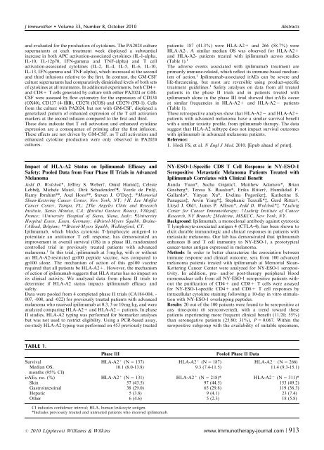
TABLE 1.<br />
Phase III<br />
Pooled Phase II Data<br />
Survival HLA-A2 + (N = 137) HLA-A2 + (N = 187) HLA-A2 (N = 266)<br />
Median OS,<br />
10.1 (8.0-13.8) 9.3 (7.4-11.5) 11.4 (9.3-15.1)<br />
months (95% CI)<br />
irAEs, no. (%) HLA-A2 + (N = 131) HLA-A2 + (N = 218)* HLA-A2 (N = 311)*<br />
Skin 57 (43.5) 97 (44.5) 153 (49.2)<br />
Gastrointestinal 38 (29.0) 65 (29.8) 119 (38.3)<br />
Hepatic 5 (3.8) 9 (4.1) 23 (7.4)<br />
O<strong>the</strong>r 6 (4.6) 5 (2.3) 18 (5.8)<br />
CI indicates confidence interval; HLA, human leukocyte antigen.<br />
*Includes previously treated and untreated patients who received ipilimumab.<br />
r 2010 Lippincott Williams & Wilkins www.immuno<strong>the</strong>rapy-journal.com | 913