Micro- and Nanoscale Deformation Measurement of Surface and ...
Micro- and Nanoscale Deformation Measurement of Surface and ...
Micro- and Nanoscale Deformation Measurement of Surface and ...

You also want an ePaper? Increase the reach of your titles
YUMPU automatically turns print PDFs into web optimized ePapers that Google loves.
60 Exp Mech (2007) 47: 51–62<br />
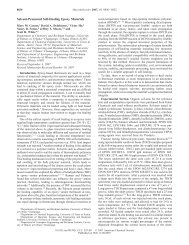
Fig. 11 <strong>Deformation</strong> fields<br />
showing Bu^ (a) <strong>and</strong> Bv^ (b)<br />
displacements near a silica<br />
microsphere (radius r=73 mm)<br />
internally embedded in a<br />
PDMS sample loaded in uniaxial<br />
tension (3.5% strain).<br />
For each displacement component,<br />
experimental DIC<br />
measurements for the airbrushed<br />
pattern are compared<br />
with the predicted<br />
analytical solution <strong>of</strong> a rigid<br />
spherical inclusion proposed<br />
by Goodier [16] for the plane<br />
z=r (where the z=0 for the<br />
sphere midplane)<br />
on the patterned layer below, as shown in Fig. 10.<br />
Experimental DIC measurements <strong>of</strong> the displacement<br />
field surrounding the microsphere were compared to<br />
the theoretically predicted field near a rigid inclusion<br />
(the z=jr plane, for z=0 at the sphere midplane) in an<br />
elastic infinite medium under a remote tensile stress as<br />
determined by Goodier [16]. The sparseness <strong>of</strong> the<br />
silica microsphere inclusions is such that we can<br />
effectively consider the microsphere isolated <strong>and</strong><br />
therefore make valid comparisons with the Goodier<br />
solution for a rigid sphere in an infinite elastic space.<br />
Furthermore, because <strong>of</strong> the large elastic property<br />
mismatch between PDMS <strong>and</strong> silica glass, the microsphere<br />
is considered rigid. Finally, at the small strains<br />
applied in the experiments the PDMS should behave<br />
in a linear elastic fashion. For the microscale pattern<br />
Fig. 12 Image focused on a<br />
fluorescently excited nanoparticle<br />
patterned layer,<br />
(a) located just below a rigid<br />
silica microsphere embedded<br />
within a PDMS sample. The<br />
exact location <strong>of</strong> the microsphere<br />
is determined by focusing<br />
at the sphere<br />
midplane under normal<br />
lighting conditions (b)<br />
SEM