Handout - STERIS University
Handout - STERIS University
Handout - STERIS University

You also want an ePaper? Increase the reach of your titles
YUMPU automatically turns print PDFs into web optimized ePapers that Google loves.
CORNEA-ANTERIOR SEGMENT<br />
the MOST COMPREHENSIVE OCT<br />
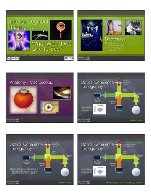
Anterior Segment<br />
Imaging….<br />
AM Option and RTVue FD-OCT<br />
Tear Film<br />
Bag and Lens<br />
Cornea Transplant<br />
Soft contact lens<br />
CK scar<br />
sule<br />
IOL<br />
Vitreous margin<br />
3D Crystalline Lens w/ SUM of C-scan display<br />
What Would We<br />
Like to See?<br />
Disclosures<br />
Financial interest:<br />
Alcon Laboratories, Inc.<br />
Abbot Medical Optics, Inc.<br />
Zeimer, Inc.<br />
MP Weikert, MD ✦ ASORN Regional Meeting ✦ San Antonio, TX ✦ August 24, 2012<br />
Part No. 43721 Rev. A<br />
Advances in<br />
Corneal Imaging<br />
Anatomy<br />
- Macroscopic Optical Coherence<br />
Tomography<br />
Scanning<br />
Reference<br />
Mirror<br />
Beam<br />
Splitter<br />
Scanning<br />
Mirror<br />
Low<br />
Coherence<br />
Source<br />
Interfering<br />
Beams<br />
“Partial Coherence”<br />
(multiple wavelengths)<br />
maintains interference<br />
over short distances<br />
Detector<br />
Target<br />
ASI - What Would<br />
We Like to See?<br />
ASI - What Would<br />
We Like to See?<br />
Interferometry<br />
Optical Coherence<br />
Tomography<br />
Scanning<br />
Reference<br />
Mirror<br />
Beam<br />
Splitter<br />
Scanning<br />
Mirror<br />
Optical Coherence<br />
Tomography<br />
Time vs. Frequency Domain:<br />
Scanning vs. Fixed<br />
Reference Mirror<br />
Beam<br />
Splitter<br />
Scanning<br />
Mirror<br />
Low<br />
Coherence<br />
Source<br />
Interfering<br />
Beams<br />
Low<br />
Coherence<br />
Source<br />
Interfering<br />
Beams<br />
Relative difference in pathlengths of<br />
reference and measurement arms<br />
determines interference pattern<br />
Detector<br />
Target<br />
“Partial Coherence”<br />
(multiple wavelengths)<br />
maintains interference<br />
over short distances<br />
Detector<br />
Target<br />
ASI - What Would<br />
We Like to See?<br />
Interferometry<br />
ASI - What Would<br />
We Like to See?<br />
Time Domain vs. Frequency Domain
Optical Coherence<br />
Tomography<br />
ASI - What Would<br />
We Like to See?<br />
“Optical B-Scan” Multiple optical A-scans<br />
combined to produce<br />
cross-sectional image @<br />
different meridians<br />
Optical Coherence Tomography (OCT)<br />
ASI - What Would<br />
We Like to See?<br />
Parameter<br />
Wavelength<br />
Penetration Depth<br />
Axial Resolution<br />
Transverse Resolution<br />
A-Scans/Line<br />
A-Scans/Second<br />
Acquisition Time<br />
Scan Diameter<br />
Scan Alignment<br />
System Design<br />
Zeiss Visante ® (Time)<br />
1310 nm<br />
3 - 6 mm<br />
18 µm<br />
60 µm<br />
128 - 512<br />
2048<br />
0.25 - 0.5 sec<br />
Approx. 12-14 mm<br />
V-Trac system aligns images on<br />
corneal vertex<br />
Dedicated Anterior Segment<br />
Zeiss Visante<br />
Zeiss Visante<br />
LASIK Flap - Mechanical µKeratome<br />
LASIK Flap - Femtosecond Laser<br />
ASI - What Would<br />
We Like to See?<br />
ASI - What Would<br />
We Like to See?<br />
Zeiss Visante<br />
Optical Coherence Tomography (OCT)<br />
Parameter<br />
Wavelength<br />
Penetration Depth<br />
Axial Resolution<br />
Transverse Resolution<br />
A-Scans/Line<br />
A-Scans/Second<br />
Acquisition Time<br />
Scan Diameter<br />
Scan Alignment<br />
System Design<br />
Zeiss Visante ® (Time)<br />
1310 nm<br />
3 - 6 mm<br />
18 µm<br />
60 µm<br />
128 - 512<br />
2048<br />
0.25 - 0.5 sec (1 scan per merid)<br />
Approx. 12-14 mm<br />
V-Trac system aligns images on<br />
corneal vertex<br />
Dedicated Anterior Segment<br />
Optovue RTVue ® (Frequency)<br />
830 nm<br />
2.3 mm<br />
5 µm<br />
5 - 10 µm<br />
1024<br />
26,000<br />
0.31 sec (5 scans per meridian)<br />
4 - 6 mm<br />
Corneal vertex<br />
Hybrid Anterior/Posterior Segment<br />
ASI - What Would<br />
We Like to See?<br />
ASI - What Would<br />
We Like to See?
the MOST COMPREHENSIVE OCT<br />
Only with the CAM Option and RTVue FD-OCT<br />
Tear Film<br />
Soft contact lens<br />
Optovue RTVue ®<br />
CK scar<br />
Cataract Wound Changes<br />
Bag and Lens<br />
Cornea Transplant<br />
Early Descemet’s<br />
Detachment<br />
Early Posterior<br />
Wound Gape<br />
Late Posterior<br />
Wound Retracton<br />
Iris<br />
LASIK Flap<br />
Corneal Transplant<br />
IOL<br />
3D Crystalline Lens w/ SUM of C-scan display<br />
Posterior capsule<br />
Vitreous margin<br />
AC Angle<br />
LASIK Interface Fluid<br />
ASI - What Would<br />
We Like to See?<br />
Pterygium<br />
ASI - What Would<br />
We Like to See?<br />
Part No. 43721 Rev. A<br />
Cataract Wound Changes<br />
% Eyes<br />
Descemet’s Membrane Detachment<br />
Posterior Wound Gape<br />
40<br />
100<br />
30<br />
75<br />
20<br />
50<br />
10<br />
25<br />
0<br />
0<br />
1 Day 1 Wk 2-3 Wk 1-3 Mo 3-12 Mo 1-2 Yr 3-15 Yr<br />
1 Day 1 Wk 2-3 Wk 1-3 Mo 3-12 Mo 1-2 Yr 3-15 Yr<br />
Time After Surgery<br />
Time After Surgery<br />
Posterior Wound Retraction<br />
% Eyes<br />
% Eyes<br />
100<br />
75<br />
50<br />
25<br />
0<br />
1 Day 1 Wk 2-3 Wk 1-3 Mo 3-12 Mo 1-2 Yr 3-15 Yr<br />
Time After Surgery<br />
VHF Ultrasound<br />
U/S<br />
Emitter/<br />
Detector<br />
Saline Bath/Coupling Medium<br />
Reflection of the incident<br />
U/S wave occurs at<br />
acoustical interfaces<br />
ASI - What Would<br />
We Like to See?<br />
Huang D, et al. (Unpublished Data)<br />
ASI - What Would<br />
We Like to See?<br />
Very High Frequency Ultrasound<br />
ArcScan Artemis ®<br />
Parameter ArcScan Artemis ®<br />
Frequency<br />
Penetration Depth<br />
Axial Resolution<br />
Transverse Resolution<br />
Meridians Scanned<br />
Acquisition Time<br />
Scan Diameter<br />
Other<br />
50 MHz<br />
3 - 6 mm<br />
1 µm<br />
200 µm<br />
4-12<br />
2-3 min<br />
Up to 15 mm<br />
ONLY system that can penetrate<br />
iris to image ciliary sulcus<br />
Off-axis imaging possible<br />
ASI - What Would<br />
We Like to See?<br />
ASI - What Would<br />
We Like to See?
ArcScan Artemis ®<br />
ArcScan Artemis ®<br />
Pre-Op Post-Op Change Layers<br />
ASI - What Would<br />
We Like to See?<br />
ASI - What Would<br />
We Like to See?<br />
Anatomy - Microscopic<br />
Confocal Microscopy<br />
Illumination<br />
Pinhole<br />
Detection<br />
↑Axial Resolution<br />
ASI - What Would<br />
We Like to See?<br />
ASI - What Would<br />
We Like to See?<br />
Confocal Microscopy<br />
Superficial<br />
Epithelium<br />
Anterior<br />
Stroma<br />
Confocal Microscopy<br />
Fuch’s Diffuse Dystrophy Lamellar LASIK Keratoconus Interface<br />
Polymegathism<br />
Acanthamoeba<br />
Keratitis<br />
Keratitis<br />
Basal<br />
Epithelium<br />
Superficial Nerve<br />
Plexus<br />
Posterior<br />
Stroma<br />
Endothelial<br />
Mosaic<br />
ASI - What Would<br />
We Like to See?<br />
ASI - What Would<br />
We Like to See?
Corneal Power/Curvature<br />
How Do We Measure Curvature?<br />
Directly - reflection principle:<br />
Keratometer<br />
Placido disc<br />
ASI - What Would<br />
We Like to See?<br />
ASI - What Would<br />
We Like to See?<br />
How Do We Measure Curvature?<br />
Directly - reflection principle:<br />
Keratometer<br />
Placido disc<br />
Indirectly - elevation measured:<br />
Scanning slit<br />
Scheimpflug<br />
Optical coherence tomography<br />
Very high frequency ultrasound<br />
Proven expertise<br />
for anterior segment care.<br />
Placido Imaging<br />
Illuminated rings<br />
(mires) projected<br />
onto corneal<br />
surface<br />
Reflected rings<br />
imaged by<br />
digital camera<br />
With revolutionary instruments such as the Stratus OCT<br />
Anterior segment imaging<br />
system, the unique technological expertise of Carl Zeiss<br />
The anterior segment can be evaluated and measured pre-<br />
Meditec has long proved invaluable in the diagnosis and<br />
and postoperatively after image acquisition using the ana-<br />
monitoring of retinal disease. Now, with the introduction of<br />
lysis mode of the Visante OCT system’s software. Practical<br />
ASI - What Would<br />
We Like to See?<br />
Narrow Angle<br />
the Visante OCT system, this same expertise is being<br />
applied to high-resolution, non-contact optical coherence<br />
tomography customized for the anterior segment.<br />
Exceptional design, greater confidence<br />
The Visante OCT system is the first to provide clear, highly<br />
tools enable planning and measurement of anterior segment<br />
ocular structures, including anterior chamber depth<br />
(ACD), anterior chamber angles and anterior chamber diameter<br />
(commonly referred to as angle-to-angle distance).<br />
Anterior chamber angle measurement results provide you<br />
with quick and reliable data for narrow-angle evaluation.<br />
ASI - What Would<br />
We Like to See?<br />
detailed, in-depth images of the anterior chamber - includ-<br />
Anterior segment images can be printed with or without<br />
ing the angle - without the need for ocular anesthesia or a<br />
measurement tools and results.<br />
Analysis Results<br />
messy, time-consuming water bath. As a result, the Visante<br />
OCT system will provide unique images and measure-ments<br />
that will dramatically expand the potential for diagnostic<br />
confidence and therapeutic precision. And, just as importantly,<br />
Visante OCT is so easy to use and efficient to operate<br />
that it will seamlessly take its place in your daily workflow,<br />
offering new clinical insights and practice opportunities<br />
from day one.<br />
Corneal imaging and pachymetry<br />
Visante OCT provides high-resolution corneal images and<br />
documentation for the anterior segment specialist to support<br />
the evaluation of ocular health. Rapid acquisition<br />
during the pachymetry scan ensures an accurate and repeatable<br />
pachymetry map result for application in refractive<br />
and glaucoma care.<br />
Versatile in application<br />
The unique, versatile Visante OCT system provides superb,<br />
highly detailed results, consistently supporting surgical<br />
Corneal Ring Segments<br />
planning and postoperative care across a range of anterior<br />
segment applications.<br />
Placido Imaging<br />
Placido Curvature Measurement<br />
Reflected rings<br />
imaged by<br />
Image processing analyzes ring digital camera<br />
spacing to determine curvature<br />
Advantages:<br />
Lots of data points<br />
Accurate curvature measurement<br />
Information on surface quality<br />
Disadvantages:<br />
Mires reflected off of tear film<br />
No true measurement of central<br />
cornea<br />
Anterior surface only<br />
(adjusted η = 1.3375)<br />
ASI - What Would<br />
We Like to See?<br />
ASI - What Would<br />
We Like to See?
Irregular Mires<br />
Decentered<br />
Placido Rings<br />
Pupil Center<br />
ASI - What Would<br />
We Like to See?<br />
ASI - What Would<br />
We Like to See?<br />
Axial Maps<br />
Measurement tied to optical<br />
axis<br />
Spherical bias (all radii share<br />
the same axis/origin)<br />
Better for central/paracentral<br />
cornea<br />
Accuracy decreased in the<br />
periphery<br />
r<br />
Optical<br />
Axis<br />
r<br />
Tangential/Instantaneous Map<br />
Optical<br />
Axis<br />
Not tied to optical axis<br />
Calculate curvature from<br />
neighboring points<br />
More accurate shape<br />
(especially in periphery)<br />
More detail to detect<br />
irregular astigmatism<br />
ASI - What Would<br />
We Like to See?<br />
ASI - What Would<br />
We Like to See?<br />
Axial vs Tangential/Instantaneous<br />
=<br />
Scheimpflug &<br />
Placido Combo<br />
Note: Sim K’s calculated from Axial Map<br />
ASI - What Would<br />
We Like to See?<br />
Axial vs. Tangential
Map Comparison<br />
Comparing corneal<br />
topographic maps can be<br />
challenging when subtle<br />
differences are present<br />
Make sure maps have the<br />
same scale when comparing<br />
them<br />
ASI - What Would<br />
We Like to See?<br />
ASI - What Would<br />
We Like to See?<br />
Elevation Data<br />
Obtained with Scheimpflug<br />
imaging, OCT, or scanning slit<br />
systems,<br />
Raw elevation data:<br />
Difficult to interpret<br />
Must be compared to a<br />
reference surface<br />
Best-fit sphere, ellipse, toroidal<br />
ellipse<br />
ASI - What Would<br />
We Like to See?<br />
ASI - What Would<br />
We Like to See?<br />
Image Plane<br />
Camera Film<br />
Lens & Image<br />
Plane are<br />
Typically Parallel<br />
Image Plane<br />
Camera Film<br />
Lens Plane<br />
Lens Plane<br />
Plane of Focus<br />
Parallel to Both<br />
Lens & Image Planes<br />
Scheimpflug<br />
Subject<br />
Plane of Focus<br />
“Coronal Image”<br />
If planar subject is<br />
parallel to Image<br />
Plane, it can<br />
coincide with the<br />
Plane of Focus &<br />
the entire subject<br />
will be in focus<br />
Plane of Focus<br />
=<br />
Subject Plane<br />
ASI - What Would<br />
We Like to See?<br />
ASI - What Would<br />
We Like to See?
Scheimpflug Intersection<br />
Image Plane<br />
Camera Film<br />
Image Plane<br />
Camera Film<br />
Lens Plane<br />
If the Subject Plane is<br />
not parallel to the<br />
Image Plane, it will be<br />
in focus only along a<br />
line that intersects the<br />
Plane of Focus<br />
Subject Plane<br />
Plane of Focus<br />
Lens Plane<br />
Subject Plane<br />
If the lens is<br />
positioned such that<br />
the Image Plane,<br />
Lens Plane, &<br />
Subject Plane all<br />
intersect, the subject<br />
(not parallel to the<br />
Image Plane) can be<br />
completely in focus<br />
ASI - What Would<br />
We Like to See?<br />
ASI - What Would<br />
We Like to See?<br />
Image Plane<br />
CCD Camera<br />
Sagital Cross-Section<br />
Lens Plane<br />
Subject<br />
Plane of Focus<br />
ASI - What Would<br />
We Like to See?<br />
ASI - What Would<br />
We Like to See?<br />
Scheimpflug Imaging<br />
Advantages:<br />
Lots of elevation data<br />
Data from central cornea<br />
Images anterior & posterior<br />
corneal surface<br />
Disadvantages:<br />
Unable to image ciliary sulcus<br />
Must compare to reference<br />
Need extremely high resolution<br />
to detect curvature differences<br />
Reference Surface<br />
Shape? - Best-fit sphere, ellipse, toric ellipsoid<br />
Different days → different reference spheres<br />
r = 8.21 mm<br />
r = 8.41 mm<br />
ASI - What Would<br />
We Like to See?<br />
ASI - What Would<br />
We Like to See?<br />
Difference = 1 D
Elevation Data &<br />
Curvature Measurement*<br />
h<br />
d<br />
45D vs. 45.25D Curvatures:<br />
7.5 mm (R2) vs. 7.99 mm (R1)<br />
R1<br />
R2<br />
Diameter (D) Height Diff. (H)<br />
1 mm 0.1 µm<br />
2 mm 0.4 µm<br />
3 mm 0.9 µm<br />
4 mm 1.6 µm<br />
5 mm 2.5 µm<br />
ASI - What Would<br />
We Like to See?<br />
*Roberts C. Corneal Topography in Refractive Surgery, 2nd edition.<br />
Dimitri Azar (ed.). Stanford, CT: Appleton & Lange.<br />
ASI - What Would<br />
We Like to See?<br />
Galilei Dual<br />
Scheimpflug Analyzer<br />
2 Scheimpflug cameras (instead of 1):<br />
Elevation data averaged from each camera<br />
Eliminates measurement errors due to<br />
angle of orientation & ocular decentration<br />
Placido Rings (20):<br />
Scheimpflug cameras occupy gaps in<br />
Placido rings<br />
2 Placido images recorded w/ cameras<br />
oriented vertically & horizontally (fills in gaps)<br />
ASI - What Would<br />
We Like to See?<br />
ASI - What Would<br />
We Like to See?
Study Results:<br />
SimK repeatability (20 eyes) = 0.19 D<br />
ASI - What Would<br />
We Like to See?<br />
ASI - What Would<br />
We Like to See?<br />
Study Results:<br />
SimK repeatability (20 eyes) = 0.19 D<br />
SimK accuracy compared w/ Humphrey Atlas, IOL Master &<br />
Manual Keratometry (20 eyes):<br />
Mean astigmatism ranged from 0.50 D to 0.60 D<br />
Mean difference b/w Galilei & IOL Master = 0.10 ± 0.07D<br />
Mean difference b/w Galilei & Atlas = 0.11 ± 0.12D<br />
Corneal thickness compared w/ ultrasonic pachymetry (USP-GP):<br />
Virgin corneas (77 eyes) - mean difference = -0.7 ± 7.1 µm<br />
Post-LASIK/PRK (39 eyes) - mean difference = -6.2 ± 9.9 µm<br />
ASI - What Would<br />
We Like to See?<br />
ASI - What Would<br />
We Like to See?<br />
IOL Calculations & Corneal Power<br />
Corneal power serves 2 functions in IOL calculations:<br />
IOL power estimation<br />
Effective lens position (ELP)<br />
Rx<br />
Obj @ -∞<br />
ηair<br />
ACD AL-ACD<br />
K<br />
ηeye<br />
PIOL<br />
Vtx AL<br />
R<br />
Holladay 1 (Based on Vergence Formula)<br />
PIOL =<br />
1336<br />
AL - - 1336<br />
ELP<br />
1336<br />
1000<br />
]-<br />
1000<br />
- Vtx (mm)<br />
Rx<br />
ELP = SF + (T + 0.3375 -<br />
K<br />
[ ] ( )<br />
√<br />
( ) 2<br />
0.3375<br />
K<br />
+ K<br />
ELP<br />
- 0.071 * AL 2<br />
ASI - What Would<br />
We Like to See?<br />
ASI - What Would<br />
We Like to See?
IOL Calculations in Normal Eyes<br />
MAE (D)<br />
0.8<br />
0.6<br />
0.4<br />
0.2<br />
0<br />
IOL MAE vs Corneal Power Method<br />
Auto K SimKp SimKp+ds TCPm TCPc<br />
Auto K - IOLMaster<br />
SimKp - Atlas<br />
SimKp+ds - Galilei<br />
TCPm - Galilei<br />
TCPc - Galilei<br />
% Eyes<br />
% Eyes<br />
100<br />
80<br />
60<br />
40<br />
20<br />
0<br />
100<br />
80<br />
60<br />
40<br />
20<br />
0<br />
% Eyes within ± 0.5D<br />
Auto K SimKp SimKp+ds TCPm TCPc<br />
% Eyes within ± 1.0D<br />
Auto K SimKp SimKp+ds TCPm TCPc<br />
*IOL calculations optimized (SF) for each method of corneal power measurement<br />
ASCRS IOL Calculator<br />
“Double-K”<br />
ASI - What Would<br />
We Like to See?<br />
*Shirayama M, et al. Cornea 2010; 29:1136-1138<br />
ASI - What Would<br />
We Like to See?<br />
http://www.ascrs.org<br />
IOL Calculation after LASIK/PRK<br />
Refractive Mean Absolute Error (MAE)<br />
MAE (D)<br />
n=17<br />
1.20<br />
1.00<br />
0.80<br />
0.60<br />
0.40<br />
0.20<br />
0<br />
1.00<br />
Clin History<br />
1.05<br />
Feiz-Mannis<br />
1.00<br />
K Bypass<br />
0.63<br />
Adj EffRP<br />
0.48<br />
Adj Atlas 0-3<br />
0.42<br />
Masket<br />
0.44<br />
Mod Masket<br />
0.59<br />
W-K-M<br />
0.60<br />
Shammas<br />
0.54<br />
Haigis-L<br />
0.65<br />
TCP-2mm<br />
0.62<br />
TCP-3mm<br />
0.60<br />
TCP-4mm<br />
0.59<br />
TCP-5mm<br />
Sample Case - Post-RK<br />
59-y/o woman:<br />
AL = 23.91 mm<br />
IOLMaster K = 38.16 D<br />
EyeSys EffRP = 38.19 D<br />
Atlas Zone 0-3 = 38.79 D<br />
Galilei TCP Zone 0-4 = 36.53 D<br />
Galilei TCP Annuli 1-4 = 36.79 D<br />
ASI - What Would<br />
We Like to See?<br />
Significantly greater MAE in methods w/ pre-LASIK K’s & ΔMR (all P
OCT Net Corneal Power<br />
Parabolic fit to central 3-mm zone<br />
Average 3 sets per meridian<br />
1.5mm<br />
R p<br />
D<br />
1.5mm<br />
R a<br />
n 0 = 1<br />
n 1 = 1.376<br />
n 2 = 1.336<br />
Net Corneal Power - combines anterior<br />
& posterior curvature measurements<br />
from 8 meridional scans<br />
IOL Calculations in Post-LASIK Eyes<br />
MAE (D)<br />
2.0<br />
1.5<br />
1.0<br />
0.5<br />
0<br />
MAE - OCT vs Other K Methods<br />
n = 14<br />
IOLM CL OR Clin Hx Haigis Orbscan OCT<br />
SD of PE (D)<br />
2.5<br />
2.0<br />
1.5<br />
1.0<br />
0.5<br />
0<br />
SD of PE - OCT vs Other K Methods<br />
n = 14<br />
IOLM CL OR Clin Hx Haigis Orbscan OCT<br />
OCT Net Corneal Power:<br />
Converted to keratometry equivalent<br />
prior to use in IOL formulas (+1.21 D)<br />
IOLM<br />
CL OR<br />
Clin Hx<br />
Haigis<br />
Orbscan<br />
OCT<br />
ASI - What Would<br />
We Like to See?<br />
Huang D, et al. (Unpublished Data)<br />
ASI - What Would<br />
We Like to See?<br />
Huang D, et al. (Unpublished Data)<br />
Posterior Corneal Astigmatism<br />
Spherical Aberration<br />
Traditional measurement of<br />
corneal astigmatism:<br />
Based on anterior surface<br />
Assumes fixed anterior-posterior<br />
curvature ratio<br />
Ignores contribution of posterior<br />
surface<br />
Retrospective study of<br />
posterior corneal astigmatism:<br />
Galilei DSA measurements<br />
Cornea exhibits (+) spherical<br />
aberration (SA):<br />
Mean +0.28 µm*<br />
Balanced by lens in youth<br />
Additive with lens in cataracts<br />
Cataract surgery:<br />
SA of crystalline lens removed<br />
SA of cornea remains<br />
Spherical IOLs have (+) SA<br />
(typically +0.18 µm)<br />
⊕<br />
⊕<br />
ASI - What Would<br />
We Like to See?<br />
*Wang L, et al. ARVO 2011<br />
ASI - What Would<br />
We Like to See?<br />
Spherical Aberration<br />
Corneal Ectasia & Ectasia Risk<br />
SA = 0.01 0.58 0.24 µm<br />
Reduction in SA:<br />
Improved contrast sensitivity<br />
Improved visual performance<br />
(e.g. night driving)<br />
Measurement of corneal SA may<br />
influence choice of IOL:<br />
Alcon AcrySof IQ (SA = -0.20 µm)<br />
AMO Tecnis (SA = -0.27 µm)<br />
B&L SofPort AO (SA = 0 µm)<br />
Standard Spherical (SA = +18 µm)<br />
ASI - What Would<br />
We Like to See?<br />
ASI - What Would<br />
We Like to See?
Abnormal Corneal Shape<br />
Determining ectasia risk is a<br />
very important part of refractive<br />
surgery screening<br />
Several criteria developed from<br />
the anterior surface<br />
Rabinowitz-McDonnell:<br />
Central K > 47.2 D<br />
Astigmatism > 1.5 D<br />
I - S > 1.4 D<br />
SRAX > 21°<br />
KISA% > 100 (Suspect 60 to 100)<br />
SRAX<br />
ASI - What Would<br />
We Like to See?<br />
ASI - What Would<br />
We Like to See?<br />
CCT: 539 µm<br />
Thinnest: 516 µm<br />
KPI: 43.4%<br />
Posterior Elevation: 35 µm<br />
ASI - What Would<br />
We Like to See?<br />
ASI - What Would<br />
We Like to See?<br />
OCT KCN Pachymetry Index (KPI)<br />
Parameter<br />
Min<br />
Min - Med<br />
Y Min<br />
S - I<br />
SN - IT<br />
KPI<br />
Definition<br />
KCN Cutoff<br />
(1 percentile)<br />
KCN<br />
Accuracy*<br />
FF KCN<br />
Accuracy*<br />
Minimum corneal thickness < 464 µm 0.94 µm 0.61 µm<br />
Minimum corneal thickness minus<br />
median corneal thickness<br />
Y coordinate of minimum corneal<br />
thickness<br />
Avg thickness of superior octant<br />
minus inferior octant<br />
Avg thickness of superonasal<br />
octant minus inferotemporal octant<br />
KPI = 0.41Min + 0.23(Min-Med) -<br />
0.14[(S-I) + (SN-IT)] + 0.22Ymin<br />
< -33 µm 0.93 µm 0.62 µm<br />
< -1.1 mm 0.82 mm 0.67 mm<br />
> 54 µm 0.84 µm 0.68 µm<br />
> 52 µm 0.90 µm 0.60 µm<br />
0.97 0.69<br />
OCT KCN Pachymetric Score (Example)<br />
23-y/o Male:<br />
-0.25 + 1.75 x 165 (20/15)<br />
SimK - 40.8/4.9 @ 16<br />
Topography<br />
OCT<br />
KISA % 96 Min (µm) 518 0<br />
K (D) 40.8 Min - Med (µm) -38 3<br />
I-S (µm) 5.7 Y Min (mm) -1.23 1<br />
AST (D) 1.1 S - I (µm) 62 2<br />
SRAX (deg) 46 SN - IT (µm) 50 1<br />
Total Pachy Score 7<br />
*Area under receiver operating curve<br />
ASI - What Would<br />
We Like to See?<br />
Huang D, et al. (Unpublished Data)<br />
ASI - What Would<br />
We Like to See?<br />
Huang D, et al. (Unpublished Data)
Conclusions<br />
Still need multiple devices to see all that we want to see<br />
Ability to image posterior cornea (accurately) is only<br />
increasing in importance<br />
“Go-to” topographer - Still Placido-based<br />
Can’t do without other devices, now that I’ve got them<br />
Probably use “imaging” features the least in current<br />
practice (lower priority)<br />
It sure is nice that we have all of this technology!<br />
Acknowledgements<br />
Douglas D. Koch, M.D.<br />
Li Wang, M.D./Ph.D.<br />
Mariko Shirayama, M.D.<br />
Shazia Ali, B.S<br />
Richard Jenkins, M.D.<br />
Zaina Al-Mohtaseb, M.D.<br />
David Huang, M.D./Ph.D.<br />
Advances in<br />
Corneal Imaging<br />
CORNEA-ANTERIOR SEGMENT<br />
the MOST COMPREHENSIVE OCT<br />
AM Option and RTVue FD-OCT<br />
Tear Film<br />
Anterior Segment<br />
Imaging….<br />
Bag and Lens<br />
Cornea Transplant<br />
Soft contact lens<br />
CK scar<br />
sule<br />
IOL<br />
Vitreous margin<br />
3D Crystalline Lens w/ SUM of C-scan display<br />
What Would We<br />
Like to See?<br />
MP Weikert, MD ✦ ASORN Regional Meeting ✦ San Antonio, TX ✦ August 24, 2012<br />
Part No. 43721 Rev. A