

Eosinophilic Abscesses of the Liver due to Enterobius Vermicularis
Eosinophilic Abscesses of the Liver due to Enterobius Vermicularis
Eosinophilic Abscesses of the Liver due to Enterobius Vermicularis

You also want an ePaper? Increase the reach of your titles
YUMPU automatically turns print PDFs into web optimized ePapers that Google loves.
Coexistence <strong>of</strong> Giant Tubercular Cyst <strong>of</strong><br />
Transverse Mesocolon along with Perforated<br />
Tubercular Ulcer <strong>of</strong> Ileum: A Rare Entity<br />
Mahesh Gupta 1 , Subhash Goyal 2 , Amit Mittal 3 , Rekha Goyal 4 , Milan Verma 5<br />
1 Associate Pr<strong>of</strong>essor, Department <strong>of</strong> Surgery, M.M. Institute <strong>of</strong> Medical Sciences and Research, Haryana, India<br />
2 Pr<strong>of</strong>essor and Head <strong>of</strong> Department <strong>of</strong> Surgery, M.M. Institute <strong>of</strong> Medical Sciences and Research, Haryana, India<br />
3 Pr<strong>of</strong>essor and Head <strong>of</strong> Department <strong>of</strong> Radiodiagnosis, M.M. Institute <strong>of</strong> Medical Sciences and Research, Haryana, India<br />
4 Pr<strong>of</strong>essor, Department <strong>of</strong> Radiodiagnosis, M.M. Institute <strong>of</strong> Medical Sciences and Research, Haryana, India<br />
5 Postgraduate Student, Department <strong>of</strong> Surgery, M.M. Institute <strong>of</strong> Medical Sciences and Research, Haryana, India<br />
ABSTRACT<br />
Mesenteric cysts are usually discovered<br />
incidentally during routine abdominal<br />
examinations and <strong>the</strong>y may present with<br />
nonspecific symp<strong>to</strong>ms. There are different<br />
types <strong>of</strong> cysts but tubercular cyst is a rare<br />
variety. The management includes complete<br />
excision <strong>of</strong> <strong>the</strong> cyst <strong>to</strong> avoid recurrence,<br />
followed by anti-tubercular drug <strong>the</strong>rapy.<br />
We report a case <strong>of</strong> tubercular cyst in<br />
relation <strong>to</strong> transverse mesocolon, which was<br />
Keywords: Giant Tubercular Cyst; Mesocolon; Tubercular Ulcer; Ileum; Peri<strong>to</strong>nitis<br />
INTRODUCTION<br />
Mesenteric cysts (MCs) are rare intra-abdominal<br />
lesions with higher incidence <strong>of</strong> small bowel than<br />
<strong>the</strong> large bowel involvement [1]. Majority <strong>of</strong><br />
<strong>the</strong>se patients present with a palpable lump and<br />
o<strong>the</strong>rs may present with nonspecific symp<strong>to</strong>ms.<br />
The tubercular mesenteric cysts are rare forms <strong>of</strong><br />
MCs and <strong>the</strong>ir coexistence with perforation <strong>of</strong><br />
ulcer at terminal ileum is extremely rare. Due <strong>to</strong><br />
such unusual coexistence, we are reporting this<br />
case.<br />
CASE REPORT<br />
incidentally found in a patient who presented<br />
with features <strong>of</strong> peri<strong>to</strong>nitis <strong>due</strong> <strong>to</strong> perforation<br />
<strong>of</strong> tubercular ulcer <strong>of</strong> terminal ileum. The<br />
coexistence <strong>of</strong> tubercular mesocolic cyst<br />
along with perforation <strong>of</strong> tubercular ulcer in<br />
<strong>the</strong> terminal ileum is a rare form <strong>of</strong><br />
presentation and both are managed surgically<br />
followed by anti-tubercular drug <strong>the</strong>rapy <strong>to</strong><br />
avoid recurrence.<br />
definite relation <strong>of</strong> <strong>the</strong> cyst could be visualized.<br />
There was free fluid in <strong>the</strong> abdomen with internal<br />
echoes suggestive <strong>of</strong> perforation <strong>of</strong> bowel.<br />
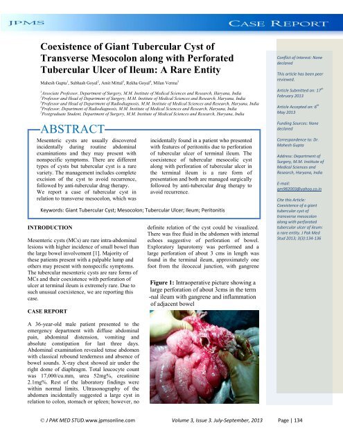
Explora<strong>to</strong>ry laparo<strong>to</strong>my was performed and a<br />
large perforation <strong>of</strong> about 3 cms in length was<br />
found in <strong>the</strong> terminal ileum, approximately one<br />
foot from <strong>the</strong> ileocecal junction, with gangrene<br />
Figure 1: Intraoperative picture showing a<br />
large perforation <strong>of</strong> about 3cms in <strong>the</strong> term<br />
-nal ileum with gangrene and inflammation<br />
<strong>of</strong> adjacent bowel<br />
Conflict <strong>of</strong> Interest: None<br />
declared<br />
This article has been peer<br />
reviewed.<br />
Article Submitted on: 17 th<br />
February 2013<br />
Article Accepted on: 6 th<br />
May 2013<br />
Funding Sources: None<br />
declared<br />
Correspondence <strong>to</strong>: Dr.<br />
Mahesh Gupta<br />
Address: Department <strong>of</strong><br />
Surgery, M.M. Institute <strong>of</strong><br />
Medical Sciences and<br />
Research, Haryana, India<br />
E-mail:<br />
gm982003@yahoo.co.in<br />
Cite this Article:<br />
Coexistence <strong>of</strong> a giant<br />
tubercular cyst <strong>of</strong><br />
transverse mesocolon<br />
along with perforated<br />
tubercular ulcer <strong>of</strong> ileum:<br />
a rare entity. J Pak Med<br />
Stud 2013; 3(3):134-136<br />
A 36-year-old male patient presented <strong>to</strong> <strong>the</strong><br />
emergency department with diffuse abdominal<br />
pain, abdominal distension, vomiting and<br />
absolute constipation for last three days.<br />
Abdominal examination revealed tense abdomen<br />
with classical rebound tenderness and absence <strong>of</strong><br />
bowel sounds. X-ray chest showed air under <strong>the</strong><br />
right dome <strong>of</strong> diaphragm. Total leucocyte count<br />
was 17,000/cu.mm, urea 52mg%, creatinine<br />
2.1mg%. Rest <strong>of</strong> <strong>the</strong> labora<strong>to</strong>ry findings were<br />
within normal limits. Ultrasonography <strong>of</strong> <strong>the</strong><br />
abdomen incidentally suggested a large cyst in<br />
relation <strong>to</strong> colon, s<strong>to</strong>mach or spleen; however, no<br />
© J PAK MED STUD.www.jpmsonline.com Volume 3, Issue 3. July-September, 2013 Page | 134
and inflammation <strong>of</strong> adjacent bowel (Figure 1).<br />
In addition, a large cyst <strong>of</strong> about 15*12 cms was<br />
found in relation <strong>to</strong> transverse mesocolon (Figure<br />
2) along with multiple, s<strong>of</strong>t <strong>to</strong> firm mesenteric<br />
lymph nodes <strong>of</strong> size 1-2 cms. The cyst was<br />
excised after separating it from adhesions and it<br />
contained whitish fluid; however, it ruptured<br />
during excision. The perforated site <strong>of</strong> ileum<br />
along with adjacent gangrenous bowel was<br />
resected and <strong>the</strong> ends were taken out as ileos<strong>to</strong>my<br />
and distal mucus fistula. Pos<strong>to</strong>perative period<br />
was uneventful and <strong>the</strong> ileos<strong>to</strong>my was closed<br />
after two and half months. His<strong>to</strong>pathological<br />
examination <strong>of</strong> <strong>the</strong> resected bowel and cyst wall<br />
showed <strong>the</strong> presence <strong>of</strong> epi<strong>the</strong>loid granulomas,<br />
histiocytes, Langerhans giant cells and caseation<br />
necrosis. The culture <strong>of</strong> <strong>the</strong> fluid from <strong>the</strong> cyst<br />
was sterile. The diagnosis <strong>of</strong> tubercular cyst and<br />
perforation was made and <strong>the</strong> patient was started<br />
on anti-tubercular drugs including isoniazid,<br />
rifampin, pyrizinamide and ethambu<strong>to</strong>l for <strong>the</strong><br />
first two months followed by isoniazid and<br />
rifampicin only for <strong>the</strong> next ten months. It is<br />
interesting <strong>to</strong> note that <strong>the</strong> patient did not have<br />
any past his<strong>to</strong>ry <strong>of</strong> tuberculosis and no such focus<br />
was evident in <strong>the</strong> lungs. The patient is doing<br />
well in <strong>the</strong> follow-up period.<br />
DISCUSSION<br />
MCs are more common than <strong>the</strong> omental cysts<br />
and according <strong>to</strong> <strong>the</strong> available literature <strong>the</strong> ratio<br />
is 4.5 <strong>to</strong> 1, with a higher incidence in females [2].<br />
MCs are more commonly chylolymphatic and<br />
enterogenous than <strong>the</strong> urogenital remnants and<br />
tera<strong>to</strong>ma<strong>to</strong>us cyst [3]. They may be unilocular or<br />
multilocular and contain fluid which varies from<br />
a clear, straw-colored liquid <strong>to</strong> a thick cheesywhite<br />
material. MCs may extend from <strong>the</strong> base <strong>of</strong><br />
mesentery in<strong>to</strong> <strong>the</strong> retroperi<strong>to</strong>neum and any part<br />
<strong>of</strong> gastrointestinal tract from <strong>the</strong> duodenum <strong>to</strong> <strong>the</strong><br />
rectum may get affected. 60% <strong>of</strong> <strong>the</strong>se cysts are<br />
found in <strong>the</strong> small intestine and 40% in <strong>the</strong> colon<br />
[1]. Cysts involving <strong>the</strong> transverse mesocolon are<br />
very rare especially tubercular mesenteric cysts<br />
[4]. The omental cysts may present as acute<br />
abdomen almost exclusively in children <strong>due</strong> <strong>to</strong><br />
<strong>to</strong>rsion and infection [5]; however, MCs rarely<br />
present as acute abdomen and majority <strong>of</strong><br />
patients present as a palpable abdominal mass [6,<br />
7]. Although <strong>the</strong> preoperative diagnosis <strong>of</strong> cysts<br />
and <strong>the</strong>ir extension can be made by ultrasound or<br />
CT scan, his<strong>to</strong>pathological examination <strong>of</strong> <strong>the</strong><br />
cysts is essential <strong>to</strong> confirm <strong>the</strong> diagnosis [8, 11].<br />
There are different treatment options such as<br />
excision, enucleation, marsupialization and drai-<br />
Figure 2: Intraoperative picture showing a<br />
large cyst <strong>of</strong> about 15*12 cms in relation<br />
<strong>to</strong> transverse mesocolon<br />
nage but excision <strong>of</strong> cyst along with preservation<br />
<strong>of</strong> surrounding vital structures is <strong>the</strong> treatment <strong>of</strong><br />
choice <strong>to</strong> avoid recurrence [9] and anti-tubercular<br />
drugs should be started pos<strong>to</strong>peratively as soon<br />
as possible in case <strong>of</strong> tubercular cysts [10]. The<br />
coexistence <strong>of</strong> tubercular mesocolic cysts<br />
alongwith perforation <strong>of</strong> tubercular ulcer in <strong>the</strong><br />
terminal ileum has not been discussed in <strong>the</strong><br />
available literature. Although generalized<br />
peri<strong>to</strong>nitis may occur in cases <strong>of</strong> perforated<br />
tubercular ulcer, explora<strong>to</strong>ry laparo<strong>to</strong>my with<br />
resection <strong>of</strong> <strong>the</strong> affected segment <strong>of</strong> small bowel<br />
and end <strong>to</strong> end anas<strong>to</strong>mosis is <strong>the</strong> treatment <strong>of</strong><br />
choice and primary closure <strong>of</strong> <strong>the</strong> perforation<br />
should be avoided <strong>due</strong> <strong>to</strong> high risk <strong>of</strong> leak and<br />
fistula formation [12].<br />
REFERENCES<br />
1. Egozi EL, Ricketts RR. Mesenteric and omental cysts in<br />
children. Am Surg 1997; 63:287-290.<br />
2. Beahrs OH, Dockerty MB. Primary omental cysts <strong>of</strong><br />
clinical importance. Surg Clin North Am 1950; 30:1073-<br />
1079.<br />
3. Beahrs OH, Judd ES, Dockertv MB. Chylous cyst <strong>of</strong> <strong>the</strong><br />
abdomen. Surg Clin North Am 1950; 30:1081-1096.<br />
4. Fahmy A, Smith R, Garner MT, Frazier HM, Walker,<br />
M. Mesenteric cyst presenting as an acute surgical<br />
abdomen. Case report. Am Surg 1966; 32:654-656.<br />
5. Oliver GA. The omental cyst: a rare cause <strong>of</strong> <strong>the</strong> acute<br />
abdominal crisis. Surgery 1964; 56:588-593.<br />
6. Caropreso PR. Mesenteric cysts: a review. Arch Surg<br />
1974; 108:242-246.<br />
7. Borisa AD, Bakhshi GD, Tayada MB, Pawar NH,<br />
Nikam NN, Gupta A. A rare case <strong>of</strong> tuberculous mesen-<br />
© J PAK MED STUD.www.jpmsonline.com Volume 3, Issue 3. July-September, 2013 Page | 135
teric cyst masquerading as chylolymphatic mesenteric<br />
cyst. Bombay Hosp J 2008; 50:496-497.<br />
8. S<strong>to</strong>upis C, Ros PR, Abbitt PL, Bur<strong>to</strong>n SS, Gauger J.<br />
Bubbles in <strong>the</strong> belly: imaging <strong>of</strong> cystic mesenteric or<br />
omental masses. Radiographics 1994; 14:729–737.<br />
9. Dequanter D, Lefebvre JC, Belva P, Takieddine M,<br />
Vaneukem P. Mesenteric cysts. A case treated by<br />
laparoscopy and a review <strong>of</strong> <strong>the</strong> literature. Surg<br />
Endosc 2002; 16:1493.<br />
10. Bhowmik D, Chiranjib, Chandira RM, Jayakar B,<br />
Kumar KPS. Recent trends <strong>of</strong> drug used treatment <strong>of</strong><br />
tuberculosis. J Chem Pharm Res 2009; 1:113-133.<br />
11. Jin XJ, Kim JM, Kim HK, Kim L, Choi SJ, Park IS, et<br />
al. His<strong>to</strong>pathology and TB-PCR kit analysis in<br />
differentiating <strong>the</strong> diagnosis <strong>of</strong> intestinal tuberculosis<br />
and Crohn’s disease. World J<br />
Gastroenterol 2010; 16:2496–2503.<br />
12. Al Salamah SM, Bismar H. Acute ileal tuberculosis<br />
perforation: a case report. Saudi J Gastroenterol 2002;<br />
8:64-66.<br />
© J PAK MED STUD.www.jpmsonline.com Volume 3, Issue 3. July-September, 2013 Page | 136


