Papillary Thyroid Carcinoma Presenting with a Right ... - PSO-HNS
Papillary Thyroid Carcinoma Presenting with a Right ... - PSO-HNS
Papillary Thyroid Carcinoma Presenting with a Right ... - PSO-HNS

Create successful ePaper yourself
Turn your PDF publications into a flip-book with our unique Google optimized e-Paper software.
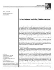
CASE REPORTS<br />
Philippine Journal Of Otolaryngology-Head And Neck Surgery Vol. 25 No. 1 January – June 2010<br />
Renato C. Pascual Jr., MD<br />
Johann F. Castañeda, MD<br />
Joel A. Romualdez, MD<br />
Department of Otorhinolaryngology<br />
Head and Neck Surgery<br />
St. Luke’s Medical Center<br />
<strong>Papillary</strong> <strong>Thyroid</strong> <strong>Carcinoma</strong> <strong>Presenting</strong> <strong>with</strong> a<br />
<strong>Right</strong> Preauricular and Intracranial Mass<br />
Correspondence: Renato C. Pascual Jr., MD<br />
Department of Otorhinolaryngology - Head and Neck Surgery<br />
St. Luke’s Medical Center<br />
279 E. Rodriguez Sr. Boulevard, Quezon City 1112<br />
Philippines<br />
Phone: (632)723-0101<br />
E-mail address: natopascual@yahoo.com<br />
Reprints will not be available from the author.<br />
No funding support was received for this study. The authors<br />
signed a disclosure that they have no proprietary or financial<br />
interest <strong>with</strong> an organization that may have a direct interest in<br />
the subject matter of this manuscript, or in any product used<br />
or cited in this study.<br />
ABSTRACT<br />
Objective: To describe a case of a papillary thyroid carcinoma presenting <strong>with</strong> a preauricular<br />
and an intracranial mass and review the literature on the metastatic nature and invasiveness of<br />
papillary thyroid carcinoma.<br />
Methods:<br />
Design: Case Report<br />
Setting: Tertiary Private Hospital<br />
Patient: One<br />
Results: A 46-year-old female <strong>with</strong> a 12-year anterior neck mass and a two-year right pre-auricular<br />
pleomorphic adenoma on fine needle aspiration biopsy was found to have an intracranial mass<br />
on CT-scan. Total thyroidectomy and section biopsy of the preauricular mass yielded a final<br />
histopathologic report of follicular variant of papillary carcinoma, thyroid gland; and metastatic<br />
papillary thyroid carcinoma, follicular type, pre-auricular mass. The condition of the patient<br />
precluded neurosurgical intervention and RAI therapy and she underwent 23 sessions of external<br />
radiotherapy using 46Gy <strong>with</strong> significant diminution in size of the intracranial metastasis.<br />
Conclusion: <strong>Papillary</strong> thyroid malignancy may be an indolent tumor but it is capable of distant<br />
metastasis. We should be alerted by host and tumor factors which can be predictors of a more<br />
radical papillary malignant disease whose management entails proper staging evaluation and<br />
good communication of prognostic data and available, realistic therapeutic options to patients<br />
using a multidisciplinary approach.<br />
Keywords: papillary thyroid carcinoma; papillary thyroid carcinoma metastasis; infratemporal<br />
metastasis; brain metastasis of papillary carcinoma.<br />
<strong>Thyroid</strong> malignancy is seventh among malignant lesions based on 1998 Philippine<br />
Department of Health statistics, 1 of which <strong>Papillary</strong> thyroid carcinoma (PTC) is the most common,<br />
representing 82% of all thyroid cancers. 2 PTC is believed to carry a good prognosis: overall<br />
mortality is 5% or less <strong>with</strong> long term follow up, 3 <strong>with</strong> a survival rate for stage I and II disease<br />
(American Joint Committee on Cancer Staging) reaching up to 95% to 100%. 4 PTC also has the<br />
lowest distant metastasis rates, ranging from 3% to 10%, <strong>with</strong> the majority of lesions affecting<br />
lungs and bone. While PTC is generally considered a malignant disease <strong>with</strong> a rather “benign”<br />
clinical character, surgeons should be aware of its dichotomous behavior. While some patients<br />
have excellent outcomes, others may surprisingly have a more aggressive disease.<br />
This case reminds us of the clinically important dimorphism of PTC. The atypical dissemination<br />
to an unlikely anatomical region made this case interesting. Moreover, the remarkably silent yet<br />
amazingly extensive distant disease and manner of spread put to question the validity of common<br />
beliefs among regarding the clinical characteristics and behaviour of PTC. This paper will review<br />
available literature associated <strong>with</strong> remote spread of disease even when clinical warning signs are<br />
Presented at Clinical Case Report Contest, Philippine Society of<br />
Otolaryngology- Head and Neck Surgery Mid-year Convention,<br />
Bohol Tropics Hotel, April 24, 2009. Philipp J Otolaryngol Head Neck Surg 2010; 25 (1): 26-30 c Philippine Society of Otolaryngology – Head and Neck Surgery, Inc.<br />
26 Philippine Journal Of Otolaryngology-Head And Neck Surgery
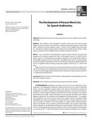
CASE REPORTS<br />
Philippine Journal Of Otolaryngology-Head And Neck Surgery Vol. 25 No. 1 January – June 2010<br />
lacking, and explore plausible mechanisms for the pathogenesis of the<br />
extraordinary dissemination observed in this case.<br />
CASE REPORT<br />
A 46-year-old female consulted for a gradually-enlarging anterior<br />
neck mass of 12 years duration and a 2-year right pre-auricular mass.<br />
Examination revealed a 7.0 cm x 5.0 cm anterior neck mass that was<br />
firm, non-tender and moved <strong>with</strong> deglutition. There were no palpable<br />
cervical lymph nodes. The firm, non-movable and non-tender preauricular<br />
mass measured 5.0 cm x 5.0 cm (Figure1). <strong>Thyroid</strong> function tests<br />
were normal. Fine needle aspiration biopsy (FNAB) of the anterior neck<br />
mass was read as follicular neoplasm, while that of the pre-auricular<br />
mass was signed out as consistent <strong>with</strong> pleomorphic adenoma.<br />
A computed tomography (CT) scan revealed a large enhancing<br />
lobulated soft tissue mass in the right pre-auricular area measuring<br />
5.9 cm. x 6.33 cm. x 6.79 cm. (Figures 2 a, b). Superiorly, the mass<br />
extended into the right temporal lobe associated <strong>with</strong> lytic destruction<br />
of the squamous and mastoid portions of the temporal bone. Inferior<br />
extension reached the level of the mandibular ramus. The masseter<br />
appeared infiltrated. There was erosive destruction of the zygomatic<br />
arch, greater sphenoid wing, right anterior skull base and anterior wall<br />
of the glenoid fossa of the temporomandibular joint. Posteriorly, there<br />
were erosive changes along the anterior wall of the mastoid bone.<br />
The lesion abutted the anterior surface of the parotid gland <strong>with</strong>out<br />
infiltration. Another similarly enhancing lesion was noted on the<br />
midoccipital region, measuring 2.96 cm. x 3.66 cm <strong>with</strong> extension into<br />
the posterior fossa, adjacent to the confluence of sinuses, associated<br />
<strong>with</strong> right anterolateral displacement of the torcula of Herophilii and<br />
lytic occipital bone destruction. The right lobe of the thyroid gland<br />
was enlarged measuring approximately 6.0 cm. x 3.6 cm. x 2.7 cm.<br />
There were hypodense lesions seen in the right lobe, some <strong>with</strong> dense<br />
peripheral calcifications; the largest measuring 3.4 cm. x 3.1 cm. x 2.2<br />
cm. The left lobe was normal in size <strong>with</strong> inhomogenous density and a<br />
focus of calcification.<br />
A total thyroidectomy and section biopsy of the preauricular<br />
mass were performed. Final histopathologic report for the thyroid<br />
gland lesion was follicular variant of papillary carcinoma <strong>with</strong> note of<br />
lymphatic and vascular invasion and thyroid capsular spread. The preauricular<br />
mass was signed out as consistent <strong>with</strong> metastatic carcinoma,<br />
follicular pattern, thyroid in origin. The final diagnosis was papillary<br />
thyroid carcinoma, right and left thyroid gland <strong>with</strong> brain (temporal and<br />
occipital lobes) metastases; T4aN0M1; Stage IVc (AJCC Classification,<br />
2002).<br />
Total body scans revealed functioning thyroid tissue remnants in<br />
the anterior neck, <strong>with</strong> no evidence of distant thyroid cancer metastasis<br />
(Figure 3). The patient was subjected to 23 sessions of external beam<br />
radiotherapy (EBRT) at 46Gy per session <strong>with</strong> focus on the right pre<br />
auricular region and mid occipital area. After one month of EBRT, the<br />
pre-auricular and occipital mass had significantly decreased in size<br />
(Figures 4 a, b).<br />
DISCUSSION<br />
The presence of distant metastasis from PTC is unusual. Dinneen<br />
et al. described the distribution of distant metastasis from PTC among<br />
a hundred cases during a five decade period. 6 The most commonly<br />
involved organs were the lungs (77%) and bone (20%). Other sites<br />
include the mediastinum (10%), adrenals (1%), skin (1%) and liver<br />
(1%). The brain is only involved in 1% of cases. Despite multiple large<br />
intracranial masses, this patient did not have any sign or symptom of<br />
increased intracranial pressure nor focal neurologic sign on examination.<br />
In a study by Chiu et al. among PTC patients <strong>with</strong> brain metastasis, 23%<br />
of the study population (11 out of 47 patients) also had no clinical<br />
evidence of distant brain metastasis, <strong>with</strong> most only diagnosed post<br />
mortem. 7 This was probably because the mass usually lysed cranial<br />
bones, the “auto-craniotomy” dissipating intracranial pressure into the<br />
more distensible scalp tissue. Most patients <strong>with</strong> clinically evident brain<br />
metastasis were those who developed deeply-seated brain lesions<br />
<strong>with</strong> practically no calvarial bone involvement, thereby increasing<br />
intracranial pressure.<br />
The mechanism of distant spread of papillary thyroid carcinoma has<br />
been postulated to be due mainly to lymphatic and vascular invasion<br />
by the tumor, following pre-formed neural and lymphovascular<br />
anatomical pathways through natural foramina, fissures and hollow<br />
fascial spaces. 9<br />
The mean time for development of distant metastasis was noted to<br />
be around seven to eight years after diagnosis of early stage disease. 8<br />
In our case, it took 10 years for distant metastasis to be evident. Dineen<br />
6<br />
showed that age at the time of distant metastasis diagnosis was a<br />
strong predictor of PTC survival after distant metastasis <strong>with</strong> a 10-year<br />
survival rate of 82% for those less than 40 years of age compared <strong>with</strong><br />
only 18% for the group aged 65 years and older at the time of distant<br />
metastasis. Gender did not significantly influence PTC survival after<br />
distant metastasis. Distant metastasis in other organs in the body<br />
may be related to brain metastasis; Dineen’s study showed that in<br />
patients who were positive for distant organ involvement, the brain<br />
had the highest risk for development for a second organ or succeeding<br />
metastases. 6 Our patient only had brain metastasis <strong>with</strong>out the more<br />
common involvement of lung and bone.<br />
The predilection of PTC to spread through the lymphatic more than<br />
the hematogenous route has been well established. Hall showed that<br />
intratumoral lymphatic proliferation and invasion of new lymphatics<br />
in and around the enlarging malignant tumor may facilitate distant<br />
spread. 9 Moreover, enormously large loads of malignant cells in the<br />
lymphatics may cause indirect vascular invasion as lymphatic channels<br />
ultimately drain into vascular channels. The intratumoral lymphatic<br />
invasion seen in our histologic specimen may explain its aggressiveness.<br />
Philippine Journal Of Otolaryngology-Head And Neck Surgery 27
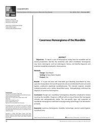
CASE REPORTS<br />
Philippine Journal Of Otolaryngology-Head And Neck Surgery Vol. 25 No. 1 January – June 2010<br />
Figure 1. Preauricular Mass. Patient had a right pre-auricular mass measuring<br />
approximately 5x5cm.<br />
Figure 2b. Contrast CTScan of Head, Coronal Section showed an enhancing soft tissue<br />
mass measuring 5.99 x 6.37 x 6.79 cm. extending superiorly to the right temporal lobe<br />
and inferiorly to the level of the mandibular ramus.<br />
Figure 2a. Contrast CTScan of Head, Axial Section showed an enhancing lobulated<br />
soft tissue mass,involving right masseter and temporalis muscles, measuring 5.99 x<br />
6.37 x 6.79cm.<br />
Figure 3. Total Body Scan (I-131). This showed intense tracer uptake in the anterior<br />
neck. Pinhole imaging in this area shows 3 foci of increased tracer deposition in<br />
midline measuring 1.6x1.1cm, 1.2x2.0cm, 1.1x1.2cm as well as a faint focus in the<br />
superior aspect. There is also a focus of tracer localization in the right thyroidal bed<br />
measuring 1.0x1.0cm. Functioning thyroid tissue remnants in the anterior neck. No<br />
evidence of distant thyroid cancer metastasis.<br />
28 Philippine Journal Of Otolaryngology-Head And Neck Surgery
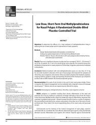
CASE REPORTS<br />
Philippine Journal Of Otolaryngology-Head And Neck Surgery Vol. 25 No. 1 January – June 2010<br />
A<br />
B<br />
Figure 4 A, B. Post-radiation Contrast CT scan of head, axial and coronal sections<br />
showed an interval decrease in size of the previously noted avidly enhancing<br />
lobulated soft tissue mass. It now measures 4.5cm x 4.3cm x 3.6cm.<br />
The aggressiveness of PTC in this case may be further related to actual<br />
vascular invasion demonstrated in our thyroid specimen. Vascular<br />
invasion in PTC occurs in only 2 to 14% of cases. Gardner et al. highly<br />
correlated vascular invasion, whether intrathyroidal or extrathyroidal,<br />
<strong>with</strong> more aggressive local disease and a greater predisposition<br />
for distant metastasis at diagnosis. 10 Their study also identified<br />
intrathyroidal evidence of vascular invasion as predictive of disease<br />
recurrence among their patient population. This mode of malignant<br />
dissemination may better explain the intracranial metastasis in our case<br />
in the absence of clinical cervical node involvement.<br />
Another factor which may explain the aggressiveness of the papillary<br />
thyroid carcinoma in this case was the extracapsular invasion. According<br />
to Machens et al 11 extrathyroidal growth consistently emerged as the<br />
most significant factor for increased likelihood of distant metastasis<br />
in PTC. Extracapsular and extrathyroidal spread give the malignant<br />
cells more avenues for lymphatic and vascular dissemination through<br />
adjacent tracheal, strap muscles, nerve tissue and other contiguous<br />
structures.<br />
The notion of follicular variant of papillary thyroid carcinoma<br />
(FVPTC) having a more aggressive clinical nature than the pure type<br />
of PTC has been disproved. 12 Even <strong>with</strong> a predominant follicular<br />
component, lesions <strong>with</strong> papillary features behave clinically as true<br />
papillary carcinoma. A clinico-pathologic analysis of 243 patients <strong>with</strong><br />
pure versus follicular variant papillary thyroid carcinoma by Jamal et al<br />
showed that pathologic and clinical behaviours of PTC and FVPTC were<br />
comparable. 12 Prognostic factors, treatment and survival also were<br />
similar between the two forms. The follicular variant seen in our patient<br />
may therefore not relate as much to tumor aggressiveness.<br />
While the usual therapeutic modality for high-risk papillary thyroid<br />
carcinoma is total thyroidectomy <strong>with</strong> radioactive iodine (RAI) ablation,<br />
the management of brain metastasis may relate more to palliation than<br />
cure. Although surgical resection has been shown to prolong survival<br />
by nearly three-fold 7 in 47 cases of thyroid malignancy <strong>with</strong> brain<br />
metastasis, our neurosurgery service advised against surgery due to<br />
multifocal brain involvement, uncertainty of complete resection and<br />
absence of neurological deficit.<br />
Initial radiation therapy prior to RAI ablation was advised because<br />
the intracranial lesion was in very close proximity to the venous<br />
confluence of sinuses which could swell <strong>with</strong> RAI and result in massive<br />
cerebral edema. Radiotherapy was probably a reasonable option as<br />
previously recommended for patients unable to undergo surgical<br />
therapy and who had disseminated or inaccessible intracranial lesions<br />
that precluded metastasectomy. 7 Our patient underwent 23 sessions of<br />
EBRT using 46Gy <strong>with</strong> subsequent remarkable decrease in the size of<br />
the brain metastatic lesions. As of this writing, she was asymptomtic<br />
and awaiting possible RAI therapy to further decrease the size of the<br />
intracranial lesions.<br />
In summary, while brain metastasis from a PTC is unusual, several<br />
features of the host (older age, long duration of disease and presence of<br />
distant metastasis) and the tumor pathology (presence of intratumoral<br />
lymphatics, vascular invasion and extrathyroidal invasion) may alert<br />
the clinician to the risk for intracranial spread and possible need for a<br />
cranial CT or MRI. While the prognosis may be dismal, surgical resection<br />
offered a significant longer survival for most patients in the literature.<br />
Philippine Journal Of Otolaryngology-Head And Neck Surgery 29
CASE REPORTS<br />
Philippine Journal Of Otolaryngology-Head And Neck Surgery Vol. 25 No. 1 January – June 2010<br />
Other palliative management such as RAI ablation and radiotherapy<br />
remain as possible options should the situation preclude surgical<br />
intervention.<br />
This case study serves to remind otolaryngologists to be more<br />
vigilant in the diagnosis and management of patients <strong>with</strong> PTC.<br />
Increasing numbers of PTC cases in our institution are displaying<br />
more aggressive behaviour. Their management entails proper staging<br />
evaluation and good communication of prognostic data and available,<br />
realistic therapeutic options to patients using a multidisciplinary<br />
approach.<br />
REFERENCES<br />
1. Department of Health, Philippines. Disease and Health condition advisories. [Updated 2009<br />
February] Available from http://www.doh.gov.ph/taxonomy/term/329?page=1<br />
2. Silva JV, Zantua RV, Arevalo AE. Profile of patients <strong>with</strong> thyroid malignancy at a university-based<br />
tertiary hospital: a six year retrospective study (1995-2000) Philipp J Otolaryngol Head Neck Surg<br />
2002; 17(3-4):163-168.<br />
3. Hay ID, Bergstralh EJ, Goellner JR, Ebersold JR, Grant CS. Predicting outcome in papillary thyroid<br />
carcinoma: development of a reliable prognostic scoring system in a cohort of 1779 patients<br />
surgically treated at one institution during 1940 through 1989. Surgery 1993 Dec;114(6):1050-7;<br />
discussion 1057-8.<br />
4. Hundahl SA, Fleming ID, Fremgen AM, Menck HR. A National Cancer Data Base report on<br />
53,856 cases of thyroid carcinoma treated in US., 1985-1995. Cancer. 1998 Dec 15; 83(12): 2638-<br />
2648<br />
5. Cohen SM, Burkey BB, Netterville JL. Surgical management of parapharyngeal space masses.<br />
Head Neck. 2005 Aug; 27(8):669-675.<br />
6. Dinneen SF, Valimaki MJ, Bergstralh EJ, Goellner JR, Gorman CA, Hay ID. Distant metastasis in<br />
papillary thyroid carcinoma: 100 cases observed at one institution during 5 decades. J Clin.<br />
Endocrinol. Metab. 1995 Jul; (80)7;2041-2045.<br />
7. Chiu AC, Delpassand ES, Sherman SI. Prognosis and treatment of brain metastases in thyroid<br />
carcinoma. J. Clin. Endocrinol. Metab.. 1997 Nov; 82(11): 3637-3642<br />
8. Mazzaferri EL, Young RL. <strong>Papillary</strong> thyroid carcinoma: a 10 year follow-up report of the impact of<br />
therapy in 576 patients. Am J Med. 1981 Mar;70(3):511–517.<br />
9. Hall FT, Freeman JL, Asa SL, Jackson DG, Beasley NJ. Intratumoral lymphatics and lymph node<br />
metastases in papillary thyroid carcinoma. Arch Otolaryngol Head Neck Surg, 2003 Jul; 129<br />
(7);716-719<br />
10. Gardner RE, Tuttle RM, Burman KD, Haddady S, Truman C, Sparling YH, et.al. Prognostic<br />
importance of vascular invasion in papillary thyroid carcinoma. Arch Otolaryngol Head Neck<br />
Surg 2000 Mar; 126(3): 309-312<br />
11. Machens A, Holzhausen HJ, Lautenshlager C, Thanh PH, Dralle H. Enhancement of lymph node<br />
metastasis and distant metastasis of thyroid carcinoma: a multivariate analysis of clinical risk<br />
factors. Cancer. 2003 Aug 15;(98)4: 712-719<br />
12. Zidan J, Karen D, Stein M, Rosenblatt E, Basher W, Kuten A. Pure versus follicular variant of<br />
papillary thyroid carcinoma: clinical features, prognostic factors, treatment, and survival.<br />
Cancer. 2003 Mar 1;(97)5: 1181-1185<br />
13. Misaki T, Masahiro I, Kanji K, Junji K. Brain metastasis from differentiated thyroid cancer in<br />
patients treated <strong>with</strong> radioiodine for bone and lung lesions. Ann Nucl Med 2000 Apr;14(2): 111-<br />
114<br />
30 Philippine Journal Of Otolaryngology-Head And Neck Surgery