SLOVENIAN VETERINARY RESEARCH
SLOVENIAN VETERINARY RESEARCH
SLOVENIAN VETERINARY RESEARCH

Create successful ePaper yourself
Turn your PDF publications into a flip-book with our unique Google optimized e-Paper software.
Slov Vet Res 2012; 49 (4): 155-203<br />
THE SCIENTIFIC JOURNAL OF THE <strong>VETERINARY</strong> FACULTY UNIVERSITY OF LJUBLJANA<br />
<strong>SLOVENIAN</strong> <strong>VETERINARY</strong> <strong>RESEARCH</strong><br />
SLOVENSKI VETERINARSKI ZBORNIK<br />
49<br />
Volume<br />
4<br />
Slov Vet Res • Ljubljana • 2012 • Volume 49 • Number 4 • 155-203
THE SCIENTIFIC JOURNAL OF THE <strong>VETERINARY</strong> FACULTY UNIVERSITY OF LJUBLJANA<br />
<strong>SLOVENIAN</strong> <strong>VETERINARY</strong> <strong>RESEARCH</strong><br />
SLOVENSKI VETERINARSKI ZBORNIK<br />
49<br />
Volume 4<br />
Slov Vet Res • Ljubljana • 2012 • Volume 49 • Number 4 • 155-203
The Scientific Journal of the Veterinary Faculty University of Ljubljana<br />
<strong>SLOVENIAN</strong> <strong>VETERINARY</strong> <strong>RESEARCH</strong><br />
SLOVENSKI VETERINARSKI ZBORNIK<br />
Previously: <strong>RESEARCH</strong> REPORTS OF THE <strong>VETERINARY</strong> FACULTY UNIVERSITY OF LJUBLJANA<br />
Prej: ZBORNIK VETERINARSKE FAKULTETE UNIVERZA V LJUBLJANI<br />
4 issues per year / izhaja štirikrat letno<br />
Editor in Chief / glavni in odgovorni urednik: Gregor Majdič<br />
Technical Editor / tehnični urednik: Matjaž Uršič<br />
Assistants to Editor / pomočnici urednika: Valentina Kubale Dvojmoč, Klementina Fon Tacer<br />
Editorial Board / uredniški odbor:<br />
Frangež Robert, Polona Juntes, Matjaž Ocepek, Seliškar Alenka, Modest Vengušt, Milka Vrecl, Veterinary Faculty University of Ljubljana /<br />
Veterinarska fakulteta Univerze v Ljubljani; Vesna Cerkvenik, Reziduum d.o.o.<br />
Editorial Advisers / svetovalca uredniškega odbora: Gita Grecs-Smole for Bibliography (bibliotekarka),<br />
Leon Ščuka for Statistics (za statistiko)<br />
Reviewing Editorial Board / ocenjevalni uredniški odbor:<br />
Ivor D. Bowen, Cardiff School of Biosciences, Cardiff, Wales, UK; Antonio Cruz, Paton and Martin Veterinary Services, Adegrove,<br />
British Columbia; Gerry M. Dorrestein, Dutch Research Institute for Birds and Exotic Animals, Veldhoven, The Netherlands; Sara Galac, Utrecht<br />
University, The Netherlands; Wolfgang Henninger, Veterinärmedizinische Universität Wien, Austria; Simon Horvat, Biotehniška fakulteta,<br />
Univerza v Ljubljani, Slovenia; Nevenka Kožuh Eržen, Krka, d.d., Novo mesto, Slovenia; Louis Lefaucheur, INRA, Rennes, France; Bela Nagy,<br />
Veterinary Medical Research Institute Budapest, Hungary; Peter O’Shaughnessy, Institute of Comparative Medicine, Faculty of Veterinary<br />
Medicine, University of Glasgow, Scotland, UK; Milan Pogačnik, Veterinarska fakulteta, Univerza v Ljubljani, Slovenia; Peter Popelka, University<br />
of Veterinary Medicine, Košice, Slovakia; Detlef Rath, Institut für Tierzucht, Forschungsbericht Biotechnologie, Bundesforschungsanstalt für<br />
Landwirtschaft (FAL), Neustadt, Germany; Henry Stämpfli, Large Animal Medicine, Department of Clinical Studies, Ontario Veterinary College,<br />
Guelph, Ontario, Canada; Frank J. M. Verstraete, University of California Davis, Davis, California, US; Thomas Wittek, Veterinärmedizinische<br />
Universität, Wien, Austria<br />
Slovenian Language Revision / lektor za slovenski jezik: Viktor Majdič<br />
Address: Veterinary Faculty, Gerbičeva 60, 1000 Ljubljana, Slovenia<br />
Naslov: Veterinarska fakulteta, Gerbičeva 60, 1000 Ljubljana, Slovenija<br />
Tel.: +386 (0)1 47 79 100, 47 79 129, Fax: +386 (0)1 28 32 243<br />
E-mail: slovetres@vf.uni-lj.si<br />
Sponsored by the Slovenian Book Agency<br />
Sofinancira: Javna agencija za knjigo Republike Slovenije<br />
ISSN 1580-4003<br />
Printed by / tisk: DZS, d.d., Ljubljana<br />
Indexed in / indeksirano v: Agris, Biomedicina Slovenica, CAB Abstracts, IVSI<br />
Urlich’s International Periodicals Directory, Science Citation Index Expanded,<br />
Journal Citation Reports/Science Edition<br />
http://www.slovetres.si/
<strong>SLOVENIAN</strong> <strong>VETERINARY</strong> <strong>RESEARCH</strong><br />
SLOVENSKI VETERINARSKI ZBORNIK<br />
Slov Vet Res 2012; 49 (4)<br />
Original Scientific Articles<br />
Farzan R, Rahimi E, Momtaz H. Virulence properties of Shiga Toxin-Producing Escherichia coli isolated<br />
from Iranian raw milk and dairy products . . . . . . . . . . . . . . . . . . . . . . . . . . . . . . . . . . . . . . . . . . . . . . . . . . . . . . . . . . . . . 159<br />
Bilandžić N, Ðokić M, Sedak M, Varenina I, Solomun Kolanović B, Končurat A, Šimić B, Rudan N. Content of five trace<br />
elements in different honey types from Koprivnica - Križevci county . . . . . . . . . . . . . . . . . . . . . . . . . . . . . . . . . . . . . . . . 167<br />
Lenhardt M, Jarić I, Cvijanović G, Kolarević J, Gačić Z, Smederevac-Lalić M, Višnjić-Jeftić Ž. Comparison<br />
of morphological characters between wild and cultured sterlet (Acipenser ruthenus L.) . . . . . . . . . . . . . . . . . . . . . . . . 177<br />
Marhold C, Slavec B, Laroucau K, Vorimore F, Račnik J, Zadravec M, Keše D, Krapež U, Dovč A. Detection<br />
of Chlamydia psittaci in cage birds in Slovenia by Real-Time PCR . . . . . . . . . . . . . . . . . . . . . . . . . . . . . . . . . . . . . . . . . 185<br />
Starič J, Nemec M, Zadnik T. Bone metabolism markers and blood minerals concentration in dairy cattle during<br />
pregnancy and lactation . . . . . . . . . . . . . . . . . . . . . . . . . . . . . . . . . . . . . . . . . . . . . . . . . . . . . . . . . . . . . . . . . . . . . . . . . . 193<br />
Author Index Volume 49, 2012. . . . . . . . . . . . . . . . . . . . . . . . . . . . . . . . . . . . . . . . . . . . . . . . . . . . . . . . . . . . . . . . . . . . . . . . . . . 201
Slov Vet Res 2012; 49 (4): 159-66<br />
UDC 579.63:616-092:579.842.1/.2:637.1<br />
Original Scientific Article<br />
Virulence properties of Shiga Toxin-Producing<br />
Escherichia coli isolated from Iranian raw milk<br />
and dairy products<br />
Rahil Farzan 1, 2 *, Ebrahim Rahimi 3 , Hassan Momtaz 4<br />
1<br />
Young researchers club, 2 Food Science & Technology, College of Agriculture, 3 Department of Food Hygine, College of Veterinary Medicine,<br />
4<br />
Department of Microbiology, College of Veterinary Medicine, Shahrekord Branch, Islamic Azad University, Shahrekord, Iran<br />
*Corresponding author, E-mail: rl.farzan@yahoo.com<br />
Summary: Shiga toxin (Stx)-producing Escherichia coli (STEC) strains are a diverse group of food-borne pathogens with<br />
various levels of virulence for humans. The main object of the present study was to determine virulence properties of STEC<br />
isolated from Iranian raw milk and dairy products. A total number of 300 samples, including sheep’s (35), goat’s (46), cow’s (106),<br />
and buffalo’s milk (21), soft cheese (42), butter (32) and ice cream (18), was obtained from farm bulk tanks, milk collection centres,<br />
and various supermarkets and retailer shops in different regions of Iran. Biochemical and molecular (PCR) method proved 26%<br />
of the samples to be E. coli positive, and among them, Stx1 and Stx2 genes were detected in 33 and 52 samples, respectively.<br />
But, eaeA and sfaS genes were not found in any of the sample. Beside Stx2, cnfI had the highest prevalence (42 isolates), and<br />
beside eaeA and sfaS, fyuA had the lowest prevalence of virulence genes. The results of this study demonstrate that there is<br />
a widespread distribution of potentially virulent E. coli strains in the environment and food that may be a cause of concern for<br />
human health.<br />
Key words: STEC; Escherichia coli; virulence factors; raw milk<br />
Introduction<br />
Raw, unpasteurized milk is consumed directly<br />
by a large number of people in rural areas,<br />
and indirectly by a much larger segment of the<br />
population via consumption of several types of<br />
cheese. One of the main reasons why people prefer<br />
raw milk and its products may be their belief<br />
in their advantages or higher value compared<br />
to pasteurized milk (1). Many microorganisms,<br />
coliforms among them, can have access to milk<br />
and dairy products, and Escherichia coli are<br />
Received: 15 February 2012<br />
Accepted for publication: 1 August 2012<br />
often used as marker organisms. Detecting and<br />
counting of E. coli is used as a reliable indicator<br />
of faecal contamination and indicates a possible<br />
presence of enteropathogenic and/or toxigenic<br />
microorganisms which constitute a public health<br />
hazard (2). E. coli, a gram negative bacillus, is<br />
naturally present in the gastrointestinal tracts<br />
of humans and animals as a part of natural<br />
microflora (3). While E. coli typically harmlessly<br />
colonizes the intestinal tract, several E. coli clones<br />
have evolved the ability to cause a variety of<br />
diseases within the intestinal tract and elsewhere<br />
in the host (4). Enterohemorrhagic Escherichia<br />
coli (EHEC) strains are a subset of Shiga toxin<br />
(Stx)-producing E. coli (STEC) strains that are
160<br />
R. Farzan, E. Rahimi, H. Momtaz<br />
isolated from human patients and are responsible<br />
for severe clinical symptoms, including diarrhea,<br />
hemorrhagic colitis, and hemolytic-uremic<br />
syndrome. These diseases are directly related<br />
to the virulence genes present in the causative<br />
agent (5,6). Four of the most commonly assayed<br />
virulence factors of STEC are the two phageencoded<br />
cytotoxins, called Shiga toxin 1 and 2<br />
(encoded by the Stx1 and Stx2 genes, respectively),<br />
the protein intimin (encoded by the chromosomal<br />
gene eae), which is responsible for the intimate<br />
attachment of the bacteria to intestinal epithelial<br />
cells, and the plasmid-encoded enterohaemolysin,<br />
also called enterohaemorrhagic E. coli haemolysin<br />
(EHEC-HlyA), encoded by the ehxA gene (3,7).<br />
Shiga toxin-producing E. coli can be transmitted<br />
through different routes, including food and water,<br />
person-to-person contact, and animal-to-person<br />
contact. Most human infections are caused by<br />
consumption of contaminated food. Domestic<br />
animals and wild ruminants, in particular cattle,<br />
are considered the main reservoir of STEC and the<br />
main source of contamination of the food supply<br />
(8,9). The presumed route of E. coli contamination<br />
of raw milk is via faecal contamination during<br />
milking (10). However, direct excretion of the<br />
organisms from the infected udder has also been<br />
reported (11).<br />
There is a lack of information on virulence<br />
properties of Shiga Toxin-producing Escherichia<br />
coli contaminating Iranian milk and dairy<br />
products. Therefore, the main goal of the present<br />
study was to evaluate these properties.<br />
Material and methods<br />
Sample collection and identification of E. coli<br />
From March 2010 to March 2011, raw bovine<br />
(n= 106), caprine (n= 46), ovine (n= 35) and<br />
buffalo (n= 21) milk samples were collected from<br />
farm bulk tanks and milk collection centres<br />
from several regions throughout Iran. Bovine<br />
and buffalo milk samples were collected in the<br />
above mentioned time period, whereas ovine<br />
and caprine milk samples were only available<br />
in the lactating periods of ewes and goats (i.e.<br />
from March through May, and from September<br />
to November of the subsequent year) within the<br />
same time frame. At each site, sampling of milk<br />
was performed according to the International<br />
Dairy Federation guidelines (IDF) (12). Samples<br />
(100 mL each, in sterile glass containers) were<br />
transported to the laboratory at ca. +4 o C within<br />
6–12 h after sampling.<br />
Among dairy products, 42 samples of soft<br />
cheese, 32 samples of soft butter and 18 samples<br />
of soft ice-cream, all made of raw milk, were<br />
purchased at the supermarkets in the city of<br />
Shiraz, Isfahan and Shahrekord. All samples<br />
were kept under refrigeration and in plastic bags.<br />
Their information about dates of production and<br />
of assigned shelf-life was not presented. Dairy<br />
product samples were collected over a period of<br />
six months between May and November 2010, and<br />
were analyzed on the day of acquisition. Samples<br />
were transported under refrigeration (+4 to +6 o C)<br />
in thermal boxes containing ice packs and were<br />
tested immediately after collection. A 25g portion<br />
of each sample was blended with 225 mL of<br />
nutrient broth (Merck, Germany) for two minutes,<br />
and at normal speed, using a Stomacher lab<br />
blender, and incubated at +37 o C for 24h. A 1 mL<br />
sample of the nutrient broth culture was mixed<br />
with 9 mL of MacConkey broth (Merck, Germany)<br />
and further incubated at +37 o C for 24h. One loop<br />
of each tube was streaked on MacConkey agar<br />
(Merck, Germany).<br />
Four colonies from each plate with typical E.<br />
coli morphology were selected and examined by<br />
biochemical tests, including hydrogen sulphide,<br />
citrate, urease and indole.<br />
All E. coli colonies were confirmed by molecular<br />
(PCR) method, determined by amplification of 16S<br />
rRNA gene region of E. coli as described by Sabat<br />
et al. (13). The identification of E. coli O157: H7<br />
was also performed with the method described<br />
by Fode-Vaughan et al. (14). PCR conditions were<br />
optimized for DPCR by recommendations reported<br />
previously. The PCR conditions for amplification<br />
of Stx1 and Stx2 were the same as used for pmoA.<br />
DNA isolation<br />
Bacterial strains were grown overnight and<br />
at +37 o C in the trypticase soy agar (TSA– Merck,<br />
German). One colony was suspended in 100 µL of<br />
sterile distilled water. The suspension was being<br />
boiled for 13 min, then frozen and subsequently<br />
centrifuged at 14,000 rpm for 15 min to pellet the<br />
cell debris (15). The supernatant was used as a<br />
template for amplification reaction.
Virulence properties of Shiga Toxin-Producing Escherichia coli isolated from Iranian raw milk and dairy products 161<br />
Polymerase chain reaction (PCR)<br />
The PCR assays, specific primer sequences<br />
and the predicted size of the amplified products<br />
for different pathogenic gene coding regions were<br />
employed as previously described (16-20). Details<br />
are shown in Table 1. For cycling, a DNA thermocycler<br />
(Eppendorf Mastercycler, Eppendorf-Nethel-<br />
Hinz GmbH, Hamburg, Germany) was used. The<br />
amplified products were visualized by ethidium<br />
bromide staining after gel electrophoresis of 10<br />
µL of the final reaction mixture in 1.5% agarose.<br />
Strains of E. coli O157:K88ac:H19, CAPM 5933<br />
and E. coli O159:H20, CAPM 6006 were used as<br />
positive controls.<br />
Table 1: Sequences and predicted lengths of PCR amplification products of the oligonucleotide primers used in<br />
this study<br />
Reference Product size (bp) Primer sequences (5’–3’) Primers name Pathogenic factor<br />
(16) 1105<br />
(17) 543<br />
(18) 366<br />
(18) 282<br />
(19) 629<br />
(16) 430<br />
(16) 720<br />
(16) 240<br />
(16) 880<br />
(16) 300<br />
(16) 290<br />
(20) 432<br />
ATCTTATACTGGATGGGATCATCTTGG<br />
GCAGAACGACGTTCTTCATAAGTATC<br />
AATCTAATTAAAGAGAAC<br />
CATGCTTTGTATATCTA<br />
AAATCGCCATTCGTTGACTACTTCT<br />
TGCCATTCTGGCAACTCGCGATGCA<br />
CGATCGTCACTCACTGGTTTCATCA<br />
GGATATTCTCCCCACTCTGACACC<br />
TGCGGCACAACAGGCGGCGA<br />
CGGTCGCCGCACCAGGATTC<br />
AAATCACCAAGAATCATCCAGTTA<br />
AAATCTCCTGCAATCATCCAGTTTA<br />
ATGGCAGTGGTGTCTTTGGTG<br />
CGTCCCACCATACGTGCTCTTC<br />
GTGGATACGACGATTACTGTG<br />
CCGCCAGCATTCCCTGTATTC<br />
TGATTAACCCCGCGACGGGAA<br />
CGCAGTAGGCACGATGTTGTA<br />
GGCTGGACATCATGGGAACTGG<br />
CGTCGGGAACGGGTAGAATCG<br />
GGTGTGGTGCGATGAGCACAG<br />
CACGGTTCAGCCATCCCTGAG<br />
CAATGCAGATGCAGATACCG<br />
CAGAGATGTCGTTGCAGCAG<br />
CNF1a<br />
CNF1b<br />
CNF2f<br />
CNF2r<br />
Stx1f<br />
Stx1r<br />
Stx2f<br />
Stx2r<br />
EAE1<br />
EAE2<br />
Cdt 1<br />
Cdt 2<br />
PapA-f<br />
PapA-r<br />
SfaS-f<br />
SfaS-r<br />
fyuA-f<br />
fyuA-r<br />
AerJ-f<br />
AerJ-r<br />
TraT-f<br />
TraT-r<br />
Hly F<br />
Hly R<br />
Cytotoxic necrotizing<br />
factor 1 (cnf1)<br />
Cytotoxic necrotizing<br />
factor 2 (cnf2)<br />
Shiga toxin 1 (stx1)<br />
Shiga toxin 2 (stx2)<br />
Enteropathogenic<br />
attachment &<br />
effacement (eaeA)<br />
Cytolethal distending<br />
factor (cdtB)<br />
P-Fimbriae (papA)<br />
S-Fimbriae adhesion<br />
(sfaS)<br />
Yersiniabactin (fyuA)<br />
Aerobactin (iutA)<br />
Serum survival (traT)<br />
Haemolysin (hlyA)<br />
Results<br />
Eventually, 78 E. coli strains (representative of 78<br />
different colony morphologies) (26%) were isolated<br />
from 300 samples (Table 2). Colony confirmation<br />
was performed by biochemical and molecular (PCR)<br />
methods. According to the obtained results, most<br />
pollution is related to cow’s milk.<br />
PCR assays were successfully developed for<br />
detection of twelve different virulence genes of<br />
genomic DNA of E. coli isolates from milk and<br />
dairy products. Agarose gel electrophoresis of PCR<br />
products revealed various bands that represent<br />
each virulence gene. In our research, 33 and 52<br />
of E. coli isolates had Stx1 and Stx2, respectively.<br />
By colony counting, 93.5% of independent isolates<br />
of Stx-positive E. coli was detected among 78<br />
E. coli isolates; and, Stx1 and Stx2 genes were<br />
detected in 33 and 52 isolates, respectively. But,<br />
eaeA gene was not found in any of the samples.
162<br />
R. Farzan, E. Rahimi, H. Momtaz<br />
The prevalence of samples containing Stx-positive<br />
and other virulence genes-positive E. coli is shown<br />
in Table 2. The prevalence of virulence genes was<br />
evaluated. 38.4% of the isolates contained only<br />
one virulence gene, 26.9% contained two, and,<br />
34.6% contained three or more virulence genes.<br />
None of the samples in this study was positive on<br />
E. coli O157:H7 (the prevalence of eaeA gene was<br />
0.0%). Our study showed that cow’s milk (41.5%),<br />
cheese and ice cream (16.6%) had the highest<br />
prevalence of STEC. PCR-amplification and<br />
individually amplified fragments of gene detection<br />
were subjected to agarose gel electrophoresis<br />
(Figure 1 and 2).<br />
Table 2: Distribution of virulence factor genes in E. coli isolated from raw milk and dairy products<br />
Sample<br />
Sheep<br />
milk<br />
Goat<br />
milk<br />
Cow<br />
milk<br />
Buffalo<br />
milk<br />
Cheese<br />
Sample<br />
No.<br />
E.coli<br />
positive (%)<br />
stxI stxII eaeA cnf1 cnf2 iutA cdtB papA hlyA traT sfaS fyuA<br />
35 6 (17.1) 3 4 - 3 3 3 1 3 1 4 - 2<br />
46 7 (15.21) 3 4 - 2 2 2 1 4 1 5 - 1<br />
106 44 (41.5) 16 35 - 26 12 12 9 18 5 14 - 6<br />
21 6 (28.5) 1 3 - 4 2 2 1 1 2 1 - -<br />
42<br />
7 (16.6) 6 5 - 2 1 1 1 4 2 1 - 1<br />
Butter<br />
32<br />
5 (15.6) 3 1 - 4 3 3 - - 1 4 - -<br />
Ice<br />
cream<br />
18 3 (16.6) 1 - - 1 - - 1 - - - - -<br />
Total 300 78 (26.0) 33 52 - 42 23 23 14 30 12 29 - 10<br />
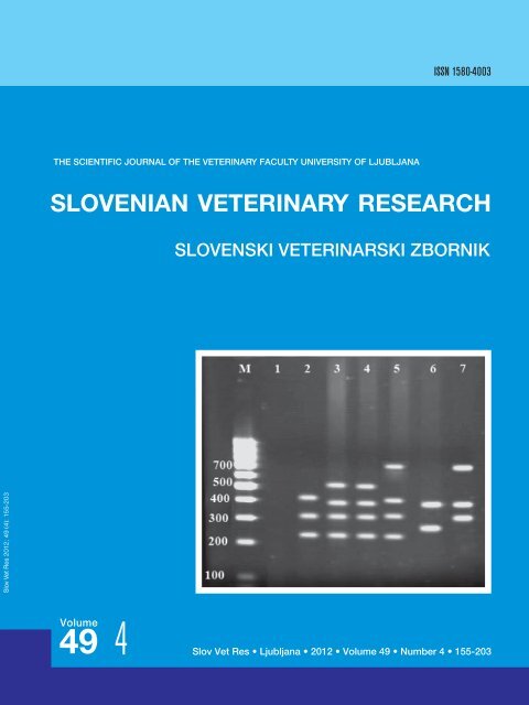
Figure 1: Agarose gel electrophoresis of PCR products<br />
amplified with PCR method for the cnf2 (543 bp), Stx1<br />
(366 bp), Stx2 (282 bp), cdtB (430 bp), papa (720 bp),<br />
iutA (300 bp) and traT (290 bp) genes in E. coli isolated<br />
from raw milk and dairy products<br />
Figure 2: Agarose gel electrophoresis of PCR products<br />
amplified with PCR method for the cnf1 (1105 bp), fyuA<br />
(880 bp) and hlyA (432 bp) genes in E. coli isolated from<br />
raw milk and dairy products
Virulence properties of Shiga Toxin-Producing Escherichia coli isolated from Iranian raw milk and dairy products 163<br />
Discussion<br />
Milk and dairy products can harbor a variety of<br />
microorganisms and can be an important source<br />
of food-borne pathogens. The presence of foodborne<br />
pathogens in milk is due to direct contact<br />
with contaminated sources in the dairy farm<br />
environment and to udder excretion of an infected<br />
animal. There are several reasons to be concerned<br />
about the microbial quality of dairy products: first,<br />
outbreaks of disease in humans have been traced<br />
back to the consumption of unpasteurized milk,<br />
and also to pasteurized milk; second, unpasteurized<br />
milk is consumed directly by dairy producers, farm<br />
employees, their families and neighbours, and<br />
raw milk advocates; third, unpasteurized milk is<br />
consumed directly by a large segment of population<br />
via consumption of several types of cheese produced<br />
from unpasteurized milk (21,22).<br />
Nowadays, public health concern associated<br />
with microbial food safety is on the increase since<br />
E. coli is not only regarded as an indicator of faecal<br />
contamination but more likely as an indicator of<br />
poor hygiene and insufficient sanitary practices<br />
during milking. Furthermore, Shiga toxinproducing<br />
strains of Escherichia coli (STEC) are<br />
now recognized as an important agent of diarrhea<br />
and other foodborne-related illnesses through<br />
ingestion of contaminated food (23,24). Hence,<br />
the prevalence of Shiga toxin-producing E. coli<br />
isolated from milk and dairy products is evaluated<br />
in the present study. Our results indicate that 78<br />
(26%) of 300 Iranian milk and dairy samples were<br />
contaminated with E. coli. 93.5% were found to<br />
be positive on two target genes, Stx1 and Stx2.<br />
To our knowledge, the main reason for such<br />
high prevalence of E. coli and their virulence<br />
factors in milk and dairy products arises from<br />
contamination with faeces. Yet other reasons are:<br />
failure to comply with relevant rules, unsanitary<br />
milking, failure to control the staff of milking room<br />
and factories and/or workshops producing butter,<br />
ice cream and cheese, and finally, the use of raw<br />
animal milk for the production of dairy products.<br />
The data of this study is older than the recent<br />
survey conducted for detection of prevalence and<br />
antimicrobial resistance of E. coli O157 isolated<br />
from Iranian traditional cheese, ice cream and<br />
yoghurt. The study reports about E. coli O157<br />
being detected in 9 (3.1%) of the 290 samples<br />
tested, 5 isolated from traditional cheese and<br />
4 isolated from traditional ice cream samples,<br />
whereas E. coli O157: H7 was not detected in<br />
any of the samples (25). One reason for this<br />
difference may be due to differences in the areas<br />
under study and the number of samples. Yet<br />
another study was conducted to investigate the<br />
presence of E. coli O157 and E. coli O157: H7<br />
strains, and the presence of virulence genes Stx1,<br />
Stx2, eaeA and ehlyA isolates derived from some<br />
Iranian traditional dairy products and minced<br />
beef meat. E. coli non-O157, E. coli O157: NM<br />
and E. coli O157:H7 were isolated from 7%, 1.5%<br />
and 0.5% of the traditional butter, cream and<br />
kashk (a dairy product, similar to sour cream),<br />
respectively. Also, all E. coli O157:H7/NM isolates<br />
were positive on eaeA and Stx1 and/or Stx2, and<br />
one E. coli O157:H7 isolate was positive on ehlyA.<br />
Of the 3 Stx-positive isolates, one and two isolates<br />
contained Stx1 and Stx2, respectively (26).<br />
Globally, several other studies reported the<br />
prevalence of E. coli and virulence properties of<br />
dairy products. For example in Italy, a polyphasic<br />
approach was evaluated for the detection of<br />
eight staphylococcal enterotoxin (SE)-encoding<br />
genes (sea, sec, sed, seg, seh, sei, sej, sel) and<br />
the Escherichia coli genes were most commonly<br />
associated with the virulence factors (eae, elt,<br />
ipaH, Stx) in traditional soft cheese, produced<br />
artisanally from raw milk in the Lombardy region.<br />
The results indicate that some of the artisanal<br />
cheese examined may constitute a potential<br />
hazard for the consumer health (27). According<br />
to the study of raw milk cheese production in<br />
Kerman, Iran, Stx genes were detected in 6.4% of<br />
the samples, but STEC strains were isolated in<br />
only 5 of them (4%) (28). Another study from Iran<br />
shows that about 21.8% of E. coli isolates from<br />
cattle were positive on ehxA, Stx1, and/or Stx2<br />
genes (29), but our study indicates that Stx1and<br />
cnf1 with incidences of 42.3% and 53.84% had<br />
the highest frequency rate of virulence genes of E.<br />
coli isolates from raw milk samples. The results<br />
offer an answer to why food contamination from<br />
animal faecal sources is so common in Iran.<br />
Yet another study of lamb food chain shows<br />
that all three virulence genes, eae, Stx1, and Stx2<br />
were the most prevalent genes in slaughterhouses<br />
(69%), processing plants (32%) and butcheries (9–<br />
10%) (30), but our results indicate that Stx2 and<br />
cnf1 had the highest prevalence of E. coli virulence<br />
genes in raw milk and dairy products.
164<br />
R. Farzan, E. Rahimi, H. Momtaz<br />
Detection of bacteria in milk samples is still<br />
important, especially in more rural areas as it<br />
is easily available and economical. Due to bad<br />
climate conditions in some geographical places<br />
of Iran, oxen, ewes and goats have unsatisfactory<br />
survival conditions, so buffaloes present the main<br />
milk source for people. Buffalo’s milk hygiene is<br />
therefore essential for human health, especially in<br />
arid and desert areas.<br />
In United States, Van Kessel et al. (31) detected<br />
Shiga toxin genes enrichments in 15.2% of the<br />
bulk tank milk samples and in 51.0% filters by<br />
real-time PCR. These data confirm those from<br />
earlier studies showing significant contamination<br />
of bulk tank milk by zoonotic bacterial pathogens,<br />
and also, that the consumption of raw milk and<br />
raw milk products present a health risk. In Spain,<br />
occurrence of STEC in 'Castellano' cheese, a noncooked<br />
and hard or semi-hard Spanish cheese<br />
made from ewe's milk, was reported. According to<br />
the report, three STEC strains were detected in two<br />
samples (2.4%) of 'Castellano' cheese, one with 2.5<br />
and the other one with 12 month-ripening period.<br />
From those STEC isolates, two were identified as<br />
E. coli O14 and one third presented an O-specific<br />
polysaccharide not-groupable serologically (ONG)<br />
(32). In Bangladesh, the prevalence of Shiga toxin<br />
(Stx)–producing Escherichia coli was found in<br />
different types of food samples, and their genetic<br />
relation to STEC strains previously isolated from<br />
animal sources was investigated. In this study,<br />
approximately 10% of raw milk and 8% of fresh<br />
juice samples were positive for Stx (33).<br />
All these data lead to conclusion that milk<br />
and dairy products represent a potential hazard<br />
for consumers, due to the potential presence<br />
of Shiga-toxin-producing E. coli, because of<br />
neglected sanitary measures during manufacture,<br />
handling and distribution of such fresh foods.<br />
Consequently, food manufacturers and specialists<br />
should design comprehensive programmes<br />
as good manufacturing practices (GMP) and<br />
implementation of HACCP system to ensure food<br />
free from the pathogens. In addition, effective<br />
heat treatments of food, providing information<br />
to food handlers and consumers, as well as<br />
implementation of strict hygienic measures during<br />
manufacture, storage and sale of these products<br />
are of significant importance in improving the<br />
quality of food and safeguarding the consumers<br />
against infections with such organisms. On the<br />
other hand, since many people still drink raw milk,<br />
especially in rural areas, this emphasises the need<br />
for educational efforts about health risks associated<br />
with consumption of raw, unpasteurized milk.<br />
Acknowledgements<br />
Our greatest appreciations go to Dr. M.D.<br />
Rahimian and Mr. M. Momeni for their valuable<br />
cooperation. Besides, this study could not have<br />
been conducted without the assisstance of Islamic<br />
Azad University Shahrekord Branch in Iran.<br />
Reference<br />
1. Altalhi AD, Hassan SA. Bacterial quality of<br />
raw milk investigated by Escherichia coli and isolates<br />
analysis for specific virulence-gene markers.<br />
Food Control 2009; 20: 913–7.<br />
2. Kaper JB, Nataro JP, Mobley HL. Pathogenic<br />
Escherichia coli. Nat Rev Microbiol 2004;<br />
2: 123–40.<br />
3. Law D. Virulence factors of Escherichia coli<br />
O157 and other Shiga toxin-producing E. coli. J<br />
Appl Microbiol 2000; 88: 729–45.<br />
4. Vidal M, Kruger E, Durán C, et al. Single<br />
multiplex PCR assay to identify simultaneously<br />
the six categories of diarrheagenic Escherichia<br />
coli associated with enteric infections. J Clin Microbiol<br />
2005; 43: 5362–5.<br />
5. Bettelheim KA. Enterohaemorrhagic Escherichia<br />
coli O157:H7: a red herring? J Med Microbiol<br />
2001; 50: 201–2.<br />
6. Elliott EJ, Robins-Browne RM, O'Loughlin<br />
EV, et al. Nationwide study of haemolytic uraemic<br />
syndrome: clinical, microbiological, and epidemiological<br />
features. Arch Dis Child 2001; 85:<br />
125–31.<br />
7. Gyles CL. Shiga toxin-producing Escherichia<br />
coli: an overview. J Anim Sci 2007; 85: E45–62.<br />
8. Erickson MC, Doyle MP. Food as a vehicle<br />
for transmission of Shiga toxin-producing Escherichia<br />
coli. J Food Prot 2007; 70: 2426–49.<br />
9. Caprioli A, Morabito S, Brugère H, et al.<br />
Enterohaemorrhagic Escherichia coli: emerging<br />
issues on virulence and modes of transmission.<br />
Vet Res 2005; 36: 289–311.<br />
10. Hussein HS, Sakuma T. Prevalence of shiga<br />
toxin-producing Escherichia coli in dairy cattle<br />
and their products. J Dairy Sci 2005; 88: 450–65.<br />
11. Lira WM, Macedo C, Marin JM. The incidence<br />
of Shiga toxin-producing Escherichia coli
Virulence properties of Shiga Toxin-Producing Escherichia coli isolated from Iranian raw milk and dairy products 165<br />
in cattle with mastitis in Brazil. J Appl Microbiol<br />
2004; 97: 861–6.<br />
12. IDF Standard 50C. Milk and milk products–<br />
guidance on methods of sampling. Brussels,<br />
Belgium: International dairy Federation, 1995.<br />
13. Sabat G, Rose P, Hickey WJ, et al. Selective<br />
and sensitive method for PCR amplification<br />
of Escherichia coli 16S rRNA genes in soil. Appl<br />
Environ Microbiol 2000; 66: 844–9.<br />
14. Fode-Vaughan KA, Maki JS, Benson JA,<br />
et al. Direct PCR detection of Escherichia coli<br />
O157:H7. Lett Appl Microbiol 2003; 37: 239–43.<br />
15. Reischl U, Youssef MT, Kilwinski J, et al.<br />
Real-time fluorescence PCR assays for detection<br />
and characterization of Shiga toxin, intimin, and<br />
enterohemolysin genes from Shiga toxin-producing<br />
Escherichia coli. J Clin Microbiol 2002; 40:<br />
2555–65.<br />
16. Johnson JR, Stell AL. Extended virulence<br />
genotypes of Escherichia coli strains from patients<br />
with urosepsis in relation to phylogeny and host<br />
compromise. J Infect Dis 2000; 181: 261–72.<br />
17. Blanco M, Blanco JE, Blanco J, et al. Polymerase<br />
chain reaction for detection of Escherichia<br />
coli strains producing cytotoxic necrotizing<br />
factor type 1 and 2 (CNF1 and CNF2). J Microbiol<br />
Methods 1996; 26: 95–101.<br />
18. Brian MJ, Frosolono M, Murray BE, et al.<br />
Polymerase chain reaction for diagnosis of enterohemorrhagic<br />
Escherichia coli infection and<br />
hemolytic-uremic syndrome. J Clin Microbiol<br />
1992; 30: 1801–6.<br />
19. Heuvelink AE, van de Kar NC, Meis JF, et<br />
al. Characterization of verocytotoxin-producing<br />
Escherichia coli O157 isolates from patients with<br />
haemolytic uraemic syndrome in Western Europe.<br />
Epidemiol Infect 1995; 115: 1–14.<br />
20. Idress M, Mussarat U, Badshah Y, et al.<br />
Virulence factors profile of drug-resistant Escherichia<br />
coli isolates from urinary tract infections in<br />
Punjab, Pakistan. Eur J Clin Microbiol Infect Dis<br />
2010; 29: 1533–7.<br />
21. Oliver SP, Jayarao BM, Almeida RA. Foodborne<br />
pathogens in milk and the dairy farm environment:<br />
food safety and public health implications.<br />
Foodborne Pathog Dis 2005; 2(2):115–29.<br />
22. Collins CH, Lyne PM, Grange J. Collins<br />
and Lyne’s microbiological methods. 7th ed. Oxford:<br />
Butterworth-Heinemann, 1995.<br />
23. Zschöck M, Hamann HP, Kloppert B, et al.<br />
Shiga-toxin-producing Escherichia coli in faeces<br />
of healthy dairy cows, sheep and goats: prevalence<br />
and virulence properties. Lett Appl Microbiol<br />
2000; 31: 203–8.<br />
24. Hayes MC, Ralyea RD, Murphy SC, et al.<br />
Identification and characterization of elevated<br />
microbial counts in bulk tank raw milk. J Dairy<br />
Sci 2001; 84: 292–8.<br />
25. Rahimi E, Shekarchian Chaleshtori S,<br />
Parsaei P. Prevalence and antimicrobial resistance<br />
of Escherichia coli O157 isolated from traditional<br />
cheese, ice cream and yoghurt in Iran. Afr<br />
J Microbiol Res 2011; 5: 3706–10.<br />
26. Rahimi E, Momtaz H, Hosseini Anari MM,<br />
et al. Isolation and genomic characterization of<br />
Escherichia coli O157:NM and Escherichia coli<br />
O157:H7 in minced meat and some traditional<br />
dairy products in Iran. Afr J Biotech 2012; 11:<br />
2328–32.<br />
27. Bernini V, Sgarbi E, Bove CG, et al. A<br />
polyphasic approach to detect enterotoxigenic<br />
Staphylococcus aureus and diarrheagenic Escherichia<br />
coli in raw milk Italian cheeses by multiplex<br />
PCR. J Food Prot 2010; 73: 2281–4.<br />
28. Mansouri-Najand L, Khalili M. Detection<br />
of shiga-like toxigenic Escherichia coli from raw<br />
milk cheeses produced in Kerman-Iran. Vet Arh<br />
2007; 77: 515–22.<br />
29. Mazhaheri Nejad Fard R, Behzadian<br />
Nezhad G. Evaluation of ehxA, stx1, and stx2 virulence<br />
gene prevalence in cattle Escherichia coli<br />
isolates by multiplex PCR. Arch Razi Ins 2005;<br />
60: 55–66.<br />
30. Osés SM, Rantsiou K, Cocolin L, et al.<br />
Prevalence and quantification of Shiga-toxin producing<br />
Escherichia coli along the lamb food chain<br />
by quantitative PCR. Int J Food Microbiol 2010;<br />
141: S163–9.<br />
31. Van Kessel JA, Karns JS, Lombard JE,<br />
et al. Prevalence of Salmonella enterica, Listeria<br />
monocytogenes, and Escherichia coli virulence<br />
factors in bulk tank milk and in-line filters from<br />
U.S. dairies. J Food Prot 2011; 74: 759–68.<br />
32. Caro I, García-Armesto MR. Occurrence of<br />
Shiga toxin-producing Escherichia coli in a Spanish<br />
raw ewe's milk cheese. Int J Food Microbiol<br />
2007; 116: 410–3.<br />
33. Islam MA, Mondol AS, Azmi IJ, et al. Occurrence<br />
and characterization of Shiga toxinproducing<br />
Escherichia coli in raw meat, raw milk,<br />
and street vended juices in Bangladesh. Foodborne<br />
Pathog Dis 2010; 7: 1381–5.
166<br />
R. Farzan, E. Rahimi, H. Momtaz<br />
VIRULENTNOST SEVA BAKTERIJE Escherichia coli, KI IZLOÈA ŠIGA TOKSIN, IZOLIRANE IZ<br />
SUROVEGA MLEKA IN MLEÈNIH IZDELKOV<br />
R. Farzan, E. Rahimi, H. Momtaz<br />
Povzetek: Sevi bakterije Escherichia coli, ki izločajo šiga (Stx) toksin (STEC), so raznolika skupina patogenov, ki jih lahko najdemo<br />
v hrani in so različno nevarni za človeka. Glavni cilj raziskave je bil določiti virulentnost STEC, izoliranih iz surovega mleka in<br />
mlečnih izdelkov v Iranu.<br />
Skupno število 300 vzorcev, ki so vključevali mleko ovac (35), koz (46), krav (106) in bivolov (21), mehki sir (42), maslo (32) in sladoled<br />
(18), je bilo pridobljenih iz zbiralnih rezervoarjev na kmetijah, zbirnih centrov za mleko, različnih trgovin in od trgovcev na drobno<br />
iz različnih področij Irana. Biokemijske in molekularne metode (PCR) so pokazale, da je bilo 26 % vzorcev pozitivnih na E. coli<br />
in med njimi smo gene Stx1 in Stx2 odkrili v 33 oziroma 52 vzorcih. Genov eaeA in sfaS nismo našli v nobenem vzorcu. Poleg gena<br />
Stx2 je imel najvišjo prevalenco (42 izolatov) gen cnfI, poleg genov eaeA in sfaS pa je imel najnižjo prevalenco izmed virulentnih<br />
genov gen fyuA. Rezultati raziskave kažejo na to, da so potencialno virulentni sevi E. coli v okolju zelo razširjeni in da je hrana lahko<br />
vzrok za okužbo ljudi.<br />
Kljuène besede: STEC; Escherichia coli, virulentni faktorji; surovo mleko
Slov Vet Res 2012; 49 (4): 167-75<br />
UDC 615.9:546.3:638.165.8<br />
Original Scientific Article<br />
CONTENT OF FIVE TRACE ELEMENTS IN DIFFERENT HONEY<br />
TYPES FROM KOPRIVNICA-KRIEVCI COUNTY<br />
Nina Bilandžić 1 , Maja Ðokić 1 , Marija Sedak 1 , Ivana Varenina 1 , Božica Solomun Kolanović 1 , Ana Končurat 2 , Branimir<br />
Šimić 3 , Nevenka Rudan 4<br />
1<br />
Laboratory for residue control, Department for Veterinary Publish Health, Croatian Veterinary Institute, Zagreb; 2 Laboratory for Culture Media<br />
Preparation and Sterilisation, Križevci Veterinary Institute, Križevci; 3 Faculty of Food Technology and Biotechnology, 4 Faculty of Veterinary<br />
Medicine, University of Zagreb, Zagreb, Croatia<br />
*Corresponding author, E-mail: bilandzic@veinst.hr<br />
Summary: Multifloral and unifloral honeys [black locust (Robinia pseudoacacia L.), chestnut (Castanea sativa Mill.), lime<br />
(Tilia spp.), indigobush (Amorfa fruticoza L.) and rapeseed (Brassica napus)] were collected from Koprivnica-Križevci County<br />
in northwestern Croatia during 2010 and 2011. The concentrations of Cd, Pb, Hg, As and Cu were determined and mean levels<br />
of elements (mg/kg) in honey samples measured were: in multifloral 1.26 for Cd, 163 for Pb, 135 for As, 1.35 for Hg and 11.7 for Cu;<br />
in black locust 1.52 for Cd, 182 for Pb, 23.2 for As, 0.46 for Hg and 7,697 for Cu; in lime 2.92 for Cd, 340 for Pb, 116 for As, 0.74 for<br />
Hg and 7,798 for Cu. Significant differences in Hg and Cu levels were observed between honey types. Average Cu levels found<br />
in lime and black locust honey types were much higher than those reported in other European countries. The highest element<br />
contents measured in different honey types were: Cd 4.0 mg/kg and As 502 mg/kg in rapeseed, Hg 6.11 mg/kg in chestnut, Pb<br />
2,159 mg/kg in black locust and Cu 79,167 mg/kg in indigobush. Lead concentrations measured in all honey types were much<br />
higher than levels obtained in Italy, Slovenia, Poland, Romania and Turkey. These indicate that special attention should be paid<br />
to ensuring positions of hive in zones of bee forage that are more distant from highways and railways. The results presented<br />
indicate a differentiation of the trace element content of honeys of different botanical origin obtained from the same area.<br />
Key words: different honey types; metals; As; Cd; Cu; Hg; Pb<br />
Introduction<br />
Honey is a sweet natural product, produced by<br />
honeybees (Apis mellifera) that collect nectar from<br />
flowers and turn it into a product considered to be a<br />
delicious food and known to be a healthier nutritional<br />
choice than sugar (1). It is mainly composed of<br />
fructose and glucose (65 %) and water (18 %), while<br />
protein contents are low (2). The mineral content<br />
in honey is low and ranges from about 0.04 % in<br />
pale honeys to 0.2 % in dark honeys (3). Honeybees<br />
are estimated to forage on plants growing over a<br />
relatively large area (more than 7 km 2 ) and when<br />
Received: 5 April 2012<br />
Accepted for publication: 31 August 2012<br />
going from flower to flower, are also in contact<br />
with air, water and soil, branches and leaves.<br />
Therefore, honey is the result of a bio-accumulative<br />
process useful for collecting information about the<br />
environment and may be considered a bioindicator<br />
of environmental pollution (4).<br />
The great variability of the trace element content<br />
in different honey types is closely related to botanical<br />
and geographical origin with regards to soil and<br />
climate characteristics (5). Environmental pollution<br />
factors that may contribute to the presence of metals<br />
in honey may be caused by non-ferrous metallurgy,<br />
industrial smelter and factories, leaded petrol,<br />
and agrochemicals such as cadmium-containing<br />
fertilizers, and organic mercury and arsenic-based<br />
pesticides still in use in some countries (6, 7).
168<br />
N. Bilandžić, M. Ðokić, M. Sedak, I. Varenina, B. Solomun Kolanović, A. Končurat, B. Šimić, N. Rudan<br />
In addition to the environmental importance,<br />
the determination of trace metals is important<br />
regarding to quality control of honey and because<br />
the fact that today’s total production of honey in<br />
the world is increasing. The European Union is<br />
the world’s largest consumer of honey (1, 8).<br />
In the past decade, different trace metals content<br />
have been determined in different honey types<br />
in European countries: France (9), Italy (10-12),<br />
Slovenia (13, 14), Poland (15), Czech Republic (16),<br />
Romania (6), Spain (17-19) and Turkey (20-23). In<br />
most of these studies, the botanical influence on<br />
honey composition has been neglected and only<br />
the differences among regions and the geographical<br />
origin of honey have been tested.<br />
In Croatia, the most common unifloral honey<br />
types are black locust (Robinia pseudoacacia L.),<br />
chestnut (Castanea sativa Mill.) and lime (Tilia<br />
spp.), while the unifloral honeys indigobush<br />
(Amorfa fruticoza L.) and rapeseed (Brassica<br />
napus) are rarer. Unifloral honeys originating<br />
from Lavandula stoechas and Salvia officinalis<br />
are also rare and are indicative of the southern<br />
geographical origin in the country.<br />
The aim of the present study is to examine the<br />
concentrations of the toxic trace metals Cd, Pb,<br />
Hg, As and Cu in different honey types collected<br />
in the same geographical region, in Koprivnica-<br />
Križevci County in northwestern Croatia.<br />
Materials and methods<br />
Sample collection<br />
A total of 14 different honey type samples were<br />
collected: 3 multifloral, 5 black locust (Robinia<br />
pseudoacacia L.), 3 lime (Tilia spp.), 1 chestnut<br />
(Castanea sativa Mill.), 1 indigobush (Amorfa<br />
fruticoza L.) and 1 rapeseed (Brassica napus).<br />
Honey samples were produced and collected<br />
during 2010 and 2011 from individual beekeepers<br />
near the towns Križevci, Đurđevac, Koprivnica and<br />
Virovitica in Koprivnica-Križevci County (Table 1).<br />
Koprivnica-Križevci County covers an area of<br />
about 1,746 km 2 and the urban centres Križevci,<br />
Đurđevac, Koprivnica and Virovitica each have<br />
populations of 20,000 to 55,000. The region is<br />
relatively clean, as economic activities are primarily<br />
agricultural with cultivated fields and woods, and<br />
a high representation of vineyards. The region has<br />
no pronounced industrial activities and vehicular<br />
traffic is rather low in comparison with European<br />
standards and the more populated, urbanized and<br />
industrialized Centre region around the capital<br />
city, Zagreb.<br />
Following collection, honey samples (500 g)<br />
were placed into clean glass bottles, labelled and<br />
brought to the laboratory and stored at 4-8°C<br />
until analysis.<br />
Table 1: Geografical and botanical origin of honey samples<br />
Sample no.<br />
Sample<br />
identification<br />
Honey type<br />
(common name)<br />
Geographical<br />
origin<br />
1 BL 1 Black Locust Đurđevac 2010<br />
2 BL 2 Black Locust Đurđevac 2011<br />
3 BL 3 Black Locust Đurđevac 2011<br />
4 BL 4 Black Locust Koprivnica 2011<br />
5 BL 5 Black Locust Križevci 2011<br />
6 L 1 Lime Đurđevac 2011<br />
7 L 2 Lime Đurđevac 2010<br />
8 L 3 Lime Koprivnica 2011<br />
9 MF 1 Multifloral Koprivnica 2011<br />
10 MF 2 Multifloral Đurđevac 2011<br />
11 MF 3 Multifloral Križevci 2011<br />
12 C Chestnut Đurđevac 2010<br />
13 I Indigobuch Virovitica 2010<br />
14 R Rapeseed Koprivnica 2010<br />
Year
Content of five trace elements in different honey types from Koprivnica-Križevci county 169<br />
Standard preparation<br />
Acid used, HNO 3<br />
and HCl were of analytical<br />
reagent grade (Kemika, Croatia). Ultra high<br />
purity water processed through a purification<br />
system NIRO VV UV UF 20 (Nirosta d.o.o. Water<br />
Technologies, Osijek, Croatia) was used for all<br />
dilutions. Calibrations were prepared with element<br />
standard solutions of 1 g/l of each element (Perkin<br />
Elmer, USA). Stock solution was diluted in HNO 3<br />
(0.2 %). In the preparation of Hg standards, 1 ml<br />
of HNO 3<br />
conc., 0.1 ml 10 % K 2<br />
Cr 2<br />
O 7<br />
, and 0.1 ml<br />
HCl conc. were added to all working standards<br />
and prepared in brown glass volumetric flasks.<br />
As matrix modifiers in each atomization for As,<br />
Cd, Cu and Pb, 0.005 mg Pd(NO 3<br />
) 2<br />
and 0.003<br />
mg Mg(NO 3<br />
) 2<br />
(Perkin Elmer, USA) were used.<br />
Plastic and glassware were cleaned by soaking<br />
in diluted HNO 3<br />
(1/9; v/v) and by subsequent<br />
rinsing with high purity water and drying prior<br />
to use.<br />
Sample preparation<br />
Honey samples (0.5 g) were digested with 4 ml<br />
HNO 3<br />
(65 % v/v) and 2 ml H 2<br />
O 2<br />
(30 % v/v) with a<br />
Multiwave 3000 microwave closed system (Anton<br />
Paar, Germany). A blank digest was carried out<br />
in the same way. The digestion programme began<br />
at a power of 500 W, ramped for 1 min and hold<br />
for 4 min. The second step began at a power of<br />
1000 W ramped for 5 min and hold for 5 min. The<br />
third step began at a power of 1400 W, ramped<br />
for 5 min and hold for 10 min. Digested samples<br />
were diluted to a final volume (50 ml) with double<br />
deionised water.<br />
Determination of metals<br />
Atomic absorption spectrometry by AAnalyst<br />
800 and AAnalyst 600 (Perkin Elmer, USA)<br />
equipped with an AS 800 and AS 600 autosamplers<br />
(Perkin Elmer, USA) was used for measurement of<br />
As, Cd, Cu and Pb concentrations. Argon was used<br />
for graphite furnace measurements. Pyrolyticcoated<br />
graphite tubes with a platform were used.<br />
The atomic absorption signal was measured in<br />
peak area mode against a calibration curve.<br />
Mercury in honey samples were quantified<br />
using the AMA-254 (Advanced Mercury Analyzer,<br />
Leco, Poland) without acid digestion by direct<br />
combustion of the sample in an oxygen-rich<br />
atmosphere. The operating parameters for working<br />
elements are presented in Table 2.<br />
Detection limits for the five metals were<br />
determined as the concentration corresponding<br />
to three times the standard deviation of twenty<br />
blanks. All samples were run in batches that<br />
included blanks, a standard calibration curve,<br />
two spiked honey samples, and one duplicate.<br />
In order to determine the method accuracy and<br />
to calculate the recovery percentage, ten honey<br />
samples were spiked with known amounts of Cd,<br />
Pb, As, Hg and Cu analytical standards.<br />
Data analysis<br />
Statistical analysis was performed using the<br />
software package Statistica 6.1 (StatSoft® Inc.,<br />
USA). One-way analysis of variance was used to<br />
test for differences in honey metal concentrations.<br />
Data were log-transformed to improve normality<br />
prior to analysis to meet the underlying<br />
assumptions of the analysis of variance; the<br />
values given are therefore geometric means. The<br />
differences between the metal concentrations in<br />
different honey types were analyzed using the<br />
t-test. A probability level of p ≤ 0.05 was considered<br />
statistically significant.<br />
Results and Discussion<br />
In this study, concentrations of heavy metals<br />
and Cu were determined in six different honey<br />
types from the same geographical origin under the<br />
same climatic conditions.<br />
The quality of data was checked by analysis of<br />
the recovery rate with spiked honey samples for<br />
Cd, Pb, Hg As and Cu and showed good accuracy,<br />
with recovery rates for metals of 95.9 % to 99.2 %<br />
(Table 3). The limits of detection (LODs, mg/kg)<br />
were: Cd 1.0, Pb 4.7, Hg 0.1, As 5.0 and Cu 1.2. The<br />
concentrations of the five elements in multifloral<br />
and unifloral honey samples are reported in Table<br />
4. Statistical analyses by one-way ANOVA showed<br />
a significant difference in Cu levels (p
170<br />
N. Bilandžić, M. Ðokić, M. Sedak, I. Varenina, B. Solomun Kolanović, A. Končurat, B. Šimić, N. Rudan<br />
In the present study, Cd content in all honey<br />
samples range from 1 to 5 μg kg −1 . No significant<br />
differences were observed between different honey<br />
types. Literature values for different honey type<br />
originating from European countries are given<br />
in Table 5. Cadmium multifloral levels observed<br />
were similar to contents obtained in different<br />
geographical regions of Turkey (23), Macedonia<br />
(3.63 mg/kg; 24) and to previously reported levels<br />
in multifloral honey samples from different regions<br />
in Croatia (1.51 mg/kg; 25). In general, Cd levels in<br />
all honey types were 2 to 10 times lower than those<br />
reported in Italy (12) and Turkey (21, 22) and more<br />
than 100 times lower than the high Cd levels found<br />
in mixed flower honey from Bologna, Italy (11).<br />
Lead is one of the most widespread metal<br />
pollutants and, like Cd, can reach humans through<br />
air, water and food. This metal had no beneficial role<br />
in human metabolism and produces a progressive<br />
toxicity and can cause health disorders such as<br />
fatigue, sleeplessness, hearing and weight loss.<br />
A provisional tolerable weekly intake (PTWI), as<br />
acceptable levels of major toxic elements that can<br />
be ingested on a weekly basis and may accumulate<br />
in the body established by WHO (The World Health<br />
Organization) and FAO (Food and Agriculture<br />
Table 2: Instrumental conditions for atomic absorption spectrometry and mercury analyzer and graphite furnace<br />
program (temperature and time) for Pb, Cd, Cu, As and Hg determination in honey samples<br />
Conditions for graphite furnace atomic absorption spectrometry<br />
Lead Cadmium Copper Arsenic<br />
Wavelength (nm) 283.3 228.8 324.8 193.7<br />
Argon flow (ml/min) 250 250 250 250<br />
Sample volume (µl) 20 20 20 20<br />
Modifier volume (µl) 5 5 5 5<br />
Heating pogram tempetature °C (ramp time (s), hold time (s))<br />
Drying 1 110 (1, 30) 110 (1, 30) 110 (1, 30) 110 (1, 30)<br />
Drying 2 130 (15, 30) 130 (15, 30) 130 (15, 30) 130 (15, 30)<br />
Ashing 900 (10, 20) 700 (10, 20) 1200 (10, 20) 1600 (10, 20)<br />
Atomisation 1850 (0, 5) 1550 (0, 5) 2000 (0, 5) 2000 (0, 5)<br />
Cleaning 2450 (1, 3) 2450 (1, 3) 2450 (1, 3) 2450 (1, 3)<br />
Conditions for determination on mercury analyzer<br />
Wavelength (nm) 253.65<br />
Drying time (s) 60<br />
Decomposition time (s) 150<br />
Wait time (s) 45<br />
Weight / volume of sample<br />
100 mg / 100 ml<br />
Working range<br />
0.05 – 600 ng<br />
Table 3: Trace metal concentrations and recoveries in spike honey samples<br />
Element Spiked value (mg/kg) Measured value (mg/kg) Recovery ( %)<br />
Cd 10 9.67 ± 0.33 96.7<br />
Pb 50 48.9 ± 6.45 97.8<br />
Hg 10 9.92 ± 0.14 99.2<br />
As 10 9.59 ± 0.27 95.9<br />
Cu 50 49.4 ± 5.23 98.8
Content of five trace elements in different honey types from Koprivnica-Križevci county 171<br />
Table 4: Concentrations of trace elements Cd, Pb, As, Hg and Cu (mg/kg) in honey samples of different botanical<br />
origin from Koprivnica-Križevci County<br />
Sample<br />
identification<br />
Cd<br />
(mg/kg)<br />
Pb<br />
(mg/kg)<br />
As<br />
(mg/kg)<br />
Hg<br />
(mg/kg)<br />
Cu<br />
(mg/kg)<br />
BL 1 4.0 270 11.0 0.90 318<br />
BL 2 1.0 134 10.1 0.62 1091<br />
BL 3 1.0 62.1 12.2 0.13 1272<br />
BL 4 2.0 2159 499 0.53 124<br />
BL 5 1.0 41.3 10.0 0.51 665<br />
Geometric mean 1.52 182 23.2 0.46 515 ab<br />
L 1 5.0 303 512 0.75 47.5<br />
L 2 2.0 364 11.1 1.21 195<br />
L 3 2.5 358 279 0.45 86.1<br />
Geometric mean 2.92 340 116 0.74 92.7 ac<br />
MF 1 2.0 438 185 2.41 10.1<br />
MF 2 1.0 134 118 0.83 12.1<br />
MF 3 1.0 73.2 112 1.22 13.2<br />
Geometric mean 1.26 163 135 1.35 11.7 bc<br />
C 1.0 1637 30.1 6.11 53.4<br />
I 1.0 159 11.1 1.62 154<br />
R 4.0 180 502 1.11 29.2<br />
Significant differences between honeys: a BL:L p < 0.05; b BL:MF p < 0.01; c L:MF p < 0.01;<br />
Table 5: Overview of the element contents in different honey types from different countries<br />
Country<br />
Honey type (reference)<br />
Element<br />
(μg/kg)<br />
Italy<br />
MF1-mixed floral (11)<br />
C-chestnut (11)<br />
MF2- multifloral (12)<br />
MF3- multifloral (10)<br />
Slovenia<br />
MM-monofloral (13)<br />
Spain<br />
MM-multifloral<br />
+ monofloral (18)<br />
Turkey<br />
MF1 -multifloral (21)<br />
MF2 -multifloral (22)<br />
MF3-multifloral (23)<br />
Cd<br />
Pb<br />
MF1 305<br />
C < 50<br />
MF2 4.25<br />
MF1 620<br />
C < 50<br />
MF2 76.4<br />
- -<br />
MM 1.86 - 4.3 -<br />
MF1 10.9 – 21.2<br />
MF2 1.1 – 17.9<br />
MF3 0.38 – 2.03<br />
MF1 17.6 – 32.1<br />
MF1 8.4 – 106<br />
MF3 1.54 – 36.7<br />
Hg MF2 < 2 - - -<br />
As MF2 6.59 MM 1.24 to 1.49 - -<br />
Cu<br />
*<br />
mg/kg<br />
MF1 890<br />
MF1 0.25 – 1.1<br />
C < 50<br />
*<br />
MM 1.4 to 2.7<br />
MF2 647<br />
* MM 0.1-1.73 * MF2 0.23 – 2.41 *<br />
MF3 9.97 – 29.5<br />
MF2 0.31 *
172<br />
N. Bilandžić, M. Ðokić, M. Sedak, I. Varenina, B. Solomun Kolanović, A. Končurat, B. Šimić, N. Rudan<br />
Organization): Cd (7 mg/kg b.w.), Pb (25 mg/kg<br />
b.w.), As (15 mg/kg b.w.) and Hg (5 mg/kg b.w.)<br />
(26-28). In 2000 the maximum residue limit (MRL)<br />
values of 0.1 mg/kg for Cd and 1 mg/kg for Pb<br />
were proposed for the European Union. However,<br />
until today there are no established MRL values<br />
for heavy metals in honey. In Croatia there is not<br />
established maximum permitted levels for the Cd,<br />
Hg, Pb, As and Cu. According to the published<br />
data, only in Macedonia maximum permitted value<br />
for Cd and Cu in honey is set to be 0.03 mg/kg<br />
for Cd and 1 mg/kg for Cu (24, 29). In the present<br />
study, obtained mean concentrations of Cd and Pb<br />
were below MRL values proposed by EU.<br />
Lead contents decreased in the following order:<br />
chestnut > black locust > lime > multifloral ><br />
rapeseed > indigobush. The lowest and highest<br />
Pb concentrations obtained were 159 mg/kg in<br />
indigobush honey and 2,159 mg/kg in black locust<br />
honey. Mean Pb concentrations measured in black<br />
locust, lime and multifloral honeys were much<br />
higher (in some cases for more that 4,000 times)<br />
than levels obtained in other countries: Italy (12),<br />
Poland (70 mg/kg; 15), Turkey (21, 22) and Romania<br />
(0.07 mg/kg; 6). However, only in multifloral honey<br />
were Pb levels for 3.8-time lower than those found<br />
in mixed flower honey from Italy (11). With regards<br />
to fact that studied region has no pronounced<br />
industrial activities and vehicular traffic is rather<br />
low it is not clear why higher concentrations of Pb<br />
were determined. It is only be assume that may be<br />
due to the position of hives in zones near highways<br />
and railways during, which is often the case.<br />
Other factor that may contribute is that in soils,<br />
and suspended air particulates, concentrations of<br />
Pb were influenced by distance from highway and<br />
direction of prevailing winds. Also it is demonstrated<br />
that Pb accumulation in and on plants next<br />
to highways were caused principally by aerial<br />
deposition and not by, at least to any great extent,<br />
absorption by the plant from contaminated soil<br />
(30). On the other hand, Pb is relatively unavailable<br />
to plants when the soil pH level is above 6.5. In case<br />
of pH values less than 6.5 there is actual increase of<br />
Pb uptake by the plant itself from soil. Recent study<br />
of soil metal content in Croatia shown that Pb is<br />
present in much higher amounts in soil (25.3–27.0<br />
μg/g d.w.) than for example Cd (0.2 μg/g d.w.) (31).<br />
In this study, As levels ranged from the lowest<br />
level of 11.1 mg/kg in indigobush honey to 502 mg/<br />
kg in rapeseed honey. In comparison to the very<br />
few literature data, As levels obtained were higher<br />
than those found in Siena County, Italy (12), but<br />
more than 9 time lower than levels in Slovenia (13).<br />
Environment pollution by mercury may be<br />
caused by industrial activities, mining and<br />
combustion and pollution from agricultural<br />
sources (32). Several reports were available for Hg<br />
concentrations in honey samples. Mercury levels<br />
ranged from 50 to 212 mg/kg in contaminated and<br />
from 1 to 3 mg/kg in uncontaminated areas in<br />
Slovakia (33). In multifloral and other honey types<br />
examined in Siena County in Italy, Hg levels were<br />
lower than the quantification level of 2 mg/kg (12).<br />
In the present study, Hg ranged from a minimal of<br />
0.13 mg/kg in black locust honey to a maximum<br />
of 6.11 mg/kg in chestnut honey.<br />
As an essential element, Cu may influence<br />
growth, skin pigmentation, bone mineralisation,<br />
gastrointestinal and heart function. However, Cu<br />
may also generate toxic effects such as dermatitis,<br />
liver cirrhosis and neurological disorders, while<br />
acute Cu poisoning causes symptoms of nausea,<br />
vomiting and abdominal and muscle pain (34).<br />
The provisional permitted daily intake for Cu<br />
determined in an average adult with 60 kg body<br />
weight is 3 mg (35). Copper can enter the food chain<br />
and also honey through the mineralisation of crops<br />
and due to its application on a large number of<br />
crop pests as a fungicide, bactericide and herbicide<br />
in the vicinity of the hives locations (36).<br />
In the present study, Cu concentrations ranged<br />
from the lowest concentration of 10.1 mg/kg<br />
measured in multifloral honey to the highest of<br />
1,272 mg/kg in black locust honey. Mean Cu levels<br />
determined in multifloral honey were significantly<br />
lower than those of lime and black locust honey<br />
types (p < 0.01, both). Also, significantly higher Cu<br />
concentration was determined in black locust than<br />
in lime honey (p < 0.05). Differences in the element<br />
content between different honey types have also<br />
been reported in previous studies (12, 16, 23, 37).<br />
In previous studies, upper Cu limit determined<br />
in other parts of the world was around 2 mg/g<br />
(38). Average Cu levels found in multifloral honey<br />
(11.7 mg/kg) were more than 25 lower than those<br />
reported in previous studies from Italy (10, 11,<br />
12), and more than 110 and 230 times lower than<br />
the highest level measured in Turkey (21, 22),<br />
Spain (18) and Slovenia (13). Also, the mean Cu<br />
level determined were more than 90 times lower<br />
than previously obtained results for multifloral<br />
honey samples from different regions in Croatia<br />
(1074 mg/kg; 25). On the other hand, the results
Content of five trace elements in different honey types from Koprivnica-Križevci county 173<br />
obtained were similar to levels found in the Black<br />
Sea region in Turkey (23).<br />
Copper levels determined in chestnut honey in<br />
this study are higher than those from Bologna,<br />
Italy (11) but for 8 times lower than those found in<br />
chestnut honey from Anatolia, Turkey (0.42 mg/<br />
kg; 39). On the other hand, Cu levels determined<br />
in monofloral honeys black locust and lime was<br />
lower that those found in Slovenia (13) and Spain<br />
(18). A soil metal study in Croatia showed low Cu<br />
levels raging form 10 to 25.4 μg/g d.w. in region<br />
studied (31).<br />
The quantities of heavy metals in honey are<br />
usually so small that even 100 g eaten daily<br />
would not contribute appreciably to dietary<br />
requirements. However, if in the environment is<br />
increased one or more elements these increased<br />
content of it in honey which then has a significant<br />
impact. As a result of present study, trace element<br />
levels of Pb and Cu determined in multifloral honey<br />
were lower than in unifloral honey black locust,<br />
lime and chestnut honeys obtained in the same<br />
geographical region. Accordingly, these results<br />
represent a differentiation of the trace element<br />
content in honeys of different botanical origin<br />
obtained from the same geographical region.<br />
Acknowledgments<br />
The authors wish to acknowledge Mirjana Hren<br />
and Tamara Nekić for their assistance in sample<br />
preparation.<br />
References<br />
1. Vanhanen LP, Emmertz A, Savage GP.<br />
Mineral analysis of mono-floral New Zealand<br />
honey. Food Chem 2011; 128: 236–40.<br />
2. Silva LR, Videira R, Monteiro AP, Valentão<br />
P, Andrade PB. Honey from Luso region (Portugal):<br />
physicochemical characteristics and mineral<br />
contents. Microchem J 2009; 93: 73–7.<br />
3. Fernández-Torres R, Pérez-Bernal JL, Bello-<br />
López MA, Callejón-Mochón M, Jiménez-Sánchez<br />
JC, Guiraúm-Pérez A. Mineral content and<br />
botanical origin of Spanish honeys. Talanta 2005;<br />
65: 686–91.<br />
4. Conti ME, Botre F. Honeybees and their<br />
products as potential bioindicators of heavy<br />
metals contamination. Environ Monit Assess<br />
2001; 69: 267–82.<br />
5. Bogdanov S, Haldimann M, Luginbühl W,<br />
Gallmann P. Minerals in honey: environmental,<br />
geographical and botanical aspects. J Apic Res<br />
2007; 46: 269–75.<br />
6. Bratu I, Georgescu C. Chemical contamination<br />
of bee honey: identifying sensor of the environment<br />
pollution. J Cent Eur Agric 2005; 6: 95–8.<br />
7. Rashed MN, El-Haty MTA, Mohamed SM.<br />
Bee honey as environmental indicator for pollution<br />
with heavy metals. Toxicol Environ Chem 2009;<br />
91: 389–403.<br />
8. Ma L. International comparison of the export<br />
competitiveness of Chinese honey. Asian Agric<br />
Res 2009; 1: 17–20.<br />
9. Devillers J, Dore JC, Marenco M, Poirier-<br />
Duchene F, Galand N, Viel C. Chemometrical<br />
analysis of 18 metallic and non metallic elements<br />
found in honeys sold in France. J Agric Food<br />
Chem 2002; 50: 5998–6007.<br />
10. Conti ME. Lazio region (central Italy) honeys:<br />
a survey of mineral content and typical quality<br />
parameters. Food Control 2000; 11: 459–63.<br />
11. Buldini PL, Cavalli S, Mevoli A, Sharma<br />
JL. Ion chromatographic and voltametric<br />
determination of heavy and transition metals in<br />
honey. Food Chem 2001; 3: 487–95.<br />
12. Pisani A, Protano G, Riccobono F. Minor<br />
and trace elements in different honey types<br />
produced in Siena County (Italy). Food Chem<br />
2008; 107: 1553–60.<br />
13. Golob T, Doberšek U, Kump P, Nečemer<br />
M. Determination of trace and minor elements<br />
in Slovenian honey by total reflection X-ray<br />
fluorescence spectroscopy. Food Chem 2005; 91:<br />
593–600.<br />
14. Kropf U, Korošec M, Bertoncelj J, et<br />
al. Determination of the geographical origin of<br />
Slovenian black locust, lime and chestnut honey.<br />
Food Chem 2010; 121: 839–46.<br />
15. Przybyłowski P, Wilczyńska A. Honey as<br />
an environmental marker. Food Chem 2001; 74:<br />
289–91.<br />
16. Lachman J, Kolihova D, Miholova´ D,<br />
Košata J, Titera D, Kult K. Analysis of minority<br />
honey components: Possible use for the evaluation<br />
of honey quality. Food Chem 2007; 101: 973–9.<br />
17. Terrab A, Recamales AF, Hernandez D,<br />
Heredia FJ. Characterization of Spanish thyme<br />
honey by their physicochemical characteristics and<br />
mineral contents. Food Chem 2004; 88: 537–42.<br />
18. Hernández OM, Fraga JMG, Jiménez AI,<br />
Jiménez F, Arias JJ. Characterization of honey from
174<br />
N. Bilandžić, M. Ðokić, M. Sedak, I. Varenina, B. Solomun Kolanović, A. Končurat, B. Šimić, N. Rudan<br />
the Canary Islands: Determination of the mineral<br />
content by atomic absorption spectrophotometry.<br />
Food Chem 2005; 93: 449–58.<br />
19. Garcia JCR, Rodriguez RI, Crecente RP,<br />
Garcia JB, Martin SG, Latorre CH. Preliminary<br />
chemometric study on the use of honey as an<br />
environmental marker in Galicia (northwestern<br />
Spain). J Agric Food Chem 2006; 54: 7206–12.<br />
20. Tuzen M. Determination of some metals<br />
in honey samples for monitoring environmental<br />
pollution. Fresenius Environ Bull 2002; 11: 366–70.<br />
21. Tuzen M, Soylak M. Heavy metal levels in<br />
microwave digested honey samples from middle<br />
Anatolia, Turkey. J Food Drug Anal 2005; 3: 343–7.<br />
22. Tuzen M, Silici S, Mendil D, Soylak M.<br />
Trace element levels in honeys from different<br />
regions of Turkey. Food Chem 2007; 103: 325–30.<br />
23. Silici S, Uluozlu O D, Tuzen M, Soylak<br />
M. Assessment of trace element levels in<br />
rhododendron honeys of Black sea Region, Turkey.<br />
J Hazard Mater 2008; 156: 612–8.<br />
24. Stankovska E, Stafilov T, Šajn R. The<br />
content of cadmium in honey from the republic<br />
of Macedonia. Ekol Zaš Živ Sred 2006/2007; 10:<br />
11–7.<br />
25. Bilandžić N, Đokić M, Sedak M, et al.<br />
Determination of trace elements in Croatian floral<br />
honey originating from different regions. Food<br />
Chem 2011; 128: 1160–4.<br />
26. WHO. Evaluation of certain food additives<br />
and contaminants. 33rd Report of the Joint<br />
FAO/WHO Expert Committee on Food Additives.<br />
Geneva, Switzerland: World Health Organization,<br />
1989. (Technical Report Series No. 776)<br />
27. WHO. Evaluation of certain food additives<br />
and contaminants. 53rd Report of the Joint<br />
FAO/WHO Expert Committee on Food Additives.<br />
Geneva, Switzerland: World Health Organization,<br />
2000. (Technical Report Series No. 896)<br />
28. 64th Meeting of the Joint FAO/WHO<br />
Expert Committee on Food Additives. Summary<br />
of evaluation performed by the Joint FAO/WHO<br />
Expert Committee on Food Additives (JECFA).<br />
Rome: FAO/WHO, 2005.<br />
29. Stankovska E, Stafilov T, Šajn R. Monitoring<br />
of trace elements in honey from the Republic of<br />
Macedonia by atomic absorption spectrometry.<br />
Environ Monit Assess 2008; 142: 117–26<br />
30. Holmgren GG, Meyer MW, Chaney RL,<br />
Daniels RB. Cadmium, lead, copper, and nickel in<br />
agricultural soils of the United States of America.<br />
J Environ Qual 1993; 22: 335–48.<br />
31. Halamić J, Miko S. Geochemical atlas of the<br />
Republic of Croatia. Zagreb: Croatian Geological<br />
Survey, 2009.<br />
32. Zhang I, Wong MH. Environmental mercury<br />
contamination in China: sources and impacts.<br />
Environ Int 2007; 33: 108–21.<br />
33. Toporcák J, Legáth J, Kul'ková J. Levels of<br />
mercury in samples of bees and honey from areas<br />
with and without industrial contamination. Vet<br />
Med 1992; 37: 405–12.<br />
34. Olivares M, Uauy R. Limits of metabolic<br />
tolerance to copper and biological basis for<br />
present recommendations. Am J Clin Nutr 1996;<br />
63: 846–52.<br />
35. 53rd meeting of the Joint FAO/WHO<br />
Expert Committee on Food Additives. Summary<br />
and conclusions performed by the Joint FAO/<br />
WHO Expert committee on food additives (JECFA).<br />
Rome, Italy: FAO/WHO, 1999.<br />
36. Provenzano MR, El Bilali H, Simeone V,<br />
Baser N, Mondelli D, Cesari G. Copper contents in<br />
grapes and wines from a Mediterranean organic<br />
vineyard. Food Chem 2010; 72: 89–95.<br />
37. Nečemer M, Košir IJ, Kump P, et al.<br />
Application of total reflection X-ray spectrometry<br />
in combination with chemometric methods for<br />
determination of the botanical origin of Slovenian<br />
honey. J Agric Food Chem 2009; 57: 4409-4414.<br />
38. Cantarelli AM, Pellerano RG, Marchevsky<br />
EJ, Camina JM. Quality of honey from Argentina:<br />
study of chemical composition and trace elements.<br />
J Argentine Chem Soc 2008; 96 (1/2): 33–41.<br />
39. Kucuk M, Kolayli S, Karaoglu S, Ulusoy<br />
E, Baltaci C, Candan F. Biological activities and<br />
chemical composition of three honeys of different<br />
types from Anatolia. Food Chem 2007; 100: 526–34.
Content of five trace elements in different honey types from Koprivnica-Križevci county 175<br />
VSEBNOST PETIH ELEMENTOV V SLEDOVIH V RAZLIÈNIH VRSTAH MEDU IZ OKROJA KOPRIVNICA-<br />
KRIEVCI<br />
N. Bilandžić, M. Ðokić, M. Sedak, I. Varenina, B. Solomun Kolanović, A. Končurat, B. Šimić, N. Rudan<br />
Povzetek: V okrožju Koprivnica-Križevci na severozahodnem delu Hrvaške so bili med leti 2010 in 2011 zbrani vzorci medu, nabrani<br />
na več rastlinah, ali sortnega medu robinje (Robinia pseudoacacia L.), kostanja (Castanea sativa Mill.), lipe (Tilia spp.), grmaste<br />
amorfe (Amorfa fruticoza L.) in kapusnice (Brassica napus). Koncentracije vsebnosti Cd, Pb, Hg, As in Cu ter srednje vrednosti<br />
elementov (µg/kg) v vzorcih medu so bile: v medu, nabranem na več rastlinah: 1,26 za Cd, 163 za Pb, 135 za As, 1,35 za Hg in<br />
11,7 za Cu, v medu z robinje: 1,52 za Cd, 182 za Pb, 23,2 za As, 0,46 za Hg in 7,697 za Cu ter v medu z lipe 2,92 za Cd, 340 za Pb, 116<br />
za As, 0,74 za Hg in 7,798 za Cu. Opažene so bile značilne razlike v nivoju Hg in Cu med vrstami medu. Povprečna raven Cu v medu<br />
lipe in robinje je bil bistveno višji kot v ostalih evropskih državah. Najvišja izmerjena vrednost elementov v različnih vrstah medu je<br />
bila: Cd 4,0 µg/kg in As 502µg/kg v medu kapusnice, Hg 6,11 µg/kg v medu kostanja, Pb 2,159 µg/kg v medu robinje in Cu 79,167<br />
µg/kg v medu grmaste amorfe. Vsebnosti svinca, izmerjenega v vseh vrstah medu, so bile bistveno višje kot izmerjene vsebnosti<br />
v Italiji, Sloveniji, Poljski, Romuniji in Turčiji. Podatki kažejo na to, da je potrebno pozorneje izbrati lokacije čebelnjakov v področjih,<br />
kjer čebele nabirajo med. Pomembno je tudi, da so oddaljena od avtocest in železnic. Rezultati kažejo na razlike v vsebnosti elementov<br />
v sledovih v vzorcih medu, ki imajo različno botanično poreklo, pridobljeno na istem področju.<br />
Kljuène besede: različni tipi medu; kovine; As; Cd; Cu; Hg; Pb
Slov Vet Res 2012; 49 (4): 177-84<br />
UDC 639.3:636.04:597.423<br />
Original Scientific Article<br />
Comparison of morphological characters between<br />
wild and cultured sterlet (Acipenser ruthenus L.)<br />
Mirjana Lenhardt 1 , Ivan Jarić 2 *, Gorčin Cvijanović 2 , Jelena Kolarević 3 , Zoran Gačić 2 , Marija Smederevac-Lalić 2 , Željka<br />
Višnjić-Jeftić 2<br />
1<br />
Institute for Biological Research, University of Belgrade, Despota Stefana 142, 2 Institute for Multidisciplinary Research, University of Belgrade,<br />
Kneza Viseslava 1, 11000 Belgrade, Serbia; 3 Nofima Marina, 6600 Sunndalsøra, Norway<br />
*Corresponding author, E-mail: ijaric@imsi.rs<br />
Summary: Sterlet (Acipenser ruthenus L.) populations in the Danube River had experienced severe decline during the 20th<br />
century, and have become dependant on stocking measures in significant area of their distribution. Despite the current wide<br />
use of stocking, there are few studies dealing with efficiency of stocking efforts, especially with impact of rearing conditions on<br />
fitness of released individuals. This study tried to assess existence of morphological changes in reared sterlet that could impact<br />
their swimming performance and thus reduce survival of stocked fish. Wild sterlet (n=45) from the Danube River were compared<br />
with sterlet from aquaculture (n=20), originating from wild Danube spawners. Statistical comparison of 15 morphological traits<br />
revealed that samples significantly differed in 11 traits, as well as that reared sterlet had significantly shorter pectoral fins and<br />
stockier body than those from the wild. Additional investigation is needed to determine if these morphological differences could<br />
affect adaptability and survival of reared sterlet after their release into the river. Period of adaptation in lotic environment, prior to<br />
stocking, could probably alleviate the influence of aquaculture rearing on the fitness of released fish.<br />
Key words: sturgeon; Acipenseridae; Danube River; Fulton factor; pectoral fin; biometry<br />
Introduction<br />
Populations of sterlet (Acipenser ruthenus L.)<br />
in the Danube River have experienced serious<br />
decline during the 20 th century (1), and sterlet<br />
has been almost extirpated from the German and<br />
Austrian section of the Danube River, where its<br />
presence depends on continuous stocking efforts<br />
(2). It has a limited distribution in the basin of<br />
the Middle and the Lower Danube (3), and there<br />
are ongoing stocking activities with the majority of<br />
fish released by Hungary, and to a small extent by<br />
Slovakia and Bulgaria (4-6). Estimated quantity of<br />
Received: 12 May 2012<br />
Accepted for publication: 2 October 2012<br />
stocked juveniles in Hungary declined from 10-100<br />
thousands of annually released specimens during<br />
the 1980s to only sporadic stocking activities<br />
in 1990s and 2000s (3). According to Guti and<br />
Gaebele (3), there were 60.000 specimens released<br />
in 2002 in the Hungarian section of the Danube<br />
River.<br />
While the stocking has been employed as a<br />
sturgeon management and rehabilitation tool for<br />
several decades, only limited information exists<br />
about the long-term effects of stocking on natural<br />
sturgeon populations (7). Significant attention of<br />
the scientific community was recently focused<br />
on the negative genetic effects of stocking on the<br />
wild sturgeon populations (8, 9), while the impact<br />
of aquaculture rearing conditions on fitness of
178<br />
M. Lenhardt, I. Jarić, G. Cvijanović, J. Kolarević, Z. Gačić, M. Smederevac-Lalić, Ž. Višnjić-Jeftić<br />
stocked individuals was not addressed in greater<br />
extent. Hatchery-cultured fish typically encounter<br />
conditions very different from those encountered<br />
by their wild counterpart, which may result in<br />
behavioural, morphological, and physiological<br />
differences (10). Hanson et al. (11) provided<br />
one of the first evidences from the wild that the<br />
morphology is correlated with the swimming<br />
activity. Swimming performance is probably the<br />
main trait determining fitness in many species of<br />
fish and other aquatic animals (12).<br />
Although the comparisons of the morphology<br />
between the reared and wild salmon stocks have<br />
been already conducted by a number of authors<br />
(13-15), such investigations are still lacking<br />
for sturgeon species. The effect of the hatchery<br />
rearing on body morphology tends to increase<br />
with the time the fish spent in the hatchery (14).<br />
In this study, we tried to determine possible<br />
morphological differences between the reared and<br />
wild juvenile sterlet.<br />
Materials and methods<br />
Sample origin and rearing conditions<br />
A total number of 45 sterlet were caught by<br />
professional fishermen during November 2002 in the<br />
Serbian part of the Danube River, near Belgrade (44°<br />
50’ 36.85”N, 20° 25’ 15.83”E), with the average total<br />
length 32.3±2.0 cm. Age of sterlet was determined<br />
from pectoral fin spine sections using a method<br />
of Stevenson and Secor (16), modified further by<br />
Lenhardt et al. (17). Average monthly temperatures<br />
of the Danube water during that period were: April<br />
11.4° C, May 19.2° C, June 22.4° C, July 25.1° C,<br />
August 21.6° C, September 19.3° C, October 13.3° C<br />
and November 8.6° C. Sterlet were frozen immediately<br />
following the capture and biometric measurements<br />
were performed on defrosted material. Analysis of<br />
pectoral fin sections showed that all wild specimens<br />
were young-of-the-year. Since the late April and<br />
early May is the spawning period for sterlet in the<br />
Danube, specimens were approximately 6 months<br />
old at the time they were captured. Diet analysis,<br />
performed by Lapkina et al. (18), showed that in<br />
July it consisted mainly of chironomids, while the<br />
leeches were dominant in August and September<br />
(70% and 100%, respectively). Daily growth in<br />
weight was 1.8 g / day in July, and 2.2 g / day in<br />
September (18).<br />
Sterlet (n=20) reared in aquaculture (Rideg &<br />
Rideg fish farm) at Homokmegy, Hungary (46°<br />
29’ 43.28”N, 19° 04’ 03.75”E), with the average<br />
total length 33.4±1.4 cm, were also used for<br />
the analysis. These specimens originated from<br />
artificially fertilized eggs of adult specimens (6<br />
females and 4 males), which were taken from the<br />
natural population in the Danube River, near<br />
Budapest. Hatching was performed between 15<br />
and 17 April 2004, and specimens were reared<br />
on the average temperature of approximately 20°<br />
C, due to the use of underground water of the<br />
same temperature for basins supply. Fish were<br />
initially fed with tubifex worms and, thereafter,<br />
only dry feed was used. Specimens were fed ad<br />
libitum. In early October sterlet were moved to<br />
an outdoor basin with the ambient temperature.<br />
The main purpose for sterlet rearing at this farm<br />
is ornamental fish production for aquariums<br />
and ponds. Before the biometric analysis was<br />
performed on 3 November 2004, live specimens<br />
were placed in a water tank and anaesthetized<br />
with few drops of oleum caranfilium in water.<br />
These specimens were about 200 days old.<br />
The distance between the sites of origin of<br />
the two fish samples (i.e., the wild fish and the<br />
broodstock that the aquaculture specimens<br />
originated from) was around 460 km of the river<br />
flow. Nevertheless, sterlet are known to move<br />
regularly over long distances of 300 km or more<br />
(19), and the recent genetic studies confirmed<br />
that the Danube sterlet should be considered as<br />
a single, panmictic population (20). As a result,<br />
we believe that the distance between the two<br />
locations produced no bias with regard to the<br />
genetic background of the studied specimens.<br />
Since the cultured sterlet were measured<br />
fresh, while those from the wild were frozen prior<br />
to measurements, additional experiment was<br />
conducted to determine if the freezing has an<br />
impact on morphological measurements. Fifteen<br />
specimens were kept frozen for one month, and<br />
morphological measurements were performed on<br />
both fresh and defrosted material. Mann-Whitney<br />
U test showed that there were no significant<br />
differences (p>0.05) in any of the measured<br />
characteristics.
Comparison of morphological characters between wild and cultured sterlet (Acipenser ruthenus L.) 179<br />
Laboratory and statistical analysis<br />
In addition to body mass (M) and total length<br />
(L t<br />
), biometric analyses of wild and cultured sterlet<br />
included 15 morphometric traits, nine of them in<br />
the head region, and three meristic traits (Figure<br />
1). Morphological variable distributions were<br />
evaluated using the Kolmogorov-Smirnov test for<br />
normality, as well as with the Shapiro-Wilk test,<br />
due to to the small sample size. As they lacked<br />
normality of distribution, Mann-Whitney U test<br />
was applied.<br />
It is usual to standardize morphometric<br />
measurements related to the head region as a<br />
proportion relative to head length, and those not<br />
related to the head region as proportions of the total<br />
length, if the growth is isometric (21-24). Therefore,<br />
a regression analysis was performed on studied<br />
specimens using allometric growth formula y = ax b ,<br />
described by Huxley (25), where a and b (slope -<br />
relative growth rates of variables) are constants. In<br />
isometric growth, the growth curve has a slope b=1.<br />
When slope b is smaller than the isometric slope, it<br />
is identified as a negative allometric growth, while<br />
it is identified as positive when b>1.<br />
Hypothesis about the equality of slopes of the<br />
pectoral fin length related to L t<br />
, between wild and<br />
reared specimens, was determined by formula<br />
t = (b 1<br />
- b 2<br />
)/S b1-b2<br />
where b 1<br />
and b 2<br />
are regression<br />
coefficients of the two samples, and S b1-b2<br />
is a<br />
standard error of the difference between regression<br />
coefficients (Figure 2) (26).<br />
Fulton’s body condition (FC) was estimated as<br />
3<br />
FC = (M / L t ) x 100 and Kolmogorov-Smirnov test<br />
was used to compare FC between groups (26, 27).<br />
Results<br />
Wild specimens ranged in M between 83.5<br />
and 278.2 g and in L t<br />
between 30 and 37.7 cm.<br />
Cultured specimens ranged in M between 80<br />
and 136 g, and in L t<br />
between 30.2 and 36.7 cm.<br />
Allometric growth formula y=ax b (25) showed that<br />
wild sterlet displayed negative allometric growth in<br />
seven morphometric traits and positive allometric<br />
growth in one trait, “Head width at barbel base”.<br />
Reared sterlet displayed negative allometric<br />
growth in 10 morphometric traits (Table 1). As a<br />
result, four traits that had isometric growth within<br />
both groups were standardized prior to statistical<br />
analysis, while the comparison of the remaining<br />
traits was performed using original measurements<br />
(see Table 1).<br />
Wild and cultured specimens differed<br />
significantly (P
180<br />
M. Lenhardt, I. Jarić, G. Cvijanović, J. Kolarević, Z. Gačić, M. Smederevac-Lalić, Ž. Višnjić-Jeftić<br />
Table 1: Mean values ± SD of 15 morphometric characters and three meristic traits, correlation coefficients (R2)<br />
and slopes (b) and results of Mann-Whitney U test, for the studied wild and cultured sterlet samples<br />
Morphometric and<br />
meristic traits (see<br />
footnote)<br />
Wild sterlet<br />
(n=45)<br />
Cultured sterlet<br />
(n=20)<br />
Mann-<br />
Whitney U<br />
test<br />
Wild<br />
sterlet<br />
(n=45)<br />
Cultured<br />
sterlet<br />
(n=20)<br />
R 2 b R 2 b P mean±SD mean±SD<br />
Standard length 0.88 1 0.84 0.82 0,702134 26.5±1.7 26.4±1<br />
Fork length 0.91 0.99 0.81 0.84 0,622832 28.5±1.8 28.5±1.1<br />
Preanal length 0.87 1 0.77 0.81 0,000793 19.3±1.3 18.1±0.7<br />
Predorsal length 0.81 1 0.86 0.89 0,000569 20.6±1.3 19.3±0.9<br />
Prepectoral length 0.58 0.92 0.53 0.95 0,000710 7.6±0.6 7.1±0.4<br />
Pectoral fin length 0.91 1 0.81 1 0,000000* 5.2±0.4 4.7±0.2<br />
Head length 0.66 0.89 0.46 0.87 0,000154 7.4±0.4 6.9±0.4<br />
Preorbital length 0.54 0.91 0.59 1 0,000884 3.7±0.4 3.5±0.3<br />
Preoral length 0.75 1 0.76 1 0,098007** 4.4±0.5 4.2±0.3<br />
Prebarbel length 0.56 1 0.57 1 0,120252** 2.9±0.3 2.7±0.3<br />
Mouth width 0.67 0.91 0.64 0.8 0,827056 1.3±0.1 1.3±0.06<br />
Distance between eyes 0.57 0.9 0.7 0.84 0,029066 1.7±0.1 1.8±0.08<br />
Maximum head width 0.68 1 0.68 1 0,332208** 3.3±0.3 3.1±0.2<br />
Head width at barbel base 0.69 1.1 0.72 0.72 0,004776 1.5±0.1 1.6±0.06<br />
Head width at mouth level 0.49 0.84 0.5 0.95 0,213410 2.4±0.2 2.3±0.1<br />
No. of dorsal scutes 0,180403 13.6±0.6 13.1±1.1<br />
No. of lateral scutes 0,837093 61.8±3.9 62.1±1.8<br />
No. of ventrolateral scutes 0,000002 14.2±0.9 13±0.9<br />
* Variable standardized prior to statistical comparison as a proportion of the total length<br />
** Variables standardized prior to statistical comparisons as a proportion relative to head length<br />
Figure 1: Sterlet morphometric and<br />
meristic traits used in analysis: 1 –<br />
Total length, 2 – Standard length, 3 –<br />
Fork length, 4 – Pre-anal length (from<br />
tip of rostrum to anterior margin to<br />
anus), 5 – Predorsal length, 6 – Prepectoral<br />
length, 7 – Head length, 8 – Preorbital<br />
length, 9 – Pectoral fin length,<br />
10 – Distance between eyes, 11 – Maximum<br />
head width, 12 – Mouth width,<br />
13 – Head width at mouth level, 14 –<br />
Head width at barbel base, 15 – Preoral<br />
length, 16 – Prebarbel length, 1a –<br />
Number of dorsal scutes, 2a – Number<br />
of lateral scutes, 3a – Number of ventrolateral<br />
scutes
Comparison of morphological characters between wild and cultured sterlet (Acipenser ruthenus L.) 181<br />
Figure 2: Pectoral fin length of the two<br />
groups (wild and reared sterlet), presented<br />
as a function of the total length.<br />
Regression: Y cultured<br />
= -0.2748 + 0.1504<br />
x; Y wild<br />
= -0.394 + 0.1734 x; ellipses represent<br />
95% confidence limits<br />
the analysis of six months old sterlet reared in<br />
warm water culture, the average value of the FC<br />
was 0.45±0.07 (ranging from 0.34 to 0.55; M.<br />
Prokeš, pers. comm.). Data obtained within this<br />
study showed that the wild specimens had a<br />
higher value of FC (0.42) than the cultured ones<br />
(0.30). This could be explained by the transfer of<br />
cultured sterlet to an outdoor basin with a lower<br />
temperature one month before sampling, as well<br />
as by a significant daily increase in wild sterlet<br />
weight during August and September, caused by<br />
a diet based on leeches (18).<br />
There is a need for research that would be focused<br />
on the best conditions of sterlet rearing. As stated<br />
by Vehanen and Huusko (34), a simple hatchery<br />
environment may delay or modify the development<br />
of morphometric characteristics that are important<br />
in a natural river environment. Differences caused<br />
by the aquaculture environment might be probably<br />
alleviated if individuals could be released in<br />
certain small and confined parts of the natural<br />
habitat, before the actual stocking, to enable<br />
their adaptation to natural conditions. Chebanov<br />
et al. (35) presented a comprehensive system of<br />
guidelines for sturgeon hatcheries, including those<br />
for the juvenile rearing for release into natural<br />
waterbodies. According to these guidelines, the<br />
key parameters that would have to be taken into<br />
consideration are the illumination regime that<br />
mimicks a natural photoperiod, thermal regime,<br />
sufficient water flow and the use of live feeds (35).<br />
Holčik et al. (6) recommended that all juveniles<br />
reared in the fish farms should be adapted to the<br />
conditions in natural water bodies before their<br />
stocking, mostly through feeding with natural diet<br />
and residing in facilities with lotic environment.<br />
Comprehensive literature exists about<br />
morphological divergence between cultured and wild<br />
juvenile salmon, and results indicated that it may<br />
affect the success of cultured specimens after their<br />
release into the wild (13, 14, 36, 37). Wild Atlantic<br />
smolts differed in the shape from hatchery-reared<br />
smolts, and this difference was less pronounced<br />
but still statistically significant when wild adults<br />
were compared with hatchery-reared adults<br />
after a year spent in the sea (14). Furthermore,<br />
Svåsand et al. (37) reviewed morphological and<br />
behavioural differences between reared and wild<br />
individuals of the Atlantic cod and European<br />
lobster, while Arechavala-Lopez et al. (38) found a<br />
clear morphological differentiation between farmed<br />
and wild individuals in a number of Mediterranean<br />
fish species. According to Sarà et al. (39), the key<br />
parameters that influence morphology of reared<br />
fish are stock density, container volume, reduced<br />
swimming performance and the food quality.<br />
Although there were some attempts to assess the<br />
performance of the stocked specimens of sturgeon<br />
species, such as the one that compared the growth<br />
between the wild and stocked European sturgeon<br />
(Acipenser sturio) juveniles (40), such studies are<br />
unfortunately still scarce. This study was the<br />
first attempt to compare morphology of the wild<br />
and reared sterlet, and it revealed the presence
182<br />
M. Lenhardt, I. Jarić, G. Cvijanović, J. Kolarević, Z. Gačić, M. Smederevac-Lalić, Ž. Višnjić-Jeftić<br />
of morphological differences. Nevertheless, this<br />
investigation had a number of limitations, especially<br />
when bearing in mind a small, but still realistic<br />
possibility that the analyzed specimens from the<br />
wild could originate from stocking activities in<br />
Hungarian section of the Danube. Such problems<br />
could be alleviated by further studies, which would<br />
involve assessment of a more representative sample<br />
and also include comparisons of adult individuals.<br />
Additional research should also deal with the<br />
assessment of inter-annual differences between<br />
cohorts in a culture system, as well as of differences<br />
among wild populations living in conditions with<br />
different water velocities.<br />
Acknowledgements<br />
This study was supported by the Ministry<br />
of Education, Science and Technological<br />
Development of the Republic Serbia, Project No.<br />
173045. The authors would like to thank Mr. Arpad<br />
Rideg for providing specimens from aquaculture<br />
in Hungary, Mr. Slobodan Batas for collecting<br />
fish from the wild, as well as to two anonymous<br />
referees for providing helpful comments and<br />
suggestions. Any experiments within this study<br />
comply with the current laws of the countries in<br />
which they were performed.<br />
References<br />
1. Lenhardt M, Jaric I, Kalauzi A, Cvijanovic<br />
G. Assessment of extinction risk and reasons for<br />
decline in sturgeon. Biodivers Conserv 2006; 15:<br />
1967–76.<br />
2. Reinartz R. Sturgeons in the Danube river.<br />
Vienna: International Association for Danube<br />
Research, 2002.<br />
3. Guti G, Gaebele T. Long-term changes of<br />
sterlet (Acipenser ruthenus) population in the<br />
Hungarian section of the Danube. Opuscula<br />
Zoologica Budapest 2009; 40(2): 17–25.<br />
4. Raikova G, Zivkov M, Vassilev M, Miloshev G,<br />
Uzunova E. Action plan for sturgeons in Bulgarian<br />
parts in the Danube river and Black sea. Report<br />
of the Ministry of the Environment and Waters.<br />
Sofia : Ministry of the Environment and Waters,<br />
2004: 189 str.<br />
5. Guti G. Past and present status of<br />
sturgeons in Hungary. In: Proceedings of the 36th<br />
International Conference of IAD. Vienna: Austrian<br />
Committee Danube Research / IAD, 2006: 143–7.<br />
6. Holčik J, Klindova A, Masar J, Meszaros J.<br />
Sturgeons in the Slovakian rivers of the Danube<br />
rver basin: an overview of their current status and<br />
proposal for their conservation and restoration. J<br />
Appl Ichthyol 2006; 22(Suppl. 1): 17–22.<br />
7. Ramsar declaration on global sturgeon<br />
conservation. J Appl Ichthyol 2006; 22(1): 5–11.<br />
8. Jager HI. Genetic and demographic<br />
implications of aquaculture in white sturgeon<br />
(Acipenser transmontanus) conservation. Can J<br />
Fish Aquat Sci 2005; 62: 1733–45.<br />
9. Ludwig A. A sturgeon view on conservation<br />
genetics. Eur J Wildl Res 2006; 52: 3–8.<br />
10. Beecham RV, Minchew CD, Parsons GR.<br />
Comparative swimming performance of juvenile<br />
pond-cultured and wild-caught channel catfish.<br />
North Am J Fish Manag 2007; 27: 729–34.<br />
11. Hanson KC, Hasler CT, Suski CD, Cooke<br />
SJ. Morphological correlates of swimming activity<br />
in wild largemouth bass (Micropterus salmoides)<br />
in their natural environment. Comp Biochem<br />
Physiol A 2007; 148: 913–20.<br />
12. Plaut I. Critical swimming speed: its<br />
ecological relevance. Comp Biochem Physiol A<br />
2001; 131: 41–50.<br />
13. Swain DP, Riddell BE, Murray CB.<br />
Morphological differences between hatchery and<br />
wild populations of coho salmon (Oncorhynchus<br />
kisutch): environmental versus genetic origin. Can<br />
J Fish Aquat Sci 1991; 48(9): 1783–91.<br />
14. von Cramon-Taubadel NV, Ling EN,<br />
Cotter D, Wilkins NP. Determination of body<br />
shape variation in Irish hatchery-reared and wild<br />
Atlantic salmon. J Fish Biol 2005; 66: 1471–82.<br />
15. Solem Ø, Berg OK, Kjøsnes AJ. Interand<br />
intra-population morphological differences<br />
between wild and farmed Atlantic salmon<br />
juveniles. J Fish Biol 2006; 69: 1466–81.<br />
16. Stevenson JT, Secor DH. Age determination<br />
and growth of Hudson River Atlantic sturgeon,<br />
Acipenser oxyrinchus. Fish Bull 1999; 97: 153–66.<br />
17. Lenhardt M, Cakic P, Kolarevic J, Mickovic<br />
B, Nikcevic M. Changes in sterlet (Acipenser<br />
ruthenus L.) catch and length frequency<br />
distribution in the Serbian part of the Danube<br />
River during the twentieth century. Ecohydrol<br />
Hydrobiol 2004; 4(2): 193–7.<br />
18. Lapkina LN, Streljnikova AP, Chuiko GM,<br />
Lenhardt M, Kolarević J. Leeches as the objects<br />
of fish feeding. In: Smirnova SL, ed. Proceedings<br />
of the International Conference “Structural-
Comparison of morphological characters between wild and cultured sterlet (Acipenser ruthenus L.) 183<br />
Functional Characteristics of the Biosystem of the<br />
North (Specimens, Populations, Communities)”.<br />
Russia, Petrozavodsk: Petrozavodsk State<br />
University, 2005: 214–7.<br />
19. Ristić MD. Migracija riba u reci Dunav<br />
i njegovim pritokama, njen uticaj na stanje i<br />
dinamiku populacija ekonomski važnih riba kao i<br />
ribolov. Ribarstvo Jugoslavije 1970; 25: 1–15.<br />
20. Reinartz R, Lippold S, Lieckfeldt D, Ludwig<br />
A. Population genetic analyses of Acipenser<br />
ruthenus as a prerequisite for the conservation<br />
of the uppermost Danube population. J Appl<br />
Ichthyol 2011; 27: 477–83.<br />
21. Negi RS, Nautiyal P. Analysis of growth<br />
pattern and variation in some morphometric<br />
characters of sympatric hill stream teleosts<br />
Barilius bendelisis and Barilius vagra. Asian Fish<br />
Sci 2002; 15: 335–46.<br />
22. Zou S, Li S, Cai W, Yang H, Jiang X. Ploidy<br />
polymorphism and morphological variation among<br />
reciprocal hybrids by Megalobrama amblycephala<br />
x Tinca tinca. Aquaculture 2007; 270: 574–9.<br />
23. Tuset VM, Piretti S, Lombarte A, González<br />
JA. Using sagittal otoliths and eye diameter for<br />
ecological characterization of deep-sea fish:<br />
Aphanopus carbo and A. intermedius from NE<br />
Atlantic waters. Sci Mar 2010; 74(4): 807–14.<br />
24. Barrera - Oro E, Eastman JT, Moreira E.<br />
Phenotypic plasticity in the Antarctic nototheniid fish<br />
Trematomus newnesi: a guide to the identification<br />
of typical, large mouth and intermediate morphs.<br />
Polar Biol 2012; 35: 1047–56.<br />
25. Huxley JS. Relative growth and form<br />
tansformation. Proc R Soc London B Biol Sci<br />
1950; 137: 465–9.<br />
26. Zar JH. Biostatistical analysis. 2nd ed. New<br />
Jersey: Prentice-Hall, 1984.<br />
27. Froese R. Cube law, condition factor<br />
and weight-length relationships: history, metaanalysis<br />
and recommendations. J Appl Ichthyol<br />
2006; 22: 241–53.<br />
28. Brannon E, Setter A, Altick J, Miller M.<br />
Columbia River white sturgeon genetics and early<br />
life history. Population segregation and juvenile<br />
feeding behavior. Project No. 1983-31600, BPA<br />
Report DOE/BP-18952-3. Portland, Oregon, USA:<br />
Bonneville Power Administration, 1987.<br />
29. Walsh MG, Bain MB, Squiers JrT, Waldman<br />
JR, Wirgin I. Morphological and genetic variation<br />
among shortnose sturgeon Acipenser brevirostrum<br />
from adjacent and distant rivers. Estuaries 2001;<br />
24: 41–8.<br />
30. Duthie GG. Observations of poor swimming<br />
performance among hatchery-reared rainbow<br />
trout, Salmo gairdneri. Environ Biol Fishes 1987;<br />
18: 309–11.<br />
31. Berejikian BA, Tezak EP. Rearing in<br />
enriched hatchery tanks improves dorsal fin<br />
quality of juvenile steelhead. North Am J Aquacult<br />
2005; 67: 289–93.<br />
32. Kalmykov VA, Ruban GI, Pavlov DS. On<br />
the populational structure of sterlet Acipenser<br />
ruthenus (Acipenseridae) from the Volga lower<br />
reaches. J Ichthyol 2010; 49(4): 339–47.<br />
33. Lenhardt M, Jaric I, Cakic P, Cvijanovic<br />
G, Gacic Z, Kolarevic J. Seasonal changes in<br />
condition, hepatosomatic index and parasitism<br />
in sterlet (Acipenser rutehnus L.). Turkish J Vet<br />
Anim Sci 2009; 33: 209–14.<br />
34. Vehanen T, Huusko A. Brown trout Salmo<br />
trutta express different morphometrics due to<br />
divergence in the rearing environment. J Fish Biol<br />
2011; 79: 1167–81.<br />
35. Chebanov M, Rosenthal H, Gessner J, et<br />
al. Sturgeon hatchery practices and management<br />
for release: guidelines. FAO Fisheries and<br />
Aquaculture Technical Paper No. 570. Ankara:<br />
FAO, 2011.<br />
36. Taylor EB. Differences in morphology<br />
between wild and hatchery populations of juvenile<br />
coho salmon. Progress Fish Culturist 1986; 48:<br />
171–6.<br />
37. Svåsand T, Skilbrei OT, Van Der Meeren GI,<br />
Holm M. Review of morphological and behavioural<br />
differences between reared and wild individuals:<br />
Implications for sea-ranching of Atlantic salmon,<br />
Salmo salar L., Atlantic cod, Gadus morhua L.,<br />
and European lobster, Homarus gammarus L.<br />
Fish Manag Ecol 1998; 5: 473–90.<br />
38. Arechavala-Lopez P, Sanchez-Jerez P,<br />
Bayle-Sempere JT, Sfakianakis DG, Somarakis<br />
S. Morphological differences between wild and<br />
farmed Mediterranean fish. Hydrobiologia 2012;<br />
679: 217–31.<br />
39. Sarà M, Favaloro E, Mazzola A. Comparative<br />
morphometrics of sharpsnout seabream (Diplodus<br />
puntazzo Cetti, 1777), reared in different<br />
conditions. Aquacult Eng 1999; 19: 195–209.<br />
40. Lochet A, Lambert P, Lepage M, Rochard E.<br />
Growth comparison between wild and hatcheryreared<br />
juvenile European sturgeons Acipenser<br />
sturio (Acipenseridae) during their stay in the<br />
Gironde estuary (France). Cybium 2004; 28(1):<br />
91–8.
184<br />
M. Lenhardt, I. Jarić, G. Cvijanović, J. Kolarević, Z. Gačić, M. Smederevac-Lalić, Ž. Višnjić-Jeftić<br />
PRIMERJAVA MORFOLOŠKIH LASTNOSTI DIVJE IN GOJENE KEÈIGE (Acipenser ruthenus L.)<br />
M. Lenhardt, I. Jarić, G. Cvijanović, J. Kolarević, Z. Gačić, M. Smederevac-Lalić, Ž. Višnjić-Jeftić<br />
Povzetek: Populacije kečige (Acipenser ruthenus L.) v reki Donavi so v 20. stoletju doživele veliko padcev in postale odvisne<br />
od stopnje gojenja v značilnem področju njihove razširjenosti. Kljub trenutni široki uporabi gojenja rib se malo raziskav ukvarja<br />
z učinkovitostjo uspeha gojitve, še posebej z vplivom rejskih pogojev na izpuščene osebke. Naša raziskava poskuša ugotoviti<br />
obstoj morfoloških sprememb pri gojenih kečigah, ki bi lahko vplivale na kakovost plavanja in tako zmanjšale preživetje gojenih<br />
rib. Divje kečige (n=45) iz reke Donave smo primerjali s kečigami iz akvakulture (n=20), ki izvira iz divjih ikrnic na Donavi. Statistična<br />
primerjava 15 morfoloških značilnosti je pokazala, da se vzorci razlikujejo v 11 značilnostih, poleg tega pa so imele gojene kečige<br />
značilno krajšo prsno plavut in bolj čokato telo v primerjavi z divjimi. Potrebne so dodatne raziskave, da bi se ugotovilo ali proučevane<br />
značilnosti lahko vplivajo na prilagodljivost in preživetje gojenih kečig po izpustu v reko. Obdobje prilagajanja v okolju pred<br />
gojitvijo lahko najbrž zmanjša vpliv akvakulturne reje na preživitvene sposobnosti izpuščenih rib.<br />
Kljuène besede: kečiga; Acipenseridae; reka Donava; Fultonov faktor; prsna plavut; biometrija
Slov Vet Res 2012; 49 (4): 185-92<br />
UDC 636.5.09:616.9-07:57.083<br />
Original Scientific Article<br />
Detection of CHLAMYDIA PSITTACI in cage birds in<br />
Slovenia by Real-Time PCR<br />
Cvetka Marhold 1 , Brigita Slavec 1 , Karine Laroucau 2 , Fabien Vorimore 2 , Jožko Račnik 1 , Marko Zadravec 1 , Darja Keše 3 ,<br />
Uroš Krapež 1 , Alenka Dovč 1 *<br />
1<br />
Institute for Health Care of Poultry, Veterinary Faculty, Gerbičeva 60, 1000 Ljubljana, Slovenia; 2 Bacterial Zoonoses Unit, French Agency for<br />
Food, Environmental & Occupational Health Safety (Anses), Maisons-Alfort, France; 3 Institute of Microbiology and Immunology, Faculty of<br />
Medicine, Zaloška 4, 1000 Ljubljana, Slovenia<br />
*Corresponding author, E-mail: alenka.dovc@vf.uni-lj.si<br />
Summary: Avian chlamydiosis is a zoonotic disease of birds caused by the bacterium Chlamydia psittaci. The highest<br />
infection rates are found in psittacine birds (Psittacidae) which are the most common cage birds. C. psittaci causes infections of<br />
the conjunctiva, respiratory tract, and digestive tract, with or without clinical signs in birds. Infected birds can shed chlamydiae<br />
through respiratory tract excretions and in faeces. Transmission of C. psittaci primarily occurs by close contact of infected bird<br />
to the susceptible bird or human.<br />
To determine the prevalence of C. psittaci in cage birds in Slovenia, oropharyngeal and cloacal swabs from 125 cage birds<br />
were examined by Chlamydiaceae-specific real-time polymerase chain reaction (real-time PCR). Two lovebirds (2/12) and a<br />
budgerigar (1/44) were positive for Chlamydiaceae by real-time PCR and were also positive for C. psittaci by an ompA-based<br />
real-time PCR assay specific for C. psittaci. Multiple loci variable number of tandem repeats analysis (MLVA) identified C. psittaci<br />
of genotype A in the positive budgerigar and C. psittaci of genotype B in the two positive lovebirds. The infected birds had no<br />
significant clinical signs of avian chlamydiosis on clinical examination. Using real-time PCR, the study showed a low prevalence<br />
(2.4 %) of C. psittaci in Slovenia.<br />
Key words: Chlamydia psittaci; real-time PCR; MLVA; cage birds; Slovenia<br />
Introduction<br />
Chlamydia psittaci (C. psittaci) is a member of<br />
the Chlamydiaceae family and is an obligatory<br />
intracellular, gram-negative bacterium, which can<br />
infect many avian species as well as a wide range<br />
of mammalian hosts (1). The disease caused by C.<br />
psittaci is designated avian chlamydiosis, originally<br />
termed psittacosis or parrot fever in psittacine<br />
birds and ornithosis in other bird species (2, 3). The<br />
clinical disease depends on the chlamydial strains<br />
Received: 15 June 2012<br />
Accepted for publication: 2 October 2012<br />
and avian host. Symptoms in the affected bird can<br />
include respiratory distress, keratoconjunctivitis,<br />
sinusitis, mucopurulent nasal discharge, diarrhea<br />
(yellow-green droppings are common), polyuria,<br />
dullness and affection of the central nervous<br />
system. Asymptomatic infections can occur<br />
with any strain of C. psittaci. A large number of<br />
chlamydiae cells can be found in respiratory<br />
secretions and faecal material of infected birds.<br />
Transmission of C. psittaci predominantly occurs<br />
through inhalation of contaminated material and<br />
sometimes through ingestion, from an infected<br />
bird to a susceptible bird. Humans can become<br />
infected by inhaling an organism shed by infected<br />
birds, mouth-to-beak contact or by handling the
186<br />
C. Marhold, B. Slavec, K. Laroucau, F. Vorimore, J. Račnik, M. Zadravec, D. Keše, U. Krapež, A. Dovč<br />
plumage and tissues of infected birds. In humans<br />
C. psittaci most often causes a respiratory infection<br />
and fever (3, 4). Person-to-person transmission of<br />
avian chlamydiosis is possible, but it is believed to<br />
be rare (5, 6, 7, 8).<br />
Avian chlamydiosis occurs world-wide. To date,<br />
C. psittaci infections have occurred in at least 467<br />
bird species, belonging to 30 different bird orders (9).<br />
The highest infection rates are found in psittacine<br />
birds (Psittacidae) and pigeons (Columbiformes) (4).<br />
Psittacidae represent major reservoirs of chlamydiae,<br />
especially under captive conditions. Psittacine cage<br />
birds should therefore be considered to be potential<br />
reservoirs of C. psittaci (4, 9).<br />
Diagnosis of chlamydial infection in birds is<br />
still a considerable challenge. Clinical changes are<br />
not pathognomonical and persistent infections<br />
can also occur. The infection can only therefore be<br />
confirmed by direct identification of the agent or<br />
indirectly by detection of specific antibodies. PCR<br />
is currently the method of choice for diagnosis of<br />
chlamydial infection. Specific and sensitive PCR<br />
methods to detect C. psittaci ompA gene have been<br />
developed (2, 10, 11).<br />
Serotyping of C. psittaci isolates, which was<br />
previously performed with a panel of serovar-specific<br />
monoclonal antibodies in an immunoflurescence<br />
test, allowed their classification into six avian<br />
serovars (A through F) (12, 13, 14). Serovar A is<br />
repeatedly isolated from psittacine birds, serovar<br />
B from pigeons and psittacine birds, serovar C<br />
from ducks and geese, serovar D from turkeys,<br />
serovar E from pigeons and ratites, and serovar<br />
F from psittacine birds and turkeys (13, 14, 15).<br />
All serovars appear to be transmissible to humans<br />
(4). Analysis of the gene encoding outer membrane<br />
protein A (ompA) is most often used to characterize<br />
avian C. psittaci strains into genotypes (16). Nine<br />
genotypes of C. psittaci are currently accepted (A-F,<br />
E/B, WC and M56) (2, 16, 17). Using comparison of<br />
serotyping results and polymerase chain reactionrestriction<br />
fragment length polymorphism (PCR-<br />
RFLP) patterns, Vanrompay et al. (12) were<br />
able to demonstrate that serovars have genetic<br />
equivalents in the corresponding genotypes<br />
(16). Other molecular tools, such as microarray<br />
analysis, multiple loci variable number of tandem<br />
repeats analysis (MLVA) and multilocus sequence<br />
typing (MLST) are also used for genotyping C.<br />
psittaci (16, 18, 19).<br />
The aim of our study was therefore to establish<br />
the excretion rate of C. psittaci in oropharyngeal<br />
and cloacal swabs from cage birds by real-time<br />
PCR in Slovenia. Samples were first verified as<br />
Chlamydiaceae and positive samples for C. psittaci<br />
were then detected and genotyped.<br />
Material and methods<br />
Clinical examination<br />
Cage birds were clinically examined at the<br />
Department for Birds, Small Mammals and<br />
Reptiles of the Institute for Health Care of Poultry,<br />
Veterinary Faculty in Ljubljana. Anamnesis<br />
was taken and different diagnostic procedures<br />
(microscopic examination of faeces and crop<br />
swabs, x-ray, molecular investigation to avian<br />
bornavirus and circovirus) were performed (data<br />
not shown) in some birds after agreement had<br />
been obtained from the owners of the birds.<br />
Clinical samples<br />
Oropharyngeal and cloacal swabs were taken<br />
during clinical examination of 125 cage birds<br />
sampled randomly, in 2010 and 2011. The order,<br />
family, genus and species of the examined birds<br />
are given in Table 1. Samples were collected<br />
with Dacron swab and stored in one millilitre of<br />
2-sucrose-phosphate based transport medium<br />
(2SP) at -20°C until processing.<br />
DNA extraction<br />
Total DNA was extracted with a QIAamp ® Mini<br />
Kit (Qiagen, Hilden, Germany), according to the<br />
manufacturer’s blood and body fluid spin protocol.<br />
Each swab was vortexed vigorously before DNA<br />
extraction. Two hundred microliters of each<br />
sample were used as starting material for the DNA<br />
extraction. Finally, DNA was eluted with 100 μl of<br />
AE buffer and stored at -20°C until examination.<br />
Detection of chlamydiae DNA by real-time<br />
PCR<br />
Generic real-time PCR was used for the<br />
amplification of the partial 23S RNA gene of<br />
Chlamydiaceae. For the amplification, the following<br />
primers Ch23S-F (5'-CTG AAA CCA GTA GCT TAT<br />
AAG CGG T-3') and Ch23S-R (5'-ACC TCG CCG
Detection of Chlamydia psittaci in cage birds in Slovenia by Real-Time PCR 187<br />
TTT AAC TTA ACT CC-3'), and probe Ch23S-p<br />
(FAM-5'-CTC ATC ATG CAA AAG GCA CGC CG-<br />
3'-TAMRA) were used (20). The reaction mix<br />
contained 4.0 µl of nuclease-free water (Promega,<br />
Madison, WI, USA), 7.5 µl of 2x QuantiFast probe<br />
PCR master mix with ROX, and 0.6 µl of each<br />
primer (0.4 μM ), 0.3 µl of specific probe and 2<br />
µl of extracted DNA. The reaction was performed<br />
by ABI 7300 thermocycler (Applied Biosystems,<br />
Foster City, CA, USA). The cycling profile included<br />
initial denaturation at 95°C for 3 min, followed<br />
by 50 cycles of denaturation at 95°C for 5 s and<br />
60°C for 30 s. The cycle threshold (Ct value) was<br />
calculated automatically. All positive samples<br />
were tested in two repeats before they were further<br />
examined by the primers CppsOMP1-F (5'-CAC<br />
TAT GTG GGA AGG TGC TTC A-3'), CppsOMP1-R<br />
(5'-CTG CGC GGA TGC TAA TGG-3') and probe<br />
CppsOMP1-S (FAM-5'-CGC TAC TTG GTG TGA<br />
C-3'-TAMRA) corresponding to the region of the<br />
ompA gene specific for C. psittaci (21). The realtime<br />
PCR reaction mix and cycling conditions<br />
were the same as for the 23S RNA gene.<br />
DNA-based characterization - multiple loci<br />
variable number of tandem repeats analysis<br />
(MLVA)<br />
C. psittaci positive samples were examined by<br />
MLVA. For the variable number of tandem repeats<br />
(VNTR) amplifications, PCR was performed in a<br />
total volume of 15 μl containing 2 μl of sample<br />
DNA, 1.5 μl 10 x PCR reaction buffer, 0.1 μl (1 U)<br />
of Hot start Taq DNA polymerase (Qiagen), 0.15<br />
μl (200 μM) of each deoxynucleotide triphosphate,<br />
and 1.5 μl (0.3 μM) of each flanking primer (18).<br />
The initial denaturation step at 95°C for 15<br />
min was followed by 40 cycles consisting of<br />
denaturation at 95°C for 30 s, annealing at 56°C<br />
for 45 s, and elongation at 72°C for 45 s. The<br />
final extension step was at 72°C for 10 min. Nine<br />
microliters of amplification product were loaded<br />
onto a 4 % standard agarose gel. Gels stained with<br />
EZ-Vision® (Interchim, Montluçon, France) were<br />
visualized under UV light and photographed. A<br />
100 bp ladder marker was used (Mbi, Euromedex,<br />
Souffelweyersheim, France).<br />
Amplicon sizes were determined manually by a<br />
recently described methodology (18). The resulting<br />
data were compared with known MLVA types (10)<br />
for determining the genotype of C. psittaci.<br />
Statistical analyses<br />
All statistical analyses were performed with<br />
GraphPad Prism 6.0 software (GraphPad Prism<br />
6.0 software, San Diego, California, USA). Fisher's<br />
exact test was used to calculate a p-value with<br />
statistical significance considered at p < 0.05.<br />
Results<br />
A total of 125 cage birds were sampled in<br />
2010 and 2011 during clinical examination<br />
(Table 1). Ninety-seven birds (77.6 %) had various<br />
clinical problems. Fifteen birds (12 %; quail,<br />
two cockatiel, two African grey parrot, Senegal<br />
parrot, two Amazon parrot, Superb parrot, four<br />
budgerigars and two Hill myna) from among<br />
them had a suspected clinical signs of having<br />
C. psittaci. While the other 28 birds (22.4 %; two<br />
cockatiels, one amazon parrot, nine lovebirds,<br />
twelve budgerigars, two canaries, one zebra finch<br />
and one java sparrow) were apparently healthy<br />
(data not shown).<br />
Three cage birds, two lovebirds and a<br />
budgerigar, were positive for Chlamydiaceae by<br />
real-time PCR for the detection of the 23S RNA<br />
gene of Chlamydiaceae and were also positive<br />
for C. psittaci by real-time PCR for the detection<br />
of the ompA gene specific for C. psittaci. The two<br />
positive samples from lovebirds showed MLVA<br />
types (ST) identical to ST 10 and the positive<br />
sample from a budgerigar was identical to ST 12.<br />
ST 10 is characteristically equivalent to genotype<br />
B, whereas ST 12 is equated with genotype A.<br />
Detailed information about the results of different<br />
diagnostic methods and clinical signs of positive<br />
cage birds are given in Table 2.<br />
In order to determine whether budgerigars and<br />
lovebirds are more susceptible to the infection<br />
with C. psittaci in a given sample, the Fisher chi<br />
- squared test was used. There was statistically<br />
significant presence of C.psittaci for lovebirds with<br />
p = 0,028, but no for budgerigars (p = 1).<br />
Discussion<br />
C. psittaci is a very important bacterial pathogen<br />
in veterinary and human medicine. It can cause<br />
a clinical disease called avian chlamydiosis in<br />
many avian and mammalian species. Birds,
188<br />
C. Marhold, B. Slavec, K. Laroucau, F. Vorimore, J. Račnik, M. Zadravec, D. Keše, U. Krapež, A. Dovč<br />
Table 1: Order, family, genus and species of examined birds. The total number of examined birds and the number<br />
of birds that tested positive for C. psittaci are shown<br />
Order Family Genus<br />
Species<br />
Birds<br />
examined<br />
(No)<br />
C. psittaci<br />
positive<br />
birds<br />
(No)<br />
Fowl (n=1) Galliformes Phasianidae Coturnix Quail* 1 0<br />
Parrots<br />
(n=112)<br />
Psittaciformes Cacatuidae Nymphicus Cockatiel 17 0<br />
Eolophus Galah 4 0<br />
Psittacidae Psittacus African grey parrot 9 0<br />
Poicephalus Senegal parrot 1 0<br />
Pionites Black-headed parrot 1 0<br />
Amazona Amazon parrot 7 0<br />
Cyanoliseus Burrowing parrot 1 0<br />
Aratinga Sun parakeet 2 0<br />
Nandayus Nanday parakeet 1 0<br />
Eclectus Eclectus parrot 1 0<br />
Alisterus Australian king parrot 1 0<br />
Polytelis Superb parrot 2 0<br />
Psittacula Ring-necked parakeet 6 0<br />
Alexandrine parakeet 2 0<br />
Agapornis Lovebird 12 2<br />
Melopsittacus Budgerigar 44 1<br />
Cyanoramphus<br />
Yellow-crowned<br />
parakeet<br />
1 0<br />
Songbirds<br />
(n=12)<br />
Passeriformes Sturnidae Gracula Hill myna 2 0<br />
Fringillidae Serinus Canary 6 0<br />
Estrildidae Taeniopygia Zebra finch 3 0<br />
Padda Java sparrow 1 0<br />
Total No 125 3<br />
*kept as a pet birds<br />
Table 2: Details of positive chlamydiae test results of cage birds with clinical signs and results of parallel diagnostic<br />
investigation from Slovenia<br />
Place of<br />
origin<br />
Bird species<br />
Real-Time PCR<br />
Chlamydiaceae<br />
(Ø Ct value)<br />
Real-Time PCR<br />
C. psittaci<br />
(Ø Ct value)<br />
OS CS OS CS<br />
MLVA<br />
Pattern<br />
Genotype of<br />
C. psittaci<br />
Ljubljana Lovebird 33.3 32.97 33.94 33.44 10 B<br />
Ljubljana Lovebird neg 30.2 neg 33.34 10 B<br />
Ljubljana Budgerigar 34.75<br />
27.02<br />
neg 30.25 12 A<br />
OS oropharyngeal swab; CS cloacal swab; neg negative; nd not done<br />
Clinical signs<br />
Depression, weight<br />
loss, anorexia,<br />
abdominal mass.<br />
With no clinical<br />
signs.<br />
Progressive weight<br />
loss.<br />
Results of parallel<br />
diagnostic<br />
investigation<br />
nd<br />
nd<br />
Macrorahbdus<br />
ornithogaster found<br />
in faeces.
Detection of Chlamydia psittaci in cage birds in Slovenia by Real-Time PCR 189<br />
especially under captive conditions, are the<br />
major reservoir of C. psittaci (10). Most chlamydia<br />
infected cage birds are parrots, especially<br />
African grey parrots, Amazon parrots, lovebirds,<br />
cockatiels and budgerigars (4, 22). The results of<br />
our study, in which the three C. psittaci positive<br />
birds (two lovebirds and a budgerigar) belong to<br />
Psittaciformes, are in agreement with this. The<br />
birds examined in our study were from the orders<br />
Galliformes (0.8 %), Psittaciformes (89.6 %) and<br />
Passeriformes (9.6 %). In general, Psittaciformes<br />
are the most frequently investigated for<br />
chlamydial infections (9). It is still not known<br />
whether Psittaciformes are more susceptible to<br />
C. psittaci than other bird species. It is possible<br />
that the clinical signs of avian chlamydiosis are<br />
more easily noticed in these birds and infection<br />
therefore more often diagnosed (9, 23, 24).<br />
Avian chlamydiosis can also occur in<br />
Passeriformes, especially in canaries and finches,<br />
but the infection is rarely diagnosed (4, 9, 22,<br />
25). Passeriformes is the largest bird order and<br />
contains more than 4000 species (9). Nevertheless,<br />
chlamydia has been detected in only 2 % of song<br />
birds in comparison to a 45 % prevalence of<br />
chlamydial infections in parrot birds (9). Based on<br />
our experience, it is very likely that parrots are<br />
the largest group among cage birds. For example,<br />
during the period of the present study, 399 cage<br />
birds (80.7 % parrots and 7.8 % song birds) had<br />
the first clinical examination at the Department<br />
for Birds, Small Mammals and Reptiles.<br />
In our study, 125 cage birds were sampled<br />
during clinical examination, in order to detect<br />
chlamydia infections by real-time PCR diagnostic<br />
methods. Oropharyngeal and cloacal swabs were<br />
taken from each bird, because infected birds<br />
can shed chlamydiae through respiratory tract<br />
excretions and in faeces (3, 4). Birds were sampled<br />
before any antibiotic treatment because antibiotic<br />
drugs can prevent shedding of chlamydiae (27).<br />
Serological monitoring was not done because the<br />
aim of our study was to establish the excretion rate<br />
of C. psittaci in cage birds through oropharyngeal<br />
and cloacal secretions.<br />
C. psittaci infections in birds may range from<br />
subclinical to infections with diverse clinical signs,<br />
such as lethargy, anorexia, ruffled feathers, ocular<br />
or nasal discharge, conjunctivitis and greenish<br />
faeces (3, 4). Among the tested birds, twenty-eight<br />
(22.4 %) showed no clinical signs, while the other<br />
97 birds (77.6 %) had a variety of clinical signs.<br />
In a budgerigar that showed progressive weight<br />
loss and was positive for C. psittaci, Macrorhabdus<br />
ornithogaster was also found by parallel diagnostic<br />
investigation. Only one of the two lovebirds that<br />
were positive for C. psittaci showed clinical signs<br />
not typical for infection with C. psittaci. The<br />
other bird appeared clinically healthy (Table 2).<br />
Anamnesis revealed that the lovebirds had been<br />
in close contact before they were tested. In the<br />
lovebird with clinical signs, C. psittaci was detected<br />
in oropharyngeal and cloacal swabs, while in the<br />
clinically healthy lovebird C. psittaci was detected<br />
only in the cloacal swab. The explanation of this<br />
may be that the shedding of C. psittaci occurs<br />
intermittently and healthy carrier birds might not<br />
excrete bacteria for extended periods (5).<br />
Three cage birds (2.4 %) were positive for<br />
Chlamydiaceae and C. psittaci by real-time PCR.<br />
Oropharyngeal and cloacal swabs were positive<br />
for C. psittaci in the lovebird with clinical signs,<br />
while in the other two birds, the clinically healthy<br />
lovebird and the budgerigar with progressive<br />
weight loss, only cloacal swabs were positive.<br />
In the budgerigar, the oropharyngeal swab was<br />
positive for Chlamydiaceae but negative for<br />
C. psittaci. The reason for this may be that the<br />
amount of investigated DNA of C. psittaci was<br />
under the detection limit of the method.<br />
Real-time PCR is one of the favored methods<br />
for the detection of DNA of Chlamydiaceae as<br />
well as for further detection of C. psittaci DNA.<br />
The method proved to be rapid and have good<br />
specificity and sensitivity as reported by Geens<br />
et al., Ehricht et al. and Pantchev et al. (2, 20,<br />
21). The prevalence of C. psittaci in cage birds<br />
of different species detected by real-time PCR<br />
described by Ehricht et al. and Pantchev et al.<br />
(20, 21) was, in our study, 2.4 % (3 of 125). One<br />
of the reasons for the low prevalence of C. psittaci<br />
infections detected in the present study may be<br />
in randomly sampled cage birds as only 12 % (15<br />
of 125) of them have had clinical signs that could<br />
be related to C. psittaci infection and no sampled<br />
birds have had history of previous infections with<br />
C. psittaci. The prevalence of the infected birds<br />
detected in the present study also correlates well<br />
with the results of previous serological monitoring<br />
of C. psittaci infection in breeding flocks of parrots,<br />
canaries and finches (25, 26). Antibodies were<br />
detected in 6.2 % of parrots, 0.8 % of canaries<br />
and 0.9 % of finches breeding flocks in the period<br />
from 1991 to 2001 (25, 26). In our study, most
190<br />
C. Marhold, B. Slavec, K. Laroucau, F. Vorimore, J. Račnik, M. Zadravec, D. Keše, U. Krapež, A. Dovč<br />
of the sampled birds (112 of 125) were parrots,<br />
of which three (2.7%) were positive for C. psittaci<br />
DNA. In canaries (4.8% of sampled birds) and in<br />
finches (3.2% of sampled birds) no infections with<br />
C. psittaci were detected.<br />
The C. psittaci detected in cloacal swabs samples<br />
from the two lovebirds and the budgerigar was<br />
typed by the MLVA method. C. psittaci of genotype<br />
A was detected in the budgerigar and C. psittaci of<br />
genotype B was detected in both lovebirds. These<br />
results correlate with previous findings, whereby<br />
genotypes A and B have been predominantly<br />
found in psittacine birds (18, 28). Surprisingly,<br />
only genotype A was detected by MLVA in samples<br />
from lovebirds in a recent study (18).<br />
Even though the statistical analyses showed<br />
that lovebirds are more susceptible to the infection<br />
with C. psittaci in a given sample it should be<br />
mentioned that both positive lovebirds were from<br />
the same household. In this case, it is very likely<br />
that this was the main reason for the infection<br />
of both birds rather than the susceptibility of the<br />
lovebirds as a species.<br />
For an avian practitioner, C. psittaci infections<br />
in cage birds, especially in the absence of clinical<br />
signs, can be difficult to diagnose. Generally,<br />
avian chlamydiosis should be suspected in any<br />
parrot that shows respiratory or gastrointestinal<br />
or neurological signs or has been in contact with<br />
infected birds. Real-time PCR is a diagnostic<br />
tool that can be used to determine infection<br />
by C. psittaci. In addition, the MLVA method<br />
can be used for typing the microorganism. If<br />
possible, serological analysis should be still used<br />
to complement the molecular diagnosis. The<br />
infected birds should be correctly treated and<br />
the transmission of the pathogen to susceptible<br />
animals and to humans should be prevented.<br />
Acknowledgements<br />
The study was supported by the Slovenian<br />
Research Agency. Authors are thankful to Katarina<br />
Zadravec and Andrej Likar for statistical analyses.<br />
We would also like to thank all the technical staff<br />
for their help in the laboratory.<br />
References<br />
1. Everett KD, Bush RM, Andersen AA.<br />
Emended description of the order Chlamydiales,<br />
proposal of Parachlamydiaceae fam. nov. and<br />
Simkaniaceae fam. nov., each containing one<br />
monotypic genus, revised taxonomy of the family<br />
Chlamydiaceae, including a new genus and five<br />
new species, and standards. Int J Syst Bacteriol<br />
1999; 49 (2): 415–40.<br />
2. Geens T, Dewitte A, Boon N, Vanrompay D.<br />
Development of a Chlamydophila psittaci speciesspecific<br />
and genotype-specific real-time PCR. Vet<br />
Res 2005; 36 (5/6): 787–97.<br />
3. Andersen AA, Vanrompay D. Avian<br />
chlamydiosis. Rev Sci Tech 2000; 19 (2): 396–404.<br />
4. Harkinezhad T, Geens T, Vanrompay D.<br />
Chlamydophila psittaci infections in birds: a<br />
review with emphasis on zoonotic consequences.<br />
Vet Microbiol 2009; 135 (1/2): 68–77.<br />
5. Vanrompay D, Ducatelle R, Haesebrouck<br />
F. Chlamydia psittaci infections: a review with<br />
emphasis on avian chlamydiosis. Vet Microbiol<br />
1995; 45 (2/3): 93–119.<br />
6. Hughes C, Maharg P, Rosario P, Herrell<br />
M, Bratt D, Howard D. Possible transmission of<br />
psittacosis. Infect Control 2012; 18 (3): 165–8.<br />
7. Ito I, Ishida T, Mishima M, et al. Familial<br />
cases of psittacosis: possible person-to-person<br />
transmission. Intern Med 2002; 41 (7): 580–3.<br />
8. McGuigan CC, McIntyre PG, Templeton<br />
K. Psittacosis outbreak in Tyside, Scotland,<br />
December 2011 to February 2012. Euro Surveill<br />
2012; 17(22): e20186.<br />
http://www.eurosurveillance.org/Viewarticle.<br />
aspx?Articleld=20186 (13.6.2012).<br />
9. Kaleta EF, Taday EM. Avian host range of<br />
Chlamydophila spp. based on isolation, antigen<br />
detection and serology. Avian Pathol 2003; 32 (5):<br />
435–61.<br />
10. Vanrompay D, Harkinezhad T, Van De<br />
Walle M, et al. Chlamydophila psittaci transmission<br />
from pet birds to humans. Emerg Infect Dis 2007;<br />
13 (7): 1108–10.<br />
11. Everett KDE, Hornung LJ, Arthur<br />
A, Andersen AA. Rapid detection of the<br />
Chlamydiaceae and other families in the order<br />
Chlamydiales: three PCR tests. J Clin Microbiol<br />
1999; 37 (3): 575-80.<br />
12. Vanrompay D, Butaye P, Sayada C,<br />
Ducatelle R, Haesebrouck F. Characterization<br />
of avian Chlamydia psittaci strains using<br />
omp1 restriction mapping and serovar-specific<br />
monoclonal antibodies. Res Microbiol 1997;<br />
148(4): 327–33.<br />
13. Vanrompay D, Andersen AA, Ducatelle R,
Detection of Chlamydia psittaci in cage birds in Slovenia by Real-Time PCR 191<br />
Haesebrouck F. Serotyping of European isolates of<br />
Chlamydia psittaci from poultry and other birds. J<br />
Clin Microbiol 1993; 31(1): 134–7.<br />
14. Andersen AA. Serotyping of Chlamydia<br />
psittaci isolates using serovar-specific monoclonal<br />
antibodies with the microimmunofluorescence<br />
test. J Clin Microbiol 1991; 29(4): 707–11.<br />
15. Andersen AA. Two new serovars of<br />
Chlamydia psittaci from North American birds. J<br />
Vet Diagn Invest 1997; 9(2): 159–64.<br />
16. Sachse K, Laroucau K, Hotzel H,<br />
Schubert E, Ehricht R, Slickers P. Genotyping<br />
of Chlamydophila psittaci using a new DNA<br />
microarray assay based on sequence analysis of<br />
ompA genes. BMC Microbiol 2008; 8: e63.<br />
17. Geens T, Desplanques A, Loock MV, et al.<br />
Sequencing of the Chlamydophila psittaci ompA<br />
gene reveals a new genotype, E/B, and the need<br />
for a rapid discriminatory genotyping method. J<br />
Clin Microbiol 2005; 43(5): 2456–61.<br />
18. Laroucau K, Thierry S, Vorimore F et al.<br />
High resolution typing of Chlamydophila psittaci<br />
by multilocus VNTR analysis (MLVA). Infect Genet<br />
Evol 2008; 8 (2): 171–81.<br />
19. Pannekoek Y, Dickx V, Beeckman DS et<br />
al. Multi locus sequence typing of Chlamydia<br />
reveals an association between Chlamydia psittaci<br />
genotypes and host species. PloS ONE 2010; 5<br />
(12): e14179.<br />
http://www.plosone.org/article/<br />
info%3Adoi%2F10.1371%2Fjournal.<br />
pone.0014179 (13.6.2012)<br />
20. Ehricht R, Slickers P, Goellner S, Hotzel<br />
H, Sachse K. Optimized DNA microarray assay<br />
allows detection and genotyping of single PCRamplifiable<br />
target copies. Mol Cell Probes 2006;<br />
20 (1): 60–3.<br />
21. Pantchev A, Sting R, Bauerfeind R, Tyczka<br />
J, Sachse K. New real-time PCR tests for speciesspecific<br />
detection of Chlamydophila psittaci and<br />
Chlamydophila abortus from tissue samples. Vet<br />
J 2009; 181 (2): 145–50.<br />
22. Fudge A. A review of methods to detect<br />
Chlamydia psittaci in avian patients. J Avian Med<br />
Surg 1997; 11: 153–65.<br />
23. Harkinezhad T, Verminnen K, Van<br />
Droogenbroeck C, Vanrompay D. Chlamydophila<br />
psittaci genotype E/B transmission from African<br />
grey parrots to humans. J Med Microbiol 2007; 56<br />
(8): 1097–100.<br />
24. Smith KA, Campbell CT. Compendium<br />
of measures to control Chlamydophila psittaci<br />
infection among humans (psittacosis) and<br />
pet birds (avian chlamydiosis), 2010 National<br />
Association of state public health veterinarians<br />
(NASPHV). J Exot Pet Med 2011; 20 (1): 32–45.<br />
25. Dovč A, Dovč P, Keše D, Vlahović K,<br />
Pavlak M, Zorman-Rojs O. Long-term study of<br />
Chlamydophilosis in Slovenia. Vet Res Commun<br />
2005; 29 (Suppl 1): 23–36.<br />
26. Dovč A, Slavec B, Lindtner-Knific R et<br />
al. Study of Chlamydophila psittaci outbreak in<br />
budgerigars. Bull Vet Inst Pulawy 2007; 51: 343–6.<br />
27. Billington S. Clinical and zoonotic aspects<br />
of psittacosis. In Pract 2005; 27 (5): 256–63.<br />
28. Mitchell SL, Wolff BJ, Thacker WL et al.<br />
Genotyping of Chlamydophila psittaci by real-time<br />
PCR and high-resolution melt analysis. J Clin<br />
Microbiol 2009; 47 (1): 175–81.
192<br />
C. Marhold, B. Slavec, K. Laroucau, F. Vorimore, J. Račnik, M. Zadravec, D. Keše, U. Krapež, A. Dovč<br />
UGOTAVLJANJE BAKTERIJE Chlamydia psittaci PRI SOBNIH PTICAH V SLOVENIJI<br />
S PCR V REALNEM ČASU<br />
C. Marhold, B. Slavec, K. Laroucau, F. Vorimore, J. Račnik, M. Zadravec, D. Keše, U. Krapež, A. Dovč<br />
Povzetek: Aviarna klamidioza je obolenje ptic, ki jo povzroča bakterija Chlamydia psittaci. Okužbe s to bakterijo najpogosteje<br />
ugotovimo pri papigah (Psittacidae), ki so hkrati tudi najštevilčnejše predstavnice sobnih ptic. Okužba ptic s C. psittaci zelo<br />
pogosto poteka brez kliničnih znakov. Pri obolelih pticah pa lahko opazimo apatičnost, neješčnost, našopirjenost, očesni in nosni<br />
izcedek ter vodene temnozelene iztrebke. Okužene ptice izločajo povzročitelja najpogosteje v presledkih z očesnimi in nosnimi<br />
izcedki ter z iztrebki. Prenos okužbe s C. psittaci je neposreden in je možen med različnimi vrstami ptic in tudi s ptic na ljudi.<br />
Za ugotavljanje prisotnosti C. psittaci pri sobnih pticah v Sloveniji smo odvzeli brise žrela in kljunske votline ter brise kloake pri 125<br />
pticah. Vzorce smo najprej pregledali na prisotnost bakterij iz družine Chlamydiaceae z metodo PCR v realnem času. Okužbo s<br />
C. psittaci smo dokazali pri dveh agapornisih (2/12) in eni skobčevki (1/44). Za ugotavljanje okužb s C. psittaci smo uporabljali<br />
PCR v realnem času, specifičen za gen ompA. S hkratno analizo večjega števila lokusov z variabilnim številom tandenskih<br />
ponovitev (MLVA) smo pri skobčevki določili C. psittaci, genotip A, pri obeh agapornisih pa C. psittaci, genotip B. Z metodo PCR v<br />
realnem času smo ugotovili nizko prevalenco (2,4 %) okužb s C. psittaci pri sobnih pticah v Sloveniji.<br />
Kljuène besede: Chlamydia psittaci; PCR v realnem času; MLVA; sobne ptice; Slovenija
Slov Vet Res 2012; 49 (4): 193-200<br />
UDC 636.2.09:618.6:612.751:616-008.82/.83<br />
Original Scientific Article<br />
BONE METABOLISM MARKERS AND BLOOD MINERALS<br />
CONCENTRATION IN DAIRY CATTLE DURING PREGNANCY<br />
AND LACTATION<br />
Jože Starič*, Marija Nemec, Tomaž Zadnik<br />
Clinic for Ruminants, Veterinary Faculty, Gerbičeva 60, 1000 Ljubljana, Slovenia<br />
*Corresponding author, E-mail: joze.staric@vf.uni-lj.si<br />
Summary: Biochemical markers of bone metabolism are substances in blood and urine that indicate how intensively bone is<br />
forming or degrading at the time of sample (blood or urine) collection. The aim of the study was to establish concentration of total<br />
calcium (Ca), inorganic phosphate (iP), magnesium (Mg), C-terminal telopeptide crosslinks of collagen I (CTx) (biochemical<br />
marker of bone resorption) and activity of total alkaline phosphatase (ALP) and bone specific alkaline phosphatase (BALP)<br />
(biochemical marker of bone formation) in blood serum of clinically healthy mature dairy cows from dry period until peak<br />
lactation. Holstein-Friesian cows (n=54) with high milk yields (average 8,463 kg/previous lactation) were enrolled in the study.<br />
Cows were divided in 5 groups according to stage of lactation: 1. approximately 1 month before calving (AP) (early dry period)<br />
(n = 10), 2. 10 to 1 day AP (close-up dry period) (n = 10), 3. 0 – 48 hours after calving (PP) (n = 10), 4. 10 – 20 days PP (n = 10) and 5.<br />
peak of lactation (35 – 55 days in milk) (n = 14). The nadir of mean Ca and iP were in the third group and significantly lower than in<br />
other groups (P
194<br />
J. Starič, M. Nemec, T. Zadnik<br />
metabolically active. Byproducts of osteoblast<br />
activity are markers of bone formation, eg. bone<br />
specific enzyme bone alkaline phospahatase<br />
(BALP) in blood serum. During bone resorption<br />
(catabolic activity), osteocalsts are metabolically<br />
more active. Byproducts of osteoclast activity<br />
are markers of bone resorption, eg. C-terminal<br />
telopeptide crosslinks of collagen I (CTx) in<br />
blood serum. Markers of bone metabolism can<br />
indicate if bone metabolism is more anabolically,<br />
catabolically or in general more or less active (1,<br />
5). In high yielding dairy cows we expect increased<br />
catabolic bone metabolism at the beginning of<br />
lactation when absorbable Ca demand suddenly<br />
increases from about 20 g to around 60 g or more<br />
per day because of colostrum and milk production<br />
concurrent with a relatively low dry matter<br />
(nutrient) intake (3, 6). Lactational osteoporosis,<br />
where around 13 % Ca can be lost from bones, can<br />
result as significant proportion of Ca is resorbed<br />
from bones at the beginning of lactation (7). This<br />
finding was confirmed also in a study conducted by<br />
Beighle (8) where cortical bone Ca concentration<br />
decreased as milk production increased. When<br />
milk production starts to decline body stores of<br />
Ca are replenished and bone metabolism is more<br />
anabolic (9). If these processes are not functioning<br />
properly bone disease and Ca metabolism<br />
disorders develop (hypocalcaemia and milk fever)<br />
which affect animal welfare, result in suboptimal<br />
production and other diseases in cattle (3). Very<br />
little is known about dynamics of biochemical<br />
markers of bone metabolism in dairy cattle during<br />
lactation and dry period.<br />
The aim of this study is to investigate bone<br />
metabolism in mature dairy cows in intensive dairy<br />
production by measuring serum bone resorption<br />
marker CTx and bone formation marker BALP<br />
along with the classic mineral metabolism panel<br />
(Ca, iP, Mg and total alkaline phosphatase (ALP))<br />
from dry period until peak lactation.<br />
Material and methods<br />
Herd and animals<br />
The study was carried out in a heard of Holstein-<br />
Friesian cattle in intensive dairy production in<br />
farm setting. Clinically healthy skeletally mature<br />
cows (n=54) in appropriate body condition for their<br />
physiological period and at least in 4 th lactation<br />
(range 4 th to 7 th lactation; mean age 7 years,<br />
median age 7 years, range 5 to 10 years) were<br />
enrolled in the study. Cows were sampled from<br />
February until mid-April when all animals were<br />
exclusively kept indoors, in tie-stall type housing<br />
system on short stalls. Average annual milk<br />
yield of investigated cows in previous lactation<br />
was 8,463 kg (rage from 7,128 kg to 10,215 kg)<br />
milk with 3.7 % milk fat and 3.17 % crude milk<br />
protein. All the cows included in the study were in<br />
dry period for 50 ± 3 days.<br />
Cows were divided in 5 groups according to<br />
their stage of lactation:<br />
1. 35 – 25 days before calving (AP) or at early<br />
dry period, n = 10,<br />
2. 10 – 1 day AP (end of close-up period –<br />
mammary gland is regenerating and secreting<br />
colostrums), n = 10.<br />
3. 0 – 48 hours after calving (PP) (the most critical<br />
time for development of milk fever (3)), n = 10,<br />
4. 10 – 20 days PP (direct influence of calving<br />
on bone metabolism is low; milk production is<br />
steeply rising), n = 10,<br />
5. peak lactation (35 – 55 days in milk), n = 14,<br />
Ration<br />
Cows were fed usual winter total mix ration<br />
(TMR), adapted to high productivity and their<br />
stage of lactation during the study, according to<br />
NRC (6) recommendations. The TMR was based<br />
on home produced forages: grass silage, maize<br />
silage, hay and straw. Anion salts were not added<br />
to feed in our study and forages were not treated<br />
in any way to reduce DCAD. DCAD of TMR was<br />
estimated to be in the range from +200 to +300<br />
mEq/kg DM.<br />
Composition of TMR is presented in table 1.<br />
The ration for lactating cows was suitable for<br />
30 kg milk yield per day. Fresh cows received<br />
about 15 kg DM of ration for peak lactation cows<br />
and hay ad libitum.<br />
Blood sampling and analytical methods<br />
Venous blood samples were collected in<br />
evacuated tubes (10 mL) without any additives<br />
(Venoject, plain silicone coated, Terumo Europe<br />
N.V., Belgium) from v. caudalis mediana<br />
according to the protocol between 9 and 11 a.m.,<br />
to avoid daily fluctuations in analytes. Cows were
Bone metabolism markers and blood minerals concentration in dairy cattle during pregnancy and lactation 195<br />
Table 1: Composition of TMR fed to cows during lactation and dry period<br />
TMR ingredient Peak lactation Early dry period Close-up dry period<br />
Grass silage (33.1 % DM) 14 kg 14 kg 5 kg<br />
Corn silage (39.2 % DM) 20 kg 7 kg 7 kg<br />
Hay (85.3 % DM) 1 kg / 3,5 kg<br />
Straw / 3 kg /<br />
Wheat 3 kg / 1 kg<br />
Commercially prepared concentrated feed<br />
(26% crude protein, 3% protected fats)<br />
Commercially prepared energy feed<br />
(9 MJ NEL, 14% crude protein)<br />
3 kg / 1 kg<br />
/ / 1kg<br />
Rape skins 1 kg 0.5 kg 0.3 kg<br />
Beet pulp 1 kg / 0.3 kg<br />
Soya bean skins 0.3 kg / 0.1 kg<br />
Mineral-vitamin supplement for lactating cows<br />
(Ca 18%, P 6%, Mg 4%, D 3<br />
80.000 IE/kg)<br />
Mineral-vitamin supplement for lactating cows<br />
(Ca 19%, P 4.5%, Mg 2.5%, D 3<br />
125.000 IE/kg)<br />
Mineral-vitamin supplement for dry cows<br />
(Ca 3%, P 8%, Mg 7%, D 3<br />
125.000 IE/kg)<br />
0.1 kg / /<br />
0.1 kg / /<br />
/ 0.1 kg /<br />
Grounded calcium carbonate 0.06 kg / /<br />
NaHCO 3<br />
0.12 kg / /<br />
Dry matter intake by animals 22 kg 11 kg 10,5 kg<br />
Ca 165.69 g 49.99 g 58.74 g<br />
P 86.14 g 39.54 g 33.70g<br />
Mg 47.75g 25.53 g 43.28 g<br />
sampled once or twice a week, during 14 sampling<br />
sessions. During this time at least 10 animals<br />
were randomly obtained for each group. Only<br />
one blood sample was taken from each animal.<br />
After blood clotting, samples were centrifuged at<br />
3000 rpm for 10 minutes then supernatants were<br />
centrifuged again at 3000 rpm for 10 minutes at<br />
room temperature. Harvested blood serum was<br />
stored at -20° C until analyses.<br />
Blood serum Ca, aP and Mg concentration<br />
and activity of total ALP were measured with<br />
automatized biochemical analyzer RX Daytona<br />
(Randox, Ireland) according to manufacturer’s<br />
instructions. Blood serum BALP activity<br />
was measured using Alkphase-B kit (Metra<br />
Biosystems, USA) by enzyme imunoanalysis<br />
according to manufacturer’s instructions on<br />
Immulite 2500 analyser (Siemens, Germany). The<br />
absorbance at the end of reaction was measured<br />
with optical reader Humareader (Human, Egypt)<br />
at 405 nm wave length. Cross reactivity of<br />
the test was validated for use in cattle (1). CTx<br />
concentration in blood serum was measured<br />
by electrochemiluminiscent imunoanalysis<br />
ECLIA. The test was conducted using Elecysis 3<br />
– CrossLaps kit on Elecys 1010 analyzer (Roche<br />
Diagnostics, USA) according to manufacturers’<br />
instructions.<br />
Statistical analysis<br />
Data were statistically analyzed using SPSS<br />
verson 15.0. software (SPSS, USA). Mean and<br />
standard deviation were calculated for measured<br />
parameters. Influence of physiological period<br />
on parameters was analyzed with analysis of<br />
variance (one-way ANOVA). All obtained values<br />
were previously normalized according to Box-<br />
Cox. When statistical differences were discovered<br />
between groups, they were ascertained with<br />
Tukey’s algorithm of multiple comparisons.<br />
Person’s correlations were calculated for all<br />
parameters. Statistical significance was set at<br />
P
196<br />
J. Starič, M. Nemec, T. Zadnik<br />
Results<br />
Descriptive statistics for Ca, iP, Mg, ALP,<br />
BALP, CTx and statistically significant differences<br />
according to stage of lactation (group) are<br />
presented in Table 2. Dynamics of investigated<br />
parameters from dry period until pick lactation<br />
are demonstrated in Graph 1 and 2.<br />
Values of Ca and iP were statistically significantly<br />
lower in 3. group compared to other groups<br />
and under physiological range for adult cattle<br />
(reference blood serum Ca of adult dairy cattle is<br />
2.2 – 2.5 mmol/L and iP 1.61 – 2.26 mmol/L (3)).<br />
All the animals had Mg within reference range for<br />
adult dairy cattle, > 0,82 mmol/L (3). The lowest<br />
values of BALP were measured in 1. group. The<br />
difference was statistically significant compared<br />
to all other groups. The highest values of CTx were<br />
measured in 4. group.<br />
Statistically significant positive Pearson’s<br />
correlations were found between total ALP and<br />
BALP (r=0.585, P
Bone metabolism markers and blood minerals concentration in dairy cattle during pregnancy and lactation 197<br />
Table 2: Mean values and standard deviations of Ca, iP, Mg, ALP, BALP and CTx at each stage of lactation and<br />
statistical significance<br />
Group n Ca (mmol/L) iP (mmol/L) Mg (mmol/L) ALP (U/L) BALP (U/L) CTx (ng/L)<br />
1 10 2.43 ± 0.12 3 2.22 ± 0.33 3,4 0.97 ± 0.08 2 32.4 ± 5.9 2.3 11.64 ±<br />
1.64 2.3.4,5 0.089 ±<br />
0.044 4,5<br />
2 10 2.44 ± 0.09 3 2.04 ± 0.19 3 0.84 ± 0.05 1.3.5 48.1 ± 7.4 1 16.29 ± 2.1 1.3 0.201 ±<br />
0.131 4,5<br />
3 10<br />
1.96 ±<br />
1.37 ±<br />
21.75 ± 0.386 ±<br />
1.02 ± 0.13<br />
0.27 1.2.4.5 0.30 2,4 52.1 ± 10.7 1 1.2.4.5 4.35 1.2.4.5 0.132 4,5<br />
4 10 2.30 ± 0.14 3,5 1.87 ± 0.17 1.3 0.88 ± 0.10 5 41.9 ± 14.7 16.91 ± 3.12 1.3 1.174 ±<br />
0.582 1,2,3<br />
5 14 2.57 ± 0.13 3.4 1.94 ± 0.23 3 1.05 ± 0.06 2.4 44.1 ± 12.3 16.14 ± 4.03 1.3 0.828 ±<br />
0.352 1,2,3<br />
Legend: 1,2,3,4,5 – statistically significant differences for parameters between groups at P
198<br />
J. Starič, M. Nemec, T. Zadnik<br />
and indicates inappropriate response of bone<br />
metabolism to hypocalcaemia. The mechanism for<br />
maintaining normocalcaemia is likely overridden<br />
by other mechanisms, associated with calving in<br />
dairy cattle beside enormous Ca drain for milk<br />
production. Cows with milk fever had lower CTx<br />
and higher BALP than healthy cows in a study by<br />
Starič and Zadnik (14). Adding anionic salts to the<br />
ration would increase bone resorption (CTx) and<br />
the concentration of ionized calcium in blood (3).<br />
High BALP activity can be associated with bone<br />
metabolism that was unprepared to provide Ca<br />
for the steep increase in demand or responded<br />
inappropriately to hypocalcaemia. Filipović et al.<br />
(10) ascertained statistically significant (P
Bone metabolism markers and blood minerals concentration in dairy cattle during pregnancy and lactation 199<br />
8. Beighle DE. The effect of gestation and lactation<br />
on bone calcium, phosphorus and magnesium<br />
in dairy cows. J S Afr Vet Assoc 1999; 70:<br />
142–6.<br />
9. Holtenius K, Ekelund A. Biochemical markers<br />
of bone turnover in the dairy cow during lactation<br />
and the dry period. Res Vet Sci 2005; 78:<br />
17–9.<br />
10. Filipović N, Stojević Z, Zdelar-Turk M,<br />
Kušec V. Plasma parathyroid hormone-related<br />
peptide and bone metabolism in periparturient<br />
dairy cows. Acta Vet Hun 2008; 56 (2): 235–44.<br />
11. Liesegang A, Eicher R, Sassi ML, et al.<br />
Biochemical markers of bone formation and resorption<br />
around parturition and during lactation<br />
in dairy cows with high and low standard milk<br />
yields. J Dairy Sci 2000; 83: 1773–81.<br />
12. Phillips CJC. Principles of cattle production.<br />
Wallingford: CABI Publishing, 2001: 31–3.<br />
13. Iwama Y, Kamiya M, Tanaka M, Shioya S.<br />
The change in dry matter intake, milk yield and<br />
bone turnover of primiparous cows as compared<br />
with multiparous cows during early lactation.<br />
Anim Sci J 2004; 75: 213–8.<br />
14. Starič J, Zadnik T. Biochemical markers<br />
of bone metabolism in dairy cows with milk fever.<br />
Acta Vet (Beogr) 2010; 60 (4): 401–10.<br />
15. Patel OV, Takenouchi N, Takahashi T, Hirako<br />
M, Sasaki N, Domeki I (1999). Plasma oestrone<br />
and oestradiol concentrations throughout<br />
gestation in cattle: relationship to stage of gestation<br />
and fetal number. Res Vet Sci 66: 129–33.<br />
16. Kurosaki N, Yamato O, Sasamoto Y, et al.<br />
Clinico-pathological findings in peripartum dairy<br />
cows fed anion salts lowering the dietary cation-anion<br />
difference: involvement of serum inorganic<br />
phosphorus, chloride and plasma estrogen<br />
concentrations in milk fever. Jpn J Vet Res 2007;<br />
55 (1): 3–12.<br />
17. Bigras-Poulin M, Tremblay A. An epidemiological<br />
study of calcium metabolism in non-paretic<br />
postparturient Holstein cows. Prev Vet Med<br />
1998; 35: 195–207<br />
18. Ramberg CF Jr, Johnson EK, Fargo RD,<br />
Kronfeld DS. Calcium homeostasis in cows, with<br />
special reference to parturient hypocalcemia. Am<br />
J Physiol 1984; 246: R698–704.<br />
19. Horst RL, Goff JP, Reinhardt TA. Calcium<br />
and vitamin D metabolism in the dairy cow. J<br />
Dairy Sci 1994; 77: 1936–51.<br />
20. Riond JL, Kocabagli N, Spichiger UE,<br />
Wanner M. The concentration of ionized magnesium<br />
in serum during the periparturient period of<br />
non-paretic dairy cows. Vet Res Commun 1995;<br />
19: 195–203.
200<br />
J. Starič, M. Nemec, T. Zadnik<br />
KAZALCI METABOLIZMA KOSTI IN KONCENTRACIJA MINERALOV V KRVI PRI KRAVAH MOLZNICAH MED<br />
PRESUŠITVIJO IN LAKTACIJO<br />
J. Starič, Nemec M, T. Zadnik<br />
Povzetek: Biokemijski kazalci metabolizma kosti so snovi v krvi in/ali urinu, ki kažejo kako intenzivno se kostnina izgrajuje<br />
oz. razgrajuje v času odvzema krvnih oz. urinskih vzorcev. Namen raziskave je ugotoviti gibanje celotnega kalcija (Ca),<br />
anorganskega fosfata (aP), magnezija (Mg), celotne alkalne fosfataze (ALP), kostno specifične alkalne fosfataze (BALP) kot<br />
biokemijskega kazalca izgrajevanja kostnine in C-terminalnih prečno povezanih telopeptidov kolagena I (CTx) kot biokemijskih<br />
kazalcev razgrajevanja kostnine v krvnem serumu odraslih krav molznic v času od presušitve do viška laktacije. V raziskavo je<br />
bilo vključenih 54 klinično zdravih črno-belih krav z visoko prirejo mleka (povprečno 8,463 kg v prejšnji laktaciji). Krave so bile<br />
razdeljene v 5 skupin glede na fazo laktacije: 1. približno 1 mesec do poroda (AP) oz. sredi presušitve (n = 10), 2. 10 do 1 dan AP (n<br />
= 10), 3. 0 do 48 ur po porodu (PP) (n = 10), 4. 10 do 20 dni PP (n = 10) in 5. višek laktacije (n = 14). Vrednosti Ca in aP so bile pri kravah<br />
v 3. skupini nižje kot pri tistih v ostalih skupinah (P
Slov Vet Res 2012; 49 (4): 201-3<br />
AUTHOR INDEX VOLUME 49, 2012<br />
Ahačič K, see Kotnik T, Ahačič K,<br />
Rostaher A, Bourdeau P. ............... 103<br />
Akçay E, Kulaksız R, Daşkin A, Çebi Ç,<br />
Tekin K. The effect of different dilution rates<br />
on post-thaw quality of ram semen frozen<br />
in two different egg-yolk free extenders. ..... 97<br />
Arsevska E, see Nikolovski G, Atanaskova<br />
Petrov E, Cokrevski S, Arsevska E,<br />
Nikolovska G. ........................ 79<br />
Atanaskova Petrov E, see Nikolovski G,<br />
Atanaskova Petrov E, Cokrevski S,<br />
Arsevska E, Nikolovska G. ............... 79<br />
Barlič-Maganja D, see Toplak I, Rihtarič D,<br />
Jamnikar Ciglenečki U, Hostnik P, Jenčič V,<br />
Barlič-Maganja D. ..................... 89<br />
Bilandžić N, Đokić M, Sedak M, Varenina I,<br />
Solomun Kolanović B, Končurat A, Šimić B,<br />
Rudan N. Content of five trace elements in<br />
different honey types from Koprivnica - Križevci<br />
county. ............................ 167<br />
Bilandžić N, Šimić B, Kmetič I. Effect of<br />
three-day ACTH administration on<br />
concentrations of cholesterol, cortisol,<br />
progesterone, testostetrone and LH<br />
in the boars. ........................ 123<br />
Blagus R, see Hosseini J, Zdovc I, Golob M,<br />
Blagus R, Kušar D, Vengušt M, Kotnik T.. .. 133<br />
Bourdeau P, see Kotnik T, Ahačič K,<br />
Rostaher A, Bourdeau P. ............... 103<br />
Çebi Ç, see Akçay E, Kulaksız R, Daşkin A,<br />
Çebi Ç, Tekin K.. ...................... 97<br />
Chakeri A, see Rahimi E, Chakeri A,<br />
Esmizadeh K.. ....................... 117<br />
Cokrevski S, see Nikolovski G, Atanaskova<br />
Petrov E, Cokrevski S, Arsevska E,<br />
Nikolovska G. ........................ 79<br />
Cvetkovikj A, see Cvetkovikj I, Dodovski A,<br />
Naletoski I, Mrenoski S, Mitrov D, Krstevski K,<br />
Dzadzovski I, Cvetkovikj A.. ............... 5<br />
Cvetkovikj I, Dodovski A, Naletoski I, Mrenoski S,<br />
Mitrov D, Krstevski K, Dzadzovski I, Cvetkovikj A.<br />
No evidence of avian influenza viruses in backyard<br />
poultry fram risk areas in R. Macedonia (Virological<br />
and molecular surveillance 2009).. .......... 5<br />
Cvijanović G, see Lenhardt M, Jarić I,<br />
Cvijanović G, Kolarević J, Gačić Z,<br />
Smederevac-Lalić M, Višnjić-Jeftić Ž. ...... 177<br />
Čebulj-Kadunc N, see Snoj T,<br />
Čebulj-Kadunc N, Nemec Svete A, Kobal S.. .. 27<br />
Čebulj-Kadunc N,see Čeh K, Ježek D,<br />
Žužek MC, Čebulj-Kadunc N, Majdič G. ..... 41<br />
Čeh K, Ježek D, Žužek MC, Čebulj-Kadunc<br />
N, Majdič G. Effects of organophosphate<br />
Chlormephos on development and function<br />
of testes and brain. .................... 41<br />
Daşkin A, see Akçay E, Kulaksız R,<br />
Daşkin A, Çebi Ç, Tekin K.. .............. 97<br />
Dodovski A, see Cvetkovikj I, Dodovski A,<br />
Naletoski I, Mrenoski S, Mitrov D, Krstevski K,<br />
Dzadzovski I, Cvetkovikj A. ............... 5<br />
Dovč A, see Marhold C, Slavec B, Laroucau<br />
K, Vorimore F, Račnik J, Zadravec M, Keše D,<br />
Krapež U, Dovč A. .................... 185<br />
Dzadzovski I, see Cvetkovikj I, Dodovski A,<br />
Naletoski I, Mrenoski S, Mitrov D, Krstevski K,<br />
Dzadzovski I, Cvetkovikj A.. ............... 5<br />
Đokić M, see Bilandžić N, Đokić M, Sedak M,<br />
Varenina I, Solomun Kolanović B, Končurat A,<br />
Šimić B, Rudan N. .................... 167<br />
Esmizadeh K, see Rahimi E, Chakeri A,<br />
Esmizadeh K.. ....................... 117<br />
Farzan R, Rahimi E, Momtaz H. Virulence<br />
properties of Shiga Toxin-Producing Escherichia<br />
coli isolated from Iranian raw milk and dairy<br />
products. ........................... 159<br />
Felipe M, see Satué K, Felipe M, Mota J,<br />
Muñoz A. ............................ 35<br />
Gačić Z, see Lenhardt M, Jarić I, Cvijanović<br />
G, Kolarević J, Gačić Z, Smederevac-Lalić M,<br />
Višnjić-Jeftić Ž. ...................... 177<br />
Golob M, see Hosseini J, Zdovc I, Golob M,<br />
Blagus R, Kušar D, Vengušt M, Kotnik T.. .. 133<br />
Gombač M, see Žižek S, Gombač M,<br />
Pogačnik M.. ........................ 149
202<br />
Author Index Volume 49, 2012<br />
Hosseini J, Zdovc I, Golob M, Blagus R, Kušar<br />
D, Vengušt M, Kotnik T. Effect of treatment with<br />
TRIS-EDTA / chlorhexidine topical solution on<br />
canine Pseudomonas aeruginosa otitis externa with<br />
or without concomitant treatment with<br />
oral fluoroquinolones .................. 133<br />
Hostnik P, see Toplak I, Rihtarič D, Jamnikar<br />
Ciglenečki U, Hostnik P, Jenčič V, Barlič-<br />
Maganja D. .......................... 89<br />
Jamnikar Ciglenečki U, see Toplak I,<br />
Rihtarič D, Jamnikar Ciglenečki U, Hostnik P,<br />
Jenčič V, Barlič-Maganja D.. ............. 89<br />
Janicki Z, Slavica A, Severin K, Konjević D.<br />
The preservation of triclabendazole in baits for<br />
free-living red deer (Cervus elaphus L.) during<br />
the pre-consummation period. ............ 73<br />
Jarić I, see Lenhardt M, Jarić I, Cvijanović<br />
G, Kolarević J, Gačić Z, Smederevac-Lalić M,<br />
Višnjić-Jeftić Ž. ...................... 177<br />
Jaśkowski JM, see Olechnowicz J,<br />
Jaśkowski JM.. ....................... 13<br />
Jenčič V, see Toplak I, Rihtarič D, Jamnikar<br />
Ciglenečki U, Hostnik P, Jenčič V, Barlič-<br />
Maganja D. .......................... 89<br />
Ježek D, see Čeh K, Ježek D, Žužek MC,<br />
Čebulj-Kadunc N, Majdič G.. ............. 41<br />
Keše D, see Marhold C, Slavec B, Laroucau K,<br />
Vorimore F, Račnik J, Zadravec M, Keše D,<br />
Krapež U, Dovč A. .................... 185<br />
Klinc P, Rath D. Improvement of sperm<br />
sorting efficiency and fertilizing capacity<br />
employing two variations of a new bull semen<br />
extender (Sexcess®). ................... 51<br />
Kmetič I, see Bilandžić N, Šimić B,<br />
Kmetič I. ........................... 123<br />
Kobal S, see Snoj T, Čebulj-Kadunc N,<br />
Nemec Svete A, Kobal S. ................ 27<br />
Kolarević J, see Lenhardt M, Jarić I,<br />
Cvijanović G, Kolarević J, Gačić Z,<br />
Smederevac-Lalić M, Višnjić-Jeftić Ž. ...... 177<br />
Končurat A, see Bilandžić N, Đokić M, Sedak M,<br />
Varenina I, Solomun Kolanović B, Končurat A,<br />
Šimić B, Rudan N ..................... 167<br />
Konjević D, see Janicki Z, Slavica A,<br />
Severin K, Konjević D. .................. 73<br />
Kotnik T, Ahačič K, Rostaher A, Bourdeau P.<br />
Canine leishmaniosis (Leishmania infantum) in<br />
Slovenia: A questionnarie-based survey. ... 103<br />
Kotnik T, see Hosseini J, Zdovc I, Golob M,<br />
Blagus R, Kušar D, Vengušt M, Kotnik T.. .. 133<br />
Krapež U, see Marhold C, Slavec B, Laroucau K,<br />
Vorimore F, Račnik J, Zadravec M, Keše D, Krapež<br />
U, Dovč A. .......................... 185<br />
Krstevski K, see Cvetkovikj I, Dodovski A,<br />
Naletoski I, Mrenoski S, Mitrov D, Krstevski K,<br />
Dzadzovski I, Cvetkovikj A.. ............... 5<br />
Kulaksız R, see Akçay E, Kulaksız R,<br />
Daşkin A, Çebi Ç, Tekin K.. .............. 97<br />
Kušar D, see Hosseini J, Zdovc I, Golob M,<br />
Blagus R, Kušar D, Vengušt M, Kotnik T ... 133<br />
Laroucau K, see Marhold C, Slavec B,<br />
Laroucau K, Vorimore F, Račnik J, Zadravec M,<br />
Keše D, Krapež U, Dovč A. .............. 185<br />
Lenhardt M, Jarić I, Cvijanović G, Kolarević<br />
J, Gačić Z, Smederevac-Lalić M, Višnjić-Jeftić Ž.<br />
Comparison of morphological characters<br />
between wild and cultured sterlet<br />
(Acipenser ruthenus L.). ................ 177<br />
Majdič G, see Čeh K, Ježek D, Žužek MC,<br />
Čebulj-Kadunc N, Majdič G.. ............. 41<br />
Marhold C, Slavec B, Laroucau K,<br />
Vorimore F, Račnik J, Zadravec M, Keše D,<br />
Krapež U, Dovč A. Detection of Chlamydia<br />
psittaci in cage birds in Slovenia by<br />
Real-Time PCR. ...................... 185<br />
Mesarič M, see Simčič M, Mesarič M,<br />
Potočnik K. ......................... 141<br />
Mitrov D, see Cvetkovikj I, Dodovski A,<br />
Naletoski I, Mrenoski S, Mitrov D, Krstevski K,<br />
Dzadzovski I, Cvetkovikj A. ............... 5<br />
Momtaz H, see Farzan R, Rahimi E,<br />
Momtaz H. .......................... 159<br />
Mota J, see Satué K, Felipe M, Mota J,<br />
Muñoz A. ............................ 35<br />
Mrenoski S, see Cvetkovikj I, Dodovski A,<br />
Naletoski I, Mrenoski S, Mitrov D, Krstevski K,<br />
Dzadzovski I, Cvetkovikj A.. ............... 5<br />
Muñoz, see A Satué K, Felipe M, Mota J,<br />
Muñoz A. ............................ 35<br />
Naletoski I, see Cvetkovikj I, Dodovski A,<br />
Naletoski I, Mrenoski S, Mitrov D, Krstevski K,<br />
Dzadzovski I, Cvetkovikj A. ............... 5<br />
Nemec M, see Starič J, Nemec M,<br />
Zadnik T.. .......................... 193<br />
Nemec Svete A, see Snoj T, Čebulj-Kadunc N,<br />
Nemec Svete A, Kobal S.. ................ 27<br />
Nikolovska G, see Nikolovski G, Atanaskova<br />
Petrov E, Cokrevski S, Arsevska E,<br />
Nikolovska G. ........................ 79<br />
Nikolovski G, Atanaskova Petrov E, Cokrevski<br />
S, Arsevska E, Nikolovska G. Bovine tuberculosis<br />
in cattle during the implementation of official<br />
control measures in Republic of Macedonia for<br />
the period 2007-2009. .................. 79
Author Index Volume 49, 2012 203<br />
Olechnowicz J, Jaśkowski JM. Somatic cell<br />
counts and total bacterial count in bulk tank<br />
milk of small ruminants. ................ 13<br />
Pelin Can Ö. The efect of thyme oil on the<br />
shelf life of chicken balls during storage<br />
period .............................. 19<br />
Pogačnik M, see Žižek S, Gombač M,<br />
Pogačnik M.. ........................ 149<br />
Potočnik K see Simčič M, Mesarič M,<br />
Potočnik K. ......................... 141<br />
Račnik J, see Marhold C, Slavec B,<br />
Laroucau K, Vorimore F, Račnik J,<br />
Zadravec M, Keše D, Krapež U, Dovč A. .... 185<br />
Rahimi E, Chakeri A, Esmizadeh K.<br />
Prevalence of Campylobacter species in fecal<br />
samples from cats and dogs in Iran.. ...... 117<br />
Rahimi E, see Farzan R, Rahimi E,<br />
Momtaz H. .......................... 159<br />
Rath D, see Klinc P, Rath D. ........... 51<br />
Richterová Z, Svobodová Z. Pyrethroids<br />
influence on fish. ...................... 63<br />
Rihtarič D, see Toplak I, Rihtarič D,<br />
Jamnikar Ciglenečki U, Hostnik P, Jenčič V,<br />
Barlič-Maganja D. ..................... 89<br />
Rostaher A, see Kotnik T, Ahačič K,<br />
Rostaher A, Bourdeau P. ............... 103<br />
Rudan N, see Bilandžić N, Đokić M,<br />
Sedak M, Varenina I, Solomun Kolanović B,<br />
Končurat A, Šimić B, Rudan N. .......... 167<br />
Satué K, Felipe M, Mota J, Muñoz A.<br />
Blood leukocytes in Carthusian mares with<br />
endometriosis or endometritis in comparison<br />
with healthy mares. .................... 35<br />
Sedak M, see Bilandžić N, Đokić M,<br />
Sedak M, Varenina I, Solomun Kolanović B,<br />
Končurat A, Šimić B, Rudan N. .......... 167<br />
Severin K, see Janicki Z, Slavica A,<br />
Severin K, Konjević D. .................. 73<br />
Simčič M, Mesarič M, Potočnik K. Analysis<br />
of conformation traits of the Posavje horse in<br />
Slovenia. ........................... 141<br />
Slavec B, see Marhold C, Slavec B, Laroucau<br />
K, Vorimore F, Račnik J, Zadravec M, Keše D,<br />
Krapež U, Dovč A ..................... 185<br />
Slavica A, see Janicki Z, Slavica A, Severin K,<br />
Konjević D. .......................... 73<br />
Smederevac-Lalić M, see Lenhardt M, Jarić I,<br />
Cvijanović G, Kolarević J, Gačić Z, Smederevac-<br />
Lalić M, Višnjić-Jeftić Ž.. ............... 177<br />
Snoj T, Čebulj-Kadunc N, Nemec Svete A,<br />
Kobal S. Determination of sex hormones in<br />
rat hair after administration of testosterone<br />
proprionate and estradiol valerate.. ........ 27<br />
Solomun Kolanović B, see Bilandžić N, Đokić<br />
M, Sedak M, Varenina I, Solomun Kolanović B,<br />
Končurat A, Šimić B, Rudan N. .......... 167<br />
Starič J, Nemec M, Zadnik T. Bone<br />
metabolism markers and blood minerals<br />
concentration in dairy cattle during pregnancy<br />
and lactation ........................ 193<br />
Svobodová Z, see Richterová Z,<br />
Svobodová Z. ......................... 63<br />
Šimić B, see Bilandžić N, Đokić M, Sedak M,<br />
Varenina I, Solomun Kolanović B, Končurat A,<br />
Šimić B, Rudan N. .................... 167<br />
Šimić B, see Bilandžić N, Šimić B,<br />
Kmetič I. ........................... 123<br />
Tekin K, see Akçay E, Kulaksız R, Daşkin A,<br />
Çebi Ç, Tekin K.. ...................... 97<br />
Toplak I, Rihtarič D, Jamnikar Ciglenečki U,<br />
Hostnik P, Jenčič V, Barlič-Maganja D. Detection<br />
of six honeybee viruses in clinically affected<br />
colonies of carniolan gray bee<br />
(Apis mellifera carnica). ................. 89<br />
Varenina I, see Bilandžić N, Đokić M, Sedak M,<br />
Varenina I, Solomun Kolanović B, Končurat A,<br />
Šimić B, Rudan N. .................... 167<br />
Vengušt M, see Hosseini J, Zdovc I, Golob M,<br />
Blagus R, Kušar D, Vengušt M, Kotnik T.. .. 133<br />
Vorimore F, see Laroucau K Marhold C,<br />
Slavec B, Laroucau K, Vorimore F, Račnik J,<br />
Zadravec M, Keše D, Krapež U, Dovč A. .... 185<br />
Zadnik T, see Starič J, Nemec M,<br />
Zadnik T.. .......................... 193<br />
Zadravec M, see Marhold C, Slavec B,<br />
Laroucau K, Vorimore F, Račnik J, Zadravec M,<br />
Keše D, Krapež U, Dovč A. .............. 185<br />
Zdovc I, see Hosseini J, Zdovc I, Golob M,<br />
Blagus R, Kušar D, Vengušt M, Kotnik T.. .. 133<br />
Žižek S, Gombač M, Pogačnik M. Occurence<br />
and effects of the bivalve-inhabiting hydroid<br />
Eugymnanthea inquilina in cultured<br />
Mediterranean mussels (Mytilus galloprovincialis)<br />
in Slovenia .......................... 149<br />
Žužek MC, see Čeh K, Ježek D, Žužek MC,<br />
Čebulj-Kadunc N, Majdič G.. ............. 41
<strong>SLOVENIAN</strong> <strong>VETERINARY</strong> <strong>RESEARCH</strong><br />
SLOVENSKI VETERINARSKI ZBORNIK<br />
Slov Vet Res 2012; 49 (4)<br />
Original Scientific Articles<br />
Farzan R, Rahimi E, Momtaz H. Virulence properties of Shiga Toxin-Producing Escherichia coli isolated<br />
from Iranian raw milk and dairy products . . . . . . . . . . . . . . . . . . . . . . . . . . . . . . . . . . . . . . . . . . . . . . . . . . . . . . . . . . . . . 159<br />
Bilandžić N, Ðokić M, Sedak M, Varenina I, Solomun Kolanović B, Končurat A, Šimić B, Rudan N. Content of five trace<br />
elements in different honey types from Koprivnica - Križevci county . . . . . . . . . . . . . . . . . . . . . . . . . . . . . . . . . . . . . . . . 167<br />
Lenhardt M, Jarić I, Cvijanović G, Kolarević J, Gačić Z, Smederevac-Lalić M, Višnjić-Jeftić Ž. Comparison<br />
of morphological characters between wild and cultured sterlet (Acipenser ruthenus L.) . . . . . . . . . . . . . . . . . . . . . . . 177<br />
Marhold C, Slavec B, Laroucau K, Vorimore F, Račnik J, Zadravec M, Keše D, Krapež U, Dovč A. Detection<br />
of Chlamydia psittaci in cage birds in Slovenia by Real-Time PCR . . . . . . . . . . . . . . . . . . . . . . . . . . . . . . . . . . . . . . . . . 185<br />
Starič J, Nemec M, Zadnik T. Bone metabolism markers and blood minerals concentration in dairy cattle during<br />
pregnancy and lactation . . . . . . . . . . . . . . . . . . . . . . . . . . . . . . . . . . . . . . . . . . . . . . . . . . . . . . . . . . . . . . . . . . . . . . . . . . 193<br />
Author Index Volume 49, 2012. . . . . . . . . . . . . . . . . . . . . . . . . . . . . . . . . . . . . . . . . . . . . . . . . . . . . . . . . . . . . . . . . . . . . . . . . . . 201