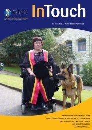
Winter 2013 In Touch - Muscular Dystrophy Association of New ...
Winter 2013 In Touch - Muscular Dystrophy Association of New ...
Winter 2013 In Touch - Muscular Dystrophy Association of New ...

Create successful ePaper yourself
Turn your PDF publications into a flip-book with our unique Google optimized e-Paper software.
Your condition in review<br />
detect the absence <strong>of</strong> essential mitochondrial<br />
enzymes in the muscle. It’s also possible<br />
to extract mitochondrial proteins from the<br />
muscle and measure their activity.<br />
Scans and electrophysiology testing<br />
<strong>In</strong> addition to the muscle biopsy, noninvasive<br />
techniques can be used to examine<br />
muscle without taking a tissue sample.<br />
For instance, a technique called muscle<br />
phosphorus magnetic resonance spectroscopy<br />
(MRS) can measure levels <strong>of</strong> phosphocreatine<br />
and ATP (which are <strong>of</strong>ten depleted in muscles<br />
affected by mitochondrial disease). CT scans<br />
and MRI scans can be used to visually inspect<br />
the brain for signs <strong>of</strong> damage, and surface<br />
electrodes placed on the scalp can be used to<br />
produce a record <strong>of</strong> the brain’s activity called<br />
an electroencephalogram (EEG).<br />
Similar techniques might be used to<br />
examine the functions <strong>of</strong> other organs<br />
and tissues in the body. For example, an<br />
electrocardiogram (EKG) can monitor the<br />
heart’s activity, and a blood test can detect<br />
signs <strong>of</strong> kidney malfunction.<br />
Genetic testing<br />
A genetic test can determine whether<br />
someone has a genetic mutation that causes<br />
mitochondrial disease. Ideally, the test is done<br />
using genetic material extracted from blood<br />
or from a muscle biopsy. It’s important to<br />
realise that, although a positive test result can<br />
confirm diagnosis, a negative test result isn’t<br />
necessarily meaningful.<br />
Special issues in mitochondrial<br />
myopathies<br />
While there is no cure for these<br />
mitochondrial myopathies there are many<br />
treatments that can help manage symptoms<br />
Ataxia and mobility issues<br />
Often, mitochondrial encephalomyopathy<br />
causes ataxia, or trouble with balance and<br />
coordination. People with ataxia are usually<br />
prone to falls. Sometimes, people with<br />
mitochondrial myopathies experience loss <strong>of</strong><br />
muscle strength in the arms or legs. These<br />
problems can be partially avoided through<br />
physical and occupational therapy, and the<br />
use <strong>of</strong> supportive aids such as railings, a<br />
walker, a cane, braces, or — in severe cases —<br />
a wheelchair.<br />
Respiratory care<br />
Sometimes, these diseases can cause<br />
significant weakness in the muscles that<br />
support breathing. A person with mild<br />
respiratory problems might require occasional<br />
respiratory support, such as pressurised air,<br />
while someone with more severe problems<br />
might require permanent support from<br />
a ventilator. Those with mitochondrial<br />
disorders should watch for signs <strong>of</strong> respiratory<br />
insufficiency (such as shortness <strong>of</strong> breath or<br />
morning headaches), and have their breathing<br />
checked regularly by a specialist.<br />
Speech and swallowing<br />
Muscle wasting in the neck region can<br />
lead to slurred speech and difficulty with<br />
swallowing. <strong>In</strong> these instances, speech<br />
therapy or changing the diet to easier-toswallow<br />
foods can be useful.<br />
Cardiac care<br />
Sometimes, mitochondrial diseases<br />
directly affect the heart. <strong>In</strong> these cases, the<br />
usual cause is an interruption in the rhythmic<br />
beating <strong>of</strong> the heart, called a conduction<br />
block. Though dangerous, this condition is<br />
treatable with a pacemaker, which stimulates<br />
normal beating <strong>of</strong> the heart. Cardiac<br />
muscle damage also may occur. People with<br />
mitochondrial disorders may need to have<br />
regular examinations by a cardiologist.<br />
Renal care<br />
Some people with mitochondrial disease<br />
experience serious kidney problems,<br />
gastrointestinal problems and/or diabetes.<br />
Some <strong>of</strong> these problems are direct effects <strong>of</strong><br />
mitochondrial defects in the kidneys, digestive<br />
system or pancreas (in diabetes), and others<br />
are indirect effects <strong>of</strong> mitochondrial defects in<br />
other tissues.<br />
Special issues in children<br />
Vision: Though PEO and ptosis typically<br />
cause only mild visual impairment in adults,<br />
they’re potentially more harmful in children<br />
with mitochondrial myopathies.<br />
Because the development <strong>of</strong> the brain<br />
is sensitive to childhood experiences, PEO<br />
or ptosis during childhood can sometimes<br />
cause permanent damage to the brain’s visual<br />
system. For this reason, it’s important for<br />
children with signs <strong>of</strong> PEO or ptosis to have<br />
their vision checked by a specialist.<br />
Developmental delays: Due to<br />
muscle weakness, brain abnormalities<br />
or a combination <strong>of</strong> both, children with<br />
mitochondrial diseases may have difficulty<br />
developing certain skills. For example, they<br />
might take an unusually long time to reach<br />
motor milestones such as sitting, crawling<br />
and walking. As they get older, they may<br />
be unable to get around as easily as other<br />
children their age, and may have speech<br />
problems and/or learning disabilities. Children<br />
who are severely affected by these problems<br />
may benefit from services. Occupational<br />
therapy is important for children with<br />
mitochondrial myopathies. Mitochondrial<br />
myopathy can lead to respiratory problems<br />
that require support from a ventilator.<br />
Research<br />
While there is no cure for these<br />
conditions scientists continue to make<br />
significant progress in their quest to fully<br />
understand mitochondrial diseases and<br />
identify the genetic mutations responsible<br />
for these conditions.<br />
Research into stem cells therapy for<br />
affected individuals to restore normal<br />
metabolic conditions and halt damage<br />
to the mitochondria is ongoing as is the<br />
use <strong>of</strong> dietary supplements. These dietary<br />
supplements based on three natural<br />
substances involved in ATP production in<br />
our cells. Although they don’t work for<br />
everyone, they do appear to help some<br />
people. One substance, creatine, normally<br />
acts as a reserve for ATP by forming a<br />
compound called creatine phosphate. When<br />
a cell’s demand for ATP exceeds the amount<br />
its mitochondria can produce, creatine can<br />
release phosphate (the “P” in ATP) to rapidly<br />
enhance the ATP supply. <strong>In</strong> fact, creatine<br />
in touch // <strong>Winter</strong> <strong>2013</strong> // PAGE 22