Fastin ELASTIN Assay Manual - Nordic Biosite
Fastin ELASTIN Assay Manual - Nordic Biosite
Fastin ELASTIN Assay Manual - Nordic Biosite

Create successful ePaper yourself
Turn your PDF publications into a flip-book with our unique Google optimized e-Paper software.
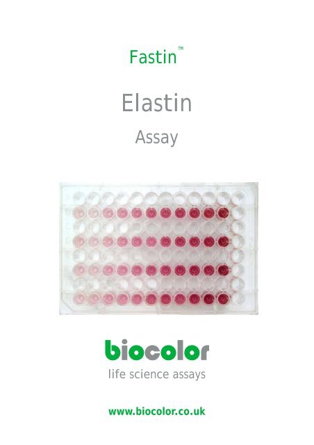
<strong>Fastin</strong> <br />
Elastin<br />
<strong>Assay</strong><br />
biocolor<br />
life science assays<br />
www.biocolor.co.uk
[1] precipitated elastin (15 minutes),<br />
then centrifuged and supernatant<br />
removed<br />
[2] elastin-dye complex after<br />
centrifuging and removing unbound<br />
dye<br />
[3] recovery of elastin bound dye, after<br />
adding Dye Dissociation Reagent<br />
Biocolor Ltd.<br />
Unit 35<br />
8 Meadowbank Road<br />
BT38 8YF<br />
UK<br />
tel: +44 (0) 2893 350 258<br />
fax: +44 (0) 2893 369 716<br />
email: info@biocolor.co.uk
<strong>Fastin</strong> Elastin <strong>Assay</strong><br />
Time Req: 4 hours<br />
Detection Limit: 5μg, (10μg/ml)<br />
0<br />
mins<br />
30<br />
mins<br />
Set up <strong>Assay</strong>:<br />
Label a set of 1.5 ml microcentrifuge tubes. If sufficient<br />
test material is available run duplicate samples.<br />
[1] Prepare Reagent Blanks: 100μl of test solution<br />
solvent, (buffer / PBS / water / 0.25M oxalic acid).<br />
[2] a-elastin standard; (suggested, 12.5, 25.0 and 50.0<br />
μl duplicate aliquots).<br />
[3] Test samples; (in vivo derived tissue extracts or in<br />
vitro cell culture medium samples). Select aliquot<br />
volumes between 10 μl and 500 μl, (to contain 5<br />
to 70 μg elastin).<br />
[4] To each tube add an equal volume of Elastin<br />
Precipitating Reagent.<br />
[5] Cap tubes and briefly vortex to mix contents;<br />
leave for 10 minutes to complete precipitation of<br />
elastin.<br />
[6] Centrifuge tubes @ >10,000 x g for 10 minutes.<br />
[7] Drain tube's liquid contents into a beaker. While<br />
the tube is still inverted remove most of the<br />
remaining fluid from the tube by tapping the<br />
inverted tube onto a single thickness absorbent<br />
paper towel.<br />
Formation of Elastin - Dye Complex:<br />
[1] To all tubes add 1.00 ml of Dye Reagent.<br />
Cap tubes and initially mix contents by inverting<br />
the tubes.<br />
Next disperse the elastin precipitate using a vortex<br />
mixer.<br />
Then place the rack of tubes on a microplate<br />
shaker unit.<br />
Allow the reaction between the elastin and the dye<br />
to proceed for 90 minutes.<br />
[9] Centrifuge the tubes @ >10,000 x g for 10 minutes.<br />
CONTINUED ON INSIDE BACK COVER OF MANUAL
140<br />
mins<br />
Recovery of elastin-dye complex:<br />
[10] Drain the tubes of unbound dye. While the tube is<br />
still inverted remove most of the remaining fluid<br />
from the tube by FIRMLY tapping the lip of the tube<br />
onto a single thickness of an absorbent paper towel.<br />
A 'cotton bud', (or Q-tip), can be useful in removing<br />
any fluid droplets from the rim of the tube. On<br />
returning the tube to the upright position, not more<br />
than 25μl of fluid should be found in the bottom of<br />
the tube.<br />
[11] The elastin-dye complex can be observed as a<br />
reddish-brown deposit in the bottom and inside lower<br />
wall of the tube.<br />
180<br />
mins<br />
Release, and recovery, of the elastin bound dye:<br />
[12] To each tube add 250 μl of Dye Dissociation Reagent.<br />
Cap tubes and release the dye into solution with the<br />
aid of a vortex mixer. Repeat the vortex mixing<br />
after 10 minutes so as to ensure all bound dye has<br />
passed into solution.<br />
[13] Transfer the contents of each tube to a well in a<br />
ninety-six well flat bottom microwell plate. Ensure<br />
that a map has been prepared in the lab notebook to<br />
record which tube contents went into which well.<br />
Elastin measurement; (dye recovered):<br />
[14] Place microwell plate into the Microplate Reader.<br />
Select wavelength or colour filter nearest to 513 nm,<br />
(blue-green colour).<br />
Plot Reference Standards, (including Reagent<br />
Blanks), graph where both Absorbance and Elastin<br />
Concentration are known.<br />
Use this graph to determine or calculate the elastin<br />
content of the Test Samples.<br />
240<br />
mins<br />
Notes:<br />
(a) Aim to achieve duplicates that do not exceed +10% of<br />
their mean absorbance value.<br />
(b) Comfort or coffee breaks can be obtained between<br />
Steps 7 & 8, during Step 8 and between Steps 11 &<br />
12.<br />
(c) Like all dyes TPPS is photo-labile. Protect tubes/<br />
microwell plate from direct sunlight or from strong<br />
overhead lighting.<br />
PLEASE READ THE MANUAL BEFORE USING THE ASSAY
<strong>Fastin</strong> <br />
<strong>ELASTIN</strong><br />
<strong>Assay</strong><br />
TECHNICAL INFORMATION<br />
Contents<br />
Test material suitable for analysis 3<br />
<strong>Assay</strong> components and storage conditions required 3<br />
Mode of action of the <strong>Fastin</strong> <strong>Assay</strong> 5<br />
Elastin <strong>Assay</strong> : 7<br />
Soluble elastin 8<br />
Insoluble elastin 12<br />
Elastin History & Information: Source References: 15<br />
1
The <strong>Fastin</strong> <strong>Assay</strong> has been designed<br />
for in vitro research work only<br />
Handle the<br />
<strong>Fastin</strong> <strong>Assay</strong> Kit<br />
using<br />
GOOD LABORATORY PRACTICE<br />
Read <strong>Manual</strong> Before Use<br />
<strong>Fastin</strong> <strong>Manual</strong><br />
Not to be reproduced in part or in whole, without written permission,<br />
unless required for personal, non‐commercial use.<br />
©Biocolor Ltd., 2007<br />
<strong>Fastin</strong> is a Trademark of Biocolor Ltd.<br />
Published by<br />
Biocolor Ltd.<br />
8 Meadowbank Road, Carrickfergus, BT38 8YF<br />
Northern Ireland.<br />
www.biocolor.co.uk<br />
6th Edition 2007<br />
2
Intended Applications:<br />
<strong>Fastin</strong> <strong>Assay</strong><br />
<strong>Manual</strong><br />
The <strong>Fastin</strong> Elastin <strong>Assay</strong> is a quantitative dye‐binding method for the analysis of elastins<br />
extracted from biological materials.<br />
The dye label employed is 5, 10, 15, 20‐tetraphenyl‐21, 23‐porphine tetra‐sulfonate (TPPS).<br />
For the structural form of the dye see Fig. 1.<br />
Test sample material:<br />
Tissue extracts and cell culture medium.<br />
Elastin forms that can be measured by the <strong>Fastin</strong> <strong>Assay</strong>:<br />
[i] soluble tropoelastins<br />
[ii] lathyrogenic elastins<br />
[iii] insoluble elastins, following solubilization to elastin polypeptides, [α‐elastin; κ‐elastin]<br />
The dye reagent binds to the 'basic' and 'non‐polar' amino acid sequences found in mammalian<br />
elastins.<br />
Due to the difficultly of obtaining sufficient quantities of tropoelastin the assay development<br />
was carried out using α‐elastin.<br />
Test sample quantities:<br />
A sample volume of between 5 and 500 μl is required, containing not less than 5 µg and not<br />
more than 70 µg elastin.<br />
Samples with elastin of >70 µg/10 μl should be diluted with water, dilute buffer or PBS.<br />
Samples with elastin of
Recommended storage conditions for <strong>Assay</strong> Kit components:<br />
Unopened;<br />
All of the reagents have long term stability (at least 6 months), when stored at room temperature.<br />
Do not freeze as complete re‐solubilisation may not occur on thawing.<br />
Avoid exposure of the <strong>Fastin</strong> Dye Reagent to direct sunlight.<br />
Opened;<br />
Reference Standard: When stored at +4ºC the α‐elastin standard is a clear transparent solution.<br />
On holding at room temperature the solution may be observed to become opalescent.<br />
This is due to the characteristic coacervation property of soluble elastin. On cooling, the<br />
process is reversible and the elastin solution again becomes transparent.<br />
The full metal seal should not be removed from the vial; the contents can be sampled as follows:<br />
[1] Remove the centre metal disc only from the vial top.<br />
[2] Obtain aliquots from the vial by using a syringe fitted with a sterile hypodermic needle.<br />
The butyl rubber seal on the vial has a thin centre disc.<br />
[3] Do not return any unused aliquots to the vial.<br />
[4] The α‐elastin standard should be discarded if the solution becomes turbid.<br />
<strong>Fastin</strong> Dye Reagent:<br />
The pH of this reagent is pH 7.5 and the dye label TPPS. To limit microbial growth, inhibitors<br />
have been added to the reagent. These agents, bromopol and sorbic acid, are compatible<br />
with the <strong>Fastin</strong> <strong>Assay</strong>; but are not 'universal' microbial inhibitors. Good laboratory practice<br />
and the storage of an opened bottle of <strong>Fastin</strong> Dye Reagent storage at 4ºC can extend the shelf<br />
life of the reagent. DO NOT FREEZE.<br />
Other components required, but not supplied:<br />
Capped 1.5 ml micro‐centrifuge tubes and a set of variable volume micro‐pipettors, with<br />
matching pipette tips.<br />
For the microcentrifuge tubes a vortex mixer, to dislodge the protein pellet and then a mechanical<br />
shaker to provide gentle mixing of the elastin and the TPPS of the tubes.<br />
A centrifuge, fitted with a 1.5 ml micro tube rotor head and capable of 10,000 x g.<br />
A Microplate Reader, with a suitable colour filter (absorbance peak of TPPS occurs at 513<br />
nm). See page 10 for further information on filter selection.<br />
Magnetic Stirrer, with hotplate. A 500 ml glass beaker containing ~300 ml water and a spinning<br />
stir bar, (to prevent water bumping at 100 0 C), is convenient set‐up for extracting elastin<br />
from tissue samples, (see page 12, section [b])<br />
4
Mode of action of the <strong>Fastin</strong> dye reagent with elastins:<br />
The <strong>Fastin</strong> Dye Reagent contains a synthetic porphrin, 5,10,15,20‐tetraphenyl‐21,23‐<br />
porphrine, that is water soluble in the sulfonate form. The TPPS molecules contain four sulfate<br />
groups.<br />
Fig. 1<br />
The visible absorbance spectrum and structural form of 5,10,15,20tetraphenyl21,23<br />
porphrine, tetrasulfonate<br />
O<br />
S O<br />
O - O<br />
S<br />
O<br />
O - O<br />
N<br />
N<br />
H<br />
H<br />
N<br />
N<br />
O<br />
S<br />
O -<br />
O<br />
S<br />
O O-<br />
The affinity of TPPS for elastin was first observed when used as a 'vital stain' on live animals.<br />
Most tissues initially took up the dye, but with time only elastin retained the TPPS molecules.<br />
[Winkelman, J. (1962), Cancer Res. 22, 589‐596; Winkelman, J & Spicer, S. (1962), Stain Technol.<br />
37, 303‐305].<br />
The mode of action of TPPS with elastin remains uncertain. It may be due to shape‐and‐fit<br />
with the acidic dye being firmly retained by the basic amino acid side chain residues of<br />
elastin.<br />
At pH 7.0 and 20ºC, TPPS has been reported to occur as a dimer, producing an out‐of‐plane<br />
type conformational change [Schneider, W. (1975) Struct. Bond. 23, 123].<br />
5
Elastin <strong>Assay</strong><br />
MEASUREMENT OF SOLUBLE <strong>ELASTIN</strong><br />
____________________________________________________________________<br />
Set up assay:<br />
To duplicate, labeled 1.5 ml microcentrifuge tubes add between 5 and 500 µl of test samples,<br />
elastin standards and reagent blanks.<br />
____________________________________________________________________<br />
Working standards:<br />
It is recommended that with each <strong>Fastin</strong> <strong>Assay</strong> Kit, the Elastin Standard is initially run in<br />
duplicate at four concentrations; (0, 12.5, 25 and 50µl aliquots).<br />
The standards along with the reagent blank can then be used to produce a straight line calibration<br />
curve from a selected Microplate Reader.<br />
In subsequent assay batches, a minimum calibration requirement is duplicate 25 µl aliquots<br />
of the elastin standard and the reagent blank. This secondary standard should give absorption<br />
values to within +5% of that defined by the standard curve.<br />
When using a different measuring system, or following instrument servicing, a new standard<br />
curve should be prepared.<br />
___________________________________________________________________<br />
Test samples:<br />
With test samples where the approximate elastin concentrations are as yet unknown, initially<br />
try single 50µl aliquots for the first trial .<br />
If the absorbance readings are found to be >1.0, (after subtraction of the reagent blank<br />
value), repeat assay using a smaller test sample aliquot.<br />
If in initial trial aliquots produced absorbance values of
START ASSAY:<br />
_______________________________________________________________________<br />
Elastin isolation:<br />
The Precipitating Reagent has been developed to perform this elastin recovery; the reagent<br />
should not be diluted with sample volumes greater than in a ratio of 1:1<br />
The reagent can be pre‐cooled to 10,000 x g for 10 minutes, to pack the precipitated elastin.<br />
[4] Remove tubes from the centrifuge, uncap and carefully invert, to drain the liquid contents<br />
into a waste beaker. While inverted remove any remaining fluid from the top of<br />
the tubes by tapping the tube onto an absorbent paper towel.<br />
A 'cotton bud',(or Q‐tip), can be useful in removing any fluid droplets from the rim of<br />
the tube.<br />
On returning the tube to the upright position, not more than 25μl of fluid should be found in<br />
the bottom of the tube.<br />
Low concentrations of elastin can be difficult to 'see' as it occurs as a translucent gel.<br />
Photographs on the outside back cover of this manual:<br />
The top photograph required 250 μg of aelastin to visually display the protein pellet.<br />
The middle and bottom photographs were obtained using 50 μg aelastin samples.<br />
7
Reaction of the elastin with the <strong>Fastin</strong> dye:<br />
____________________________________________________________________<br />
[5] Add 1.0ml of the <strong>Fastin</strong> Dye Reagent to each tube.<br />
[6] Cap the tubes and use a vortex mixer to bring the elastin gel precipitate into<br />
solutionwith the Dye Reagent.<br />
During centrifugation (>10,000 x g) the precipitate will have been compacted and<br />
may require 2 or 3 short periods with the vortex mixer to bring the elastin into<br />
solution.<br />
Lower g forces (10,000 x g for 10 minutes).<br />
[9] The tubes are uncapped and the supernatants discarded. Any remaining fluid is<br />
removed by firm tapping of the inverted tubes onto a paper towel.<br />
While inverted remove any remaining fluid from the top of the tubes by tapping the<br />
tube onto an absorbent paper towel. While the tube is still inverted remove most of<br />
the remaining fluid from the tube by FIRMLY tapping the lip of the tube onto a single<br />
thickness of an absorbent paper towel. A 'cotton bud', (or Q‐tip), can be useful in<br />
removing any fluid droplets from the rim of the tube.<br />
Visual inspection should reveal a red residue within the elastin standard tubes and,<br />
hopefully also in the test sample tubes.<br />
_______________________________________________________________________<br />
8
Release of the elastin bound dye:<br />
[10] To each tube add 250 ul of Dye Dissociation Reagent.<br />
Cap the tubes and bring the elastin‐bound dye into solution using a vortex mixer.<br />
Two brief mixing periods are usually more effective than one long mixing period.<br />
Tubes should not be uncapped until transfer to the wells of a 96‐well microplate for<br />
absorbance measurement.<br />
The dye extract is stable for several hours, but if readings are to be delayed store the tube<br />
rack containing the microcentrifuge tubes in a light‐proof container or cupboard.<br />
____________________________________________________________________<br />
Elastin measurement:<br />
The elastin content of the assayed samples is determined by the amount of bound dye<br />
released from the elastin.<br />
Transfer the complete 250 μl contents of the labeled micro‐centrifuge tubes to wells of a<br />
96 well microplate, (with flat‐bottom wells to reduce light scatter effects).<br />
The absorbance peak of TPPS in the Dye Dissociation Reagent occurs at 513 nm.<br />
Although the instrument can be set to zero using the reagent blank, it is usually better to<br />
determine the absorbance of the blank as a quality control check of the assay.<br />
Duplicates (blanks, standards and test samples) should not exceed + 10%f duplicate means.<br />
With practice the analyst can usually obtain this after processing one or two batches of<br />
assays.<br />
When any set of duplicate test sample readings exceed +10% of their mean value the sample<br />
should be checked for clarity and re‐assayed.<br />
Check the colour filter options that are available for the Microplate Reader; a blue green<br />
filter will probably be found to be suitable.<br />
Initially also try the filters on either side of the selected filter; read the blank and standards<br />
with each of the three filters.<br />
Plot standard curves for each of the filters and select the optimum curve based on that which<br />
gave the highest absorbance readings of the standards, and produced a straight line fit for the<br />
standard readings.<br />
For further information on colour filter selection see Fig. 3 (page 14)<br />
____________________________________________________________________<br />
9
Elastin concentration in Test Samples:<br />
[1] The duplicate absorbance readings of the reagent blank and the three, or more, sets<br />
of elastin standards are used to produce a standard curve.<br />
[2] Subtract the reagent blank mean absorbance value from the absorbance means of<br />
each duplicate set of elastin standards and test samples.<br />
[3] Plot the elastin standard values; vertical axis 'Absorbance' and horizontal axis;<br />
'elastin concentration; (µg)'.<br />
[4] Obtain the best fit line through these plotted points, [Fig. 2].<br />
The line should pass through zero (0.0 absorbance, 0 µg elastin).<br />
Do not extend the line beyond the highest plotted point.<br />
The plot points should be within +5% (absorbance) of the fitted line.<br />
[5] The concentration of elastin present in the test samples can now be determined.<br />
Take the mean of each duplicate test sample after subtracting the reagent blank<br />
value.<br />
The equivalent elastin concentration for the absorbance value obtained is then read<br />
from the standard curve.<br />
[6] The quantity of elastin found in the aliquot taken for analysis can then be multiplied<br />
to give the elastin concentration present, either in 100 µl sample or 1ml test sample.<br />
[7] As the total extract volume has been logged, (of the pooled extracts), from a known<br />
weight of tissue, (wet or dry milligrams). It is possible to express the elastin concentration<br />
as; ‘μg elastin per mg tissue’<br />
____________________________________________________________________<br />
10
Fig. 2.<br />
Elastin Standard Curve;<br />
Microplate Reader 96 well format, 250 μl/well.<br />
For further information see Fig. 3.<br />
11
MEASUREMENT OF INSOLUBLE <strong>ELASTIN</strong><br />
The <strong>Fastin</strong> <strong>Assay</strong> can be used to measure insoluble cross‐linked elastin. Insoluble elastin is<br />
extracted from tissue in the form of soluble cross‐linked polypeptide elastin fragments.<br />
Insoluble elastin can be converted into either soluble α‐elastin or κ‐elastin. After extraction<br />
the samples can be assayed by the procedure described for soluble elastin.<br />
Conversion of insoluble elastin to water soluble α‐elastin<br />
[a] Tissue samples are weighed (decide if the elastin content is to be expressed as wet<br />
weight or dry weight, (μg/mg), of tissue).<br />
Cut tissue into small fragments for extraction.<br />
Place the weighed samples into centrifuge tubes (~10 x 2 cm) and add ~20 volumes<br />
of 0.25 M oxalic acid (assuming tissue density is 1.00 gm/cm3).<br />
[b] The tubes are then placed into a boiling water‐bath (or a metal heating block with the<br />
thermostat set at 100ºC), for 60 minutes. Do not tighten tube caps.<br />
[c] Remove the tubes from the heat and cool to room temperature.<br />
Centrifuge at ~3000 rpm for 10 minutes.<br />
Pipette off the liquid and retain this extract in labeled containers.<br />
[d] To the residual tissue in the tubes add a further 20 volumes of 0.25 M oxalic acid and<br />
again heat for 60 min. Up to three heat extractions should be initially used to check<br />
that complete solubilisation of the tissue elastin has occurred<br />
[e] Some tissue material, such as that from foetal or immature animals, can be solubilised<br />
after one or two extractions. Tissue from mature or old animals, including aged human<br />
tissues, may require up to three extractions.<br />
[f] Initially when using new test material retain each of the oxalic acid extracts separately<br />
and analyze each for elastin; to establish that elastin extraction was quantitative.<br />
The last extract should contain no elastin.<br />
Elastin extracted from mature tissue, (elderly adult human tissue), will often produce<br />
yellow coloured extracts.<br />
[g] The tissue elastin in the form of α‐elastin which has an average molecular weight of<br />
60‐84 KDa. The extract can now be directly assayed using the procedure described<br />
for soluble elastins.<br />
Retain a record of the extract volumes to permit calculation of the tissue elastin content.<br />
12
Conversion of insoluble elastin to soluble κelastin:<br />
In this method, insoluble elastin is solubilised using ethanolic potassium hydroxide, (1 M<br />
KOH in 80% ethanol)<br />
The elastin solubilisation reaction is carried out at 37ºC for two hours only. The excess alkali<br />
in the pooled extracts is then removed by dialysis against water.<br />
The solubilised elastin recovered has been termed Kappa‐elastin (κ‐elastin). This high<br />
molecular weight fraction, (< 50KDa), is composed of elastin polypeptides that have ‘similar’<br />
properties to that of α‐elastin.<br />
Further details on how to prepare κ‐elastin can be found in the following reference Moczar,<br />
M., Moczar, E. & Robert, L. (1980), Frontiers Matrix Biol. 8, 174.<br />
Tropoelastin, the native monomer form of elastin:<br />
Tropoelastin, as exported from mammalian cells, has a molecular weight between 62 – 72<br />
KDa.<br />
This is the elastin form that maybe found in cell culture medium during invitro cell culture.<br />
Tropoelastin avidly binds to a co‐exported microfibril glycoprotein to form ‘elastic matrix’.<br />
This associated glycoprotein is therefore a common component of many tropoelastin<br />
preparations.<br />
13
Fig. 3.<br />
Microplate Reader colour filter selection<br />
The graph displays the results obtained, using two batches of Elastin Standard. These<br />
reference solution were used to produce the ‘standard curves’ plotted in the figure.<br />
Each sets of ‘standards’ were run in duplicate and the ‘Absorbance’ measured using two<br />
different Microplate Readers;<br />
[1] Awareness Technology Inc., model Multi‐well Reader II: Filter selected; 545 nm<br />
[2] Bio‐Tek Instruments Inc., Model EL 311: Filter selected; 550 nm.<br />
The [1] data has been plotted using white symbols.<br />
For [2] data was plotted using black symbols.<br />
The ‘Reagent blank’ mean absorbance value was 0.385 for [1] and 0.162 for [2], when these<br />
values were subtracted from the ‘standard readings’ the blank corrected absorbance values<br />
of the elastin standards were similar for both Microplate Readers.<br />
The photograph of the front cover of the manual is a visual image of the microwell plate<br />
that was used to produce this graph.<br />
14
<strong>ELASTIN</strong> SOURCE REFERENCES<br />
Biochemistry, biophysics; preparation, extraction & analysis<br />
Cunningham, L.W. & Frederiksen, D.W. [1982], Eds of 'Elastin Structure and Biosynthesis'.<br />
Methods of Enzymology, 82, 559‐743.<br />
Kreis, T & Vale, R. [1993] Eds of 'Guidebook to the Extracellular Matrix and Adhesion Proteins'.<br />
Oxford University Press, Oxford.<br />
Mecham, R.P. [1986] Ed of 'Regulation of Matrix Accumulation'. Academic Press, New York.<br />
Robert, L. & Hornebeck, W. [1989], Eds of 'Elastin and Elastases' in two volumes. CRC<br />
Press Inc. Boca Raton, Florida.<br />
Royce, P.M. & Steinmann, B.U. [1993] Eds of 'Connective tissue and its heritable disorders;<br />
molecular, genetic and medical aspects'. Wiley‐Liss, New York.<br />
Sandberg, L.B., Gray, W.R. & Franzblau, C. [1977] Eds of 'Elastin and Elastic Tissue',<br />
(Advances Exper. Med. Biol. Series, Vol 79), Plenum Press, ,New York.<br />
Uitto, J., & Perejda, A. J. [1987], Eds of 'Connective Tissue Disease. Molecular Pathology of the<br />
Extracellular Matrix'. Marcel Dekker, New York.<br />
15
Other <strong>Assay</strong>s available from Biocolor<br />
APOPercentage Apoptosis <strong>Assay</strong><br />
For mammalian cells that have a conventional phospholipid composition membrane. <strong>Assay</strong><br />
is not recommended for non‐mammalian cells and is not suitable for neural cells. Requires<br />
an inverted microscope, (magn. x 100), for detection and a microplate reader for measurement.<br />
Digital microphotograph analysis option using Adobe Photoshop.<br />
<strong>Assay</strong> sensitivity; single apoptotic cell<br />
<strong>Assay</strong> run time; 1 hour<br />
Sircol Soluble Collagen <strong>Assay</strong><br />
Suitable for monitoring collagen production during invitro cell culture or of recent invivo<br />
synthesis from tissue samples; such as wound healing and skin grafts research. Fibrillar collagens,<br />
[type I, II, III, V & XI] and basement membrane collagen, [type IV]. assay for acidsoluble,<br />
(cold 0.5 M acetic acid), mammalian collagens.<br />
<strong>Assay</strong> sensitivity; 2.5ug<br />
<strong>Assay</strong> run time; 1 hour<br />
Blyscan Sulfated Glycosaminoglycans <strong>Assay</strong><br />
For analysis of sulfated glycosaminoglycan components of proteoglycans including decorin,<br />
biglycan, fibromodulin, aggrecan, syndecan, betaglycan. Galactosaminoglycans: chondroitin<br />
sulfates, dermatan sulfate. Glucosaminoglycans: heparan sulfate, heparin, keratan sulfate.<br />
Test material; cell culture medium, amniotic fluid, urine, synovial fluid and tissue extracts.<br />
<strong>Assay</strong> sensitivity; 0.5ug<br />
<strong>Assay</strong> run time; 1 hour<br />
FOR FURTHER INFORMATION AND UPDATES VISIT OUR WEBSITE<br />
www.biocolor.co.uk<br />
16