

Spontaneous Calf Hematoma - The Podiatry Institute
Spontaneous Calf Hematoma - The Podiatry Institute
Spontaneous Calf Hematoma - The Podiatry Institute

You also want an ePaper? Increase the reach of your titles
YUMPU automatically turns print PDFs into web optimized ePapers that Google loves.
258 CHAPTER +5<br />
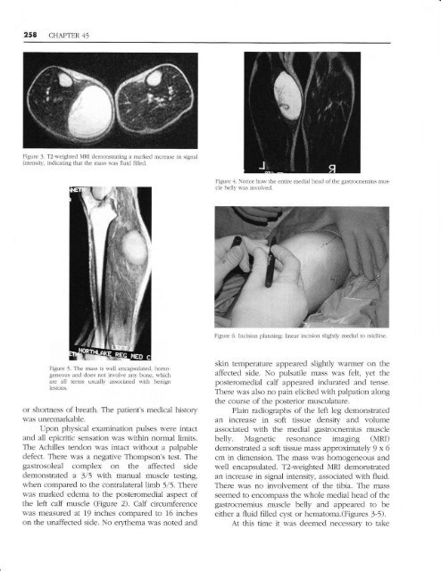
Figure J. T2-r,eighted NIRI demonstrating a nrarkt:cl incre:Lse in signai<br />
intensit),. indicating that the mass r\ras fluicl filled.<br />
Figure 4. Notice horl. the entire meclial heacl of the gastrocnemius nrus<br />
cle 1te111. rras invohed.<br />
F-igure 6. hrcision planning: lineal incision slightlv medial to rnidline.<br />
Figure 5. <strong>The</strong> mass is well encapsulated, homo<br />
geneolrs and does not involve any bone. n-hiclt<br />
are all terms usuall1. associated l,'ith benign<br />
iesions.<br />
or shofiness of breath. <strong>The</strong> patient's medical history<br />
was unremarkable.<br />
Upon physical examination pulses were intact<br />
and al1 epicritic sensation was within normal limits.<br />
<strong>The</strong> Achilles tendon was intact without a palpable<br />
defect. <strong>The</strong>re was a negative Thompson's test. <strong>The</strong><br />
gastrosoleal complex on the affected side<br />
demonstrated a 3/5 with manual muscle testing,<br />
when compared to the contralateral limb 5/5. <strong>The</strong>re<br />
was marked edema to the posteromedial aspect of<br />
the left calf muscle (Figure 2). <strong>Calf</strong> circumference<br />
was measufed at 19 inches compafed to 16 inches<br />
on the unaffected side. No erythema was noted and<br />
skin temperature appeared slightly warmer on the<br />
affected side. No pulsatile mass was felt, yet the<br />
posteromedial calf appeared indurated and tense.<br />
<strong>The</strong>re was also no pain elicited with palpation along<br />
the course of the posterior musculature.<br />
Plain radiographs of the left 1eg demonstrated<br />
an increase in soft tissue density and volume<br />
associated with the medial gastrocnemius muscle<br />
be1ly. Magnetic resonance imaging (MRI)<br />
demonstrated a soft tissue mass approximately 9 x 6<br />
cm in dimension. <strong>The</strong> mass was homogeneous and<br />
well encapsulated. T2-weighted MRI demonstrated<br />
an increase in signal intensity, associated nith fluid.<br />
<strong>The</strong>re was no involvement of the tibia. <strong>The</strong> mass<br />
seemed to encompass the whole medial head of the<br />
gastrocnemius muscle be1ly and appeared to be<br />
either a fluid filled cyst or hematom2.(Figures 3-5).<br />
At this time it w-as deemed necessary to take


