

closure of deep fascia in ankle fracture surgery - The Podiatry Institute
closure of deep fascia in ankle fracture surgery - The Podiatry Institute
closure of deep fascia in ankle fracture surgery - The Podiatry Institute

You also want an ePaper? Increase the reach of your titles
YUMPU automatically turns print PDFs into web optimized ePapers that Google loves.
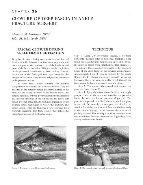
C H A P T E R 2 6CLOSURE OF DEEP FASCIA IN ANKLEFRACTURE SURGERYMeagan M. Jenn<strong>in</strong>gs, DPMJohn M. Schuberth, DPMFASCIAL CLOSURE DURINGANKLE FRACTURE FIXATIONDeep <strong>fascia</strong>l <strong>closure</strong> dur<strong>in</strong>g open reduction and <strong>in</strong>ternalfixation <strong>of</strong> <strong>ankle</strong> <strong>fracture</strong>s is an important step <strong>in</strong> the s<strong>of</strong>ttissue reapproximation and coverage <strong>of</strong> the hardware andbone <strong>of</strong> the lateral malleolus. This process also capitalizeson the periosteal contribution to bone heal<strong>in</strong>g. Further,restoration <strong>of</strong> the fascio-periosteal layer ma<strong>in</strong>ta<strong>in</strong>s the<strong>in</strong>tegrity <strong>of</strong> the lateral compartment and prevents herniation<strong>of</strong> the peroneal muscles.<strong>The</strong> <strong>deep</strong> <strong>fascia</strong>l fibers cover<strong>in</strong>g the anteriorcompartment are oriented <strong>in</strong> a transverse fashion. <strong>The</strong>y areattached to the anterior border and lateral surface <strong>of</strong> thefibula and are usually disrupted by the <strong>fracture</strong> trauma, thesurgical exposure, or both. Even with meticulous dissectionand m<strong>in</strong>imal stripp<strong>in</strong>g <strong>of</strong> the s<strong>of</strong>t tissues, the lateral s<strong>of</strong>ttissues are <strong>of</strong>ten shredded. As such, it is impractical to usestandard suture techniques to restore this anatomy. <strong>The</strong>senior author (JMS) has developed a new technique for arout<strong>in</strong>ely successful <strong>deep</strong> <strong>fascia</strong> <strong>closure</strong> that utilizes thenatural arrangement <strong>of</strong> the local anatomy.TECHNIQUEStep 1. Us<strong>in</strong>g 2-0 absorbable sutures, a modifiedhorizontal mattress stitch is fashioned. Start<strong>in</strong>g on the<strong>fascia</strong>l-periosteal flap from the posterior aspect <strong>of</strong> the fibula,the suture is passed from superficial to <strong>deep</strong> (Figure 1).<strong>The</strong> suture is then placed perpendicular to the transversefibers <strong>of</strong> the <strong>deep</strong> <strong>fascia</strong> <strong>of</strong> the anterior compartment.Approximately 1-cm <strong>of</strong> tissue is captured by the needle(Figure 2). By plac<strong>in</strong>g the suture vertically across thehorizontal fibers, the suture is unable to pull through the<strong>fascia</strong> unless the <strong>fascia</strong> is separated from the fibula.Step 2. <strong>The</strong> suture is then passed back through theposterior <strong>fascia</strong> (Figure 3).Step 3. Ty<strong>in</strong>g the suture allows the surgeon to applyproper tension to the stitch and mobilize the posterior<strong>fascia</strong>l flap over the lateral hardware (Figure 4). <strong>The</strong>process is repeated <strong>in</strong> a distal direction until the plateis covered. Occasionally, as one proceeds distally theanterior <strong>fascia</strong>l flap has separated from the fibula (usually<strong>in</strong> the zone <strong>of</strong> <strong>in</strong>jury). In this <strong>in</strong>stance the plate is leftpartially exposed. This technique provides a consistent andreliable solution for <strong>deep</strong> <strong>closure</strong> <strong>of</strong> the fragile <strong>deep</strong> <strong>fascia</strong>dur<strong>in</strong>g <strong>ankle</strong> <strong>fracture</strong> fixation.Figure 1. <strong>The</strong> suture is passed from superficial to <strong>deep</strong>.Figure 2. <strong>The</strong> suture is placed vertically across the horizontal fibers.
132CHAPTER 26Figure 3. Pass<strong>in</strong>g the suture back through the posterior <strong>fascia</strong>.Figure 4. <strong>The</strong> suture is tied.


