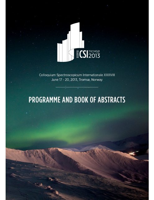
CSI2013_Program_and_Abstract_Book
CSI2013_Program_and_Abstract_Book
CSI2013_Program_and_Abstract_Book

Create successful ePaper yourself
Turn your PDF publications into a flip-book with our unique Google optimized e-Paper software.
XXXVIII CSI 2013<br />
Table of Content<br />
TABLE OF CONTENT<br />
Table of Content ................................................................................................................. 1<br />
Welcome! .............................................................................................................................. 2<br />
Organising <strong>and</strong> Scientific Committee ...................................................................... 3<br />
International Advisory Board ....................................................................................... 4<br />
Continuation Committee ................................................................................................ 4<br />
General Information ......................................................................................................... 5<br />
Social <strong>Program</strong>me ............................................................................................................. 8<br />
Scientific <strong>Program</strong>me ................................................................................................... 10<br />
Liability ................................................................................................................................ 10<br />
Sponsors <strong>and</strong> Exhibitors .............................................................................................. 11<br />
Correspondence after the conference ................................................................. 12<br />
Schedule of Events ......................................................................................................... 13<br />
Daily <strong>Program</strong>me ............................................................................................................ 15<br />
Poster Presentations ..................................................................................................... 25<br />
Plenary Lecture <strong>Abstract</strong>s .......................................................................................... 30<br />
Lecture <strong>Abstract</strong>s ........................................................................................................... 67<br />
Address list ...................................................................................................................... 241<br />
-1 -
XXXVIII CSI 2013<br />
Welcome Letter<br />
WELCOME!<br />
On behalf of the Norwegian Chemical Society, University of Tromsø <strong>and</strong> the Organising<br />
Committee it is an honour <strong>and</strong> pleasure to welcome you to Tromsø <strong>and</strong> the Colloquium<br />
Spectroscopicum Internationale XXXVIII in Tromsø, Norway, June 17 – 20, 2013. This<br />
conference provides both an international <strong>and</strong> a regional forum by which researchers <strong>and</strong><br />
users have the opportunity to share their knowledge <strong>and</strong> exchange ideas.<br />
We know that the natural beauty of the area will captivate you, but we also hope that the<br />
conference excursions, social events <strong>and</strong> farewell dinner may complement the scientific<br />
endeavours.<br />
Yngvar Thomassen Jon Øyvind Odl<strong>and</strong> Walter Lund<br />
-2 -
XXXVIII CSI 2013<br />
Committees<br />
ORGANISING AND SCIENTIFIC COMMITTEE<br />
ORGANISERS<br />
The Colloquium Spectroscopicum Internationale XXXVIII is organised by the Norwegian<br />
Chemical Society <strong>and</strong> the University of Tromso <strong>and</strong> in collaboration with International Union<br />
of Pure <strong>and</strong> Applied Chemistry.<br />
YNGVAR THOMASSEN<br />
(Chairman)<br />
National Institute of Occupational Health <strong>and</strong><br />
Norwegian University of Life Sciences, Ås, Norway<br />
WALTER LUND<br />
(Vice-Chairman)<br />
University of Oslo, Norway<br />
ELIN GJENGEDAL<br />
Norwegian University of Life Sciences Ås, Norway<br />
IVAR MARTINSEN<br />
GE Healthcare, Oslo, Norway<br />
ARNE ÅSHEIM<br />
(Exhibition Coordinator)<br />
Molab AS, avd. Porsgrunn, Norway<br />
SVERRE OMANG<br />
(Treasurer)<br />
Oslo, Norway<br />
ODDVAR RØYSETH<br />
NIVA, Norwegian Institute of Water Research, Oslo, Norway<br />
GEORG BECHER<br />
Norwegian Institute of Public Health, Oslo<br />
BALÁZS BERLINGER<br />
(Secretary)<br />
National Institute of Occupational Health, Oslo<br />
JON ØYVIND ODLAND<br />
University of Tromsø<br />
ANNE REGINE LAGER<br />
University Hospital, Tromso, Norway<br />
-3 -
XXXVIII CSI 2013<br />
Committees<br />
INTERNATIONAL ADVISORY BOARD<br />
Freddy Adams<br />
Michael Bolshov<br />
Maria Luisa de Carvalho<br />
Albert Gilmutdinov<br />
Detlef Günther<br />
Klaus Heumann<br />
Gary Hieftje<br />
Alex<strong>and</strong>er A. Kamnev<br />
Ryszard Lobinski<br />
Robert McCrindle<br />
Alfredo Sanz Medel<br />
János Mink<br />
Harpal Minhas<br />
Lars-Otto Reiersen<br />
Bernhard Welz<br />
Gyula Záray<br />
Belgium<br />
Russia<br />
Portugal<br />
Russia<br />
Switzerl<strong>and</strong><br />
Germany<br />
USA<br />
Russia<br />
France<br />
South-Africa<br />
Spain<br />
Hungary<br />
UK<br />
Norway<br />
Brazil<br />
Hungary<br />
CONTINUATION COMMITTEE<br />
Bernhard Welz<br />
Marco Aurélio Zezzi Arruda<br />
Yngvar Thomassen<br />
Balázs Berlinger<br />
Maria Luisa de Carvalho<br />
Joaquim dos Santos<br />
Department of Chemistry, UFSC, Brazil<br />
Institute of Chemistry, UNICAMP, Brazil<br />
National Institute of Occupational Health, Norway<br />
National Institute of Occupational Health, Norway<br />
Atomic Physics Center, University of Lisbon, Portugal<br />
Department of Physics, University of Coimbra, Portugal<br />
-4 -
XXXVIII CSI 2013<br />
General Information<br />
GENERAL INFORMATION<br />
Conference Desk<br />
Participants are requested to register as soon as possible upon arrival, preferably at Radisson<br />
Blu Hotel.<br />
The conference desk will operate as follows:<br />
Sunday, June 16<br />
16:00 - 20:00 Lobby of Radisson Blu Hotel<br />
Monday, June 17<br />
07:00 - 08:30 Lobby of Radisson Blu Hotel<br />
Monday, June 17<br />
13:00 - 18:00 Faculty of Health Sciences, First floor HE-building<br />
Tuesday, June 18<br />
08:30 - 18:00 Faculty of Health Sciences, First floor HE-building<br />
Wednesday, June 19<br />
08:30 - 18:00 Faculty of Health Sciences, First floor HE-building<br />
Thursday, June 20<br />
08:30 - 15:00 Faculty of Health Sciences, First floor HE-building<br />
-5 -
XXXVIII CSI 2013<br />
General Information<br />
Conference Venue<br />
All oral sessions will be held in the various auditoriums of University of Tromsø:<br />
The morning plenary sessions will be held in Auditorium 1, Teorifagbygget, House 1.<br />
The afternoon sessions will be held in the Auditoriums at the Faculty of Health Sciences,<br />
First floor HE-Building, Lysgården.<br />
"Lysgården"<br />
-6 -
XXXVIII CSI 2013<br />
General Information<br />
Bus transportation from Tromsø City to University Campus<br />
Conference buses will leave from outside the main entrance of Radisson Blu Hotel every<br />
morning from Monday, June 17, including Thursday, June 20, at 08:00 <strong>and</strong> 08:30.<br />
Please consult the conference program for the bus return schedule.<br />
The public bus connection between the centre of the city <strong>and</strong> the university campus is quite<br />
good. Bus no. 20/21 leaves from Fr. Langes gate (F1) in the centre of the city, <strong>and</strong> from the<br />
university just below building 23 on the campus map. This trip takes approximately 10 minutes.<br />
The bus leaves every 15 mins from the centre of the city on weekdays. You can also take bus no.<br />
34 – but this route takes you on a longer journey round the southern parts of the isl<strong>and</strong> (nice, if<br />
you are not in a hurry).<br />
-7 -
XXXVIII CSI 2013<br />
Social <strong>Program</strong>me<br />
SOCIAL PROGRAMME<br />
Monday, June 17, 19:00-24:00, the CSI-Club<br />
Sponsored by Agilents Technologies <strong>and</strong> Matriks AS<br />
<strong>and</strong><br />
Tuesday, June 18, 19:00-24:00, the CSI-Club<br />
The Ølhallen pub in Tromsø opened in1928 in the cellar of Macks Bryggeri, the most northerly<br />
brewery in the world. Here, in a world of what appears to be eternal c<strong>and</strong>le-lit night, perched on<br />
wooden stools, every Tom, Dick <strong>and</strong> Harry in Tromsø takes his beer.This is no cool, trendy bar –<br />
Ølhallen is an original, in a class of its own.<br />
As a CSI-participant you go to Ølhallen to drink beer, Mack beer. Mack’s entire selection is<br />
available, on draught, in bottles, or both. Regular Gullmack Pilsner, the heavier Håkon beer, the<br />
dark, malty Bayer beer <strong>and</strong> all the new kinds of beer are offered, but Ølhallen’s regulars swear by<br />
Bl<strong>and</strong>ing: two parts dark Bayer <strong>and</strong> one part light Pilsner.<br />
Each CSI-participant is granted minimum two 0.5 L drafts free of charge! Cheaper beer is not<br />
available in town!<br />
A selection of Peppes famous pizzas will also be served free of charge.<br />
Ølhallen is at the southern end of Tromsø’s main street, Storgata 4, <strong>and</strong> is easy to find.<br />
-8 -
XXXVIII CSI 2013<br />
Social <strong>Program</strong>me<br />
Wednesday, June 19, 19:45: Conference Dinner at the Fram Centre<br />
Sponsors: Agilents Technologies <strong>and</strong> Matriks AS<br />
After an introduction to the scientific activities at the FRAM CENTRE a seafood buffet dinner will be<br />
served. Non-alcoholic <strong>and</strong> alcoholic beverages are included.<br />
NOK 500 (not included in the registration fee)<br />
The Fram Centre is based in Tromsø, <strong>and</strong> consists of about 500 scientists from 20 institutions<br />
involved in interdisciplinary research in the fields of natural science, technology <strong>and</strong> social<br />
sciences. FRAM contributes to maintaining Norway’s prominent status in the management of<br />
environment <strong>and</strong> natural resources in the North.<br />
-9 -
XXXVIII CSI 2013<br />
Scientific <strong>Program</strong>me<br />
SCIENTIFIC PROGRAMME<br />
Oral Presentations<br />
Invited plenary lectures <strong>and</strong> submitted oral contributions will be 30 <strong>and</strong> 20 minutes in length,<br />
respectively (including discussion).<br />
Video projectors <strong>and</strong> computers will be provided in all lecture rooms.<br />
Posters<br />
The posters should be mounted Monday June 17, in the poster area located at the Faculty of<br />
Health Sciences, First floor HE-Building, Lysgården. Materials for poster mounting are available<br />
either from the Conference Desk or in the poster mounting area.<br />
All posters will be exhibited throughout the whole conference.<br />
Language<br />
The working language of the conference is English.<br />
LIABILITY<br />
The Organising Committee declines any responsibility whatsoever for injuries or damages to<br />
persons or their property during the Conference.<br />
-10 -
XXXVIII CSI 2013<br />
Sponsors <strong>and</strong> Exhibitors<br />
SPONSORS AND EXHIBITORS<br />
The exhibition of scientific instrumentation, literature <strong>and</strong> consumables is located next to the<br />
auditoriums of Faculty of Health Sciences, First floor HE-Building, Lysgården.<br />
The following companies have registered for display <strong>and</strong> demonstration:<br />
AGILENT<br />
ANALYTIK JENA<br />
BRUKER<br />
MILESTONE SRL<br />
SHIMADZU<br />
THERMO SCIENTIFIC<br />
KAISER OPTICAL SYSTEMS<br />
PERKIN ELMER<br />
PANALYTICAL<br />
CPI INTERNATIONAL<br />
GAMMADATA<br />
MAGRITEK GMBH<br />
POSTNOVA<br />
-11 -
CORRESPONDENCE AFTER THE CONFERENCE<br />
-12 -
XXXVIII CSI 2013<br />
Schedule of Events<br />
SCHEDULE OF EVENTS<br />
June 16-20, 2013<br />
Time<br />
Sunday,<br />
June 16 Monday, June 17 Tuesday, June 18<br />
09:00<br />
Opening Ceremony<br />
09:15<br />
Plenary Lecture<br />
Aud. 1, Teorifagbygget, House 1<br />
09:30<br />
PL7-PL9<br />
09:45<br />
Aud. 1, Teorifagbygget, House 1<br />
10:00<br />
10:15<br />
Inaugural Lecture<br />
PL1<br />
10:30<br />
Coffee Break,10:30-11:00<br />
10:45<br />
Coffee Break, 10:45-11:15<br />
11:00<br />
11:15<br />
11:30<br />
Plenary Lecture<br />
11:45<br />
PL10-PL12<br />
Plenary Lecture<br />
12:00<br />
PL2-PL4<br />
12:15<br />
12:30<br />
12:45<br />
13:00<br />
Lunch, Lysgården<br />
12:30-13:30<br />
13:15<br />
13:30<br />
Lunch, Lysgården,<br />
13:00-14:00<br />
13:45<br />
14:00<br />
Lecture Lecture Lecture<br />
Plenary Lecture PL5-PL6,<br />
14:15<br />
L25-L29 L30-L34 L35-L38<br />
Store Auditorium, Lysgården<br />
14:30<br />
Store Aud Aud. 3 Aud. 4<br />
14:45<br />
Lecture Lecture Lecture<br />
15:00<br />
L1-L4, L9-L12 L18-L21<br />
15:15<br />
Aud. 3 Store Aud. 4 Coffee Break, 15:20-15:40<br />
15:30<br />
Aud.<br />
15:45<br />
16:00<br />
Coffee Break, 16:00-16:20<br />
CSI award<br />
16:15<br />
Store Auditorium<br />
16:30<br />
Lecture Lecture Lecture<br />
16:45<br />
L5-L8 L13-L17 L22-L24<br />
17:00<br />
Aud. 3 Store Aud. 4<br />
NKS award, Store Auditorium<br />
17:15<br />
Aud.<br />
17:30<br />
17:45<br />
18:00<br />
18:15<br />
Registration<br />
Radisson<br />
Blu Hotel Bus Departures, 18:15<br />
Award Reception<br />
Lysgården<br />
Bus Departures, 18:15<br />
18:30<br />
18:45<br />
19:00<br />
19:15<br />
19:30<br />
19:45<br />
20:00<br />
The CSI-Club<br />
Ølhallen<br />
The CSI-Club<br />
Ølhallen<br />
-13 -
XXXVIII CSI 2013<br />
Schedule of Events<br />
Lecture<br />
L39-L44<br />
Aud. 3<br />
Wednesday, June 19 Thursday, June 20 Time<br />
09:00<br />
09:15<br />
09:30<br />
Plenary Lecture<br />
Plenary Lecture<br />
09:45<br />
PL13-PL16<br />
PL21-PL24<br />
10:00<br />
Aud. 1, Teorifagbygget, House 1<br />
Aud. 1, Teorifagbygget, House 1<br />
10:15<br />
10:30<br />
10:45<br />
Coffee Break, 11:00-11:20 Coffee Break 11:00-11:20 11:00<br />
Plenary Lecture PL17-PL19<br />
11:20-12:50<br />
Lunch,<br />
Lysgården, 12:50-13:50<br />
Plenary Lecture PL20<br />
Store Aud., 13:50-14:20<br />
Lecture<br />
L45-L50<br />
Store<br />
Aud.<br />
Lecture<br />
L51-L56<br />
Aud. 4<br />
Poster Discussions<br />
16:20 (16:50) -18:30<br />
Lecture<br />
L56-L63<br />
Aud. 2<br />
Plenary Lecture PL25-PL26<br />
11:20-12:20<br />
Lunch,<br />
Lysgården, 12:20-13:20<br />
Plenary Lecture PL27-PL28<br />
13:20-14:20<br />
Lecture<br />
L64-L67<br />
Store Aud.<br />
Lecture<br />
L68-L70<br />
Aud. 3<br />
11:15<br />
11:30<br />
11:45<br />
12:00<br />
12:15<br />
12:30<br />
12:45<br />
13:00<br />
13:15<br />
13:30<br />
13:45<br />
14:00<br />
14:15<br />
14:30<br />
14:45<br />
15:00<br />
15:15<br />
15:30<br />
15:45<br />
Closing Ceremony<br />
16:00<br />
15:45<br />
16:15<br />
Bus Departures, 16:30 16:30<br />
16:45<br />
17:00<br />
17:15<br />
17:30<br />
17:45<br />
18:00<br />
18:15<br />
Bus Departures, 18:30 18:30<br />
18:45<br />
19:00<br />
19:15<br />
19:30<br />
Conference Dinner, Fram Centre, 19:45<br />
19:45<br />
20:00<br />
-14 -
XXXVIII CSI 2013<br />
Daily <strong>Program</strong>me<br />
DAILY PROGRAMME<br />
Monday, June 17, 2013<br />
Time Abs.<br />
09:00 Opening Ceremony<br />
Auditorium 1, Teorifagbygget, House 1<br />
Chairman: Yngvar Thomassen<br />
09:45 PL1 Inaugural Lecture<br />
Hubble Space Telescope <strong>and</strong> Its Discoveries<br />
Richard E. Griffiths, Carnegie Mellon University, Pittsburgh, USA/NASA HQ<br />
10:45 Coffee Break<br />
Climate Change <strong>and</strong> Spectroscopy<br />
Auditorium 1, Teorifagbygget, House 1<br />
Chairman: Yngvar Thomassen<br />
11:15 PL2 Climate Change <strong>and</strong> Mitigation: Carbon Capture – A Bridge Into a Low Carbon Economy<br />
Claus Jørgen Nielsen, University of Oslo, Norway<br />
12:00 PL3 Molecular - Level Analysis <strong>and</strong> Photochemical Aging of Atmospheric Organics in Ambient Particles<br />
<strong>and</strong> Aqueous Droplets<br />
Sergey A. Nizkorodov, University of California, USA<br />
12:30 PL4 Characterization of Atmospheric Aerosol Particles by Electron Microscopy<br />
Stephan Weinbruch, Technical University Darmstadt, Germany<br />
13:00 Lunch<br />
Faculty of Health Sciences, Lysgården.<br />
Store Auditorium – Lysgården<br />
14:00 PL5 Basic Research for Chemical Absorption of Carbon Dioxide Using NMR Spectroscopy<br />
Cristina Perinu, Telemark University College, Porsgrunn, Norway<br />
14:20 PL6 Molecular Analysis of the Organic <strong>and</strong> Elemental Carbon Fractions (EC/OC) of Ambient Particulate<br />
Matter by Coupling of a Thermal Carbon Analyzer to Photo-Ionization Mass Spectrometry<br />
Ralf Zimmermann, University of Rostock, Germany<br />
Time Abs.<br />
I. MATERIAL CHARACTERISATION<br />
Auditorium 3 – Lysgården<br />
Chairman: Irina Snigireva<br />
14:40 L1 X-Ray Refractive Optics: Present Status <strong>and</strong> New Developments<br />
Anatoly Snigirev, European Synchrotron Radiation Facility, France<br />
15:00 L2 Characterisation of Nanoparticles With Synchrotron X-Ray St<strong>and</strong>ing Wave Fluorescence<br />
Rol<strong>and</strong> Hergenröder, Leibniz-Institut für Analytische Wissenschaften, Dortmund, Germany<br />
15:20 L3 NEXAFS Spectroscopic Studies of Anodized Ti-6Al-4V Alloy With the Aide of First Principles<br />
Calculations<br />
Toshihiro Okajima, Kyushu Synchrotron Light Research Center, Japan<br />
15:40 L4 Microscale Mineral Analysis of Clay Rock Thin Sections After Sorption Experiment Using SRXRF<br />
Szabina Török, HAS Energy Research Institute, Budapest, Hungary<br />
16:20 Coffee Break<br />
16:20 L5 Synthesis <strong>and</strong> Characterization of Metal Nanoclusters: A New Generation of Luminescent Labels<br />
Laura Trapiella-Alfonso, University of Oviedo, Spain<br />
16:40 L6 Advanced Inspection Technologies in Extreme Scenarios: St<strong>and</strong>off LIBS <strong>and</strong> Underwater LIBS<br />
Javier J. Laserna, University of Málaga, Spain<br />
17:00 L7 The UMS: A New Tool for Multi-Angle UV-VIS-NIR Photometric Spectroscopy<br />
Jan Wuelfken, Agilent Technologies,Waldbronn, Germany<br />
17:20 L8 57Fe-Mössbauer Study of Electrically Conductive Lithium Iron Vanadate Glass<br />
Shiro Kubuki, Tokyo Metropolitan University, Japan<br />
17:40<br />
18:10 Bus Departures<br />
-15 -
XXXVIII CSI 2013<br />
Daily <strong>Program</strong>me<br />
Monday, June 17, 2013<br />
Time Abs.<br />
II. PLASMA SPECTROCHEMISTRY<br />
Store Auditorium – Lysgården<br />
Chairman: Maria Montes-Bayón<br />
14:40 L9 Breakthrough in Sensivity for Quadrupole ICP-MS<br />
Meike Hamester, Bruker Daltonics, Germany<br />
15:00 L10 Determination of Rare Earth Elements in Extracts from Oil Refinary Spent Catalyst by ICP-MS with<br />
a Reaction Cell<br />
Jessee Severo Azevedo Silva, Universidade Federal de Santa Catarina, Brazil<br />
15:20 L11 Some Procedures of Reducing Matrix Effects in ICP-MS Analysis of Biological Samples<br />
Konstantin Ossipov, Lomonosov Moscow State University, Russia<br />
15:40 L12 Development of an Analytical Method for Cd, Co, Cr, Cu, Ni <strong>and</strong> Pb Determination in Cosmetic<br />
Samples<br />
Edenir Rodrigues Pereira-Filho, Federal University of São Carlos, Brazil<br />
16:00 Coffee Break<br />
16:20 L13 The New 8800 ICP-QQQ: H<strong>and</strong>ling the Most Difficult Samples With Ease<br />
Uwe Noetzel, Agilent Technologies, Germany<br />
16:40 L14 A Comparison of Conventional (Off-Line) <strong>and</strong> On-Line Isotope Dilution ICP-MS for the<br />
Determination of Total Selenium in Human Serum<br />
Petru Jitaru, Institut Polytechnique LaSalle Beauvais, Beauvais cedex, France<br />
17:00 L15 From 2D Towards 3D Elemental Imaging by Laser Ablation ICP-MS - A Study of Archaeological<br />
Glass<br />
Vid S. Šelih, National Institute of Chemistry, Ljubljana, Slovenia<br />
17:20 L16 Direct Solid Quantitative Analysis of Battery Components Using LA-ICP-MS <strong>and</strong> Development of<br />
Custom Made Solid St<strong>and</strong>ard Materials Analysis of Battery Materials<br />
Björn Hoffmann, University of Münster, Germany<br />
17:40 L17 Direct Elemental Analysis of Nanodiamonds With ICP-OES<br />
Dmitry S. Volkov, Lomonosov Moscow State University, Russia<br />
18:10 Bus Departures<br />
Time Abs.<br />
III. THEORETICAL, STRUCTURAL AND MODELLING STUDIES<br />
Auditorium 4 – Lysgården<br />
Chairman: Arne Bengtson<br />
14:40 L18 Experimental Spectroscopic <strong>and</strong> Quantum Chemical Studies of the Reactivity of Alkylresorcinols in<br />
Redox Processes<br />
Alex<strong>and</strong>er A. Kamnev, Institute of Biochemistry <strong>and</strong> Physiology of Plants <strong>and</strong> Microorganisms, Saratov,<br />
Russia<br />
15:00 L19 Vibrational Spectral Analysis of the Isotopic Species of Hydrogen Sulphide, Hydrogen Selenide <strong>and</strong><br />
Water Using the U(4) Algebraic Model<br />
Nirmal Kumar Sarkar, Karimganj College, India<br />
15:20 L20 Theoretical Investigation of the CW Absorption, Resonance Raman <strong>and</strong> REMPI Spectroscopy of the<br />
S1 <strong>and</strong> S2 States of cis-1,3,5-Hexatriene <strong>and</strong> trans-1,3,5-Hexatriene<br />
Clemens Woywod, University of Tromsø, Norway<br />
15:40 L21 Basis Set Extrapolation for High Resolution Spectroscopy<br />
Kiran Sankar Maiti, University of Gothenburg, Sweden<br />
16:00 Coffee Break<br />
16:20 L22 Exploring Structure <strong>and</strong> Ultrafast Dynamics of Protein <strong>and</strong> Peptide Using Two Color 2D IR<br />
Spectroscopy<br />
Susmita Roy, University of Gothenburg, Sweden<br />
16:40 L23 Fat Determination of Intact Food Samples with Time-Domain Nuclear Magnetic Resonance<br />
Spectroscopy <strong>and</strong> Chemometrics<br />
Fabiola Manhas Verbi Pereira, Embrapa Instrumentation, São Carlos, Brazil<br />
17:00 L24 Diode Laser Absorption Spectrometry as a Tool for Contactless Diagnostic of a Hot Zone<br />
Michael A. Bolshov, Institute for Spectroscopy RAS, Moscow, Russia<br />
18:10 Bus Departures<br />
-16 -
XXXVIII CSI 2013<br />
Daily <strong>Program</strong>me<br />
Time<br />
Abs.<br />
Tuesday, June 18, 2013<br />
Pristine Environments <strong>and</strong> Spectroscopy<br />
Auditorium 1, Teorifagbygget, House 1<br />
Chairman: Klaus Heumann<br />
09:00 PL7 Arctic – From Cold War to Arctic Melt Down: 20 Years of Arctic Monitoring <strong>and</strong> Assessment of<br />
Pollutants <strong>and</strong> Climate Change by AMAP<br />
Lars Otto Reiersen, Artic Monitoring <strong>and</strong> Assessment <strong>Program</strong>me, Oslo, Norway<br />
09:30 PL8 Spectroscopy under Ice<br />
Carlo Barbante, University of Venice, Italy<br />
10:00 PL9 Bioluminescent Ecological Assay. Features <strong>and</strong> Scope of Applications<br />
Nadezhda Kudryasheva, Siberian Federal University, Krasnoyarsk, Russia<br />
10:30 Coffee Break<br />
11:00 PL10 FTIR Spectroscopy in Microbial Ecology: ‘Shedding IR Light’ on Cellular Metabolic Responses to<br />
Environmental Factors<br />
Alex<strong>and</strong>er A. Kamnev, Institute of Biochemistry <strong>and</strong> Physiology of Plants <strong>and</strong> Microorganisms, Saratov,<br />
Russia<br />
11:30 PL11 Atmospheric Deposition of Trace Elements on the Local <strong>and</strong> Regional Scale Studied by ICP-MS<br />
Analysis of Moss Samples<br />
Eiliv Steinnes, Norwegian University of Science <strong>and</strong> Technology, Trondheim, Norway<br />
12:00 PL12 Mass Spectrometry Made Easy: The Quest for Simplicity in Forensic, Food, Pharmaceutical,<br />
Environmental, Medical <strong>and</strong> Biochemical Analysis<br />
Marcos Eberlin, Universidade Estadual de Campinas, Brazil<br />
12:30 Lunch<br />
Faculty of Health Sciences, Lysgården.<br />
Time Abs<br />
IV. MASS SPECTROMTRY<br />
Store Auditorium – Lysgården<br />
Chairman: Georg Becher<br />
13:30 L25 Liquid Chromatography Mass-Spectrometry as a Tool for Detection of Chemicals Connected with<br />
Chemical Warfare Agents in Environmental <strong>and</strong> Bio Samples<br />
Igor A. Rodin, Moscow State University, Russia<br />
14:00 L26 Collision-Induced Dissociation of Hydroxylated Polycyclic Aromatic Hydrocarbons in an Ion Trap<br />
T<strong>and</strong>em Mass Spectrometer<br />
Xue Li, ETH Zürich, Switzerl<strong>and</strong><br />
14:20 L27 Electrospray Ionization Mass Spectrometry Assisted by Inductively Coupled Plasma Mass<br />
Spectrometry as a Tool to Study the Se/S Substitution in Methionine <strong>and</strong> Cysteine in Se-Enriched<br />
Yeast<br />
Katarzyna Bierła, CNRS/UPPA, Laboratoire de Chimie Analytique Bio-inorganique et Environnement<br />
(LCABIE), Pau, France<br />
14:40 L28 Identification of Original Sources of Vermilion in Antiquity Using Sulfur Isotope Ratio Analysis<br />
Takeshi Minami, Kinki University, Osaka, Japan<br />
15:00 L29 Study of Interactions Between Reactive Gas Species <strong>and</strong> Microorganisms by Nano-Resolution Mass<br />
Spectrometry Imaging<br />
Jean-Nicolas Audinot, Centre de Recherche Public Gabriel Lippmann, Belvaux, Luxembourg<br />
15:20 Coffee Break<br />
15:40<br />
CSI AWARD<br />
Store Auditorium – Lysgården<br />
Introduced by Yngvar Thomassen <strong>and</strong> Gary Hieftje<br />
16:40 NORWEGIAN CHEMICAL SOCIETY-DIVISION OF ANALYTICAL CHEMISTRY HONORARY AWARD<br />
Store Auditorium – Lysgården<br />
Introduced by Elin F. Gjengedal <strong>and</strong> Freddy Adams<br />
17:15<br />
Award Reception<br />
Faculty of Health Sciences, Lysgården.<br />
18:15 Bus Departures<br />
-17 -
XXXVIII CSI 2013<br />
Daily <strong>Program</strong>me<br />
Tuesday, June 18, 2013<br />
Time Abs.<br />
V. ATOMIC ABSORPTION SPECTROMTRY<br />
Auditorium 3 – Lysgården<br />
Chairman: Robert McCrindle<br />
13:30 L30 Solid Sampling Techniques for the Direct Elemental or Isotopic Analysis of Dried Matrix Spots<br />
Martín Resano, University of Zaragoza, Spain<br />
14:00 L31 Low Resolution Continuum Source Electrothermal Atomic Absorption Spectrometry: Clarification of<br />
Analytical Potential<br />
Dmitri Katskov, Tshwane University of Technology, Pretoria, South Africa<br />
14:20 L32 Trace Determination of Metals by In-Atomizer Hydride Trapping AAS: Method Development,<br />
Validation <strong>and</strong> Analytical Applications<br />
Jan Kratzer, Institute of Analytical Chemistry of the ASCR, Brno, Czech Republic<br />
14:40 L33 Optimization Study on Determination of Inorganic Arsenic Species in Hot Chilli Pepper <strong>and</strong> Tomato<br />
Varieties by Using Microwave Assisted Digestion Followed by Flow Injection-Hydride Generation<br />
Atomic Absorption Spectrometry<br />
Saksit Chanthai, Khon Kaen University, Thail<strong>and</strong>.<br />
15:00 L34 Preconcentration of Mercury from Natural Waters by Amalgamation of Hg 2+ on Copper Powder <strong>and</strong><br />
Hg 0 on Gold Nanoparticles.<br />
Nikolay Panichev, Tshwane University of Technology, Pretoria, South Africa<br />
15:20 Coffee Break<br />
Time Abs.<br />
VI. GLOW DISCHARGE<br />
Auditorium 4 – Lysgården<br />
Chairman: Volker Hoffmann<br />
13:30 L35 Sampling of Liquids in Atomic Emission Spectrometry Using a Helium Atmospheric Pressure Glow<br />
Discharge<br />
José A.C. Broekaert, University of Hamburg, Germany<br />
14:00 L36 GD TOFMS with Pulsed Combined Hollow Cathode for Direct Analysis of Dielectric Samples<br />
Alex<strong>and</strong>er Ganeev, St. Petersburg State University, Russia<br />
14:20 L37 Selective Excitation in Analytical Glow Discharges – Its Relevance in GD-OES Analysis<br />
Edward B. M. Steers, London Metropolitan University, UK<br />
14:40 L38 Molecular Emission in GD-OES Revisited – Strategies for Background Correction<br />
Arne Bengtson, Swerea KIMAB, Kista, Sweden<br />
15:20 Coffee Break<br />
15:40<br />
CSI AWARD<br />
Store Auditorium – Lysgården<br />
Introduced by Yngvar Thomassen <strong>and</strong> Gary Hieftje<br />
16:40 NORWEGIAN CHEMICAL SOCIETY-DIVISION OF ANALYTICAL CHEMISTRY HONORARY AWARD<br />
Store Auditorium – Lysgården<br />
Introduced by Elin F. Gjengedal <strong>and</strong> Freddy Adams<br />
17:15<br />
Award Reception<br />
Faculty of Health Sciences, Lysgården.<br />
18:15 Bus Departures<br />
-18 -
XXXVIII CSI 2013<br />
Daily <strong>Program</strong>me<br />
Time<br />
Abs.<br />
Wednesday, June 19, 2013<br />
Human Health <strong>and</strong> Spectroscopy<br />
Auditorium 1, Teorifagbygget, House 1<br />
Chairman: Alex<strong>and</strong>er Kamnev<br />
09:00 PL13 Confocal Spectral Imaging Technique in the Development of Photo- <strong>and</strong> Neutronsensitizers for<br />
Anticancer Therapy<br />
Alexey V. Feofanov, Lomonosov Moscow State University, Russia<br />
09:30 PL14 New Developments in Disease Recognition by Infrared <strong>and</strong> Raman Spectroscopy <strong>and</strong> Microscopy:<br />
Present Status <strong>and</strong> Future Promises<br />
János Mink, Institute of Molecular Pharmacology, Research Centre for Natural Sciences, Budapest,<br />
Hungary<br />
10:00 PL15 Metabolic Fingerprinting via Mass Spectrometric Analysis of Exhaled Breath<br />
Renato Zenobi, ETH Zürich, Switzerl<strong>and</strong><br />
10:30 PL16 Accurate Measurement of Iron Metabolism Biomarkers: New Tools <strong>and</strong> Remaining Challenges<br />
Maria Montes-Bayón, University of Oviedo, Spain<br />
11:00 Coffee Break<br />
11:20 PL17 SALDI Mass-Spectrometry: Principles <strong>and</strong> Application for Drug Analysis<br />
Alex<strong>and</strong>er Grechnikov, Vernadsky Institute of Geochemistry <strong>and</strong> Analytical Chemistry of RAS, Moscow,<br />
Russia<br />
11:50 PL18 SIMS <strong>and</strong> the Single Cell<br />
Vic Norris, University of Rouen, France<br />
12:20 PL19 High Resolution MALDI Imaging: Reliable Molecular Information at Cellular Resolution<br />
Andreas Römpp, University of Giessen, Germany<br />
12:50 Lunch<br />
Faculty of Health Sciences, Lysgården<br />
Time Abs.<br />
Progress in Mass Spectrometry<br />
Store Auditorium – Lysgården<br />
Chairman: Carlo Barbante<br />
13:50 PL20 New Approaches, Plasmas, <strong>and</strong> Instrumentation for Atomic Spectrometry<br />
Steven J. Ray, Indiana University, Bloomington, USA<br />
-19 -
XXXVIII CSI 2013<br />
Daily <strong>Program</strong>me<br />
Wednesday, June 19, 2013<br />
Time Abs.<br />
VII. ENVIRONMENTAL APPLICATIONS I<br />
Auditorium 3 – Lysgården<br />
Chairman: Janos Mink<br />
14:20 L39 Chemical Characterization of Dekati ® Low Pressure Impactor (DLPI) Wall Deposits<br />
Thibaut Dur<strong>and</strong>, Institut National de Recherche et de Sécurité, V<strong>and</strong>oeuvre-lès-Nancy, France<br />
14:40 L40 Chemical Characterization <strong>and</strong> Oxidative Potential of PM2.5 Collected in Office Buildings in Greece<br />
<strong>and</strong> The Netherl<strong>and</strong>s: A Cooperative Study<br />
Tamás Szigeti, Eötvös Loránd University, Budapest, Hungary<br />
15:00 L41 FTIR (DRIFT) Spectroscopic Analysis of Accumulation <strong>and</strong> Structural Features of Poly-3-<br />
Hydroxybutyrate in Cells of Azospirillum Brasilense: Effects of Copper(II)<br />
Anna V. Tugarova, Laboratory of Biochemistry, Institute of Biochemistry <strong>and</strong> Physiology of Plants <strong>and</strong><br />
Microorganisms, RAS, Saratov, Russia<br />
15:20 L42 A New Hybrid Fluorometer-Spectrophotometer for Water Quality Analysis of Oil, Chromophoric<br />
Dissolved Organic Matter, Chlorophyll <strong>and</strong> -NHX<br />
Adam M. Gilmore, HORIBA Instruments Inc., Edison, USA<br />
15:40 L43 Occurrence of Polychlorinated Biphenyls (PCBs) <strong>and</strong> Polybrominated Diphenylethers (PBDEs) in<br />
Different Fish Species from Ilha Gr<strong>and</strong>e Bay, Southeastern Brazil.<br />
Isabel Moreira, Pontifícia Universidade Católica do Rio de Janeiro, Brazil<br />
16:00 L44 Application of Multi-Reflection, High Resolution Time-of-Flight-Mass Spectrometry as Detector for<br />
One- <strong>and</strong> Two-Dimensional Gas Chromatography: Characterization of Complex Mixtures<br />
Ralf Zimmermann, University of Rostock, Germany<br />
16:20 Poster Discussions<br />
18:30 Bus Departures<br />
19:45 Conference Dinner<br />
Time Abs.<br />
VIII. IMAGING AND MODELLING<br />
Store Auditorium – Lysgården<br />
Chairman: José Broekaert<br />
14:20 L45 New Imaging Capabilities Using LA-ICP-TOF Mass Spectrometry<br />
Detlef Günther, ETH Zürich, Switzerl<strong>and</strong><br />
14:40 L46 Further Developments of an Energy- <strong>and</strong> Position-Sensitive XRF Imaging System Based on a<br />
THCOBRA Detector<br />
Analusia L.M. Silva, University of Aveiro, Portugal<br />
15:00 L47 Pharmaceutical Images Harvesting <strong>and</strong> Comparison<br />
Tomáš Pekárek, Zenitva, k.s., Prague, Czech Republic<br />
15:20 L48 Novel Multispectral Imaging Approaches For Recovering of Degraded Archaeological Wall<br />
Paintings<br />
Vincenzo Palleschi, CNR Area della Ricerca del CNR, Pisa, Italy<br />
15:40 L49 LA-ICPMS Imaging And Nuclear Forensics: Pu Isotope Ratios In Sediments From Mayak PA, Russia<br />
Simone Cagno, Norwegian University of Life Sciences, Ås, Norway<br />
16:00 L50 Physicochemical Investigation of The Wall Paintings of Petros Paulos Church, Ethiopia<br />
Kidane Fanta Gebremariam, Norwegian University of Science <strong>and</strong> Technology, Trondheim, Norway<br />
16:20 Poster Discussion<br />
18:30 Bus Departures<br />
19:45 Conference Dinner<br />
-20 -
XXXVIII CSI 2013<br />
Daily <strong>Program</strong>me<br />
Wednesday, June 19, 2013<br />
Time Abs.<br />
IX. HUMAN HEALTH<br />
Auditorium 4 – Lysgården<br />
Chairman: Elin Gjengedal<br />
14:20 L51 Comparison Between SR-XRF <strong>and</strong> ICP-AES<br />
Jun Kawai, Kyoto University, Japan<br />
14:40 L52 Analysis of Bones for Forensic Studies<br />
Vincenzo Palleschi, CNR Area della Ricerca del CNR, Pisa, Italy<br />
15:00 L53 Printable Surface Enhanced Raman Scattering Strips with In-Situ Growth of Gold Nanoparticles<br />
Wei Ju, Liao, National Yang-Ming University, Taiwan<br />
15:20 L54 Speciation <strong>and</strong> Cobalt Toxicity on Human Lung Cells: An Interdisciplinary Study<br />
Carole Bresson, Laboratoire de Développement Analytique Nucléaire, Isotopique et Elémentaire, Gifsur-Yvette,<br />
France.<br />
15:40 L55 Fast <strong>and</strong> Non-Destructive Quantification of Dapivirine in Hiv Preventive Intravaginal Rings by<br />
Raman Spectroscopy<br />
Lotte B. Lyndgaard, University of Copenhagen, Denmark<br />
16:00 L56 Titanium Measurement in Biofluids by ICP-OES (Simultaneous Inductively Coupled Plasma Optical<br />
Emission) <strong>and</strong> He-CC KED Quadrupole ICP-MS<br />
János Fucskó, NMS Labs, Willow Grove, USA<br />
16:20 Poster Discussion<br />
18:30 Bus Departures<br />
19:45 Conference Dinner<br />
Time<br />
Abs.<br />
X. ENVIRONMENTAL APPLICATIONS II<br />
Auditorium 2 – Lysgården<br />
Chairman: Eiliv Steinnes<br />
14:20 L57 Distribution <strong>and</strong> Source of Metals in Contaminated Sediments from Rivers in Coal Fields<br />
Rob McCrindle, Tshwane University of Technology, Pretoria, South Africa<br />
14:40 L58 Interpretation of the Plastic Life Cycle Using FTIR-ATR <strong>and</strong> ICP-OES Spectrometry<br />
Albert van Oyen, CARAT GmbH, Bocholt, Germany<br />
15:00 L59 Advanced Techniques for Environmental Analysis Using ICP-MS<br />
Shona McSheehy Ducos, Thermo Scientific, Bremen, Germany<br />
15:20 L60 Quantitative Analysis of Heavy Elements <strong>and</strong> Semi-Quantitative Evaluation of Heavy Mineral<br />
Compositions of Stream Sediments in Japan for Construction of a Forensic Soil Database Using<br />
Synchrotron Radiation X-Ray Analyses<br />
Izumi Nakai, Tokyo University of Science, Japan<br />
15:40 L61 Effect of Metal Stress on Pigments in Copper-Hyperaccumulating Lichens<br />
Hiromitsu Nakajima, Yokohama National University, Japan<br />
16:00 L62 High Sensitivity <strong>and</strong> Extended Scan Speed for Dedicated Isotope Ratio Determinations<br />
René Chemnitzer, Bruker Daltonics, Bremen, Germany<br />
16:20 L63 Determination of Heavy Metals at Ultralow Concentration Levels in Pristine Polar Snow <strong>and</strong> Ice<br />
Claude F. Boutron, University Joseph Fourier of Grenoble, France<br />
16:50 Poster Discussion<br />
18:30 Bus Departures<br />
19:45 Conference Dinner<br />
-21 -
XXXVIII CSI 2013<br />
Daily <strong>Program</strong>me<br />
Thursday, June 20, 2013<br />
Time Abs.<br />
Material Characterisation <strong>and</strong> Spectroscopy<br />
Auditorium 1, Teorifagbygget, House 1<br />
Chairman: Maria Luisa de Carvalho<br />
09:00 PL21 Pulsed Glow Discharge Time of Flight Mass Spectrometry: A Powerful <strong>and</strong> Versatile Tool for<br />
Elemental <strong>and</strong> Molecular Depth Profile Analysis<br />
Rosario Pereiro, University of Oviedo, Spain<br />
09:30 PL22 Coherent High Energy X-Ray Microscopy: A New Tool to Study Mesoscopic Materials<br />
Irina Snigireva, European Synchrotron Radiation Facility, Grenoble, France<br />
10:00 PL23 Analysis of Topical Biomedical <strong>and</strong> Technological Samples by Photothermal <strong>and</strong> Photoacoustic<br />
Spectroscopies Using Signal-Enhancement Techniques <strong>and</strong> Selective Reactions<br />
Mikhail A. Proskurnin, Lomonosov Moscow State University, Russia<br />
10:30 PL24 Progress <strong>and</strong> Dem<strong>and</strong>s in Analytical Glow Discharges<br />
Volker Hoffmann, Institute for Complex Materials, IFW Dresden, Germany<br />
11:00 Coffee Break<br />
11:20 PL25 Development <strong>and</strong> Characterization of Materials for Advanced Power Plants<br />
Hubertus Nickel, Research Centre Jülich/University of Technology Aachen, Germany<br />
11:50 PL26 Application of Synchrotron Microprobe Techniques to Speciation of Plutonium in Argillaceous Rocks<br />
Tobias Reich, Johannes Gutenberg-Universität Mainz, Germany<br />
12:20 Lunch<br />
Faculty of Health Sciences, Lysgården<br />
Progress in Analytical Spectrometry<br />
Store Auditorium – Lysgården<br />
Chairman: Martín Resano<br />
13:20 PL27 An Analytical Technique on Its Way to Adulthood: Current Status <strong>and</strong> Future Perspectives of Mass<br />
Spectrometry Imaging<br />
Andreas Römpp, University of Giessen, Germany<br />
13:50 PL28 “Think Big! – Optimization of a Spectrometry Lab on the Industrial Scale<br />
Heiko Egenolf, BASF SE, Competence Center Analytics, Ludwigshafen, Germany<br />
XI. ENERGY STORAGE CHARACTERISATION<br />
Store Auditorium – Lysgården<br />
Chairman: Michael Bolshov<br />
14:20 L64 Characterization of Decomposition Products in Energy Storage Materials by Chromatographic<br />
Methods<br />
Sascha Nowak, University of Münster, Germany<br />
14:40 L65 Lock-In Thermography – A Novel In-Situ Measurment Methode to Support Surface Spectroscopy for<br />
Lithium-Ion Cells<br />
Mathias Reichert, University of Münster, Germany<br />
15:00 L66 Characterization of the Decomposition Products of a Utilized Battery Electrolyte from a<br />
Commercial Available Hybrid Vehicle with Purposeful Analytical Methods<br />
Martin Grützke, University of Münster, Germany<br />
15:20 L67 Analysis of the Manganese Dissolution <strong>and</strong> Deposition in LiMn2O4/Li4Ti5O12 Based Lithium Ion<br />
Batteries<br />
Markus Börner, University of Münster, Germany<br />
15:45 Closing Ceremony<br />
Store Auditorium – Lysgården<br />
16:30 Bus Departures<br />
-22 -
XXXVIII CSI 2013<br />
Daily <strong>Program</strong>me<br />
Thursday, June 20, 2013<br />
Time Abs.<br />
XII. SPECIATION ANALYSIS<br />
Auditorium 3 – Lysgården<br />
Chairman: Yngvar Thomassen<br />
14:20 L67B UV-Photochemical Volatile Species Generation Employed as a Derivatization Technique Between<br />
HPLC Separation <strong>and</strong> AAS Detection within Speciation Analysis of Mercury(II), Methylmercury(I),<br />
Ethylmercury(I) <strong>and</strong> Phenylmercury(I)<br />
Vaclav Cerveny, Charles University in Prague, Czech Republic<br />
14:40 L68 Chemical Vapor Generation for Trace Analysis - Recent Developments<br />
Aless<strong>and</strong>ro D’Ulivo, Institute of Chemistry of Organometallic Compounds, U.O.S. of Pisa, Italy<br />
15:00 L69 Influence of Selenium Species in Aquaculture Feeds on the Selenium Status of Farmed Rainbow<br />
Trout Fry<br />
Simon Godin, Université de Pau et des Pays de l’Adour, France<br />
15:45 Closing Ceremony<br />
Store Auditorium – Lysgården<br />
16:30 Bus Departures<br />
-23 -
-24 -
XXXVIII CSI 2013<br />
Poster Presentations<br />
POSTER PRESENTATIONS<br />
<strong>Abstract</strong><br />
P1 DETERMINATION OF TRACE SULFUR IN BIODIESEL AND DIESEL STANDARD REFERENCE<br />
MATERIALS BY ISOTOPE DILUTION SECTOR FIELD INDUCTIVELY COUPLED PLASMA MASS<br />
SPECTROMETRY<br />
Renata S. Amais, Stephen E. Long, Joaquim A. Nóbrega <strong>and</strong> Steven J. Christopher<br />
P2 DETERMINATION OF CHROMIUM SPECIES IN THE WORKPLACE AIR<br />
M. Stanisławska, B. Janasik; R. Brodzka; W. Wąsowicz<br />
P3 A NEW APPROACH FOR THE DETERMINATION OF ARSENIC IN THE WORKPLACE AIR. THE<br />
POSSIBILITY OF USING LA-ICP-MS TECHNIQUE.<br />
R. Brodzka, B. Janasik, M. Stanisławska, M. Trzcinka-Ochocka, W. Wąsowicz<br />
P4 DETERMINATION OF MERCURY SPECIES IN FISH USING HPLC-ICP/MS<br />
Syr-Song Chen, Che-Lun Hsu, Wei-Min Fu, Cheng-Ming, Chu, Su-Hsiang Tseng, Ya-Min Kao, Lih-<br />
Ching Chiueh <strong>and</strong> Yang-Chih Shih<br />
P5 DEVELOPMENT OF A QUANTUM DOT-BASED IMMUNOASSAY FOR SCREENING OF<br />
TETRACYCLINES IN BOVINE MUSCLE<br />
Jenifer García-Fernández, Laura Trapiella-Alfonso, José M. Costa, Rosario Pereiro, Alfredo Sanz-<br />
Medel<br />
P6 ICP-MS-BASED ISOTOPIC MEASUREMENTS OF ATMOSPHERIC LEAD IN POLAR REGIONS<br />
Marco Grotti, Andrea Bazzano 1 <strong>and</strong> Mery Mal<strong>and</strong>rino<br />
P7 SIGNS OF SPATIAL HETEROGENEITIES WITHIN XLPE CABLE INSULATION PROBED BY<br />
SOLID STATE 1 H-NMR<br />
Jobby Paul, Eddy W. Hansen, Sissel Jørgensen, Bjørnar Arstad <strong>and</strong> Aud Bouzga<br />
P8 DELTA ( 13 C) DETERMINATION ON BIOFUELS AND BIOPLASTIC APPLYING CAVITY RING-<br />
DOWN SPECTROSCOPY<br />
Gisele Birman Tonietto, Jose M. Godoy, Julianna M. Martins , Walquiria R. S. Ribeiro, Mara A. Silva<br />
P9 THE STRUCTURAL AND MAGNETO-RESONANCE PROPERTIES OF THE POR-INP<br />
Suchikova Y.<br />
P10 57FE-MÖSSBAUER, XANES AND HR-TEM STUDIES OF ELECTRICALLY CONDUCTIVE BAO-<br />
FE 2O 3-V 2O 5 GLASES<br />
Satoru Yoshioka, Shiro Kubuki, Hitomi Masuda, Kazuhiko Akiyama, Kazuhiro Hara <strong>and</strong> Testuaki<br />
Nishida<br />
P11 MEASUREMENT OF ELEMENTAL CONTENTS IN HEMOLYMPH, MALPIGHIAN<br />
TUBULES, GUT AND URINE OF RHODNIUS PROLIXUS INVESTIGATED BY SR-TXRF<br />
Andrea Mantuano, Arissa Pickler, Regina C. Barroso, Liebert P. Nogueira, Carla L. Mota, André P. de<br />
Almeida, Delson Braz, Simone C. Cardoso, Marcelo S. Gonzalez, Eloi S. Garcia <strong>and</strong> Patricia<br />
Azambuja<br />
P12 CHARACTERIZATION OF SILVER NANOPARTICLES BY PAGE-LA-ICP-MS<br />
Maria S. Jimenez, Maria T. Gomez, Carmen Diez, Lluis Arola, M.Josepa Salvadó, Cinta Bladé, Juan R.<br />
Castillo<br />
P13 MERCURY LEVELS IN A POLLUTED RIVER ECOSYSTEM IN EAST BOHEMIA: FROM LONG-<br />
TERM MONITORING OF TOTAL CONTENT TO SPECIATION ANALYSIS<br />
Miroslav Soukup, Inga Petry-Podgórska, Stanislav Lusk, Lukáš Vetešník, Jan Zíka, Vlasta Korunová<br />
<strong>and</strong> Jan Kratzer<br />
-25 -
XXXVIII CSI 2013<br />
Poster Presentations<br />
<strong>Abstract</strong><br />
P15 ICP-MS METHODOLOGY FOR BLOOD TRACE ELEMENTS COMPOSITION ANALYSIS FOR<br />
PATIENTS WITH DIFFERENT STAGES OF TUMOR<br />
O.V. Kovalenko, I.V. Boltina, E.O. Pisarev, G. A.Liubchenko, L.S. Kyolodna, N.Ya. Gridina , I.V.<br />
Kalinitchenko<br />
P16 THE ISAS- X-RAY FLUORESCENCE BEAM LINE AT DELTA: POSSIBILITIES AND<br />
APPLICATIONS<br />
Rol<strong>and</strong> Hergenröder, Alex von Bohlen <strong>and</strong> Martin Brücher<br />
P17 CHEMICAL SPECIATION OF INORGANIC BERYLLIUM FOR WORKING AREAS PARTICULATE<br />
MATTER SAMPLES: SEQUENTIAL EXTRACTION PROCEDURE DEVELOPMENT AND<br />
APPLICATION<br />
Thibaut Dur<strong>and</strong> <strong>and</strong> Davy Rousset<br />
P18 SELECTIVE AND NON-SELECTIVE EXCITATION/IONIZATION PROCESSES IN AR/HE MIXED<br />
PLASMAS<br />
Sohail Mushtaq, Edward B. M. Steers, Juliet C. Pickering <strong>and</strong> Karol Putyera<br />
P19 THE MICROWAVE PHOTOCHEMICAL REACTOR FOR THE ON-LINE OXIDATIVE<br />
DECOMPOSITION OF P-HYDROXYMERCURYBENZOATE (PHMB)-TAGGED PROTEINS AND<br />
THEIR DETERMINATION BY LC-COLD VAPOUR GENERATION ATOMIC FLUORESCENCE<br />
DETECTION.<br />
Beatrice Campanella, Jose González Rivera, Carlo Ferrari, Massimo Onor, Emanuela Pitzalis,<br />
Aless<strong>and</strong>ro D’Ulivo <strong>and</strong> Emilia Bramanti<br />
P20 STUDY OF THE INTERACTION OF CHLORINATED AND SULFOCHLORINATED PARAFFINS<br />
WITH GELATIN B AND SKIN POWDER. A MODEL FOR FATTENING IN THE LEATHER<br />
TANNING PROCESS<br />
Valentina Della Porta, Susanna Monti, Massimo Onor, Aless<strong>and</strong>ro D’Ulivo, Emanuela Pitzalis, Alice<br />
D’Allara <strong>and</strong> Emilia Bramanti<br />
P21 OPTIMIZATION OF ANALYTICAL TESTS FOR THE CHARACTERIZATION AND VALIDATION<br />
OF MERCURY-SORBENT MATRICES<br />
Massimo Onor, Emanuela Pitzalis, Aless<strong>and</strong>ro D’Ulivo, Valentina Della Porta, Marco Carlo<br />
Mascherpa <strong>and</strong> Emilia Bramanti<br />
P22 IMPROVEMENTS IN THE DETERMINATION OF SULFIDE, CYANIDE AND THIOCYANATE BY<br />
CHEMICAL VAPOR GENERATION COUPLED WITH HS-GC-MS<br />
Massimo Onor, Sara Ammazzini, Enea Pagliano, Emanuela Pitzalis, Emilia Bramanti <strong>and</strong> Aless<strong>and</strong>ro<br />
D’Ulivo<br />
P23 DEVELOPMENT AND EVALUATION OF DESOLVATION SYSTEM FOR DROPLET DIRECT<br />
INJECTION NEBULIZER<br />
Yuki Kaburaki, Tomokazu Kozuma, Akito Nomura, Takahiro Iwai, Hidekazu Miyahara, Akitoshi<br />
Okino<br />
P24 STUDY OF THE EXCITATION PROCESSES INVOLVING OXYGEN AS AN ADDED GAS IN A<br />
NEON ANALYTICAL GLOW DISCHARGE PLASMA<br />
Sohail Mushtaq, Edward B. M. Steers <strong>and</strong> Juliet C. Pickering<br />
P25 INVESTIGATIONS ON THE USE OF AMMONIA AS A REACTION GAS TO OVERCOME<br />
INTERFERENCES IN RARE EARTH ELEMENTS BY ICP-MS<br />
Jessee Severo Azevedo Silva, Tatiane de Andrade Maranhão, Daniel L. Galindo Borges, Vera Lucia<br />
A. Frescura <strong>and</strong> Adilson José Curtius<br />
P26 STUDY OF TETRACYCLINE FRAGMENTATION WITH LC-MS<br />
Martin Šala, Drago Kočar, Tadeja Lukežič, Gregor Kosec <strong>and</strong> Hrvoje Petkovič<br />
-26 -
XXXVIII CSI 2013<br />
Poster Presentations<br />
<strong>Abstract</strong><br />
P27 METHOD DEVELOPMENT FOR THE ANALYSIS OF ORGANOPHOSPHORUS COMPOUNDS IN<br />
LIPF 6-BASED ELECTROLYTES<br />
Vadim Kraft, Martin Grützke, Martin Winter <strong>and</strong> Sascha Nowak<br />
P28 IN-SITU MÖSSBAUER SPECTROSCOPY AS A NON-DESTRUCTIVE TOOL TO ANALYZE<br />
LITHIUM-ION BATTERY AGING<br />
Sascha Weber, Thorsten Langer, Falko Schappacher, Rainer Pöttgen <strong>and</strong> Martin Winter<br />
P29 COMPARISON OF VARIOUS SPECTROSCOPIC IMAGING TECHNIQUES FOR<br />
INVESTIGATION OF HG AND SE METABOLISM IN PLANT TISSUES<br />
Marta Debeljak, Johannes Teun van Elteren 1 , Katarina Vogel-Mikuš, Aless<strong>and</strong>ra Gianoncelli, David<br />
Jezeršek<br />
P30 SIZE CHARACTERISATION OF METALS IN TUNNEL WASH WATER AS A FUNCTION OF<br />
TIME AND DETERGENT<br />
Jon-Henning Aasum, Elin Gjengedal <strong>and</strong> Sondre Mel<strong>and</strong><br />
P31 DIRECT DETERMINATION OF BROMINE IN PLASTIC MATERIALS BY MEANS OF SOLID<br />
SAMPLING HIGH-RESOLUTION CONTINUUM SOURCE GRAPHITE FURNACE MOLECULAR<br />
ABSORPTION SPECTROMETRY<br />
María R. Flórez, E. García-Ruiz, Martín Resano<br />
P32 CHANGES IN CHEMICAL COMPOSITION OF URBAN PM 2.5 BETWEEN 2010 AND 2013 IN<br />
HUNGARY<br />
Tamás Szigeti, Mihály Óvári, Franco Lucarelli, Gyula Záray, Victor G. Mihucz<br />
P33 DETERMINATION OF FLUORINE USING HIGH RESOLUTION CONTINUUM SOURCE<br />
MOLECULAR ABSORPTION SPECTROMETRY (HR-CS MAS)<br />
René Nowka <strong>and</strong> Heike Gleisner<br />
P34 DETERMINATION OF TRACE ELEMENTS IN BLACK AND WHITE PEPPERS BY XRF<br />
SPECTROMETER EQUIPPED WITH POLARIZATION OPTICS AND ITS DEVELOPMENT TO<br />
IDENTIFICATION OF THEIR PRODUCTION AREA<br />
Akiko Hokura, Megumi Shibasawa <strong>and</strong> Noriko Kuze<br />
P35 DETERMINATION OF SELENIUM USING CHEMICAL AND PHOTOCHEMICAL VOLATILE<br />
COUMPOUNDS GENERATION COUPLED WITH ATOMIC ABSORPTION SPECTROMETRY<br />
Marcela Rybinova, Vaclav Cerveny <strong>and</strong> Petr Rychlovsky<br />
P36 EFFECT OF ZINC IN HISTORICAL IRON BASED INK CONTAINING DOCUMENTS: A MULTI-<br />
SPECTROSCOPIC APPROACH<br />
Marta Manso, Ana Mafalda Cardeira, Tânia Rosado, Mara Silva, Agnès Le Gac, Sofia Pessanha,<br />
Mauro Guerra, Stéphane Longelin, Ana Teresa Caldeira, António C<strong>and</strong>eias <strong>and</strong> Maria Luísa<br />
Carvalho<br />
P37 EVALUATION OF CALCIUM AND PHOSPHORUS IN TOOTH ENAMEL EXPOSED TO<br />
BLEACHING GEL<br />
Godinho J., Pessanha S., Silveira J, Mata A., Carvalho M.L.<br />
P38 ASSESSMENT OF ESSENTIAL ELEMENTS AND HEAVY METALS CONTENT ON MYTILUS<br />
GALLOPROVINCIALIS FROM RIVER TAGUS ESTUARY<br />
I. Santos, M. Diniz, M. L. Carvalho, J. P. Santos<br />
P39 CHARACTERIZATION OF CALCIUM SULPHATE AND GLUE SIZING UNDER CALCIUM<br />
CARBONATE GROUND LAYERS IN FLEMISH AND LUSO-FLEMISH PAINTINGS - ANALISYS<br />
BY SEM-EDS AND µXRD<br />
Vanessa Antunes, Maria José Oliveira, Helena Vargas , António C<strong>and</strong>eias , Maria Luísa Carvalho,<br />
Ana Isabel Seruya, João Coroado, Luís Dias, José Mirão, Vitor Serrão<br />
-27 -
XXXVIII CSI 2013<br />
Poster Presentations<br />
<strong>Abstract</strong><br />
P40 TRACE ELEMENT ENRICHMENT OF LIVING NOURISHMENT AQUATIC ORGANISMS AND<br />
DETERMINATION OF THEIR UPTAKE BY ATOMIC ABSORPTION SPECTROMETRY<br />
Milán Fehér, Edina Baranyai, Edina Simon, István Szűcs, Péter Bársony, József Posta, László Stündl<br />
P41 APPLICATION OF NON-MEMBRANE ELECTROLYTIC CELL FOR ELECTROCHEMICAL<br />
VOLATILE SPECIES GENERATION OF TRANSITION METALS<br />
Jakub Hraníček, Andrea Kobrlová, Václav Červený, Tomáš Vacek, Tomáš Matoušek <strong>and</strong> Petr<br />
Rychlovský<br />
P42 ASSESSMENT OF NUTRIENTS OF ESCAMOLES ANT EGGS LIMOTEPUM APICULATUM M BY<br />
SPECTROSCOPY METHODS<br />
Virginia Melo, Tomas Quirino, Concepción Calvo, Karina Sánchez <strong>and</strong> Horacio S<strong>and</strong>oval<br />
P43 SPECTROSCOPIC STUDY OF THE AGEING PROCESSES IN TANNIN DYED TEXTILES<br />
S. Legnaioli, G.H. Cavalcanti G. Lorenzetti, V. Palleschi, E. Grifoni, I. Degano, M. P. Colombini, E.<br />
Ribechini<br />
P44 SPECTROSCOPIC STUDIES OF XII-XIV CENTURY ITALIAN GOLD COINS BY X-RAY<br />
FLUORESCENCE<br />
M. Baldassarri, G.H. Cavalcanti, M. Ferretti, A. Gorghinian, E. Grifoni, S. Legnaioli, G. Lorenzetti, L.<br />
Marras, E. Violano <strong>and</strong> V. Palleschi<br />
P45 SPECTROSCOPIC STUDIES ON ETRUSCAN ARCHAEOLOGICAL FINDINGS<br />
G. Sorrentino, S. Giuntoli, M. Lezzerini, S. Legnaioli, G. Lorenzetti, G.H. Cavalcanti <strong>and</strong> V.Palleschi<br />
P46 DETERMINATION OF METALS IN THE FOOD CHAIN USING THE HIGH SPEED SELF<br />
REVERSAL METHOD FOR BACKGROUND COMPENSATION<br />
Oppermann, Uwe <strong>and</strong> van Oyen, Albert<br />
P47 LO-RAY-LIGH ® DIFFRACTION GRATINGS IN UV-VIS SPECTROSCOPY<br />
U. Oppermann, M. Egelkraut-Holtus, <strong>and</strong> T. Fujiwara<br />
P48 PLASTC WASTE IN THE ENVIRONMENT – A NEW CHALLENGE IN SPECTROSCOPY<br />
Oppermann, Uwe <strong>and</strong> van Oyen, Albert<br />
P49 XRF DETERMINATION OF SILICON IN ALUMINA<br />
Michele Cowley <strong>and</strong> Johann Fischer<br />
P50 XRF DETERMINATION OF SULPHUR IN IRON OXIDE<br />
Michele Cowley , Johann Fischer <strong>and</strong> Willemien van Schalkwyk<br />
P51 ELEMENTAL MAPPING OF MOROCCAN ENAMELED TERRACOTTA TILE WORKS (ZELLIJ)<br />
BASED ON X-RAY MICRO-ANALYSES<br />
R. Bendaoud, A. Guilherme, A. Zegzouti, M. Elaatmani, J. Coroado, A. Le Gac,4, S. Pessanha, M.<br />
Manso, M. L. Carvalho <strong>and</strong> I. Queralt<br />
P52 DETERMINATION OF METALS IN LARVAE USING ICP-OES<br />
Blanca Paz,.Ciro Márquez, Olga Cabrera; Lydia Romero, Carlos Enrique Díaz<br />
P53 CHALLENGING SPATIAL RESOLUTION LIMITS OF LASER ABLATION INDUCTIVELY<br />
COUPLED PLASMA MASS SPECTROMETRY (LA-ICP-MS) IN ELEMENTAL DISTRIBUTION<br />
MAPPING APPLICATIONS EMPLOYING ACTIVE 2-VOLUME CELL TECHNOLOGY<br />
Dhinesh Asogan, Damon Green, John Roy, Stephen Shuttleworth, Bill Spence <strong>and</strong> Peter Winship<br />
P54 LOW VOLUME SAMPLE INJECTION FOR TRACE ELEMENT ANALYSIS EMPLOYING<br />
INDUCTIVELY COUPLED PLASMA MASS SPECTROMETRY (ICP-MS)<br />
David Clarke, Bill Spence <strong>and</strong> Peter Winship<br />
P55 APPLICATION OF LEAD ISOTOPE RATIO MEASUREMENTS FOR THE ORIGIN ASSESSMENT<br />
OF MARINE POLLUTION<br />
Emilia Vassileva <strong>and</strong> Anna Maria Orani<br />
-28 -
XXXVIII CSI 2013<br />
Poster Presentations<br />
<strong>Abstract</strong><br />
P56 APPLICATION OF HIGH RESOLUTION SECTOR FIELD ICP- MS FOR DETERMINATION OF<br />
LOW LEVEL PLUTONIUM IN MARINE SAMPLES<br />
Emilia Vassileva, Eunmi Han <strong>and</strong> Isabel Levy<br />
P57 FTIR EMISSION SPECTROSCOPIC STUDY OF<br />
ALUMINA-SILICATE BASED BLACK POWDER WITH PECULIAR PROPERTIES<br />
J. Mink, J. Mihály, Cs. Németh<br />
P58 NEW IMAGING CAPABILITIES USING LA-ICPTOF MASS SPECTROMETRY<br />
H.A.O. Wang, C. Giesen, D. Grolimund, B. Bodenmiller, D. Günther<br />
P59 TRACE ELEMENT DISTRIBUTION OF EXTRACELLULAR PROTEINS DETERMINED IN HUMAN<br />
SERUM BY MP-AES AND GFAAS<br />
Edina Baranyai, Csilla Noémi Tóth, Mihály Braun, Tünde Tarr, István Csípő, Margit Zeher, József<br />
Posta<br />
P60 THE ANALYSIS OF DDTS AND CHLORDANES AND THEIR METABOLITES BY GAS<br />
CHROMATOGRAPHY TANDEM MASS SPECTROMETRY: COMPARING ATMOSPHERIC<br />
PRESSURE IONISATION WITH ELECTRON IMPACT AND CHEMICAL IONISATION<br />
S<strong>and</strong>ra Huber, Nicholas A. Warner, Therese Haugdahl Nøst, Ole-Martin Fuskevåg <strong>and</strong> Jan Brox<br />
P61 PALM-TOP EPMA USING PYROELECTRIC ELECTRON BEAM FOR 100 MICROMETER BEAM SIZE<br />
Jun Kawai, Akira Imanishi, Susumu Imashuku<br />
-29 -
XXXVIII CSI 2013<br />
Plenary Lecture <strong>Abstract</strong>s<br />
PLENARY LECTURE ABSTRACTS<br />
(PL1)<br />
HUBBLE SPACE TELESCOPE AND ITS DISCOVERIES<br />
Richard E. Griffiths,<br />
Carnegie Mellon University, Pittsburgh, USA/NASA HQ<br />
-30 -
XXXVIII CSI 2013<br />
Plenary Lecture <strong>Abstract</strong>s<br />
(PL2)<br />
CLIMATE CHANGE MITIGATION.<br />
CARBON CAPTURE – A BRIDGE INTO A LOW CARBON ECONOMY<br />
Claus Jørgen Nielsen,<br />
Department of Chemistry, University of Oslo<br />
Climate is often defined as “average weather” <strong>and</strong> described in terms of the mean <strong>and</strong> variability of<br />
observed temperature, precipitation <strong>and</strong> wind over a period of time. The most fundamental climate<br />
descriptor is probably the Earth annual average surface temperature. This temperature is controlled<br />
by the solar energy input <strong>and</strong> the surface reflectivity of the Earth. Climate is constantly changing due<br />
to dynamic interactions between atmosphere, l<strong>and</strong> surface, snow, ice, oceans, rivers, lakes, biota,<br />
<strong>and</strong> due to changes in external factors (forcings) including natural phenomena such as volcanic<br />
eruptions <strong>and</strong> solar variations, as well as human-induced changes in atmospheric composition. The<br />
radiation balance of the Earth will change as a result of changes in the incoming solar radiation,<br />
changes in the fraction of solar radiation that is reflected, <strong>and</strong> changes in greenhouse gas (GHG)<br />
concentrations.<br />
The increasing emissions of GHGs from human activities have led to a marked increase in<br />
atmospheric concentrations of the long-lived GHG gases CO 2 , CH 4 , N 2 O, SF 6 , perfluorocarbons<br />
(PFCs), hydrofluorocarbons (HFCs), chlorofluorocarbons (CFCs), hydrochlorofluorocarbons<br />
(HCFCs) <strong>and</strong> halons, <strong>and</strong> the human-induced radiative forcing of the Earth’s climate is largely due<br />
to the atmospheric increases in these.<br />
Global energy use is projected to continue to grow. With no substantial change in policies, the<br />
energy sources to run the global economy will essentially remain unchanged – the majority of our<br />
energy supply will be based on fossil fuels, with consequent implications for GHG emissions.<br />
Carbon Capture <strong>and</strong> Storage (CCS) is seen as one way to mitigate climate change, <strong>and</strong> one of the<br />
more mature post combustion CO 2 capture technologies is based on amine absorbents. Given the<br />
scale of implementation of post-combustion CCS, it is likely that there will be relatively small but<br />
still significant discharges of amines to the atmosphere during operation. Quantitative knowledge<br />
about the atmospheric fate of amines including their partitioning to particles <strong>and</strong> droplets <strong>and</strong> their<br />
contribution to the formation of new particles, is therefore important to an environmental impact<br />
assessment of amine-based CO 2 capture.<br />
The CO 2 Technology Centre Mongstad (TCM) is the world’s largest facility for testing <strong>and</strong><br />
improving CO 2 capture. The knowledge gained will prepare the ground for CO 2 capture initiatives to<br />
combat climate change. TCM is a joint venture between the Norwegian state, Statoil, Shell <strong>and</strong><br />
Sasol. It is located at the West coast of Norway, north of the city Bergen. The main ambitions of<br />
TCM are: (1) to test, verify <strong>and</strong> demonstrate CO 2 capture technology owned <strong>and</strong> marketed by<br />
vendors; (2) to reduce cost, technical, environmental <strong>and</strong> financial risks; (3) to encourage the<br />
development of the market for carbon capture technology, <strong>and</strong> (4) to stimulate international<br />
development. The centre was officially opened on May 7 th 2012, <strong>and</strong> consists of two CO 2 capture<br />
demonstration plants <strong>and</strong> a utility system. One plant is an amine plant designed by Aker Clean<br />
Carbon, <strong>and</strong> the other is a chilled ammonia plant designed by Alstom.<br />
-31 -
XXXVIII CSI 2013<br />
Plenary Lecture <strong>Abstract</strong>s<br />
(PL3)<br />
MOLECULAR-LEVEL ANALYSIS AND PHOTOCHEMICAL AGING OF<br />
ATMOSPHERIC ORGANICS IN AMBIENT PARTICLES AND AQUEOUS DROPLETS<br />
Sergey A. Nizkorodov<br />
Department of Chemistry, University of California, Irvine, CA 92697-2025, USA<br />
Organic aerosols make up a significant fraction of the atmospheric particulate matter. They affect air<br />
quality, visibility, <strong>and</strong> regional <strong>and</strong> global climate. What makes the representation of organic<br />
aerosols in climate <strong>and</strong> air pollution models especially challenging is their dynamic nature – they<br />
continuously change their chemical composition <strong>and</strong> physical properties as a result of various<br />
“aging” processes. This presentation will discuss the effects of particle-phase photochemical <strong>and</strong><br />
dark reactions on the molecular level chemical composition of biogenic organic aerosols. This<br />
question will be addressed with a combination of novel methods of high resolution mass<br />
spectrometry <strong>and</strong> photochemistry.<br />
-32 -
XXXVIII CSI 2013<br />
Plenary Lecture <strong>Abstract</strong>s<br />
(PL4)<br />
CHARACTERIZATION OF ATMOSPHERIC AEROSOL PARTICLES BY ELECTRON<br />
MICROSCOPY<br />
Stephan Weinbruch, Martin Ebert, Konrad K<strong>and</strong>ler, <strong>and</strong> Nathalie Benker<br />
Institute of Applied Geosciences, Technical University Darmstadt, Darmstadt, Germany<br />
Characterization of atmospheric aerosol particles is of special importance in a number of fields in<br />
environmental science including:<br />
climate research (e.g., optical properties of aerosols, cloud formation),<br />
ecology (e.g., input of organic <strong>and</strong> inorganic pollutants into ecosystems),<br />
public health (e.g., adverse health effects of aerosol particles),<br />
cultural heritage preservation (e.g., degradation of monument surfaces).<br />
In these applications, characterization of individual particles by electron microscopy (scanning <strong>and</strong><br />
transmission electron microscopy) <strong>and</strong> related spectroscopic techniques (X-ray microanalysis,<br />
electron energy loss spectroscopy) complements or even replaces bulk chemical techniques.<br />
Information obtained by electron microscopy includes size, shape, morphology, nanostructure,<br />
fractal geometry, chemical composition (elemental composition <strong>and</strong> in selected cases oxidation<br />
state), phase composition, <strong>and</strong> mixing state of particles. In addition, environmental scanning <strong>and</strong><br />
transmission electron microscopy can be used to study thermal, hygroscopic <strong>and</strong> ice-forming<br />
properties of individual particles in situ. Application of electron microscopy is especially suited for<br />
small sample amounts (e.g., airborne sampling with high time resolution), nanoparticles (where it<br />
may difficult to obtain an appropriate mass for bulk analysis), <strong>and</strong> for investigation of processes that<br />
are scaled with the particle number or particle surface (e.g., heterogeneous ice nucleation, systemic<br />
effects after inhalation of ultrafine particles).<br />
In the present contribution, an overview of the capability of electron microscopy in particle<br />
characterization is given first. In the second part, examples of recent applications from our research<br />
group in the context of climate research are given. These examples include the role of aerosol<br />
particles in heterogeneous ice nucleation <strong>and</strong> source apportionment of soot.<br />
In the atmosphere, homogeneous ice nucleation is only observed at temperatures below -39 °C. At<br />
higher temperatures, ice nucleation requires the presence of aerosol particles which act as ice nuclei<br />
(IN). Minerals dust (especially clay minerals) <strong>and</strong> biological particles are efficient IN. In addition,<br />
lead compounds present as heterogeneous inclusions (often with diameters of a few nanometer only)<br />
seem to enhance the ice forming capability of aerosol particles substantially. Environmental<br />
scanning electron microscopy was used to determine the ice nucleation behavior different minerals<br />
in laboratory experiments (Zimmermann et al., 2007, 2008). In addition, scanning <strong>and</strong> transmission<br />
electron microscopy are used for identification of IN nuclei in various field experiments (Cziczo et<br />
al. 2009; Ebert et al., 2011).<br />
Soot (black carbon) is a strongly absorbing aerosol component. Therefore, soot deposited on snow<br />
will strongly reduce the albedo of the snow, <strong>and</strong> may also lead to increased melting. On a global<br />
scale, biomass burning, coal burning <strong>and</strong> traffic are the major sources of soot. Based on<br />
nanostructure, minor element contents <strong>and</strong> the presence of heterogeneous inclusions it is attempted<br />
to develop a fingerprint for the different soot sources.<br />
-33 -
XXXVIII CSI 2013<br />
Plenary Lecture <strong>Abstract</strong>s<br />
References:<br />
Cziczo D., Stetzer O., Worringen A., Ebert M., Weinbruch S., Kamphus M., Gallavardin S.J., Curtius J.,<br />
Borrmann S., Froyd K.D., Mertens S., Möhler O., <strong>and</strong> Lohmann U. (2009): Inadvertent climate modification<br />
due to anthropogenic lead., Nature Geoscience 2, 333-336.<br />
Ebert M., Worringen A., Benker N., Mertes S., Weingartner E., <strong>and</strong> Weinbruch S. (2011): Chemical<br />
composition <strong>and</strong> mixing-state of ice residuals sampled within mixed phase clouds., Atmospheric Chemistry<br />
<strong>and</strong> Physics, 11, 2805-2816.<br />
Zimmermann F., Ebert M., Worringen A., Schütz L., <strong>and</strong> Weinbruch S. (2007): Environmental scanning<br />
electron microscopy (ESEM) as a new technique to determine the ice nucleation capability of individual<br />
atmospheric aerosol particles., Atmos. Environ. 41, 8219-8227.<br />
Zimmermann F., Weinbruch S., Schütz L., Hofmann H., Ebert M., K<strong>and</strong>ler K., <strong>and</strong> Worringen A. (2008): Ice<br />
nucleation properties of the most abundant mineral dust phases., J. Geophys. Res. 113, D23204, doi:<br />
10.1029/2008JD010655.<br />
-34 -
XXXVIII CSI 2013<br />
Plenary Lecture <strong>Abstract</strong>s<br />
(PL5)<br />
BASIC RESEARCH FOR CHEMICAL ABSORPTION OF CARBON DIOXIDE USING<br />
NMR SPECTROSCOPY<br />
Cristina Perinu, 1 Bjørnar Arstad, 2 Aud M. Bouzga, 2 Klaus J. Jens 1<br />
1 Faculty of Technology, Telemark University College, Kjølnes ring 56, 3901 Porsgrunn, Norway<br />
2 SINTEF Materials <strong>and</strong> Chemistry, Forskningsveien 1, 0314 Oslo, Norway<br />
e-mail: cristina.perinu@hit.no<br />
Nowadays, there is strong concern with the anthropogenic CO 2 (carbon dioxide) emission activities, mainly<br />
the combustion of fossil fuels <strong>and</strong> chemical transformation, which is driving the global warming. Several<br />
options for the reduction of CO 2 emissions have been proposed. Among them, post-combustion capture<br />
(PCC) technology based on the chemical absorption of CO 2 in aqueous amines is considered the most feasible<br />
<strong>and</strong> robust technology to be applied in short-term on a larger scale. The process of chemical absorption in<br />
PCC involves the reaction of CO 2 with an amine solvent to form an intermediate compound which, with the<br />
application of heat, will be regenerated to give the original solvent <strong>and</strong> a CO 2 stream (that will be transported<br />
for storage). However, the main drawbacks still limiting the application on an industrial scale are represented<br />
by the high energy dem<strong>and</strong> for CO 2 release <strong>and</strong> amine regeneration, the corrosivity of the amine solution <strong>and</strong><br />
the tendency of degradation. In order to develop novel absorbents <strong>and</strong> improve the efficiency of PCC<br />
technology, an accurate underst<strong>and</strong>ing of the fundamental chemical processes (such as equilibriums, kinetics<br />
<strong>and</strong> thermodynamics) involved in the capture <strong>and</strong> release of CO 2 in aqueous amine solvents is of paramount<br />
importance. Reliable estimates of the liquid phase composition (identification <strong>and</strong> quantification, called<br />
speciation) are a prerequisite both to perform chemical investigations <strong>and</strong> develop theoretical models for<br />
kinetic <strong>and</strong> thermodynamic studies. In the present context, speciation is rather involved since several parallel<br />
reactions that give rise to a large number of species occur. For primary (RNH 2 ) <strong>and</strong> secondary amines, the<br />
following main equilibrium reactions are considered to take place in the liquid phase:<br />
2 RNH 2 + CO 2(aq) RNHCOO - +<br />
+ RNH 3 (1)<br />
RNHCOO - -<br />
+ H 2 O RNH 2 + HCO 3 (2)<br />
-<br />
HCO 3 + H 2 O CO 2- 3 + H 3 O + (3)<br />
RNH + 3 + H 2 O RNH 2 + H 3 O + (4)<br />
Within the analytical techniques used to chemically investigate these multi-equilibrium systems, Nuclear<br />
Magnetic Resonance (NMR) is considered to be the most valuable <strong>and</strong> successful tool because direct<br />
qualitative <strong>and</strong> quantitative information about all the species formed during the absorption <strong>and</strong> desorption of<br />
CO 2 can be gathered (including unknown compounds, degradation <strong>and</strong>/or secondary products). By speciation,<br />
relationships between amine structures can be unveiled, hypothesis on reaction mechanisms may be proposed<br />
<strong>and</strong> other information on factors influencing reactions may be obtained.<br />
In the present work, a NMR investigation on a series of aqueous primary alkalonamines as absorbents for<br />
CO 2 capture is carried out in order to underst<strong>and</strong> the influence of the amine chemical structure on the<br />
absorption of CO 2 . By proper optimization of NMR parameters, extensive quantitative 13 C NMR experiments<br />
to determine the concentrations of all the species at the equilibrium <strong>and</strong> consequentially the carbamate<br />
hydrolysis constant (2) are performed. These data will be used for the establishment of the linear free-energy<br />
relationships between the different amines. Moreover, further NMR experiments are also acquired to support<br />
the quantitative results in revealing relationship between amine structures <strong>and</strong> deriving hypothesis on reaction<br />
mechanisms.<br />
In the context of the present contribution, the methods used to carry out quantitative NMR experiments <strong>and</strong><br />
the main results showing the influence of structural change on the activities of amines in CO 2 capture will be<br />
presented. Moreover, some considerations on the important contribution that NMR can give in the<br />
investigation of chemical equilibriums to improve amine characteristics <strong>and</strong> rationalize the development of<br />
high performance CO 2 absorbents will be discussed.<br />
-35 -
XXXVIII CSI 2013<br />
Plenary Lecture <strong>Abstract</strong>s<br />
(PL6)<br />
MOLECULAR ANALYSIS OF THE ORGANIC AND ELEMENTAL CARBON<br />
FRACTIONS (EC/OC) OF AMBIENT PARTICULATE MATTER BY COUPLING OF A<br />
THERMAL CARBON ANALYZER TO PHOTO-IONIZATION MASS SPECTROMETRY<br />
J.Grabowski a , T.Streibel a , J.Chow b , L.-W. Chen b , M.Sklorz a , J. Passig a H. Czech a , O. Sippula a <strong>and</strong><br />
R.Zimmermann a<br />
Joint Mass Spectrometry Centre of University of Rostock, Chair of Analytical Chemistry,<br />
Rostock/Germany <strong>and</strong> Helmholtz Zentrum München,CMA, Neuherberg/Germany (O.S. on leave<br />
from University of Eastern Finl<strong>and</strong>), Contact: ralf.zimmermann@uni-rostock.de<br />
DRI-Desert Research Institute, Reno, NV, USA<br />
Carbonaceous material in airborne particulate matter (PM) is of increasing interest due its adverse<br />
health effects <strong>and</strong> its potential influence on the climate. Its analytical ascertainment on a molecular<br />
level is still challenging. Hence, analysis of carbonaceous fractions for many studies is often solely<br />
carried out by determining sum parameters such as the overall content of organic carbon (OC) <strong>and</strong><br />
elemental carbon (EC) as well as the total carbon content, TC (sum of OC <strong>and</strong> EC). The used<br />
thermal analyzing procedure, however, allows to get additional interesting information: By defining<br />
different thermal OC fractions (i.e. temperature steps in the thermal analyzer) also information on<br />
the refractory properties of the carbonaceous material is obtained. In this context it is particularly<br />
interesting to investigate the release <strong>and</strong> formation behaviors of the evolved molecular species<br />
responsible for the different OC <strong>and</strong> EC fractions. Thus after initial promising results of a pre-study<br />
[1] in the current work a thermal EC/OC carbon analyzer (Model DRI 2000) <strong>and</strong> a homebuilt photoionization<br />
time-of-flight mass spectrometer (PI-TOFMS) were hyphenated [2] to investigate<br />
individual organic compounds in particular from the different OC fractions The carbon analyzer<br />
enables the stepwise heating of quartz filter samples loaded with PM <strong>and</strong> provides the sum values of<br />
the carbon release (Used temperature steps: “Improve protocol” [2]: OC1 - 120 °C, OC2 - 250°C,<br />
OC3 - 450°C OC4 - 550°C). With the on-line coupled PI-TOFMS now in addition the organic<br />
compounds which are released during the thermal profile are detectable in real time. This is possible<br />
by the soft photo ionization methods (SPI - single photon ionization <strong>and</strong> REMPI - resonanceenhanced<br />
multi photon ionization) which are suppressing fragmentation upon ionization. The two<br />
instruments were coupled by a newly developed interface <strong>and</strong> characterized with st<strong>and</strong>ard<br />
substances. The final thermal EC/OC-analyzer-PI-TOFMS hyphenated instrument then was applied<br />
to several types of PM filter samples, such as ambient aerosol, gasoline/diesel emissions <strong>and</strong> wood<br />
combustion emission. Ambient filter samples e.g. showed a strong impact of organic wood<br />
combustion markers. This was revealed by comparison to the thermal release signatures of pure<br />
cellulose, lignin <strong>and</strong> wood combustion PM with samples of ambient air PM. At higher temperatures<br />
(450 °C) often a shift to smaller molecules is visible in the mass spectra. This is due to the thermal<br />
decomposition of larger oligomeric or polymeric molecular structures comparable to lignocelluloses<br />
<strong>and</strong> similar oxygenated humic-like substances. PM from vehicle exhaust (gasoline <strong>and</strong> diesel with<br />
10% biodiesel) was analyzed. Gasoline PM exhibited large polycyclic aromatic hydrocarbons,<br />
whereas diesel PM showed a much higher total organic content in the REMPI measurements. The<br />
detected pattern (SPI) revealed a strong influence of the biodiesel content on the nature of the<br />
organic PM material. Finally the added value of this specialized thermal analysis technology for the<br />
field of environmental research is discussed.<br />
The health effects of organic fractions in anthropogenic aerosols are currently further investigated in<br />
the framework of the Helmholtz Virtual Institute of Complex Molecular Systems-Aerosols <strong>and</strong> Healt<br />
in Environmental Health, HICE (www.hice-vi.eu).<br />
References: [1] T. Streibel et al., (2006). Anal. Chem. 78, 5354-5361; [2] J. Grabowsky et al. (2011). Anal Bioanal<br />
Chem 401, 3153–3164<br />
-36 -
XXXVIII CSI 2013<br />
Plenary Lecture <strong>Abstract</strong>s<br />
(PL7)<br />
ARCTIC – FROM COLD WAR TO ARCTIC MELT DOWN<br />
20 YEARS OF ARCTIC MONITORING AND ASSESSMENT OF POLLUTANTS AND<br />
CLIMATE CHANGE BY AMAP<br />
Lars Otto Reiersen,<br />
Artic Monitoring <strong>and</strong> Assessment <strong>Program</strong>me, Oslo, Norway<br />
Introduction<br />
AMAP was initiated in 1991 by Ministers from the eight Arctic countries (Canada, Denmark,<br />
Finl<strong>and</strong>, Icel<strong>and</strong>, Norway, Russia, Sweden <strong>and</strong> USA) that today is the Arctic Council. AMAP was<br />
tasked to monitor <strong>and</strong> assess the pollution of the Arctic, including effects on ecosystems <strong>and</strong><br />
humans. In 1993, AMAP was asked to include assessments of climate change - including UV/ozone<br />
<strong>and</strong> its effects on Arctic ecosystems <strong>and</strong> humans.<br />
Since 1993 AMAP has delivered more than 30 scientific <strong>and</strong> technical assessments. During the first<br />
ten years the main focus was on contaminants like persistent organic pollutants (POPs), heavy<br />
metals (especially mercury, lead <strong>and</strong> cadmium), radionuclides, acidification (especially forest death<br />
around smelters) <strong>and</strong> petroleum hydrocarbons. During the following ten years the climate change got<br />
increasing priority, <strong>and</strong> today it is the combined effects of climate, pollutants <strong>and</strong> other stressors that<br />
have the main focus. Over all the years, the effects due to pollution <strong>and</strong> climate change on humans,<br />
especially the Arctic indigenous peoples has been a priority area in relation to their food security <strong>and</strong><br />
life style <strong>and</strong> to provide advice to health workers.<br />
Materials <strong>and</strong> methods<br />
AMAP has developed strict Guidelines for the work to be done, both for the monitoring part, the<br />
assessment <strong>and</strong> data h<strong>and</strong>ling. AMAP recommend methodologies accepted by international<br />
scientific communities <strong>and</strong> other international organization to achieve comparable data. There are<br />
special requirements for QA/QC programmes <strong>and</strong> reporting to Thematic Data Centers. The<br />
Guidelines are updated when new components are added or new methods accepted. All of these<br />
Guidelines are available from AMAP web site www.amap.no .<br />
Results<br />
The assessments have documented that a range of contaminants: POPs, mercury, radionuclides <strong>and</strong><br />
acidifying components have been <strong>and</strong> still are transported into the Arctic area <strong>and</strong> deposited in the<br />
environment. Some of these contaminants accumulate in food chains, <strong>and</strong> some also bio-magnify in<br />
species that feed high in the food chain, including humans. POPs accumulate mainly in the marine<br />
food web <strong>and</strong> are bound to lipids (fat), with potential to affect high trophic level predators such as<br />
polar bears <strong>and</strong> killer whales, but also indigenous human populations that consume marine mammals<br />
as part of their traditional diet. The same goes for mercury which also accumulates in fish <strong>and</strong><br />
marine mammals, whereas for radionuclides, the terrestrial food chain is most affected <strong>and</strong><br />
indigenous people living on reindeer meat are the most exposed groups.<br />
Effects due to the exposure to POPs <strong>and</strong> methyl mercury have been documented in Arctic animals<br />
(e.g. polar bear <strong>and</strong> glaoucous gulls) <strong>and</strong> humans. This paradoxical situation –- where Arctic people<br />
that hardly used <strong>and</strong> had little benefit from products containing these harmful contaminants, are<br />
among the most highly exposed groups to these contaminants anywhere on the planet - has resulted<br />
in the so-called “the Arctic dilemma”. The marine food chain is rich in fat, which provides energy as<br />
well as essential vitamins for humans. POPs accumulate in the blubber <strong>and</strong> mercury in the meat of<br />
Arctic marine organisms - two main components of a diet that can be a key to survival under the<br />
harsh conditions that exist in the Arctic.<br />
The last eight years have been among the warmest years ever recorded in the Arctic since 1880. The<br />
-37 -
XXXVIII CSI 2013<br />
Plenary Lecture <strong>Abstract</strong>s<br />
melting of the Greenl<strong>and</strong> ice sheet <strong>and</strong> Arctic mountain glaciers <strong>and</strong> ice caps have increased over the<br />
last decades, the permafrost is thawing <strong>and</strong> the sea ice has been thinner <strong>and</strong> the extent of the summer<br />
sea ice reduced since satellite observations started in the 70-ties. Combined effects with Short Lived<br />
Climate Forces (black carbon, ozone, etc.) <strong>and</strong> feedback mechanisms (reduced albedo <strong>and</strong> increased<br />
heat adsorption) have been documented in the latest reports. Climate models predict temperature<br />
increases that may have a dramatic effect on the ice <strong>and</strong> snow conditions in large parts of the Arctic<br />
on time scales as short as a few decades. The melting of multiyear l<strong>and</strong> ice <strong>and</strong> thawing of<br />
permafrost is remobilizing contaminants that has been deposited. AMAP has also reported an<br />
interesting link between climate change <strong>and</strong> the transport of contaminant <strong>and</strong> precipitation over the<br />
Arctic – combined effects. In May this year AMAP delivered the first assessment on Arctic Ocean<br />
Acidification (AOA).<br />
The climate change is affecting the Northern areas <strong>and</strong> it is creating challenges <strong>and</strong> opportunities.<br />
Challenges for the local people to continue the traditional life style, e.g. when sea ice is no longer<br />
close to the coast bring seals to the hunters, <strong>and</strong> then change in snow <strong>and</strong> permafrost are affecting<br />
terrestrial ecosystems <strong>and</strong> thereby herding, hunting <strong>and</strong> food storage. Opportunities are for increased<br />
sea transport, mining <strong>and</strong> oil <strong>and</strong> gas exploration <strong>and</strong> exploitation, tourism, etc.<br />
The results from AMAP have played a significant role in the establishment of international protocols<br />
<strong>and</strong> conventions to h<strong>and</strong>le pollution, e.g. the Århus protocol <strong>and</strong> the Stockholm convention. AMAP<br />
has established a close cooperation with UNEP Chemicals to achieve a better control for global<br />
emission of mercury <strong>and</strong> a new global agreement to reduce emission of mercury will be signed this<br />
autumn – the Minamata agreement.<br />
AMAP is at present implementing a scientific project assessing the combined effects of climate<br />
change <strong>and</strong> contaminants on human health – the ArcRisk project to be presented in January 2014.<br />
AMAP together with international organizations has put priority on analysing the combined effects<br />
of several stressors or drivers on Arctic ecosystems, the human health <strong>and</strong> the societies. We intend to<br />
perform a significant work on the effects <strong>and</strong> adaptation to the Arctic Change issue the next five<br />
years.<br />
All AMAP assessment reports are available from the AMAP web site www.amap.no .<br />
-38 -
XXXVIII CSI 2013<br />
Plenary Lecture <strong>Abstract</strong>s<br />
(PL8)<br />
SPECTROSCOPY UNDER ICE<br />
Carlo Barbante<br />
- Institute for the Dynamics of Environmental Processes –CNR ,University of Venice, Italy<br />
- Department of Environmental Sciences, Informatics <strong>and</strong> Statistics, University of Venice, Italy<br />
- Centro Linceo interdisciplinare B. Segre, Accademia Nazionale dei Lincei, Rome, Italy<br />
Polar ice caps are among the best archives of atmospheric composition of the past. Analysing the<br />
snow layers continuously deposited during centuries <strong>and</strong> millennia, it is possible to reconstruct the<br />
chemical composition of the atmosphere of our planet. Many chemical species are entrapped in<br />
gaseous or particulate phases into the snow <strong>and</strong> ice <strong>and</strong> thanks to sophisticated analytical techniques<br />
we are able to quantify their fluxes on the Earth’s surface. In addition, ice caps sometime conceal<br />
enormous undisclosed subglacial lakes, buried under hundreds meters of ice, which have preserved<br />
fossil liquid water for millions of years.<br />
The analysis of these matrices, the purest water on the Earth’s surface, poses a real challenge to<br />
analytical chemists that have to develop ultrasensitive analytical methods <strong>and</strong> stringent protocols to<br />
avoid sample contamination.<br />
In this talk I will review the recent developments in the speciation analyses of these ultra-clean<br />
matrices. In particular I’ll present a novel method coupling a high-performance liquid<br />
chromatography with ion chromatography <strong>and</strong> inductively coupled plasma mass spectrometry,<br />
which allows the determination of iodine (I) <strong>and</strong> bromine (Br) species (IO 3 − , I − , Br − , BrO 3 − ). Iodine<br />
<strong>and</strong> bromine species participate in key atmospheric reactions including the formation of cloud<br />
condensation nuclei <strong>and</strong> ozone depletion. Additional examples on the state of the art in this field of<br />
research will also include the iron speciation analysis using Collision Reaction Cell-Inductively<br />
Coupled Plasma-Mass Spectrometry (CRC-ICP-MS) that we have recently applied to Antarctic ice<br />
samples. This has shown the importance of moving a step forward from the traditional elemental<br />
analyses in snow <strong>and</strong> ice cores, applying the elemental speciation approach to these extremely<br />
diluted chemical matrices.<br />
-39 -
XXXVIII CSI 2013<br />
Plenary Lecture <strong>Abstract</strong>s<br />
(PL9)<br />
BIOLUMINESCENT ECOLOGICAL ASSAY. FEATURES AND SCOPE OF<br />
APPLICATIONS<br />
Nadezhda Kudryasheva 1,2<br />
1 Institute of Biophysics SB RAS, Akademgorodok, 50/50, Krasnoyarsk, 660036, Russia<br />
2 Siberian Federal University, Svobodniy Pr., 79, Krasnoyarsk, 660041, Russia<br />
e-mail: n_qdr@yahoo.com<br />
Luminous marine bacteria can emit green light as a result of enzymatic chemiluminescent (i.e.<br />
bioluminescent) reactions involved in metabolic processes. Bacterial bioluminescent spectra are<br />
wide <strong>and</strong> asymmetric, their maxima are around 490-500 nm. The spectral shape is stable, but<br />
luminescent intensity is highly sensitive to toxic compounds. This is why bacterial bioluminescent<br />
assays have been widely used to monitor environmental toxicity for more than forty years, <strong>and</strong> now<br />
they are conventional <strong>and</strong> important biotechnological applications of the bioluminescence<br />
phenomenon. The tested parameter here is the luminescent intensity that can be easily measured<br />
instrumentally. The advantages of bioluminescent assays are high sensitivity, simplicity <strong>and</strong> rapidity<br />
of measurements (1–3 min), <strong>and</strong> availability of simple devices to register toxicity. Bioluminescent<br />
assay systems can be based on biological objects of different levels of organization – bacteria-based<br />
or enzyme-based bioassays, providing for a study of the effects of toxic compounds on cells or<br />
enzymes, respectively.<br />
Bioluminescent assays are classified as biological assays. Interrelations between biological <strong>and</strong><br />
chemical assays have been intensively discussed till now.<br />
As a matter of fact, the main feature of all bioassays is an integral response that accounts for nonadditivity<br />
of the effects of numerous environmental pollutants <strong>and</strong> natural components. It implies<br />
that the toxic effect of a sum of compounds can be higher or lower than the sum of the effects of<br />
these compounds taken separately. Chemical analyses per se do not evaluate hazard to living<br />
organisms; they take no account of non-additivity of the effects <strong>and</strong> differences in the sensitivity of<br />
various organisms. Additionally, in should always be implied that chemical assays were initially<br />
calibrated using a st<strong>and</strong>ard biological test system under st<strong>and</strong>ard environmental conditions.<br />
It is supposed that only a combination of chemical <strong>and</strong> biological methods can provide complete<br />
information on the ecological state of a medium. This combination should involve bioassays of<br />
different sensitivity to toxic compounds. Analyses of complex results of biological <strong>and</strong> chemical<br />
assays require underst<strong>and</strong>ing the mechanisms of toxic effects in organisms.<br />
It is known that toxic effects of exogenous compounds are determined by physicochemical<br />
characteristics of the compounds in aqueous solutions; the effects can be classified as physical,<br />
chemical <strong>and</strong>/or biochemical ones in the bioluminescent assay systems.<br />
Basing on a broad investigation of effects of model toxic exogenous compounds on bioluminescent<br />
assay systems, classification of the effects on the bioluminescent enzyme reaction is suggested. Five<br />
mechanisms are discussed (Fig.1): (1) change in electron-excited states’ population <strong>and</strong> energy<br />
transfer, (2) change in the efficiency of the S-T conversion in the presence of an external heavy<br />
atom, (3) change in the rates of coupled reactions, (4) interactions with enzymes <strong>and</strong> variation of the<br />
enzymatic activity, (5) nonspecific effects of electron acceptors.<br />
-40 -
XXXVIII CSI 2013<br />
Plenary Lecture <strong>Abstract</strong>s<br />
Chemical reaction<br />
S 2<br />
*<br />
T 2<br />
*<br />
Effects of exogenous compounds:<br />
1. electron-excited states population<br />
2. S-T conversion<br />
3. coupled reactions<br />
4. enzymes<br />
5. electron density distribution (all stages<br />
of the bioluminescent process)<br />
S 1<br />
*<br />
hv<br />
T 1<br />
S 0<br />
Bioluminescent emitter<br />
Fig.1. Classification of effects of exogenous compounds at different stages of bioluminescent<br />
process.<br />
Effects of different groups of exogenous compounds were discussed according to the classification<br />
suggested. Energy transfer processes (mechanism 1) contribute to the bioluminescent intensity<br />
change in the presence of exogenous compounds with the energy of electron-excited states lower<br />
than that of the bioluminescent emitter. Fluorescent exogenous compounds of this kind can provoke<br />
changes in bioluminescent spectra. Iodine- <strong>and</strong> bromine-substituted exogenous compounds change<br />
the efficiency of the S-T conversion (mechanism 2), but the contribution of this mechanism is much<br />
lower than that of mechanism (4). Organic <strong>and</strong> inorganic oxidizers (quinones, metals with variable<br />
oxidation numbers, etc.) produce specific changes in bioluminescence kinetics: a bioluminescent<br />
induction period appears; its value depends on concentration <strong>and</strong> redox potential of the oxidizers. A<br />
competition of the oxidizers with FMN for NADH in the reaction of NADH:FMN-oxidoreductase is<br />
responsible for these changes (mechanism 3). Such specific kinetic changes make a bioluminescent<br />
enzymatic assay specific to oxidizers: oxidative toxicity of solutions can be evaluated using the<br />
bioluminescent induction period, while general toxicity – using the maximal luminescent intensity.<br />
Interactions with enzymes (mechanism 4) is a prevalent mechanism for most exogenous compounds;<br />
its efficiency depends on the hydrophobicity of organic exogenous compounds, atomic weight of<br />
haloid substituents, or electron-accepting properties of metal ions. Interactions of exogenous<br />
compounds with enzymes studied using time-resolved fluorescent techniques are discussed.<br />
Mechanism 5 is specific for solutions of polar exogenous compounds, e.g., metal salts.<br />
Bioluminescent assays were found to be sensitive to alpha- <strong>and</strong> beta-radionuclides. The role of<br />
peroxides (mechanism 3) <strong>and</strong> electron transfer (mechanism 5) in bioluminescence activation <strong>and</strong><br />
inhibition in radionuclide’ solutions are under discussion.<br />
Acknowledgements: The work was supported by Grant from the RFBR (N 13-04-01305) <strong>and</strong> the<br />
<strong>Program</strong>me ‘Molecular & Cellular Biology’ of the Russian Academy of Sciences.<br />
-41 -
XXXVIII CSI 2013<br />
Plenary Lecture <strong>Abstract</strong>s<br />
(PL10)<br />
FTIR SPECTROSCOPY IN MICROBIAL ECOLOGY: ‘SHEDDING IR LIGHT’ ON<br />
CELLULAR METABOLIC RESPONSES TO ENVIRONMENTAL FACTORS<br />
Alex<strong>and</strong>er A. Kamnev,<br />
Laboratory of Biochemistry, Institute of Biochemistry <strong>and</strong> Physiology of Plants <strong>and</strong><br />
Microorganisms, Russian Academy of Sciences, 410049 Saratov, Russia<br />
e-mail: aakamnev@ibppm.sgu.ru; a.a.kamnev@mail.ru<br />
Infrared (IR) spectroscopy, from its early decades up to now, has been used as a routine technique<br />
for structural analysis in materials science. The main advantage of this type of vibrational<br />
spectroscopy techniques representing, on the other h<strong>and</strong>, its complicacy is that virtually all major<br />
chemical functional groups of the sample under study contribute to the resulting spectrum. The latter<br />
includes not only b<strong>and</strong>s featuring all possible vibration modes ‘visible’ in the IR, but also reflects all<br />
possible non-covalent interactions between functional groups in a sample which affect bonding<br />
energies <strong>and</strong>, therefore, vibrational frequencies. Moreover, IR spectroscopy is, in my opinion, one of<br />
the most ‘provocative’ spectroscopic techniques. While in most others a wrong methodology or<br />
sample preparation would result in obtaining a clearly poor or no spectrum, a ‘rich’ IR spectrum<br />
could almost always be recorded. However, in order for the IR spectrum to represent the real ‘state<br />
of the matter’, both the sample preparation <strong>and</strong> the methodology used for recording a spectrum<br />
should be absolutely adequate <strong>and</strong> ‘compatible’ with each other. Finally (last but not at all least!),<br />
the most creative <strong>and</strong> often complicated part is the interpretation of the spectrum thus obtained,<br />
which may appear to be quite a challenging task.<br />
All the aforementioned features of IR spectroscopy are even more important in modern Fourier<br />
transform IR (FTIR) spectroscopy featured by significantly improved instrumental capabilities,<br />
greatly enhanced sensitivity <strong>and</strong> signal-to-noise ratio. Its applications in biological fields, commonly<br />
characterised by extremely sophisticated <strong>and</strong> highly heterogeneous samples (from<br />
biomacromolecules to cells <strong>and</strong> tissues), yet steadily grow. For instance, while the first IR<br />
spectroscopic studies of microbiological objects appeared already in the middle of the XX century,<br />
the real development of efficient bioanalytical applications in microbiology started after the 1980s<br />
with FTIR spectrometers becoming more available.<br />
In our laboratory, for over a decade we have been conducting collaborative research on microbial<br />
cellular metabolic responses to various environmental factors using FTIR spectroscopy as the main<br />
analytical tool [1–4] or in combination with other techniques [5–7]. In this talk, representative<br />
examples will be given illustrating the fascinating possibilities of FTIR spectroscopy in microbial<br />
ecology related to analysing fine structural <strong>and</strong> quantitative modifications of major cellular<br />
components which reflect microbial adaptation to unfavourable conditions. These include<br />
modifications of cellular proteins with a redistribution of their secondary structure components as a<br />
response to stress factors or molecular signals; biosynthesis <strong>and</strong> intracellular accumulation of<br />
biopolyesters, fine changes in their structure <strong>and</strong> degree of crystallinity as a microbial adaptation<br />
strategy to nutritional <strong>and</strong> other stresses.<br />
The FTIR methodology used in these studies included the diffuse reflectance (DRIFT) mode. This<br />
method allows the microbiological samples under study, usually dry biomass, to be analysed without<br />
using highly polar matrices such as KBr <strong>and</strong> thus to avoid uncontrollable <strong>and</strong> often quantitatively<br />
unpredictable shifts of vibrational b<strong>and</strong>s of some polar functional groups, especially those involved<br />
-42 -
XXXVIII CSI 2013<br />
Plenary Lecture <strong>Abstract</strong>s<br />
in H-bonding. This is most important for analysing intracellular proteinaceous components, the<br />
secondary structure of which is formed by various types of H-bonds between polypeptide chains.<br />
However, for microbial intracellular polyester granules, both conventional strong H-bonding<br />
(involving water molecules remaining within the granules as a result of polycondensation) <strong>and</strong> weak<br />
C–H...O bonds play a significant role.<br />
Our studies mostly involved bacteria of the genus Azospirillum [1–6] which are capable of<br />
establishing effective associative symbioses with higher plants <strong>and</strong> therefore belong to the diverse<br />
family of plant-growth-promoting rhizobacteria (PGPR). This feature, together with their high<br />
adaptability <strong>and</strong> resistance to stresses, is of significant importance for agricultural biotechnology,<br />
including PGPR-assisted phytoremediation of contaminated soils. Thus, these bacteria have been<br />
attracting the steadily increasing attention of researchers already for over three decades. Another<br />
advantage of these microorganisms is that some of their ubiquitous species, e.g. A. brasilense,<br />
comprise different strains which occupy different ecological niches in the rhizosphere of host plants.<br />
Such strains may be used as model microorganisms exhibiting different ecological behaviour under<br />
similar stress conditions owing to their different adaptation strategies.<br />
The author’s research related to FTIR spectroscopy in microbiology has been supported in part<br />
within the recent years by grants from NATO (Projects LST.NR.CLG.981092 <strong>and</strong> ESP.NR.NRCLG<br />
982857), from The Siberian Health International LLC (Novosibirsk, Russia; Call for Projects, 2012)<br />
as well as under the Agreement on Scientific Collaboration between the Russian <strong>and</strong> Hungarian<br />
Academies of Sciences for 2011–2013 (Project # 28).<br />
1. Kamnev A.A., Sadovnikova J.N., Tarantilis P.A., Polissiou M.G., Antonyuk L.P. Microb. Ecol.,<br />
2008, 56: 615-624.<br />
2. Kamnev A.A. Spectroscopy Int. J., 2008, 22: 83-95.<br />
3. Tugarova A.V., Kamnev A.A., Tarantilis P.A., Polissiou M.G. Eur. Biophys. J., 2011, 40 (Suppl.<br />
1): S240.<br />
4. Kamnev A.A., Tugarova A.V., Tarantilis P.A., Gardiner P.H.E., Polissiou M.G. Appl. Soil Ecol.,<br />
2012, 61: 213-216.<br />
5. Kamnev A.A., Tugarova A.V., Antonyuk L.P., Tarantilis P.A., Kulikov L.A., Perfiliev Yu.D.,<br />
Polissiou M.G., Gardiner P.H.E. Anal. Chim. Acta, 2006, 573-574, 445-452.<br />
6. Kamnev A.A., Tugarova A.V., Kovács K., Kuzmann E., Biró B., Tarantilis P.A., Homonnay Z.<br />
Anal. Bioanal. Chem., 2013, 405: 1921-1927.<br />
7. Kamnev A.A., Tugarova A.V., Selivanova M.A., Tarantilis P.A., Polissiou M.G., Kudryasheva<br />
N.S. Spectrochim. Acta Part A: Mol. Biomol. Spectrosc., 2013, 100: 171-175.<br />
-43 -
XXXVIII CSI 2013<br />
Plenary Lecture <strong>Abstract</strong>s<br />
(PL11)<br />
ATMOSPHERIC DEPOSITION OF TRACE ELEMENTS ON THE LOCAL AND<br />
REGIONAL SCALE STUDIED BY ICPMS ANALYSIS OF MOSS SAMPLES<br />
Eiliv Steinnes 1 , Hilde Thelle Uggerud 2 , Torunn Berg 1 <strong>and</strong> Katrine Aspmo Pfaffhuber 2<br />
1<br />
Department of Chemistry, Norwegian University of Science <strong>and</strong> Technology, NO-7491<br />
Trondheim, Norway<br />
2 Norwegian Institute for Air Research, NO-2027 Kjeller, Norway<br />
e-mail: eiliv.steinnes@chem.ntnu.no<br />
Naturally growing moss is a useful substrate for large-scale studies of atmospheric deposition of<br />
trace elements. Starting in 1977 the temporal trends of trace element deposition has been studied<br />
every five years in Norway using this approach. Since 1990 ICPMS has been the analytical<br />
technique of choice for this purpose, <strong>and</strong> in the most recent survey in 2010 the geographical trends<br />
were reported for as much as 53 elements in Hylocomium splendens moss samples from 464 sites<br />
situated all over the country. Since 2000 results from these campaigns are reported to the joint<br />
European deposition survey employing moss biomonitoring. Extensive contamination by a number<br />
of metals in the south of Norway from trans-boundary pollution has been substantially reduced over<br />
time.<br />
Starting in 2000 the moss sampling technique was also used for mapping metal deposition patterns<br />
around 15 major industrial point sources, including aluminium <strong>and</strong> ferroalloy smelters, cement mills,<br />
<strong>and</strong> copper-nickel smelters. The most severe contamination was observed in the vicinity of two<br />
Russian smelters situated close to the Norwegian border. Appreciable deposition of some metals was<br />
also observed locally around metal industries at Mo i Rana <strong>and</strong> Odda.<br />
Results from such multi-element studies of moss samples do not necessarily represent contribution<br />
from air pollution only. Previous studies by the authors comparing concentrations in moss with<br />
atmospheric deposition rates from precipitation sampling over a corresponding time period showed<br />
significant positive correlations for trace elements such as V, As, Cu, Zn, Mo, Cd, Sb, Tl, Pb, <strong>and</strong><br />
Bi. For some elements the content in moss is affected by uptake from the growth substrate, as in the<br />
case of K, Ca, Mn, Rb, Cs, Ba, <strong>and</strong> partly Cu <strong>and</strong> Zn. Another source is local windblown soil dust,<br />
such as for Ti, Fe, REE, Th, <strong>and</strong> U. Finally contribution from the marine environment is important<br />
for the supply of some elements to the moss, such as B, Na, Mg, <strong>and</strong> Sr. Principal component factor<br />
analysis is a convenient tool for distinguishing contributions of elements from different pollution<br />
sources <strong>and</strong> separating natural <strong>and</strong> anthropogenic contributions to the element distribution in moss<br />
samples.<br />
-44 -
XXXVIII CSI 2013<br />
Plenary Lecture <strong>Abstract</strong>s<br />
(PL12)<br />
MASS SPECTROMETRY MADE EASY: THE QUEST FOR SIMPLICITY IN FORENSIC,<br />
FOOD, PHARMACEUTICAL, ENVIRONMENTAL, MEDICAL AND BIOCHEMICAL<br />
ANALYSIS<br />
Marcos N. Eberlin<br />
Universidade Estadual de Campinas, Brazil<br />
Mass spectrometry is generally viewed as a highly complex <strong>and</strong> dem<strong>and</strong>ing technique, full of<br />
difficulties <strong>and</strong> apprehensions, particularly for the non expert. Ease <strong>and</strong> simplicity are therefore<br />
infrequently used descriptors of MS but a series of revolutionary developments are turning a<br />
complex technique into a model of simplicity making MS easier than ever. Focusing on spray-based<br />
ambient desorption/ionization techniques, I will illustrate using applications in forensic, food,<br />
pharmaceutical, environmental, medical <strong>and</strong> biochemical analysis, that previously unthinkable goals<br />
for MS, that is:<br />
a) to bring it to the real world open atmosphere environment;<br />
b) to perform fast, selective <strong>and</strong> highly sensitive chemical <strong>and</strong> biochemical MS analysis with ease<br />
while<br />
c) avoiding pre-separation <strong>and</strong> sample work-up for samples at their natural environment<br />
And therefore, at the end, that the task of making MS accessible at wherever MS is needed <strong>and</strong> by<br />
whoever needs it – has become fully feasible.<br />
The applications that will be highlighted will illustrate that, without compromising the unique<br />
combination of high speed, selectivity, sensitivity <strong>and</strong> separation competences, simplicity has<br />
become a new MS attribute – a 5th “S” in the unique 5S set of MS trademark features.<br />
Immediate MS can now also be performed by non-specialists with ease <strong>and</strong> simplicity using no preseparation<br />
<strong>and</strong> sample work-up protocols. We can now offer mass spectrometry to the “masses” –<br />
wherever it is needed <strong>and</strong> to whoever needs it.<br />
-45 -
XXXVIII CSI 2013<br />
Plenary Lecture <strong>Abstract</strong>s<br />
(PL13)<br />
CONFOCAL SPECTRAL IMAGING TECHNIQUE IN THE DEVELOPMENT OF PHOTO-<br />
AND NEUTRONSENSITIZERS FOR ANTICANCER THERAPY<br />
Alexey V. Feofanov 1,2 , Anastasija V. Efremenko 1,2 , Anastasija A. Ignatova 1,2 , George V. Sharonov 1<br />
<strong>and</strong> M.V. Astapova 1<br />
1 Shemyakin-Ovchinnikov Institute of Bioorganic Chemistry, Russian Academy of Sciences, ul.<br />
Miklukho-Maklaya 16/10, 117997 Moscow, Russia<br />
2 Biological Faculty, Lomonosov Moscow State University, Vorobyevi Gori 1, Moscow, 119992,<br />
Russia<br />
e-mail: avfeofanov@y<strong>and</strong>ex.ru<br />
Photosensitizers <strong>and</strong> neutronsensitizers are compounds that are non-toxic themselves for cells, but<br />
able to accumulate in cancer cells <strong>and</strong> kill them in the course of irradiation with photons <strong>and</strong><br />
neutrons, respectively. Photosensitizers are essential component of photodynamic therapy (PDT).<br />
Photoinduced cell death occurs because of generation of reactive oxygen species by a<br />
photosensitizer. Boron-bearing neutronsensitizers are used for boron neutron capture therapy<br />
(BNCT). Neutronsensitizer that contains nonradioactive 10 B isotopes interacts with thermal<br />
neutrons, <strong>and</strong> excited boron nuclei break into high energy α <strong>and</strong> 7 Li particles, which destroy cells.<br />
Efficiency <strong>and</strong> selectivity of cancer treatment depends critically on enhanced accumulation of such<br />
sensitizers in cancer cells as compared to normal cells. Development of new improved photo- <strong>and</strong><br />
neutron- sensitizers is an actual pharmaceutical task.<br />
photons<br />
3 O 2<br />
H<br />
H 3 C<br />
NH<br />
N<br />
H<br />
H 3 CO 2 C<br />
H 3 CO 2 C<br />
neutrons<br />
N<br />
HN<br />
HN<br />
linker<br />
O<br />
Reactive oxygen species<br />
other than 1 O 2<br />
fluorescen<br />
1 O 2<br />
Photodynamic therapy<br />
10 В<br />
α<br />
7 Li<br />
γ<br />
Boron neutron capture<br />
680 700 720 740 760 780<br />
, nm<br />
680 700 720 740 760 780<br />
, nm<br />
Photosensitizers are fluorophores that absorb light <strong>and</strong> fluoresce in red <strong>and</strong> far region (650-850 nm).<br />
Therefore, fluorescence microscopy can provide useful data about accumulation <strong>and</strong> distribution of<br />
photosensitizers in cells <strong>and</strong> tissues, can greatly help in a structural optimization of new<br />
photosensitizers.<br />
Since many porphyrin-based photosensitizers were found to accumulate in malignant cells, an idea<br />
appeared to conjugate boron nanoparticles with porphyrin derivatives for improved boron delivery in<br />
cancer cells. And again, fluorescence microscopy can assist in the development of such fluorescent<br />
nanoconjugates-neutronsensitizers.<br />
Microenvironment <strong>and</strong> molecular interactions of fluorophores affect quantum yield, maximum <strong>and</strong><br />
shape of fluorescence spectra. Therefore, spectral characteristics of fluorescent sensitizers should be<br />
-46 -
XXXVIII CSI 2013<br />
Plenary Lecture <strong>Abstract</strong>s<br />
known in biological environment in order to use correctly fluorescence techniques in the studies of<br />
sensitizer cellular uptake, localization, pharmacodynamics <strong>and</strong> pharmacokinetics. A powerful tool<br />
being able to resolve this problem is a confocal spectral imaging (CSI) technique.<br />
The CSI technique measures a two-dimensional set of spectra with a three-dimensional spatial<br />
resolution from a tissue section or an intact living cell treated with a sensitizer. After, a<br />
decomposition procedure is applied: each original spectrum is decomposed into a sum of the<br />
reference spectra with appropriate coefficients. The reference spectra come from detailed in vitro<br />
study of spectral changes induced by environmental factors <strong>and</strong> molecular interactions of fluorescent<br />
sensitizer. The decomposition coefficients are used to create spectral images describing subcellular<br />
(tissue) quantitative distribution of the sensitizer.<br />
The CSI technique is utilized by us in order to:<br />
analyze <strong>and</strong> compare ability of sensitizers to penetrate in cancer cells;<br />
identify <strong>and</strong> map molecular interactions of sensitizers in cells;<br />
quantify accumulation, localization <strong>and</strong> retention of sensitizers in cells <strong>and</strong> tissues;<br />
reveal chemical modifications of sensitizers in cellular environment;<br />
analyze photostability of sensitizers.<br />
The CSI technique helps us to perform a multiparametric selection of leading sensitizers, opens a<br />
way to their structure-functional optimization. Features of the CSI technique are illustrated with our<br />
data obtained in the course of development <strong>and</strong> studies of advanced sensitizers for photodynamic<br />
<strong>and</strong> boron neutron-capture anticancer therapies.<br />
-47 -
XXXVIII CSI 2013<br />
Plenary Lecture <strong>Abstract</strong>s<br />
(PL14)<br />
NEW DEVELOPMENTS IN DISEASE RECOGNITION BY INFRARED AND RAMAN<br />
SPECTROSCOPY AND MICROSCOPY: PRESENT STATUS AND FUTURE PROMISSES<br />
János Mink 1,2 , Veronika Gombás 2 , Judith Mihály 1 , Csaba Németh 1 <strong>and</strong> László Hajba 2<br />
1 Institute of Molecular Pharmacology, Research Centre for Natural Sciences, Hungarian Academy<br />
of Sciences, H-1525 Budapest, Hungary.<br />
2<br />
Research Institute of Chemical <strong>and</strong> Process Engineering, Faculty of Information<br />
Technology, University of Pannonia, H-8201 Veszprém, Hungary.<br />
e-mail: jmink@chemres.hu<br />
The fast development of infrared <strong>and</strong> Raman spectroscopic instrumentation during the past decades<br />
has made possible their application to complex problems of biology <strong>and</strong> medicine. With the<br />
availability of ultraviolet <strong>and</strong> near infrared laser excitation to replace the traditional visible lasers,<br />
the problem of background fluorescence of biological Raman samples can be avoided. The new<br />
methods of spectral manipulation <strong>and</strong> decoding the enormous informational content of vibrational<br />
spectra is aided by the availability of high-power computer workstations <strong>and</strong> advanced algorithms<br />
for data processing. The progress has been especially manifested in the field of cancer diagnostic. In<br />
this paper we try to summarise the progress achieved over the past ten years in applying vibrational<br />
spectroscopy to problems of medical diagnostic including our novel achievements.<br />
It was recently discovered in our laboratory that the infrared spectra of human skin (measured on<br />
forearm) <strong>and</strong> hair fibre showed definite correlations with the general physiological condition of the<br />
human organism. Especially strong spectral deviations were observed in case of cancerous patients<br />
even in very early stage of the illness. More than 3000 patients have been examined <strong>and</strong> based on<br />
our spectral library <strong>and</strong> developed special spectral data processing it can be suggested as a good<br />
screening methodology adequate for mass measurements. Both the developing process of the certain<br />
illness <strong>and</strong> the process of recovery can be clearly monitored. Future perspectives <strong>and</strong> limitations will<br />
be discussed.<br />
Beside the “screening“capabilities of the general health condition unique illness specific infrared<br />
signals have been observed by skin measurement of diabetic patients. Due to the great interest in<br />
dermatology <strong>and</strong> in cosmetic industry, reflective FTIR spectroscopic studies of human skin have<br />
been widely published ([1-3] <strong>and</strong> references therein). Accordingly to our best knowledge there are<br />
no publications about detection of diabetes based on mid-infrared spectra of human skin but great<br />
number of papers report about human blood measurements.<br />
Infrared spectra were compared for 39 patients suffering from diabetes <strong>and</strong> 59 healthy persons by<br />
principal component analysis (PCA). The chemometric analysis results in separation of healthy <strong>and</strong><br />
diabetic patient’s spectra, i.e. the spectral features of healthy <strong>and</strong> diabetic patients resulted in plot of<br />
scores that fall in two sets within their respective group. The ‘infrared diagnosis’ results were<br />
compared with the genuine medical diagnosis, <strong>and</strong> among the 39 patients’ skin samples no one was<br />
misclassified. In some cases diabetes was detected in very early stage. Consequently FTIR detection<br />
can be used for prevention, early diagnosis <strong>and</strong> monitoring the status of diabetes as well.<br />
1. Ch. Xiao, D.J. Moore, C.R. Flach, R. Mendelsohn, Vibrational Spectroscopy, 38, 151-158, 2005.<br />
2. R. Mendelsohn, C. R. Flach <strong>and</strong> D. J. Moore, Biochimica et Biophysica Acta (BBA) -<br />
Biomembranes, 1758, 923-933, 2006.<br />
3. A. N. Crowson, Modern Pathology 19, S155–S163, 20064. G. Hosafci, O. Klein, G. Oremek, W.<br />
Mantele, Anal. Bioanal. Chem., 387, 1815-1822, 2006.<br />
-48 -
XXXVIII CSI 2013<br />
Plenary Lecture <strong>Abstract</strong>s<br />
(PL15)<br />
METABOLIC FINGERPRINTING VIA MASS SPECTROMETRIC ANALYSIS OF<br />
EXHALED BREATH<br />
Renato Zenobi, Pablo Martinez-Lozano Sinues, Lukas Bregy, Xue Li, <strong>and</strong> Jingjing He<br />
Department of Chemistry <strong>and</strong> Applied Biosciences, ETH Zurich, CH-8093 Zurich, Switzerl<strong>and</strong>.<br />
e-mail: zenobi@org.chem.ethz.ch<br />
Ambient ionization mass spectrometry offers attractive new perspectives for real-time, on-line<br />
metabolic fingerprinting of the exhaled breath of patients as well as healthy individuals. Ion<br />
formation at ambient pressure avoids problems with limited detection sensitivity <strong>and</strong> scope arising<br />
from introducing breath into a low pressure environment for ionization. We are using a nanoESIbased<br />
secondary electrospray ionization (SESI) as well as a dielectric barrier discharge (DBD)<br />
plasma ionization [1] maintained in an active sampling capillary to achieve efficient ambient<br />
ionization of compounds in exhaled breath. ppb … ppt limits of detection can be achieved, <strong>and</strong><br />
compounds with molecular weights up to 400 Da are observed.<br />
With this technology, several interesting questions about metabolic signatures in the body can be<br />
addressed: Is there a core pattern for individual phenotypes visible in mass spectrometric<br />
“breathprints” [2]? Can diurnal changes be monitored via exhaled breath [3]? Can diseases be<br />
diagnosed via exhaled breath, <strong>and</strong> if yes, which ones? Can proper drug use (or drug abuse) be<br />
detected via analysis of the chemical composition of exhaled breath? These studies have obvious<br />
ramification for using exhaled breath as a non-invasive alternative to the analysis of blood or urine<br />
in medical diagnosis, doping control, forensics, <strong>and</strong> other areas.<br />
Figure: photograph<br />
of the DBD source<br />
mounted on the inlet<br />
of a Synapt G2S<br />
instrument.<br />
Capillary<br />
containing<br />
DBD<br />
Breath<br />
inlet<br />
References:<br />
[1] M.M. Nudnova, L. Zhu, <strong>and</strong> R. Zenobi, Active Capillary Plasma Source for Ambient Mass<br />
Spectrometry, Rapid Commun. Mass Spectrom. 26, 1447-1452 (2012).<br />
[2] P. Martinez-Lozano Sinues, M. Kohler, <strong>and</strong> R. Zenobi, Human Breath Analysis May Support the<br />
Existence of Individual Metabolic Phenotypes. PLoS ONE 8(4): e59909.<br />
doi:10.1371/journal.pone.0059909 (2013).<br />
[3] P. Martinez-Lozano Sinues, M. Kohler, <strong>and</strong> R. Zenobi, Monitoring Diurnal Changes in Exhaled<br />
Human Breath, Anal. Chem. 85, 369-373 (2013).<br />
-49 -
XXXVIII CSI 2013<br />
Plenary Lecture <strong>Abstract</strong>s<br />
(PL16)<br />
ACCURATE MEASUREMENT OF IRON METABOLISM BIOMARKERS: NEW TOOLS<br />
AND REMAINING CHALLENGES<br />
Maria Montes-Bayón, Tobias Konz, Alfredo Sanz-Medel.<br />
Department of Physical <strong>and</strong> Analytical Chemistry. Faculty of Chemistry. University of Oviedo.<br />
Iron balance is tightly regulated in our organism <strong>and</strong> such regulation occurs exclusively at the site of<br />
absorption, as there is no physiological process to excrete the iron excess. The majority of iron<br />
absorption occurs via enterocytes in the proximal small intestine <strong>and</strong> is then transported to other<br />
sites within the body by transferrin, the main Fe transporter in human serum. Ferroportin is a newly<br />
identified iron efflux pump that mediates the export of iron from the enterocyte into the blood stream<br />
for transferrin binding. While much remains to be discovered regarding the control of iron balance,<br />
hepcidin, a recently discovered 25 amino acid protein, is believed to be critical to this process.<br />
Hepcidin serves as a negative regulator, <strong>and</strong> when elevated, results in reduced intestinal iron<br />
absorption <strong>and</strong> macrophage iron release [1].<br />
Tranferrin carries Fe to, among others, the liver where the metal is stored in Ferritin. Ferritin makes<br />
iron available for critical cellular processes while protecting lipids, DNA, <strong>and</strong> proteins from the<br />
potentially toxic effects of iron. Alterations in ferritin are seen commonly in clinical practice, often<br />
reflecting perturbations in iron homeostasis or metabolism. Therefore, for a better underst<strong>and</strong>ing of<br />
the iron metabolism disorders the development of analytical strategies that permit the specific<br />
monitoring of new biomarkers with high accuracy <strong>and</strong> precision in m<strong>and</strong>atory. In this work we will<br />
illustrate the new tools for characterization of hepcidin based on mass spectrometric (MS) <strong>and</strong><br />
immunochemical assays. In addition, the quantitative determination of ferritin in human serum as<br />
well as its Fe binding characteristics will be introduced as potential specific biomarkers of iron<br />
metabolic diseases.<br />
[1] Kroot JJC, Tjalsma H, Fleming RE, Swinkels DW (2011) Clin Chem 57:1650-166<br />
-50 -
XXXVIII CSI 2013<br />
Plenary Lecture <strong>Abstract</strong>s<br />
(PL17)<br />
SALDI MASS-SPECTROMETRY: PRINCIPLES AND APPLICATION FOR DRUG<br />
ANALYSIS<br />
Alex<strong>and</strong>er Grechnikov<br />
Vernadsky Institute of Geochemistry <strong>and</strong> Analytical Chemistry of RAS, Kosygin str., 19, 119991<br />
Moscow, Russia<br />
e-mail: grechnikov@geokhi.ru<br />
The development of laser-assisted mass spectrometric techniques has resulted in new opportunities,<br />
approaches, <strong>and</strong> methods. Surface-Assisted Laser Desorption-Ionization (SALDI) is an excellent<br />
example, which holds great promise for analytical applications. In SALDI the gas-phase ions are<br />
formed from molecules deposited on a particular surface substrate that is irradiated with a pulsed<br />
laser. This process does not require the entrainment action of an added matrix compound for<br />
desorption <strong>and</strong> ionization.<br />
This lecture consists of three parts. In the first part the main factors determining the analytical<br />
performance of SALDI are reviewed <strong>and</strong> analyzed. These factors are the following:<br />
- electronic structure of SALDI active materials;<br />
- laser irradiation parameters;<br />
- surface chemistry of SALDI active substrates;<br />
- basicity of analyte.<br />
The results <strong>and</strong> experiences from rather extensive SALDI experiments are reported. A very strong<br />
dependence of SALDI ion generation on the electronic, physical, <strong>and</strong> chemical properties of the<br />
substrates are demonstrated. The sensitivity of silicon based SALDI for about 50 compounds with<br />
varying proton affinity ranging from 800 to 1000 kJ/mol was determined, <strong>and</strong> it was found that the<br />
ion signal showed an exponential dependence on compound proton affinity. Based on experimental<br />
results <strong>and</strong> quantum chemical calculations, a model for SALDI ion generation from silicon surfaces<br />
is proposed.<br />
The details about the current state of SALDI instrumentation will be discussed in the second part of<br />
the lecture. Main concepts of SALDI realization in a comparatively simple, affordable, <strong>and</strong> highly<br />
efficient instrument are reviewed. The traditional approach for SALDI analysis is the use of a<br />
st<strong>and</strong>ard MALDI ion source. Two novel strategies for the construction <strong>and</strong> application of SALDI<br />
instruments are presented. The first strategy is based on a combined gas chromatography (GC) –<br />
SALDI time-of-flight mass-spectrometry. The second one includes novel methods of surface<br />
activation <strong>and</strong> sample deposition with the rotating ball inlet. It is shown that SALDI has excellent<br />
potential both for powerful liquid chromatography-SALDI MS <strong>and</strong> gas-phase SALDI MS analysis.<br />
The SALDI technique is applicable to a broad range of analytical problems <strong>and</strong> areas, including<br />
ambient air analysis, organic mass spectrometry <strong>and</strong> biochemical analysis. The third part of the<br />
lecture is focused mainly on drug analysis. Various examples including the rapid screening of<br />
pharmaceutical products without any sample pretreatment, GC-SALDI mass spectrometry of<br />
amphetamine-like drugs, protein analysis <strong>and</strong> the determination of metal-organic complexes are<br />
presented. It is shown that with simple preparation steps, biological samples can be analyzed using<br />
the SALDI technique with the detection limits at a subfemtomole level.<br />
This work was partially supported by the <strong>Program</strong> for basic researches of the Presidium of RAS №9.<br />
-51 -
XXXVIII CSI 2013<br />
Plenary Lecture <strong>Abstract</strong>s<br />
(PL18)<br />
SIMS AND THE SINGLE CELL<br />
Vic Norris 1 Ghislain Gangwe Nana 2 , Armelle Cabin 2 , Jean-Nicolas Audinot 3 , David Gibouin 2 ,<br />
Rabah Boukherroub 4 , <strong>and</strong> Camille Ripoll 2<br />
1 Theoretical Biology Unit, University of Rouen, 76821, Rouen, France<br />
2 Equipe Assemblages Moléculaires: Modélisation et Imagerie SIMS, Laboratoire MERCI EA 3829,<br />
University of Rouen, 76821, Rouen, France<br />
3<br />
Institut de Recherche Interdisciplinaire (IRI, USR-3078) <strong>and</strong> Institut d’Electronique, de<br />
Microélectronique et de Nanotechnologie (IEMN, CNRS-8520), Cité Scientifique, Avenue<br />
PoincaréB.P. 60069, 59652 Villeneuve d’Ascq, France<br />
4<br />
Département Science et Analyse des Matériaux, Centre de Recherche Public Gabriel Lippmann,<br />
4422 Belvaux, Luxembourg<br />
e-mail: Victor.Norris@univ-rouen.fr<br />
Secondary Ion Mass Spectrometry (SIMS) is particularly suited for addressing a number of<br />
fundamental problems in biology. One of these problems is how cells reconcile the conflicting<br />
strategies required to grow in favourable conditions <strong>and</strong> to survive in harsh conditions. Using the<br />
model bacteria, Bacillus subtilis <strong>and</strong> Escherichia coli, we find evidence that a partial solution to this<br />
problem occurs at the level of the population. Cells were labelled with 13 C-glucose <strong>and</strong> 15 N-<br />
ammonium chloride as sources of carbon <strong>and</strong> nitrogen for times ranging from a tenth of a generation<br />
to three generations; this was followed by the use of a NanoSIMS 50 to locate the incorporated<br />
isotopes <strong>and</strong> to obtain isotope ratios. This dynamic SIMS analysis revealed a remarkable metabolic<br />
heterogeneity consistent with (1) some cells growing rapidly to profit from the availability of<br />
nutrients <strong>and</strong> (2) other cells growing slowly in readiness for an environmental challenge. These<br />
results complement other results revealing population heterogeneity obtained using fluorescence<br />
microscopy to localise RNA <strong>and</strong> proteins, as in the case of the toponome, <strong>and</strong> using optical<br />
microscopy <strong>and</strong> nanodevices to study growth rates, as in the case of the suspended microchannel<br />
resonator.<br />
Where does this heterogeneity come from? This question is related to the more general problem of<br />
how cells manage to generate coherent, reproducible behaviours (i.e., phenotypes) at all; the root of<br />
the problem here is that even bacterial cells can produce tens of thous<strong>and</strong>s of types of molecules <strong>and</strong><br />
macromolecules <strong>and</strong> therefore appear to be able to generate hyperastronomical numbers of<br />
combinations of these products; the result ought to be a phenotype space so vast that coherent<br />
phenotypes rarely recur.<br />
A partial solution to this problem of phenotype space may involve the extended assemblies of<br />
molecules <strong>and</strong> macromolecules, known as hyperstructures, which have been proposed to constitute a<br />
level of intracellular organisation between the lower level of the gene or protein <strong>and</strong> the higher level<br />
of the cell itself. This solution would help with the phenotype problem by attributing behaviour to<br />
the action of a much smaller number of combinations of scores of hyperstructures. Again, using 15 N-<br />
labelling <strong>and</strong> detection on the 50 nm scale by dynamic SIMS, we find evidence in a population of E.<br />
coli growing in steady state that hyperstructures exist in different metabolic states within the same<br />
cells. We argue that this is consistent not only with the hypothesis that hyperstructures limit<br />
phenotype space but also with the hypothesis that hyperstructures control the bacterial cell cycle.<br />
The cell cycle in bacteria consists essentially of three events: the initiation of replication of the<br />
chromosome, the segregation of the chromosomes, <strong>and</strong> division of the cell. Although the cell cycle<br />
-52 -
XXXVIII CSI 2013<br />
Plenary Lecture <strong>Abstract</strong>s<br />
has been intensively studied, surprisingly, the nature of the fundamental control of the cell cycle<br />
remains unknown. One possibility is that this control is based on the dynamics of hyperstructures<br />
<strong>and</strong>, further, that the cell cycle itself serves to generate the heterogeneous population needed to<br />
ensure growth in heaven <strong>and</strong> survival in hell. By combining the isotope labelling of DNA, the<br />
combing of this DNA on silicon surfaces, caesium flooding <strong>and</strong> analysis by SIMS, we have<br />
developed a technique that may allow, on the scale of 150 base pairs, the quantification of DNA<br />
replication in individual chromosomes <strong>and</strong> the localisation of the molecules <strong>and</strong> macromolecules<br />
that bind to the origin of replication <strong>and</strong> to other regions on the DNA. This techique should therefore<br />
facilitate the testing of hypotheses for the control of the initiation of DNA replication.<br />
-53 -
XXXVIII CSI 2013<br />
Plenary Lecture <strong>Abstract</strong>s<br />
(PL19)<br />
(HIGH RESOLUTION) 2 MALDI IMAGING: RELIABLE MOLECULAR INFORMATION<br />
AT CELLULAR RESOLUTION<br />
Andreas Römpp <strong>and</strong> Bernhard Spengler<br />
Institute of Inorganic <strong>and</strong> Analytical Chemistry, Justus Liebig University, Schubertstrasse 60, D-<br />
35392 Giessen, Germany<br />
e-mail: Andreas.Roempp@anorg.chemie.uni-giessen.de<br />
Mass spectrometry imaging is a versatile <strong>and</strong> powerful analytical technique. Our work is focused on further<br />
increasing the biologically relevant information that can be obtained by mass spectrometry imaging. Here we<br />
present a number of improvements in instrumentation, sample preparation, measurement parameters <strong>and</strong> data<br />
processing. The discussion will be based on phospholipids in mammalian samples, but the method was also<br />
successfully used for other applications including insect <strong>and</strong> plant specimen.<br />
MS imaging experiments were performed with a high resolution atmospheric-pressure imaging source<br />
[Römpp et al., 2010] attached to ‘LTQ Orbitrap’, ‘Exactive Orbitrap’ or ‘Q Exactive’ mass spectrometers<br />
(Thermo Scientific GmbH, Bremen). Pixel size was between 2 <strong>and</strong> 10 µm. Mass accuracy was better than 2<br />
ppm (root mean square) under imaging conditions. Tentative identification based on accurate mass was<br />
confirmed by on-tissue MS/MS experiments.<br />
A dedicated sample preparation protocol was established for the analysis of cell cultures. Phospholipids <strong>and</strong><br />
smaller metabolites such as nucleic acids <strong>and</strong> cholesterol were imaged in single cells. A full metabolic profile<br />
was obtained from a single 7 µm pixel. Phospholipids were investigated in detail in mouse brain <strong>and</strong> human<br />
tumor samples. Proteins were analyzed after on-tissue tryptic digestion at 25 μm pixel size. MS image<br />
analysis for all these experiments showed excellent agreement with histological staining evaluation. In<br />
addition it provided highly specific molecular information. In many cases signals with very similar mass<br />
(∆m/z
XXXVIII CSI 2013<br />
Plenary Lecture <strong>Abstract</strong>s<br />
(PL20)<br />
NEW APPROACHES, PLASMAS, AND INSTRUMENTATION FOR ATOMIC<br />
SPECTROMETRY<br />
Steven J. Ray 1 , Alex Graham 1 , Elise Dennis 1 , Christie Enke 2 , Andrew Schwartz 1 , Charles Barinaga 3 ,<br />
Anthony Carado 3 , David Koppenaal 3 , <strong>and</strong> Gary M. Hieftje 1 .<br />
1 Department of Chemistry, Indiana University, Bloomington, IN 47405<br />
2 Department of Chemistry, University of New Mexico, Albuquerque, NM 87131<br />
3 Pacific Northwest National Laboratory, Richl<strong>and</strong>, WA, 99352<br />
e-mail: sjray@indiana.edu<br />
It is a truth universally acknowledged that the introduction of any new <strong>and</strong> successful<br />
analytical technique will not only address some current need of chemical measurement science, but<br />
will also cause a new <strong>and</strong> even more challenging set of chemical questions to be formulated. In<br />
many instances, these new chemical questions will require novel analytical approaches to be<br />
developed to address them, thus closing one of the catalytic loops that drives science forward. In<br />
this presentation, several new analytical instrumentation approaches under developed to speak to<br />
pressing needs in atomic spectroscopy will be examined.<br />
The first example examined will be a new type of mass analyser known as the distance-offlight<br />
mass spectrometer (DOFMS). The concept behind DOFMS is best explained by comparison<br />
with traditional time-of-flight mass spectrometry (TOFMS). Time-of-flight mass analyzers measure<br />
the m/z of an ion by imparting the same energy to all ions <strong>and</strong> then measuring the time required for<br />
each m/z to traverse a known distance <strong>and</strong> arrive at a single detector. In contrast, DOFMS measures<br />
the m/z of an ion by measuring the distance each ion travels during a set time period. More<br />
specifically, ions accelerated to a constant momentum separate in space according to their various<br />
m/z-dependent velocities, with ions of lower m/z traveling longer distances than ions of greater m/z.<br />
At a specific instant after acceleration, all m/z will achieve a sharp spatial focus <strong>and</strong> can then be<br />
directed onto the surface of a position-sensitive ion detector where their m/z is determined based<br />
upon location.<br />
The DOFMS<br />
strategy offers a number of<br />
significant benefits. Like<br />
TOFMS, DOFMS is<br />
architecturally simple, rapid,<br />
<strong>and</strong> has an unlimited m/z<br />
range. However, DOFMS is<br />
able to employ new solidstate<br />
array ion detectors to<br />
great advantage, providing<br />
greater detection efficiency<br />
<strong>and</strong> dynamic range than<br />
typical TOFMS, <strong>and</strong><br />
obviating the need for fast<br />
electronics <strong>and</strong> timing<br />
circuits. As important, the<br />
DOFMS strategy permits<br />
new experiments to be<br />
performed not possible with<br />
Figure 1: The Distance of Flight mass spectrometer<br />
typical TOFMS. The theory<br />
-55 -
XXXVIII CSI 2013<br />
Plenary Lecture <strong>Abstract</strong>s<br />
of operation <strong>and</strong> experimental advantages of DOFMS will be discussed, <strong>and</strong> the analytical<br />
performance of this new type of mass spectrometer will be described<br />
In a second discussion, a novel alternative to the conventional plasma excitation sources commonly<br />
used for atomic emission spectroscopy will be examined. The solution-cathode glow discharge<br />
(SCGD) is a novel direct-current atmospheric pressure glow discharge source sustained in the<br />
ambient atmosphere under ambient conditions. The simple approach obviates the need for complex<br />
electronics <strong>and</strong> expensive plasma gases, <strong>and</strong> makes the SCGD particularly attractive for use in<br />
remote or low-cost instrumentation. Illustrated in Figure 2, the SCGD is an atmospheric pressure<br />
glow discharge that is sustained in the open atmosphere between an anode pin <strong>and</strong> a sample solution,<br />
which acts as the discharge cathode. Because the plasma lies directly atop the liquid surface, the<br />
plasma itself is responsible for sampling analyte from the solution. This same plasma then<br />
subsequently desolvates <strong>and</strong> atomizes the analyte <strong>and</strong> excites the constituent atoms to be observed<br />
by atomic emission spectroscopy.<br />
The SCGD offers a number of significant advantages over<br />
conventional plasma emission techniques in AES. Introduction of<br />
the analyte solution into the plasma occurs by a unique <strong>and</strong> direct<br />
sputtering mechanism, which obviates the need for a sample<br />
nebulization step. This direct solution sampling promises high<br />
efficiency <strong>and</strong> rapid response, <strong>and</strong> because the liquid surface of the<br />
flowing solution cathode is self-renewing, memory effects are<br />
reduced. Moreover, <strong>and</strong> unlike many conventional commercialized<br />
plasma sources, the SCGD operates in the open atmosphere,<br />
employs a very simple experimental setup, requires no compressed<br />
gases, <strong>and</strong> consumes only 70W of power. Thus, it is well suited for<br />
portable instrumentation, <strong>and</strong> its simple construction <strong>and</strong> glow<br />
Figure 2: A Photograph of<br />
the SCGD During the<br />
Analysis of a Tl solution.<br />
discharge structure make it amenable to miniaturization.<br />
Experimental construction, spectroscopic characteristics, <strong>and</strong><br />
analytical figures of merit of this SCGD-AES instrument will be<br />
presented.<br />
-56 -
XXXVIII CSI 2013<br />
Plenary Lecture <strong>Abstract</strong>s<br />
(PL21)<br />
PULSED GLOW DISCHARGE TIME OF FLIGHT MASS SPECTROMETRY: A<br />
POWERFUL AND VERSATILE TOOL FOR ELEMENTAL AND MOLECULAR DEPTH<br />
PROFILE ANALYSIS<br />
Rosario Pereiro 1 , Beatriz Fernández 1 , Nerea Bordel 2 <strong>and</strong> Alfredo Sanz-Medel 1<br />
1 Department of Physical <strong>and</strong> Analytical Chemistry, Faculty of Chemistry, University of Oviedo. C/<br />
Julian Claveria, 8. 33006 Oviedo. Spain.<br />
2 Department of Physics, University of Oviedo, c/ Gonzalo Gutiérrez Quirós. 33600 Mieres. Spain.<br />
e-mail: mrpereiro@uniovi.es<br />
The coupling of radiofrequency pulsed glow discharges (RF-PGDs) to time of flight mass<br />
spectrometry (TOFMS) provides an amazing amount of data because the TOFMS time-gated<br />
detection permits to collect complete mass spectra produced by different ionization mechanisms<br />
within each single glow discharge pulse cycle. This emerging technique makes possible to obtain<br />
multielemental depth-profiles with high depth resolution <strong>and</strong> good sensitivity, isotope ratio<br />
measurements, as well as the simultaneous production of elemental, structural <strong>and</strong> molecular<br />
information. As a result, the use of RF-PGD-TOFMS allows the analytical characterization of a high<br />
variety of samples. Moreover, the analytical capabilities of the RF-PGD-TOFMS for some<br />
applications can be considered as really unique.<br />
Considering the promising analytical performance brought about by RF-PGD-TOFMS, further<br />
research is still welcomed to achieve the best performance of this direct solid analysis tool. In this<br />
context, <strong>and</strong> having in mind that the development of an optimum GD design is crucial, the<br />
evaluation of different GD sources will be presented. Also, the analytical potential of RF-PGD-<br />
TOFMS will be shown through an overview of recent applications including:<br />
glasses coated with films a few nanometers thick,<br />
photovoltaic materials including thin film solar cells based on amorphous silicon, <strong>and</strong> quantitative<br />
depth profiling of ultra low energy implants,<br />
nanostructured materials, such as metallic nanowires, grown by template-assisted electrochemical<br />
deposition procedures in self-ordered nanoporous alumina,<br />
polymeric samples, e.g. screening of polymer-based coatings containing brominated flame retardants<br />
<strong>and</strong> analysis of conductive polymeric coatings.<br />
It is expected that in the next years the routine use of RF-PGD-TOFMS will become commonplace.<br />
The traditional application field of GD spectrometry is elemental direct solid analysis. Here, RF-<br />
PGD-TOFMS is expected to become a common tool for the characterization of innovative materials<br />
allowing for the fast quantitative multielemental (major, minor <strong>and</strong> trace elements) <strong>and</strong> isotopic<br />
depth-profile analysis of thin films <strong>and</strong> dopants distribution, <strong>and</strong> to investigate the presence of<br />
surface contaminants. Also, the capabilities of RF-PGD-TOFMS to provide molecular information<br />
directly from the solid-state call for further research to better define its application niche in those<br />
important fields.<br />
-57 -
XXXVIII CSI 2013<br />
Plenary Lecture <strong>Abstract</strong>s<br />
(PL22)<br />
COHERENT HIGH ENERGY X-RAY MICROSCOPY: A NEW TOOL TO STUDY<br />
MESOSCOPIC MATERIALS<br />
Irina Snigireva <strong>and</strong> Anatoly Snigirev<br />
European Synchrotron radiation Facility (ESRF), 38043 Grenoble France<br />
e-mail: irina@esrf.fr<br />
In this talk we present a novel X-ray microscopy technique to study mesoscopically structured<br />
materials, employing compound refractive lenses. The obvious advantage of using lens methodology<br />
is the possibility to retrieve high resolution diffraction pattern <strong>and</strong> real-space images in the same<br />
experimental setup [1-3]. Methodologically the proposed approach is similar to the studies of<br />
crystals by high resolution transmission electron microscopy.<br />
The microscope operates under a coherent illumination where a diffraction pattern of the specimen is<br />
formed in the back focal plane of the condenser <strong>and</strong> an inverted two-dimensional image of the object<br />
is formed by objective lens in the image plane [4]. The diffraction mode is used to investigate the<br />
crystal structure over the macroscopic distances <strong>and</strong> to orient the crystals parallel to the low index<br />
direction to perform high resolution imaging on the local scale. The structural image formation relies<br />
on phase contrast since it is made by interference of several diffracted beams.<br />
The microscope was realized at the MOTB ID06 beamline using X-rays from 10 to 30 keV. It<br />
consists of the condenser, the objective lens <strong>and</strong> two X-ray CCD cameras – large area detector for<br />
diffraction <strong>and</strong> high resolution CCD for imaging. Condenser <strong>and</strong> objective assemblies were<br />
comprised of Beryllium parabolic refractive lenses. Switching from the diffraction mode to the<br />
imaging was achieved by placing the objective lens into the beam, <strong>and</strong> the chosen detector (Fig. 1).<br />
The tunable objective lens provides a magnification between 10 <strong>and</strong> 30. At maximum magnification<br />
a resolution of 100 nm was achieved. The microscope was applied for structural characterization of<br />
mesoscopic materials such as natural <strong>and</strong> synthetic opals, metal inverted photonic crystals. As an<br />
example figure 2 shows X-ray enlarged image of Ni inverted photonic crystal recorded at different<br />
crystal orientations.<br />
The proposed approach provides the ultimate tool for the efficient studies of real structure of<br />
mesoscopic materials. Short acquisition times with modern area detectors allow the method to be<br />
extended to time-resolved studies <strong>and</strong> combined 3-D real/reciprocal space mapping. As immediate<br />
practical application of the microscope is the in-situ characterization of the real crystal structure<br />
during photonic crystal growth.<br />
References<br />
1. A. Snigirev, V. Kohn, I. Snigireva, B. Lengeler, Nature (1996), 384, 49-51.<br />
2. V. Kohn, I. Snigireva, A. Snigirev, Opt. Comm. (2003), 216, 247-260.<br />
3. M. Drakopoulos, A. Snigirev, I. Snigireva, J. Schilling, Appl. Phys. Lett. (2005), 86, 014102.<br />
4. A. Bosak, I. Snigireva, K. Napolskii, A. Snigirev, Adv. Mater., (2010), 22, 3256-3259.<br />
-58 -
XXXVIII CSI 2013<br />
Plenary Lecture <strong>Abstract</strong>s<br />
(PL23)<br />
ANALYSIS OF TOPICAL BIOMEDICAL AND TECHNOLOGICAL SAMPLES<br />
BY PHOTOTHERMAL AND PHOTOACOUSTIC SPECTROSCOPIES<br />
USING SIGNAL-ENHANCEMENT TECHNIQUES AND SELECTIVE REACTIONS<br />
Mikhail A. Proskurnin, Ivan V. Mikheev, Kristina V. Lobko, Dmitry S. Volkov,<br />
Ivan M. Pelivanov<br />
Lomonosov Moscow State University, 119991, Leninskie gori, MSU, Moscow, Russia<br />
e-mail: proskurnin@gmail.com<br />
We will survey the main trends in photothermal <strong>and</strong> photoacoustic spectroscopies related, directly or<br />
indirectly, to analytical chemistry <strong>and</strong> applied chemical analysis of biomedical <strong>and</strong> technological<br />
samples. Since the 1980s, these methods had been used in various analytical applications including<br />
highly sensitive photometric optical measurements <strong>and</strong> versatile detection schemes. Recently, much<br />
attention is paid to the microfluidic applications (µ-TAS) of these methods, which begin to play a<br />
key role in analytical chemistry [1]. Still, their potentialities in analytical chemistry <strong>and</strong><br />
(bio)chemical analysis are even wider. The advantages (see the figure) <strong>and</strong> drawbacks of<br />
photothermal spectroscopy will be considered. Particular attention will be given to the coupling of<br />
photothermal spectroscopy to other analytical techniques <strong>and</strong> advances over conventional methods.<br />
Measurement of biomedical samples <strong>and</strong> disperse systems <strong>and</strong> the prospects for the practical<br />
analysis will be discussed.<br />
Drawbacks of<br />
spectrophotometry<br />
Shifting to<br />
photothermal methods<br />
is expedient...<br />
Photothermal<br />
procedure classes<br />
New features of<br />
photothermal techniques<br />
Features of<br />
photothermal<br />
spectroscopy<br />
Sensitivity of<br />
spectrophotometry is<br />
relatively low<br />
Spectrophotometry<br />
provides the<br />
information on the<br />
sample transmittance,<br />
not absorption<br />
If no sensitive<br />
procedure for this<br />
substance exists<br />
If the thermal response<br />
of electromagnetic<br />
radiation should be<br />
known<br />
If direct measurements<br />
of light absorption are<br />
required<br />
If high sensitivity is<br />
dictated by low<br />
contents of the test<br />
substance in the<br />
sample<br />
Development of novel<br />
procedures for lowly<br />
absorbing substances<br />
Studies of diffusion,<br />
thermal diffusion <strong>and</strong><br />
related processes<br />
Simultaneous<br />
determination of light<br />
absorption <strong>and</strong><br />
photochemical or<br />
radiative (fluorescence)<br />
processes<br />
High-precision low<br />
molar <strong>and</strong> linear<br />
absorptivities<br />
Remaking of existing<br />
photometric<br />
procedures with due<br />
account for trace level<br />
of the analyte <strong>and</strong><br />
matrix effects<br />
Variability of solvent<br />
composition for enhancing<br />
the sensitivity<br />
Time-resolved <strong>and</strong> steadystate<br />
signal dependences<br />
bear complementary<br />
information on the sample<br />
Excitation power density can<br />
be adjusted depending on<br />
the study aim (microcopy,<br />
photochemical or biological<br />
samples)<br />
Power can be adjusted for<br />
enhancing the sensitivity<br />
Signal depends on<br />
photothermal<br />
properties of the<br />
sample<br />
Signal obeys the<br />
Bouguer-Lambert-<br />
Beers’ law<br />
The sensitivity of optical<br />
detection of<br />
photothermal effects<br />
than transmittance<br />
measurements in<br />
spectrophotometry<br />
A power-based method<br />
Spectrophotometry<br />
Photothermal spectroscopy<br />
Figure. Advances of photothermal spectroscopy over conventional absorption methods.<br />
-59 -
XXXVIII CSI 2013<br />
Plenary Lecture <strong>Abstract</strong>s<br />
As photothermal <strong>and</strong> photoacoustic spectroscopies are ‘not just highly sensitive photometry’, their<br />
field of application for heterogeneous materials (solid or liquid), including dynamically appearing<br />
<strong>and</strong> changing heterogeneities, develops rapidly. The examples will include the formation of<br />
nanoparticles, crystalline <strong>and</strong> amorphous residues <strong>and</strong> protein-lipid complexes. Photothermics shows<br />
wide potentialities both for the determining the absorption-b<strong>and</strong> parameters of proteins <strong>and</strong> for<br />
detecting laser-induced photochemical reactions; high precision of the measurements of absorption<br />
spectra is observed both in solutions <strong>and</strong> in cellular structures.<br />
The application of photothermal spectroscopy for the quantification, size estimation of heme<br />
proteins <strong>and</strong> carbon nanomaterials (nanodiamonds, fullerenes) will be discussed. The examples of<br />
multi-wavelength photothermal <strong>and</strong> photoacoustic imaging <strong>and</strong> determination of carbon<br />
nanomaterials <strong>and</strong> heme proteins (including <strong>and</strong> photochemical processes <strong>and</strong> in vivo analysis) will<br />
be discussed. We will discuss multiwavelength photothermal analysis (including flow applications)<br />
in blood <strong>and</strong> cytometry with multiple dyes in vivo <strong>and</strong> in real-time mode.<br />
Some examples of new relevant analytical applications based on well-known <strong>and</strong> novel selective<br />
photometric reactions will be given, especially those of optical chemical sensors [2]. Novel sensible<br />
materials — specially designed polymer matrices or surface-enhanced glasses/films with grafted or<br />
absorbed photometric reagents — can be used in the form of transparent chips, films, or resins.<br />
These materials serve as transducers in novel gas/aerosol/solute optical sensors <strong>and</strong> in microfluidics<br />
<strong>and</strong> state-of-the-art separation/preconcentration analytical methods. Photothermal spectroscopy is<br />
used for a considerable increase in the sensitivity of photometric measurements of such materials.<br />
The examples that will be given include trace metal determination, classical <strong>and</strong> enzyme kinetic<br />
indicator systems for phenolic compounds, immunoassays, <strong>and</strong> nanoparticle-assisted sensible<br />
materials. Moreover, lasers can be used for photoinduced synthesis of nanoparticles in polymer<br />
matrices with simultaneous online photothermal control.<br />
Finally, some schematics under discussion involve differential <strong>and</strong> wide-beam schemes <strong>and</strong> those<br />
combining several photothermal methods (with laser <strong>and</strong> non-laser excitation sources), fluorescence,<br />
scattering, photoacoustics, etc., relevant for analytical practice (aerosol analysis, table-top analytical<br />
instruments, etc.) will be discussed. The possibilities of photoacoustic spectroscopy for the chemical<br />
analysis of highly concentrated samples as ‘scattering-proof’ high-accuracy technique for the<br />
determination <strong>and</strong> estimation of physicochemical parameters will be discussed <strong>and</strong> exemplified.<br />
Acknowledgements. This work is supported by the RFBR, grants nos. 12-03-00653-а <strong>and</strong> 12-03-<br />
31569-mol_a <strong>and</strong> the MST of Russian Federation, contract no. 16.740.11.0471.<br />
References<br />
[1] S.E. Bialkowski “Photothermal spectroscopy methods for chemical analysis”. New York:<br />
(1996).<br />
[2] R. Narayanaswamy <strong>and</strong> O. S. Wolfbeis, Eds., “Optical Sensors. Industrial, Environmental<br />
<strong>and</strong> Diagnostic Applications” Berlin, Heidelberg, New York (2004)<br />
-60 -
XXXVIII CSI 2013<br />
Plenary Lecture <strong>Abstract</strong>s<br />
(PL24)<br />
PROGRESS AND DEMANDS IN ANALYTICAL GLOW DISCHARGES<br />
Volker Hoffmann<br />
IFW Dresden, Institute for Complex Materials, P.O. Box 270116, D-01171 Dresden, Germany<br />
e-mail: V.Hoffmann@IFW-Dresden.de<br />
Glow Discharge Optical Emission Spectrometry (GD-OES) is one of the most frequently applied<br />
techniques for the direct analysis of solid materials, including the main components <strong>and</strong> the<br />
impurities. The dynamic range (µg/g 100%), high sample throughput (≈ 5 min/sample), good<br />
depth resolution (z/z ≈ 5%) over the range from several nm to > 100 µm depth <strong>and</strong> the multielement<br />
capability (including the light elements) make this method unique among the other<br />
techniques for elemental analysis <strong>and</strong> has led to the introduction of this method for quality control in<br />
many industrial laboratories.<br />
Since the introduction of radio-frequency (rf) discharges the analysis of non-conductors became<br />
possible. In combination with the feature to analyze extremely thin layers in the nm range, new<br />
dem<strong>and</strong>s for the glow discharge sources <strong>and</strong> power supplies exist. Sources for small <strong>and</strong> non-flat<br />
samples are needed <strong>and</strong> sputter craters with diameters up to 0.3 mm are possible nowadays. The<br />
investigation of the electrical characteristics, sputtering rates <strong>and</strong> crater shapes helps to find<br />
similarities <strong>and</strong> differences e.g. with the well-tried 4 mm sources (see Fig. 1).<br />
Rel. Intensity<br />
5,0<br />
4,5<br />
4,0<br />
Al<br />
Rel. Intensity<br />
5,0<br />
4,5<br />
4,0<br />
Al<br />
3,5<br />
3,0<br />
3,5<br />
3,0<br />
Ni<br />
2,5<br />
Ni<br />
2,5<br />
2,0<br />
1,5<br />
1,0<br />
P<br />
Ar<br />
Ar<br />
2,0<br />
1,5<br />
1,0<br />
P<br />
Ar<br />
Ni<br />
Ar<br />
P<br />
0,5<br />
Ni<br />
Al<br />
P<br />
0<br />
0 10 20 30 40 50 60 70 80 90 100<br />
Time [s]<br />
0,5<br />
Al<br />
0<br />
0 5 10 15 20 25 30 35 40<br />
Time [s]<br />
Figure 1: DC-GD-OES intensity-time profile of a hard drive at 1000 V, 3 hPa<br />
left: 4 mm, 56 mA, right: 0.3 mm, 0.25 mA<br />
For the analysis of non-conductors many investigations about rf-discharges <strong>and</strong> the corresponding<br />
equipment were going on, because the discharge should be stable in milliseconds. After the<br />
application of matched rf-generators also free running generators are now commercially applied.<br />
More recently IFW <strong>and</strong> Ingenieurbüro Birus have developed a frequency controlled system. Its<br />
advantage for special applications will be shown.<br />
More <strong>and</strong> more pulsed dc- <strong>and</strong> rf-discharges are applied in GD-OES <strong>and</strong> -MS. On the one h<strong>and</strong>,<br />
pulsed discharges reduce the thermal stress of the sample. On the other h<strong>and</strong>, this type of discharge<br />
offers a lot of additional possibilities - e.g. to optimize the crater shape or to get molecular <strong>and</strong><br />
elemental information in glow discharge mass spectrometry (GD-MS). Up to now only pulsed dcdischarges<br />
are commercially available in GD-MS <strong>and</strong> rf at all is offered by just one GD-MS<br />
-61 -
XXXVIII CSI 2013<br />
Plenary Lecture <strong>Abstract</strong>s<br />
producer. Applications of pulsed rf-GD-OES in the field of solar cell development will be shown.<br />
The measurement of the electrical discharge parameters proved to be very essential for the<br />
optimization of the equipment <strong>and</strong> for the evaluation of the analytical results. As an example in Fig.<br />
2 the influence of the duty cycle on the discharge conditions is shown for pulsed rf at constant<br />
voltage <strong>and</strong> pressure.<br />
Figure 2: Ionic part of the I-U characteristics of an rf-discharge at 3.4 MHz at different pressures <strong>and</strong><br />
duty cycles (50 µs constant pulse length, cycle during the last µs).<br />
Those people, who deal with electron microscopy, know that existing sample preparation methods<br />
such as polishing, chemical etching, <strong>and</strong> ion beam etching are laborious <strong>and</strong> time consuming.<br />
Sputtering in the glow discharge has a great number of attractive features <strong>and</strong> thus lends itself to a<br />
fast <strong>and</strong> simple sample preparation. The low energy of the sputtering ions <strong>and</strong> atoms together with<br />
the high erosion rate make glow discharges useful for the preparation of samples, which are<br />
afterwards analyzed by Scanning Electron Microscopy (SEM), Transmission Electron Microscopy<br />
(TEM), Electron BackScatter Diffraction (EBSD) etc. This part of the presentation is dedicated to<br />
possibly new applications of the glow discharge sputtering. A new source for the preparation of<br />
TEM-samples will be presented.<br />
-62 -
XXXVIII CSI 2013<br />
Plenary Lecture <strong>Abstract</strong>s<br />
(PL25)<br />
DEVELOPMENT AND CHARACTERIZATION OF MATERIALS<br />
FOR ADVANCED POWER PLANTS<br />
Hubertus Nickel<br />
Research Centre Jülich <strong>and</strong> University of Technology Aachen, Germany<br />
The development of modern combined cycle power plants with high thermal efficiency requires<br />
constructional materials of high strength <strong>and</strong> improved resistance against aggressive service<br />
atmospheres. After giving an introduction into the actual situation of energy resources world wide<br />
the talk concerns questions of material research for combined steam <strong>and</strong> gas turbine cycle power<br />
plants.<br />
1) An increase of thermal efficiency of fossil fuel fired steam power plants can be achieved by<br />
increasing steam temperature <strong>and</strong> pressure. This requirement has provided the incentive for the<br />
development of 9 wt.-% chromium steels towards improved creep rupture strength as well as<br />
oxidation resistance in combustion gases. During the last decades such steels have been developed<br />
for power plant construction.<br />
Results of experiments using 18O isotopes <strong>and</strong> simulated combustion gases will be discussed with<br />
respect to oxidation mechanism of 9 wt.-% Cr steels. Also the investigation to improve these 9 wt.-<br />
% Cr steels <strong>and</strong> the use of Ni-based alloys like Inconel 617 for applying higher steam temperature<br />
<strong>and</strong> pressure will be shown.<br />
2) The increase of efficiency of large l<strong>and</strong>-based stationary gas turbines for electric power<br />
generation requires increased gas inlet temperature. Gas temperature higher than 1300 °C can only<br />
be h<strong>and</strong>led using air-cooled structures to keep the metal temperature below 1000°C. Only columnar<br />
grained directionally solidified (DS) or single crystal (SC) superalloys possess required creep<br />
strength <strong>and</strong> thermo-mechanical fatigue resistance. Superalloys like IN 792 DS or CMSX-4 SC with<br />
excellent mechanical properties have been developed.<br />
But the increase of component surface temperatures leads to an enhanced oxidation attack in<br />
stationary gas turbines of thermal barrier coating consisting usually of ZrO2+8-wt.-%Y2O3 on top<br />
of a MCrAlY (with M=Ni <strong>and</strong>/or Co) bond coat. Air plasma spraying (APS) <strong>and</strong> electron beam<br />
physical vapour deposition (EB-PVD) are discussed with respect to new coating materials <strong>and</strong><br />
improved corrosion resistance.<br />
One of our research was directed to the improvement of the high temperature properties of MCrAlY<br />
alloys by addition of minor alloying elements to clarify the effect of yttrium, silicon <strong>and</strong> titanium<br />
additions on corrosion resistance.<br />
The corrosion mechanisms as well as the microstructure stability of the high temperature materials<br />
<strong>and</strong> the protective coatings were studied by combining the results of kinetic studies with extensive<br />
analytical investigations using techniques like SNMS, SIMS, SEM, TEM, Rutherford backscattering<br />
(RBS), Laser Raman Spectroscopy (LRS) as well as X-ray diffraction.<br />
-63 -
XXXVIII CSI 2013<br />
Plenary Lecture <strong>Abstract</strong>s<br />
(PL26)<br />
APPLICATION OF SYNCHROTRON MICROPROBE TECHNIQUES TO SPECIATION<br />
OF PLUTONIUM IN ARGILLACEOUS ROCKS<br />
Tobias Reich 1 , Ugras Kaplan 1 , Samer Amayri 1 , Jakob Drebert 1 <strong>and</strong> Daniel Grolimund 2<br />
1 Institute of Nuclear Chemistry, Johannes Gutenberg-Universität Mainz, 55099 Mainz, Germany<br />
2 Swiss Light Source, Paul Scherrer Institut, 5232 Villigen PSI, Switzerl<strong>and</strong><br />
e-mail: tobias.reich@uni-mainz.de<br />
In several European countries, e.g., France, Germany <strong>and</strong> Switzerl<strong>and</strong>, argillaceous rocks are<br />
considered as a potential host rock for the construction of nuclear waste repositories. Detailed<br />
information on the interaction between the clay <strong>and</strong> plutonium, which is a major contributor to the<br />
radiotoxicity of spent nuclear fuel after storage times of more than 1,000 years, is required for the<br />
safety assessment of future nuclear waste repositories.<br />
The interaction between redox-sensitive plutonium <strong>and</strong> natural clay has been investigated in batch<br />
<strong>and</strong> diffusion experiments <strong>and</strong> by X-ray techniques. Opalinus Clay (OPA, Mont Terri Rock<br />
Laboratory, Switzerl<strong>and</strong>), which was used as a reference for natural clay, consists of different clay<br />
minerals (66%), quartz (14%), calcite (13%), iron(II) minerals (4%) <strong>and</strong> traces of other minerals <strong>and</strong><br />
organics. Due to this mineralogical heterogeneity, a combination of different synchrotron based<br />
techniques with micrometer resolution was used to study the sorption <strong>and</strong> diffusion of Pu(V) <strong>and</strong><br />
Pu(VI), which have a high solubility at near neutral pH conditions. Different thin sections of OPA<br />
were contacted with 20 µM Pu(V) or Pu(VI) solutions at pH 7.6 under aerobic conditions for several<br />
days. In the diffusion experiment, plutonium was allowed to diffuse from a reservoir with 20 µM<br />
Pu(V) into an intact OPA bore core during one month before it was removed from the diffusion cell.<br />
The thin sections <strong>and</strong> small pieces of the bore core were investigated at the microXAS beamline at<br />
the Swiss Light Source using monochromatic synchrotron radiation with a beam size of typically<br />
3 1.5 µm 2 .<br />
Micro-X-ray fluorescence (µ-XRF) mappings of both sorption <strong>and</strong> diffusion samples showed<br />
heterogeneous distributions of plutonium <strong>and</strong> other heavy elements (Ca, Mn, Fe, Sr) contained in<br />
OPA. Regions with high concentrations of plutonium, which frequently occurred in close vicinity to<br />
areas enriched in iron, were investigated by Pu L III -edge micro-X-ray absorption near-edge structure<br />
(µ-XANES) spectroscopy. By comparing these XANES spectra to those of reference spectra of<br />
plutonium in different oxidation states, it was shown that the highly soluble Pu(V) <strong>and</strong> Pu(VI)<br />
species were reduced to the less soluble tetravalent oxidation state in all investigated samples. In<br />
some areas also Pu(III) was identified, although Pu(IV) remained the dominating oxidation state. To<br />
obtain further information about the minerals present in areas enriched in plutonium, micro-X-ray<br />
diffraction (µ-XRD) was applied. In addition to the clay mineral illite, siderite (FeCO 3 ) could be<br />
identified near plutonium spots by µ-XRD in several thin section samples, indicating that this Fe(II)<br />
mineral acts as a reducing agent <strong>and</strong> causes the immobilization of plutonium in OPA. The examples<br />
given in this presentation illustrate that µ-XRF, µ-XANES, <strong>and</strong> µ-XRD are a powerful combination<br />
of techniques for studying the speciation <strong>and</strong> migration behaviour of actinides <strong>and</strong> other heavy toxic<br />
metals in heterogeneous natural systems like argillaceous rocks with high spatial resolution.<br />
-64 -
XXXVIII CSI 2013<br />
Plenary Lecture <strong>Abstract</strong>s<br />
(PL27)<br />
AN ANALYTICAL TECHNIQUE ON ITS WAY TO ADULTHOOD: CURRENT STATUS<br />
AND FUTURE PERSPECTIVES OF MASS SPECTROMETRY IMAGING<br />
Andreas Römpp<br />
Institute of Inorganic <strong>and</strong> Analytical Chemistry, Justus Liebig University, Schubertstrasse 60,<br />
D-35392 Giessen, Germany<br />
e-mail: Andreas.Roempp@anorg.chemie.uni-giessen.de<br />
Mass spectrometry imaging (MS imaging) is the method of scanning a sample of interest <strong>and</strong><br />
generating an image of the intensity distribution of a specific analyte ion. A full mass spectrum is<br />
generated for each position sequentially. In contrast to most histochemical techniques, mass<br />
spectrometry imaging can differentiate (amino acid) modifications <strong>and</strong> does not require labelling of<br />
compounds.<br />
Matrix-assisted laser desorption/ionization (MALDI) <strong>and</strong> secondary ion mass spectrometry (SIMS)<br />
are the most widely used ionization techniques for mass spectrometry imaging, but a number of<br />
alternative techniques have been developed in recent years. In particular, atmospheric pressure<br />
ionization techniques such as desorption electrospray (DESI), low-temperature plasma (LTP) <strong>and</strong><br />
laserspray are increasingly operated in imaging mode. Each of these techniques has specific<br />
analytical capabilities that are highly complementary.<br />
While SIMS has been routinely used in material science for many years, the situation is much more<br />
challenging for MS imaging of biological (tissue) samples. Mass spectrometry imaging has seen<br />
impressive instrumental <strong>and</strong> methodological progress in the last decade. The number of groups who<br />
are working on this topic is constantly increasing. However, the field is very diverse <strong>and</strong> results are<br />
often highly dependent on the experience of the operator. In order to become a (more) routinely used<br />
method, the MS imaging workflow needs to be more robust <strong>and</strong> user-friendly. An activity that<br />
targets these issues is the EU-funded COST action (European Cooperation in Science <strong>and</strong><br />
Technology) „Mass Spectrometry Imaging: New Tools for Healthcare Research” (BM1104). This<br />
network, which includes more than 30 MS imaging groups, aims to establish MS imaging as a<br />
routine method in clinical research. This includes setting up best practice examples for sample<br />
preparation <strong>and</strong> measurement protocols as well as by providing reference sample material for quality<br />
control.<br />
Mass spectrometry imaging produces large <strong>and</strong> complex data sets, <strong>and</strong> thus efficient strategies for<br />
processing <strong>and</strong> analyzing data are essential. A common data format for the flexible exchange <strong>and</strong><br />
processing of mass spectrometry imaging data was developed: imzML (www.imzml.org). Data sets<br />
from more than 10 different MS imaging platforms (including MALDI, SIMS, DESI <strong>and</strong> LA-ICP)<br />
have been converted into imzML so far. A growing number of software tools support imzML<br />
(including commercial <strong>and</strong> open-source software packages). This allows for choosing from a variety<br />
of options to display <strong>and</strong> process MS imaging data. Users are no longer limited to proprietary<br />
software, but are able to use the processing software best suited for a specific question or<br />
application. A common data format is not a mere technical detail, but has significant impact on<br />
practical work: fully searchable mass spectrometry imaging data can be shared with collaborators in<br />
biological or clinical labs without being restricted to proprietary vendor software.<br />
Additional aspects of MS imaging that will be discussed include quantitation, on-tissue digest of<br />
proteins, compound identification <strong>and</strong> ‘round-robin’ experiments.<br />
-65 -
XXXVIII CSI 2013<br />
Plenary Lecture <strong>Abstract</strong>s<br />
(PL28)<br />
“THINK BIG! – OPTIMIZATION OF A SPECTROMETRY LAB ON THE INDUSTRIAL<br />
SCALE<br />
Heiko Egenolf<br />
BASF SE, Competence Center Analytics, GMC/E - M320, 67056 Ludwigshafen, Germany<br />
e-mail: heiko.egenolf@basf.com<br />
BASF is the world’s leading chemical company: The Chemical Company. Its portfolio ranges from<br />
chemicals, plastics, performance products <strong>and</strong> crop protection products to oil <strong>and</strong> gas. We combine<br />
economic success with environmental protection <strong>and</strong> social responsibility. Through science <strong>and</strong><br />
innovation, we enable our customers in nearly every industry to meet the current <strong>and</strong> future needs of<br />
society. Our products <strong>and</strong> solutions contribute to conserving resources, ensuring nutrition <strong>and</strong><br />
improving quality of life. We have summed up this contribution in our corporate purpose: We create<br />
chemistry for a sustainable future. BASF had sales of €72.1 billion in 2012 <strong>and</strong> more than 110,000<br />
employees as of the end of the year. Further information on BASF is available on the Internet at<br />
www.basf.com.<br />
With the Competence Center Analytics, BASF operates a central unit with 350 employees that are<br />
solving all analytical problems of research, development, <strong>and</strong> production. Three factors determine<br />
the excellence of an analytical service provider; quality, speed <strong>and</strong> analysis costs. Short turnover<br />
times in compliance with the requisite quality st<strong>and</strong>ards at reasonable prices are key factors<br />
determining long-term success in the analytical market.<br />
The high amount of analysis samples processed at the Competence Center Analytics – approx.<br />
250,000 per year – requires efficient organization of the structures <strong>and</strong> workflows in the laboratories.<br />
Using the example of the atomic spectrometry lab, some insights will be given into the method<br />
portfolio covering “routine” tasks as well as customized solutions for special analytical problems. In<br />
addition, the optimization of an industrial analysis lab is explained. One key feature has been the<br />
development <strong>and</strong> construction of several robotic analysis systems for sample preparation <strong>and</strong><br />
measurement. The modes of operation for automated sample preparation of both organic <strong>and</strong><br />
inorganic samples are demonstrated, <strong>and</strong> the benefits of the robotic systems are explained.<br />
-66 -
XXXVIII CSI 2013<br />
Lecture <strong>Abstract</strong>s<br />
LECTURE ABSTRACTS<br />
(L1)<br />
X-RAY REFRACTIVE OPTICS: PRESENT STATUS AND NEW DEVELOPMENTS<br />
Anatoly Snigirev<br />
European Synchrotron Radiation Facility (ESRF), 38043 Grenoble, France<br />
e-mail: snigirev@esrf.fr<br />
Soon after the first experimental demonstration of X-ray focusing by refractive lenses at the ESRF in<br />
1996, it became immediately clear that the refraction-based methods traditionally used in the visible<br />
light regime can be successfully applied for X-rays. The use of X-ray refractive optics has rapidly<br />
exp<strong>and</strong>ed <strong>and</strong> they are now in common use on various beamlines at 15 synchrotrons in 10 countries.<br />
Firstly, this dramatic progress was driven by the unprecedented properties of X-ray beams delivered<br />
by third generation synchrotron sources such as very low emittance coupled with high brilliance.<br />
Secondly, refractive optics offers number of advantages over the existing optics taking into account<br />
applicability, tunability <strong>and</strong> diversity in terms of energy range, focal distance <strong>and</strong> focal spot.<br />
The talk will give a short overview of ongoing <strong>and</strong> future activities on the development of refractive<br />
optics at the ESRF. Over the past, most of our developments were driven by the dem<strong>and</strong>s of the<br />
Upgrade <strong>Program</strong>. The significant progress has been made in extending the diapason of applications<br />
of the refractive lenses in terms of energy range <strong>and</strong> tunability. The applicability of Be lenses has<br />
been extended to softer X-rays - down to ~2 keV opening new opportunities for spectroscopy <strong>and</strong><br />
diffraction applications. On the nano-beam front we have taken steps to improve the performance of<br />
the planar Si lenses. The resolution below 100 nm has been demonstrated at 30-50 keV.<br />
Finally we will shortly present the latest developments of new techniques based on refractive optics:<br />
A coherent diffraction microscopy for mesoscopic materials, photonic <strong>and</strong> colloidal crystals.<br />
A new concept of a dark-field diffraction microscopy for 3D stress mapping of single grains.<br />
An in-line multi-lens interferometry for nano-scale measurements <strong>and</strong> coherence diagnostics.<br />
-67 -
XXXVIII CSI 2013<br />
Lecture <strong>Abstract</strong>s<br />
(L2)<br />
CHARACTERISATION OF NANOPARTICLES WITH SYNCHROTRON X-RAY<br />
STANDING WAVE FLUORESCENCE<br />
Rol<strong>and</strong> Hergenröder <strong>and</strong> Martin Brücher<br />
Leibniz-Institut für Analytische Wissenschaften-ISAS, Interface Processes, Bunsen-Kichhoff<br />
Str.11, 44139 Dortmund, Germany<br />
e-mail: rol<strong>and</strong>.hergenroeder@isas.de<br />
Since the development of total reflection X-ray fluorescence (TXRF) spectroscopy in 1971,<br />
measurements basing on the generation of a St<strong>and</strong>ing Waves interference pattern produced by total<br />
X-ray reflection (XSW) have found a wide range of applications. The main advantage of this<br />
powerful technique is the extremely low detection limit of the analyte, made possible by a greatly<br />
reduced spectral background compared to conventional X-ray fluorescence analysis. Furthermore,<br />
the absence of any matrix effects makes TXRF suited for exact quantitative analyses.<br />
If the TXRF set-up is equipped with a high resolution goniometer <strong>and</strong> if a coherent X-ray radiation<br />
source like a synchrotron can be applied, then the method can be extended to provide high resolution<br />
in-depth profiles of elemental distributions of surfaces. The sub-nanometre wavelength domain of<br />
X-ray provides a spatial resolution on molecular scale. Main advantages of this technique are low<br />
detection limit <strong>and</strong> sub-nanometer resolution, enabling space-resolved measurements on molecular<br />
scale. [1-3]<br />
In X-ray st<strong>and</strong>ing wave (XSW) experiments need a beam of coherent <strong>and</strong> monochromatic X-rays,<br />
hitting a flat surface under grazing incidence that is totally reflected. In the overlap of incoming <strong>and</strong><br />
reflected beam, an interference pattern of alternating minima (nodes) <strong>and</strong> maxima (antinodes), the<br />
X-ray St<strong>and</strong>ing Waves field, is generated. (see Figure 1)<br />
Figure 1 Angular <strong>and</strong> vertical distribution of intensity in a 15 keV X-ray St<strong>and</strong>ing<br />
Waves field generated inside a poymer layer on a reflecting substrate. At c polymer <br />
0.07°, the incoming X-rays are refracted into the layer <strong>and</strong> interfere after reflection at<br />
the layer/substrate interface. (b) The scheme visualizes the refraction <strong>and</strong> reflection of<br />
-68 -
XXXVIII CSI 2013<br />
Lecture <strong>Abstract</strong>s<br />
Atoms or ions within the interference volume are excited to fluorescence depending on the local<br />
intensity I(α,z) of the XSW field. The position of the fluorescence maxima depends on the incident<br />
angle α, so by recording fluorescence spectra with a stepwise variation of α between the single<br />
measurements, the distribution of elements above the reflecting surface is scanned vertically. The<br />
XSW method has successfully been applied to the investigation of particles, layered structures,<br />
electric double layers, adsorbates <strong>and</strong> biofilms.<br />
Here, the versatility of this method is demonstrated by examples from<br />
Determination of element-specific nanoparticle size distributions.<br />
Metallic nanoparticles produced by laser ablation under varying liquid conditions are dried on a<br />
silicon wafer. The median size of such particle is known to be under 20 nm und therefore difficult to<br />
determine. XSW enables a direct measurement of the vertical mass distribution on the surface which<br />
then can be fitted to different assumed distribution of spheres. As a result a best-fit size distribution<br />
function is provided. No not verifiable ad-hoc assumption about the distribution is necessary.<br />
Because fluorescence is element specific the additional advantage to measure elementally mixed<br />
particle distribution is provided.<br />
Measurements of elemental distribution at the solid/ liquid are presented.<br />
Measuring the distribution of elements contained in a liquid contacting directly a solid surface is<br />
another interesting application. Numerous natural <strong>and</strong> technological processes are taking place on<br />
surface. Bulk concentration of a liquid gives only limited information of the relevant concentration<br />
of compounds in proximity to the surface. Electrostatic interaction may considerably modify the<br />
concentration (e.g. electric double layer, stern layer) <strong>and</strong> an in-situ measurement of the distribution<br />
above the surface is of great interest. Measurement of hydrophobic/ hydrophilic <strong>and</strong> chemically<br />
(“soft”) surface is presented.<br />
A.von Bohlen, M. Krämer, C. Sternemann, M. Paulus, Spectrochim. Acta, Part B 2009.<br />
P. Wobrauschek, H. Aiginger, Analytical Chemistry 1989, 47, 852.<br />
M. J. Bedzyk, D. H. Bilderback, G. M. Bommarito, M. Caffrey, J. S. Schildkraut, Science 1988, 241,<br />
1788.<br />
-69 -
XXXVIII CSI 2013<br />
Lecture <strong>Abstract</strong>s<br />
(L3)<br />
NEXAFS SPECTROSCOPIC STUDIES OF ANODIZED Ti-6Al-4V ALLOY WITH THE<br />
AIDE OF FIRST PRINCIPLES CALCULATIONS<br />
Toshihiro Okajima 1,2 , Yoshiteru Mizukoshi 3 <strong>and</strong> Naoya Masahashi 3<br />
1 Kyushu Synchrotron Light Research Center, 8-7 Yayoigaoka, Tosu, Saga, 741-0005, Japan<br />
2 Research Center for Synchrotron Light Applications, Kyushu University, 6-1 Kasugakohen,<br />
Kasuga, Fukuoka 816-8505, Japan<br />
3 Kansai Center for Industrial Materials Research, Institute for Materials Research, Tohoku<br />
University, 1-2 Gakuen-cho, Naka-ku, Sakai, Osaka, Japan<br />
e-mail: okajima@saga-ls.jp<br />
Introduction<br />
Titanium dioxide (TiO 2 ) is a well-known photocatalyst. Many efforts including doping ions to TiO 2<br />
have been paid for the improvement in the visible light response of the photocatalytic activities.<br />
More recently, Mizukoshi et. al. reported that the visible light response was improved in the<br />
anodized Ti-6Al-4V (hereafter described as Ti64) alloy [1], which is one of the commonly used Ti<br />
alloys due to its balanced workability <strong>and</strong> strength. The anodization can lead TiO 2 containing V, but<br />
the details of V are not elucidated. In the present study, we investigated the local structures of V ion<br />
in the anodized Ti64 alloy by a combination of NEXAFS spectroscopy <strong>and</strong> first-principles DFT<br />
calculations.<br />
Experimental <strong>and</strong> computational procedures<br />
Anodization of Ti64 plate was carried out in an aqueous solution of acetic acid (2M) using a Pt mesh<br />
electrode as the cathode. The oxidation was conducted for 0.5 h. The anodized Ti64 was annealed at<br />
723 K for 5 h in air [1].<br />
V <strong>and</strong> Ti K-edge NEXAFS spectra were measured at the beamline BL11 of Kyushu Synchrotron<br />
Light Research Center with convergent electron yield (CEY) mode for Ti64 plates before <strong>and</strong> after<br />
anodization <strong>and</strong> with transmission mode for reference samples. The theoretical V K-edge NEXAFS<br />
spectra were also obtained with GGA using full-potential augmented plane wave plus local orbitals<br />
(APW + lo) calculations [2]. The core-hole effects were fully taken into account in the present<br />
calculations. The theoretical spectral profiles were obtained by a product of the radial part of the<br />
transition matrix element <strong>and</strong> the corresponding projected partial density of state. Each of calculated<br />
spectrum was broadened by the Gaussian function of =1.0 eV full width at half maximum.<br />
Results <strong>and</strong> Discussions<br />
The measured Ti K-edge <strong>and</strong> V K-edge NEXAFS spectra of before <strong>and</strong> after anodized Ti64 plates<br />
are shown in Figures 1 <strong>and</strong> 2, respectively. The Ti K-edge spectra obtained from some reference<br />
samples are also shown in the Figure 1. The Figure 2 (c) shows the calculated V K-edge NEXAFS<br />
spectrum for V substitutional model. In the model, a Ti 4+ ion in the 2×2×1 surpercell of an anatase<br />
type TiO 2 unit cell were substituted by a V 5+ ion. The V ion is 4-fold coordinated with O ions in the<br />
alloy.<br />
In Figure 1, the spectral features of Ti64 alloy before <strong>and</strong> after anodization are drastically changed,<br />
<strong>and</strong> are similar to those of Ti metal <strong>and</strong> anatase type TiO 2 , respectively. These results show that Ti<br />
metals in Ti64 alloy are oxidized to anatase type TiO 2 by anodization. This result consistent with<br />
that obtained from XRD shown in previous paper [1]. The spectral features in V K-edge NEXAFS<br />
region shown in Figure 2 (a) <strong>and</strong> (b) also change between before <strong>and</strong> after the anodization. The V in<br />
Ti64 alloy before anodization exists in a metal from the spectral features. On the other h<strong>and</strong>, it is<br />
-70 -
XXXVIII CSI 2013<br />
Lecture <strong>Abstract</strong>s<br />
thought that the V in Ti64 alloy after anodization is oxidized. However, the V ion is not simply<br />
oxidized state such as VO 2 , V 2 O 3 <strong>and</strong> V 2 O 5 from the comparisons of the spectral features previously<br />
reported for reference samples [3]. On the comparison of V K-edge region between the anodized<br />
Ti64 alloy <strong>and</strong> the calculation, the spectral features around 5490 to 5510 eV are similar between<br />
both spectra. However, the differences are seen clearly at pre-edge region around 5470 eV between<br />
both spectra. This result shows that the V substitutional model is not suitable to explain the local<br />
structures of V ions in the anodized Ti64 alloy.<br />
We will discuss the local structures of V ion in the anodized Ti64 alloy <strong>and</strong> also discuss the<br />
mechanism of improvement in the visible light response of the photocatalytic activities in the<br />
presentation.<br />
Acknowledgment<br />
This work was supported in part by Izumi Science <strong>and</strong> Technology Foundation.<br />
References<br />
[1] Y. Mizukoshi <strong>and</strong> N. Masahashi, Chem. Lett., 41, (2012), 544.<br />
[2] P. Blaha, et. al., WIEN2k, an augmented plane wave +local orbitals program for calculating<br />
crystal properties, Karlheinz, Achwarz, TechnicalUniversitat Wien, Austria, 2001.<br />
[3] J. Wong, F.W. Lytle, R.P. Messmer <strong>and</strong> D.H. Maylotte, Phys. Rev. B30, (1984), 5596.<br />
-71 -
XXXVIII CSI 2013<br />
Lecture <strong>Abstract</strong>s<br />
(L4)<br />
MICROSCALE MINERAL ANALYSIS OF CLAY ROCK THIN SECTIONS AFTER<br />
SORPTION EXPERIMENT USING SRXRF<br />
János Osán 1 , Annamária Kéri 1 , Daniel Breitner 1 , Margit. Fábián 1 , Rainer Dähn 2 <strong>and</strong><br />
Szabina. Török 1<br />
1 Environmental Physics, HAS Energy Research Institute, H-1121 Budapest, Hungary<br />
2 Laboratory for Waste Management , Paul Scherrer Institute, CH-5232 Villigen PSI, Switzerl<strong>and</strong><br />
e-mail: szabina.torok@energia.mta.hu<br />
Argillaceous rock of the Boda Claystone Formation (BCF) is considered in Hungary as suitable host<br />
rock formations for the deep geological disposal of radioactive waste. The most important chemical<br />
characteristics in this respect is their generally strong radionuclide retention property due to the<br />
high sorption capacity. Consequently, the physico-chemical parameters of this process have to be<br />
studied in great detail. Synchrotron based XRF <strong>and</strong> XRD have sufficient sensitivity to study these<br />
processes on the microscale without the need to use radioactive substances. The present study<br />
focuses on the interaction of escaped ions with the host-rock surrounding the planned high-level<br />
radioactive waste (HLW) repository.<br />
The aim of our study is to investigate the uptake mechanisms of key ions presents at high<br />
conectration in such wastes. For this reason, combined synchrotron-radiation micro-XRF mapping,<br />
micro-XRD measurements were performed on thin sections subjected to sorption experiments<br />
using 5 µm spatial resolution at the ANKA FLUO beamline (Karlsruhe, Germany). Inactive Cs(I),<br />
Ni(II ) <strong>and</strong> natural U(VI) were selected for the experiments chemically representing key<br />
radionuclides.<br />
The thin sections were prepared from geochemically characterized cores from drillings of BCF. Thin<br />
sections were prepared on 350-µm thick high-purity silicon wafers as acceptable backing for XRD.<br />
The average thicknesses of the sections are 30-60 µm. Samples were subjected to 72-hour sorption<br />
experiments with one ion of interest added, using synthetic porewater solution.<br />
The elemental micro-XRF maps taken usually on several thous<strong>and</strong> pixels indicate a correlation of<br />
Cs, Ni <strong>and</strong> with Fe- <strong>and</strong> K-rich regions suggesting that these elements are predominantly taken up<br />
by these phases. Micro-XRD can identy the mineral composition of the particular interesting pixel.<br />
However, XRD takes at least one order of magnitude longer counting time so it can be only<br />
performed on limited number of pixels. When multivariate mathematical statistics is used for the<br />
processing of elemental maps significant information can be obtained in most cases that can be<br />
related to the uptake of element by a particular mineral phase.<br />
Factor profiles obtained by positive matrix factorization show which elements of the rock-forming<br />
minerals are present together with the (sorbed) element (of interest), delivering information on the<br />
mineral phases responsible for the uptake. In addition, cluster analysis was found to be a useful tool<br />
for semi-quantitative calculation of the uptake capacity of regions dominated by different mineral<br />
phases. The presence of mineral phases was verified by micro-XRD.<br />
-72 -
XXXVIII CSI 2013<br />
Lecture <strong>Abstract</strong>s<br />
(L5)<br />
SYNTHESIS AND CHARACTERIZATION OF METAL NANOCLUSTERS: A NEW<br />
GENERATION OF LUMINESCENT LABELS<br />
Laura Trapiella-Alfonso 1,2 , Mónica Fernández-Ujados 1 , Mario Menéndez-Mir<strong>and</strong>a 1 , José M. Costa 1 ,<br />
Rosario Pereiro 1 , M.A. Habeeb Muhammed 2,3 , Hedi Mattoussi 2 , Alfredo Sanz-Medel 1 .<br />
1<br />
Department of Physical <strong>and</strong> Analytical Chemistry, University of Oviedo, Avd. Julián Clavería 8,<br />
33006 Oviedo, Spain.<br />
2<br />
Department of Chemistry <strong>and</strong> Biochemistry, Florida State University, 95 Chieftan Way,<br />
Tallahassee, Florida 32306, United States.<br />
3 Present address: Department für Physik <strong>and</strong> CeNS, Ludwig-Maximilians-Universtität München,<br />
Amalienstr. 54, D-80799 München, Germany<br />
e-mail: trapiellalaura@uniovi.es<br />
The advance in nanomaterials research has brought about a new class of fluorescent nanoprobes<br />
known as metal nanoclusters (NCs). Being composed of several to tens of atoms NCs present<br />
distinct optical, electrical, <strong>and</strong> chemical properties as compared to other large-scale nanomaterials or<br />
bulk materials with the same chemical composition. NCs exhibit luminescence from ultraviolet to<br />
the near infra-red regions, large Stokes shifts, two-photon absorption, lack of intermittency, low<br />
photobleaching, electroluminescence <strong>and</strong> huge surface-to-volume ratio. Moreover, their tunable<br />
emission is size, core composition <strong>and</strong> environment dependent. All those features make them<br />
promising materials for developing a new generation of sensors, catalysts, optoelectronic devices or<br />
for biological applications. Thus, NCs can be treated as alternatives to quantum dots <strong>and</strong> organic<br />
dyes, being highly attractive for bio-imaging <strong>and</strong> bio-labeling applications due to their smaller size<br />
<strong>and</strong> because they do not present the stigma of potential toxicity often attributed to some luminescent<br />
quantum dots. As this is a new nanomaterial class, they also offer a great <strong>and</strong> challenging<br />
fundamental problem to underst<strong>and</strong> cluster growth, stability, <strong>and</strong> functionality.<br />
In this communication we present a simple <strong>and</strong> efficient synthetic route approach for the preparation<br />
of Ag 1 <strong>and</strong> Cu nanoclusters in aqueous media. For both type of nanomaterials, the synthesis process<br />
is based on the direct reduction of the metal in the presence of thiotic acid (TA)-appended<br />
poly(ethylene glycol) (PEG) lig<strong>and</strong>s, requiring to ensure slight different experimental conditions,<br />
depending on the nanocluster composition (Cu or Ag), such as different metal:lig<strong>and</strong> ratios,<br />
temperature <strong>and</strong> reaction times. The TA-PEG lig<strong>and</strong> acts as a strong metal affinity anchor onto the<br />
metal surface while promoting aqueous compatibility over a broad range of conditions (pH, ionic<br />
strength media, cell culture media) <strong>and</strong> reduce significantly the nonspecific interaction in biological<br />
systems. The above route also allows easy surface-functionalization of the NCs with reactive groups<br />
(e.g. carboxylic acid or amine). We will describe in detail the proposed synthetic route along with<br />
the structural, optical <strong>and</strong> spectroscopic characterization of these materials.<br />
Additionally, some insights about the mechanism involved in the growth of these nanomaterials<br />
could be provided (see Fig. 1). These studies were carried out by three different ways: (1) optical<br />
monitoring of the synthesis; (2) size focusing methodology; (3) asymmetrical flow field-flow<br />
fractionation technique (AF4) coupled to several detectors (UV-vis, Fluorimeter <strong>and</strong> ICP-MS). The<br />
results obtained are in agreement among them, confirming that the synthesis of the metal clusters<br />
was driven by the previous formation of the corresponding metal nanoparticle.<br />
Finally, photostability <strong>and</strong> long-term storage stability of the new synthesized fluorescent<br />
nanoclusters were investigated, in order to establish its manipulation <strong>and</strong> its possible applications.<br />
Different behaviours versus UV irradiation were observed depending on the core composition.<br />
-73 -
XXXVIII CSI 2013<br />
Lecture <strong>Abstract</strong>s<br />
While AgNCs loss their optical properties after 30 min of UV irradiation, CuNCs seem to be highly<br />
photostable, retaining their optical properties after 24 hours of continuous UV exposition. Observed<br />
AgNCs optical properties degradation could be a result of three possible issues: (1) the liberation of<br />
free silver ions from the core that implies the total destruction of the NC; (2) the aggregation of the<br />
NCs catalized by UV light or (3) the formation of a higher-size nanostructure driven by a<br />
photoreaction. To try to elucidate which process is the responsible, we make use of the AF4<br />
technique, coupled to different molecular <strong>and</strong> elemental detectors, <strong>and</strong> the HR-TEM as confirmatory<br />
analysis. We can conclude that the most probable mechanism explaining the reduced photostability<br />
of AgNCs could be a fusion of nanoclusters, catalyzed by UV irradiation, resulting in larger-size<br />
non-fluorescent silver nanostructures.<br />
Figure 1. Proposed mechanism for the synthesis of metal nanocluster.<br />
So we can conclude that these new nanoprobes have a great promising applicability in different<br />
fields such as sensors, catalyst or labels for bioanalytical applications.<br />
1 M.A. Habeeb Muhammed, Fadi Aldeek, Goutam Palui, Laura Trapiella-Alfonso, Hedi Mattoussi, Growth of<br />
in-situ functionalized luminescent silver nanoclusters by direct reduction <strong>and</strong> size focusing, ACS Nano, 2012,<br />
6, 8950-8961<br />
-74 -
XXXVIII CSI 2013<br />
Lecture <strong>Abstract</strong>s<br />
(L6)<br />
ADVANCED INSPECTION TECHNOLOGIES IN EXTREME SCENARIOS: STANDOFF<br />
LIBS AND UNDERWATER LIBS<br />
J.J. Laserna, J. Moros, F.J. Fortes, I. Gaona, S. Guirado <strong>and</strong> J. Serrano<br />
Department of Analytical Chemistry, University of Málaga, Málaga, Spain<br />
e-mail: Laserna@uma.es<br />
Development <strong>and</strong> application of advanced laser technologies for materials characterization offers<br />
quick-turn-around, cost-effective solutions to a variety of research <strong>and</strong> technical problems in diverse<br />
application areas, including process monitoring technologies, environment, cultural heritage,<br />
defence <strong>and</strong> security, <strong>and</strong> steel products <strong>and</strong> processes. Among the techniques available for field<br />
operation, laser-induced breakdown spectroscopy (LIBS) constitutes one of the most advanced<br />
solutions for real world problems. St<strong>and</strong>off inspection of materials at distances up to 100 m based on<br />
LIBS will be presented. Also, systems for underwater analysis of materials at depth of up to 50 m in<br />
the ocean will be discussed. The underlying technology used in both approaches <strong>and</strong> a survey of its<br />
applications in the inspection of cultural heritage assets <strong>and</strong> in the detection of explosives <strong>and</strong> other<br />
threats will be presented.<br />
-75 -
XXXVIII CSI 2013<br />
Lecture <strong>Abstract</strong>s<br />
(L7)<br />
THE UMS: A NEW TOOL FOR MULTI-ANGLE UV-VIS-NIR PHOTOMETRIC<br />
SPECTROSCOPY<br />
David L Death 1 , Robert J Francis 1 , Cameron Bricker 1 , Travis Burt 1 <strong>and</strong> Jan Wuelfken 2<br />
1 Agilent Technologies Australia, 679 Springvale Road, Mulgrave VIC 3170, Australia<br />
David.Death@agilent.com<br />
2 Agilent Technologies Sales & Service GmbH & Co. KG, Hewlett-Packard Straße 8, 38179<br />
Waldbronn, Germany<br />
jan.wuelfken@agilent.com<br />
<strong>Abstract</strong>: The UMS is a new, high performance, Cary UV-Vis-NIR spectrophotometer system<br />
which provides a wide range of functionality for the routine measurement of both reflectance <strong>and</strong><br />
transmittance of specular <strong>and</strong>/or diffuse samples.<br />
1. Introduction<br />
The Universal Measurement Spectrophotometer (UMS) is a versatile new system designed for<br />
Multi-Angle Photometric Spectroscopy applications. Multi-angle Photometric Spectroscopy (MPS)<br />
measures the reflectance <strong>and</strong>/or transmittance of a sample across a range of angles from near<br />
normal 1 to oblique incidence. The UMS combines both reflection <strong>and</strong> transmission measurements<br />
from the same patch of a sample’s surface in a single automated platform for angles of incidence in<br />
the range 5°≤| i |≤85° 2 (I.E. angles on either side of beam normal noted as +/-). The UMS also<br />
functions as a goniometer providing further capability for diffuse reflectance measurements of nonspecular<br />
surfaces <strong>and</strong> diffuse transmittance measurements of translucent materials. The addition of<br />
an automated polarizer further enables accurate measurement at S, P or user specified polarization<br />
angles.<br />
2. Optical Characterization of thin films<br />
Reflection (R) <strong>and</strong> transmission (T) are the most fundamental measurements available for<br />
characterizing optical materials <strong>and</strong> coatings. Historically the characterization of optical materials<br />
<strong>and</strong> coatings for precision optics has been largely accomplished on the basis of normal <strong>and</strong> near<br />
normal incidence measurements due to the experimental simplicity of such an approach. This<br />
simplicity, however, is not without compromise. Normal incidence transmission measurements are<br />
typically conducted in the sample chamber of a spectrophotometer whilst near normal reflectance<br />
measurements require the use of a suitable reflectance accessory. A consequence of this approach is<br />
that there is never any guarantee that reflectance <strong>and</strong> transmission measurements are made from<br />
exactly the same patch on the sample due to sample repositioning during the significant changes in<br />
instrument configuration between R <strong>and</strong> T measurements.<br />
The Agilent Technologies Universal Measurement Spectrophotometer provides multi-angle<br />
measurement capability for both specular reflectance <strong>and</strong> direct transmittance measurements in a<br />
single fully automated unit without the need to reposition the sample. For example figure 2 shows<br />
sequential measurements made on the same patch of sample of 1 mm thick fused silica without<br />
repositioning of the sample. This simple measurement of the transmission <strong>and</strong> reflection from a<br />
1mm thick plate of fused silica was conducted at angles of incidence ranging from 0 to 82° in<br />
transmission <strong>and</strong> 6 to 82° in reflection in both S <strong>and</strong> P polarization. The physical size of the silica<br />
1 Near normal for reflectance, Normal incidence for transmittance.<br />
2 In Reflection. 0°≤| i |≤85° direct transmission.<br />
-76 -
XXXVIII CSI 2013<br />
Lecture <strong>Abstract</strong>s<br />
sample limited the measurable range of angle of incidence to 82° without the incident beam falling<br />
off the sample surface. The measurements shown include contributions from both the front <strong>and</strong><br />
internal rear surface reflections <strong>and</strong> transmissions. The individual points represent the measured<br />
values <strong>and</strong> the underlying solid lines indicate the total reflectance <strong>and</strong> total transmittance predicted<br />
by the Fresnel equations.<br />
Figure 1. Reflectance <strong>and</strong> transmittance of a 1mm thick silica sample plate as a function of angle of<br />
incidence. The solid lines are calculated from the Fresnel equations the symbols are values measured<br />
using the UMA. Measurement wavelength: 500nm; Physical size of the Silica sample limited AOI<br />
range to 0-82°.<br />
For materials <strong>and</strong> coatings routinely used in normal or near normal incidence applications it makes<br />
sense to analyze their performance at or near normal incidence. However the analysis of optical<br />
coatings routinely employed over a range of oblique angles requires a degree of extrapolation from<br />
data collected solely at near normal incidence. Further as the complexity of the coatings increase to<br />
include multiple stacks <strong>and</strong>/or absorbing layers the ambiguity introduced by this extrapolation<br />
becomes progressively worse [1]. Such analysis is typically used to fine tune coating parameters<br />
<strong>and</strong>/or reverse engineer coatings to enable a better underst<strong>and</strong>ing of those parameters <strong>and</strong> to improve<br />
production of desired performance [2]. The accurate characterization of the optical parameters of<br />
materials used in thin film production is thus a critical factor for improving thin film performance.<br />
Recent work has demonstrated the utility of increasing the range of data available for use in reverse<br />
engineering films to extract optical parameters. The inclusion of MPS data at angles greater than<br />
normal <strong>and</strong> near normal incidence lessens ambiguities in analysis providing better reverse<br />
engineering of complex thin films [2]. Additionally multi-angle photometric spectroscopic data has<br />
also provided insight into oscillations in the total losses in thin dielectric films [3].<br />
This talk will further examine the utility of multi-angle photometric spectroscopy data provided by<br />
the UMS for the accurate characterization of a number of different sample types.<br />
3. Conclusion<br />
The measurement of spectral data across a wide range of angles of incidence (multi-angle<br />
photometric spectroscopy) provides for better characterization of the performance of materials <strong>and</strong><br />
coatings employed for precision optics. These data can also assist in the validation of optical coating<br />
designs by reducing the uncertainties encountered in reverse engineering of coating parameters. The<br />
UMS is a new, high performance, Cary UV-Vis-NIR spectrophotometer which provides a wide<br />
range of functionality for the routine measurement of multi-angle photometric spectra for both<br />
reflectance <strong>and</strong> transmittance of specular <strong>and</strong>/or diffuse samples. It will prove to be a valuable tool<br />
for the characterization of optical materials, coatings <strong>and</strong> components in a wide range of industrial<br />
<strong>and</strong> laboratory applications.<br />
-77 -
XXXVIII CSI 2013<br />
Lecture <strong>Abstract</strong>s<br />
(L8)<br />
57 FE-MÖSSBAUER STUDY OF ELECTRICALLY CONDUCTIVE LITHIUM IRON<br />
VANADATE GLASS<br />
Shiro Kubuki 1 , Koken Matsuda 1 , Kazuhiko Akiyama 1 <strong>and</strong> Testuaki Nishida 2<br />
1 Department of Chemistry, Graduate School of Science <strong>and</strong> Engineering, Tokyo Metropolitan<br />
University, Minami-Osawa 1-1, Hachi-Oji, Tokyo 192-0397, JAPAN<br />
2 Department of Biological <strong>and</strong> Environmental Chemistry, Faculty of Humanity-Oriented Science<br />
<strong>and</strong> Engineering, Kinki University, Kayanomori 11-6, Iizuka, Fukuoka 820-8555, JAPAN<br />
e-mail: kubuki@tmu.ac.jp<br />
Vanadate glass has an electrical conductivity () of 10 -7 -10 -5 Scm -1 caused by small polaron hopping<br />
from V IV to V V . Nishida et al. firstly reported that a remarkable increase in took place from the<br />
order of 10 -8 to 10 -3 Scm -1 in K 2 O-Fe 2 O 3 -V 2 O 5 glasses after isothermal annealing at temperatures<br />
between glass transition temperature (T g ) <strong>and</strong> crystallization temperature (T c ) [1]. Kubuki et al.<br />
revealed that xLi 2 O•(20-x)BaO•10Fe 2 O 3 •70V 2 O 5 glass showed a larger increase in from the order<br />
of 10 -4 to 10 -1 S cm -1 after isothemal annealing [2]. Structural relaxation of these vanadate glasses,<br />
i.e., a decrease in the local distortion of Fe III O 4 <strong>and</strong> VO 4 tetrahedra constituting the glass network<br />
was successfully elucidated by 57 Fe-Mössbauer study [1-3]. Vanadate glass with higher will<br />
contribute to the development of cathode-active material for secondary battery of high performance.<br />
In order to reveal the relationship between <strong>and</strong> structural change of vanadate glass in more detail,<br />
characterization of 20Li 2 O•10Fe 2 O 3 •70V 2 O 5 glass (20LFV glass) was carried out by means of 57 Fe-<br />
Mössbauer spectroscopy <strong>and</strong> dc-probe method.<br />
In Fig. 1, of 20LFV glass is plotted against different annealing temperatures. When 20LFV was<br />
annealed for 100 min at 400, 410, 430 <strong>and</strong> 450 o C, increased from 2.4×10 -2 to 2.1×10 -2 , 5.8×10 -2<br />
<strong>and</strong> 1.0 ×10 -1 S cm -1 , respectively. On the other h<strong>and</strong>, a gradual decrease in was observed from<br />
3.4×10 -2 to 7.5×10 -2 , 2.0×10 -2 , 1.0×10 -2 <strong>and</strong> 1.2×10 -2 S cm -1 when it was annealed at 460, 470, 480,<br />
490 <strong>and</strong> 500 o C. These results indicate that change of in annealed 20LFV glass depends on the<br />
annealing temperature.<br />
Figure 1 Electrical conductivity () of 20Li 2 O•10Fe 2 O 3 •70V 2 O 5 glass annealed<br />
for 100 min at different temperatures.<br />
-78 -
XXXVIII CSI 2013<br />
Lecture <strong>Abstract</strong>s<br />
Mössbauer spectra of 20LFV glass annealed at temperature for 100 min between 400 <strong>and</strong> 500 o C are<br />
shown in Fig. 2. One paramagnetic doublet due to distorted Fe III O 4 tetrahedra was observed with<br />
identical isomer shift () value of 0.39 mm s -1 <strong>and</strong> decreasing quadrupole splitting () from 0.67 to<br />
0.52 mm s -1 when the annealing temperature was changed from 400 to 450 o C (Fig. 2 a-d). On the<br />
other h<strong>and</strong>, three paramagnetic doublets with <strong>and</strong> of 0.40 <strong>and</strong> 0.25 mm s -1 , 0.38 <strong>and</strong> 0.60 mm s -1 ,<br />
<strong>and</strong> 0.31 <strong>and</strong> 1.11 mm s -1 , respectively were observed when annealed for 100 min above 460 o C<br />
(Fig. 2 e-h). In these samples, absorption area of outer-most <strong>and</strong> inner-most peaks was increased<br />
(a)<br />
(e)<br />
(b)<br />
(f)<br />
(c)<br />
(g)<br />
(d)<br />
(h)<br />
with Figure an 2 increase 57 Fe-Mössbauer of annealing spectra temperature of 20Li 2 O•10Fe <strong>and</strong> became 2 O 3 •70V identical 2 O 5 glass to each annealed other, for which 100 min is ascribed at to<br />
the precipitation (a) 400, of semiconducting<br />
(b) 410, (c) 430, (d) 450, (e) 460, (f) 470, (g) 490 <strong>and</strong> (h) 500 o C.<br />
FeVO 4 [4, 5]. These results indicate that isothermal annealing of 20LFV glass at temperature higher<br />
than 460 o C results in the precipitation of FeVO 4. It is considered that isochronal annealing of<br />
20LFV glass at 400-450 o C causes an increase of due to the structural relaxation, as was confirmed<br />
from the decrease in (Fig. 2a-d), while it decreases owing to the precipitation of semiconducting<br />
FeVO 4 particles when annealed above 460 o C.<br />
References<br />
[1] T. Nishida, J. Kubota, Y. Maeda, F. Ichikawa <strong>and</strong> T. Aomine, J. Mater. Chem., 6 [12], 1889-<br />
1896 (1996).<br />
[2] S. Kubuki, Y. Tomota, R. Yoshimura, Z. Homonnay, K. Sinkó, E. Kuzmann <strong>and</strong> T. Nishida,<br />
Journal of Physics: Conference Ser., 217, 012026 (2010).<br />
[3] S. Kubuki, H. Sakka, K. Tsuge, Z. Homonnay, K. Sinkó, E. Kuzmann, H. Yasumitsu <strong>and</strong> T.<br />
Nishida, J. Ceram. Soc. Jpn., 115 [11], 776-779 (2007).<br />
[4] L. M. Levinson <strong>and</strong> B. M. Wanklyn, J. Solid State Chem., 3, 131-133 (1971).<br />
[5] V.D. Nithya <strong>and</strong> R. Kalai Selvan, Physica B, 406 (2011) 24–29.<br />
-79 -
XXXVIII CSI 2013<br />
Lecture <strong>Abstract</strong>s<br />
(L9)<br />
BREAKTHROUGH IN SENSIVITY FOR QUADRUPOLE ICP-MS<br />
Meike Hamester 1, René Chemnitzer 1 <strong>and</strong> Søren Dalby 2<br />
1 Bruker Daltonics, Fahrenheitstrasse 4, 28359 Bremen, Germany<br />
2 Bruker Daltonics, Kallerupvej 39 d, 2640 Hedehusene, Denmark<br />
e-mail: meike.hamester@bdal.de<br />
Quadrupole ICP-MS became a robust analytical instrument to measure almost all elements of the<br />
periodic table <strong>and</strong> in almost any matrix. Sensitivity specifications increased with every new<br />
generation but high resolution magnetic sector field ICP-MS still exceeded sensitivities of<br />
quadrupole ICP-MS by far. However, the combination of a high efficient interface, an sophisticated<br />
ion optical system with a robust plasma could demonstrate for the first time ever that quadrupole<br />
ICP-MS (Bruker aurora Elite) can get clearly ahead of sensitivity specifications of magnetic sectorfield<br />
ICP-MS. More important: without compromising other parameter such as background, spectral<br />
interferences, or stability.<br />
The presentation will describe the technical layout of the high sensitivity ICP-MS (Bruker aurora<br />
Elite) <strong>and</strong> will elaborate for which applications such high sensitivity is beneficial (e.g. nanoparticles,<br />
laser ablation but also isotope ratio analysis.<br />
-80 -
XXXVIII CSI 2013<br />
Lecture <strong>Abstract</strong>s<br />
(L10)<br />
DETERMINATION OF RARE EARTH ELEMENTS IN EXTRACTS FROM OIL<br />
REFINARY SPENT CATALYST BY ICP-MS WITH A REACTION CELL<br />
Jessee Severo Azevedo Silva 1 , Tatiane de Andrade Maranhão 2 , Fern<strong>and</strong>o J. S. Oliveira 3 , Vera Lucia<br />
A. Frescura 1<br />
1 Universidade Federal de Santa Catarina, Florianópolis, SC, Brazil<br />
2 Universidade Federal do Rio Gr<strong>and</strong>e do Norte, Natal, RN, Brazil<br />
3<br />
Petróleo Brasileiro S.A, Gerência de Meio Ambiente, Rio de Janeiro, RJ, Brazil<br />
e-mail: jesseesevero@yahoo.com.br<br />
One primary process used in petroleum refining industry is the fluid catalytic cracking (FCC). Many<br />
catalysts are impregnated with rare earth elements (REE) in order to improve their cracking<br />
characteristics. With repetitive usage, the catalyst loses its activity <strong>and</strong> is rejected as a waste. Every<br />
year, hundreds of thous<strong>and</strong>s of spent catalysts are produced <strong>and</strong> their applications <strong>and</strong> re-use are<br />
proposed, mostly as filler in cement. The use of FCC spent catalyst as a source of REE could be a<br />
very interesting application, considering the recovery <strong>and</strong> recycling concept. In order to evaluate the<br />
potential application of FCC spent catalyst as a REE source, studies were performed both to<br />
determine the concentration of REE in spent catalyst samples <strong>and</strong> the feasibility of their extraction.<br />
The FCC spent catalyst samples were digested in a microwave oven in a medium containing nitric<br />
<strong>and</strong> hydrochloric acids, hydrogen peroxide <strong>and</strong> HF. After the digestion procedure has been carried<br />
out, boric acid was added in excess <strong>and</strong> a second procedure, assisted by microwave irradiation, was<br />
carried out aiming at the dissolution of the REE complexes <strong>and</strong> to assure the complete elimination of<br />
fluoride ions. This treatment guaranteed the complete digestion of the samples <strong>and</strong> eliminated the<br />
risks of damage to the ICP-MS sample introduction system. Prior to the determination by ICP-MS,<br />
studies using a reaction cell with ammonia as the reaction gas were conducted. Four elements, Gd,<br />
Lu, Nd <strong>and</strong> Yb, required the use of ammonia to overcome polyatomic interferences. The optimized<br />
flow rates were 0.4 mL min -1 for Nd <strong>and</strong> Yb <strong>and</strong> 0.6 mL min -1 for Gd <strong>and</strong> Lu. The average<br />
concentration, in µg g -1 , for ten FCC spent catalyst samples are shown in Figure 1.All sixteen REE<br />
found in nature were present in the samples. The most significant element present in spent catalyst<br />
samples was lanthanum, with an average concentration of about 2.4% m/m. Other REE, as for<br />
instance Ce, Nd <strong>and</strong> Gd, were found in significantly high concentrations.<br />
Since FCC catalysts are impregnated with REE, it should be expected that obtaining REE from these<br />
samples will imply in a less expensive <strong>and</strong> easier process, when compared to their extraction from<br />
natural sources. In this sense, studies of extraction media, acid concentration, extraction time <strong>and</strong><br />
temperature were carried out. After each study the analyte concentrations were measured by ICP-MS<br />
using the same method described before. The four media evaluated, HNO 3 , HCl, HNO 3 :HCl (1:3)<br />
<strong>and</strong> HNO 3 :HCl (3:1), at a temperature of 100 ºC for three hours, were very efficient in extracting the<br />
analytes, but nitric acid was the medium with the higher extraction potential as can be seen in Figure<br />
2. In this medium, the extraction percentage, compared with the concentration obtained for the<br />
digested sample, was higher than 80% for nine elements, including three elements for which the<br />
percentage was higher than 95% (La, Gd <strong>and</strong> Nd).<br />
-81 -
XXXVIII CSI 2013<br />
Lecture <strong>Abstract</strong>s<br />
24000<br />
Average concentration, g g -1<br />
250<br />
200<br />
150<br />
100<br />
50<br />
Extraction percentage<br />
100<br />
80<br />
60<br />
40<br />
20<br />
La; Ce; Nd; Gd;<br />
Pr; Sm; Y; Others<br />
0<br />
La Ce Nd Gd Pr Sm Sc Y Others<br />
Figure 1. REE average concentrations, in µg g -1 ,<br />
for ten FCC spent catalyst sample by ICP MS.<br />
0<br />
HNO 3<br />
HCl HNO 3<br />
:HCl 3:1 HNO 3<br />
:HCl 1:3<br />
Extraction media<br />
Figure 2. Extraction media evaluation using one<br />
FCC sample at a temperature of 100 ºC.<br />
Another study was carried out to evaluate if the extraction would be advantageous at lower<br />
concentration of HNO 3 . The same sample was submitted to extraction in nitric acid at concentrations<br />
of 1.4, 3.5, 7.0 <strong>and</strong> 10.5 mol L -1 for three hours at a temperature of 100 ºC. The results presented in<br />
Figure 3 show that even at low concentrations of HNO 3 a good recovery of REE from FCC spent<br />
catalyst sample can be achieved.<br />
Extraction percentage<br />
100 La; Ce; Nd; Gd;<br />
Pr; Sm; Y; Others<br />
80<br />
60<br />
40<br />
20<br />
Extraction percentage<br />
60<br />
50<br />
40<br />
30<br />
20<br />
10<br />
La; Ce; Nd; Gd;<br />
Pr; Sm; Y; Others<br />
0<br />
1.4 3.5 7.0 10.5 14.0<br />
HNO 3<br />
Concentration, mol L -1<br />
Figure 3. HNO 3 concentration study for REE<br />
extraction in a FCC sample at a temperature of<br />
100 ºC.<br />
0<br />
30 60 90 120 150 180<br />
Extraction time (at 30 0 C), min<br />
Figure 4. Study of REE extraction time using a<br />
FCC spent catalyst sample in a 1.4 mol L -1 HNO 3<br />
medium, at a temperature of 30 ºC.<br />
Two other studies were performed. Firstly, the extraction was carried out using the lower<br />
concentration of HNO 3 , 1.4 mol L -1 , at 100 ºC in intervals from 30 min to 180 min. The time was not<br />
an important factor in the extraction process, since no significant difference in the concentrations<br />
obtained within the studied time range was observed. Good recoveries were accomplished even<br />
after 30 min extraction. The second study was similar to the first, but at a temperature of 30 ºC. The<br />
results, shown in Figure 4, demonstrate that the temperature is a key factor in extracting REE from<br />
FCC spent catalyst samples. However, the previous conditions, which employ low acid<br />
concentration, low temperature <strong>and</strong> a short time result in significant extraction of the analytes, <strong>and</strong> it<br />
may appear as a more attractive approach, particularly considering that the process could be repeated<br />
one or two times to increase the extraction. Taking into account that hundreds of thous<strong>and</strong>s of FCC<br />
spent catalyst are generated every year as waste, requiring investments in their treatment, the use of<br />
this material could be a very interesting <strong>and</strong> cheap a source of REE, especially La, <strong>and</strong> also be in<br />
agreement with the recycling concept.<br />
-82 -
XXXVIII CSI 2013<br />
Lecture <strong>Abstract</strong>s<br />
(L11)<br />
SOME PROCEDURES OF REDUCING MATRIX EFFECTS IN ICP-MS ANALYSIS OF<br />
BIOLOGICAL SAMPLES<br />
Konstantin Ossipov 1 , Irina F. Seregina 1 , Mikhail A. Bolshov 1, 2<br />
1 Chemistry Department, Lomonosov Moscow State University, 1-3 Leninskie Gory, 119991<br />
Moscow, Russia<br />
2 Institute for Spectroscopy, Russian Academy of Sciences, 142190 Moscow, Troitsk, Fizicheskaya<br />
5, Russia<br />
e-mail: ossipovk@y<strong>and</strong>ex.ru<br />
Inductively coupled plasma – mass spectrometry (ICP-MS) has already established itself as a routine<br />
method for analysis of biological samples. ICP-MS is widely used in clinical laboratories on account<br />
of indisputable advantages: multielemental determination, high sensitivity, high throughput <strong>and</strong><br />
possibility of small volume sample analysis. However, spectral interferences <strong>and</strong> matrix effects can<br />
significantly complicate the determination of elements in such samples as blood, plasma, urine <strong>and</strong><br />
hair <strong>and</strong> affect the accuracy of the results. In spite of great number of investigations the new<br />
approaches to suppress or account for the interferences in bio-samples analysis are still actual.<br />
Two techniques of bio-samples pre-treatment are mostly used – 1) acid mineralization (in open or<br />
closed vessels or MW digestion), <strong>and</strong> 2) dilution by the mixture of reagents (Triton X-100-EDTAbutanol)<br />
for whole blood or 1% HNO 3 1:10 for urine <strong>and</strong> plasma. The second technique is evidently<br />
simpler <strong>and</strong> less time consuming <strong>and</strong> successfully applied for the analysis of urine, plasma,<br />
lyophilized blood. But there are number of biological samples, which cannot be diluted - hair, dried<br />
blood, etc.<br />
The goal of this work is the investigation of the factors affecting the accuracy of the analysis of such<br />
bio-samples by MW digestion-ICP-MS technique.<br />
First experiments showed the noticeable (up to 30-40%) decrease of the Cu <strong>and</strong> Zn concentrations<br />
measured in blood samples after mineralization as compared to simple dilution of the same samples.<br />
Internal st<strong>and</strong>ards (IS) with different first ionization potentials were used to overcome this effect. It<br />
was shown that application of IS Rh (7.46 eV) is preferable only for elements with approximately<br />
the same IP (Co, Cu, Mn, Pb, Cr, Al) under conditions of optimal sensitivity (RF power 1440 W <strong>and</strong><br />
nebulizer gas flow rate 1.2 L min -1 ). The discrepancy for elements with high IP (As, Se, Zn, Cd)<br />
cannot be fully eliminated even using IS Be (9.32 eV), while for Co, Cu, Mn, Pb, Cr, Al a 40-50%<br />
increase in relative signals is observed.<br />
The main reason of remaining difference between mineralization <strong>and</strong> dilution techniques might be<br />
high acidity of the digested samples. It was estimated that the residual content of nitric acid in<br />
digested solutions is 10-15% (v/v) for blood <strong>and</strong> 10% (v/v) for urine samples while 1% HNO 3<br />
solutions are used for dilution. The variation of ICP <strong>and</strong> sample introduction parameters is known as<br />
the strategy to overcome the acidic effect. Contrary to the published recommendations the decrease<br />
of RF power to 900 W had no effect on the measured analytes concentrations. The nebulizer gas<br />
flow rate did affect the analytical signals. The maximal RF power of 1440 W <strong>and</strong> the nebulizer gas<br />
flow rate of (0.8 – 1.0) L min -1 were found as the optimal parameters. Combination of application of<br />
the optimal parameters <strong>and</strong> IS provides adequate accuracy of the determination for Al, Cr, Mn, Co,<br />
Cu, Zn, As, Se, Cd, Pb in blood <strong>and</strong> hair samples. The accuracy of the developed strategy was<br />
proved by the analysis of the st<strong>and</strong>ard reference samples of blood (Seronorm Trace Elements Whole<br />
Blood, Norway) <strong>and</strong> hair (GBW 07601, China).<br />
-83 -
XXXVIII CSI 2013<br />
Lecture <strong>Abstract</strong>s<br />
(L12)<br />
DEVELOPTMENT OF AN ANALYTICAL METHOD FOR Cd, Co, Cr, Cu, Ni <strong>and</strong> Pb<br />
DETERMINATION IN COSMETIC SAMPLES<br />
Am<strong>and</strong>a dos Santos Augusto, Érica Ferreira Batista <strong>and</strong> Edenir Rodrigues Pereira-Filho<br />
Federal University of São Carlos, Chemistry Department, P.O. Box 676, Zip Code 13565-905 São<br />
Carlos – São Paulo State, Brazil<br />
e-mail: erpf@ufscar.br<br />
This study presents a method for metals determination in some cosmetic samples such as, blush <strong>and</strong><br />
eye shadow products. Elements as Cd, Co, Cr, Cu, Ni <strong>and</strong> Pb were determined using a ICAP 6000<br />
series, Thermo Scientific ICP OES (Inductively Coupled Plasma Optical Emission Spectrometry)<br />
<strong>and</strong> the most favourable working conditions were identified using desirability function [1] combined<br />
with a fractional factorial design (2 9-5 = 16 experiments). An aqueous multi element st<strong>and</strong>ard<br />
solution containing 1 mg/L of each analyte was introduced in the equipment <strong>and</strong> two responses were<br />
observed: signal area <strong>and</strong> relative st<strong>and</strong>ard deviation (RSD) for each analyte in the 16 experiments.<br />
Nine variables were investigated <strong>and</strong> a total of 78 responses were recorded. Four variables presented<br />
significant contrasts (Integration time for low wavelengths, flow rate during the analyses, radio<br />
frequency power <strong>and</strong> flow rate of nebulisation gas). The most favourable condition was identified<br />
when the highest signal <strong>and</strong> the lowest RSD were observed. Table 1 shows the final conditions.<br />
Table 1: Experimental conditions obtained for Cd, Co, Cr, Cu, Ni <strong>and</strong> Pb determination in ICP OES.<br />
Variables Most favourable (nm) in Axial <strong>and</strong> Radial<br />
condition<br />
views<br />
Integration time for low (variable 1)<br />
<strong>and</strong> high (2) wavelengths<br />
5 s for both Cd: 228.802;<br />
Co: 228.616;<br />
Flow rate of sample introduction (3) 4.2 mL/min<br />
Cr: 357.869;<br />
Flow rate during the analyses (4) 2.1 mL/min<br />
Cu: 324.754;<br />
Time for stabilization pump (5) 25 s<br />
Ni: 341.476;<br />
Radio frequency power (6)<br />
1150 W<br />
Pb: 220.353.<br />
Flow rate of auxiliary gas (7) 0.25 L/min<br />
Flow rate of nebulisation gas (8) 0.65 L/min<br />
Flow rate of coolant gas (9) 16 L/min<br />
The sample preparation procedure was performed using a microwave oven system (Berghof,<br />
Speedwave) <strong>and</strong> the conditions were also studied using a full factorial design (2 4 = 16 experiments).<br />
Sixteen experiments were performed in order to see the effects of four variables: HNO 3<br />
concentration (2 or 7 mol/L), sample mass (150 or 250 mg), power <strong>and</strong> temperature microwave oven<br />
program. Two millilitres of H 2 O 2 was also added in each experiment, the microwave oven power<br />
<strong>and</strong> temperature did not present significant effects in the studied range <strong>and</strong> the following conditions<br />
were fixed: between 800 <strong>and</strong> 1400W for power <strong>and</strong> between 180 <strong>and</strong> 210 o C for temperature. Nitric<br />
acid concentration <strong>and</strong> sample mass presented significant effects <strong>and</strong> these variables were fixed at 2<br />
mol/L <strong>and</strong> 250 mg, respectively when residual carbon content (RCC), residual acidity <strong>and</strong> analytes<br />
recovery were observed. Residual carbon content was calculated after C determination using ICP<br />
OES ( = 193.091 nm) [2] at a linear range of 15 to 1000 mg/L (LOD = 5.0 mg/L in Axial <strong>and</strong><br />
Radial views). The RCC observed for the best condition was 2.8 ± 0.4 % (m/v, n = 5) <strong>and</strong> the<br />
residual acidity ranged between 90 to 105%. Cobalt, Cr, Ni <strong>and</strong> Pb concentration were not observed<br />
-84 -
XXXVIII CSI 2013<br />
Lecture <strong>Abstract</strong>s<br />
in any sample. Copper <strong>and</strong> Cd were detected in blush <strong>and</strong> eye shadow samples, respectively. Table 2<br />
shows the results obtained for the determinations of Cu <strong>and</strong> Cd.<br />
Table 2: Cu <strong>and</strong> Cd concentrations obtained for blush <strong>and</strong> eye shadow samples.<br />
Sample [Analyte] (mg/kg), n = 3 Recovery (%) LOQ (mg/kg)<br />
Blush Cu = 0.96 ± 0.01 (Axial) 114 (Conc. added = 6 0.2<br />
µg/L)<br />
Eye shadow Cd = 1.04 ± 0.06 (Axial)<br />
Cd = 1.15 ± 0.09 (Radial)<br />
87 (Axial)<br />
96 (Radial)<br />
(Conc. added = 20 µg/L)<br />
0.01 (Axial) <strong>and</strong> 0.1<br />
(Radial)<br />
References:<br />
[1] Derringer, G.; Suich, R.; J. Qual. Technol.; 1980 12, 214.<br />
[2] Gouveia, S. T.; Silva, F. V.; Costa, L. M.; Nogueira, A. R. A.; Nóbrega, J. A.; Anal. Chim. Acta;<br />
2001, 445, 269;<br />
Acknowledgments:<br />
The authors are grateful to Thermo Scientific for the instruments, Fapesp (12/10680-6 <strong>and</strong><br />
12/01769-3) <strong>and</strong> CAPES for the financial support.<br />
-85 -
XXXVIII CSI 2013<br />
Lecture <strong>Abstract</strong>s<br />
(L13)<br />
THE NEW 8800 ICP-QQQ: HANDLING THE MOST DIFFICULT SAMPLES WITH EASE<br />
Uwe Noetzel 1 <strong>and</strong> Ed McCurdy 2<br />
1 Agilent Technologies, Germany,<br />
2 Agilent Technologies, UK.<br />
e-mail: uwe_noetzel@agilent.com<br />
ICP-MS has been widely accepted for inorganic analysis in reason to its fast, sensitive <strong>and</strong> multielemental<br />
capacities. However, the plasma, solvent <strong>and</strong> sample matrix give rise to polyatomic<br />
interferences on many analytes, so modern quadrupole ICPMS instruments employ a<br />
collision/reaction cell (CRC) to reduce these interferences. Indeed collision mode (using He cell gas)<br />
is used successfully in ICP-QMS to remove polyatomic interferences in high matrix samples since<br />
many years. However He mode cannot provide effective removal of intense interferences on some<br />
key analytes such as P, S <strong>and</strong> Se, or enable ppt-level analysis in high-purity materials. For ultra-trace<br />
analysis with ICP-QMS, reaction mode (using a reactive cell gas) can be more efficient than He<br />
mode, but often gives erratic results due to unpredictable reaction processes <strong>and</strong> cell-formed product<br />
ions. The 8800 employs MS/MS to solve these problems, enabling reaction mode to be used to its<br />
full potential.<br />
The 8800 ICP-QQQ features an additional quadrupole mass analyser (Q1) in front of the ORS 3 cell<br />
<strong>and</strong> analyser quadrupole (Q2). This configuration is called MS/MS or t<strong>and</strong>em MS.<br />
Combining the proven capabilities of ICP-MS with the unique power of MS/MS, the 8800 ICP-<br />
QQQ is a new analytical tool that can h<strong>and</strong>le even the most difficult samples <strong>and</strong> applications with<br />
ease.<br />
In the current presentation, few examples will be given to illustrate the increase of performances that<br />
can be expected from the new 8800 ICP-QQQ.<br />
-86 -
XXXVIII CSI 2013<br />
Lecture <strong>Abstract</strong>s<br />
(L14)<br />
A COMPARISON OF CONVENTIONAL (OFF-LINE) AND ON-LINE ISOTOPE<br />
DILUTION ICP-MS FOR THE DETERMINATION OF TOTAL SELENIUM IN HUMAN<br />
SERUM<br />
Petru Jitaru 1,2 , Caroline Oster 2 , Guillaume Labarraque 2 , Maria Estela del Castillo, Sophie Vaslin-<br />
Reimann 2 <strong>and</strong> Paola Fisicaro 2<br />
1<br />
HydrISE, Institut Polytechnique LaSalle Beauvais, 19 rue Pierre Waguet, 60026 Beauvais cedex,<br />
France<br />
2<br />
Laboratoire National de Métrologie et d’Essais (LNE), Department of Biomedical <strong>and</strong> Inorganic<br />
Chemistry, 1 rue Gaston Boissier, 75024 Paris Cedex 15, France<br />
Selenium is one of the most widely investigated of all the trace element nutrients in the last decades,<br />
mainly due to its role in cancer prevention. It has been demonstrated that most biological functions<br />
attributed to Se are mediated by the selenoproteins. For biomedical applications, the serum<br />
selenoproteins, namely glutathione peroxidase (GPx) <strong>and</strong> selenoprotein P (SelP) as well as the Secontaining<br />
protein, selenoalbumin (SeAlb) are of main interest for the assessment of the Se status.<br />
Accurate determination of the selenoproteins is still challenging, especially because of the lack of<br />
st<strong>and</strong>ards commercially available as well as the difficulty of their preparation in-house. In these<br />
circumstances, the only method nowadays available for the quantification of selenoproteins is the<br />
on-line (post-column) isotope dilution (ON-IDMS) after their HPLC separation <strong>and</strong> further (on-line)<br />
detection by ICP-MS. Briefly, this technique relies on spiking the eluted species post column with<br />
an isotopically enriched material (‘spike’), which is generally an inorganic species of the analyte. It<br />
is worth to emphasize that ON-IDMS is not recognized as a primary method of chemical analysis, as<br />
it is the case of the conventional off-line IDMS (OFF-IDMS) where the analytes (the total element or<br />
specific chemical species) are mixed with the spikes prior to the ICP-MS (total element) or HPLC-<br />
ICP-MS (species-specific) measurements. Therefore, validation of ON-IDMS is required, especially<br />
when applied to bio-inorganic speciation analysis. Nevertheless, for such applications, the validation<br />
of ON-IDMS can be tentatively carried out only against a primary method such as OFF-IDMS<br />
applied for total element determination.<br />
This study presents a comparison between OFF-IDMS (primary method) <strong>and</strong> the ON-IDMS for the<br />
determination of total selenium in human serum. Firstly, the serum was analyzed OFF-ID-ICP-MS<br />
using two sample preparation procedures. In a first instance, the serum was digested by an acidic<br />
microwave treatment in order to destroy the proteins <strong>and</strong> hence obtain the (total) inorganic selenium.<br />
In parallel, the serum was diluted (1:25) with ultra-pure water before total selenium quantification<br />
by OFF-ID-ICP-MS. The latter approach, apart from being considerably more simple <strong>and</strong> rapid,<br />
provided also a 7 times signal enhancement hence leading to significant increase in the precision of<br />
the isotopic ratios measurements. Therefore, in view of comparison of OFF-IDMS <strong>and</strong> ON-IDMS<br />
methods, the whole serum was diluted with ultrapure water <strong>and</strong> analyzed further on. Each analysis<br />
step was repeated at least three times, <strong>and</strong> a complete uncertainty budget was evaluated for each<br />
analysis.<br />
The two methods developed in this study, namely OFF-IDMS <strong>and</strong> ON-IDMS were applied to the<br />
analysis of three commercially available serums with certified (BCR-637, IRMM, Geel, Belgium) or<br />
indicative (Seronorm Trace Elements-Level 1 <strong>and</strong> Level 2 serums, SERO AS, Billingstad, Norway)<br />
values for total selenium. Both methods provided results in good agreement with the certified <strong>and</strong><br />
indicative values, respectively. In addition, the exp<strong>and</strong>ed uncertainty was calculated for the analysis<br />
of each serum both by OFF-IDMS <strong>and</strong> ON-IDMS with emphasis on the most predominant<br />
uncertainty sources in each case.<br />
This work comparing OFF- <strong>and</strong> ON-IDMS from a metrological perspective could be considered as a<br />
case study providing pro evidence for the use of ON-IDMS, either for speciation analysis of biomolecules<br />
or for the total element determination in biological fluids, due to its simplicity (no sample<br />
preparation required at all), versatility <strong>and</strong> accuracy.<br />
-87 -
XXXVIII CSI 2013<br />
Lecture <strong>Abstract</strong>s<br />
(L15)<br />
FROM 2D TOWARDS 3D ELEMENTAL IMAGING BY LASER ABLATION ICP-MS - A<br />
STUDY OF ARCHAEOLOGICAL GLASS<br />
Vid S. Šelih 1 , Johannes T. van Elteren 1 , Martin Šala 1 , Andrei Izmer 2 , Frank Vanhaecke 2 , Emilio F.<br />
Orsega 3 , Serena Panighello 3 <strong>and</strong> Norman H. Tennent 4<br />
1 Analytical Chemistry Laboratory, National Institute of Chemistry, Hajdrihova 19, SI-1000<br />
Ljubljana, Slovenia,<br />
2 Department of Analytical Chemistry, Ghent University, Krijgslaan 281-S12, B-9000 Ghent,<br />
Belgium<br />
3 Department of Molecular Sciences <strong>and</strong> Nanosystems, University Ca'Foscari Venezia, Calle Larga<br />
S. Marta 2137, IT-30123 Venice, Italy<br />
4<br />
Faculty of Humanities, University of Amsterdam, Hobbemastraat 22, Amsterdam 1071 ZC<br />
The Netherl<strong>and</strong>s<br />
e-mail: vid.selih@ki.si<br />
Archaeological glass has been a subject of many studies that try to reveal ancient glass<br />
manufacturing techniques, sources of materials used during production (s<strong>and</strong>s, fluxes, colourizers,<br />
opacifiers, etc.) <strong>and</strong> provenance studies as well as studies that focus on mechanisms of degradation<br />
of glass artefacts during time. Various techniques of elemental analysis, such as e.g. SEM-EDS,<br />
EMPA, TEM, micro-XRF, SIMS, LA-ICP-MS have been used for such studies <strong>and</strong> they mainly<br />
focus on bulk or localized microanalysis of the sample. However useful the bulk or localized<br />
analyses are, even more information can be retrieved from surface (2D) <strong>and</strong> depth (3D) elemental<br />
distributions. Laser ablation ICP-MS is particularly suitable for such studies, as it is very versatile<br />
with a very large dynamic range, low detection limits <strong>and</strong> the potential for imaging on scales from<br />
hundreds of μm 2 to tens of mm 2 .<br />
We developed a two-dimensional LA-ICP-MS imaging procedure that succesfully extracts the<br />
sample's surface elemental features. For this purpose the laser beam is scanned across the surface of<br />
the sample along parallel lines <strong>and</strong> the data obtained are manipulated so that a “map” of the element<br />
distributions is obtained. Aided by sum-normalization calibration approaches 1 such element<br />
distributions can be converted to quantitative 2D elemental images. In this way maps for 54<br />
elements (oxides) in archaeological glass 2 were obtained (Figure 1).<br />
To further study processes such as weathering <strong>and</strong> degradation of surface layers of archaeological<br />
glass, 2D surface distributions are not enough <strong>and</strong> a novel three-dimensional LA-ICP-MS analysis<br />
approach was very developed recently 3 . This approach is a combination of rastering <strong>and</strong> depth<br />
profiling, <strong>and</strong> allows for direct 3D mapping of “hard” materials such as glass but it might also be<br />
used for similar (or even "soft") materials.<br />
The 3D mapping procedure (see Figure 2) features slow laser drilling on a virtual grid on the surface<br />
of the sample <strong>and</strong> ultrafast ICP-MS acquisition to resolve individual ablation pulses. Acquired data<br />
is then manipulated in such way that 3D quantitative elemental (volume) images are created. These<br />
can be visualized as 3D volume images or time-lapse movies.<br />
We successfully tested this approach to investigate mechanisms behind the degradation of a<br />
medieval, weathered glass artifact using colocalization analysis of selected cross-sectional 2D<br />
elemental images constructed in arbitrary planes of the acquired 3D volume images.<br />
-88 -
XXXVIII CSI 2013<br />
Lecture <strong>Abstract</strong>s<br />
(L16)<br />
DIRECT SOLID QUANTITATIVE ANALYSIS OF BATTERY COMPONENTS USING LA-<br />
ICP-MS AND DEVELOPMENT OF CUSTOM MADE SOLID STANDARD MATERIALS<br />
ANALYSIS OF BATTERY MATERIALS<br />
Björn Hoffmann, Martin Winter, Sascha Nowak *<br />
* (sascha.nowak@uni-muenster.de)<br />
University of Münster, MEET Battery Research Centre, Corrensstraße 46, 48149 Germany<br />
Laser ablation is a method that allows direct sampling of solids. This is done by evaporating small<br />
spots of the sample with a high powered pulsed laser in an argon-helium-atmosphere. The aerosol is<br />
transported into an inductively coupled plasma <strong>and</strong> subsequently analyzed in a mass spectrometer.<br />
This approach retains spatial information, within certain limitations <strong>and</strong> can be used to record three<br />
dimensional concentration profiles.<br />
Lithium-ion batteries still experience a loss in performance over time. Although the technology is<br />
state of the art in portable consumer electronics, widespread application in automobiles requires<br />
improvement beyond the current capabilities. Underst<strong>and</strong>ing deterioration processes <strong>and</strong><br />
mechanisms requires the examination of aged cells. As electrochemical cells do not age<br />
homogeneously <strong>and</strong> electrochemistry in general is heavily dependent on surface properties, bulk<br />
analysis is not suitable for investigating these phenomena. LA-ICP-MS promises to be a suitable<br />
tool to study these issues.<br />
Our initial studies show that battery materials are challenging samples that require thorough<br />
optimization of the ablation conditions. Due to analysis of the undiluted <strong>and</strong> undissolved solid<br />
sample, laser ablation offers detection limits comparable to liquid analysis with easier sample<br />
preparation <strong>and</strong> most importantly offers spatial information on analyte concentrations.<br />
-89 -
XXXVIII CSI 2013<br />
Lecture <strong>Abstract</strong>s<br />
(L17)<br />
DIRECT ELEMENTAL ANALYSIS OF NANODIAMONDS WITH ICP-OES<br />
Dmitry S. Volkov, Mikhail A. Proskurnin, <strong>and</strong> Mikhail V. Korobov<br />
Lomonosov Moscow State University, Chemistry Department, 119991, 1-build. 3, Leninskie gori,<br />
MSU, Moscow, Russia<br />
e-mail: dmsvolkov@gmail.com<br />
Detonation nanodiamonds (DNDs) gain much attention for biomedical <strong>and</strong> clinical applications [1],<br />
<strong>and</strong> high-technology purposes [2-7] due to their unique properties. However, many DND properties,<br />
which are relevant for such uses (aggregate size, surface groups, optical <strong>and</strong> colloidal properties,<br />
etc.), depend on their production <strong>and</strong> purification technology. In particular, it is very important to<br />
know, reliably <strong>and</strong> precisely, the DND impurity composition — content of metals <strong>and</strong> nonmetal<br />
elements first — because some chemical elements may be hazardous even in trace quantities,<br />
especially if we deal with nanomaterials. In addition, they also change the DND properties, e.g.<br />
thermal <strong>and</strong> oxidative resistance [8, 9]. Thus, the replicability of DND technology is a topical<br />
problem in the industry [10].<br />
The development of reliable chemical analytical control is the obvious first step to render traceable<br />
DND properties <strong>and</strong> to improve <strong>and</strong> advance the required technologies. This usually means sensitive<br />
<strong>and</strong> selective multielement analysis because the impurities in DNDs have various nature <strong>and</strong><br />
quantity level — metal-oxide nanoparticles, carbides, silicon dioxide, insoluble salts as well as<br />
cations <strong>and</strong> anions adsorbed at DND surface [11-13]. They appear due to the interactions in the<br />
detonation reaction chamber (Fe <strong>and</strong> Cr) or from the explosion initiation (Cu, Pb, <strong>and</strong> Hg) [12], or<br />
adsorbed [14] on already formed DNDs from liquids (acids <strong>and</strong> water) used for their isolation from<br />
the detonation-chamber charge [13, 15]. To the best of our knowledge, the problem of quantitative<br />
multielement analysis of DNDs was not sufficiently investigated.<br />
In our opinion, analysis of NDs can be reliably solved using the state-of-the-art method of atomic<br />
spectroscopy — optical-emission analysis with inductively coupled plasma (ICP-OES). This method<br />
is de facto st<strong>and</strong>ard for various environmental, high-technology, clinical <strong>and</strong> pharmaceutical<br />
analysis. In this paper, we will discuss the analytical possibilities of ICP-OES for accurate <strong>and</strong><br />
precise quantifications of various elements in DNDs.<br />
We developed a technique for quantitative multielement analysis of DND impurities by ICP-OES<br />
using a slurry nebulization technique. We found out that the most of analysed DNDs contain<br />
relatively high amounts of Fe, Na, Si; Cu, B, Ni, Al (>100 ppm), while Pb, Zn, K, Mn, B, Cr, Mg,<br />
Mo, Sn, W, Ba, Sb, Co, Sr are in low but significant amounts. Moreover, we measured generalised<br />
indicator property — the ash mass after combustion — <strong>and</strong> found that all incombustible impurities<br />
comprise 1–3% of the total DND mass. In commercially available «deeply purified» DND samples,<br />
we detected much lower impurities of Сu, Ni, Al <strong>and</strong> slightly lower amounts of Fe, while other<br />
elements were almost at the same average level as in not so thoroughly purified DND species. This<br />
means that the purification process is possible, but needs improvement. We have found that DNDs<br />
from different manufacturers contain very different impurities <strong>and</strong> even for a single product type,<br />
they change from lot to lot. This means that DND purity needs to be monitored, <strong>and</strong> additional<br />
purification might be made if necessary<br />
Acknowledgements. This work was supported by the RFBR, grants nos. 12-03-00653-а <strong>and</strong> 12-03-<br />
31569-mol_a <strong>and</strong> the MST of Russian Federation, contract no. 16.740.11.0471.<br />
-90 -
XXXVIII CSI 2013<br />
Lecture <strong>Abstract</strong>s<br />
(L18)<br />
EXPERIMENTAL SPECTROSCOPIC AND QUANTUM CHEMICAL STUDIES OF THE<br />
REACTIVITY OF ALKYLRESORCINOLS IN REDOX PROCESSES<br />
Alex<strong>and</strong>er A. Kamnev 1 , Roman L. Dykman 1 , Alexei G. Shchelochkov 1 , Krisztina Kovács 2 , Ernő<br />
Kuzmann 2 <strong>and</strong> Alexei N. Pankratov 3<br />
1 Laboratory of Biochemistry, Institute of Biochemistry <strong>and</strong> Physiology of Plants <strong>and</strong><br />
Microorganisms, Russian Academy of Sciences, 410049 Saratov, Russia<br />
2 Laboratory of Nuclear Chemistry, Institute of Chemistry, Eötvös Loránd University, P.O.Box 32,<br />
H-1512, Budapest, Hungary<br />
3 Division of Analytical Chemistry <strong>and</strong> Chemical Ecology, Institute of Chemistry, N.G.<br />
Chernyshevskii Saratov State University, 410012 Saratov, Russia<br />
e-mail: aakamnev@ibppm.sgu.ru; a.a.kamnev@mail.ru<br />
The physical chemistry <strong>and</strong> reactivity of extracellular signalling molecules, which are used by<br />
microbial consortia as a “chemical language”, is of importance for underst<strong>and</strong>ing <strong>and</strong> predicting<br />
abiotic impacts of the environmental conditions on microbial communication [1]. Any possible<br />
chemical transformations of such molecular signals (e.g., hydrolysis, complexation or oxidation<br />
reactions) would result in their exclusion from the signalling pathways, which is equivalent to a<br />
“message non-delivery”.<br />
In our earlier studies, it was shown using a variety of spectroscopic techniques that indolic<br />
phytohormones of the auxin series (e.g. indole-3-acetic acid synthesized by many soil bacteria <strong>and</strong><br />
excreted into the environment), involved in plant-microbe interactions <strong>and</strong> signalling, can be<br />
gradually oxidised in moderately acidic media in the presence of iron(III) [2, 3].<br />
In this work, chemical reactivity under similar conditions (simulating acidic soil environment) was<br />
studied for alkylresorcinols (ARs; 1,3-dihydroxybenzenes with alkyl substituents in positions 4 or<br />
5), chemical analogues of extracellular microbial autoinducers with adaptogenic functions [4, 5].<br />
The kinetics of iron(III) reduction by ARs with different molecular structures <strong>and</strong> the ironcontaining<br />
products formed both in the solutions <strong>and</strong> in the dried solids were studied using freezequench<br />
57 Fe Mössbauer spectroscopy. Soluble products in the course of ARs oxidation were<br />
monitored using UV spectrophotometry [4, 5] <strong>and</strong> analysed using GC-MS <strong>and</strong> FTIR spectroscopy.<br />
The results obtained have shown that the AR redox-reaction rate strongly depends on the length <strong>and</strong><br />
position of the alkyl substituent in the aromatic ring. Thus, at pH~3, 4-n-hexylresorcinol is much<br />
more rapidly (within minutes) oxidised than 5-methylresorcinol under similar conditions. The results<br />
of GC-MS <strong>and</strong> FTIR spectroscopic analyses of 4-n-hexylresorcinol oxidation products have shown<br />
that the first step of AR oxidation in the presence of iron(III) in weakly acidic aqueous media<br />
involves an additional hydroxylation of the benzene ring in the position C6. This abiotic oxidation<br />
pathway is similar to enzymatic oxidation of ARs reported in the literature. The observed chemical<br />
transformations show possible routes of mutual influence of Fe III in acidic soils <strong>and</strong> organic<br />
biomolecules of microbial origin, including extracellular molecular signals. These routes are of<br />
importance both for soil microbial ecology <strong>and</strong> for biogeochemistry of iron.<br />
In order to obtain theoretical evaluations of the ARs reactivities in redox processes, quantum<br />
chemical computations at the B3LYP/6-311++G(3d,3p) level were carried out to elucidate the steric<br />
<strong>and</strong> electronic structures of resorcinol as well as its 4-n-hexyl- <strong>and</strong> 5-methyl-substituted analogues.<br />
-91 -
XXXVIII CSI 2013<br />
Lecture <strong>Abstract</strong>s<br />
The results have shown that 4-n-hexylresorcinol has the highest propensity for oxidation as<br />
compared with 5-methylresorcinol, while resorcinol has the lowest propensity. This trend is in line<br />
with our experimental data showing that 4-n-hexylresorcinol is much more rapidly oxidised at pH~3<br />
in the presence of iron(III) than 5-methylresorcinol, while non-alkylated resorcinol is not prone to<br />
oxidation under similar conditions (see [1] <strong>and</strong> references therein). Regioselectivity of the<br />
hydroxylation process in the positions C4 <strong>and</strong>/or C6 of the benzene ring for resorcinol <strong>and</strong> its<br />
alkylated analogues has been shown to correlate with the calculated spin density values at the atoms<br />
in the corresponding cation radicals. This implies a homolytic mechanism of the hydroxylation<br />
process.<br />
Acknowledgements. This work was supported in part by NATO Grant ESP.NR.NRCLG 982857<br />
<strong>and</strong> under the Agreement on Scientific Cooperation between the Russian <strong>and</strong> Hungarian Academies<br />
of Sciences for 2011–2013 (Project # 28).<br />
1. Kamnev A.A., Kovács K., Kuzmann E., Vértes A. (2009) J. Mol. Struct. 924, 131-137.<br />
2. Kovács K., Sharma V.K., Kamnev A.A., Kuzmann E., Homonnay Z., Vértes A. (2008) Struct.<br />
Chem. 19, 109-114.<br />
3. Kovács K., Kamnev A.A., Mink J., Nemeth Cs., Kuzmann E., Megyes T., Grosz T.,<br />
Medzihradszky-Schweiger H., Vértes A. (2006) Struct. Chem. 17, 105-120.<br />
4. Kamnev A.A., Kovács K., Dykman R.L., Kuzmann E., Vértes A. (2010) Bull. Russ. Acad. Sci.<br />
Phys. 74, 394-398.<br />
5. Kamnev A.A., Dykman R.L., Kovács K., Kuzmann E. (2013) Bull. Russ. Acad. Sci. Phys. 77 (6),<br />
in press (DOI: 10.7868/S0367676513060161).<br />
-92 -
XXXVIII CSI 2013<br />
Lecture <strong>Abstract</strong>s<br />
(L19)<br />
VIBRATIONAL SPECTRAL ANALYSIS OF THE ISOTOPIC SPECIES OF HYDROGEN<br />
SULPHIDE, HYDROGEN SELENIDE AND WATER USING THE U(4) ALGEBRAIC<br />
MODEL<br />
Nirmal Kumar Sarkar<br />
Department of Physics, Karimganj College, Karimganj – 788710, India<br />
Email : nksarkar49@gmail.com<br />
With the development of more powerful experimental techniques, molecular spectroscopy is now<br />
going through an exciting time of renewed interest. Tunable, stable <strong>and</strong> powerful lasers are now<br />
available to create highly excited levels with unprecedented resolution. Further more, new detection<br />
techniques are constantly being developed with sensitivities far exceeding the limits of detectors<br />
used just a few years back. To maintain resonance with the rapid development of sophisticated<br />
experimental approaches, theoretical physics has also been constantly tested to provide a collection<br />
of satisfactory models that can account for the experimental observations. It should be noted that<br />
molecular spectroscopy is undergoing a radical change not simply due to the development of more<br />
powerful experimental techniques. Also, one should realize that as a consequence of new (<strong>and</strong> quite<br />
often unexpected) experimental results, unprecedented efforts towards constructing alternative<br />
theoretical models have taken place in recent years.<br />
A comprehensive theoretical treatment for most aspects of molecular spectroscopy necessarily has to<br />
rely on a Hamiltonian formulation. The typical theoretical procedure used to study a given molecule<br />
consists of (i) separating the electronic <strong>and</strong> nuclear motions (assuming the Born-Oppenheimer<br />
approximation ) <strong>and</strong> (ii) solving the Schrödinger equation in the potential surface for the rovibrating<br />
nuclei. For large molecule (larger than a diatomic), the potential energy surface is a very complex<br />
function, composed of a discouragingly large number of coordinates. To this problem, a st<strong>and</strong>ard<br />
approach involves approximating the potential energy surface by convenient analytical functions.<br />
Various approaches have been used so far in the study of molecular spectra. Out of these, two<br />
important approaches are – ( i ) Dunham expansion[1] <strong>and</strong> ( ii ) potential approach[2]. A simple<br />
analysis of molecular rovibrational spectra is provided by the Dunham expansion. This is an<br />
expansion of the energy levels in terms of vibration-rotation quantum numbers. This expansion,<br />
however, does not contain any information about the wave functions of individual states. Thus,<br />
matrix elements of operators cannot be directly calculated. In the Dunham expansion, one needs a<br />
large number of parameters to account for a large polyatomic molecule. Further, these parameters<br />
have to be adjusted by a fitting procedure over a conveniently large experimental database, which is<br />
not always available. This is another serious drawback for this approach. Compared to the Dunham<br />
expansion, a better analysis is provided by the potential approach in the study of molecular spectra.<br />
Energy levels are obtained by solving the Schrödinger equation with an inter atomic potential.<br />
The potential is exp<strong>and</strong>ed in terms of inter atomic variables. The solution of the<br />
Schrödinger equation here provides wave functions from which matrix elements of various operators<br />
can be calculated. In this approach, all manipulations are either differentiations or integrations.<br />
Though the potential approach is better compared to the Dunham expansion, one should note that<br />
this approach also encounters difficulties as soon as we consider highly excited levels. Once more, a<br />
large number of parameters are needed here to achieve meaningful results for a large polyatomic<br />
molecule.<br />
Since the last part of the 20 th century, an algebraic approach[3-4] has been used in the study of<br />
molecular spectra. The algebraic approach attracted a wider scientific community in recent years for<br />
-93 -
XXXVIII CSI 2013<br />
Lecture <strong>Abstract</strong>s<br />
the analysis <strong>and</strong> interpretation of experimental rovibrational spectra of small <strong>and</strong> medium-sized<br />
molecules. This approach is based on the idea of dynamical symmetry <strong>and</strong> is expressed through the<br />
language of Lie algebras. This approach can account for any specific mechanism relevant for the<br />
correct characterization of the molecular dynamics <strong>and</strong> spectroscopy. Applying algebraic techniques,<br />
in this approach, one obtains an effective Hamiltonian operator that conveniently describes the<br />
rovibrational degrees of freedom of the physical system. Unlike the more familiar differential<br />
operators of the potential approach, the Hamiltonian used in the algebraic approach is<br />
algebraic <strong>and</strong> so are all the operations in the method. The technical advantage of the<br />
algebraic approach is the comparative ease of algebraic operations. Equally important is the<br />
results, obtained by comparison with experiment. For this approach, another important<br />
advantage is that there are general forms of algebraic Hamiltonians <strong>and</strong> that entire classes of<br />
molecules can be described by a common Hamiltonian where only the ( typically, linear )<br />
parameters are different for the different molecules. Furthermore, in algebraic approach, we can<br />
have a good accuracy in the study of vibrational spectra of a molecule, by using only a fewer<br />
parameters than in the traditional approaches.<br />
Till today, the algebraic models have been applied successfully in the study of vibrational spectra of<br />
linear triatomic, linear tetratomic <strong>and</strong> some other small <strong>and</strong> medium-sized molecules[5-10]. A<br />
limited number of successful attempts[5,9-10] has been reported so far in the application of<br />
algebraic models to study the vibrational spectra of bent XY 2 <strong>and</strong> bent XYZ molecules. For<br />
algebraic models, a large sector of bent triatomic molecules has been remained unattended till today.<br />
In this study, a successful application of U(4) algebraic model has been reported in the vibrational<br />
spectral analysis of the isotopic species of hydrogen sulphide, hydrogen selenide <strong>and</strong> water. The<br />
inclusion of intermode couplings in algebraic models has been stated to give a deep insight into<br />
detailed spectroscopy of the molecules. With a detail spectral analysis, in this study it has been<br />
shown that the isotopic species of hydrogen sulphide, hydrogen selenide <strong>and</strong> water can be<br />
approximated very well using the U(4) algebraic model.<br />
References<br />
[1] J. L. Dunham, Phys. Rev. 1932, 41, 721-731.<br />
[2] G. Herzberg, Spectra of Diatomic Molecules, Van Nostr<strong>and</strong>, Toronto, (1950).<br />
[3] R. D. Levine <strong>and</strong> C. E. Wulfman, Chem.Phys.Lett. 1979, 60, 372-376.<br />
[4] F. Iachello, Chem. Phys. Lett. 1981, 78, 581-585.<br />
[5] F. Iachello, R. D. Levine, Algebraic Theory of Molecules, Oxford University Press,<br />
Oxford, 1995.<br />
[6] Nirmal Kumar Sarkar, Joydeep Choudhury <strong>and</strong> Ramendu Bhattacharjee, Mol. Phys.<br />
2006, 104, 3051-3055; Nirmal Kumar Sarkar, Joydeep Choudhury, Srinivasa Rao<br />
Karumuri <strong>and</strong> Ramendu Bhattacharjee, Mol.Phys. 2008, 106, 693-702.<br />
[7] Nirmal Kumar Sarkar, Joydeep Choudhury, Srinivasa Rao Karumuri <strong>and</strong> Ramendu<br />
Bhattacharjee, Eur. Phys. J. D. 2009, 53, 163-171.<br />
[8] Srinivasa Rao Karumuri, Nirmal Kumar Sarkar, Joydeep Choudhury <strong>and</strong> Ramendu<br />
Bhattacharjee, J. Mol. Spectrosc. 2009, 255, 183-188.<br />
[9] Nirmal Kumar Sarkar, Joydeep Choudhury, Srinivasa Rao Karumuri <strong>and</strong> Ramendu<br />
Bhattacharjee, Vib. Spectrosc. 2011, 56, 99-104.<br />
[10]Nirmal Kumar Sarkar, Joydeep Choudhury, Ramendu Bhattacharjee, Vib. Spectrosc.,<br />
2012, 60, 63-67.<br />
-94 -
XXXVIII CSI 2013<br />
Lecture <strong>Abstract</strong>s<br />
(L20)<br />
THEORETICAL INVESTIGATION OF THE CW ABSORPTION, RESONANCE RAMAN<br />
AND REMPI SPECTROSCOPY OF THE S 1 AND S 2 STATES OF CIS-1,3,5-HEXATRIENE<br />
AND TRANS-1,3,5-HEXATRIENE<br />
Clemens Woywod 1<br />
1 Centre for Theoretical <strong>and</strong> Computational Chemistry,<br />
Chemistry Department, University of Tromsø, N-9037 Tromsø<br />
The 2 1 A g <strong>and</strong> 1 1 B u states of trans-1, 3, 5-hexatriene (THT) are vibronically interacting to first order<br />
via modes of b u symmetry. Likewise, vibrations transforming according to the b 1 irreducible<br />
representation are linearly coupling the 2 1 A 1 <strong>and</strong> 1 1 B 1 states of cis-1, 3, 5-hexatriene (CHT). We<br />
have developed vibronic coupling Hamiltonians for a description of the photoinduced excited state<br />
dynamics of both isomers based on ab initio electronic structure information [1,3,5,6]. Solution of<br />
the vibronic Schr¨odinger equation for each system allows for the simulation of continuous wave<br />
ultraviolet photoabsorption <strong>and</strong> resonance Raman spectra [2, 4]. In addition, the observed resonance<br />
enhanced multiphoton ionization (REMPI) spectrum of the 2 1 A 1 state of CHT has been modeled.<br />
The results of the calculations provide evidence for strong S 1 –S 2 vibronic coupling in both<br />
molecules <strong>and</strong> an explanation for the elusiveness of the 2 1 A g state of THT to observation.<br />
[1] C. Woywod, W. C. Livingood <strong>and</strong> J. H. Frederick, J. Chem. Phys. 112, 613 (2000).<br />
[2] C. Woywod, W. C. Livingood <strong>and</strong> J. H. Frederick, J. Chem. Phys. 112, 626 (2000).<br />
[3] C. Woywod, W. C. Livingood <strong>and</strong> J. H. Frederick, J. Chem. Phys. 114, 1631 (2001).<br />
[4] C. Woywod, W. C. Livingood <strong>and</strong> J. H. Frederick, J. Chem. Phys. 114, 1645 (2001).<br />
[5] C. Woywod, J. A. Snyder <strong>and</strong> J. H. Frederick, J. Phys. Chem. A. 105, 2903 (2001).<br />
[6] C. Woywod, Chem. Phys. 311, 321 (2005).<br />
-95 -
XXXVIII CSI 2013<br />
Lecture <strong>Abstract</strong>s<br />
(L21)<br />
BASIS SET EXTRAPOLATION FOR HIGH RESOLUTION SPECTROSCOPY<br />
Kiran Sankar Maiti<br />
Department of Chemistry <strong>and</strong> Molecularbiology, University of Gothenburg, PO Box 462, SE-40530<br />
Gothenburg, Sweden<br />
e-mail: kiran.maiti@gu.se<br />
Recent development on experimental method of coherent multi–dimensional infrared spectroscopy<br />
provides a powerful new tool to study structure <strong>and</strong> dynamics of proteins <strong>and</strong> other biomolecules<br />
with a temporal resolution down to the sub–picosecond regime. The observed multidimensional<br />
spectra contain structural <strong>and</strong> dynamical information in terms of diagonal <strong>and</strong> cross–peak shapes,<br />
locations, <strong>and</strong> intensities <strong>and</strong> their respective temporal evolution. Extensive theoretical modeling is<br />
required to invert this data to obtain insight into the molecular dynamics.<br />
The important spectral features are due to anharmonicities in the vibrational Hamiltonian <strong>and</strong><br />
nonlinearities in the dipole operator of the molecule. The accuracy of the anharmonic spectra depend<br />
upon the accurate potential energy surface (PES) calculation through the internal degrees of<br />
freedom. The recent development of high level quantum chemical ab initio method reached to<br />
spectroscopic accuracy for small molecules. The highly accurate ab initio calculation requires the<br />
basis functions to be as large as possible. As the basis function increases, the computational expense<br />
grows much faster than the rate at which the accuracy is improved, which makes the method<br />
unaffordable for the highly accurate PES calculation. On the other h<strong>and</strong>, spectroscopic accuracy can<br />
not be achieved by the smaller basis sets. In such a situation extrapolation of the energy to the basis<br />
set limit may speed up the PES calculation if there is a general extrapolation scheme.<br />
It is well known that Hartree-Fock (HF) energy converges very quickly <strong>and</strong> essentially reaches the<br />
basis set limit with the small basis set. Therefore spectroscopic accuracy only depends upon the<br />
accurate calculation of the correlation energy, which is known to converge very slowly. Two point<br />
extrapolation of the correlation energy, proposed by Halkier et al. reached to the spectroscopic<br />
accuracy but this method suggests to extrapolate from two larger basis sets, which is not feasible for<br />
large molecules. Inclusion of fifth order term from Schwartz’s formula provides an efficient route to<br />
extrapolate the correlation energy from small basis sets <strong>and</strong> reduces the computational expenses by<br />
two to three order of magnitude to reach the basis set limit correlation energy.<br />
-96 -
XXXVIII CSI 2013<br />
Lecture <strong>Abstract</strong>s<br />
(L22)<br />
EXPLORING STRUCTURE AND ULTRAFAST DYNAMICS OF PROTEIN AND PEPTIDE<br />
USING TWO COLOR 2DIR SPECTROSCOPY<br />
Susmita Roy <strong>and</strong> Kiran Sankar Maiti<br />
Department of Chemistry <strong>and</strong> Molecularbiology, University of Gothenburg, PO Box 462, SE-40530<br />
Gothenburg, Sweden<br />
e-mail: kiran.maiti@gu.se<br />
A significant technological development on laser science <strong>and</strong> spectroscopy is going on during last<br />
couple of decads. Specially generation <strong>and</strong> control over the femtosecond infrared pulse, permits to<br />
visualize the time-dependent structural changes of fundamental physical processes in complex<br />
material <strong>and</strong> biological system. One of these development is two-dimensional infrared (2DIR)<br />
vibrational echo spectroscopy which has essentially sufficient time resolution to observe the<br />
structure <strong>and</strong> dynamics of proteins <strong>and</strong> peptides. Mainly three sequence of ultrafast pulses with<br />
phase match conditions are employed experimentally. Depending upon the sequence <strong>and</strong> data<br />
collection, the technique varies <strong>and</strong> named differently, but the 2DIR spectra looks same. The<br />
structural information is typically present in terms of the position, strength, <strong>and</strong> shape of the offdiagonal<br />
cross-peaks in the 2D spectrum. These are directly related to the anharmonic vibrational<br />
structure of the molecule.<br />
-97 -
XXXVIII CSI 2013<br />
Lecture <strong>Abstract</strong>s<br />
(L23)<br />
FAT DETERMINATION OF INTACT FOOD SAMPLES WITH TIME-DOMAIN<br />
NUCLEAR MAGNETIC RESONANCE SPECTROSCOPY AND CHEMOMETRICS<br />
Fabiola Manhas Verbi Pereira <strong>and</strong> Luiz Alberto Colnago<br />
Embrapa Instrumentation, St Quinze de Novembro, 1452, 13561-260 São Carlos/São Paulo state,<br />
Brazil<br />
e-mail: fmverbi@uol.com.br<br />
The analytical technique time-domain nuclear magnetic resonance (TD-NMR) is a potential<br />
alternative to distinguish between fat <strong>and</strong> water molecules, mainly in food products. The high<br />
specificity of this technique for total fat or oil content is based on signal from protons, in our study<br />
hydrogen protons, that is recorded as relaxation decays. For instance, the decays acquired using the<br />
sequence of pulses Carr-Purcell-Meiboom-Gill (CPMG) comprise analytical information from oil<br />
<strong>and</strong> water of samples. The differences within signals are directly related to the fat/water composition<br />
<strong>and</strong> 1H transverse relaxation time (T2) is usually applied to differentiate both components, because<br />
this physical property depend on molecular mobility. The sequence known as Continuous Wave-<br />
Free Precession (CWFP) is able to measure longitudinal relaxation time (T1) <strong>and</strong> T2 in a single <strong>and</strong><br />
fast experiment. The main objective of this study is to develop accurate models for prediction of the<br />
fat content in mayonnaise <strong>and</strong> beef cattle samples. This method was previously applied to measure<br />
moisture on frozen-thawing meat <strong>and</strong> to classify plums according to sugar content. The direct<br />
application is to help the consumers to better choice the food product according to their needing <strong>and</strong><br />
diet.<br />
The tested mayonnaise samples were purchased at the local markets (São Carlos, São Paulo State,<br />
Brazil). Twenty one samples were from the same lot <strong>and</strong>, 10 remaining samples from another lot<br />
were used to test the prediction ability of the models. All samples were taken of three worldrenowned<br />
manufacturers. The number of samples varying in fat according to the label product was:<br />
3 of 10.0 g 100 g-1, 19 of 15.0-22.5 g 100 g-1 <strong>and</strong> 9 of 30.8-55.8 g 100 g-1. The volume of the packing<br />
was between 107 <strong>and</strong> 109 cm3, corresponding to net weight up 400 to 500 g. Sixty-one Bonsmara<br />
heifers were separated into five groups according to genetic (breeding composition) <strong>and</strong> feed system<br />
(grain <strong>and</strong> grass feed). After harvest <strong>and</strong> chilling, a portion of each left side strip loin (Longissimus<br />
dorsi muscle) was collected <strong>and</strong> vacuum packaged. The through-package signal acquisition of intact<br />
mayonnaises were recorded using NMR spectrometer (Spinlock Magnetic Resonance Solution,<br />
Cordoba, Argentina) with a permanent Halbach magnet of 0.23 T (9 MHz for 1H), 10 cm bore, 50<br />
cm of analytical magnet <strong>and</strong> 120 cm of pre-polarizer magnet. The applied sequence of pulses for<br />
CPMG was π/2 <strong>and</strong> π pulses of 9.2 <strong>and</strong> 18.08 µs, respectively; echo times τ = 200 µs <strong>and</strong> 2500<br />
echoes. After thawing, each raw beef sample was separated into three cylindrical slices using a<br />
cylinder cutter with a diameter of 2 cm. For beef cattle measurements, a benchtop SLK 100 TD-<br />
NMR spectrometer (Spinlock Magnetic Resonance Solution, Cordoba, Argentina) equipped with a<br />
0.23 T permanent magnet (9 MHz for 1H) <strong>and</strong> a 13 x 30 mm probe head was applied to collect<br />
CPMG <strong>and</strong> CWFP decay signals. The CPMG sequence was executed using π/2 <strong>and</strong> π pulses of 6.28<br />
<strong>and</strong> 12.56 µs, respectively, <strong>and</strong> echo times of τ = 300 µs with a total of 1500 echoes. The dead time<br />
was approximately 50 µs. The CWFP [6] sequence also used π/2 <strong>and</strong> π pulses of 6.28 <strong>and</strong> 10.6 µs,<br />
echo times of τ = 141.56 µs <strong>and</strong> 1501 echoes. The frequency offset was 5 KHz. Each signal for both<br />
sequences was the result of an average of four scans. The room temperature was held constant at 23<br />
°C for every measurement. Three replicates for each sample were carried out for the extraction of<br />
lipids using the Bligh <strong>and</strong> Dyer method. Firstly,<br />
the signals of TD-NMR CPMG were investigated using the principal component analysis (PCA).<br />
-98 -
XXXVIII CSI 2013<br />
Lecture <strong>Abstract</strong>s<br />
The univariate model for fat prediction was performed using the transverse relaxation time (T2)<br />
values exponentially fitted to a function available on Origin 8.1 (OriginLab, Northampton, MA,<br />
USA). The chemometric technique partial least squares (PLS) was used to compute the multivariate<br />
model. PCA <strong>and</strong> PLS are available at Pirouette 4.0 rev. 2 software (Infometrix, Bothell, USA).<br />
The exploratory analysis of mayonnaise data using PCA shows the tendency of samples to spread<br />
along a first principal component (PC1) according to the fat range with 99.5% of total explained<br />
variance for the PC1 <strong>and</strong> PC2, as shown in Figure 1a. On right side of the plot of Figure 1a, the<br />
scores represent the samples with low values of total lipids (g 100 g-1) up 10.0 (dark circles) <strong>and</strong> on<br />
opposite side the scores with dark triangles symbols are the highest ones up 30.8 to 55.8 g 100 g-1.<br />
No outliers were observed in this data. The loadings plot related to independent variables was<br />
represented in Figure 1b. The best pre-processing applied to the signals was mean-centering. In this<br />
case, the auto-scaling of the independent variables (X matrix) was not adequate, because the spectral<br />
nature of data. The profiles of the signals represent more or less short decays according to fat content<br />
on samples under analysis.<br />
A very good linear fitting was verified between the reference values <strong>and</strong> those predicted by PLS<br />
with values of 0.93 <strong>and</strong> 0.97 for training <strong>and</strong> validation data sets. The total fat content of 10<br />
remaining samples was also successfully predicted by PLS with high linear correlation coefficient of<br />
0.93. For PLS model, the highest values of r prove better fat predictability than univariate model.<br />
The results from PLS model shown indicates a potential of TD-NMR for industrially applicable in<br />
food analysis considering the high linearity <strong>and</strong>, regarding the signals were obtained from entire<br />
content of packing of mayonnaise. Otherwise, the bulk material was used for lipids extraction.<br />
For beef cattle, the potential of these multivariate models was also confirmed by the low values for<br />
RMSEP, between 1.16 (CPMG) <strong>and</strong> 1.55 (CWFP). Thus, the multivariate models can help predict<br />
the studied properties by measuring of fat content using TD-NMR. The correlation coefficients (r)<br />
between the values of reference methods <strong>and</strong> those predicted by the PLS models for CPMG is very<br />
promising with r value of -0.91. Otherwise, CWFP signals have higher correlation for fat content (r<br />
= 0.99) than CPMG.<br />
The main advantage verified here is the no-invasive measurement of fat content performed for intact<br />
packing of food products. The high linear correlation coefficients between the reference values from<br />
Bligh <strong>and</strong> Byer lipids extraction <strong>and</strong> those predicted by PLS model evidences the accuracy of<br />
multivariate model against the univariate fitting with the discrete T2 values.<br />
-99 -
XXXVIII CSI 2013<br />
Lecture <strong>Abstract</strong>s<br />
(L24)<br />
DIODE LASER ABSORPTION SPECTROMETRY AS A TOOL FOR CONTACTLESS<br />
DIAGNOSTIC OF A HOT ZONE<br />
M. A. Bolshov, Yu. A. Kuritsyn, V. V. Liger, <strong>and</strong> V. R. Mironenko<br />
Institute for Spectroscopy RAS, 5 Fizicheskaya str., 142190, Moscow, Troitsk, Russia<br />
mbolshov@mail.ru<br />
Temperature is the critical parameter of any combustion process. In particular, temperature of the<br />
combustion zone in mixing flows of oxidant <strong>and</strong> fuel characterize the efficiency of a jet <strong>and</strong> fuel<br />
consumption. Absorption spectrometry with tunable diode lasers (TDLAS) is a powerful tool for<br />
contactless measurements of gas concentration <strong>and</strong> temperature in hot zones. The technique is based<br />
on the registration of the experimental transient absorption spectra of water molecules <strong>and</strong> fitting of<br />
the experimental spectra by the simulated ones constructed using the spectroscopic data bases.<br />
Wavelength scale, frequency, temperature, pressure, base line <strong>and</strong> the concentration of water<br />
molecules were the parameters of the fitting. Specific technique of transient data processing <strong>and</strong><br />
algorithm of fitting will be discussed.<br />
The advantages <strong>and</strong> limitations of the developed technique will be discussed in the talk with the<br />
emphasis on the problems of the experimental spectra fitting <strong>and</strong> different fitting algorithms. The<br />
efficiency of the developed technique was exemplified by the measurement of the temperature <strong>and</strong><br />
water vapor concentration in the hot zone of plasma-assisted combustion in air-fuel supersonic flow.<br />
The combustion is ignited <strong>and</strong> sustained by the pulsed electric discharge in experimental<br />
aerodynamic tube, characterized by rather strong fluctuations, vibrations <strong>and</strong> different optical <strong>and</strong><br />
electrical noises. The air flow parameters were: Mach = 2, total pressure 150-300 torr. Air was used<br />
as the oxidant, hydrogen or ethylene were used as the fuel.<br />
The mean temperature of the hot tail of the combustion zone varied within (800-1200)K for<br />
hydrogen <strong>and</strong> (700-1000)K for ethylene, the water concentration varied within 10-20 Torr interval.<br />
The high signal-to-noise ratio enabled to obtain the temporal profile of both parameters with the<br />
resolution of ~ 1 ms. Precision of the temperature evaluation was estimated to ~ 40 K.<br />
-100 -
XXXVIII CSI 2013<br />
Lecture <strong>Abstract</strong>s<br />
(L25)<br />
LIQUID CHROMATOGRAPHY MASS-SPECTROMETRY AS A TOOL FOR DETECTION<br />
OF CHEMICALS CONNECTED WITH CHEMICAL WARFARE AGENTS IN THE<br />
ENVIRONMENTAL AND BIO SAMPLES<br />
Igor A. Rodin<br />
Chemistry Department Moscow State University 119991 Leninsky Gory 1-3 Moscow Russia<br />
e-mail: Rodin@analyt.chem.msu.ru<br />
The development, production, stockpiling <strong>and</strong> use of chemical weapons are prohibited under the<br />
Chemical Weapons Convention [1]. In cases of alleged use of chemical warfare (CW) agents,<br />
environmental samples may be collected <strong>and</strong> analyzed for agents <strong>and</strong> their degradation products<br />
presented as a supporting evidence of a CW attack. Biomedical samples, e.g. blood <strong>and</strong> urine, may<br />
be analyzed for biological markers of poisoning as evidence that individuals have been exposed to a<br />
CW agent. Biomedical sample analysis also has applications in exposure monitoring, e.g. in<br />
individuals engaged in demilitarization activities, <strong>and</strong> for the diagnosis of poisoning prior to the<br />
administration of medical countermeasures. Common technique for organic toxicants is a gas<br />
chromatography-mass spectrometry.<br />
In general, chemical warfare agents are rather volatile <strong>and</strong> easy to degrade. Once exposed to the<br />
environment chemical warfare readily degrade by rapid hydrolysis or oxidation to the corresponding<br />
degradation chemicals which never exist in nature. These chemicals are highly polar <strong>and</strong> unsuitable<br />
to direct separation by gas chromatography. Liquid chromatography is a good alternative to<br />
separation for low volatile <strong>and</strong> highly polar compounds, <strong>and</strong> mass-spectrometry detection provides<br />
excellent sensitivity <strong>and</strong> selectivity.<br />
Several novel analytical methods for chemicals connected with various types of chemical warfare<br />
agents detection (nerve agents <strong>and</strong> vesicants) were developed using liquid chromatography massspectrometry.<br />
Development, validation <strong>and</strong> using in OPCW Proficiency Tests of techniques for<br />
sulfur mustard metabolites detection [3], nerve agents metabolites detection (alkylmethylphosphonic<br />
acids <strong>and</strong> dyalkyltaurines) [4-5] <strong>and</strong> lewisite metabolites detection [6] will be reported.<br />
References<br />
Convention on the Prohibition of the Development. Production. Stockpiling <strong>and</strong> Use of Chemical<br />
Weapons <strong>and</strong> on their Destruction. Technical Secretariat for the Organization for the Prohibition of<br />
Chemical Weapons. The Hague. 1997.<br />
Rodin, I. A., Braun, A. V., Savelieva, E. I., Rybalchenko, I. V., Ananieva, I. A., & Shpigun, O. A.<br />
(2011). Rapid method for the detection of metabolite of sulfur mustard 1,1'-sulfonylbis[2-S-(Nacetylcysteinyl)ethane]<br />
in plasma <strong>and</strong> urine by liquid chromatography-negative electrospray-t<strong>and</strong>em<br />
mass spectrometry. Journal of Liquid Chromatography <strong>and</strong> Related Technologies, 34(16), 1676.<br />
Rodin, I. A., Braun, A. V., Anan'eva, I. A., Shpigun, O. A., Savel'eva, E. I., Rybal'chenko, I. V.,<br />
(2011). Detection of nerve agent markers by liquid chromatography - mass spectrometry. Journal of<br />
Analytical Chemistry, 66(14), 1417.<br />
I. Rodin, A. Stavrianidi, R. Smirnov, A. Braun, O. Shpigun, I. Rybalchenko (2013) New Techniques<br />
for Nerve Agent Oxidation Products Determination in Environmental Water by HPLC-MS <strong>and</strong> CE<br />
with Direct UV Detection. Environmental Forensics, 2(14). In press.<br />
Rodin, I., Braun, A., Stavrianidi, A., Shpigun, O., & Rybalchenko, I. (2011). Lewisite metabolites<br />
detection in urine by liquid chromatography-t<strong>and</strong>em mass spectrometry. Journal of Chromatography<br />
B: Analytical Technologies in the Biomedical <strong>and</strong> Life Sciences, 879(32), 3788.<br />
(L26)<br />
-101 -
XXXVIII CSI 2013<br />
Lecture <strong>Abstract</strong>s<br />
COLLISION-INDUCED DISSOCIATION OF HYDROXYLATED POLYCYCLIC<br />
AROMATIC HYDROCARBONS IN AN ION TRAP TANDEM MASS SPECTROMETER<br />
Xue Li 1,2 <strong>and</strong> Renato Zenobi 2<br />
1 Institute of Environmental Pollution <strong>and</strong> Health, Shanghai University, Shanghai 200444, PR China<br />
2 Department of Chemistry <strong>and</strong> Applied Biosciences, ETH Zurich, CH-8093 Zurich, Switzerl<strong>and</strong><br />
e-mail: xue.li@org.chem.ethz.ch<br />
Hydroxylated polycyclic aromatic hydrocarbons (OH-PAHs) are important biomarkers of<br />
carcinogenic PAHs <strong>and</strong> widely used for assessing health risks due to exposure to PAHs. T<strong>and</strong>em<br />
mass spectrometry (MS/MS) based methods have advantages in both sensitivity <strong>and</strong> selectivity for<br />
the detection of OH-PAHs; however, MS/MS fragmentation efficiency still needs to improve due to<br />
the stable fused-ring aromatic structure of OH-PAHs.<br />
100<br />
6000 NCE = 60%@AQ = 0.45<br />
239<br />
Rel. Int. (%)<br />
50<br />
Intensity<br />
4000<br />
2000<br />
3-Hydroxybenzo[a]pyrene<br />
267<br />
267<br />
CO (28 Da)<br />
0<br />
180 200 220 240 260<br />
m/z<br />
0<br />
Instrument<br />
Default Value<br />
0.0 0.1 0.2 0.3 0.4 0.5 0.6 0.7<br />
AQ<br />
Fig.1. Relative intensity variation of 3-OHBaP fragment ion ([M–H–CO] – ) as a function of AQ<br />
In this study, eight OH-PAHs (with 2 to 5 rings) were selected as target compounds, including 1-/2-<br />
naphthol (1-/2-OHNap), 3-/4-/9-hydroxyphenanthrene (3-/4-/9-OHPhe), 1-hydroxypyrene (1-<br />
OHPyr), 6-hydroxychrysene (6-OHChr) <strong>and</strong> 3-hydroxybenzo[a]pyrene (3-OHBaP). Collisioninduced<br />
dissociation (CID) MS/MS analyses were performed using a LCQ Deca XP ion trap mass<br />
spectrometer (Thermo, San Jose, CA, US). The effects of CID parameters, such as the activation Q<br />
(AQ) <strong>and</strong> normalized collision energy (NCE) on fragmentation efficiency of all eight OH-PAHs<br />
were systematically investigated.<br />
By optimizing NCE <strong>and</strong> AQ in the CID experiments, the fragment ion [M–H–CO] – generated from<br />
the parent ion [M–H] – was monitored. Compared with previous results, 3-OHBaP was efficiently<br />
fragmented (Fig.1), <strong>and</strong> the MS/MS fragmentation efficiencies for 1-OHNap, 3-/4-/9-Phe <strong>and</strong> 6-<br />
OHChr were clearly improved, too. AQ was found to be the critical parameter for fragmenting OH-<br />
PAHs in the LCQ ion trap t<strong>and</strong>em mass spectrometer. A possible explanation is that as AQ is raised<br />
above a threshold value, parent ions of each OH-PAHs can be efficiently trapped even at increased<br />
NCE, <strong>and</strong> thus ion internal energy of the parent ion can be greatly increased, resulting in effective<br />
fragmentation (Fig. 1).<br />
(L27)<br />
-102 -
XXXVIII CSI 2013<br />
Lecture <strong>Abstract</strong>s<br />
ELECTROSPRAY IONIZATION MASS SPECTROMETRY ASSISTED BY<br />
INDUCTIVELY COUPLED PLASMA MASS SPECTROMETRY AS A TOOL TO STUDY<br />
THE SE/S SUBSTITUTION IN METHIONINE AND CYSTEINE IN SE-ENRICHED YEAST<br />
Katarzyna Bierła, Juliusz Bianga, Laurent Ouerdane <strong>and</strong> Joanna Szpunar<br />
CNRS/UPPA, Laboratoire de Chimie Analytique Bio-inorganique et Environnement (LCABIE),<br />
UMR5254, Hélioparc, 2, av. Pr. Angot, 64053 Pau, France<br />
e-mail: katarzynabierla@wp.pl<br />
Selenium-enriched yeast produced by growing Saccharomyces cerevisiae in the presence of selenite<br />
is a popular food supplement <strong>and</strong> an established ingredient of Se-enriched feed premixes <strong>and</strong><br />
finished feed product. The latest patents issued indicate possible emerging uses of the Se-yeast <strong>and</strong><br />
its extracts for cultivating mammalian cell cultures <strong>and</strong> altering cell functions for therapeutic<br />
applications. The development of suitable methods for speciation analysis is necessary to underst<strong>and</strong><br />
the Se-involving biochemical processes during Se-rich yeast production, the optimization of the Seincorporation<br />
during the yeast growth <strong>and</strong> the characterization of the final products in terms of the<br />
selenium chemical forms present which is requested by regulatory agencies.<br />
Till now most of the methodological developments concerning analysis of Se-rich yeast have been<br />
focused on the quantitative determination of selenomethionine (SeMet) leading to several round<br />
robin exercises culminated at the issue of a certified reference material (SELM-1). The improvement<br />
of analytical methods allowed the SeMet content above a certain value to be accepted as an<br />
indispensable parameter of the quality control of marketed products. The other mainstream of<br />
analytical chemistry developments included the identification of the myriad of selenium metabolites<br />
allowing the quasi-complete characterisation of the water soluble part of the Se metabolome.<br />
Nevertheless, none of these approaches allowed to balance an account of selenium species.<br />
We developed a proteomics approach (based on 2D gel electrophoresis followed by capillary HPLC<br />
with ICP MS <strong>and</strong> electrospray Orbitrap MS/MS parallel detection) allowing an investigation of the<br />
replacement <strong>and</strong> the degree of the Se/S substitution in methionine <strong>and</strong> cysteine <strong>and</strong> quantification of<br />
Se-compounds in Se-rich yeast. Mass spectrometry enabled to demonstrate for the first time a<br />
considerable incorporation of selenocysteine (SeCys) in proteins of the yeast proteome despite the<br />
absence of the UGA codon. The SeMet/Met <strong>and</strong> SeCys/Cys ratios were determined in a large<br />
number of peptides (57 <strong>and</strong> 26, respectively) issued from the tryptic digestion of 19 Se-containing<br />
proteins located in the gel by laser ablation - ICP MS imaging. The average Se/S substitution in<br />
methionine was 42.9 ± 35.0 <strong>and</strong> was protein dependent with ratios ranging from 5 to 160 for<br />
individual peptides. The substitution of sulphur in cysteine (14.1 ± 4.8 %) in the cysteine -<br />
containing peptides was relatively similar (ratios from 9 to 23). Taking into account that the<br />
cysteine/methionine average ratio (2:1) in the yeast protein fraction, the study allowed the<br />
conclusion that 10-15% of selenium present in Se-enriched yeast is in the form of selenocysteine<br />
making up the mass balance of selenium species.<br />
-103 -
XXXVIII CSI 2013<br />
Lecture <strong>Abstract</strong>s<br />
(L28)<br />
IDENTIFICATION OF ORIGINAL SOURCES OF VERMILION IN ANTIQUITY USING<br />
SULFUR ISOTOPE RATIO ANALYSIS<br />
Takeshi Minami 1 , Bálint Péterdi 2 , <strong>and</strong> Miguel Angel Cau 3<br />
1 School of Science & Engineering, Kinki University 3-4-1 Kowakae, Higashi-osaka, Osaka 577-<br />
8502 Japan<br />
2 Geological <strong>and</strong> Geophysical Collections, Geological <strong>and</strong> Geophysical Institute of Hungary,<br />
Stefánia út 14, Budapest 1143, Hungary<br />
3 Institució Catalana de Recerca i Estudis Avançats (ICREA)/director of Equip de Recerca<br />
Arqueològica i Arqueomètrica, Universitat de Barcelona (ERAAUB), Department de Prehistòria,<br />
Història Antiga i Arqueologia, Montalegre, 6-8, 08001 Barcelona, Spain<br />
e-mail: minamita@life.kindai.ac.jp<br />
Vermilion is a vivid red colour pigment used for decorating in ancient times worldwide. The<br />
chemical identification for vermilion is mercuric sulfide, <strong>and</strong> it was purified from cinnabar ore. A<br />
fine vermilion increases the vividness of the red colour. Cinnabar ores might have been collected<br />
from the outcrop of mines, <strong>and</strong> it is believed that a large amount of vermilion was necessary for<br />
decorating. Therefore, it is thought that there were not so many mines where a large amount of<br />
vermilion could have been collected in ancient times.<br />
We observed that the ratio of sulfur isotope of cinnabar ore varies according to the main mines of<br />
Europe. The samples were taken from the collections of the Geological <strong>and</strong> Geophysical Institute of<br />
Hungary. The ratio of sulfur isotope is measured by using Elemental Analyzer (Euro EA HEKAtech<br />
GmbH, Germany) <strong>and</strong> IsoPrism High Performance Stable Isotope Ratio MS (GV Instrument Ltd,<br />
Germany). As shown in Table 1, the sulfur isotope ratio of cinnabar ore from Almaden mine was<br />
very different from the value of other European mine ores. Then, we measured the ratio of sulfur<br />
isotope of vermilion used in Roman paintings collected from two Spanish Roman cities, Clunia<br />
(Burgos) <strong>and</strong> Baetulo (Badalona, Catalonia), in order to examine whether it was possible to<br />
determine the original source.<br />
The results of the analysis <strong>and</strong> the calculation of the ratio of sulfur isotope for the four samples<br />
collected from Clunia gave a value of +9.85 ± 0.94 ‰ <strong>and</strong> the ratio for the three samples from<br />
Badalona was +13.11 ± 2.67 ‰. From these results,<br />
vermilion used in Clunia could have been originally collected Table 1. Sulfur isotope ratio of cinnabar ores of<br />
from the Almaden mine. However, the value of vermilion European main mines<br />
used in Badalona is slightly higher than the value of the ore Mine Country mean SD (n)<br />
of the Almaden mine. The possibility of the use of ore from Almaden Spain +8.78 ± 1.20 (9)<br />
another mine ─but not the other mines shown in Table 1─ in<br />
Monteamiata Italy -0.97 ± 0.51 (2)<br />
order to produce the vermilion used in the paintings of<br />
Erzberg Austria +0.65 ± 3.47 (3)<br />
Badalona cannot be excluded.<br />
Idria Slovenia -1.33 ± 0.50 (6)<br />
In conclusion, the analysis of sulfur isotope ratio in vermilion<br />
Rudnany Slovakia -1.60 ± 0.16 (2)<br />
is an effective method to identify the original sources of<br />
δ 34 S(‰)<br />
vermilion used in ancient times.<br />
-104 -
XXXVIII CSI 2013<br />
Lecture <strong>Abstract</strong>s<br />
(L29)<br />
STUDY OF INTERACTIONS BETWEEN REACTIVE GAZ SPECIES AND<br />
MICROORGANISMS BY NANO-RESOLUTION MASS SPECTROMETRY IMAGING<br />
Jean-Nicolas Audinot 1 , David Duday 1 , Franck Clément 2 , Elodie Lecoq 1 , Christian Penny 1 , Thierry<br />
Belmonte 3 , Kinga Kutasi 4 , Henry-Michel Cauchie 1 , Patrick Choquet 1<br />
1 Centre de Recherche Public Gabriel Lippmann – SAM & EVA, 41 rue du Brill, L-2422 Belvaux,<br />
Luxembourg<br />
2 Pau University UPPA – IPREM UMR 5254 – LCABIE, Plasmas & Applications, 2 Avenue du<br />
Président Angot, F-64000 Pau, France<br />
3 Nancy University – Institut Jean Lamour UMR CNRS 7198, Chemistry <strong>and</strong> Physics of Solids <strong>and</strong><br />
Surfaces, CS 14234, F-54042 NANCY Cedex, France<br />
4 Research Institute for Solid State Physics <strong>and</strong> Optics - Hungarian Academy of Sciences, POB 49,<br />
H-1525 Budapest, Hungary<br />
e-mail: audinot@lippmann.lu<br />
By working with a plasma reactor under gas flow conditions, it is possible to obtain a reactive gaz<br />
which keeps high reactivity on materials surfaces. Nowadays, these plasma reactors are commonly<br />
used for decontamination applications by acting on various micro-organisms (bacteria, virus, …).<br />
The interaction mechanism of the reactive species with microorganisms <strong>and</strong> living tissues is<br />
currently one of the most active fields of research in the plasma community [1].<br />
Several innovative characterization methods are currently developed in order to study thoroughly the<br />
modifications induced by the reactive species (e.g. Reactive Oxygen Species (ROS) on plasmatreated<br />
microorganisms. The method used in this work consists of determining the effect of<br />
hydrogen, oxygen <strong>and</strong> nitrogen coming from the plasma on Escherichia coli bacteria. In order to<br />
follow the treatment effect on bacteria, we used isotopically labelled 15 N 2 , 18 O 2 <strong>and</strong> 2 H 2 containing-<br />
Ar gas mixtures to produce the plasma. To localize <strong>and</strong> quantify the amount of reactive species on<br />
the bacteria, we used the unique mass spectrometry technique allowing working at the cellular scale:<br />
the NanoSIMS50 [2]. This mass spectrometer has been developed for high-resolution imaging (with<br />
an optimized lateral resolution down to 50 nm) allowing the investigation of subcellular structures<br />
<strong>and</strong> measurement of isotopic compositions with an excellent limit of detection in the analyzed<br />
nanoscale volume [3-4].<br />
Different exposure times (1, 5, 10 <strong>and</strong> 15 min) were used to treat the biological samples under<br />
different experimental plasma conditions. The NanoSIMS50 analyses deliver isotopic images of<br />
cellular structures <strong>and</strong> we measured gas fixation pixel by pixel in different intracellular areas for<br />
each individual E. coli. Once the acquisition done, we calculated the values of the isotopic ratio <strong>and</strong><br />
the percentage of penetration of labelled gas for individual bacteria [5]. For example, an increase in<br />
isotopic oxygen incorporated into the structure as a function of the exposure time was observed,<br />
until a saturation time. These results were compared with other treatments performed with nitrogen<br />
<strong>and</strong> hydrogen labelled gases ( 15 N, 2 H).<br />
-105 -
XXXVIII CSI 2013<br />
Lecture <strong>Abstract</strong>s<br />
(L30)<br />
SOLID SAMPLING TECHNIQUES FOR THE DIRECT ELEMENTAL OR ISOTOPIC<br />
ANALYSIS OF DRIED MATRIX SPOTS<br />
Martín Resano, 1 M. Aramendía, 2 L. Rello, 3 E. García-Ruiz 1<br />
1 Department of Analytical Chemistry, University of Zaragoza, Zaragoza, Spain, 50009<br />
2 Centro Universitario de la Defensa-Academia General Militar de Zaragoza, Carretera de Huesca<br />
s/n, 50090, Zaragoza, Spain<br />
3 Department of Clinical Biochemistry, “Miguel Servet” Universitary Hospital, Paseo Isabel La<br />
Católica 1-3, 50009, Zaragoza, Spain<br />
e-mail: mresano@unizar.es<br />
The deposition of biological fluids onto clinical filter paper, producing a dried matrix spot (DMS), is<br />
a methodology that has become increasingly popular in the years to date <strong>and</strong> is nowadays deployed<br />
in a wide variety of bioanalytical contexts, such as screening for metabolic diseases in newborns,<br />
therapeutic drug monitoring, pharmacokinetic or toxicological <strong>and</strong> forensic studies. 1,2 This<br />
popularity is a consequence of the significant advantages brought by this methodology, that permits<br />
the development of minimally or non-invasive collection approaches, <strong>and</strong> results in specimens<br />
(DMS) that are very stable <strong>and</strong> can be easily transported <strong>and</strong> stored.<br />
However, there is still a very limited number of works exploring its potential for elemental <strong>and</strong>/or<br />
isotopic analysis. In part, this can be explained considering that the transformation of a liquid sample<br />
(e.g., blood or urine) into a solid one (DMS) often implies additional problems for the analyst, such<br />
as lower sensitivities or enhanced matrix effects. Fortunately, there are analytical techniques that<br />
permit direct analysis of these types of solid samples <strong>and</strong> are capable of offering an excellent<br />
performance.<br />
This work discusses the use of solid sampling high-resolution graphite furnace atomic absorption<br />
spectrometry (SS HR CS GFAAS) <strong>and</strong> laser ablation-inductively coupled plasma mass spectrometry<br />
(LA-ICPMS) for the direct analysis of dried blood spots <strong>and</strong> dried urine spots, <strong>and</strong> its application to<br />
a variety of situations, such as i) fast screening of large populations; 1 ii) monitoring of chronic<br />
patients, 2,3 <strong>and</strong> iii) early detection of disease associated with the metabolism of some metals. 4<br />
References<br />
1. M. Resano, L. Rello, E. García-Ruiz <strong>and</strong> M. A. Belarra, J. Anal. At. Spectrom., 2007, 22, 1250–<br />
1259<br />
2. L. Rello, A. C. Lapeña, M. Aramendía, M. A. Belarra <strong>and</strong> M. Resano, Spectrochim. Acta Part B,<br />
2013, 81, 11–19.<br />
3. M. Aramendía, L. Rello, F. Vanhaecke <strong>and</strong> M. Resano, Anal. Chem., 2012, 84, 8682−8690.<br />
4. M. Resano, M. Aramendía, L. Rello, M. L. Calvo, S. Bérail <strong>and</strong> C. Pécheyran, J. Anal. At.<br />
Spectrom., 2013, 28, 98–106<br />
-106 -
XXXVIII CSI 2013<br />
Lecture <strong>Abstract</strong>s<br />
(L31)<br />
LOW RESOLUTION CONTINUUM SOURCE ELECTROTHERMAL ATOMIC<br />
ABSORPTION SPECTROMETRY: CLARIFICATION OF ANALYTICAL POTENTIAL<br />
Dmitri Katskov 1 <strong>and</strong> Svetlana Kurilko 2<br />
1 Department of Chemistry, Tshwane University of Technology, Private Bag X680, Pretoria 001,<br />
South Africa<br />
2<br />
Department of Optical Information Technologies, State Technical University, 20, K. Marx av.<br />
Novosibirsk 630073, Russia<br />
e-mail: katskovda@tut.ac.za<br />
The methodology of simultaneous multi-element electrothermal atomic absorption spectrometry<br />
(SMET AAS) [1-3] is based on pulse vaporization of the sample in a graphite tube atomizer, CCD<br />
detection of transitory radiation from the continuum source (CS) within broad wavelength range <strong>and</strong><br />
calculation of absorption at the resonance lines of the elements to be determined. Relatively small<br />
number of lines in atomic absorption compared to that in respective emission spectra makes possible<br />
to employ low resolution (LR) spectral instrument, which can provide fast data acquisition within<br />
wavelength range 200-400 nm where most part of resonance lines is located.<br />
Providing simultaneous access to the absorption spectra of various elements LR, however, causes<br />
substantial sensitivity loss compared to traditional AAS <strong>and</strong> non-linearity of calibration curves. In<br />
SMET AAS the sensitivity loss is partially compensated by high pulse density of atomic vapor in the<br />
fast heated tube atomizer with narrow absorption volume. The calculation algorithm provides<br />
background (BG) correction, linearization of function absorption vs. concentration of atomic vapor<br />
<strong>and</strong> integration of the modified signals. Simultaneous determination of individual analytes is<br />
performed within 3-5 orders of magnitude concentration range. The methodology has been applied<br />
to the analysis of underground water <strong>and</strong> coal slurry [2, 3].<br />
In this work the investigation of analytical potential of SMET AAS continued with focus on<br />
instrumental <strong>and</strong> software improvement as well as on methodology of multi-element calibration.<br />
The D 2 <strong>and</strong> Xe arc lamps for 190-350 <strong>and</strong> 220-410 nm ranges were employed combined with Ocean<br />
Optics HR4000 CCD spectrometer (3600 pixels) <strong>and</strong> fast heated tube atomizer (25 <strong>and</strong> 2.5 mm in<br />
length <strong>and</strong> internal diameter, respectively). High CS intensity permitted increasing of number of<br />
spectra collected during 1 s atomization time, from 80 to 200. Together with the increased sensitivity<br />
on account of longer tube this helped to reach limits of detection equal or for some elements better<br />
than it is claimed for ICP instruments. The calculation algorithm permits BG correction of sharp<br />
molecular b<strong>and</strong>s using reference spectra obtained independently. The calculation results are<br />
presented directly in the concentration units using permanent calibration obtained for single element<br />
solutions or can be justified by the measurements with multi-element st<strong>and</strong>ard addition. Instant<br />
observation of 3-d or temporary integrated spectra helps to select conditions for chemical<br />
modification of the sample or determination of non-metals using molecular b<strong>and</strong>s.<br />
References:<br />
D.A.Katskov, G.E.Kanye, S.Afr.J.Chem. 63 (2010) 45-57<br />
G.Jim, D.Katskov, S.Afr.J.Chem. 64 (2011)71-78<br />
D.Katskov, Trends in Applied Spectroscopy, 9 (2013) 17-40<br />
-107 -
XXXVIII CSI 2013<br />
Lecture <strong>Abstract</strong>s<br />
(L32)<br />
TRACE DETERMINATION OF METALS BY IN-ATOMIZER HYDRIDE TRAPPING<br />
AAS: METHOD DEVELOPMENT, VALIDATION AND ANALYTICAL APPLICATIONS<br />
Libor Průša 1,2 , Stanislav Musil 1 , Miloslav Vobecký 1 , Jiří Dědina 1 <strong>and</strong> Jan Kratzer 1<br />
1 Institute of Analytical Chemistry of the ASCR, v.v.i., Veveří 97, Brno, CZ 602 00, Czech Republic<br />
2 Charles University in Prague, Faculty of Science, Department of Analytical Chemistry, Albertov 8,<br />
Prague 2, CZ 128 43 Czech Republic<br />
e-mail: jkratzer@biomed.cas.cz<br />
Hydride generation coupled to atomic absorption detector (HG-AAS) is due to its simplicity,<br />
selectivity <strong>and</strong> sensitivity a powerful <strong>and</strong> favorite analytical technique. Analyte conversion to the<br />
gaseous hydride has two advantages – firstly, the analyte is separated from the matrix <strong>and</strong> secondly,<br />
analyte can be preconcentrated from the gaseous phase. Employing the preconcentration step, the<br />
HG-AAS limit of detection (LOD) can be lowered to meet the requirements of the ultratrace analysis<br />
being often inevitably requested for hydride forming elements either by law regulations or by the<br />
customers. Providing that the preconcentration step is simple, fast <strong>and</strong> efficient, this inexpensive<br />
approach based on hydride trapping-AAS can substitute or even surpass conventional approaches to<br />
elemental <strong>and</strong> speciation analysis based on the liquid phase sampling inductively coupled plasma<br />
mass spectrometry (ICP-MS), which generally serves as a trademark of unparalleled sensitivity. The<br />
most common approach to hydride preconcentration is in-situ trapping in graphite furnaces.<br />
However, also quartz <strong>and</strong> metal preconcentration devices have been designed <strong>and</strong> successfully<br />
employed for ultratrace determination of hydride forming elements with detection by AAS.<br />
A compact trap-<strong>and</strong>-atomizer device based on a quartz multiatomizer has been constructed in our<br />
laboratory recently. It was shown to be a powerful tool for ultratrace determination of hydride<br />
forming elements (As, Sb, Bi, Se, Pb <strong>and</strong> Sn) since it allows rapid <strong>and</strong> efficient in-atomizer<br />
preconcentration of the analyte prior to its detection by AAS. Oxygen rich atmosphere is used in the<br />
collection step of the preconcentration procedure to remove hydrogen evolved during the chemical<br />
hydride generation of the analyte to reach efficient trapping. On the contrary, hydrogen rich<br />
atmosphere is used in the volatilization step to efficiently release trapped species. The optimum<br />
preconcentration conditions for individual hydride forming elements as well as the corresponding<br />
preconcentration efficiencies are summarized in Tab. 1.<br />
Optimization of generation, preconcentration <strong>and</strong> atomization conditions will be discussed using tin<br />
as a model analyte. Complete analyte collection followed by quantitative release of trapped tin<br />
species is reached under the optimum preconcentration conditions, i.e. collection temperature of 500<br />
°C <strong>and</strong> volatilization temperature of 800 °C. Hydrogen flow rate of 50 ml.min -1 was found to be<br />
optimum with respect to sensitivity <strong>and</strong> peak shape (FWHM 0.7 s).<br />
-108 -
XXXVIII CSI 2013<br />
Lecture <strong>Abstract</strong>s<br />
Tab. 1 Optimum preconcentration conditions <strong>and</strong> corresponding preconcentration efficiencies of<br />
hydride forming elements in the quartz trap-<strong>and</strong>-atomizer device<br />
element T collection, (°C) T volatilization , (°C) H 2(volatilization) flow rate,<br />
(ml min -1 )<br />
As 80 - 650 650 - 800 100 50<br />
Sb 400 - 1000 900 - 1050 75 100<br />
Bi 80 - 1000 800 - 1100 100 100<br />
Se 80 - 300 550 - 650 50 70<br />
Pb 100 - 300 700 - 900 100 100<br />
Sn 200 - 600 750 - 900 50 100<br />
preconcentration<br />
efficiency, (%)<br />
Analytical figures of merit reached by the proposed method for ultratrace determination of Sn by<br />
stannane generation in-atomizer trapping AAS will be presented. Whereas LOD for the on-line<br />
atomization mode (no preconcentration, 2 ml sample) reached 0.14 ng.ml -1 Sn, LOD for 30 s<br />
preconcentration (2 ml sample) was 0.05 ng ml -1 Sn. Preconcentration period can be further<br />
increased without any change in the preconcentration efficiency indicating sufficient stability of the<br />
trapped Sn form (no losses). Preconcentration period of 2 min (sample consumption 8 ml) was<br />
chosen as a compromise between sample throughput <strong>and</strong> sensitivity (LOD 0.03 ng ml -1 Sn).<br />
Interferences of other hydride forming elements on Sn determination were investigated employing<br />
analyte to interferent ratios up to 1:1000. Both the on-line atomization (no preconcentration) as well<br />
as the preconcentration modes have been found free of significant interferences of other hydride<br />
forming elements (As, Sb, Bi <strong>and</strong> Se). Analyses of real samples (tinned food) will be discussed.<br />
Radiotracer technique is a very effective, reliable <strong>and</strong> powerful tool to verify the newly developed<br />
analytical procedure independently of the detector used during the development. This fact will be<br />
demonstrated employing lead as model analyte. The use of radiotracers enables not only to quantify<br />
the efficiency of single steps (generation, collection, volatilization) of the newly developed<br />
procedure but also to visualize the spatial distribution of the analyte in the apparatus. Lead<br />
preconcentration on quartz surface after plumbane generation was studied by means of laboratory<br />
prepared 212 Pb radioactive indicator. Trapping capacity, preconcentration efficiency as well as Bi<br />
interference on Pb preconcentration were characterized by radiometry <strong>and</strong> autoradiography. The<br />
mechanism of Bi interference was understood thanks to the use of the radioactive indicator - it<br />
appears in the volatilization step of the preconcentration procedure.<br />
This work was supported by the Czech Science Foundation (grant No. P206/11/P002), Institute of<br />
Analytical Chemistry of the ASCR, v.v.i. (project no. RVO: 68081715) <strong>and</strong> the Ministry of<br />
Education, Youth <strong>and</strong> Sports of the Czech Republic (project MSM 0021620857).<br />
-109 -
XXXVIII CSI 2013<br />
Lecture <strong>Abstract</strong>s<br />
(L33)<br />
OPTIMIZATION STUDY ON DETERMINATION OF INORGANIC ARSENIC SPECIES<br />
IN HOT CHILLI PEPPER AND TOMATO VARIETIES BY USING MICROWAVE<br />
ASSISTED DIGESTION FOLLOWED BY FLOW INJECTION-HYDRIDE GENERATION<br />
ATOMIC ABSORPTION SPECTROMETRY<br />
Nittaya Thaharn 1 , Suchila Techawongstien 2 <strong>and</strong> Saksit Chanthai 1 *<br />
1 Department of Chemistry <strong>and</strong> Center of Excellence for Innovation in Chemistry, Faculty of<br />
Science, Khon Kaen University, Khon Kaen, 40002 Thail<strong>and</strong>.<br />
2 Department of Plant Science <strong>and</strong> Agricultural resources, Faculty of Agriculture, Khon Kaen<br />
University, Khon Kaen, 40002 Thail<strong>and</strong>.<br />
*e-mail: sakcha2@kku.ac.th<br />
An analytical procedure for inorganic arsenic (As) species in hot chilli pepper <strong>and</strong> tomato fruits at<br />
red-ripe stage using microwave assisted digestion (MAD) followed by flow injection - hydride<br />
generation atomic absorption spectrometry (FI-HGAAS) was presented. The optimum conditions of<br />
both acid digestion method <strong>and</strong> arsenic hydride (AsH 3 ) determination were studied in details. The<br />
plant sample (0.5 g) was digested with 5 mL of concentrate nitric acid by MAD programmed at 900<br />
W for 25-35 min. Regarding on speciation analysis, arsenite, As(III), in the acid digests could only<br />
be analyzed by FI-HGAAS using 1.0% (v/v) HCl as a carrier solution <strong>and</strong> 0.5% (w/v) NaBH 4 in<br />
0.04% (w/v) NaOH as a reducing agent, while total As content was determined after pre-reduction<br />
of arsenate, As(V), to be As(III) with 2% (w/v) thiourea prior to measurement by the instrument.<br />
The concentration of As(V) was then calculated by the difference. Detection limits for As(III) <strong>and</strong><br />
As(V) were 0.004 <strong>and</strong> 0.006 μg L -1 , respectively. Relative st<strong>and</strong>ard deviation (RSD) of the data was<br />
less than 5.32% (n = 10). Recovery study (98-103%) of the real samples spiked with 10 µg L -1 As<br />
was satisfactorily obtained. The proposed method was applied for the trace determination of As in<br />
six varieties of hot chilli pepper (0.55-0.88 µg As(III) g -1 & 0.22-0.93 µg As(V) g -1 ) <strong>and</strong> seven<br />
varieties of tomato (0.36-0.64 µg As(III) g -1 & As(V): 0.22-0.60 µg As(V) g -1 ) samples.<br />
Keywords: Arsenic speciation, Chilli, Tomato, Microwave assisted digestion, FI-HGAAS<br />
-110 -
XXXVIII CSI 2013<br />
Lecture <strong>Abstract</strong>s<br />
(L34)<br />
PRECONCENTRATION OF MERCURY FROM NATURAL WATERS BY<br />
AMALGAMATION OF HG 2+ ON COPPER POWDER AND HG 0 ON GOLD<br />
NANOPARTICLES<br />
Nikolay Panichev, Khakhathi M<strong>and</strong>iwana <strong>and</strong> Merimee. Kalumba<br />
Department of Chemistry, Tshwane University of Technology, P.O. Box 56208, Arcadia 0007,<br />
Pretoria, South Africa<br />
e-mail: PanichevN@tut.ac.za<br />
Determination of total amount of mercury (Hg) in natural waters is usually carried out after chemical<br />
restoration of Hg +2 ions to elemental mercury Hg 0 , using cold vapour generation technique with<br />
atomic absorption detection (CV-AAS). The limit of detection (LOD) of Hg determination in natural<br />
waters using CV-AAS is approximately 0.2 µg L -1 . At present, to reduce the value of LOD of Hg<br />
determination in natural waters is only possible by analytical methods in which a step of<br />
preconcentration is involved. The most promising method of preconcentration could be the sorption<br />
of Hg 0 on golden nanoparticles. For this, all Hg +2 ions in water samples must be reduced to<br />
elemental mercury. This step of chemical pretreatment is connected with additional time of analysis<br />
<strong>and</strong> could be the reason of either losses of Hg or contamination of samples.<br />
To avoid the problem of samples chemical pretreatment, we proposed the method of determination<br />
of Hg in water samples by preconcentration of Hg +2 on copper particles <strong>and</strong> Hg 0 on golden<br />
nanoparticles impregnated into membrane filters. The reaction between different chemical forms of<br />
mercury with Cu <strong>and</strong> Au are the following:<br />
Hg 2+ + Cu → Hg + Cu 2+ (1)<br />
Hg + Cu → CuHg AM (2)<br />
Hg + Au → AuHg AM (3)<br />
The addition of golden nanoparticles to membrane filters for Hg 0 collection was connected with their<br />
high efficiency for amalgamations, which in combination with high efficiency of Hg+2 collection by<br />
copper powder created the possibility of total Hg (Hg +2 + Hg 0 ) determination.<br />
Cu powder<br />
Cu Hg<br />
20000<br />
15000<br />
Intensity<br />
10000<br />
5000<br />
0<br />
20 40 60 80<br />
Fig.1 SEM image of copper powder on membrane filter (A) <strong>and</strong> pattern XRD of copper powder with<br />
accumulated Hg 0 (B)<br />
The measurements of Hg were carried out by thermal desorption of Hg from filters, using a RA<br />
915+ mercury analyzer (Lumex, Russia). The determination of Hg by thermal desorption does not<br />
2<br />
-111 -
XXXVIII CSI 2013<br />
Lecture <strong>Abstract</strong>s<br />
involve any additional chemical reagents <strong>and</strong> sharply reduces risk of samples contaminations.<br />
The LOD calculated using the calibration graph function (y=332x + 2; R 2 = 0.9983) <strong>and</strong><br />
corresponding st<strong>and</strong>ard deviation (3.2 %) for the calibration line was found to be 0.02 ng (absolute<br />
mass). It should be noted that the LOD in concentration units is inversely proportional to the sample<br />
volume processed. In our study, the relative LOD (0.2 ng L –1 ) was calculated for 0.1 L of water<br />
sample.<br />
The validation of method was done by analysis of several CRM’s <strong>and</strong> by the recovery test. For<br />
example, the result of Hg determination in CRM TORT-2 (National Research Council of Canada),<br />
0.28 ± 0.02 µg g-1, which was obtained after its chemical decomposition in HNO 3 + H 2 O 2 using<br />
microwave, Mars-5 were in a good agreement with certified value 0.27 ± 0.06 µg g-1. The results of<br />
recovery test, which was conducted with samples of tap water collected at in Pretoria city, were<br />
between 94-104 % for Hg concentrations 0.5 <strong>and</strong> 1.0 ng L -1 .<br />
The greatest advantage of this method is the possibility of samples collection on the sport, without<br />
transportation of large amounts of water for the analysis in laboratories due to high efficiency of<br />
simultaneous collection of mercury ions Hg +2 <strong>and</strong> elemental mercury Hg 0 from aqueous phase on<br />
membrane filters.<br />
-112 -
XXXVIII CSI 2013<br />
Lecture <strong>Abstract</strong>s<br />
(L35)<br />
SAMPLING OF LIQUIDS IN ATOMIC EMISSION SPECTROMETRY USING A HELIUM<br />
ATMOSPHERIC PRESSURE GLOW DISCHARGE<br />
José A.C. Broekaert, Katharina K. Moß <strong>and</strong> Klaus-Georg Reinsberg<br />
Department of Chemistry, University of Hamburg, Martin-Luther-King-Platz 6, D-20146 Hamburg,<br />
Germany<br />
e-mail: jose.broekaert@chemie.uni-hamburg.de<br />
Atmospheric pressure glow discharges (APGD) during the last years have been recognized as<br />
powerful radiation <strong>and</strong> ion sources for atomic emission <strong>and</strong> mass spectrometry, respectively. One of<br />
the many possible systems is the He operated dc APGD with hollow electrodes, as it has been<br />
developed <strong>and</strong> optimized with respect to its geometry <strong>and</strong> working conditions by Gielniak et al. [1]:<br />
Initially for this source the excitation <strong>and</strong> rotational temperatures <strong>and</strong> electron number densities were<br />
determined. Further, this source has been found useful to excite dry element vapours, as in the case<br />
of Hg vapour released in the Hg cold vapour technique, but also for the excitation of gas<br />
chromatography effluents, as shown for halogenated hydrocarbons [2]. The source in this<br />
contribution has been studied to elutriate its possibilities to take up wet aerosols for spectral analysis<br />
purposes.<br />
At a current of 40 mA <strong>and</strong> a He flow of 500 mL.min -1 the source was found to be able to accept<br />
aerosols generated with a conventional pneumatic nebulizer positioned in a double pass spray<br />
chamber according to Scott as well as with a custom built drop on dem<strong>and</strong> system based on printer<br />
cartridges [3]. In the case of pneumatic nebulization ca. 170 µL.min -1 was consumed at an efficiency<br />
of some %. The current-voltage characteristics were found to be normal both in the case of dry as<br />
well as with wet aerosols. However, the burning voltage in the case of a change from dry to wet<br />
aerosols was found to considerably increase. Indeed, in the case of pneumatic nebulization the<br />
burning voltage at 40 mA was found to increase from 630 to 840 V but therefor in the case of the<br />
drop on dem<strong>and</strong> nebulizer only from 630 to 750 V. Further, the influence of the water load on the<br />
rotational temperatures measured on OH b<strong>and</strong>s <strong>and</strong> on the excitation temperatures measured with<br />
the aid of Fe atomic lines was studied. In the case of dry He for this purpose ferrocene vapour was<br />
lead into the plasma <strong>and</strong> in the case of wet aerosols aqueous solutions of Fe were used. Whereas the<br />
rotational temperatures were found to be at the 1500 K level <strong>and</strong> the excitation temperatures at 4500<br />
K both for a dry plasma, the introduction of the sample liquids was found to decrease the rotational<br />
temperatures by 500 K. The excitation temperatures were found to decrease by 400 K for pneumatic<br />
nebulization <strong>and</strong> 240 K for the drop on dem<strong>and</strong> system. The electron number densities as determined<br />
from Stark broadening of the H ß line are of the order of 1 x 10 14 cm -3 <strong>and</strong> they were found to be<br />
mainly unaffected by the introduction of moisture into the plasma.<br />
As all influences of the introduction of wet aerosols discussed above were found to be moderate, the<br />
analytical figures of merit of the He APGD in the case of its use as radiation source for atomic<br />
emission spectrometry using both ways of sample liquid introduction were studied. For these<br />
experiments a 0.55 m Czerny-Turner monochromator with a 2400 lines.mm -1 grating (Triax550,<br />
Horiba Jobin-Yvon) equipped with a iDus 420-OE CCD camera (Andor Technology Ltd., Belfast,<br />
UK) was used. It was found that in the spectra almost only atomic lines <strong>and</strong> hardly any ion lines<br />
were found. The detection limits for the case of pneumatic nebulization for some elements were: Cd<br />
228.802 nm: 19; Cu 324.754 nm: 140; Mg 285.213 nm: 110; Mn 279.482 nm: 130 <strong>and</strong> Na 588.995<br />
nm: 10 µg.L -1 . For Na in the case of the drop on dem<strong>and</strong> nebulizer a detection limit of the same<br />
range of concentration was found. The calibration curves with both liquid sampling systems were<br />
found to be linear over the range of 1 – 100 mg.L -1 . With OES using the He dc APGD a<br />
concentration of Na in tap water of 18.9 + 0.3 mg.L -1 was found with the drop on dem<strong>and</strong> system. In<br />
-113 -
XXXVIII CSI 2013<br />
Lecture <strong>Abstract</strong>s<br />
the case of the pneumatic nebulization system the analysis result was: 20.8 + 0.9 mg.L -1 Both values<br />
were in the range of the concentration determined with ICP-OES (20.2 + 0.4 mg.L -1 ).<br />
The results show that the He APGD developed <strong>and</strong> optimized within the frame of this work for the<br />
case of the introduction of minute flows of aqueous solutions, as it is e.g. the case in elementspecific<br />
detection in liquid chromatography, might be useful.<br />
[1] B. Gielniak, T. Fiedler <strong>and</strong> J.A.C. Broekaert, Spectrochim. Acta, Part B 2011, 66: 21-27.<br />
[2] Unpublished work.<br />
[3] J.O. Orl<strong>and</strong>ini v. Niessen, J.N. Schaper, J.H. Petersen <strong>and</strong> N.H. Bings, J. Anal. At. Spectrom.<br />
2011, 26: 1781-1789.<br />
-114 -
XXXVIII CSI 2013<br />
Lecture <strong>Abstract</strong>s<br />
(L36)<br />
GD TOFMS WITH PULSED COMBINED HOLLOW CATHODE FOR DIRECT<br />
ANALYSIS OF DIELECTRIC SAMPLES<br />
Alex<strong>and</strong>er Ganeev 1,2 , Anna Gubal l , Ilja Ivanov 1 , Sergey Potapov 2 <strong>and</strong> Volker Hoffmann 3<br />
1 Faculty of Chemistry, St. Petersburg State University, Universitetsky pr., 26, St. Petergof,<br />
St. Petersburg, 198504, Russia<br />
2 Lumass Ltd., Obukhovskoy Oborony pr. 70, bld.2, St. Petersburg, 192029, Russia<br />
3 Institute for Complex Materials, Leibniz Institute for Solid State <strong>and</strong> Materials Research Dresden,<br />
Helmholtzstrasse 20, 01069 Dresden, Germany<br />
e-mail: Ganeev@lumex.ru<br />
Now GD MS is used both for bulk analysis <strong>and</strong> for depth profiling. One of the important<br />
applications of GD MS is the direct determination of the elements (including radioactive) in pressed<br />
dielectric powders (first of all oxides), nuclear fuel, spent nuclear fuel, different glasses <strong>and</strong> besides<br />
semiconductors with large b<strong>and</strong> gap. But the analysis of non-conductors with the traditionally used<br />
Grimm cell is seriously more difficult than that of conductors. Using the DC mode it is impossible<br />
to perform the direct analysis of such non-conducting samples without a secondary (or hollow)<br />
cathode. The use of RF power allows the analysis of dielectric samples, but one cannot avoid<br />
problems with overheating of the sample <strong>and</strong> capacitive loss of voltage <strong>and</strong> consequently of the<br />
sensitivity. The first problem is easily solved by pulsing the RF power supply, but lower erosion<br />
rates <strong>and</strong> thus longer times for the analysis are caused.<br />
The combination of the combined hollow cathode discharge cell <strong>and</strong> pulsed powering of the<br />
discharge was found to overcome these limitations. From the one h<strong>and</strong> pulsed glow discharges help<br />
to get a significant increase of the analytical signal, do not lead to an overheating of the sample <strong>and</strong><br />
besides they are easy to couple with a fast mass analyzer <strong>and</strong> have more independent parameters to<br />
tune for optimization. Moreover using the pulsed mode helps to reduce the accumulation of surface<br />
charge. At the same time by the use of the combined hollow cathode one can additionally get a<br />
considerable increase in sensitivity due to the so called “hollow cathode effect”.<br />
A number of non-conducting samples including sapphire, polycor (Al 2 O 3 ), GaN, SiC, Si, pressed<br />
oxides of rare elements, iron <strong>and</strong> uranium were used for investigations. GD OES <strong>and</strong> GD TOFMS<br />
techniques were used in the experiments. The Pulsed GD hollow cathode (PGD HC) source was<br />
shown to give considerably higher signal intensities comparing with the RF Grimm source. It is<br />
worth mentioning that the use of the hollow cathode effect does not give a considerable advantage at<br />
equal power. The only benefit was found at thick dielectric samples since for the HC system no<br />
decrease of signal with sample thickness was observed. A principle difference between sputtering<br />
processes in HC cell <strong>and</strong> Grimm cell was found to take place. The mechanism of sputtering of nonconductors<br />
in direct current mode was investigated <strong>and</strong> established to be connected with the<br />
formation of a thin conductive surface layer. Thus it is possible to sputter non-conducting samples<br />
even in DC mode <strong>and</strong> thus the signal intensity practically does not depend on sample thickness in<br />
contrast to the RF discharge in a Grimm type cell. Considering the nature of this layer both, cathode<br />
material deposition <strong>and</strong> the formation of the “enriched” surface layer produced when the component<br />
with high sputtering rate leaves the surface faster than the component with lower sputtering rate<br />
were found to take place. As for the disadvantages of the HC system it should be noted that due to<br />
the introduction of the additional component this system is more complicated to operate <strong>and</strong><br />
optimize <strong>and</strong> is less stable than the Grimm source. Sputtering processes were found to depend<br />
strongly on cathode material. Aluminum, copper, <strong>and</strong> tantalum cathodes were considered <strong>and</strong><br />
compared.<br />
The present work includes the results of the application of PGD HC TOFMS system for the analysis<br />
of various non-conducting sample. Possibilities <strong>and</strong> limitations of the system are discussed.<br />
-115 -
XXXVIII CSI 2013<br />
Lecture <strong>Abstract</strong>s<br />
(L37)<br />
SELECTIVE EXCITATION IN ANALYTICAL GLOW DISCHARGES – ITS RELEVANCE<br />
IN GD-OES ANALYSIS<br />
Edward B. M. Steers, London Metropolitan University, 166-220 Holloway Road, London, N7 8DB,<br />
UK<br />
e-mail: e.steers@londonmet.ac.uk<br />
All analytical glow discharges are relatively low power sources, running in low pressure plasma<br />
gases (Grimm type sources: ~15 W, 5 hPa, VG9000 type sources: ~5 W, 1 hPa). Such sources are<br />
not in local thermodynamic equilibrium, <strong>and</strong> line intensities <strong>and</strong> ion signals depend on the excitation<br />
<strong>and</strong> ionization processes populating the relevant levels.<br />
For energy levels produced by electron excitation, the intensity will depend on the optical excitation<br />
function for that particular level <strong>and</strong> the electron energy distribution function (EEDF) so that<br />
changes in the EEDF produced by the plasma gas composition will affect the intensity of the various<br />
spectral lines differently. However, the main selective processes for ionisaton <strong>and</strong> excitation are<br />
Penning excitation (PE), Penning ionisation (PI), both resulting from collisions with metastable<br />
atoms of the plasma gas, <strong>and</strong> asymmetric charge transfer (ACT), caused by collisions with plasma<br />
gas ions.<br />
PE: A o + B m A* + B o + ΔE<br />
PI: A o + B m A + o + B o + e + ΔE<br />
or A o + B m A + * + B o + e + ΔE,<br />
ACT: A o + B + o A + * + B o + ΔE,<br />
A o , B o are ground state atoms, e.g. A from the sputtered sample, <strong>and</strong> B from the plasma gas<br />
* denotes an excited state<br />
B m is a metastable atom, A +<br />
o a ground state ion<br />
<strong>and</strong> ΔE is the kinetic energy released in the collision<br />
In all cases, ΔE must be positive, or small if negative. For PE <strong>and</strong> ACT, conservation of energy <strong>and</strong><br />
momentum mean that ΔE must be small even when positive – these are resonant processes. For PI,<br />
the release of an electron allows the conservation laws to be satisfied for larger values of ΔE. Thus<br />
all the selective excitation processes depend on the arrangement of the energy levels of the colliding<br />
atoms <strong>and</strong> ions, <strong>and</strong> vary greatly from element to element. Moreover, the presence of foreign gases<br />
in the plasma gas can cause major changes in the number density of plasma gas ions <strong>and</strong> metastable<br />
atoms <strong>and</strong> so affect the intensities of the spectral lines. The most significant process affecting the<br />
intensities of spectral lines is probably ACT – not only must there be ionic states with appropriate<br />
energy to be excited, but spin must also be conserved, at least for the strongest interactions. Since<br />
the change from ionised to atomic ground state in a noble gas involves a spin change of ½, the<br />
element to be excited must have ionic states of appropriate energy differing in spin by ½ from the<br />
spin of the atomic ground state or possibly of low lying metastable atomic states – i.e the<br />
multiplicity must differ by 1. This latter condition is satisfied, e.g. for copper, iron <strong>and</strong> manganese<br />
for Ar-ACT, but not for chromium.<br />
ACT in particular can be seriously affected by small amounts of hydrogen or oxygen in the plasma<br />
gas – this may come from gas trapped in the sample or may arise from a constituent of the sample,<br />
e.g. an oxide or hydride. For example, the presence of hydrogen in argon causes a dramatic fall in<br />
the number density of Ar + ions <strong>and</strong> their replacement by ArH + ions. Oxygen also reduces the<br />
number of Ar + ions but to a lesser extent. In both cases the intensity of lines excited by Ar-ACT is<br />
-116 -
XXXVIII CSI 2013<br />
Lecture <strong>Abstract</strong>s<br />
reduced. On the other h<strong>and</strong>, both hydrogen <strong>and</strong> oxygen can excite ionic levels having a total<br />
excitation energy (i.e. ionisation energy + excitation energy) close to but less than 13.6 eV.<br />
We have carried out extensive experimental work, deliberately introducing hydrogen or oxygen into<br />
the plasma gas at low levels ~ 0.1% v/v, corresponding to the amount that may arise e.g. from a<br />
hydride layer <strong>and</strong> at higher levels so that the effect may be more clearly seen. All the results to be<br />
presented have been obtained using a “Grimm-type” source with “st<strong>and</strong>ard excitation conditions”,<br />
i.e. constant voltage (700 V) <strong>and</strong> current (20 mA). The pressure used therefore had to be changed,<br />
depending on the cathode (sample) material <strong>and</strong> the plasma gas. Further, the addition of small<br />
amounts of molecular gases (H 2 , O 2 or N 2 ) required a change in pressure to maintain st<strong>and</strong>ard<br />
conditions. In the majority of cases, the spectra were recorded using the Imperial College high<br />
resolution vis-vuv Fourier transform spectrometer.<br />
Examples of selective excitation will be presented for various elements, including the effect of<br />
impurities, e.g. hydrogen, <strong>and</strong> the analytical implications discussed. For example, Figs 1 & 2 show<br />
effects produced by hydrogen with a manganese cathode<br />
5.0 – Pure Ar<br />
Line Intensity (AU)<br />
4.0 –<br />
Line Intensity (AU)<br />
250<br />
Ar + 0.15% H 2<br />
275<br />
Fig. 1. A section of the manganese spectrum<br />
excited in argon <strong>and</strong> in an argon/hydrogen plasma<br />
Intensity Ratio Ar+H2 / Ar<br />
1.8<br />
1.6<br />
1.4<br />
1.2<br />
1.0<br />
0.8<br />
0.6<br />
0.4<br />
0.2<br />
0.0<br />
257.610 12.245 eV 272.210 15.697 eV<br />
259.372 12.213 eV 271.003 15.427 eV<br />
0 0.1 0.2 0.3 0.4 0.5<br />
Hydrogen concentration, %<br />
Fig. 2 Intensities of some Mn II lines with various argon/<br />
hydrogen mixtures, normalised to intensity with pure argon.<br />
di /h d l<br />
Mn II lines with total excitation energy ~ 15.8 eV are<br />
normally strongly excited by Ar-ACT <strong>and</strong> fall<br />
dramatically in intensity when hydrogen is introduced.<br />
-117 -
XXXVIII CSI 2013<br />
Lecture <strong>Abstract</strong>s<br />
(L38)<br />
MOLECULAR EMISSION IN GD-OES REVISITED – STRATEGIES FOR BACKGROUND<br />
CORRECTION<br />
Arne Bengtson 1 <strong>and</strong> Mats R<strong>and</strong>elius 1<br />
1 Swerea KIMAB, Isafjordsgatan 28A, SE-164 07 Kista, Sweden<br />
e-mail: arne.bengtson@swerea.se<br />
Previous work has shown the existence of molecular emission in Glow Discharge Optical Emission<br />
Spectroscopy (GD-OES). The most prominent emission b<strong>and</strong>s originate from the diatomic radicals<br />
CO, OH, NH <strong>and</strong> CH, but emission from C 2 <strong>and</strong> a few metal oxides has also been observed. The<br />
molecular emission is always present in the very beginning of the discharge, but when sputtering<br />
organic coatings, oxides etc. the emission often persist throughout the layer. From the analytical<br />
point of view, the molecular emission is a problem in compositional depth profile analysis (CDP)<br />
since it is a source of spectral background at several of the atomic emission lines used for elemental<br />
analysis. Although this problem has been well known for several years, there is not yet a well<br />
developed technique to perform background correction for molecular emission. In principle a<br />
conventional “line interference correction” algorithm is applicable, but there is no easy way to<br />
determine the correction factors. In most CDP work with GD-OES, the molecular emission problem<br />
is simply ignored, which undoubtedly gives rise to “false” depth profiles of certain elements.<br />
In this work, the temporal behaviour of emission lines from CO, OH, NH <strong>and</strong> CH is studied. A Leco<br />
GDS 850 spectrometer fitted with channels for these molecules was used. Since the spectrometer is a<br />
conventional multichannel instrument with PMT detectors, only the emission from single vibrational<br />
– rotational lines within broad “emission b<strong>and</strong>s” from each molecule is detected. In addition,<br />
simultaneous observations of complete low – resolution spectra of the molecular b<strong>and</strong>s were made<br />
with miniature CCD spectrometers, coupled directly to the glow discharge. The temporal profiles of<br />
the molecular emission signals were compared with profiles of several atomic emission lines,<br />
located within the spectral width of the corresponding molecular b<strong>and</strong> structure. It was shown that<br />
while the expected correlation between the molecular channels <strong>and</strong> the spectral background of the<br />
atomic lines is obvious, the temporal behaviour can be different when comparing initial signals (< 2<br />
s) with the long-term signals through e.g. polymer coatings. This indicates that the relative<br />
intensities of the different parts of a molecular b<strong>and</strong> are affected by the discharge conditions, which<br />
inevitably takes a finite time to stabilise after ignition of the discharge. Possible strategies to deal<br />
with this problem by implementing “dynamic” background correction for molecular emission will be<br />
discussed.<br />
-118 -
XXXVIII CSI 2013<br />
Lecture <strong>Abstract</strong>s<br />
(L39)<br />
CHEMICAL CHARACTERIZATION OF DEKATI ® LOW PRESSURE IMPACTOR (DLPI)<br />
WALL DEPOSITS<br />
Thibaut Dur<strong>and</strong>, Yves Morele, Denis Bemer, <strong>and</strong> Davy Rousset<br />
Institut National de Recherche et de Sécurité, Rue du Morvan CS60027, 54519 V<strong>and</strong>oeuvre-lès-<br />
Nancy, France<br />
e-mail: Thibaut.Dur<strong>and</strong>@inrs.fr<br />
Cascasde impactors are widely used to assess aerodynamic particle size distribution of airborne<br />
particles. It is possible from each impaction stage to determine mass concentration <strong>and</strong> chemical<br />
composition of particles according to size fractions. Geometry of these impactors however induces<br />
inner deposit phenomena, which can be due to particle bounce on impacting support, or to diffusion<br />
phenomena for fine particles, or because of the particles charges for specific impactors (for instance<br />
in the electrical low pressure impactor). Previous studies have highlighted wall depositions for<br />
several impactors but these have never been properly studied on low pressure impactors which are<br />
dedicated for ultrafine particles characterization <strong>and</strong> monitoring. In this study, characterization of<br />
wall deposits was carried out with the Dekati ® Low Pressure Impactor (DLPI), which separates<br />
airborne particles into 13 size fractions from < 30 nm up to > 10 μm (filter stage configuration). In<br />
the DLPI, many surfaces could be subjected to particles collection, in addition to impaction filter.<br />
Airborne particles were generated by a metal coating process, an electric arc gun with zinc wires.<br />
This process is widely used for coating <strong>and</strong> generates large quantities of ultrafine particles. The<br />
aerosol produced was bi-modal (80 nm <strong>and</strong> 5µm) <strong>and</strong> thus covered the whole DLPI collection range.<br />
In front of the gun spray, aerosol was driven through a tunnel at a constant flow rate. The sampling<br />
point was situated few meters down the tunnel from the emission point so that the aerosol stabilized.<br />
This sampling point was connected to a 10 fold diluter with 4 outputs among which two was<br />
connected to the DLPI <strong>and</strong> to a 47 mm filter holder (PVC filters) as a reference in mass<br />
concentration. The diluter was used because of the high particle concentration (~300 mg.m -3 ). This<br />
concentration would imply too short sampling duration without dilution. For the first generation, an<br />
additional Electrical Low Pressure Impactor (ELPI) was connected with the DLPI <strong>and</strong> the filter to<br />
assess the difference in collection efficiency for two similar impactors. No wall characterization was<br />
performed for the ELPI.<br />
Two different amounts of zinc were investigated (N=3) to check the influence of the particle<br />
concentration on the deposition phenomena for two collection duration (5 <strong>and</strong> 20 minutes). For each<br />
generation, gravimetric <strong>and</strong> chemical analyses were performed. Before each generation, the DLPI<br />
was entirely cleaned, by cleaning each part in an ultrapure water sonicated bath, then by isopropyl<br />
alcohol wiping.<br />
To quantify wall deposits, 37 mm mixed cellulose ester (MCE) membranes were wetted with<br />
ultrapure water to wipe inner surface of the impactor. One MCE membrane was used for each<br />
collection plate <strong>and</strong> one for each jet-plate. Each MCE was first cut into four parts in order to wipe<br />
each side of a specific piece at least twice. Once used, the four parts of the membrane were stored in<br />
clean tube before acid digestion. Blanks were performed before <strong>and</strong> after each run to verify cleaning<br />
procedure, <strong>and</strong> the wall particles collection efficiencies. Digestion of MCE membranes was<br />
performed directly in the storage tube with HClO 4 :HNO 3 (1:6) acid mixture.<br />
25 mm PVC filters were used as collection medium in the DLPI impaction stage. They were<br />
previously Triton-X washed, vaseline greased, <strong>and</strong> weighed before generation. After generation,<br />
they were weighted again <strong>and</strong> then digested with ultrapure nitric acid using a microwave digestion<br />
system. Chemical composition from both PVC filters <strong>and</strong> MCE membranes were determined either<br />
by inductively coupled plasma optical emission spectrometry (ICP-OES) or by inductively coupled<br />
-119 -
XXXVIII CSI 2013<br />
Lecture <strong>Abstract</strong>s<br />
plasma mass spectrometry (ICP-MS), according to particle loading.<br />
Results showed a good repeatability between the different generations regarding the mass collected<br />
on the PVC impaction filters. Moreover gravimetric results <strong>and</strong> chemical analysis match well. Wall<br />
deposits were more scattered among generations although a similar whole trend could be noticed.<br />
Considering the relative amount of Zn on walls per stage, three main deposition sites could be<br />
distinguished. The first one was in a small size range, around the fourth <strong>and</strong> fifth collection plate<br />
(~200 nm), the second one in the intermediate size range (~1 µm), <strong>and</strong> the third one in a large range,<br />
on the thirteenth stage (~10 µm). These maximums were not related to the mode of the aerosol.<br />
Amounts of zinc were higher on jet-plate parts than onto corresponding collection plates, which is<br />
consistent regarding the surface wiped. No significant differences regarding the relative amount of<br />
wall deposits were observed between 5 <strong>and</strong> 20 minutes sampling duration. For the whole DLPI<br />
column, in average, about 13 % of the total mass collected was found on surfaces rather than onto<br />
the PVC impaction filters. These wall losses could be considered as negligible, but locally for some<br />
stages, wall losses could rise up to 30%, which could lead to an underestimation of a specific size<br />
fraction of the airborne particles.<br />
Results from the ELPI experiments showed a large difference between ELPI <strong>and</strong> DLPI: ELPI<br />
collected 75 <strong>and</strong> 59 % of particles less than DLPI did respectively, when comparing chemistry <strong>and</strong><br />
gravimetric results. This cannot be explained only because of the charge of particles. More<br />
investigation should be done to explain the phenomenon.<br />
In conclusion, characterization of DLPI wall deposits using a zinc aerosol containing ultrafine<br />
particles mainly showed that:<br />
Wall deposits were not specifically related to aerosol modes<br />
Collection times were not influent on the relative quantity deposited on walls<br />
Main wall deposits were found on jet-plate nozzles<br />
-120 -
XXXVIII CSI 2013<br />
Lecture <strong>Abstract</strong>s<br />
(L40)<br />
CHEMICAL CHARACTERIZATION AND OXIDATIVE POTENTIAL OF PM 2.5<br />
COLLECTED IN OFFICE BUILDINGS IN GREECE AND THE NETHERLANDS: A<br />
COOPERATIVE STUDY<br />
Tamás Szigeti 1 , Philomena M. Bluyssen 2* , Henricus J.M. Cornelissen 2 , Chrissi Dunster 3 , Krystallia<br />
K. Kalimeri 4 , Frank J. Kelly 3 , Yvonne de Kluizenaar 2 , Franco Lucarelli 5 , Ioannis Sakellaris 4 , Dikaia<br />
E. Saraga 4 , John Bartzis 4 , Gyula Záray 1 , Victor G. Mihucz 1<br />
1<br />
Department of Analytical Chemistry, Eötvös Loránd University, 1117 Budapest, Hungary<br />
2 TNO, PO Box 49, 2600 AA Delft, The Netherl<strong>and</strong>s<br />
2* Current address: Department of Architectural Engineering <strong>and</strong> Technology, Delft University of<br />
Technology, Delft, 2628 BL, The Netherl<strong>and</strong>s<br />
3 MRC-HPA Centre for Environment <strong>and</strong> Health, King’s College London, SE1 9NH London, UK<br />
4 Department of Mechanical Engineering, University of Western Macedonia, 50100 Kozani, Greece<br />
5 Department of Physics <strong>and</strong> Astronomy, University of Florence/INFN, 50019 Sesto Fiorentino,<br />
Italy<br />
e-mail: tamas.szigeti@yahoo.com<br />
Indoor air quality has a high importance in the everyday life of people of the 21 st century, as people<br />
spend a large proportion of their time indoors. One of the objectives of the European collaborative<br />
project called OFFICAIR is the characterization of indoor <strong>and</strong> outdoor PM 2.5 (particles with an<br />
aerodynamic diameter smaller than 2.5 μm) in <strong>and</strong> around modern office buildings.<br />
PM 2.5 sampling was performed in 1 office in 5 different buildings in Greece <strong>and</strong> 1 office in 2<br />
buildings in The Netherl<strong>and</strong>s with low-volume aerosol samplers operating for about 100 hours (from<br />
Monday morning to Friday afternoon) onto 47 mm quartz fibre filters (Whatman QM-A) in summer<br />
2012 <strong>and</strong> in the same offices in winter 2012/2013. All sampling locations were < 15 km far from<br />
sea). Thus, in Greece the 5 office buildings were in Athens <strong>and</strong> its metropolitan area, while in The<br />
Netherl<strong>and</strong>s, one sampling location was by the sea <strong>and</strong> the other in Delft (about 15 km far from the<br />
sea). Outdoor sampling was also undertaken close to the air intake of the of HVAC system (4<br />
buildings out of the 7) or at ground floor (3 out of the 7). Filters had been conditioned at 20 1 °C<br />
<strong>and</strong> 50 5% relative humidity. Field blanks were also employed.<br />
In summer, indoor PM 2.5 mass concentration values ranged between 5.2 μg m -3 <strong>and</strong> 16.8<br />
μg m -3 whereas outdoor concentrations were always higher, ranging between 10.6 μg m -3 <strong>and</strong> 31.2<br />
μg m -3 . In winter, indoor PM 2.5 mass concentration values ranged between 5.0 μg m -3 <strong>and</strong> 18.5 μg m -<br />
3 <strong>and</strong> the outdoor concentration ranged between, 6.1 μg m -3 <strong>and</strong> 33.6 μg m -3 . Indoor / outdoor (I/O)<br />
mass concentration ratios varied between 0.42 <strong>and</strong> 0.86. This outcome is in good agreement with<br />
literature data: 0.37-0.88 (Horemans et al., 2008). However, the summer <strong>and</strong> winter I/O ratios<br />
differed for each monitored building.<br />
Every one third part of the filters were subjected to microwave-assisted digestion using closed<br />
Teflon vessels with quartz inlets filled with 5 mL of aqua regia <strong>and</strong> immersed in a mixture of 10 mL<br />
hydrogen peroxide <strong>and</strong> deionized water (1: 4 v/v). After the samples were evaporated close to<br />
dryness, they were dissolved into 5 mL of 5 % w/v HNO 3 <strong>and</strong> subjected to inductively coupled<br />
plasma mass spectrometric (ICP-MS) analysis. Another third part of the samples were sonicated in 5<br />
mL of deionized water for 150 min. Then, the acidified samples also underwent to ICP-MS analysis.<br />
The anion <strong>and</strong> cation content of the filter samples subjected to water sonication were further diluted<br />
-121 -
XXXVIII CSI 2013<br />
Lecture <strong>Abstract</strong>s<br />
five times <strong>and</strong> determined by ion chromatography equipped with suppressor columns for the<br />
determination of anions. Major ion sources of the samples were: SO 4<br />
2-<br />
, NH 4 + , Ca 2+ , Na + <strong>and</strong> NO 3<br />
-<br />
ions. Chloride, K + <strong>and</strong> Mg 2+ can be considered as minor ions in the samples. Generally, the major<br />
anion in the samples was sulphate (40.3-67.8 % of the total ions determined) in all samples<br />
independently of their provenance for the summer campaign. In the air-conditioned Dutch offices,<br />
the nitrate concentration was lower in summer than outdoor by 4-10 times when the outdoor<br />
temperature was lower than the indoor value. The contribution of sea-salt-sulphate to the PM 2.5 mass<br />
was higher in the Netherl<strong>and</strong>s than in Greece. Contribution of ions to PM 2.5 mass varied between<br />
25% <strong>and</strong> 63%.<br />
Seventeen elements (Al, V, Cr, Mn, Fe, Co, Ni, Cu, Zn, Ga, Rb, Mo, Cd, Sn, Sb, Pt, Pd) could be<br />
determined in the filter samples independently of the provenance <strong>and</strong> sampling site in the<br />
pg m -3 - ng m -3 concentration range. The traffic density was reflected in the trace element<br />
concentration: the traffic-related element concentration was higher in the urban areas compared to<br />
rural or suburban sampling sites. Iron, Al <strong>and</strong> Zn were the major trace elements in the samples<br />
independently of the site of collection. Considerable differences were observed in the concentration<br />
of some elements in the water-soluble fraction of the samples compared to the aqua regia<br />
extractable part. For example, Fe concentration in the water-soluble part was 4%-24% of the aqua<br />
regia extractable part. For V, the same percentage varied between 59% <strong>and</strong> 99%. Some elements<br />
(i.e., Cr, Fe, Cu, Rb, Cd, Pb) were accumulated in the indoor environment. Generally, the water<br />
soluble metal concentration/aqua regia extractable metal concentration ratios were higher for indoor<br />
samples than for the outdoor ones.<br />
Samples were also subjected to oxidative potential analysis (Godri et al., 2011). This consisted of a<br />
4-h incubation at 37 °C of 5-mm discs cut from the loaded filters in 0.5 mL of a model respiratory<br />
tract lining fluid containing the antioxidants of urate (UA), ascorbate (AA) <strong>and</strong> glutathione each at<br />
200 µmol dm -3 . After centrifugation, the remaining amounts of UA <strong>and</strong> AA were determined by<br />
reversed-phase high performance liquid chromatography with electrochemical detection.<br />
Glutathione (GSX, GSSG, GSH) was determined by enzyme-linked 5,5'-dithio-bis(2-nitrobenzoic<br />
acid) (DTNB) assay by using a microplate reader. Indoor PM 2.5 generally had increased oxidative<br />
potential compared with the equivalent outdoor sample <strong>and</strong> this appeared to be related to the<br />
increased concentrations of some trace elements in the indoor PM 2.5 .<br />
This work was supported from the project “OFFICAIR” (On the reduction of health effects from<br />
combined exposure to indoor air pollutants in modern offices) funded by the European Union 7 th<br />
Framework (Agreement 265267) under Theme: ENV.2010.1.2.2-1. The financial support through<br />
grant TÁMOP-4.2.2/B-10/1-2010-0030 is also, hereby, acknowledged.<br />
Horemans, B., Worobiec, A., Buczynska, A., Van Meel, K. <strong>and</strong> Van Grieken, R. (2008) J. Environ.<br />
Monitor. 10, 867-876.<br />
Godri, K.J., Harrison, R.M., Evans, T., Baker, T., Dunster, C., Mudway, I.S., Kelly, F.J. (2011)<br />
PLoS ONE 6, e21961. doi:10.1371<br />
-122 -
XXXVIII CSI 2013<br />
Lecture <strong>Abstract</strong>s<br />
(L41)<br />
FTIR (DRIFT) SPECTROSCOPIC ANALYSIS OF ACCUMULATION AND STRUCTURAL<br />
FEATURES OF POLY-3-HYDROXYBUTYRATE IN CELLS OF AZOSPIRILLUM<br />
BRASILENSE: EFFECTS OF COPPER(II)<br />
Anna V. Tugarova 1 , Alex<strong>and</strong>er A. Kamnev 1 , Petros A. Tarantilis 2 , Olga P. Grigoryeva 3 <strong>and</strong><br />
Alex<strong>and</strong>er M. Fainleib 3<br />
1 Laboratory of Biochemistry, Institute of Biochemistry <strong>and</strong> Physiology of Plants <strong>and</strong><br />
Microorganisms, Russian Academy of Sciences, 410049 Saratov, Russia<br />
2 Laboratory of Chemistry, Department of Science, Agricultural University of Athens, 11855 Athens,<br />
Greece<br />
3 Department of Heterochain Polymers <strong>and</strong> Interpenetrating Polymer Networks, Institute of<br />
Macromolecular Chemistry, National Academy of Sciences of Ukraine, Kyiv 02160, Ukraine<br />
e-mail: tugarova_anna@mail.ru; a.a.kamnev@mail.ru<br />
Unfavourable environmental factors can induce the accumulation of polyhydroxyalkanoates (PHAs)<br />
in many bacteria in the form of intracellular granules serving as energy <strong>and</strong> carbon reserve materials.<br />
In the agriculturally important phytostimulating rhizobacteria of the genus Azospirillum, PHAs are<br />
known to be represented by a homopolymer, poly-3-hydroxybutyrate (PHB). This biopolyester,<br />
playing a role in bacterial stress endurance, is also of industrial <strong>and</strong> biotechnological significance as<br />
an environmentally friendly biodegradable plastic.<br />
For the last decade, we have been studying metabolic responses of Azospirillum brasilense, one of<br />
the most widely studied species, to various stresses using Fourier transform infrared (FTIR)<br />
spectroscopy. Using this technique in the diffuse reflectance mode (DRIFT), which is sensitive to<br />
fine structural modifications of major cellular biomacromolecules, PHB accumulation was analysed<br />
in cells of A. brasilense (strain Sp7). Stress conditions included bound nitrogen limitation (high C:N<br />
ratio inducing PHB accumulation) <strong>and</strong>, as an additional stress factor, the presence of 0.1 mM Cu 2+ in<br />
the medium. It has been found that Cu 2+ induced a 1.6-fold increase in PHB accumulation (up to 40<br />
wt.% of dry biomass) at an earlier phase of growth (after 2 days). By the 9 th day, in both cases PHB<br />
content reached ca. 50 wt.%. Analysis of DRIFT spectra has shown that prolonged stressed<br />
conditions induced a shift in some main PHB-related vibrational b<strong>and</strong>s reflecting the accumulation<br />
of PHB of lower crystallinity. A more amorphous PHB fraction is known to be more rapidly<br />
enzymatically hydrolysed (i.e., more readily ‘digestible’ by starving cells). In the presence of Cu 2+ ,<br />
this effect was already noticeable after 2 days of PHB accumulation, reflecting cellular response to<br />
the double stress.<br />
PHB samples extracted with CHCl 3 from biomasses after 9 days of growth (with <strong>and</strong> without Cu 2+ )<br />
were also comparatively studied as dried films using FTIR spectroscopy, differential scanning<br />
calorimetry (DSC) <strong>and</strong> thermogravimetric analysis (TGA). Some differences in the PHB structure,<br />
lower thermostability of the PHB sample obtained from Cu 2+ -stressed cells <strong>and</strong> its lower degree of<br />
crystallinity (partly remaining after extraction <strong>and</strong> film formation) were detected. The results<br />
obtained show the possibility to regulate PHB accumulation <strong>and</strong> its structural features by varying the<br />
nature, severity, duration <strong>and</strong> combination of stress factors.<br />
This work was supported in part by a research grant from The Siberian Health International LLC<br />
(Novosibirsk, Russia; Call for Projects, 2012).<br />
-123 -
XXXVIII CSI 2013<br />
Lecture <strong>Abstract</strong>s<br />
(L42)<br />
A NEW HYBRID FLUOROMETER-SPECTROPHOTOMETER FOR WATER QUALITY<br />
ANALYSIS OF OIL, CHROMOPHORIC DISSOLVED ORGANIC MATTER,<br />
CHLOROPHYLL AND -NH X<br />
Adam M. Gilmore 1<br />
1 HORIBA Instruments Inc., 3880 Park Avenue, Edison, NJ 08820<br />
e-mail: adam.gilmore@horiba.com<br />
This presentation describes a new water quality analysis instrument that can continuously <strong>and</strong><br />
simultaneously measure the absorbance spectrum <strong>and</strong> fluorescence excitation-emission matrix<br />
(EEM). Key applications include oils <strong>and</strong> polycyclic aromatic hyrdocarbons, chromophoric<br />
dissolved organic matter (CDOM), chlorophyll <strong>and</strong> –NH x containing compounds as well as oxygen<br />
dem<strong>and</strong> parameters. The concentration of CDOM compounds in the humic <strong>and</strong> fulvic acid classes<br />
are globally regulated for drinking water treatment because they represent disinfection by-product<br />
precursors (DBPPs) that can generate toxic disinfection by-products during halogen disinfection<br />
treatments for microorganisms such as Giardia spp. CDOM thus plays an important role in the new<br />
Stage 2 USEPA regulations for drinking water sterilization. The instrument allows for continuous<br />
<strong>and</strong> rapid (seconds to minutes) monitoring of the DBPPs to facilitate accurate prediction of the<br />
trihalomethane formation potential (THMFP) <strong>and</strong> dissolved organic carbon (DOC) concentrations.<br />
Conventional tests for THMFP <strong>and</strong> DOC require long <strong>and</strong> tedious protocols that are prone to errors<br />
<strong>and</strong> prevent rapid responses to the natural dynamics of CDOM concentrations. Further,<br />
conventional total organic carbon meters <strong>and</strong> online ultraviolet absorbance methods have proven to<br />
indicate THFMP with poor correlative capacity due to lack of specific information on key DBPPs.<br />
The fluorescence data corrections for the spectral response, sample concentration-dependent<br />
absorption <strong>and</strong> for both the excitation beam <strong>and</strong> fluorescence signals are certified by st<strong>and</strong>ard<br />
reference materials <strong>and</strong> protocols that are either NIST <strong>and</strong> or ISO traceable. The data collection <strong>and</strong><br />
processing is fully automated through to a component ID <strong>and</strong> concentration table by means of a<br />
selection of validated multivariate tools including principal components analysis (PCA), parallel<br />
factor analysis (PARAFAC) <strong>and</strong> classical least squares (CLS). Notably the wavelength range of the<br />
instrument is also compatible with USEPA approved absorbance <strong>and</strong> fluorescence methods for<br />
monitoring chlorophyll concentrations as these relate algae that produce odor <strong>and</strong> taste compounds<br />
as well as toxins associated with algal blooms. The absorbance data further can be used to quantify<br />
concentrations of N –NH 2 <strong>and</strong> N–NH 3 compounds simultaneously to the fluorescence EEM<br />
information. The system also has proven potential to quantify oxygen dem<strong>and</strong> parameters as well as<br />
identify <strong>and</strong> quantify a wide range of pollutants.<br />
-124 -
XXXVIII CSI 2013<br />
Lecture <strong>Abstract</strong>s<br />
(L43)<br />
OCCURRENCE OF POLYCHLORINATED BIPHENYLS (PCBS) AND<br />
POLYBROMINATED DIPHENYLETHERS (PBDES) IN DIFFERENT FISH SPECIES<br />
FROM ILHA GRANDE BAY, SOUTHEASTERN BRAZIL<br />
Ricardo Lav<strong>and</strong>ier 1 , Natalia Quinete 2 , Rachel Ann Hauser-Davis 1 , Patrick Simões Dias 3 , Satie Taniguchi 3 ,<br />
Rosalinda Montone 3 <strong>and</strong> Isabel Moreira 1<br />
1<br />
Department of Chemistry, Pontifícia Universidade Católica do Rio de Janeiro (PUC-Rio), Rua Marquês de<br />
São Vicente, 225, Gávea – Rio de Janeiro, RJ 22453-900, Brazil<br />
2 Environmental Analysis Research Laboratory, Southeast Environmental Research Center (SERC), Florida<br />
International University, Biscayne Bay Campus, Marine Sciences Building, 3000 NE 151st, North Miami, FL<br />
33181, USA<br />
3 Institute of Oceanography, São Paulo University (USP), Praça do Oceanográfico 191, Butantã, São Paulo,<br />
SP 05508-900, Brazil<br />
e-mail: isabel@puc-rio.br<br />
Polybrominated Diphenyls Ethers (PBDEs) <strong>and</strong> Polychlorinated biphenyls (PCBs) are<br />
environmental contaminants that have been the aim of several recent investigations. They have<br />
similar physicochemical properties <strong>and</strong> may present up to 209 different congeners. These<br />
compounds bioaccumulate throughout the food web. Both are endocrine disrupters <strong>and</strong> can cause<br />
reproductive alterations, as well as neurotoxic <strong>and</strong> carcinogenic effects. Humans are subject to a<br />
high risk of contamination by these compounds. PBDEs <strong>and</strong> PCBs have been increasingly studied in<br />
the northern hemisphere, but few studies have been conducted in the southern hemisphere. In the<br />
present study, PBDEs <strong>and</strong> PCBs levels were determined in three different fish species from the Ilha<br />
Gr<strong>and</strong>e Bay, located in the state of Rio de Janeiro, Brazil. This area is considered a reference area,<br />
since previous investigations have indicated no sources of contamination regarding organochlorine<br />
pesticides <strong>and</strong> metals. The analyzed fish species were Mugil liza - mullet (n=15), Micropogonias<br />
furnieri - croaker (n=25) <strong>and</strong> Trichiurus lepturus - scabbardfish (n=21), the latter in two different<br />
seasons (dry <strong>and</strong> wet). Fish were sexed, individually measured (±1.0 mm), weighed (±0.01 g) <strong>and</strong><br />
dissected to obtain muscle <strong>and</strong> liver samples. Samples were immediately frozen at -80 ºC, freezedried<br />
<strong>and</strong> stored until analysis. Sample extraction consisted of four different steps: saponification,<br />
extraction, clean-up <strong>and</strong> chromatographic analysis by GC-MS. Lipid content was also determined.<br />
The GC–MS analyses was conducted in an electron capture negative ionization mode (GC/MS-<br />
ECNI) <strong>and</strong> operated in selected ion monitoring (SIM) mode. PCB-53 was used as an internal<br />
st<strong>and</strong>ard. The carrier gas was helium with a constant flow of 1.1 mL min -1 <strong>and</strong> 1 μL of sample<br />
extract was injected in the splitless mode. The conditions for PCBs determination were the<br />
following: the column oven was programmed for an initial temperature of 75 ºC for 3 min <strong>and</strong> a rate<br />
of increase of 15 ºC min -1 from 75 to 150 ºC, then at a rate of 2 ºC min -1 the temperature was raised<br />
to 260 ºC. Finally, the temperature was increased at a rate of 20 ºC min -1 –300 ºC <strong>and</strong> was held for 10<br />
min. The conditions for PBDE determination were the following: the column oven was programmed<br />
for an initial temperature of 70 ºC for 1 min <strong>and</strong> a rate of 12 ºC min -1 from 75 to 154 ºC, then at a<br />
rate of 2 ºC min -1 the temperature was raised to 210 ºC. Finally, the temperature was increased at a<br />
rate of 3 ºC min -1 –300 ºC <strong>and</strong> was held for 5 min. PBDEs levels were very low, with values below<br />
the limit of quantification. PCBs concentrations ranged from 2.29 to 27.60 ng g -1 ww in muscle <strong>and</strong><br />
from 3.41 to 34.22 ng g -1 ww in liver of the three investigated fish species. Values for the daily<br />
intake of fish by the human population around the Ilha Gr<strong>and</strong>e area were calculated <strong>and</strong> levels of<br />
PCBs were above the maximum allowed under Brazilian law, the FDA/EPA norms <strong>and</strong> the Italian<br />
legislation, therefore raising great concern regarding PCB contamination of fish products consumed<br />
in the area. Correlations were established between the concentration of PCBs <strong>and</strong> biometric<br />
variables of the fish individuals, such as length <strong>and</strong> fat content, <strong>and</strong> a statistical variation due<br />
seasonality was also observed only for croaker, with higher PCBs concentrations during the wet<br />
season.<br />
-125 -
XXXVIII CSI 2013<br />
Lecture <strong>Abstract</strong>s<br />
(L44)<br />
APPLICATION OF MULTI-REFLECTION, HIGH RESOLUTION TIME-OF-FLIGHT-<br />
MASS SPECTROMETRY AS DETECTOR FOR ONE- AND TWO-DIMENSIONAL GAS<br />
CHROMATOGRAPHY: CHARACTERIZATION OF COMPLEX MIXTURES<br />
Ralf Zimmermann a , Thomas Gröger a , Marie Schäffer a , Benedikt Weggler a , Jürgen Wendt b ,<br />
Martin Sklorz a , Theo Schwemer a<br />
Joint Mass Spectrometry Centre of University of Rostock, Chair of Analytical Chemistry,<br />
Rostock/Germany <strong>and</strong> Helmholtz Zentrum München,CMA, Neuherberg/Germany<br />
Contact: ralf.zimmermann@uni-rostock.de<br />
LECO Instrumente GmbH/Mönchengladbach/Germany<br />
Complex samples e.g of petrochemical origin require highly selective analytical methods for<br />
comprehensive analysis. Gas chromatography-mass spectrometry (GC-MS) <strong>and</strong> comprehensive twodimensional<br />
gas chromatography-time-of-flight mass spectrometry (GCxGC-TOFMS) are current<br />
st<strong>and</strong>ard approaches for resolving the “petrochemical complexity”. On the other h<strong>and</strong>, ultra-high<br />
mass resolution mass spectrometry became prominent in elucidating the complexity of<br />
petrochemical samples (e.g. by direct infusion FTICR- or Orbitrap-MS measurements) by<br />
determination of elemental compositions via exact mass measurement. This approach, however,<br />
usually has limitations in generality (AP ionization selectivities/matrix effects) <strong>and</strong> discrimination of<br />
isomeric compounds. By coupling of high resolution (gas-) chromatography <strong>and</strong> high massresolution<br />
mass spectrometry, the knowledge e.g. on the “chemical space” of SVOC in petroleum<br />
fractions can be increased. A novel TOFMS-system with a multi-reflection-time-of-flight<br />
technology using periodic ion focusing lenses (HRT, LECO Inc, St. Joseph, USA), allows the<br />
detection of gas chromatographic transients at high mass-resolution (R = 50.000) with good mass<br />
accuracy (< 1 ppm) <strong>and</strong> at a very fast acquisition rate (200 Hz) without compromising the detection<br />
sensitivity. As the HRT-TOFMS is capable to follow accurately very fast changing chromatographic<br />
transients it is particularly well suited for coupling to comprehensive two-dimensional gas<br />
chromatography (GCxGC, GC: Agilent Inc., USA, Modulator: Zoex Inc., USA). The HRT-TOFMS<br />
(electron ionization, 70 eV) was applied e.g. to the analysis of petrochemical samples (one- <strong>and</strong><br />
two-dimensional comprehensive gas chromatography), including a B5 biodiesel (~ 5 % fatty acid<br />
methyl esters (FAME) content). The high-resolution mass spectra can be used for target compound<br />
identification/verifications in the case of one dimensional gas chromatography. For comprehensive<br />
two-dimensional gas chromatography the high resolution MS mode was used to improve the<br />
selectivity of a no-targeted compounds-class identification scheme, called “scripting” [1]. The<br />
scripting approach uses on the one h<strong>and</strong> two-dimensional retention-time information (i.e. specific<br />
“areas” in the GCxGC - 2D-retention time-space, which can be translated into a physical-chemical<br />
parameter-space characterizing e.g. polarity <strong>and</strong> volatility of compounds) <strong>and</strong> on the other h<strong>and</strong><br />
substance-class specific EI-fragmentation pattern rules for classification of peaks to substance<br />
classes. The high mass resolution enables an improved scripting approach, using the exact massvalues<br />
of specific fragments to suppress the any accidental contribution of matrix (fragment-)peaks<br />
with different elemental composition. Note that this interfering “matrix”-peak contribution is usually<br />
quite large in complex samples. It is demonstrated that the “exact mass filtering” in combination<br />
with the high chromatographic resolution of GCxGC can have significant impact on a better<br />
underst<strong>and</strong>ing of complex molecular mixtures. Finally the approach is also compared to another GC-<br />
HRMS technique using FT-ICR.<br />
[1] W. Welthagen, J. Schnelle-Kreis, R.Zimmermann; J. Chromatography A 1019 (2003) 233-249<br />
-126 -
XXXVIII CSI 2013<br />
Lecture <strong>Abstract</strong>s<br />
(L45)<br />
NEW IMAGING CAPABILITIES USING LA-ICPTOF MASS SPECTROMETRY<br />
H.A.O. Wang, 1,3 C. Giesen, 2 D. Grolimund, 3 B. Bodenmiller, 2 D. Günther 1<br />
1 Trace Element <strong>and</strong> Micro Analysis Group, ETH Zurich, 8093 Zurich<br />
2<br />
Institute of Molecular Life Sciences, University of Zürich, 8057 Zürich<br />
3<br />
microXAS Beamline Project, Swiss Light Source, PSI, 5232 Villigen PSI<br />
e-mail: guenther@inorg.chem.ethz.ch<br />
This work demonstrates an advanced multiplexing imaging setup based on Laser Ablation (LA)<br />
coupled to a sector field-ICP-MS <strong>and</strong> a commercial Mass Cytometer. The current measurements<br />
successfully demonstrate sub-cellular (~1 μm) spatial resolution imaging of biological tissue thin<br />
sections. One of the key improvements is attributed to a laser ablation cell. 1 This novel LA cell has a<br />
short washout time <strong>and</strong> enhances the signal to noise ratio of the LA transient signal. It enables<br />
complete separation of single shot signals generated by high frequency (>20 Hz) laser ablation <strong>and</strong><br />
furthermore acquisition of analytes in the sample aerosol produced by 1 μm laser single shot. As<br />
shown in a case study, multiplexing biomarkers with metal-tagged antibodies were imaged in thin<br />
sections of breast cancer tissue. The resulted high spatial resolution high sensitivity biomarker<br />
images were acquired simultaneously by the mass cytometer (CyTOF®). Finally, the experimental<br />
setup helps biologists investigate various breast cancer sub-types, <strong>and</strong> better underst<strong>and</strong> cancer<br />
metastasis mechanisms. The presented imaging setup will open new research opportunities for<br />
pathologists <strong>and</strong> pharmacologists <strong>and</strong> some of the potential applications will be discussed.<br />
1 Wang, H.A.O. et al. Fast Chemical Imaging at High Spatial Resolution by Laser Ablation<br />
Inductively Coupled Plasma Mass Spectrometry. Submitted to Analytical Chemistry (2013).<br />
-127 -
XXXVIII CSI 2013<br />
Lecture <strong>Abstract</strong>s<br />
(L46)<br />
FURTHER DEVELOPMENTS OF AN ENERGY- AND POSITION-SENSITIVE XRF<br />
IMAGING SYSTEM BASED ON A THCOBRA DETECTOR<br />
A.L.M. Silva 1 , M.L. Carvalho 2 , J.M.F. dos Santos 3 , J.F.C.A. Veloso 1<br />
1 I3N – Physics Department of University of Aveiro, 3810-193 Aveiro, Portugal<br />
2 Atomic Physics Centre, University of Lisbon, 1649-003 Lisboa, Portugal<br />
3 GIAN—Physics Department of University of Coimbra, 3004-516, Coimbra, Portugal<br />
e-mail: analuisa.silva@ua.pt<br />
New developments of a full-field <strong>and</strong> large area energy dispersive X-ray fluorescence (XRF)<br />
imaging system for elemental analysis, are presented. The system offers an alternative instrument<br />
for today's X-ray fluorescence imaging needs in different fields of science. The system presents<br />
moderate both energy <strong>and</strong> spatial resolution which are sufficient for many applications which<br />
require the combination of these two detector modes <strong>and</strong> large detection area.<br />
The XRF imaging system is based on a new Micropattern Gaseous Detector called 2D-THCOBRA.<br />
The 2D-THCOBRA is a combination of the THGEM with the 2D-MHSP, benefiting from the<br />
robustness <strong>and</strong> low cost of the first structure <strong>and</strong> from the position discrimination, together with the<br />
two charge multiplication stages of the second structure The micropatterned structure uses a simple<br />
position readout based on resistive lines <strong>and</strong> has an active area of about 10x10 cm2 allowing large<br />
area of analysis.<br />
The sample is excited by a broad X-ray beam <strong>and</strong> the resulting X-ray fluorescence photons are<br />
detected, through a pin-hole lens, by the full-field energy dispersive detector, the 2D-THCOBRA.<br />
The single-photon counting detector allows for energy resolved X-ray imaging (22% FWHM for 5.9<br />
keV X-rays) with a spatial resolution of about 0.5 mm (FWHM), <strong>and</strong> a counting rate capability of up<br />
to 0.5 MHz limited by the present electronic readout configuration.<br />
Results will be presented <strong>and</strong> discussed concerning the XRF system performance <strong>and</strong> the different<br />
analyzed samples.<br />
Acknowledgements: This work was partially supported by projects CERN/FP/123604/2011 <strong>and</strong><br />
PTDC/FIS/110925/2009 through COMPETE, FEDER <strong>and</strong> FCT (Lisbon) programs. A.L.M. Silva,<br />
was supported by a doctoral grant from FCT (Lisbon) with the reference SFRH/BD/61862/2009.<br />
-128 -
XXXVIII CSI 2013<br />
Lecture <strong>Abstract</strong>s<br />
(L47)<br />
PHARMACEUTICAL IMAGES HARVESTING AND COMPARISON<br />
Tomáš Pekárek 1 , Martina Mudrová 2 , Tomáš Chvojka 1 <strong>and</strong> Aleš Procházka 2<br />
1 Development Dept., Zenitva, k.s., CZ-10237 Prague 10, Czech Republic<br />
2 Department of Computing <strong>and</strong> Control Engineering, Institute of Cemical Technology Prague, CZ-<br />
16628 Prague 6, Czech Republic<br />
e-mail: Tomas.Pekarek@zentiva.cz<br />
Infrared <strong>and</strong> Raman mapping <strong>and</strong> imaging can result in very valuable information, not only in the<br />
generic pharmaceutical development for originator’s manufacturing procedure estimation, but also<br />
for counterfeit detection, as many counterfeits have different particles’ size <strong>and</strong>/or their distribution.<br />
Obtained images are currently usually evaluated <strong>and</strong> compared manually. In order to make the<br />
comparison <strong>and</strong> evaluation more objective, reliable <strong>and</strong> person independent, a new approach<br />
employing the in-house build software was developed.<br />
Firstly, it is necessary to extract as much information as possible from the initial map (Fig. 1)<br />
obtained by IR or Raman mapping or imaging of pharmaceutical tablet cross-section. Such crucial<br />
information is the individual components’ area, number <strong>and</strong> size of individual components <strong>and</strong> the<br />
neighborhood of individual components.<br />
Fig. 1. IR map – different colors represent individual components of a pharmaceutical tablet.<br />
Once the data mentioned above are known, two or more maps can be simple compared in selected<br />
parameters (Fig. 2) to see how (dis)similar are the maps, which parameter contributes the most to the<br />
map differentiation, etc. Up to five parameters can be easily displayed: three axes, mark size, mark<br />
color. As there is a large amount of observed properties, Principal Component Analysis can be used<br />
for dimension reduction (Fig. 3). The influence of each parameter to the map differentiation can be<br />
investigated, as well.<br />
Using such method it is possible to make the generic backward-engineering easier <strong>and</strong> cheaper <strong>and</strong><br />
to detect counterfeits in case of originators’ litigation.<br />
-129 -
XXXVIII CSI 2013<br />
Lecture <strong>Abstract</strong>s<br />
Selected Properties - Comp. No.2<br />
Mark Size ~ Particle Size (8-con.)<br />
Component Occurence<br />
30<br />
20<br />
10<br />
100 0<br />
8<br />
80<br />
60<br />
8-Neighbour - Comp. No.3<br />
40<br />
11<br />
7<br />
20<br />
0<br />
10<br />
10<br />
Fig. 2. Comparison of different maps in selected parameters: Properties of the investigated<br />
component are marked (Component’s occurrence, its neighborhood with other two selected<br />
components <strong>and</strong> average particle size).<br />
20<br />
3<br />
2 4<br />
30<br />
9<br />
40<br />
5<br />
6<br />
50<br />
1<br />
60<br />
8-Neighbour - Comp. No.1<br />
70<br />
100<br />
(a) Original Data<br />
50<br />
(b) Data Transformed<br />
50<br />
0<br />
0<br />
2 4 6 8 10<br />
(c) PCA Result<br />
-50<br />
2 4 6 8 10<br />
Marker Size ~ 4th PC<br />
10<br />
3rd PC<br />
5<br />
0<br />
20<br />
-5<br />
10<br />
2nd PC<br />
0<br />
-10<br />
8<br />
7<br />
-60<br />
11<br />
-40<br />
5<br />
1 3<br />
-20<br />
9<br />
6<br />
10<br />
2<br />
4<br />
0<br />
1st PC<br />
20<br />
Fig. 3. Example of reduction of a number of observed parameters by means of PCA: Six selected<br />
properties (a) were transformed into four new parameters (b). Result obtained is presented in (c).<br />
-130 -
XXXVIII CSI 2013<br />
Lecture <strong>Abstract</strong>s<br />
(L48)<br />
NOVEL MULTISPECTRAL IMAGING APPROACHES FOR RECOVERING OF<br />
DEGRADED ARCHAEOLOGICAL WALL PAINTINGS<br />
S. Legnaioli 1 , G. Lorenzetti 1 , G.H. Cavalcanti 2 , E. Grifoni 3 , L. Marras 3 , A. Tonazzini 4 , E. Salerno 4 ,<br />
P. Pallecchi 5 , G. Giachi 5 <strong>and</strong> V. Palleschi 1,6<br />
1<br />
Institute of Chemistry of Organometallic Compounds<br />
Research Area of National Research Council<br />
Via G. Moruzzi, 1 – 56124 Pisa (ITALY)<br />
2<br />
Instituto de Fìsica, Universidade Federal Fluminense<br />
Av. Gal. Milton Tavares de Souza, s/nº - Campus da Praia Vermelha - CEP 24210-346 - Niterói –<br />
Rio de Janeiro (BRAZIL)<br />
3<br />
Art-Test s.a.s.<br />
Via del Martello, 14 - 56121 Pisa (ITALY)<br />
4<br />
Institute of Sciences <strong>and</strong> Technology of Information<br />
Research Area of National Research Council<br />
Via G. Moruzzi, 1 – 56124 Pisa (ITALY)<br />
5<br />
Soprintendenza per i Beni Archeologici della Toscana<br />
Via della Pergola, 65 - 50121 Firenze (ITALY)<br />
6<br />
Department of Civilizations <strong>and</strong> Forms of Knowledge<br />
University of Pisa Via G. Galvani, 1 – 56126 Pisa (ITALY)<br />
e-mail: vincenzo.palleschi@cnr.it<br />
New approaches in the application of multispectral imaging to the recovery of archeological wall<br />
paintings are presented, based on blind deconvolution techniques <strong>and</strong> on a novel method of image<br />
treatment (Chromatic Derivative Imaging – ChromaDI) which offers a way of visualizing in color<br />
the information coming from more than three spectral b<strong>and</strong>s. The methods are applied to the<br />
recovery of some wall paintings from the Tomb of the Monkey, an Etruscan tomb in the necropolis<br />
of Poggio Renzo, near the city of Chiusi (Siena), dated around 480-470 BC. It is shown that a<br />
number of otherwise invisible details emerge from the multispectral image set, which can be<br />
enhanced <strong>and</strong> evidenced using the techniques described.<br />
-131 -
XXXVIII CSI 2013<br />
Lecture <strong>Abstract</strong>s<br />
(L49)<br />
LA-ICPMS IMAGING AND NUCLEAR FORENSICS: PU ISOTOPE RATIOS IN<br />
SEDIMENTS FROM MAYAK PA, RUSSIA<br />
Simone Cagno 1,2 , Kevin Hellemans 2 , Ole Christian Lind 1 , Lindis Skipperud 1 , Koen Janssens 2 , Brit<br />
Salbu 1<br />
1 CERAD, Department of Plant <strong>and</strong> Environmental Science, Norwegian University of Life Sciences,<br />
P.O. Box 5003, N-1432 Ås, Norway<br />
2 Department of Chemistry, University of Antwerp, Groenenborgerlaan 171, B-2020 Antwerpen,<br />
Belgium<br />
e-mail: simone.cagno@umb.no<br />
Laser Ablation – Inductively Coupled Plasma Mass Spectrometry (LA-ICPMS) was initially<br />
intended as the combination of a well-established trace-ultra trace analytical technique (ICPMS),<br />
plus a tool for representative solid sampling, i.e. the laser ablation system. The development of fast<br />
washout ablation cells has recently allowed great advances in 2D <strong>and</strong> 3D isotope mapping [1].<br />
In this paper we focus on the application of LA-ICPMS on reservoir sediments originating from the<br />
MAYAK Production Association, Ural region, Russia. There, weapon production took place from<br />
1948, <strong>and</strong> reprocessing of civil sources took place from mid 1980s. Both sources generated d vast<br />
radioactive contamination at the site, downstream the river <strong>and</strong> in the surrounding region [2]. The<br />
samples of investigation originate from the 1996 joint Russian-Norwegian monitoring campaign. At<br />
that time, the activity concentrations of plutonium isotopes ( 239,240 Pu) were determined using by<br />
alpha spectrometry (AS). Sediments were ashed <strong>and</strong> radiochemical separations were performed prior<br />
to electrodeposition of Pu on steel planchettes [3]. In alpha spectrometry, 239 Pu <strong>and</strong> 240 Pu cannot be<br />
distinguished due to similar alpha energies. However, the 240 Pu/ 239 Pu ratio is a highly useful tool for<br />
source identification, i.e. to distinguish civil sources (high ratio, e.g. reprocessing), global fallout<br />
(intermediate ratios) <strong>and</strong> weapon grade material (low ratio). Large archives of electrodeposited alpha<br />
sources, usually planchettes, are stored worldwide. With the development of new techniques such as<br />
LA-ICPMS, these samples could be subject to further measurements, allowing information on<br />
individual Pu isotopes. The MAYAK samples have therefore been reanalysed with the aim of<br />
demonstrating the feasibility of LA-ICPMS mapping of Pu isotopes, allowing the comparison of the<br />
LA-ICPMS results with those previously obtained by AS <strong>and</strong> AMS on the same samples [4-5].<br />
The LA-ICPMS analyses were performed at the University of Antwerp, with a NWR193 ArF<br />
excimer laser, combined with an Agilent 7700X <strong>and</strong> a Varian 820 ICPMS. The results show<br />
minimal or no presence of other elements in the electrodeposited samples, thus confirming high<br />
efficiency of the separation procedure. The Pu electrodeposition was fairly inhomogeneously<br />
distributed on the steel surface, however, LA-ICP-MS allowed the measurements of individual Pu<br />
isotopes, <strong>and</strong> the obtained 240 Pu/ 239 Pu ratios demonstrated the presence of global fallout Pu as well<br />
as weapon grade Pu. Based on a series of planchettes from the Mayak reservoir 10, the 240 Pu/ 239 Pu<br />
ratios in the deep sediments were low, reflecting the Pu weapon production activities of MAYAK<br />
PA in the 1940-50s.<br />
[1] Van Elteren JT et al. (2013) J Anal Atom Spectrom DOI: 10.1039/C3JA30362D<br />
[2] Christensen, GC et al. (1997) Sci Total Environm 202: 237-248<br />
[3] Ketterer ME et al. (2004) J Anal Atom Spectrom 19:241–245<br />
[4] Skipperud L et al. (2005) Health Physics 89: 255-266<br />
[5] Oughton DH et al. (2000) Environ. Sci. Technol. 34: 1938–1945<br />
-132 -
XXXVIII CSI 2013<br />
Lecture <strong>Abstract</strong>s<br />
(L50)<br />
PHYSICOCHEMICAL INVESTIGATION OF THE WALL PAINTINGS OF PETROS<br />
PAULOS CHURCH, ETHIOPIA<br />
Kidane Fanta Gebremariam,<br />
Norwegian University of Science <strong>and</strong> Technology, Trondheim, Norway<br />
-133 -
XXXVIII CSI 2013<br />
Lecture <strong>Abstract</strong>s<br />
(L51)<br />
COMPARISON BETWEEN SR-XRF AND ICP-AES<br />
Jun Kawai<br />
Kyoto University, Department of Materials Science <strong>and</strong> Engineering, Sakyo-ku, Kyoto 606-8501,<br />
Japan<br />
e-mail: kawai.jun.3x@kyoto-u.ac.jp<br />
Sensitivity of elemental analysis between SR-XRF (synchrotron radiation X-ray fluorescence) <strong>and</strong><br />
ICP-AES (inductively coupled plasma atomic emission spectrometry) is compared using data<br />
submitted to court for the arsenic poisoning case in Japan. The key evidence was that Sn, Sb, <strong>and</strong> Bi<br />
were found as impurity elements in the arsenic stored at kitchen of a saleswoman (she used it as<br />
pesticide), <strong>and</strong> paper cup near the poisoned curry pot. It was generally accepted that the Sn, Sb, <strong>and</strong><br />
Bi were trace impurities <strong>and</strong> thus were possible to be detected only by the SR-XRF method.<br />
However when I compared SR-XRF <strong>and</strong> ICP-AES results, the concentration of Sn, <strong>and</strong> Sb were 20<br />
ppm levels, <strong>and</strong> Bi 50 ppm level. Additionally ICP-AES could detect Ca, Fe, Zn, Se, <strong>and</strong> Pb. Ca<br />
(>10000 ppm) was not detected by SR-XRF; Fe (30 ppm) <strong>and</strong> Zn (200 ppm) were too weak in SR-<br />
XRF spectra; Se (100 ppm) was overlapped by major element As (74 wt%) in XRF; Pb (200 ppm)<br />
was high blank intensity because of the shielding material of the synchrotron beamline. SR-XRF<br />
found Mo as a key element of murder (ICP-AES did not analyse Mo), <strong>and</strong> thus finally the<br />
saleswoman was sentenced to death due to the SR-XRF analysis. However, if I include Fe, Zn, <strong>and</strong><br />
Ba for identification, the saleswoman’s arsenic was significantly different from that found in the<br />
paper cup. The saleswoman is now in the death row <strong>and</strong> this issue is now discussed in Japanese<br />
journalism.<br />
-134 -
XXXVIII CSI 2013<br />
Lecture <strong>Abstract</strong>s<br />
(L52)<br />
SPECTROSCOPIC ANALYSIS OF BONES FOR FORENSIC STUDIES<br />
A. D’Ulivo 1 , L. Pardini 2 , M. Borrini 3 , L. Marchesini 3 , M. Tofanelli 1 , F. Bartoli 4 ,<br />
E. Pitzalis 1 , M.C. Mascherpa 1 , S. Legnaioli 1 , G. Lorenzetti 1 , G.H. Cavalcanti 5 <strong>and</strong> V.Palleschi 1<br />
1<br />
Institute of Chemistry of Organometallic Compounds, CNR<br />
Area della Ricerca del CNR di Pisa<br />
Via G. Moruzzi, 1 – 56124 PISA (Italy)<br />
2<br />
Institut für Physik und IRIS Adlershof<br />
Humboldt-Universität zu Berlin<br />
Zum Großen Windkanal 6, 12489 Berlin (Germany)<br />
3<br />
Italian Accademy of Forensic Sciences<br />
Viale Regina Margherita 9/D - 42124 REGGIO EMILIA (Italy)<br />
4<br />
Department of Biology<br />
Via A. Volta, 4 - 56126 PISA (Italy)<br />
5<br />
Instituto de Fìsica, Universidade Federal Fluminense<br />
Av. Gal. Milton Tavares de Souza, s/nº<br />
Campus da Praia Vermelha - CEP 24210-346 - Niterói – RIO DE JANEIRO (Brazil)<br />
e-mail: vincenzo.palleschi@cnr.it<br />
The elemental analysis of human bones can give information about the dietary habits of the<br />
deceased, especially in the last years of their lives, which can be useful for forensic studies. The<br />
most important requirement that must be satisfied for this kind of analysis is that the concentrations<br />
of analyzed elements is the same as ante mortem. In this work, a set of bones was analyzed using<br />
Laser-Induced Breakdown Spectroscopy (LIBS) <strong>and</strong> validated using Inductively Coupled Plasma –<br />
Optical Emission Spectroscopy (ICP-OES), in order to compare those two techniques <strong>and</strong> to<br />
investigate the effect of possible alterations in the elemental concentrations' proportion resulting<br />
from the treatment usually applied for preparing the bones for traditional forensic analysis. The<br />
possibility that elemental concentrations’ changes would occur after accidental or intentional<br />
burning of the bones was also studied.<br />
-135 -
XXXVIII CSI 2013<br />
Lecture <strong>Abstract</strong>s<br />
(L53)<br />
PRINTABLE SURFACE ENHANCED RAMAN SCATTERING STRIPS WITH IN-SITU<br />
GROWTH OF GOLD NANOPARTICLES<br />
Wei Ju, Liao <strong>and</strong> Surojit Chattopadhyay*<br />
Institute of Biophotonics, National Yang-Ming University, Taiwan (ROC)<br />
* Corresponding author: e-mail: sur@ym.edu.tw<br />
Surface enhanced Raman scattering (SERS) is being developed to challenge existing conventional<br />
technologies used for biomolecular sensing. SERS strips are printed on paper with in-situ growth of<br />
gold nanoparticles (AuNPs) on printed patterns. Plasmons generated in these AuNPs when irradiated<br />
with suitable light increase the scattering cross-section for Raman spectroscopy of selected analytes.<br />
Components of human sweat were used as a bio-ink to reduce HAuCl 4 forming the AuNPs on paper.<br />
These AuNPs on the strip’s surface enhances the local electric field to increase the Raman signal.<br />
SERS strips are capable of absorbing analyte molecules readily <strong>and</strong> firmly. Development of this<br />
inexpensive SERS strips for detection of toxic chemical will be important for health applications.<br />
We demonstrate detection of Rhodamine 6G, a st<strong>and</strong>ard Raman analyte, at μM levels, on these strips<br />
to show its efficacy in sensor applications.<br />
Figure 1. (A) UV-Vis absorption spectrum on the AuNP printed pattern (Black) <strong>and</strong> bare paper<br />
(RED) showing the surface plasmon absorption of AuNPs at 530 nm only for the black curve. Inset<br />
shows a camera image of the SERS strip with printed patterns of AuNPs (purple). (B) Raman<br />
spectra of R6G (M) on the AuNP pattern (black) <strong>and</strong> bare paper (Red), showing strong signals<br />
originating from the AuNP patterns.<br />
-136 -
XXXVIII CSI 2013<br />
Lecture <strong>Abstract</strong>s<br />
(L54)<br />
SPECIATION AND COBALT TOXICITY ON HUMAN LUNG CELLS: AN<br />
INTERDISCIPLINARY STUDY<br />
Carole Bresson, 1 Carine Darolles, 2 Asuncion Carmona, 3, 4 Céline Gautier, 5 Nicole Sage, 2<br />
Stéphane Roudeau, 3, 4 Richard Ortega, 3, 4 Eric Ansoborlo, 6 Véronique Malard. 2<br />
1<br />
CEA, DEN, DPC, SEARS, Laboratoire de développement Analytique Nucléaire, Isotopique et Elémentaire, F-91191<br />
Gif-sur-Yvette, France.<br />
2<br />
CEA, DSV, IBEB, Lab Biochim System Perturb, Bagnols-sur-Cèze, F-30207, France.<br />
3 Univ. Bordeaux, CENBG, UMR 5797, F-33170 Gradignan, France.<br />
4 CNRS, IN2P3, CENBG, UMR 5797, F-33170 Gradignan, France.<br />
5<br />
CEA, DEN, DPC, SEARS, Laboratoire d’Analyse en Soutien aux Exploitants, F-91191 Gif-sur-Yvette, France.<br />
6<br />
CEA, DEN, DRCP, CETAMA, Marcoule F-30207, Bagnols-sur-Cèze, France.<br />
e-mail: carole.bresson@cea.fr<br />
Underst<strong>and</strong>ing the toxicity mechanisms of radionuclides towards living organisms in case of<br />
contamination is crucial, in order to develop strategies for prevention, diagnosis <strong>and</strong> design efficient<br />
<strong>and</strong> selective decorporants. Cobalt is used in industrial <strong>and</strong> nuclear sectors <strong>and</strong> occupational risks of<br />
exposure, particularly by inhalation, are known. In this framework, we investigated the impact of a<br />
soluble cobalt compound, CoCl 2 .6H 2 O, on the BEAS-2B lung epithelial cell line, as well as its<br />
impact on metal homeostasis. Cobalt speciation study in various culture media was performed by<br />
modeling <strong>and</strong> experimentally, highlighting the influence of the culture medium composition on the<br />
precipitation level of cobalt. The cytotoxic effects of cobalt on the cells were also assessed. Upon in<br />
vitro exposure of BEAS-2B cells to cobalt, intracellular accumulation of cobalt <strong>and</strong> zinc was shown<br />
using in situ microchemical analysis based on ion micro-beam techniques <strong>and</strong> analysis after cell<br />
lysis by inductively coupled plasma mass spectrometry (ICP-MS). Microchemical imaging revealed<br />
that cobalt was rather homogeneously distributed in the nucleus <strong>and</strong> in the cytoplasm whereas zinc<br />
was more abundant in the nucleus. Combined in vitro exposure of the cells to zinc <strong>and</strong> cobalt<br />
induced a clear synergistic increase in toxicity <strong>and</strong> a substantial increase in intracellular zinc<br />
amount. A decrease in ZnT1 expression, involved in zinc efflux, seems to be the origin of this<br />
significant increase. This work highlights the considerable contribution of analytical techniques to<br />
the underst<strong>and</strong>ing of complex toxicity mechanisms at the cellular <strong>and</strong> molecular level.<br />
-137 -
XXXVIII CSI 2013<br />
Lecture <strong>Abstract</strong>s<br />
(L55)<br />
FAST AND NON-DESTRUCTIVE QUANTIFICATION OF DAPIVIRINE IN HIV<br />
PREVENTIVE INTRAVAGINAL RINGS BY RAMAN SPECTROSCOPY<br />
Lotte B. Lyndgaard 1) , Rolf Spångenberg 2) , Ann-Charlotte Jägervall 2) Christopher Gilmour 3) <strong>and</strong><br />
Frans van den Berg 1)<br />
1 Department of Food Science, Faculty of Science, University of Copenhagen, DK-1958<br />
Frederiksberg, Denmark<br />
2 Qpharma, Malmø, Sweden<br />
3 International Partnership for Microbicides (IPM), Silver Spring, MD, USA<br />
e-mail: lottebs@life.ku.dk<br />
An intravaginal ring (IVR) is a polymeric delivery device designed to provide controlled release of<br />
drugs to the vagina over an extended period of time. The international partnership for microbicides<br />
(IPM) is investigating the use of the active pharmaceutical ingredient (API) Dapivirine in a silicone<br />
matrix vaginal ring delivery system, with the potential to reduce female vaginal HIV infection rates.<br />
Several phase I <strong>and</strong> II studies have proved the ring as a safe viable drug method. The ring is intended<br />
as a 28 day use device for women as a complementary prevention technology to safer sex practices.<br />
The ring formulation will be used continuously by HIV-uninfected women for the prevention of<br />
male to female vaginal transmission of HIV-1 infection.<br />
The objective of the study was to test Raman spectroscopy as a fast alternative to the current<br />
st<strong>and</strong>ard reference method (High-performance liquid chromatography) for quantifying Dapivirine in<br />
IVRs. By Raman spectroscopy IVRs can be measured directly, without any tedious <strong>and</strong> time<br />
consuming sample preparation, <strong>and</strong> non-destructively. The Raman signal of Dapivirine is strong, but<br />
the silicone-based polymer matrix also has a high signal which interferes with the quantification.<br />
Measurements on rotating rings using a wide area illumination probe with a spot size of 6 mm<br />
allowed that almost the entire ring were sampled. Both reference rings containing 50-150% of the<br />
nominal concentrations of Dapivirine <strong>and</strong> rings from six different production batches were<br />
evaluated. Raman spectra were baseline corrected using asymmetric least squares smoothing.<br />
We demonstrate how a calibration model for Dapivirine concentration can be conducted <strong>and</strong> used<br />
for the prediction of concentrations in rings collected from the production. The task turned out to not<br />
be a straight forward partial least squares (PLS) regression on the Raman signals, predominantly<br />
due to batch-to-batch variations in the silicone sample matrix. Different data analytical approaches<br />
proved to be more of less effective <strong>and</strong> will be discussed: spectroscopic knowledge based<br />
quantification using the ratios of selected b<strong>and</strong> heights, b<strong>and</strong> fitting using Cauchy-Lorentz functions,<br />
interval based normalization, orthogonalization according to silicone elastomer <strong>and</strong> estimation of<br />
interference using multivariate curve resolution (MCR).<br />
-138 -
XXXVIII CSI 2013<br />
Lecture <strong>Abstract</strong>s<br />
(L56)<br />
TITANIUM MEASUREMENT IN BIOFLUIDS BY ICP-OES (SIMULTANEOUS<br />
INDUCTIVELY COUPLED PLASMA OPTICAL EMISSION) AND HE-CC KED<br />
QUADRUPOLE ICP-MS<br />
Balázs Berlinger 2 , Dr. Lee Blum 1 , Nathaniel Rieders 1 <strong>and</strong> János Fucskó 1<br />
1 NMS Labs, 3701 Welsh Road, Willow Grove, PA 19090<br />
2 National Institute of Occupational Health, Gydas vei 8., N-0363 Oslo, Norway<br />
<strong>Abstract</strong><br />
Titanium measurement in biofluids by ICP-MS – the preferred way for trace metal analysis in<br />
biological matrices - involves multiple challenges: interferences from polyatomic species like PO + ,<br />
SO + ClC + , direct isobaric overlap of 48 Ca with the major isotope of 48 Ti, <strong>and</strong> typically very low<br />
concentration of Ti in urine, serum <strong>and</strong> blood. While titanium is not known to be particularly toxic,<br />
quantification at concentrations of < 20 ng/mL is desirable. These challenges have been<br />
acknowledged by many clinical laboratories during the last decade, <strong>and</strong> sector field ICP-MS<br />
appeared to be the most accurate way to measure Ti below the 2 ng/mL typical levels in bio-fluids.<br />
Experiment<br />
A He-CC KED quadrupole ICP-MS method on Agilent 7700 ICP-MS <strong>and</strong> a simultaneous ICP-OES<br />
method on Thermo iCAP6500Duo ICP-OES were developed to measure <strong>and</strong> confirm Ti levels in<br />
urine, serum <strong>and</strong> blood at > 2 ng/mL concentrations. Multiple internal st<strong>and</strong>ards – Sc, Ga, Y, Rh, In,<br />
Zr - were evaluated <strong>and</strong> compared.<br />
While good tuning conditions would suppress polyatomic interference from S <strong>and</strong> below the 1<br />
ng/mL Ti BEC level in urine, serum <strong>and</strong> blood samples, an interference correction method for PO +<br />
<strong>and</strong> SO + interferences using S <strong>and</strong> P calibration with the He-CC KED ICP-MS <strong>and</strong> subsequent<br />
measurement of P <strong>and</strong> S in biofluids was developed <strong>and</strong> used to obtain more accurate data. The<br />
interference correction provided good spike recovery down to <strong>and</strong> below 10 ng/mL Ti<br />
concentrations. The urine, serum <strong>and</strong> blood blank <strong>and</strong> spike levels measured by He-KED ICP-MS<br />
<strong>and</strong> simultaneous ICP-OES were also compared to sector field ICP-MS data, at the National Institute<br />
of Occupational Health in Norway, for more accurate quantitative determination of interferences of<br />
major components, like P, S, Cl, C, Li, Be, alkali <strong>and</strong> alkaline earth metals.<br />
Summary<br />
Both the He-KED-quadrupole ICP-MS method <strong>and</strong> the simultaneous ICP-OES method are adequate<br />
for measurement of >5-10 ng/mL Ti concentration in biofluids. Thus, a higher cost sector field ICP-<br />
MS instrument may not be needed for routine analysis if appropriate interference correction is used<br />
for the quadrupole ICP-MS method. The ICP-OES method is particularly promising for urine<br />
measurement, due to the higher available sample volume, lower dilution ratio required <strong>and</strong> the lack<br />
of polyatomic interferences causing elevated BEC by ICP-MS.<br />
-139 -
XXXVIII CSI 2013<br />
Lecture <strong>Abstract</strong>s<br />
(L57)<br />
DISTRIBUTION AND SOURCE OF METALS IN CONTAMINATED SEDIMENTS FROM<br />
RIVERS IN COAL FIELDS<br />
Rob McCrindle 1 , Stanley Moyo 1 , Ntebogeng Mokgalaka 1 <strong>and</strong> Jan G Myburgh 2<br />
1 Department of Chemistry, Tshwane University of Technology, Pvt. Bag X680, Pretoria 0001,<br />
Republic of South Africa.<br />
2<br />
Department of Paraclinical Sciences, Faculty of Veterinary Science, University of Pretoria, P Bag<br />
X04, Onderstepoort, 0110, South Africa<br />
E-mail: mccrindleri@ tut.ac.za<br />
A sequential extraction procedure was applied to identify the species of Cd, Co, Cr, Pb, Ti, V <strong>and</strong><br />
Fe-Mn-oxides present in sediments from the Olifants, Klein Olifants <strong>and</strong> Wilge Rivers, South<br />
Africa. The three rivers pass through the most active coal fields in the country. A four step<br />
sequential extraction procedure partitioned the metals into CH 3 COOH exchangeable/extractable;<br />
NH 2 OH HCI carbonate extractable <strong>and</strong> easily reducible; H 2 O 2 -HNO 3 organic extractable/oxidisable<br />
<strong>and</strong> HF, HNO 3 residual/acid soluble. Most of the elements were found to exist in the residual<br />
fraction, characterized by stable compounds in the sediments. The non-residual fractions, total of the<br />
first three fractions, were analyzed since they are more bioavailable than the residual amount.<br />
Correlation analysis <strong>and</strong> two multivariate analysis techniques (factor <strong>and</strong> cluster analysis) were<br />
applied to underst<strong>and</strong> the associations between the non-residual phase of the trace metals <strong>and</strong> Fe-<strong>and</strong><br />
Mn-oxides within the sediments, since Fe-<strong>and</strong> Mn-oxides play a critical role in the adsorption of<br />
trace metals within aquatic environment.<br />
-140 -
XXXVIII CSI 2013<br />
Lecture <strong>Abstract</strong>s<br />
(L58)<br />
INTERPRETATION OF THE PLASTIC LIFE CYCLE USING FTIR-ATR AND ICP-OES<br />
SPECTROMETRY<br />
Van Oyen, Albert 1 <strong>and</strong> Oppermann, Uwe 2<br />
1 CARAT GmbH, Harderhook, D-46395 Bocholt, Germany<br />
2 Shimadzu Europa GmbH, Albert- Hahn- Str. 6-10, D-47269 Duisburg, Germany<br />
e-mail: Albert.van.oyen@carat-lab.com<br />
The worldwide production of plastics in 2008 was 245 million tons <strong>and</strong> has reached an all-time high<br />
[1]. It is estimated that in the European Union (incl. 27 member states) around 25 Mt of plastic waste<br />
was generated in 2008; 12.1 Mt (48.7%) was l<strong>and</strong>filled while 12.8 Mt (51.3%) went to recovery [2],<br />
<strong>and</strong> only 5.3 Mt (21.3%) was recycled [3].<br />
On the waste management side, collection <strong>and</strong> sorting of waste from electric <strong>and</strong> electronic<br />
equipment (WEEE) <strong>and</strong> plastics provide the greatest job opportunities, with a total of 40 <strong>and</strong> 15.6<br />
jobs respectively being created per 1 000 tons of material processed. Plastic recycling alone has the<br />
potential to create 162 018 jobs in the EU if the recycling rate increases up to a level of 70% by<br />
2020 [4].<br />
Plastic is mostly used in packaging as a low-cost one-way product that is most often not reusable or<br />
not foreseen for reuse. The plastics converting market is dominated by plastic packaging (40.1%)<br />
followed by the building <strong>and</strong> construction sector (20.4%). The plastics industry expects a long-term<br />
growth of around 4% globally, well ahead of expected global GDP growth [5]. Europe is still a net<br />
exporter of plastic products with a value of 13 billion Euro in 2009, but Chinese production has<br />
reached similar levels since 2008 [6].<br />
There are hundreds different kinds of plastic [7]. Most of these plastics aren’t mixable, this means<br />
that the plastic varieties have to be separated before the recycling process cab start. Furthermore the<br />
additives are playing an important role in the quality of the recyclates, especially the presence of<br />
heavy metals <strong>and</strong> flame retardants.<br />
Another problem is degradation. Plastics are not inert but degrade under influence of UV-radiation,<br />
heat <strong>and</strong> other external influences. From a thermodynamic point of view plastics are meta-stable,<br />
which means that after extra energy exposure (e.g. heat), the plastic will degrade in unknown rest<br />
products.<br />
FTIR spectrometry is a useful tool for determination <strong>and</strong> identification the kind of plastic.<br />
Experimental work has been done using the Shimadzu IRPrestige-21 FTIR spectrometer in<br />
combination with commercial libraries, which are available on the market, containing usually the<br />
spectra of most used plastics. For identifying the kind of plastic from pre-consumer waste this<br />
method is quick <strong>and</strong> accurate.<br />
Identifying post-consumer plastic is more difficult because the plastic could have several signs of<br />
degradation. This becomes visible only by some extra peaks in the spectrum. Furthermore the heavy<br />
metal concentration in plastics is of interest, as it is nowadays restricted by RoHS <strong>and</strong> the European<br />
packaging directive. This requires precise analytical systems such as X-ray fluorescence, ICP-OES-,<br />
<strong>and</strong> atomic absorption spectrometers. For the quantitative determination of hazardous substances<br />
such as cadmium <strong>and</strong> lead the EDX-720P energy dispersive x-ray fluorescence spectrometer <strong>and</strong><br />
-141 -
XXXVIII CSI 2013<br />
Lecture <strong>Abstract</strong>s<br />
ICPE-9000 simultaneous ICP-OES spectrometer have been used.<br />
Experimental data for post consumer recyclate samples will be presented – advantages <strong>and</strong><br />
limitations will be discussed.<br />
References<br />
[1] BIOIS, Plastic waste in the environment, Final Report EU Commission 2010<br />
http://ec.europa.eu/environment/waste/studies/pdf/plastics.pdf<br />
[2] Member State's statistics do generally only report on plastic packaging. The actual amount of<br />
plastic waste can be assumed to be higher. See: FORWAST, 2010, Policy recommendations, p. 43.<br />
(http://forwast.brgm.fr/Documents/Deliverables/Forwast_D63.pdf).<br />
[3] (BIOIS) Plastic waste in the Environment, loc. cit., p. 73.<br />
[4] Friends of the Earth, Report of September 2010, more jobs, less waste, p. 16, p. 31<br />
[5] Plastic Europe, plastics – the facts, 2012 p.5.<br />
[6] Plastic Europe, plastics – the facts, 2012 p.12.<br />
[7] Wikipedia: plastics http://en.wikipedia.org/wiki/Plastic<br />
-142 -
XXXVIII CSI 2013<br />
Lecture <strong>Abstract</strong>s<br />
(L59)<br />
ADVANCED TECHNIQUES FOR ENVIRONMENTAL ANALYSIS USING ICP-MS<br />
Jianfeng Cui, Daniel Kutscher, Shona McSheehy Ducos, Lothar Rottmann, Tomoko Vincent, Julian<br />
Wills<br />
Thermo Scientific, Bremen, Germany<br />
e-mail: shona.mcsheehy@thermofisher.com<br />
Environmental monitoring has become a key strategy in determining how industrial activities <strong>and</strong><br />
pollution affect our water supplies <strong>and</strong> ecosystems. Policies for example, such as the Water<br />
Framework Directive <strong>and</strong> Cleaner Air for Europe (CAFÉ) provide the framework for st<strong>and</strong>ards <strong>and</strong><br />
objectives for pollutants that can damage health <strong>and</strong> the environment. Additionally, research<br />
activities investigate other activities or analytes that could have detrimental effects. Analytically<br />
meeting the requirements in legislation <strong>and</strong> research can be challenging due to sampling, sample<br />
h<strong>and</strong>ling, matrix effects <strong>and</strong> the ever decreasing levels of analytes that need to be measured.<br />
ICP-MS is a highly sensitive, accurate <strong>and</strong> rapid technique that efficiently meets the dem<strong>and</strong>s of<br />
environmental analysis. This paper outlines some approaches for ICP-MS analysis that offer<br />
flexibility for the analysis of challenging samples <strong>and</strong> environmental compartments. Trace elemental<br />
speciation is a growing field that offers information about the chemical forms of analytes in the<br />
environment <strong>and</strong> thus information on pathways <strong>and</strong> cycling of elements. The IC-ICP-MS approach<br />
presented for speciation is based on a completely metal free flow path that enables contamination<br />
free speciation in natural waters, fauna <strong>and</strong> flora. For h<strong>and</strong>ling of high matrix samples, an<br />
autodilution system is presented that enables prescriptive <strong>and</strong> intelligent dilution of samples as well<br />
as on-line preparation of calibration st<strong>and</strong>ards. Challenges in air monitoring are overcome using a<br />
Gas Exchange Device (GED). The GED substitutes atmospheric gases from air samples with argon<br />
for direct introduction of particulate matter in the air or direct analysis of toxic gases in air after an<br />
on-line chemical reaction.<br />
-143 -
XXXVIII CSI 2013<br />
Lecture <strong>Abstract</strong>s<br />
(L60)<br />
QUANTITATIVE ANALYSIS OF HEAVY ELEMENTS AND SEMI-QUANTITATIVE<br />
EVALUATION OF HEAVY MINERAL COMPOSITIONS OF STREAM SEDIMENTS IN<br />
JAPAN FOR CONSTRUCTION OF A FORENSIC SOIL DATABASE USING<br />
SYNCHROTRON RADIATION X-RAY ANALYSES<br />
Izumi Nakai 1 , Shunsuke Furuya 1 ,Willy Shun Kai Bong 1 , Yoshinari Abe 1 , Keiichi Osaka 2 , Takuya<br />
Matsumoto 2 , Masayoshi Itou 2 , Atsuyuki Ohta 3 , <strong>and</strong> Toshio Ninomiya 2<br />
a. Department of Applied Chemistry, Tokyo University of Science, Shinjuku,Tokyo 162-8601<br />
b. Japan Synchrotron Radiation Research Institute(JASRI), SPring-8,Kouto,Hyo-go 679-5198<br />
c. Institute of Geology <strong>and</strong> Geoinformation, National Institute of Advanced Industrial Science <strong>and</strong><br />
Technology (AIST), Higashi, Tsukuba, Ibaraki 305-8567, Japan<br />
e-mail: inakai@rs.kagu.tus.ac.jp<br />
Soil materials are routinely encountered in a crime scene <strong>and</strong> used as evidence by police <strong>and</strong><br />
forensic staff, <strong>and</strong> thus the forensic analysis of soils has a long history. However, characterization of<br />
soil materials requires high level of training <strong>and</strong> expertise. We have therefore started to construct a<br />
nationwide forensic soil sediment database for Japan, which can be used by non-expert forensic<br />
staff. The database is based on the heavy mineral <strong>and</strong> trace heavy element compositions of stream<br />
sediments collected at 3,024 points across Japan.<br />
Heavy element compositions of the samples were determined by high-energy synchrotron X-ray<br />
fluorescence analysis (HE-SR-XRF) utilising 116keV X-rays from Wiggler source (Fig.1). Heavy<br />
mineral compositions were determined by high-resolution synchrotron X-ray powder diffraction<br />
(SR-XRD) utilizing a Deby-Scherrer camera with a radius of 286.5mm (Fig. 2). After optimization<br />
of the measurement conditions, the measurements were carried out at SPring-8, JASRI in Japan. The<br />
automated sampling systems allow the measurements of 130 powder diffraction patterns <strong>and</strong> 100<br />
XRF spectra per day using sediment samples of 2 milligrams, which enabled us to construct a<br />
database of a large number of samples.<br />
The concentrations of heavy elements such as rare earth elements, Cs, <strong>and</strong> W from 1 ppm to<br />
500 ppm levels in a soil sediment can be determined by the calibration curve method using HE-SR-<br />
XRF. A heavy element concentration map superimposed on a geographical map of Japan was<br />
successfully prepared from these analytical data. The powder diffraction data were recorded on IP.<br />
The heavy mineral compositions were quantitatively evaluated using the peak intensity of<br />
characteristic X-ray diffraction peaks of the component minerals. The obtained data have been<br />
expressed by heavy mineral map. This study demonstrates that XRF <strong>and</strong> XRD data collected from<br />
the sediments of Shizuoka Prefecture closely reflect the geological <strong>and</strong> geographical signature of the<br />
sediment samples, which can be used for the provenance determination of soil evidence from a<br />
crime scene. This will<br />
become the first forensic<br />
database constructed by SR<br />
X-ray analyses.<br />
Fig.1 HE-SR-XRF system at BL08W<br />
Fig.2 SR-XRD system at BL19B2<br />
-144 -
XXXVIII CSI 2013<br />
Lecture <strong>Abstract</strong>s<br />
(L61)<br />
EFFECT OF METAL STRESS ON PIGMENTS IN COPPER-HYPERACCUMULATING<br />
LICHENS<br />
Hiromitsu Nakajima <strong>and</strong> Kiminori Itoh<br />
Graduate School of Environment <strong>and</strong> Information Sciences, Yokohama National University,<br />
Tokiwadai 79-7, Hodogayaku, Yokohama 240-8501, Japan<br />
e-mail: h-nakaji@ynu.ac.jp<br />
To underst<strong>and</strong> the ecology <strong>and</strong> physiology of metal-accumulating lichens growing in Cu-polluted<br />
sites, we investigated lichens near temple <strong>and</strong> shrine buildings with Cu roofs in Japan <strong>and</strong> found that<br />
Stereocaulon japonicum (Fig. 1) <strong>and</strong> Cladonia humilis (Fig. 2) grow in Cu-polluted sites. Metal<br />
concentrations in the lichen samples collected at some of these sites were determined by inductively<br />
coupled plasma mass spectroscopy (ICP-MS). UV-vis absorption spectra of pigments extracted from<br />
the lichen samples were measured, <strong>and</strong> the pigment concentrations were estimated from the spectral<br />
data using equations from the literature. We found that S. japonicum <strong>and</strong> C. humilis are Cuhyperaccumulating<br />
lichens. Differences in pigment concentrations <strong>and</strong> their absorption spectra were<br />
observed between the Cu-polluted <strong>and</strong> control samples of the 2 lichens. However, no correlation was<br />
found between Cu <strong>and</strong> pigment concentrations. We observed a positive correlation between Al <strong>and</strong><br />
Fe concentrations <strong>and</strong> unexpectedly found high negative correlations between Al <strong>and</strong> pigment<br />
concentrations. This suggests that Al stress reduces pigment concentrations.<br />
Fig. 1. Stereocaulon japonicum on rockwall.<br />
Fig. 2. Cladonia humilis on soil.<br />
-145 -
XXXVIII CSI 2013<br />
Lecture <strong>Abstract</strong>s<br />
(L62)<br />
HIGH SENSITIVITY AND EXTENDED SCAN SPEED FOR DEDICATED ISOTOPE<br />
RATIO DETERMINATIONS<br />
René Chemnitzer 1 <strong>and</strong> Meike Hamester 1<br />
1 Bruker Daltonics, Fahrenheitstrasse 4, 28359 Bremen, Germany<br />
e-mail: Rene.Chemnitzer@bruker.com<br />
Scientific disciplines like Food Chemistry, Geochemistry, environmental sciences <strong>and</strong> Paleontology<br />
are not only interested in the total concentration of an element but also in the isotope ratio of either<br />
two or more isotopes of the same element or isotopes of different elements. Researchers are<br />
interested in isotope ratios of elements such as Selenium, Strontium/Rubidium, Lead <strong>and</strong> Uranium.<br />
ICP-MS is a powerful technique for the determination of trace elements in various matrices. Beyond<br />
that ICP-MS is able to determine isotope ratios with high accuracy <strong>and</strong> precision. The sensitivity of<br />
an ICP-MS is an indispensable performance characteristic <strong>and</strong> will enable the instrument to achieve<br />
highest isotope ratio precisions even at low, down to single digit ppt levels. On the other h<strong>and</strong> high<br />
sensitivity is important for single collector instrumentation to enable short integration times without<br />
sacrificing precision due to counting statistical limitations.<br />
An additional challenge is the isotope ratio measurement of spectrally interfered isotopes. Effective<br />
interference management with high sensitivity in interference mode is required to ensure precision<br />
<strong>and</strong> accuracy of spectrally interfered isotope ratios.<br />
The presentation will illustrate the layout of a high sensitive ICP-MS instrumentation (Bruker<br />
Daltonics). With a 90° reflecting ion optic <strong>and</strong> an optimized ion extraction the instrument is capable<br />
to obtain sensitivities of > 10 6 cps/ppb - without plasma shielding, <strong>and</strong> > 10 5 cps/ppb in collision<br />
mode for spectrally interfered isotopes. The key parameters to improve the precision for isotope<br />
ratio analysis with ICP-QMS are discussed. Geochemical applications using Laserablation-ICP-MS<br />
as well as the isotope ratio analysis of liquid samples will be presented.<br />
-146 -
XXXVIII CSI 2013<br />
Lecture <strong>Abstract</strong>s<br />
(L63)<br />
DETERMINATION OF HEAVY METALS AT ULTRALOW CONCENTRATION LEVELS<br />
IN PRISTINE POLAR SNOW AND ICE<br />
Claude F. BOUTRON<br />
Honoray Professor at University Joseph Fourier of Grenoble ( Laboratory of Glaciology <strong>and</strong><br />
Geophysics of the Environment ) , 13 rue Messidor , 05000 Gap , France .<br />
E-mail : claudeboutron@orange.fr<br />
Dated snow <strong>and</strong> ice layers deposited in Antarctica <strong>and</strong> Greenl<strong>and</strong> are unique archives of the<br />
changing occurrence of heavy metals in the atmosphere . Ancient ice allows to assess past natural<br />
levels of these metals <strong>and</strong> their changes during the successive glacial - interglacial periods . More<br />
recent ice <strong>and</strong> snow allow to evaluate man - induced changes during the Greek-Roman Antiquity<br />
<strong>and</strong> from the Industrial Revolution to present . Deciphering these frozen archives is unfortunately<br />
extremely difficult especially because of the extreme purity of polar snow <strong>and</strong> ice . As an illustration<br />
, Pb concentration in Antarctic ice dated 130 000 years ago is about 0.4 picogram per gram , while<br />
Ir concentration in Greenl<strong>and</strong> ice dated 5000 years ago is about 0.3 femtogram per gram . Such<br />
extremely low concentrations make it m<strong>and</strong>atory to carefully control contamination problems from<br />
field sampling to laboratory analysis , <strong>and</strong> use highly sensitive analytical techniques .<br />
Surface <strong>and</strong> shallow depth samples can be collected cleanly from the walls of snow pits h<strong>and</strong> dug by<br />
operators wearing clean room clothing far away from local contamination sources such as scientific<br />
stations , using sampling tools extensively cleaned with ultrapure water <strong>and</strong> acids . Deep ice ( depths<br />
up to several kilometers ) can only be obtained as ice cores whose outside is heavily contaminated<br />
during drilling operations , especially because of the use of wall-retaining fluids which are necessary<br />
to counterbalance the enormous pressure encountered at great depths . The core sections must then<br />
be decontaminated by chiselling successive veneer layers of ice from the contaminated outside to the<br />
pristine inner part of the core . The efficiency of the decontamination procedure has to be assessed<br />
by determining changes in concentrations from the outside to the center of each core section . A<br />
clear plateau of concentrations in the central part of the core section will indicate that concentrations<br />
measured in the center of the core do represent the original concentrations in the ice .<br />
Special clean laboratories flushed with air filtered through high efficiency particle air filters have to<br />
be used for the analysis of these very valuable samples . Ultrapure water <strong>and</strong> acids are required ,<br />
together with the use of sophisticated cleaning procedures . It is also essential to perform extensive<br />
blank determinations in order to accurately determine the amount of each heavy metal added to the<br />
samples during each successive step of the analytical procedure , especially by processing artificial<br />
ice cores made by freezing ultrapure water whose composition is known beforeh<strong>and</strong> .<br />
The analytical techniques used for the determination of heavy metals in these very valuable samples<br />
include Isotope Dilution with Thermal Ionization Mass spectrometry ( IDMS ) , Laser Excited<br />
Atomic Fluorescence Spectrometry ( LEAFS ) <strong>and</strong> Inductively Coupled Plasma Sector Field Mass<br />
Spectrometry ( ICP - SFMS ) .<br />
As an illustration , some examples of the data obtained so far will be presented . Especially , Pb <strong>and</strong><br />
Pb isotopes data obtained for Greenl<strong>and</strong> ice dated from the Greek-Roman Antiquity , which show<br />
evidence of an early large scale pollution of the atmosphere of the Northern Hemisphere 2000 years<br />
ago at the time of the flourishing of the Roman Republic <strong>and</strong> Empire . Also , past natural changes of<br />
various heavy metals in Antarctic ice during the past 600 000 years , with pronounced natural<br />
variations during the successive interglacial <strong>and</strong> glacial periods .<br />
-147 -
XXXVIII CSI 2013<br />
Lecture <strong>Abstract</strong>s<br />
(L64)<br />
CHARACTERIZATION OF DECOMPOSITION PRODUCTS IN ENERGY STORAGE<br />
MATERIALS BY CHROMATOGRAPHIC METHODS<br />
Sascha Nowak*, Lydia Terborg <strong>and</strong> Martin Winter<br />
* (sascha.nowak@uni-muenster.de)<br />
University of Münster, MEET Battery Research Centre, Corrensstraße 46, 48149 Germany<br />
The electrolyte system in lithium ion batteries consists of organic carbonates <strong>and</strong> a conducting salt.<br />
For most commercial available batteries lithium hexafluorophosphate (LiPF 6 ) is established as the<br />
conducting salt. Based on the high hygroscopicity of LiPF 6 , such systems are always contaminated<br />
with a certain amount of water that causes decomposition of the conducting salt to LiF <strong>and</strong> PF 5 ,<br />
which subsequently may release hydrofluoric acid (HF). The decomposition products, especially HF<br />
<strong>and</strong> PF 5 , have a negative influence on the performance of the lithium ion batteries, such as the<br />
deterioration of the SEI <strong>and</strong> can further act as catalysts for the decomposition of the electrolyte. Due<br />
to the fact, that the reaction mechanism of the hydrolysis of LiPF 6 in the system is still not fully<br />
understood, this work focuses on the decomposition of the conducting salt <strong>and</strong> additionally its<br />
interaction with the organic carbonates.<br />
Therefore, the decomposition products were analyzed by ion chromatography coupled with<br />
inductively coupled plasma optical emission spectrometry (IC/ICP-OES) <strong>and</strong> verified by ion<br />
chromatography coupled with electrospray ionisation mass spectrometry (IC/ESI-MS). Furthermore,<br />
the interaction of the decomposition products of the conducting salt with the organic carbonates was<br />
investigated by means of GC-MS <strong>and</strong> Headspace-GC-MS.<br />
-148 -
XXXVIII CSI 2013<br />
Lecture <strong>Abstract</strong>s<br />
(L65)<br />
LOCK-IN THERMOGRAPHY – A NOVEL IN-SITU MEASURMENT METHODE TO<br />
SUPPORT SURFACE SPECTROSCOPY FOR LITHIUM-ION CELLS<br />
Mathias Reichert, Christian Wendt, Chrisitan Herkt-Bruns, Falko Schappacher, Stefano Passerini<br />
<strong>and</strong> Martin Winter<br />
University of Muenster, MEET Battery Research Centre, Corrensstraße 46, 48149 Muenster,<br />
Germany<br />
e-mail: mathias.reichert@uni-muenster.de<br />
Surface spectroscopy methods <strong>and</strong> imaging techniques like SEM, EDX, XPS <strong>and</strong> Raman are<br />
important tools to investigate aging <strong>and</strong> failure mechanism in lithium-ion cells. Usually the cell is<br />
dismantled in a contamination free environment <strong>and</strong> samples were taken from the electrolyte, the<br />
electrodes <strong>and</strong> the separator. This proceeding is called “post-mortem analysis”. One drawback of<br />
this procedure is the decisions were to take the sample, because in most cases it is not possible to<br />
spot interesting areas by optical survey, so samples were taken r<strong>and</strong>omly or by a fix pattern.<br />
To identify areas of interest for the post-mortem analysis of lithium-ion cells we are introducing the<br />
highly sensitive Lock-In (IR-) Thermography as a new non-destructive <strong>and</strong> non-contacting (in-situ)<br />
measurement method. This method is already known <strong>and</strong> used in the semiconductor technology to<br />
detect failures, like short circuits <strong>and</strong> oxide-pinholes in integrated circuits.<br />
Fig. 1: Left: Adapted Lock-In Thermography measurement setup.<br />
Right: Detailed amplitude picture with the active part marked as (A) <strong>and</strong> the inactive<br />
electrode site marked as (B). Electrode size: 5 x 5 cm.<br />
The principle of lock-in thermography consists of introducing periodically modulated current into an<br />
object <strong>and</strong> monitoring only the periodic surface temperature modulation phase-referred to the<br />
modulated heat supply. The information of each pixel of the IR image is processed as if it were fed<br />
into a lock-in amplifier.<br />
Measurement Setup – We adapted <strong>and</strong> optimized this method for the use in lithium-ion<br />
battery research. Our measurement setup is described in Fig. 1. The measurements were carried out<br />
with an InSb IR camera.<br />
The sample was a st<strong>and</strong>ard pouch cell design with a lithium-nickel-manganese-cobalt-dioxide<br />
(NMC) / cathode <strong>and</strong> a graphite anode in a full cell arrangement. The pouch cell surface was<br />
blackened with beamless graphite paint (Graphit 33 from Kontakt Chemie).<br />
The lock-in measurement was carried out with an electric excitation frequency of 1 Hz between<br />
4.2 V <strong>and</strong> 2.8 V; 300 single experiments were carried out during a five minute measurement time.<br />
-149 -
XXXVIII CSI 2013<br />
Lecture <strong>Abstract</strong>s<br />
Fig. 2: Left: 3D-SEM picture taken from a sample from inactive area (B) of the separator<br />
between anode <strong>and</strong> cathode.<br />
Centre: SEM picture taken from a sample from active area (A) of the graphite<br />
anode.<br />
Right: SEM picture taken from a sample from inactive area (B) of the graphite<br />
anode.<br />
Results – The received amplitude image from the lock-in thermography measurement is<br />
shown in (Fig. 1 / Left). The amplitude image correlate with the temperature change in the cell, were<br />
the bright spots represent the highest temperature rise (max. 1.2 mK) <strong>and</strong> the dark blue areas no<br />
temperature increase.<br />
The inhomogeneity of the temperature distribution in the active material is clearly visible in the<br />
amplitude picture (Fig. 2 / Centre). In the middle of the squared electrode an area with no heat<br />
production <strong>and</strong> therefore also electrochemical inactive is detected. Samples are taken from the active<br />
(A) <strong>and</strong> inactive (B) areas from the electrodes.<br />
The samples were analyzed with a scanning electron microscope (SEM). The thermal active site<br />
(Fig. 2 / Centre) of the anode has normal graphite composite electrode morphology, were the<br />
inactive part (Fig. 2 / Right) is covered with a dense <strong>and</strong> thick surface film. Parts of this film are<br />
found on the anode facing side of the separator, as shown in the 3D-SEM picture (Fig. 2 / Left).<br />
This heavy coating can lead to a very high charge-transfer resistance, which can explain the<br />
inactivity of area B, as shown in the thermal amplitude picture (Fig. 1 / Left).<br />
Conclusion – The lock-in thermography as a novel in-situ measurement method for lithium-ion cells<br />
is a good addition to existing analyse methods, by detecting the areas of interest for the sample<br />
preparation for various surface spectroscopy methods. It is also a promising tool to investigate the<br />
local conductivity, electrolyte wetting, electrode quality <strong>and</strong> aging / abuse mechanism.<br />
-150 -
XXXVIII CSI 2013<br />
Lecture <strong>Abstract</strong>s<br />
(L66)<br />
CHARACTERIZATION OF THE DECOMPOSITION PRODUCTS OF A UTILIZED<br />
BATTERY ELECTROLYTE FROM A COMMERCIAL AVAILABLE HYBRID VEHICLE<br />
WITH PURPOSEFUL ANALYTICAL METHODS<br />
Martin Grützke, Vadim Kraft, Britta Vortmann, Björn Hoffmann, Martin Winter <strong>and</strong> Sascha Nowak<br />
University of Münster, MEET - Battery Research Centre, Corrensstr. 46, 48149 Germany<br />
Email: martin.gruetzke@uni-muenster.de, sascha.nowak@uni-muenster.de<br />
In consideration of the global warming <strong>and</strong> delimited fossil fuels, alternative sources of energy <strong>and</strong><br />
new techniques for mobility are required. One possibility are regenerative energies like wind, water<br />
<strong>and</strong> solar energy. However all this technologies have one main drawback: they are not constantly<br />
available. Therefore a temporary storage of the energy is necessary. Furthermore the mobility based<br />
on fossil fuels as we know it nowadays could be replaced by electric vehicles or at least reduced by<br />
hybrid vehicles fueled by electricity from regenerative energies. The lithium-ion battery is a<br />
promising energy storage device to implement this revolution because of its high energy density <strong>and</strong><br />
high reversibility without a memory effect.<br />
Unfortunately also lithium-ion batteries have their disadvantages. The lifetime is limited due to<br />
internal decomposition reactions of the electrolyte solvent, the conducting salt, the active materials<br />
<strong>and</strong> other battery parts during the electrochemical cyclization. It is necessary to identify the<br />
decomposition products of the different battery parts <strong>and</strong> to clarify the interactions between them to<br />
underst<strong>and</strong> how the cell works <strong>and</strong> why its lifetime is limited. Not till then it is possible to improve<br />
the battery performance <strong>and</strong> the lifetime.<br />
The investigations were focused on the ion conducting liquid, the electrolyte. Usually the electrolyte<br />
of commercial available lithium-ion batteries is a solution of different linear <strong>and</strong> cyclic organic<br />
carbonates like for example dimethyl carbonate <strong>and</strong> ethylene carbonate, the conducting salt LiPF 6<br />
<strong>and</strong> some additives.<br />
In this study the decomposition products of a utilized battery electrolyte solution from a commercial<br />
available hybrid vehicle were characterized. The whole battery pack was dismounted into single<br />
cells. We drilled up one cell <strong>and</strong> collected the electrolyte which showed a lightly yellow color.<br />
The organic carbonates <strong>and</strong> other volatile compounds were identified <strong>and</strong> quantified with GC <strong>and</strong><br />
GC-MS. The electrolyte solution consists of (37.5 ± 2.1) % dimethyl carbonate, (27.5 ± 1.4) % ethyl<br />
methyl carbonate, (32.6 ± 0.6) % ethylene carbonate <strong>and</strong> (2.3 ± 1.0) % cyclohexyl benzene.<br />
Furthermore, the decomposition product PO(OMe) 3 could be detected with this method.<br />
The only cation found with IC (ion chromatography) was Li + in a concentration of (1.32 ± 0.02)<br />
-<br />
mol/L. PF 6 <strong>and</strong> its degradation <strong>and</strong> decomposition products were investigated with the anion<br />
column of the IC system <strong>and</strong> a coupled ESI-MS. Beside PF - 6 , F - <strong>and</strong> H 2 PO - 4 , a series of fluoro- <strong>and</strong><br />
alkyl phosphates could be identified with MS/MS fragmentation experiments.<br />
To complete the squad of decomposition products, further investigations with coupled IC-ICP-OES<br />
<strong>and</strong> IC-ICP-MS systems were carried out.<br />
-151 -
XXXVIII CSI 2013<br />
Lecture <strong>Abstract</strong>s<br />
(L67)<br />
ANALYSIS OF THE MANGANESE DISSOLUTION AND DEPOSITION IN<br />
LIMN 2 O 4 /LI 4 TI 5 O 12 BASED LITHIUM ION BATTERIES<br />
Markus Börner, Sebastian Klamor, Björn Hoffmann, Melanie Schroeder, Martin Winter, Falko<br />
Schappacher<br />
University of Münster, MEET Battery Research Centre, Corrensstraße 46, 48149 Germany<br />
E-Mail: markus.boerner@uni-muenster.de<br />
Lithium ion batteries have been extensively investigated in the last decade due to the steadily<br />
increasing dem<strong>and</strong>s for the application in portable electric devices <strong>and</strong> the use in electric vehicles.<br />
Among the various cathode materials, LiMn 2 O 4 (LMO) is one of most promising c<strong>and</strong>idates for the<br />
use in commercial lithium ion batteries. Apart from its high potential of 4 V vs. Li/Li + its benefits<br />
are the low costs <strong>and</strong> the environmental benignity compared to other cathode materials containing<br />
for example Cobalt <strong>and</strong> Nickel. However it suffers from capacity fading during cycling due to the<br />
dissolution of Manganese into the electrolyte.<br />
Several research groups investigated this fading mechanism <strong>and</strong> ascribed the dissolution of the<br />
active material to different effects. Therefore various theoretical <strong>and</strong> experimental approaches have<br />
been reported to clarify the dependence of the Manganese dissolution on the discharge/charge rate<br />
<strong>and</strong> the temperature. To overcome the issues of these complex theoretical models <strong>and</strong> experimental<br />
setups, the most suitable approach was found to be the use of a Li 4 Ti 5 O 12 (LTO) anode material.<br />
With its potential of 1.5 V vs. Li/Li + it does not feature the formation of a solid electrolyte interface<br />
(SEI) compared to carbonaceous anodes. Thus the Manganese dissolution was investigated by<br />
analyzing the deposition of the dissolved Manganese on the LTO anode.<br />
The dissolved Manganese ions migrate to the anode <strong>and</strong> do not form a surface layer but discrete<br />
particles as shown in Fig. 1. Scanning electron microscopy (SEM) <strong>and</strong> Energy-dispersive X-ray<br />
spectroscopy (EDX) revealed the particles to consist of Manganese <strong>and</strong> Oxygen. Further<br />
investigations were performed to determine the structure <strong>and</strong> composition of the Manganese oxide<br />
particles. Since single crystal XRD measurements did not yield a diffraction pattern, the particles<br />
seem to be either amorphous or show a short range order. The corresponding Raman spectrum<br />
features different b<strong>and</strong>s that can be attributed to Manganese oxide vibration modes <strong>and</strong> besides<br />
clearly deviate from the LTO spectrum. Potential Manganese oxide compositions were analyzed by<br />
Raman spectroscopy to create a database of Raman spectra to determine the exact composition of<br />
the deposited Manganese oxide particles.<br />
EDX mappings of larger areas of the sample showed that Manganese oxide particles grow at several<br />
spots on the LTO anode. The comparison of the EDX mappings for samples cycled with different<br />
discharge/charge rates at 23°C, 35°C <strong>and</strong> 45°C suggest general tendencies concerning the quantity<br />
<strong>and</strong> size of the particles. Therefore Raman mappings were performed to examine the temperature<br />
dependence of the amount of deposited Manganese oxide particles. However, since SEM, EDX <strong>and</strong><br />
Raman spectroscopy are very surface sensitive, the measurements do not give information about the<br />
actual amount of deposited Manganese oxide. Thus flat particles <strong>and</strong> tower-shaped particles with the<br />
same cross section would yield the same result. The analysis of the amount of deposited Manganese<br />
was accomplished by Laser Ablation measurements coupled with ICP-MS. These Laser Ablation<br />
mapping experiments clarified the discharge/charge rate dependence of the Manganese dissolution at<br />
the LMO cathode as well as the deposition behavior at the LTO anode. Moreover the different<br />
shapes of the Manganese oxide particles were confirmed by three-dimensional SEM images.<br />
-152 -
XXXVIII CSI 2013<br />
Lecture <strong>Abstract</strong>s<br />
Fig. 1: SEM image of a Manganese oxide particle on a LTO anode.<br />
-153 -
XXXVIII CSI 2013<br />
Lecture <strong>Abstract</strong>s<br />
(L67B)<br />
UV-PHOTOCHEMICAL VOLATILE SPECIES GENERATION EMPLOYED AS A<br />
DERIVATIZATION TECHNIQUE BETWEEN HPLC SEPARATION AND AAS<br />
DETECTION WITHIN SPECIATION ANALYSIS OF MERCURY(II),<br />
METHYLMERCURY(I), ETHYLMERCURY(I) AND PHENYLMERCURY(I)<br />
Vaclav Cerveny 1 , Ondrej Linhart 1 , Jan Kratzer 2 , Jakub Hranicek 1 <strong>and</strong> Petr Rychlovsky 1<br />
1 Charles University in Prague, Faculty of Science, Department of Analytical Chemistry, Albertov 6,<br />
CZ-12843 Prague 2, Czech Republic<br />
2 Institute of Analytical Chemistry of the AS CR, v.v.i., detached workplace - Department of Trace<br />
Element Analysis, Videnska 1083, CZ-14220 Prague 4, Czech Republic<br />
e-mail: vaclav.cerveny@natur.cuni.cz<br />
After some ecological accidents, increased mercury concentration can be found not only in water but<br />
also in living organisms, e.g. fishes. Consequently, toxic effects can be caused by drinking of<br />
contaminated water or by eating of poisoned food. The information about content of individual<br />
mercury species in a sample is important from the point of view of toxicity <strong>and</strong> can be obtained also<br />
by employing a connection of cheap <strong>and</strong> easy available analytical techniques as it will be explained<br />
in this contribution.<br />
HPLC on reverse phases on a GEMINI column was chosen for separation of inorganic mercury(II),<br />
methylmercury(I), ethylmercury(I) <strong>and</strong> phenylmercury(I). Separated mercury species needed to be<br />
transformed from a liquid phase in the effluent into gaseous phase in which atomic mercury<br />
absorption at 254 nm can be measured using atomic absorption spectrometer. A derivatization stepvolatile<br />
species generation was needed between these two common analytical techniques.<br />
Therefore, a derivatization unit was made in our laboratory by wrapping of Teflon tubing around a<br />
mercury fluorescent lamp without inner luminofor layer. This fluorescent lamp was fixed in a cheap<br />
kitchen light source. Additionally, other tubing materials like quartz tubing of various wall<br />
thicknesses with different transmittance at 254 nm were tested. Mercury volatile species generation<br />
(derivatization) occurs in this UV-photochemical generator in media of acetic acid solution.<br />
Comparable signals were obtained for all the four compounds selected for this study when gaseous<br />
hydrogen was added prior to the atomizer. This addition could be eliminated when using some other<br />
additives directly in the generation electrolyte.<br />
It was found that some additives like e.g., 2-mercaptoethanol <strong>and</strong>/or ethanol in the mobile phase<br />
caused positive effects like higher signal measured <strong>and</strong> better resolution of peaks obtained. On the<br />
other h<strong>and</strong>, some (mostly of) other substances tested caused signal depression or changes in<br />
individual species content ratio in a sample.<br />
Detection limits of proposed method for four selected species are between 25 <strong>and</strong> 50 ng/ml,<br />
quantification limits were found around 0.1 mg/ml <strong>and</strong> the linear dynamic range finished at 2 mg/ml<br />
for inorganic mercury (II) <strong>and</strong> 2.5 mg/ml for other species.<br />
Proposed method was tested also on real samples of two types: tap water with unknown mercury<br />
content <strong>and</strong> fish tissue which contained methylmercury without doubt <strong>and</strong> for which the total<br />
mercury content was determined by Advanced Mercury Analyzer AMA 254. Unfortunately, the<br />
extraction of mercury species from these samples was not carried out satisfactory because used<br />
extraction agents speeded up species degradation or decreased signals of st<strong>and</strong>ards too much when<br />
considering for routine use. Many of these tested insufficient extraction ways were assumed from<br />
literature.<br />
For sure, the total mercury content was determined very sensitively by AMA 254. Unfortunately,<br />
this device equipped with combustion cell, amalgamator <strong>and</strong> two absorption cells can not resolute<br />
between individual mercury species <strong>and</strong> it also does not allow measurements in real time.<br />
-154 -
XXXVIII CSI 2013<br />
Lecture <strong>Abstract</strong>s<br />
Improving of the extraction step will be the aim of future studies.<br />
Acknowledgements:<br />
The authors thank for financial support to the Ministry of Education, Youth <strong>and</strong> Sports for project<br />
MSM 0021620857, to the Charles University in Prague (projects SVV267215 <strong>and</strong> UNCE<br />
204025/2012).<br />
-155 -
XXXVIII CSI 2013<br />
Lecture <strong>Abstract</strong>s<br />
(L68)<br />
CHEMICAL VAPOR GENERATION FOR TRACE ANALYSIS.<br />
RECENT DEVELOPMENTS<br />
Aless<strong>and</strong>ro D’Ulivo 1 , Massimo Onor 1 , Enea Pagliano 2 , Emanuela Pitzalis 1 , Emilia Bramanti 1<br />
1 C.N.R., Institute of Chemistry of Organometallic Compounds, U.O.S. of Pisa,<br />
Via G. Moruzzi, 1, 56124 Pisa, Italy.<br />
2 National Research Council Canada, Ottawa, Ontario K1A 0R9, Canada<br />
e-mail: dulivo@pi.iccom.cnr.it<br />
Chemical vapor generation (CVG) coupled with atomic <strong>and</strong> mass spectrometry is one of the most<br />
powerful analytical tools for determination <strong>and</strong> speciation of trace elements. The most popular<br />
applications are those concerning the determination of volatile hydrides <strong>and</strong> mercury but in the last<br />
few years its scope has been exp<strong>and</strong>ed to many other elements, such as Cu, Zn, Ag, Au, Ni <strong>and</strong><br />
several transition <strong>and</strong> noble metals. This presentation will be focused on some recent developments<br />
concerning both fundamental <strong>and</strong> applicative aspects of CVG for trace analysis.<br />
The mechanisms of generation of volatile hydrides <strong>and</strong> volatile species by using aqueous NaBH 4 has<br />
reached a reasonable degree of rationalization, which is confirmed by recent experimental evidence<br />
which have been achieved on the CVG of arsane, methylarsane <strong>and</strong> dimethylarsane.<br />
Concerning with analytical applications, the scope of CVG has been exp<strong>and</strong>ed to determination of<br />
ultra trace amounts of simple inorganic anionic species such as chloride, bromide, iodide, cyanide,<br />
thiocyanide, sulfide, nitrite <strong>and</strong> nitrate, by gas chromatography mass spectrometry (GC-MS). In this<br />
case the derivatizing reaction is the aqueous phase alkylation with trialkyloxonium salts, R 3 O + [X] <br />
(R = Me, Et; X = BF 4 ), which converts the anionic species to their volatile, stable alkyl derivatives<br />
RX (X=Cl, Br, I, CN, SCN), R 2 S, RO-NO <strong>and</strong> RO-NO 2 . More recently, the use of Et 3 O + [FeCl 4 ] <br />
allowed the CVG of fluoride as a stable, volatile ethyl fluoride derivative, which can be determined<br />
by GC-MS at sub-micromolar level.<br />
Mechanistic aspect of CVG of anionic species, at trace level, by aqueous phase alkylation with R 3 O +<br />
salts has not been investigated, but their knowledge is important for the optimization of reaction<br />
conditions <strong>and</strong> for the control of interferences. The first approach is to identify all the possible<br />
reactions involved in the generation <strong>and</strong> in the liquid-vapor phase transfer of the volatile analytical<br />
derivative, RX. They can be classified as analytical reactions, competitive reactions <strong>and</strong> interfering<br />
reactions according to the scheme reported below.<br />
Analytical reactions<br />
R 3 O + + X RX(aq) + R 2 O (1) primary derivatization reaction<br />
RX(aq) ⇌ RX(g) (2) phase transfer of analytical derivative<br />
Competitive reactions<br />
R 3 O + + H 2 O ROH + R 2 O + H + (3) reagent hydrolysis<br />
X + H + ⇌ HX(aq) (4) protonation of analytical substrate<br />
HX(aq) ⇌ HX(g) (5) phase transfer of protonated analyte<br />
RX + R 3 O + R 2 X + + R 2 O (6) alkylation of analytical derivative<br />
Interfering reactions<br />
X + M n+ ⇌ MX (n-1)+ (7) metal complex formation<br />
R 3 O + + Y RY + R 2 O (8) alkylation of anionic species other than X <br />
R 3 O + + L RL + + R 2 O (9) alkylation of lig<strong>and</strong>/donor species<br />
-156 -
XXXVIII CSI 2013<br />
Lecture <strong>Abstract</strong>s<br />
The optimization of experimental conditions should take into account the those experimental<br />
parameters which play a role in controlling all the possible reactions above mentioned. The first<br />
approach is to consider temperature, time, acidity <strong>and</strong> amount of reagent as the most critical<br />
parameters which play a major role in controlling the analytical reaction system. The choice of<br />
chemical additives (buffers, masking agents) represents a critical aspect of this derivatization<br />
procedure due to the reactivity of trialkyloxonium salts with other anionic species or lig<strong>and</strong>/donor<br />
species.<br />
-157 -
XXXVIII CSI 2013<br />
Lecture <strong>Abstract</strong>s<br />
(L69)<br />
INFLUENCE OF SELENIUM SPECIES IN AQUACULTURE FEEDS ON THE SELENIUM<br />
STATUS OF FARMED RAINBOW TROUT FRY<br />
Simon Godin 1 , Stéphanie FONTAGNE-DICHARRY 2 , Maïté BUENO 1 , Philippe TACON 3 , Brice<br />
BOUYSSIERE 1 , Françoise MEDALE 2<br />
1<br />
LCABIE, Université de Pau et des Pays de l’Adour, IPREM UMR CNRS 5254, Technopôle<br />
Hélioparc Pau Pyrénées. 2, avenue du Président Angot, F- 64053 Pau Cedex 09, France<br />
2 INRA, UR1067 Nutrition, Métabolisme, Aquaculture, F-64310 Saint-Pée-sur-Nivelle, France<br />
3 Lesaffre Feed Additives, F-59700 Marcq-en-Barœul, France<br />
e-mail : simon.godin@univ-pau<br />
Nowadays, the extensive use of material from marine origin in aquaculture feeds takes a nonnegligible<br />
part in threatening marine resources [1]. Thus, the development of a sustainable<br />
aquaculture calls for new fish feeds mainly based on plant-ingredients, but also requires that the<br />
nutritional value of the final product is kept. Indeed, compounds such as polyunsaturated fatty acids<br />
(PUFAs), which are commonly found in fish, are proved to efficiently protect against cardiovascular<br />
diseases <strong>and</strong> breast cancer [2,3]. Also, selenium is a micronutrient essential to animals <strong>and</strong> has a<br />
decisive role in the regulation of the antioxidative capacity of the body. Therefore, there is a growing<br />
interest in the supplementation of plant-based diets with selenium, which would offer an alternative<br />
to marine-based feeds, <strong>and</strong> are moreover expected to fulfill the requirements of fish during its<br />
growth, especially during the early developmental stages.<br />
In this study, a 12-week feeding trial has been conducted with Se supplemented (selenite or Seyeast)<br />
<strong>and</strong> non-supplemented diets from marine or vegetal origin. The determination of major<br />
selenium species in two experimental diets has been carried out using anion exchange<br />
chromatography - inductively coupled plasma mass spectrometry (LC - ICP MS). Significant<br />
increase was observed in the total selenium level of rainbow trout fry depending on the Se diet level.<br />
After enzymatic digestion, the selenoaminoacids levels in fry fed with each diet have been<br />
investigated at the end of the trial. Growth rate <strong>and</strong> mortality were also monitored during the feeding<br />
trial, <strong>and</strong> the activity of some antioxidant enzymes was measured.<br />
References<br />
[1] L. Deutsch, S. Gräslund, C. Folke, M. Troell, M. Huitric, N. Kautsky, L. Lebel, Global<br />
Environmental Change 17 (2006) 238<br />
[2] H. Bartsch, J. Nair, R. Wyn Owen, Carcinogenesis 20 (1999), 2209<br />
[3] P. C. Calder, Clinical Science 107 (2004), 1<br />
-158 -
XXXVIII CSI 2013<br />
Poster <strong>Abstract</strong>s<br />
(P1)<br />
DETERMINATION OF TRACE SULFUR IN BIODIESEL AND DIESEL STANDARD<br />
REFERENCE MATERIALS BY ISOTOPE DILUTION SECTOR FIELD INDUCTIVELY<br />
COUPLED PLASMA MASS SPECTROMETRY<br />
Renata S. Amais 1 , Stephen E. Long 2 , Joaquim A. Nóbrega 1 <strong>and</strong> Steven J. Christopher 2 *<br />
1 Group of Applied Instrumental Analysis, Department of Chemistry, Federal University of São<br />
Carlos, 13565-905, São Carlos, SP, Brazil<br />
2 Chemical Sciences Division, National Institute of St<strong>and</strong>ards <strong>and</strong> Technology, Charleston, SC,<br />
United States<br />
* e-mail: steven.christopher@nist.gov<br />
Sulfur emissions are related to the formation of acid rain <strong>and</strong> atmospheric pollution, <strong>and</strong><br />
environmental concerns about sulfur emissions from the combustion of fossil fuels, have resulted<br />
in governmental agencies regulating the maximum sulfur content in biodiesel <strong>and</strong> diesel fuel. 1 The<br />
US Environmental Protection Agency (EPA) denominated low sulfur diesel with a sulfur content<br />
less than 500 mg kg -1 <strong>and</strong>, more recently in 2010 m<strong>and</strong>ated the use of ultra low sulfur diesel<br />
(ULSD) which must have less than 15 mg kg -1 sulfur. 2 In addition, biodiesel has been added to<br />
diesel as an alternative to reduce S <strong>and</strong> other pollutant emissions, <strong>and</strong> the maximum sulfur content<br />
allowed in biodiesel can be as low as 10 mg kg -1 such as that established by the Brazilian<br />
Government Petroleum, Natural Gas <strong>and</strong> Biofuels Agency (ANP) <strong>and</strong> the European Union (EU)<br />
Euro V st<strong>and</strong>ard. In this work, a method is described for quantification of sulfur at low mass<br />
fractions in the order of µg g -1 in biodiesel <strong>and</strong> diesel fuels using double isotope dilution <strong>and</strong><br />
sector field inductively coupled plasma mass spectrometry (ID-SF-ICP-MS). A SF-ICP–MS<br />
(Element XR, Thermo Scientific, Waltham, MA, USA) was used for all isotope ratio<br />
measurements. The sample introduction system comprised a concentric nebulizer <strong>and</strong> an ESI<br />
(Elemental Scientific, Omaha, NE, USA) stable sample introduction spray chamber which<br />
combines cyclonic <strong>and</strong> double-pass Peltier-cooled spray chamber. The radio frequency applied<br />
power was 1.35 kW, the monitored isotopes were 32 S <strong>and</strong> 34 S, <strong>and</strong> the number of spectra per<br />
sample was 250. All data for samples, blanks <strong>and</strong> calibration solutions were corrected for<br />
experimental mass bias, typically about 1.5 % per amu. The correction factor applied was based<br />
on comparing measured 32/34 ratios to an isotopic st<strong>and</strong>ard generated from SRM 3154 (Sulfur<br />
St<strong>and</strong>ard Solution) having known isotopic abundances. Medium resolution conditions were<br />
employed to eliminate isobaric interferences at 32 S <strong>and</strong> 34 S related to polyatomic phosphorus <strong>and</strong><br />
oxygen species. Closed vessel microwave digestion was employed using a diluted nitric acid <strong>and</strong><br />
hydrogen peroxide decomposition medium to reduce sample dilution volumes. Biodiesel <strong>and</strong><br />
diesel fuel digestion was performed using a high-pressure microwave oven (Microwave 3000,<br />
Anton Paar, Graz, Austria). A volume of 5.0 mL of nitric acid with a concentration of 7 or 14 mol<br />
L -1 , plus 3.0 mL hydrogen peroxide was added to each vessel containing 0.25 g fuel sample <strong>and</strong> an<br />
accurately weighed aliquot of the spike solution. The heating program consisted of one power<br />
ramp step (40 min to 1.4 kW) <strong>and</strong> a hold step for 30 min at the maximum pressure setting of 80<br />
bar. A conservative 35 W/min ramp rate was used to avoid a rapid increase of pressure. The same<br />
analytical process was applied for the procedure blanks. The final volume of all digests was made<br />
up to 50 mL with deionized water. The limit of detection (LOD) <strong>and</strong> the limit of quantification<br />
(LOQ) were calculated as three times <strong>and</strong> ten times the st<strong>and</strong>ard deviation of the mean<br />
concentration of S determined in 10 procedure blanks, respectively. The analytical method yielded<br />
LOD <strong>and</strong> LOQ values of 0.7 <strong>and</strong> 2.5 µg g -1 (in the fuel sample), respectively. Digests obtained<br />
using 7 <strong>and</strong> 14 mol L -1 nitric acid presented accurate results in both systems, 10.64 ± 0.13 mg kg -1<br />
<strong>and</strong> 10.73 ± 0.17 mg kg -1 respectively, by comparing with the certified value 10.81 ± 0.47 mg kg -1 .<br />
-159 -
XXXVIII CSI 2013<br />
Poster <strong>Abstract</strong>s<br />
No statistically significant difference at a 95 % confidence level could be observed between the<br />
certified value <strong>and</strong> both results using diluted <strong>and</strong> concentrated HNO 3 . Thus, 7 mol L -1 nitric acid<br />
was used in all further determinations. As can be seen in Table 2, the measured sulfur values for<br />
B100 Biodiesel (Animal-Based) <strong>and</strong> NIST SRM 2723a Sulfur in Diesel Fuel Oil agree well with<br />
certified data <strong>and</strong> no statistically significant difference at a 95 % confidence level was observed<br />
between measured <strong>and</strong> certified values for control samples confirming method accuracy. In<br />
addition, the analysis of the DRM-272b Sulfur in Diesel demonstrates that the proposed method is<br />
also feasible to measure high sulfur content in diesel (Table 2).<br />
Table 2. Sulfur determination in SRMs using diluted nitric acid at sample preparation step. The<br />
values are expressed as mean ± exp<strong>and</strong>ed uncertainty (n = 6).<br />
Sample Certified value (µg g -1 ) Found (µg g -1 )<br />
B100 Biodiesel animal-based (SRM 2773) 7.39 ± 0.39 7.42 ± 0.32<br />
Sulfur in diesel fuel oil (SRM (2723a) 10.91 ± 0.32 10.85 ± 0.30<br />
Sulfur in diesel fuel oil (DRM 272b) 409.2 ± 8.6 412.7 ± 4.1 a<br />
a n=5<br />
SRM 2723a was used as a control for the c<strong>and</strong>idate SRM 2723b value assignment measurement<br />
process. Both the control <strong>and</strong> the c<strong>and</strong>idate SRM were measured in the same analytical sequence<br />
<strong>and</strong> the same spike solution (9.9110 µg g -1 ± 0.0076 µg g -1 34 S, n=4, mean ± st<strong>and</strong>ard deviation)<br />
was added to each. The individual components of uncertainty for S in each sample were<br />
determined according to ISO guidelines. 3 The total sulfur measured in the material was 9.05 µg g -1<br />
± 0.13 µg g -1 , <strong>and</strong> the exp<strong>and</strong>ed uncertainty is under 1.5 % relative.<br />
It was demonstrated in this work that it is feasible to use diluted nitric acid solution <strong>and</strong><br />
microwave-assisted digestion, followed by ID-SF-ICP-MS for biodiesel <strong>and</strong> diesel fuel<br />
measurements of sulfur. Accurate results were obtained for SRM control materials <strong>and</strong> it was<br />
possible to make accurate total sulfur measurements in c<strong>and</strong>idate SRM 2723b Sulfur in Diesel<br />
Fuel Oil.<br />
Acknowledgement<br />
The authors would like to thank the Fundação de Amparo à Pesquisa do Estado de São Paulo<br />
(FAPESP) for scholarship <strong>and</strong> fellowship provided (2010/17387-7 <strong>and</strong> 2012/00920-0). The<br />
authors are also grateful to the Conselho Nacional de Desenvolvimento Científico e Tecnológico<br />
(CNPq).<br />
References<br />
1 S. F. Boulyga, J. Heilmann, K. G. Heumann. Anal. Bioanal. Chem., 382: 1808. 2005.<br />
2 Environmental Protection Agency. www.epa.gov Access March, 2013.<br />
3 Guide to the Expression of Uncertainty in Measurement”, ISBN 92-67-10188-9, 1st ed. ISO,<br />
Geneva, Switzerl<strong>and</strong>, 1993.<br />
-160 -
XXXVIII CSI 2013<br />
Poster <strong>Abstract</strong>s<br />
(P2)<br />
DETERMINATION OF CHROMIUM SPECIES IN THE WORKPLACE AIR<br />
M. Stanisławska, B. Janasik; R. Brodzka; W. Wąsowicz<br />
Nofer Institute Of Occupational Medicine, Department of Toxicology <strong>and</strong> Carcinogenesis,<br />
Biomonitoring Laboratory, 91-348, Lodz, Pol<strong>and</strong>;<br />
e-mail:beatajan@imp.lodz.pl<br />
Occupational exposure to welding fumes is a known health hazard. There is substantial<br />
evidence that some chromium (Cr) containing substances are toxic for humans; furthermore<br />
hexavalent chromium is known to be carcinogenic. IARC has classified chromium VI (CrVI) as<br />
carcinogenic to humans (group 1). Exposure to Cr VI has been known to be responsible for the<br />
damage to the airways, irritating skin disease, asthma, bronchopulmonary cancer. According to the<br />
welding process, stainless steel welders may be exposed to all chemical species of chromium<br />
(metallic chromium, chromium III <strong>and</strong> chromium VI compounds). The toxicity depends on the<br />
chemical forms in which the chromium is present. It is necessary to quantify the individual<br />
oxidation states of chromium for an accurate assessment of their impact.<br />
The objective of this work was to application of high performance liquid chromatography<br />
(HPLC) coupled to inductively plasma mass spectrometry (ICP-MS) to measure the chromium<br />
species in the workplace air.<br />
The study was carried out in two plants in Pol<strong>and</strong> performing welding operations. Stainless<br />
steel welders chosen for the study had worked in welding using three processes: manual metal arc<br />
(MMA), metal inert gas (MIG) <strong>and</strong> tungsten inert gas (TIG). Personal samples (one sample per<br />
shift) to assess time-weighted average (TWA) concentrations were continuously collected from<br />
the welders’ breathing zone. All the samples covered 6-7 hours out of the 8-hr work shift<br />
(including breaks). Airborne particulate sampling was performed on glass filters (Whatman GF/A,<br />
diameter 37mm), <strong>and</strong> membrane filters (Sartorius 11304, 0,8 µm, diameter 37 mm).<br />
The concentration of welding fumes (total particulate) were determined by weighing the filter<br />
<strong>and</strong> calculated in milligrams per cubic meter. Concentration the total chromium was determination<br />
by ICP-MS. The total hexavalent chromium were analyzed by visible absorption<br />
spectrophotometry with the diphenyl carbazide (DPC). The water-soluble chromium species were<br />
analyzed by HPLC-ICP-MS. The content of water-insoluble chromium (VI) species in a sample<br />
was determined from the difference of total chromium (VI) content <strong>and</strong> content of water-soluble<br />
chromium (VI).<br />
Detection of chromium species was accomplished with ELAN DRC-e (Perkin Elmer, SCIEX,<br />
USA). Methane was used in the Dynamic Reaction Cell (DRC) to reduce potential polyatomic<br />
interferences on Cr + at m/z 52. Separation was accomplished using HPLC Series<br />
200(PerkinElmer, SCIEX, USA). A 3 cm pecosphere column with 3µm C8 packing (Perkin<br />
Elmer) was used for separation. The mobile phase consisted of 1mM tetrabutyloammonium<br />
hydroxide (TBAH) <strong>and</strong> 0.6 mM ethylenediaminetetraaceticacid dipotassium salt (EDTA) <strong>and</strong> 5%<br />
methanol. Total chromium concentrations were measured using conventional nebulization into the<br />
ICP-MS.<br />
Linear regression analysis established the response function from the reagent blank <strong>and</strong> the<br />
series of five st<strong>and</strong>ard solutions (0.05; 0.1; 0.5; 0.75 <strong>and</strong> 1.0 µg/ml). Each species yielded an<br />
r>0.999.<br />
Time weighted average concentrations of the welding fumes <strong>and</strong> its elements at the worker’s<br />
breathing zone were respectively (mg/m 3 ): dust 0.14–10.7; iron 0.004–2.9; manganese 0.001–<br />
1.12; nickel < 0.001–0.2; chromium < 0.002–0.85 (mainly Cr (III) <strong>and</strong> insoluble Cr (VI)). The<br />
maximum admissible limits for workplace pollutants (TLV®-TWA) were exceeded for<br />
-161 -
XXXVIII CSI 2013<br />
Poster <strong>Abstract</strong>s<br />
manganese <strong>and</strong> for insoluble chromium Cr (VI). For Cr (III) the limit was exceeded in individual<br />
cases. The concentration of MIG/ total Cr fumes in air was, arithmetic mean 317µg/m 3 . It is also<br />
known that MIG welding generates more particulate matter than TIG welding. The percent<br />
contenof iron in welding fumes at workplace of the stainless steel welders was about 30 percent<br />
(MIG) <strong>and</strong> 15 percent (MMA) <strong>and</strong> 5 percent (TIG). The contribution of manganese in the welding<br />
fumes was about 10 percent (MIG) <strong>and</strong> 5 percent (MMA) <strong>and</strong> 1 percent (TIG). The nickel was at<br />
the level of about 1 – 2 percent (MIG, TIG). The contributions of the chromium compounds in the<br />
welding fumes was about 6 percent (MIG) <strong>and</strong> 1 percent (TIG), while those of hexavalent<br />
chromium was below 1 percent including water-soluble chromium(VI). In the MIG process the<br />
proportion of total soluble chromium is low; almost 100% of total chromium emitted in MIG<br />
process is the water-insoluble chromium compounds.<br />
The results from this work demonstrated that HPLC-ICP-DRC-MS technique can serve as a<br />
rapid, sensitive, precision system to determined chromium species in the workplace air samples.<br />
-162 -
XXXVIII CSI 2013<br />
Poster <strong>Abstract</strong>s<br />
(P3)<br />
A NEW APPROACH FOR THE DETERMINATION OF ARSENIC IN THE<br />
WORKPLACE AIR. THE POSSIBILITY OF USING LA-ICP-MS TECHNIQUE<br />
R. Brodzka, B. Janasik, M. Stanisławska, M. Trzcinka-Ochocka, W. Wąsowicz<br />
Nofer Institute of Occupational Medicine, Department of Toxicology <strong>and</strong> Carcinogenesis,<br />
Laboratory of Biological Monitoring, St. Teresy 8, 91-348 Lodz.<br />
e-mail: brodzka@imp.lodz.pl<br />
Assessment of occupational exposure to trace elements occurring in the workplace is based on the<br />
determination of metals concentrations in the air in the breathing zone of the employee. Arsenic,<br />
lead, cadmium <strong>and</strong> chromium are particularly harmful to health. They may pose carcinogenic<br />
effects to the human body. Currently, the method most commonly used to assess the exposure in<br />
the workplace is based on analyzing individual air samples collected on filters in the breathing<br />
zone of the workers (Polish Norm PN-Z-04008-7). Air filter samples are then mineralized in<br />
mixtures of acids. The ways of digestion depend mostly on the type of a filter used for air<br />
sampling (e.g. membrane filters). Therefore in present paper, a fast, easy <strong>and</strong> reliable qualitative<br />
<strong>and</strong> quantitative analysis of the content of metals in air samples from the workplace should be<br />
developed.<br />
The introduction of the LA-ICP-MS (mass spectrometry with inductively coupled plasma<br />
combined with laser ablation) technique for determination of air samples offers great opportunities<br />
for eliminating pretreatment steps, like mineralization process. Reducing execution time <strong>and</strong> low<br />
cost saving necessary to conduct research of environmental work would be possible.<br />
This study was aimed to assess of the applicability of LA-ICP-MS technique (LXS-500, Cetac,<br />
USA, ELAN DRC-e, Perkin Elmer, SCIEX) for the determination of metals in air samples in the<br />
occupational settings <strong>and</strong> compare techniques: LA-ICP-MS (quartz filters) <strong>and</strong> ICP-MS (by<br />
extraction from quartz filters <strong>and</strong> digestion membrane filters).<br />
Air samples were collected via the individual dosimetry method in the workers’ breathing zone<br />
continuously throughout a 6 – 7-hour period of time in accordance with the sample collecting<br />
strategy contained in the Polish Norm PN-Z-04008-7 (PN-Z-04008-7:2002).<br />
All analysis were conducted using three methods: LA-ICP-MS in quartz filters, ICP-MS in quartz<br />
filters after extraction 10ml 1% HNO 3 <strong>and</strong> LA-ICP-MS in membrane filters after mineralization.<br />
The study included 16 air quartz filters collected at work stations in Copper Smelter.<br />
Methods for metals determination in quartz filters by LA-ICP-MS <strong>and</strong> ICP-MS were developed<br />
<strong>and</strong> optimized. Validation parameters: linearity, range, limit of detection, limit of quantification,<br />
precision, accuracy, repeatability <strong>and</strong> recovery for maximum admissible concentration (MAC)<br />
levels in Pol<strong>and</strong> were evaluated.<br />
After comparing this method with the mineralization method (regarded as a our reference analysis)<br />
adapted by NIOSH No. 7303, it can be concluded that the examined LA-ICP-MS method could be<br />
used for rapid pre-monitoring of the work environment based on the determination of metals in the<br />
workplace air.<br />
On the basis of analysis by LA-ICP-MS <strong>and</strong> validation set of parameters can be considered that<br />
analytical methodologies are correct. Results indicate that, LA-ICP-MS can be used in the future<br />
to monitoring of work environment. Application of this technique would be more promising when<br />
appropriate reference materials are available <strong>and</strong> technical problems (e.g. heterogeneity of dust<br />
covering the surface of the filter or lability of dust on the filter) would be established by<br />
appropriate of analytical strategy.<br />
-163 -
XXXVIII CSI 2013<br />
Poster <strong>Abstract</strong>s<br />
(P4)<br />
DETERMINATION OF MERCURY SPECIES IN FISH USING HPLC-ICP/MS<br />
Syr-Song Chen, Che-Lun Hsu, Wei-Min Fu, Cheng-Ming, Chu,<br />
Su-Hsiang Tseng, Ya-Min Kao, Lih-Ching Chiueh <strong>and</strong> Yang-Chih Shih<br />
Food <strong>and</strong> Drug Administration Department of Health<br />
No.161-2, Kunyang St, Nangang District, Taipei City 115-61, Taiwan (R.O.C)<br />
e-mail: jerlun@fda.gov.tw<br />
A method was developed for determination of inorganic, methy-, ethyl- <strong>and</strong> propylmercury in<br />
seafood. Mercury species were extracted from 1.0 g of edible seafood by adding 5 mL of alkaline<br />
solution (20% tetramethyl ammonium hydroxide in H 2 O) <strong>and</strong> using focused microwave digestion<br />
system. The condition of irradiation temperature of 70℃ <strong>and</strong> heating time of 8 min could have the<br />
optimal extraction efficiency without decomposing methy-, ethyl- <strong>and</strong> propyl mercury mercury.<br />
Mercury species in filtered extract were separated by reverse-phase high performance liquid<br />
chromatograph using a Phenomenex Synergi Hydro-RP column with gradient elution by aqueous<br />
(0.1% cysteine + 0.1% cysteine-HCl) <strong>and</strong> organic(methanol) mobile phase at room temperature<br />
<strong>and</strong> were detected by inductively coupled plasma/mass spectrometer at mass-to-charge 202.<br />
Optimum conditions allowed sample through out to be controlled by increasing the organic<br />
composition of the mobile phase, <strong>and</strong> the instrumental analysis time was near 10 min per sample.<br />
The recoveries of inorganic, methy-, ethyl- <strong>and</strong> propylmercury were 94.0 - 113.3%, 102.5 -<br />
107.2%, 85.6 - 87.8% <strong>and</strong> 98.9 - 110.8%, respectively. The limits of quantitation of mercury<br />
species were found 0.01 ppm for inorganic <strong>and</strong> methylmercury, <strong>and</strong> 0.005 ppm for ethyl- <strong>and</strong><br />
propylmercury. The proposed method was finally validated by the analysis of biological certified<br />
reference material DORM-3 fish protein. The detected <strong>and</strong> certified value of methylmercury were<br />
0.329 ± 0.019 mg/kg <strong>and</strong> 0.355 ± 0.056 mg/kg respectively. It indicated that the method was well<br />
available to detect the mercury species in fish. Twelve samples of fish muscle purchased from<br />
markets were analyzed. The results showed the levels of methylmercury were in the range of ND -<br />
0.924 mg/kg, those of inorganic mercury were ND - 0.011 mg/kg, <strong>and</strong> ethyl- <strong>and</strong> propylmercury<br />
were not detected.<br />
Key words:seafood, mercury species, high performance liquid chromatograph, inductively<br />
coupled plasma mass spectrometer<br />
-164 -
XXXVIII CSI 2013<br />
Poster <strong>Abstract</strong>s<br />
(P5)<br />
DEVELOPMENT OF A QUANTUM DOT-BASED IMMUNOASSAY FOR SCREENING<br />
OF TETRACYCLINES IN BOVINE MUSCLE<br />
Jenifer García-Fernández, Laura Trapiella-Alfonso, José M. Costa, Rosario Pereiro, Alfredo Sanz-<br />
Medel.<br />
Department of Physical <strong>and</strong> Analytical Chemistry, University of Oviedo, Avd. Julián Clavería 8,<br />
33006 Oviedo, Spain.<br />
e-mail: trapiellalaura@uniovi.es<br />
The family of tetracyclines, (TC: tetracycline, chlortetracycline, oxytetracycline <strong>and</strong><br />
doxytetracycline) is a group of antibiotics that have been widely used in human <strong>and</strong> veterinary<br />
medicine. These drugs have also been applied as growth promoters in animal husb<strong>and</strong>ry.<br />
However, the presence of TC residues in food has harmful effects in human health, so maximum<br />
residue levels have been set, being 100 µg/Kg in bovine muscle 1 . Although up to date, there are<br />
different analytical strategies available for the determination of TC (based on chromatography,<br />
capillary electrophoresis, photoluminescence or immunochemical techniques), nowadays exists a<br />
great dem<strong>and</strong> of developing simple, fast <strong>and</strong> low cost screening methodologies for the<br />
discrimination of such contaminants. Immunoassays with its inherent selective recognition <strong>and</strong><br />
binding of the analyte <strong>and</strong> simple sample treatment are specially indicated for such<br />
determinations.<br />
In this communication we present the development of a new screening strategy for tetracyclines<br />
based on a quantum dot (QD) fluorescent competitive immunoassay. In this format of assay the<br />
antigen (analyte) <strong>and</strong> the labeled antigen (tracer) compete for the limited binding sites of the<br />
immobilized antibody. We will present our work in the following steps:<br />
Synthesis <strong>and</strong> characterization of the label: CdSe/ZnS QDs.<br />
Preparation of the TC-BSA conjugate <strong>and</strong> further bio-conjugation with QDs to obtain the tracer<br />
TC-BSA-QDs that gives the analytical signal in the immunoassay. Characterization of the tracer.<br />
Development of the immunoassay <strong>and</strong> the screening curve; here, unspecific absorptions, cross<br />
reactivity, <strong>and</strong> additive studies are evaluated.<br />
Application to muscle tissue by extraction of TC from the bovine muscle.<br />
The results obtained showed the positive discrimination between non-contaminated <strong>and</strong><br />
potentially TCs contaminated meat samples. Moreover, the cut-off level for the screening curve<br />
turned out to be about two orders of magnitude lower than the permitted legislation levels. Finally,<br />
it should be stressed that the strategy proposed in this contribution could be exp<strong>and</strong>ed easily to the<br />
adventageous analysis of other food contaminants or other food samples of interest.<br />
1 Commission Regulation (EU) Nº 37/2010 on pharmacologically active substances <strong>and</strong> their classification regarding<br />
maximum residue limits in food stuffs of animal origin.<br />
-165 -
XXXVIII CSI 2013<br />
Poster <strong>Abstract</strong>s<br />
(P6)<br />
ICP-MS-BASED ISOTOPIC MEASUREMENTS OF ATMOSPHERIC LEAD IN POLAR<br />
REGIONS<br />
Marco Grotti 1 , Andrea Bazzano 1 <strong>and</strong> Mery Mal<strong>and</strong>rino 2<br />
1 Department of Chemistry <strong>and</strong> Industrial Chemistry, University of Genoa, Italy<br />
2 Department of Chemistry, University of Turin, Italy<br />
e-mail: grotti@unige.it<br />
The lead isotopic composition of aerosols reaching the polar regions potentially contains valuable<br />
information on the source <strong>and</strong> long-range transport of atmospheric particulate <strong>and</strong> associated<br />
contaminants, which will complement that from meteorological <strong>and</strong> elemental composition<br />
studies. In the frame of the Italian polar research programmes, samples of atmospheric aerosols<br />
have been systematically collected in both Norwegian Arctic (Ny Älesund, Svalbard Isl<strong>and</strong>s) <strong>and</strong><br />
Antarctica (Terra Nova Bay, Victoria L<strong>and</strong>) <strong>and</strong> analysed for elemental composition <strong>and</strong> stable<br />
lead isotope ratios ( 206 Pb/ 207 Pb, 208 Pb/ 207 Pb).<br />
Accurate <strong>and</strong> precise determination of lead isotope ratios in digests of particulate samples has<br />
been achieved by inductively coupled plasma mass spectrometry (ICP-MS), using the dynamic<br />
reaction cell to improve the precision. The relevant operating parameters have been optimized in a<br />
multivariate way, according to the empirical modelling <strong>and</strong> experimental design concepts. This<br />
allowed the full consideration of mutual interactions among the factors <strong>and</strong> simultaneous<br />
minimization of %RSD-values <strong>and</strong> mass discrimination effects.<br />
After determining the detector dead time, the following instrumental parameters have been<br />
studied: settling time, the rod offset voltages of both the quadrupole <strong>and</strong> the reaction cell, the<br />
Mathieu stability parameters of the cell’s quadrupole, the cell path <strong>and</strong> the axial field voltages,<br />
dwell time <strong>and</strong> number of sweeps. Moreover, the use of argon, methane <strong>and</strong> ammonia as the<br />
collision gas was explored <strong>and</strong> the measured isotope ratio precision compared to that attainable<br />
with st<strong>and</strong>ard quadrupole-based ICP-MS <strong>and</strong> theoretical values.<br />
The main figures of merit of the optimized method have been determined according to the IUPAC<br />
recommendations, with special attention to the limit of quantification at a given precision<br />
threshold <strong>and</strong> robustness.<br />
The main results of the method optimization <strong>and</strong> validation <strong>and</strong> preliminary data for the analysis<br />
of particulate samples will be reported <strong>and</strong> discussed.<br />
-166 -
XXXVIII CSI 2013<br />
Poster <strong>Abstract</strong>s<br />
(P7)<br />
SIGNS OF SPATIAL HETEROGENEITIES WITHIN XLPE CABLE INSULATION<br />
PROBED BY SOLID STATE 1 H-NMR<br />
Jobby Paul 1 , Eddy W. Hansen 1 , Sissel Jørgensen 1 , Bjørnar Arstad 2 <strong>and</strong> Aud Bouzga 2<br />
1<br />
Department of Chemistry, UiO, P. O. Box 1033 Blindern, N-0315 Oslo, Norway<br />
2 SINTEF Materials <strong>and</strong> Chemistry, P.O. Box 124 Blindern, N-0314 Oslo, Norway<br />
e-mail: eddywh@kjemi.uio.no<br />
A number of cylindrical samples were drilled out from the inner-, middle- <strong>and</strong> outer regions of a<br />
commercial cross-linked polyethylene (XLPE) cable insulation material as illustrated:<br />
Inner<br />
Middle<br />
Outer<br />
r<br />
During ageing in an air circulating oven at 130 o C for a period of 2 months samples were taken out<br />
regularly from the oven, cooled to 55 0 C, <strong>and</strong> analyzed by NMR. As can be inferred from<br />
18 days<br />
22 days<br />
26 days<br />
34 days<br />
Inner<br />
Middle<br />
Outer<br />
Figure 1. The colour of the inner, middle <strong>and</strong> outer layers of the XLPE cable insulation disc aged<br />
in air at 130 0 C for 18, 22, 26 <strong>and</strong> 34 days, respectively. Images are taken at room temperature.<br />
the sample color in Figure 1, a significant change in some (unspecified) properties of the polymer<br />
occurs during ageing. More specifically, 1 H-NMR analysis of the Free Induction Decays (FID)<br />
during ageing of the outer region of the samples demonstrates the crystalline (C) component <strong>and</strong><br />
the two amorphous components – the amorphous soft (A S ) <strong>and</strong> the amorphous rigid (A r ) - change<br />
during ageing at 130 0 C, as noted on Figure 2 [1] .<br />
-167 -
XXXVIII CSI 2013<br />
Poster <strong>Abstract</strong>s<br />
Figure 2. Fractional intensity as a function of ageing time within the outer layer of the XLPE<br />
cable insulation. C, A r <strong>and</strong> A s represent the crystalline, the amorphous rigid <strong>and</strong> the amorphous<br />
solid fractions, respectively. The vertical, dotted lines represent the start (t I ) <strong>and</strong> the end (t II ) of the<br />
fast oxidation time period (period II), as determined for the C-fraction.<br />
Of particular interest is the spatial dependence of the spin-lattice relaxation rate R 1 =1/T 1 which<br />
occurs during ageing, as suggested by the results presented in Figure 3.<br />
Figure 3. Spin lattice relaxation rate R 1 (=1/T 1 ) as a function of ageing time within the inner-,<br />
middle- <strong>and</strong> outer layers of the XLPE cable insulation. The measurements were all performed at<br />
55 o C, after cooling the sample from 130 o C to room temperature.<br />
Also, NMR spin-spin relaxation time measurements (not shown) together with sample mass<br />
measurements are all consistent <strong>and</strong> suggest that the native cable insulation material is spatially<br />
inhomogeneous. These spatial inhomogeneities are believed to originate during the processing of<br />
the material.<br />
Acknowledgement. A PhD-grant (Jobby Paul) financed through the project "Dem<strong>and</strong>ing<br />
Polyolefin Applications" (project partners Nexans, Bredero Shaw SINTEF, Norner, NTNU <strong>and</strong><br />
UiO) is greatly acknowledged. The project is financed by Nexans, Bredero Shaw, Borealis <strong>and</strong><br />
the Research Council of Norway (179945/I40).<br />
[1]. Hansen EW, Roots J. Determination of crystallinity in polyethylene from 1 H-NMR FID<br />
analysis. Effect of non-curie temperature behaviour. International Journal of Research <strong>and</strong><br />
Reviews in Applied Sciences. 2010 (5) 207-212.<br />
-168 -
XXXVIII CSI 2013<br />
Poster <strong>Abstract</strong>s<br />
(P8)<br />
DELTA ( 13 C) DETERMINATION ON BIOFUELS AND BIOPLASTIC APPLYING<br />
CAVITY RING-DOWN SPECTROSCOPY<br />
Gisele Birman Tonietto 1, 2 , Jose M. Godoy 1 , Julianna M. Martins 1 , Walquiria R. S. Ribeiro 1 , Mara<br />
A. Silva 1<br />
1 Pontifícia Universidade Católica do Rio de Janeiro – Chemistry Department<br />
2 Petrobras – Research Center – Cenpes - Chemistry Department<br />
email: giselebt@puc-rio.br<br />
Cavity ring-down (CRDS) spectroscopy is a direct absorption technique, which can be performed<br />
with pulsed or continuous light sources <strong>and</strong> has a significantly higher sensitivity than obtainable in<br />
conventional absorption spectroscopy. The CRDS technique is based upon the measurement of the<br />
rate of absorption rather than the magnitude of absorption of a light pulse confined in a closed<br />
optical cavity with a high Q factor. The advantage over normal absorption spectroscopy results<br />
from, firstly, the intrinsic insensitivity to light source intensity fluctuations <strong>and</strong>, secondly, the<br />
extremely long effective path lengths (many kilometres) that can be realized in stable optical<br />
cavities. In the last decade, it has been shown that the CRDS technique is especially powerful in<br />
gas-phase spectroscopy for measurements of either strong absorptions of species present in trace<br />
amounts or weak absorptions of abundant species.<br />
Cavity ring-down spectroscopy (CRDS) is a laser-based absorption spectroscopy technique that is<br />
starting to <strong>and</strong> extensive application as a consequence of the very high sensitivity of the method<br />
compared with more traditional absorption spectroscopy techniques.<br />
The present study aimed to obtain the 13 C/ 12 C isotopic signature in solid <strong>and</strong> liquid samples using<br />
a laser analyzer. An isotopic analysis method using a total organic carbon analyzer coupled to a<br />
cavity ring-down spectrometer (iTOC-CRDS) was developed <strong>and</strong> implemented. The results were<br />
compared with those obtained by IRMS. The method performance was evaluated by the<br />
parameters of linearity; accuracy, using st<strong>and</strong>ard reference materials; precision, using parameters<br />
of repeatability <strong>and</strong> reproducibility <strong>and</strong> by calculating the associated uncertainties. The analyzed<br />
samples were biofuel <strong>and</strong> bio-plastic.<br />
We determined stable carbon isotopic compositions (δ13C) of plastics to discriminate between<br />
plant- <strong>and</strong> petroleum-derived plastics. The δ13C values of plastics derived from C4 plants are<br />
significantly higher than those of petroleum-derived plastics. These results suggest that the stable<br />
isotope analysis would be useful in discrimination of plant-derived plastics from petroleumderived<br />
plastics.<br />
-169 -
XXXVIII CSI 2013<br />
Poster <strong>Abstract</strong>s<br />
(P9)<br />
THE STRUCTURAL AND MAGNETO-RESONANCE PROPERTIES OF THE POR-INP<br />
Suchikova Y. 1<br />
1 Berdyansk State Pedagogical University, str. Schmidt 4, Berdyansk, Ukraine<br />
e-mail: yanasuchikova@mail.ru<br />
Low-dimensional semiconductors are a subject of intensive investigations due to their modified<br />
optical <strong>and</strong> electric properties caused by the quantum-dimensional effects being inherent to<br />
nanostructures. Among such the semiconductors a special attention is given to the indium<br />
phosphite (InP) as the technologically impotent material in manufacturing the lasers, diodes, solar<br />
batteries etc. The single-crystal InP plates are used as the substrates in growing different<br />
heterostructures in making the effective radiation sources (injection lasers, light-emitting diodes)<br />
<strong>and</strong> high-speed photodetectors for fiber-optics communication lines. Today, the indium phosphite<br />
is the most probable material for chips mass-production. The properties of porous InP are<br />
interesting since in contrast to the cilicon, for example, the indium phosphite is the direct gap<br />
semiconductor that allows one to use such semiconductor for manufacturing the high-performance<br />
light-emitting devices. Finally, the InP is sufficiently inert material, oxidize weakly in the open air<br />
<strong>and</strong> does not interact virtually with acids. Due to that one can expect that the porous based layers<br />
will be more stable ones <strong>and</strong> less subjected to the environmental activity than the porous cilicon.<br />
In this paper the structural <strong>and</strong> magneto-resonance properties of the InP samples doped by the<br />
sulfur or zinс with n- <strong>and</strong> p- polarity of conductivity <strong>and</strong> the charge carries concentration<br />
N=2.3*10 18 cm -3 are investigated.<br />
The InP samples with n- <strong>and</strong> p- polarity of conductivity with different surface orientation <strong>and</strong> with<br />
charge carries concentration N=2.3*10 18 cm -3 were selected by us the subject under test. The<br />
crystal lattice of those is similar to the zinc blende structure. The atom coupling in the lattice has<br />
mainly the covalent nature with some part (up to 15%) of the ion component. The samples are<br />
doped by sulfur (S) or zinc (Zn).<br />
The single-crystalls of InP have been produced by Chohralskii method in the Laboratory of<br />
«Molecular Technology GmbH» company (Berlin, Germany). The sample thickness is 1mm. The<br />
plates were cut out perpendicularly to the axis growth <strong>and</strong> polished with both sides.<br />
Electrochemical etching was performed on the st<strong>and</strong>ard set up in the electrolytic cell with a<br />
platinum on the cathode. In this case the hydrofluoric <strong>and</strong> hydrochloric acid solutions with<br />
different concentration were employed as the electrolyte. The morphology of the porous samples<br />
under test has been investigated by means of the scanning electron microscope JSM-6490.<br />
The analysis of sample's morphology has shown that the active cavitation was observed in all the<br />
cases virtually. It has been revealed that a steady-state configuration of the porous layer surface is<br />
formed at the current density maximization under conditions when the cavitation becomes as<br />
dominating electrochemical process leaking at the given value of the polarizing voltage on the<br />
single-crystal semiconductor anode. In the row of halogenide-ions the minimum voltage value of<br />
beginning the structure formation corresponds always to the fluorine anion. The morphology of<br />
porous samples being obtained by employing the hydrofluoric acid demonstrates a grid of mesoor<br />
macropores. The formation of such pores is explained frequently by the emergence of defects<br />
<strong>and</strong> dislocations on the crystall surface. Here, the substantial surface over-pickling is observed<br />
(Fig.1a). As can be seen from the Figure, the re-etching areas are clearly visible that is evidence of<br />
strongly “hard” etching conditions. The porous surface demonstrates the extended morphology<br />
with generated massive etching holes. The surface like that has the dangerous effective area as<br />
compared with the single-crystal analogue. However, the aforementioned surface is not to be<br />
-170 -
XXXVIII CSI 2013<br />
Poster <strong>Abstract</strong>s<br />
sufficiently perfect for the heterostructure substrate employing. Therefore, it seems to be advisable<br />
to change the etching regime.<br />
HCl-based etching agents allow one to obtain a layer composed basically of the nanopores (Fig.<br />
1b) demonstrates the ordered assembly of pores formed on the substrate from the single-crystal<br />
InP under etching conditions in 5% hydrochloric acid solution. In this case the pores inter-grow<br />
along the full ingot surface. The pore size is about 40nm that is evidence of that the given structure<br />
is the nanostructure. The wall size between the pores is in the limits of 5-10nm. The result like that<br />
is technologically important since a quality of the porous films is determined by the nanostructure<br />
dimensions, by the range of porosity <strong>and</strong> by the uniform pores distribution along the sample<br />
surface. Here, the smaller pores size <strong>and</strong> more porosity ratio the porous structure is more perfect.<br />
porosity ratio. The range of porosity is about 60% from the total sample are.<br />
Figure 1. Morphology of n-InP (111), electrolyte HF: H2O=1:1, j=80 mА/сm2, t=10min (a);<br />
<strong>and</strong> of the porous n-InP (100) being obtained in the 5% HCl, t=5min (b)<br />
The measured EPR spectra are sufficienty broad (200 -300 Gauss). They undergo minor changes<br />
in the temperature range from 300К to 77К. Here, g-factor is changed within the limits 1.97-1.98<br />
for different samples. The observable differences in the EPR spectra of single-crystal samples <strong>and</strong><br />
porous ones are caused by changing in the surface properties as a result of electrochemical<br />
treatment of the crystals. Among the most probable reasons can be next factors:<br />
the partial disorder of the crystal lattice; the fast phosphorus sublattice etching leads to the<br />
stoichiometry shifting towards the excess of indium atoms; due to the nanocrystallites formation<br />
the surface becomes more "relief", i.e. the surface loses both the homogeneity <strong>and</strong> the uniformity;<br />
quite possible the formation of additional disturbed bonds which can also effect on the EPR<br />
spectra.<br />
The results of investigations of the structural <strong>and</strong> magneto-resonance properties of both the singlecrystals<br />
<strong>and</strong> porous InP samples doped by S <strong>and</strong> Zn with the charge carries concentration<br />
N=2.3*10 18 cm -3 have been presented in this paper. As it follows from the ample's morphology the<br />
active cavitation was observed for all the samples under test. It has been shown that in order to<br />
decrease the electrolyte effect on the porous surface formation it is advisable to change the etching<br />
regime (the time <strong>and</strong> the current density) on a more soft one or to use the more diluted solution of<br />
the etching agent. Really, the HCl-based etching agents allow one to obtain a layer composed<br />
basically of the nanopores that determines in the final analysis the porous films quality.<br />
-171 -
XXXVIII CSI 2013<br />
Poster <strong>Abstract</strong>s<br />
(P11)<br />
57 FE-MÖSSBAUER, XANES AND HR-TEM STUDIES OF ELECTRICALLY<br />
CONDUCTIVE BAO-FE 2 O 3 -V 2 O 5 GLASES<br />
Satoru Yoshioka 1 , Shiro Kubuki 2 , Hitomi Masuda 2 , Kazuhiko Akiyama 2 , Kazuhiro Hara 1<br />
Testuaki Nishida 3<br />
<strong>and</strong><br />
1 Department of Applied Quantum Physics <strong>and</strong> Nuclear Engineering, Graduate School of<br />
Engineering, Kyushu University, Moto-oka 744, Nishi-ku, Fukuoka 819-0395, JAPAN<br />
2 Department of Chemistry, Graduate School of Science <strong>and</strong> Engineering, Tokyo Metropolitan<br />
University, Minami-Osawa 1-1 Hachi-Oji, Tokyo 192-0397, JAPAN<br />
3 Department of Biological <strong>and</strong> Environmental Chemistry, Faculty of Humanity-Oriented Science<br />
<strong>and</strong> Engineering, Kinki University, Kayanomori 11-6, Iizuka, Fukuoka 820-8555, JAPAN<br />
e-mail: kubuki@tmu.ac.jp<br />
Vanadate glass having electrical conductivity () of 10 -7 -10 -5 Scm -1 is classified as a semi-conductor.<br />
Kubuki et al. revealed that 20BaO•10Fe 2 O 3 •70V 2 O 5 glass showed larger value of 10 0 Scm -1 when it<br />
was annealed at around the crystallization temperature (T c ) of 500 o C [1]. Vanadate glass could be a<br />
fascinating c<strong>and</strong>idate for cathode active material of secondary battery [2]. Structural change<br />
occurring in vanadate glass, i.e., a decrease in the local distortion of Fe III O 4 <strong>and</strong> VO 4 tetrahedra was<br />
elucidated by 57 Fe-Mössbauer study [1,2]. In this study, 57 Fe-Mössbauer, X-ray absorption near edge<br />
structure (XANES) <strong>and</strong> high-resolution transmitting electron microscopy (HR-TEM) were carried<br />
out, in order to reveal the structural change of xBaO•10Fe 2 O 3 •(90-x)V 2 O 5 glass, abbreviated as xBFV<br />
glass, caused by isothermal annealing.<br />
Elemental mapping obtained from HR-TEM images of 20BFV glass showed that Ba 2+ , V IV <strong>and</strong> V V<br />
ions were homogeneously dispersed in the glass matrix before <strong>and</strong> after isothermal annealing at 500<br />
o C for 100 min, as shown in Fig. 1 (left <strong>and</strong> center). On the other h<strong>and</strong>, it proved that Fe III was<br />
aggregated after annealing at 500 o C for 100 min (Fig. 1, right). From DTA study, activation energy<br />
for crystallization (E a ) of 20BFV glass was estimated to be 2.3 eV [1], which is comparable to Fe-O<br />
chemical bond energy of 2.6 eV [3]. These results suggest that Fe III primarily migrated during the<br />
partial crystallization of 20BFV glass. In order to estimate the dimension of crystallization, Johnson-<br />
Mehl-Avrami (JMA) equation [4] was applied to the change of quadrupole splitting () in the<br />
Mössbauer spectra due to isothermal annealing, i.e.<br />
Ba<br />
V<br />
Fe<br />
Figure 1. Elemental mapping obtained from TEM image of 20BaO•10Fe 2 O 3 •70V 2 O 5 glass<br />
after annealing at 500 o C for 100 min.<br />
-172 -
XXXVIII CSI 2013<br />
Poster <strong>Abstract</strong>s<br />
ln [−ln(1 − x)] = n ln t + ln k (1),<br />
1− x = ( − t ) / (2),<br />
where, x, n, t, k <strong>and</strong> t are fraction of crystallization, Avrami index, annealing time, rate constant<br />
<strong>and</strong> quadrupole splitting value obtained after isothermal annealing for t min, respectively. As a result<br />
of JMA plot with obtained for 20BaO•10Fe 2 O 3 •70V 2 O 5 glass annealed at 450 o C, n value was<br />
estimated to be 1.5, indicating that the crystallization proceeded three dimensionally. This result is<br />
consistent with the result of elemental mapping of Fe (Fig. 1, right).<br />
(a)<br />
(b)<br />
(c)<br />
(d)<br />
(e)<br />
V 2 O 5<br />
VO 2<br />
V 2 O 3<br />
(a)<br />
(b)<br />
(c)<br />
(d)<br />
(e)<br />
Fe 2 O 3<br />
Fe 3 O 4<br />
FeO<br />
(A)<br />
(B)<br />
Photon energy / eV<br />
Photon energy / eV<br />
Figure 2. (A) V-K <strong>and</strong> (B) Fe-K edges of XANES profiles of xBaO•10Fe 2 O 3 •(90-x)V 2 O 5<br />
glass with ‘x’ of (a) 40, (b) 35, (c) 30, (d) 25 <strong>and</strong> (e) 20 after annealing at 500<br />
o C for 100 min. Dashed lines are guide to the eyes.<br />
XANES profiles of xBFV glass before annealing compose of a shoulder peak of 5483 eV for V-K<br />
edge <strong>and</strong> two peaks of 7128 <strong>and</strong> 7135 eV for Fe-K edge. As shown in Fig. 2, V-K edge XANES<br />
profiles of xBFVglass after the annealing became similar to that of VO 2 , having a shoulder peaks at<br />
the photon energy of 5483 eV, with an increase of V 2 O 5 content (Fig. 2(A) (a)→(e)). On the other<br />
h<strong>and</strong>, Fe-K edge profiles showed a decrease in the peak intensity of 7135 eV <strong>and</strong> became similar to<br />
that of Fe 3 O 4 , having a characteristic peak at the photon energy of 7128 eV (see Fig. 2(B) (a)→(e)).<br />
These results indicate that high electrical conductivity of xBFV glass is associated with the mixed<br />
valence states of V IV , V V , Fe II <strong>and</strong> Fe III , affected by isothermal annealing.<br />
References<br />
[1] S. Kubuki, H. Sakka, K. Tsuge, Z. Homonnay, K. Sinkó, E. Kuzmann, H. Yasumitsu <strong>and</strong> T.<br />
Nishida, J. Ceram. Soc. Jpn., 115 [11], 776-779 (2007).<br />
[2] T. Nishida, Y. Yoshida, Y. Takahashi, S. Okada <strong>and</strong> J. Yamaki, J. Radioanal. Nucl. Chem.,<br />
275[2], 417-422 (2008).<br />
[3] K. H. Sun, J. Am. Ceram. Soc., 30, 277-281 (1947).<br />
[4] M. Avrami, J. Chem. Phys., 7, 1103-1112 (1939).<br />
-173 -
XXXVIII CSI 2013<br />
Poster <strong>Abstract</strong>s<br />
(P11)<br />
MEASUREMENT OF ELEMENTAL CONTENTS IN HEMOLYMPH, MALPIGHIAN<br />
TUBULES, GUT AND URINE OF RHODNIUS PROLIXUS INVESTIGATED BY SR-<br />
TXRF<br />
Andrea Mantuano 1 , Arissa Pickler 1 , Regina C. Barroso 1 , Liebert P. Nogueira 1 , Carla L. Mota 2 ,<br />
André P. de Almeida 2 , Delson Braz 2 , Simone C. Cardoso 3 , Marcelo S. Gonzalez 4 , Eloi S.<br />
Garcia 5 <strong>and</strong> Patricia Azambuja 5<br />
1<br />
Physics Institute/State University of Rio de Janeiro, Rua São Francisco Xavier 524/3007B, Zip<br />
code: 20550-900, Rio de Janeiro, RJ, Brazil<br />
2 Nuclear Engineering <strong>Program</strong>/COPPE/Federal University of Rio de Janeiro, Brazil.<br />
3<br />
Physics Institute/Federal University of Rio de Janeiro, Brazil<br />
4 Departamento de Biologia Geral/Federal Fluminense University, Rio de Janeiro, Brazil<br />
5 Laboratory of Biochemistry <strong>and</strong> Physiology of Insects/Oswaldo Cruz Foundation, Rio de<br />
Janeiro, Brazil<br />
e-mail: mantuano<strong>and</strong>rea@gmail.com<br />
Rhodnius prolixus is a blood-sucking insect well known by its importance as vector of<br />
Trypanosoma cruzi (parasite causative agent of Chagas’ disease) principally in Latin America.<br />
The insect feeds on blood infected with trypomastigote forms which transform into epimastigotes<br />
<strong>and</strong> spheromastigotes in stomach. The gut of triatomines is the first environment for the<br />
transformation of the Trypanosoma cruzi where the epimastigote multiply increasing the<br />
population of parasites. In the rectum, the epimastigotes transform into metacyclic trypomastigotes<br />
which are eliminated with the faeces <strong>and</strong> urine [1] .<br />
The establishment of Trypanosoma cruzi infection in the gut of the insect vector may be<br />
dependent on, <strong>and</strong> possibly regulated by, a range of biochemical <strong>and</strong> physiological factors.<br />
Some nutrients contained in the body of Rhodnius prolixus can influence on the parasite<br />
development.<br />
In this work, special attention is given to the elemental contents in fluid secretion by Malpighian<br />
tubules <strong>and</strong> gut of the uninfected control insects. The Malpighian tubules filter hemolymph <strong>and</strong><br />
secrete a liquid that is often compared with the primary urine in vertebrates. The transport<br />
regulation of Malpighian tubules is certainly one of the key points for insect homeostasis <strong>and</strong>,<br />
certainly, it is also important for Trypanosoma cruzi transmission. We have investigated Cl, Ca<br />
<strong>and</strong> K elemental changes observed in gut, Malpighian tubules, hemolymph <strong>and</strong> urine of fifth-stage<br />
Rhodnius prolixus on different days after feeding rabbit blood. The analytical approach of<br />
Synchrotron Radiation Total Reflection X-ray Fluorescence (SR-TXRF) technique was used to<br />
investigate trace elements of these parts of the insect. It was facility at the X-ray Fluorescence<br />
beamline in Brazilian Synchrotron Light Laboratory LNLS/Brazil.<br />
The point to note from the results is that basically, largest part of Ca <strong>and</strong> K is deposited in the<br />
Malpighian tubules, some is retained in the hemolymph <strong>and</strong> relatively little is eliminated from the<br />
body. The results agree in showing that much the greater part of the Ca in the diet is retained in the<br />
body.<br />
Our results reveal that the concentrations, in µg mL -1 , of Cl, K <strong>and</strong> Ca in the gut, in comparison<br />
with hemolymph, urine <strong>and</strong> Malpighian tubules, are low statistically significative. We concluded<br />
that the Cl <strong>and</strong> K are extremely excreted in the first two days. The accumulation of K <strong>and</strong> Ca<br />
increased (P
XXXVIII CSI 2013<br />
Poster <strong>Abstract</strong>s<br />
[1] E. S. Garcia, Journal of Insect Physiology, 53, 11–21, 2007.<br />
(P12)<br />
CHARACTERIZATION OF SILVER NANOPARTICLES BY PAGE-LA-ICP-MS<br />
Maria S. Jimenez 1 , Maria T. Gomez 1 , Carmen Diez 1 , Lluis Arola 2 , M.Josepa Salvadó 2 , Cinta<br />
Bladé 2 , Juan R. Castillo 1<br />
1<br />
Analytical Spectroscopy <strong>and</strong> Sensors Group, Institute of Environmental Sciences, University of<br />
Zaragoza, Zaragoza, Spain<br />
2 Group of Nutrigenomics, University Rovira i Virgili, Campus Sescelades, Tarragona, Spain.<br />
e-mail: jimenezm@unizar.es<br />
The numerous possibilities of engineered nanomaterials (ENMs) have led to fast growth of<br />
industries utilizing ENMs. This creates a need to determine physicochemical properties of each<br />
material <strong>and</strong> to evaluate the biological response due to exposure to the developed ENMs. In<br />
particular, Silver nanoparticles (Ag NPs) are of interest because of the unique properties which<br />
can be incorporated into anti microbial applications, biosensor materials, composite fibers,<br />
cryogenic superconducting materials, cosmetic products <strong>and</strong> electronic components.<br />
When NPs enter a biological fluid, proteins <strong>and</strong> other biomolecules rapidly compete for binding to<br />
the nanoparticle surface, leading to the formation of a dynamic protein corona that critically<br />
defines the biological identity of the particle. The biophysical properties of such a particle-protein<br />
complex often differ significantly from those of the formulated particle. Hence, the further<br />
biological responses of the body as well as the particle´s biodistribution are significantly<br />
influenced by the nanoparticle-protein complex, potentially contributing also to unwanted<br />
biological side-effects. There are many factors influencing the detailed nature of the NP<br />
biomolecule corona, with NP size, shape, surface charge, <strong>and</strong> solubility all playing a role in the<br />
interaction of the NPs with proteins.<br />
In this study, characterization of different AgNPs in a biological culture media by applying<br />
electrophoretic techniques (1D-PAGE <strong>and</strong> Agarose Gel Electrophoresis ) <strong>and</strong> detection of Ag<br />
species associated to proteins by Laser Ablation(LA)-ICP-MS will be shown. AgNPs st<strong>and</strong>ards of<br />
different sizes <strong>and</strong> market products were used as Ag NPs; Dulbecco’s Modified Eagle’s Medium<br />
supplemented with Fetal Bovine Serum was used as biological culture media. Based on our<br />
previous research [1], different electrophoretic methods have been applied for protein separation<br />
<strong>and</strong> NPs characterization. Evaluation of protein corona formation has been studied <strong>and</strong><br />
investigations for successful detection of Ag forms by LA-ICP-MS have been carried out.<br />
[1] M. S.Jiménez, L. Rodríguez L, Juan R. Bertolin, M.T. Gomez MT, J.R. Castillo JR, Anal. Bioanal.<br />
Chem. 405 (2013) 359-368.<br />
Acknowledgement This work was sponsored by the by Spanish Ministry of Science <strong>and</strong><br />
Technology, project no CTQ 2012-38091-CO2.<br />
-175 -
XXXVIII CSI 2013<br />
Poster <strong>Abstract</strong>s<br />
(P13)<br />
MERCURY LEVELS IN A POLLUTED RIVER ECOSYSTEM IN EAST BOHEMIA:<br />
FROM LONG-TERM MONITORING OF TOTAL CONTENT TO SPECIATION<br />
ANALYSIS<br />
Miroslav Soukup 1,2 , Inga Petry-Podgórska 1 , Stanislav Lusk 3 , Lukáš Vetešník 3 , Jan Zíka 1,4 , Vlasta<br />
Korunová 1 <strong>and</strong> Jan Kratzer 1<br />
1 Institute of Analytical Chemistry of the ASCR, v.v.i., Veveří 97, 602 00 Brno, Czech Republic<br />
2 Secondary Industrial School, Havlíčkova 426, 470 01 Česká Lípa, Czech Republic<br />
3 Institute of Vertebrate Biology of the ASCR, v.v.i., Květná 8, 603 65 Brno, Czech Republic<br />
4 Charles University in Prague, Faculty of Science, Department of Analytical Chemistry, Albertov<br />
8, Prague 2, CZ 128 43 Czech Republic<br />
e-mail: jkratzer@biomed.cas.cz<br />
Total mercury content in fish samples (muscle <strong>and</strong> liver tissue) have been yearly monitored in the<br />
river Tichá Orlice in the vicinity of the town Králíky (East Bohemia) for almost 20 years. This<br />
region was selected because there had been a factory producing fluorescent lamps since 1970´s till<br />
1987. Moreover, there is a disposal of the fluorescent lamps wasters <strong>and</strong> other dangerous waste<br />
originating from the factory located about two kilometers far from the former factory.<br />
Undoubtedly, both the factory <strong>and</strong> the disposal area had significantly contributed to the<br />
environmental pollution by mercury in the past as demonstrated in this study. Whereas the<br />
mercury level in the vicinity of the factory has decreased by an order of magnitude in last 20 years<br />
thanks to its closure, the disposal is still a local source of pollution.<br />
Brown trout (salmo trutta) species were collected annually in autumn <strong>and</strong> were characterized in<br />
terms of age, sex, body weight <strong>and</strong> length. Samples of liver <strong>and</strong> muscle tissue were taken <strong>and</strong><br />
stored frozen (-20 °C). Frozen samples were found stable for at least one year as verified<br />
experimentally, no mercury losses or cross-contamination was observed. Total mercury content<br />
was determined without any sample pretreatment employing advanced mercury analyzer (AMA<br />
254) produced by Altec Czech Republic. This single purpose atomic absorption spectrometer<br />
AMA 254 is based on in situ dry ashing of the sample in the stream of oxygen followed by<br />
amalgamation of Hg cold vapors on gold surface with subsequent detection by AAS.<br />
Natural (background) levels of mercury content in fish samples were estimated to be between 0.1<br />
<strong>and</strong> 0.2 mg.kg -1 . Samples of brown trout species were collected in Czech rivers Divoká Orlice <strong>and</strong><br />
Punkva in localities were neither industrial, nor agricultural influence was expected. This<br />
estimated level of natural Hg content in river ecosystem is in a good agreement with other studies.<br />
The average mercury content in muscle <strong>and</strong> liver tissue in trouts living in the vicinity of the<br />
factory is compared in years 1995 (7 years after its closure) <strong>and</strong> 2012 (25 years after its closure),<br />
respectively. A 15 km long part of the river downstream the factory was monitored <strong>and</strong> the results<br />
are summarized in Tab. 1. The results in Tab. 1 demonstrate that average mercury level in fish<br />
muscle tissue has significantly decreased between years 1995 <strong>and</strong> 2012, i.e. after the closure of the<br />
factory. The decrease by a factor of ten was observed in the factory neighbourhood. At least 15 km<br />
of the river downstream the factory was significantly influenced by the factory in 1995.<br />
Interestingly, Czech limit for Hg content in muscle tissue of predatory fish was 0.5 mg.kg -1 at that<br />
time (EU limit 1.0 mg.kg -1 ). As a result, the trout muscle tissue was not safe to eat in 1995.<br />
Nowadays, 25 years after the closure of the factory, the mercury content in the monitored area is<br />
not significantly higher than natural background in the Czech Republic. The only exception is the<br />
locality Borikovice (see Tab. 1), 2 km far from the former factory, where the ground water from<br />
-176 -
XXXVIII CSI 2013<br />
Poster <strong>Abstract</strong>s<br />
the disposal area merges the river stream. Whereas the factory does not contribute to the<br />
environmental pollution 25 years after its closure, the nearby disposal of Hg-containing wastes is<br />
still the source from where mercury leaks in to the river ecosystem.<br />
Tab. 1 Average total mercury content in muscle <strong>and</strong> liver tissue in 1995 <strong>and</strong> 2012 in the<br />
longitudinal river profile from 0 to 15 km downstream the factory<br />
Average Hg content (mg.kg -1 )<br />
year 1995 2012<br />
Locality/distance downstream muscle liver muscle liver<br />
the factory (km)<br />
Králíky/ 0 1.44 2.83 0.13 0.14<br />
Boříkovice/2.2 0.52 0.82 0.31 0.46<br />
Celné/11.4 0.87 2.10 0.14 0.20<br />
Těchonín/14.4 0.44 0.83 0.11 0.17<br />
A method for mercury speciation analysis was validated for fish tissue. Mercury species can be<br />
completely extracted from the sample by 0.2 % L-Cystein hydrochloride solution (2 hours at 60<br />
°C). Chromatographic separation was carried out with Agilent 1200 HPLC setup using Agilent<br />
Zorbax Eclipse XDB-C18 column (4.6 x 150 mm, 5 m) <strong>and</strong> 0.1 % L-Cystein hydrochloride with<br />
2% methanol as a mobile phase. An Agilent 7700 Series ICP-MS was employed as a detector.<br />
Applicability of this method will be demonstrated on analysis of certified reference material<br />
DOLT-4 (dogfish liver). Results of speciation analysis of selected brown trout samples will be<br />
discussed.<br />
Further monitoring in this area will continue since remediation of the disposal is planned in the<br />
near future. The effect of the remediation works on the mobility <strong>and</strong> concentration levels of<br />
mercury species in the ecosystem (during the works <strong>and</strong> after the remediation) will be<br />
investigated.<br />
This work was supported by Institute of Analytical Chemistry of the ASCR, v.v.i. (project no.<br />
RVO: 68081715), the Ministry of Education, Youth <strong>and</strong> Sports of the Czech Republic (project<br />
MSM 0021620857) <strong>and</strong> Academy of Sciences of the Czech Republic (project Orlice).<br />
Miroslav Soukup is grateful to “The Open Science III Project“ (registration number<br />
CZ.1.07/2.3.00/35.0023) – the systematic integration of talented secondary-school students in<br />
scientific-research activities. This project was approved within the Operational <strong>Program</strong>me<br />
Education for Competitiveness, Support Area 2.3, co-financed from the state budget of the Czech<br />
Republic <strong>and</strong> the European Social Funds.<br />
-177 -
XXXVIII CSI 2013<br />
Poster <strong>Abstract</strong>s<br />
(P14)<br />
METAL, METALLOTHIONEIN AND REDUCED GLUTATHIONE LEVELS<br />
INDICATING OXIDATIVE STRESS IN BLUE CRABS (CALLINECTES SP.) AND<br />
NOVEL DATA REGARDING OXIDATIVE STRESS IN EGGS<br />
Raquel Teixeira Lavradas 1 ; Rachel Ann Hauser-Davis 2 ; Ricardo Lav<strong>and</strong>ier 1 ; Rafael Christian<br />
Chávez Rocha 1 ; Tatiana D. Saint’ Pierre 1 ; Tércia Seixas 1 ; Helena Kehrig 1 ; Isabel Moreira 1<br />
1 Pontifícia Universidade Católica do Rio de Janeiro (PUC-Rio), Departamento de Química,<br />
Laboratório de Bioanalítica, Rua Marquês de São Vicente, 225, Gávea, CEP: 22453-900, Rio de<br />
Janeiro, RJ, Brazil.<br />
2<br />
Universidade Estadual de Campinas (UNICAMP), Instituto de Química, Grupo de<br />
Espectrometria, Preparo de amostras e Mecanização - GEPAM, C.Postal 6154, Campinas, CEP:<br />
13084-971, São Paulo, SP, Brazil.<br />
e-mail: isabel@puc-rio.br<br />
Metal concentrations were determined in muscle, gills, soft tissues <strong>and</strong> eggs in Callinectes sp.<br />
(n=26) from a reference site in Southeastern Brazil in males, ovigerous <strong>and</strong> non-ovigerous<br />
females, by inductively coupled plasma mass spectrometry (ICP-MS). Metallothionein (MT) <strong>and</strong><br />
reduced glutathione (GSH) levels were also determined in all tissues <strong>and</strong>, to the best of our<br />
knowledge, this is the first study regarding both MT <strong>and</strong> GSH levels in Callinectes sp. eggs. For<br />
metal determination, samples were decomposed in an acid medium using a digestion block, by<br />
weighing 0.250 g of each composite sample in 5 mL of subboiled bidistilled nitric acid at 100 ° C<br />
for 5 h. After cooling, the volume was adjusted to 20 mL with ultrapure water. A 2 mL aliquot<br />
was removed <strong>and</strong> diluted with 2 mL of ultrapure water for subsequent mass spectrometry analysis<br />
using inductively coupled plasma mass spectrometry (ICP-MS). Metals were quantified using an<br />
ICP-MS in the st<strong>and</strong>ard mode, without the use of a reaction cell. For metallothionein<br />
determination, approximately 100 mg of each tissue were extracted according to the thermal<br />
extraction procedure described by Erk et al. (Erk et al., 2002). Metallothioneins were quantified<br />
using a spectrophotometric method conducting an Ellman's reaction, treated with 1 mol L -1 HCl<br />
containing 0.004 mol L -1 EDTA <strong>and</strong> 2M NaCl containing 0.00043 mol L -1 5,5'-dithiobis-2-<br />
nitrobenzoic acid (DTNB) buffered with 0.2 mol L -1 sodium phosphate, pH 8 <strong>and</strong> centrifuged at<br />
3000 xg for 5 min. The supernatant absorbance was then measured at 412 nm in a microplate<br />
reader <strong>and</strong> the MT concentration was estimated using a calibration curve plotted with GSH as<br />
external st<strong>and</strong>ard. MT levels were then estimated assuming a ratio of 1 mol MT corresponds to 20<br />
mol GH as described by Kagi (Kagi, 1991). For GSH determination, each sample (approximately<br />
200 mg) was homogenized in 2 mL of sodium phosphate buffer 0.1 mol L -1 pH 7 containing<br />
sucrose 0.25 mol L -1 . The samples were then centrifuged at 13,500 rpm for 30 min at 4 °C. The<br />
supernatants were then carefully removed <strong>and</strong> transferred to other sterile 2 mL tubes. DTNB 0.1<br />
mol L -1 in phosphate buffer pH 8.0 was then added to the sample supernatant at a 1:1 ratio. The<br />
samples were incubated for 15 min in the dark <strong>and</strong> their absorbance was measured in a microplate<br />
reader at 412 nm. Results demonstrated a significant trophic transfer of Zn <strong>and</strong> Cd from ovigerous<br />
females to eggs. Metallothionein showed increasing levels in the following order: muscle > gills ><br />
soft tissues > eggs, indicating higher oxidative stress <strong>and</strong> metal detoxification in the latter. GSH<br />
followed the same trend as MT, further corroborating the higher oxidative stress in eggs.<br />
Regarding human consumption, metal concentrations were lower than the maximum permissible<br />
levels established by international <strong>and</strong> Brazilian regulatory agencies, indicating that this species is<br />
safe for human consumption concerning this parameter. The presence of metals in Callinectes sp.,<br />
however, is of great importance considering that this is a key species within the studied ecosystem<br />
<strong>and</strong>, therefore, plays a major role in the transference of pollutants to higher trophic levels. In<br />
-178 -
XXXVIII CSI 2013<br />
Poster <strong>Abstract</strong>s<br />
addition, the presence of significant metals concentrations found in eggs must be considered in<br />
this context, since crab eggs are eaten by several other species, such as shorebirds, seabirds, <strong>and</strong><br />
fish. The increasing oxidative stress levels observed in eggs are also of concern since they may be<br />
related to developmental issues in this species.<br />
-179 -
XXXVIII CSI 2013<br />
Poster <strong>Abstract</strong>s<br />
(P15)<br />
ICP-MS METHODOLOGY FOR BLOOD TRACE ELEMENTS COMPOSITION<br />
ANALYSIS FOR PATIENTS WITH DIFFERENT STAGES OF TUMOR<br />
O.V. Kovalenko 1 , I.V. Boltina 1 , E.O. Pisarev 1 , G. A.Liubchenko 2 , L.S. Kyolodna 2 , N.Ya. Gridina 3<br />
, I.V. Kalinitchenko 4<br />
1 Medved`s Institute of Ecohygiene <strong>and</strong> Toxicology, Ministry of Health, Kyiv, Ukraine<br />
2 Taras Shevchenko National University of Kyiv, Ukraine<br />
3<br />
Institute of Neurosurgery, National Academy of Medical Science, Ukraine<br />
4 Bruker, 3500 West Warren Ave, Fremont, CA, USA<br />
e-mail: Kovalenko.Olesya@gmail.com<br />
It is known that about 8% of total tumors have genetic origins. The rest (about 92%) could be<br />
related to poor habits such as smoking, drinking, unhealthy life style, iatrogenic <strong>and</strong> environment<br />
factors. Deviations in optimal chemical elemental composition of blood could be a reason for or<br />
consequence of different health disorders, particularly cancerous tumors.<br />
We have been researching trace elements composition deviation between reference group of<br />
people - healthy patients <strong>and</strong> groups of those with a compromised medical history (oncologic<br />
pathology) <strong>and</strong> brain tumors (meningioma of different malignancy grades).<br />
Bruker 820 ICP-MS equipped with Ion Mirror optics <strong>and</strong> CRI mini-Collision Cell technology has<br />
been used for the trace element analysis due to its superior sensitivity <strong>and</strong> excellent interference<br />
suppression. One of the requirements for this work was to setup a reliable method for<br />
microelements quantitative <strong>and</strong> qualitative determination based on minimal sample preparation<br />
routine due to extra-large number of samples.<br />
The ICP-MS results obtained were compared to parallel cytogenetic study of peripheral blood<br />
lymphocytes in all chosen groups of patients. Lymphocytes cultivation <strong>and</strong> chromosome specimen<br />
preparation was carried out by a st<strong>and</strong>ard semi-micromethod.<br />
Samples <strong>and</strong> materials: Blood samples taken from group (aged 29 -55y.o.) treated for benign <strong>and</strong><br />
malignant meningioma at Romodanov Institute of Neurosurgery (Ukraine) were analyzed at the<br />
ICP-MS trace element laboratory at Medved`s Institute of Ecohygiene <strong>and</strong> Toxicology.<br />
Results:<br />
Benign meningioma patients: increase [times] in concentration for Se-3.2, Fe-1.2 , Cr-6, Ni-4, Al-<br />
6, <strong>and</strong> I-3.2 <strong>and</strong> decrease for Zn-1.7, Mn-4, Sr-1.6, Ca-1.7 compared to the reference group.<br />
Malignant meningioma patients: increase [times] in concentration: Se-4.8, Cr-5, Ni-2.7, Al-9.6,<br />
Fe-1.7, I- 6.7, Mn-4 <strong>and</strong> decrease Cu-1.4, Zn-6, Sr-1.8, Ca-1.8 found in blood serum under the<br />
conditions of malignant meningioma development.<br />
-180 -
XXXVIII CSI 2013<br />
Poster <strong>Abstract</strong>s<br />
(P16)<br />
THE ISAS- X-RAY FLUORESCENCE BEAM LINE AT DELTA: POSSIBILITIES AND<br />
APPLICATIONS<br />
Rol<strong>and</strong> Hergenröder, Alex von Bohlen <strong>and</strong> Martin Brücher<br />
Leibniz-Institut für Analytische Wissenschaften-ISAS, Interface Processes, Bunsen-Kichhoff<br />
Str.11, 44139 Dortmund, Germany<br />
e-mail: rol<strong>and</strong>.hergenroeder@isas.de<br />
Recently, the Leibniz Institut für Analytische Wissenschaften started to maintain an X-ray<br />
fluorescence beam line at Delta a synchrotron located at the Technical University Dortmund. The<br />
experiment is set-up at a bending magnet. The double crystal monochromator equipped with either<br />
InSb(111) or Si(111) has a usable energy range from 1.5keV to 8keV. The beam line is designed<br />
for X-ray experiments under grazing incidence geometry <strong>and</strong> is equipped for experiments like X-<br />
ray fluorescence, X-ray absorption, X-ray reflectivity, XANES, etc.<br />
Spectrum of the polychromatic beam of BL2-the ISAS line.<br />
This contribution presents the principles <strong>and</strong> the possibilities of synchrotron-based X-ray grazing<br />
incidence for the measurements of element distribution profiles at surfaces <strong>and</strong> interfaces.<br />
Different experiments are presented ranging from element-specific nanoparticle size distribution<br />
measurements to elucidate element distribution along model bio-membranes.<br />
This presentation is an invitation to colleagues from all disciplines to use the possibilities of the<br />
ISAS beamline.<br />
-181 -
XXXVIII CSI 2013<br />
Poster <strong>Abstract</strong>s<br />
(P17)<br />
CHEMICAL SPECIATION OF INORGANIC BERYLLIUM FOR WORKING AREAS<br />
PARTICULATE MATTER SAMPLES: SEQUENTIAL EXTRACTION PROCEDURE<br />
DEVELOPMENT AND APPLICATION<br />
Thibaut Dur<strong>and</strong> <strong>and</strong> Davy Rousset<br />
Institut National de Recherche et de Sécurité, Rue du Morvan CS60027, 54519 V<strong>and</strong>oeuvre-lès-<br />
Nancy, France<br />
e-mail: Thibaut.Dur<strong>and</strong>@inrs.fr<br />
It is well known that beryllium (Be) exposure is a concern in working areas <strong>and</strong> can lead to Be<br />
sensitization <strong>and</strong> chronic Be disease (CBD). Usually the method for determining occupational<br />
beryllium exposure requires to collect airborne particles by sampling a known volume of air<br />
through a filter disposed in a sampling device, for instance a closed-faced-cassette (CFC).<br />
Collected matter is digested in acid mixture prior to analysis of beryllium by spectrometry:<br />
electrothermal atomic absorption spectrometry (ETAAS), inductively coupled plasma atomic<br />
emission spectrometry (ICP-AES) or inductively coupled plasma mass spectrometry (ICP-MS).<br />
ICP-MS is particularly useful due to its very low detection limit capability with regards to low<br />
beryllium occupational exposure limits (50 ng.m -3 , ACGIH 2010). However this method only<br />
allows total beryllium quantification, whereas Be-induced toxicity may be not only related to total<br />
concentration but also to particle size, surface area, number <strong>and</strong> chemical form 1,2 . In occupational<br />
environments, different beryllium species with various solubility could be encountered, from<br />
easily soluble beryllium salts to poorly soluble beryllium oxides. Difference in solubility will lead<br />
in different residence time in the body <strong>and</strong> different toxicity. It is then of prime importance to<br />
assess the proportion of beryllium species according to their solubility in occupational atmosphere.<br />
A four step sequential extraction procedure has been already developed for beryllium speciation 4 :<br />
it allows the separation of soluble salts, metallic beryllium, oxides <strong>and</strong> residual Be-containing<br />
silicates, using different extraction solutions. This procedure has been developed on bulk sample<br />
but it would not be directly applicable to CFC because it would require the removal of the filter<br />
from the cassette which could induce a bias by loosing material during transfer <strong>and</strong> by not taking<br />
into account cassette wall deposits. In this study, an improvement of the leaching procedure was<br />
proposed to have a direct <strong>and</strong> quick analytical method to perform speciation in solubility of<br />
inorganic beryllium collected on mixed cellulose ester (MCE) membranes disposed in CFC. The<br />
protocol consists on separating soluble salts, metal, oxides, <strong>and</strong> residual species with four specific<br />
extraction solutions (10 mL HCl 0.01M, 10 mL CuSO 4 0.1M (in HCl 0.1M), 10 mL NH 4 HF 2 1%<br />
(ABF), 15 mL HCLO 4 :HNO 3 :HCl:HF mixture) directly introduced subsequently into the CFC to<br />
take into account wall deposits. Four different beryllium species were investigated: soluble<br />
beryllium salts (BeF 2 <strong>and</strong> BeSO 4 ), beryllium metal (Be(0)), <strong>and</strong> beryllium oxide. These species,<br />
except for BeSO 4 , were tested separately, then in mixture. BeSO 4 were only tested in mixture to<br />
highlight some problems of selectivity. After reaction with the corresponding extraction solution<br />
introduced in CFC, the solution is filtered with an adapted filtration device <strong>and</strong> the quantification<br />
of beryllium is performed by ICP-MS with matrix-matched calibration solutions. Influence of<br />
parameters such as solid/liquid ratio, extraction duration, temperature, solution concentration, was<br />
also investigated.<br />
Limits of detection were calculated for the four extraction solution-induced matrices by repeated<br />
analyses of blank solutions which have gone through the whole extraction procedure. Obtained<br />
LODs are low, ≤ 4.6 ng.m -3 , (calculated for sampling duration of 8 h at flowrate of 2 L.min -1 ),<br />
lower than the tenth of the ACGIH limit value of 50 ng.m -3 . Single specie tests have led to good<br />
-182 -
XXXVIII CSI 2013<br />
Poster <strong>Abstract</strong>s<br />
recovery rates with at least 90% for each extraction solution, while mixture tests exhibited<br />
recoveries depending on the beryllium specie ratio initially introduced. Statistical analysis results<br />
for mixture have showed influence of each specie on the final result.<br />
Optimized method has been tested on samples from French factories for which a potential risk of<br />
beryllium exposure is suspected. Inhalable <strong>and</strong> respirable sampling devices have been used.<br />
Results were consistent regarding processes investigated.<br />
1 KENT M.S., ROBINS T.G., MADL A.K. - Is total mass or mass of alveolar-deposited airborne<br />
particles of beryllium a better predictor of disease? A preliminary study of a beryllium processing<br />
facility. Applied Occupational <strong>and</strong> Environmental Hygiene, 2001, 16, pp. 539-558.<br />
2 KELLEHER P.C., MARTYNY J.W., MROZ M.M., MAIER L.A., RUTTENBER A.J., YOUNG<br />
D.A., NEWMAN L.S - beryllium particulate exposure <strong>and</strong> disease relations in a beryllium<br />
machining plant. Journal of Occupational <strong>and</strong> Environmental Medicine, 2001, 43, pp. 238-249<br />
3<br />
PAUSTENBACH D.J., MADL A.K., GREENE J.F. - Identifying an appropriate occupational<br />
exposure limit (OEL) for beryllium: data gaps <strong>and</strong> current research initiatives. Applied<br />
Occupational <strong>and</strong> Environmental Hygiene, 2001, 16, pp. 527-538.<br />
4<br />
PROFUMO A., SPINI G., CUCCA L., PESAVENTO M. – Determination of inorganic beryllium<br />
species in the particulate matter of emissions <strong>and</strong> working areas. Talanta, 2002, 57, pp. 929-934.<br />
-183 -
XXXVIII CSI 2013<br />
Poster <strong>Abstract</strong>s<br />
(P18)<br />
SELECTIVE AND NON-SELECTIVE EXCITATION/IONIZATION PROCESSES IN AR/HE<br />
MIXED PLASMAS<br />
Sohail Mushtaq 1 , Edward B. M. Steers 1 , Juliet C. Pickering 2 <strong>and</strong> Karol Putyera 3<br />
1 London Metropolitan University, 166-220 Holloway Road, London, N7 8DB, UK<br />
2 Blackett Laboratory, Imperial College London, London SW7 2BW, UK<br />
3 Evans Analytical Group, 103 Commerce Blvd, Liverpool, NY 13088, USA<br />
E-mail: s.mushtaq@londonmet.ac.uk<br />
In low pressure analytical glow discharges (GD) such as Grimm-type sources, the plasma gas plays a<br />
vital role in the excitation <strong>and</strong> ionization processes in the discharge. As part of a wide study to<br />
identify unusual excitation processes affecting particular lines in GD, helium was added up to 60 %<br />
v/v to an argon plasma. The additional gas can drastically influence the emission intensities of the<br />
main plasma gas <strong>and</strong> sputtered analyte (Cu or Fe) due to selective excitation processes which are<br />
mainly dependant on the nature of the plasma gas <strong>and</strong> analyte. In order to underst<strong>and</strong> better the<br />
excitation processes occurring, we have investigated the effect of Ar/He mixed plasmas on the<br />
electrical parameters, pressure required, sputter rate <strong>and</strong> emission intensities of sample (cathode)<br />
material <strong>and</strong> plasma gas. ‘St<strong>and</strong>ard conditions’ (20 mA & 700 V with a 4 mm inner diam. anode<br />
tube) were used throughout. Spectral measurements were made using the Imperial College high<br />
resolution uv-vis Fourier transform spectrometer <strong>and</strong> involved over 370 spectral lines in the spectral<br />
range 200-900 nm. We discuss here the significance of the intensity changes recorded as the amount<br />
of helium in the plasma was varied.<br />
Ar I emission lines: It is shown in Fig. 1 that<br />
when helium is added to the plasma gas,<br />
significant changes in the normalized profiles<br />
of the Ar I 811.531 nm line occur, clearly due<br />
to an increase in self-absorption. In general, as<br />
the helium concentration is increased, argon<br />
atomic lines in the 600 – 900 nm region<br />
(transitions to the 4s levels) show increasing<br />
amounts of self-absorption <strong>and</strong> self reversal<br />
suggesting an increase in the population of the<br />
metastable 4s levels. A significant increase in<br />
the number density of argon atoms in these<br />
states would cause major changes in the<br />
excitation processes in the discharge.<br />
Ar II emission lines: 53 argon ionic lines between 345 – 510 nm were studied in various<br />
argon/helium mixtures. In Fig. 2 the intensity ratios of argon ionic lines (I Ar+He /I Ar ) are plotted<br />
against the Ar II excitation energy for 70% Ar + 30% He. The enhanced intensities of argon ionic<br />
emission lines with excitation energy ~ 20 eV are excited selectively by Penning excitation of<br />
ground state argon ions by helium metastable atoms, viz<br />
Ar o + + He m → Ar + * + He o + ΔE (1)<br />
Normalised intensity<br />
1.0<br />
Pure Ar<br />
90Ar + 10e<br />
0.8<br />
70 Ar + 30e<br />
Ar I 811.531 nm<br />
40 Ar + 60e<br />
0.6<br />
0.4<br />
0.2<br />
0.0<br />
12318.50 12318.75 12319.00 12319.25 12319.50<br />
Wavenumber/ cm -1<br />
Fig. 1 Normalized line profiles showing<br />
changes in self-reversal for various He<br />
-184 -
XXXVIII CSI 2013<br />
Poster <strong>Abstract</strong>s<br />
As there are two particles before <strong>and</strong> after the<br />
collision, conservation of energy <strong>and</strong> momentum<br />
can only be satisfied if the kinetic energy change,<br />
ΔE is small, so Penning excitation is a resonant<br />
reaction. The intensity of argon ionic lines from this<br />
energy group in particular strongly increases with<br />
the increase of helium concentration in the plasma.<br />
Fe II emission lines: 90 iron ionic lines between 230<br />
<strong>and</strong> 285 nm were identified. In general their<br />
intensity does not change greatly, but Fig. 3 shows<br />
that a group of iron ionic lines, upper energy near to<br />
19 eV, are excited by the non-selective Penning<br />
ionization by helium metastable atoms of ground<br />
state iron atoms, viz:<br />
Fe o + He m → Fe + * + He o + e - + ΔE (2)<br />
In this case, the additional particle produced allows<br />
energy <strong>and</strong> momentum conservation for a large<br />
range of positive values of ΔE. The process appears<br />
selective in Fig. 3 as the only lines emitted from<br />
levels closer to the helium metastable level are in<br />
VUV region below the lower wavelength limit of<br />
the Fourier transform spectrometer <strong>and</strong> therefore<br />
could not observed in this work.<br />
Fig. 2 Intensity ratios for argon ionic lines<br />
measured in 70%Ar + 30% He as a<br />
function of excitation energy (Cu sample).<br />
0<br />
12 13 14 15 16 17 18 19 2<br />
Excitation energy/ eV<br />
Cu II emission lines: With a copper cathode, 64<br />
copper ionic lines were studied in the range 200 –<br />
900 nm; a large number within 460 – 580 nm range<br />
were observable in Ar-He plasmas. It is suggested<br />
that these lines are predominately excited by<br />
asymmetric charge transfer between helium ions<br />
<strong>and</strong> ground state copper atoms (He-ACT) which is again a selective process.<br />
Fig. 3 Intensity ratios for iron ionic<br />
emission lines measured in 40%Ar +<br />
60% He as a function of excitation<br />
energy using Fe sample.<br />
Cu o + He + → Cu + * + He o + ΔE (3)<br />
Several intense copper ionic lines with upper energies in the range 16.2 – 16.5 e\V were also<br />
recorded using Ar-He mixtures. It appears that these intense copper ionic lines may be the results of<br />
cascade effects. The increase in the population of highly excited levels of copper ionic lines by He-<br />
ACT strongly affects the intensity of lines from lower levels.<br />
It was expected that these selective <strong>and</strong> non-selective processes would have significant effects on<br />
analytical applications of GD mass spectrometry. However, our MS studies using a Thermo Fisher<br />
Element GD MS showed a gradual increase in ion signal intensities of various minor <strong>and</strong> major<br />
constituents of matrix in Ar/He mixtures. The changes in intensities were not as dramatic as was<br />
expected from the considerable signal enhancements due to changes in excitation <strong>and</strong> ionization<br />
processes shown by our OES studies.<br />
Intensity ratio (I Ar+He / I Ar )<br />
10<br />
8<br />
6<br />
4<br />
2<br />
(b)<br />
Fe II emission lines<br />
He m<br />
-185 -
XXXVIII CSI 2013<br />
Poster <strong>Abstract</strong>s<br />
(P19)<br />
THE MICROWAVE PHOTOCHEMICAL REACTOR FOR THE ON-LINE OXIDATIVE<br />
DECOMPOSITION OF P-HYDROXYMERCURYBENZOATE (PHMB)-TAGGED<br />
PROTEINS AND THEIR DETERMINATION BY LC-COLD VAPOUR GENERATION<br />
ATOMIC FLUORESCENCE DETECTION<br />
Beatrice Campanella 1 , Jose González Rivera 2 , Carlo Ferrari 3 , Massimo Onor 1 , Emanuela Pitzalis 1 ,<br />
Aless<strong>and</strong>ro D’Ulivo 1 <strong>and</strong> Emilia Bramanti 1<br />
1 C.N.R., Institute of Chemistry of Organometallic Compounds, UOS of Pisa, Via Moruzzi, 1,<br />
56124 Pisa, Italy.<br />
2 Chemical Engineering Department, University of Guanajuato, Noria Alta s/n 36050, Guanajuato,<br />
Gto. Mexico.<br />
3 National Research Council of Italy, C.N.R., Istituto Nazionale di Ottica, INO – UOS Pisa, Area<br />
di Ricerca, Via G. Moruzzi 1, 56124 Pisa (Italy).<br />
e-mail: dulivo@pi.iccom.cnr.it<br />
Chemical labelling in combination with mass spectrometry is appointed as a modern approach for<br />
quantifying biopolymers, especially proteins. Protein labelling approaches are generally based on<br />
elemental mass spectrometry techniques, specifically inductively coupled plasma-mass<br />
spectrometry (ICP-MS).<br />
In this work we present a novel method for the characterization <strong>and</strong> determination of proteins<br />
labeled with p-hydroxymercurybenzoate (pHMB, an organic mercury species widely used for<br />
mercaptan <strong>and</strong> thiolic compound labeling), based on the on line oxidation of pHMB-labelled<br />
proteins with a novel on-line UV/microwave (MW) photochemical reactor, followed by cold<br />
vapour generation atomic fluorescence spectrometry (CVG-AFS) detection [1,2]. MW/UV<br />
process leaded to the quantitative conversion of pHMB <strong>and</strong> protein-pHMB complexes to Hg(II),<br />
with a yield between 89±0.5% without using chemical oxidating reagents <strong>and</strong> avoiding the use of<br />
toxic carcinogenic compounds. The MW/UV oxidation system <strong>and</strong> the CVGAFS detection<br />
system has been hyphenated with reversed phase (RP) <strong>and</strong> size exclusion (SEC) liquid<br />
chromatography. Acetonitrile (ACN), one of the typical organic eluents in RPC, does not affect<br />
sensitivity when MW/UV oxidation system is used.<br />
LC-MW/UV-CVGAFS method has been applied to the characterization, separation <strong>and</strong><br />
determination of several thiolic proteins (albumins, beta-lactoglobulin <strong>and</strong> k-casein) using SEC<br />
<strong>and</strong> RPC. Several denaturation systems (8 M urea, 3 M GdmSCN, 0.2% SDS, thermal<br />
denaturation, 20% methanol, 50% trifluoroethanol) have been employed to study pHMBovalbumin<br />
complexes, using ovalbumin as a model protein. The maximum number of titrated SHgroups<br />
for OVA has been obtained denaturing the protein with 0.2% SDS at room temperature<br />
(2.7 ±0.3 SH- groups).<br />
SEC-MW/UV-CVGAFS has also been applied to the study of thiolic proteins in human plasma<br />
from normal donors <strong>and</strong> from patients affectd by amyloidosis. Finally, the potentialities of RPC-<br />
MW/UV-CVGAFS have been explored for the characterization of proteic tryptic digests.<br />
REFERENCES<br />
[1] V. Angeli, C Ferrari, I. Longo, M. Onor, A. D’Ulivo <strong>and</strong> E. Bramanti. Analytical Chemistry 83<br />
(2011) 338-343.<br />
[2] Valeria Angeli, Simona Biagi, Silvia Ghimenti, Massimo Onor, Aless<strong>and</strong>ro D'Ulivo, Emilia<br />
Bramanti. Spectrochimica Acta B, 66 (2011) 799–804.<br />
-186 -
XXXVIII CSI 2013<br />
Poster <strong>Abstract</strong>s<br />
(P20)<br />
STUDY OF THE INTERACTION OF CHLORINATED AND SULFOCHLORINATED<br />
PARAFFINS WITH GELATIN B AND SKIN POWDER. A MODEL FOR FATTENING IN<br />
THE LEATHER TANNING PROCESS 1<br />
Valentina Della Porta 1 , Susanna Monti 1 , Massimo Onor 1 , Aless<strong>and</strong>ro D’Ulivo 1 , Emanuela<br />
Pitzalis 1 , Alice D’Allara 2 <strong>and</strong> Emilia Bramanti 1<br />
1 C.N.R., Institute of Chemistry of Organometallic Compounds, UOS of Pisa, Via Moruzzi, 1, 56124 Pisa, Italy.<br />
2 ENEA, UTTMATF, Via Ravegnana 186 48018 Faenza.<br />
e-mail: pitzalis@pi.iccom.cnr.it<br />
Fattening is crucial to confer exceptional softness, smoothness <strong>and</strong> elasticity on leather. Indeed, at<br />
the end of the tanning process leather does not contain enough lubricant to prevent it from drying<br />
into a hard mass. Fatting agents have the role of lubricating both the surface of collagen fibers <strong>and</strong><br />
interfibrillar spaces by replacing water.<br />
Chlorinated <strong>and</strong> sulfo chlorinated paraffins (CPs <strong>and</strong> SCPs) are common fatting agents employed<br />
in the leather industry because it is supposed that they interact effectively with the collagen<br />
matrix.<br />
The most abundant collagen type consists of three polypeptide chains arranged in tight triplehelical<br />
structures where the conformation of each chain depends on the presence of glycine (GLY)<br />
<strong>and</strong> the high content of proline (PRO) <strong>and</strong> hydroxyproline (HPR). Fibrillar collagens contain<br />
uninterrupted sequences of GLY-X-Y triplets which are flanked by terminal globular domains<br />
(telopeptides). A careful analysis of the primary structure of collagen reveals the presence of some<br />
patterns <strong>and</strong> motifs, which may consist of a periodic distribution of certain sequences or features.<br />
For example, polar <strong>and</strong> hydrophobic residues are periodically clustered along the sequence of<br />
collagen I every 234 residues.<br />
Differently from fibrillar collagen, gelatin is a heterogeneous mixture of water-soluble proteins of<br />
high average molecular masses. These proteins are extracted by boiling skin, tendons, ligaments,<br />
bones, etc. in water.<br />
Little is known about the interactions of CPs/SCPs with collagen <strong>and</strong> gelatin.<br />
In this work we have performed experimental (FTIR spectroscopy) <strong>and</strong> computational (MD<br />
simulations) studies of the interaction of collagen, gelatin <strong>and</strong> skin powder with CPs <strong>and</strong> SCPs.<br />
The investigation of the reaction mechanisms involved in CPs/SCPs action on collagen can be a<br />
good starting point for the development of natural environmental-friend fatting agents1 <strong>and</strong> the<br />
definition of more effective <strong>and</strong> efficient industrial processes.<br />
(1) Life + EU Project ENV/IT/364 “ECOFATTING”<br />
-187 -
XXXVIII CSI 2013<br />
Poster <strong>Abstract</strong>s<br />
(P21)<br />
OPTIMIZATION OF ANALYTICAL TESTS FOR THE CHARACTERIZATION AND<br />
VALIDATION OF MERCURY-SORBENT MATRICES 1<br />
Massimo Onor, Emanuela Pitzalis, Aless<strong>and</strong>ro D’Ulivo, Valentina Della Porta, Marco Carlo<br />
Mascherpa <strong>and</strong> Emilia Bramanti<br />
1 C.N.R., Institute of Chemistry of Organometallic Compounds, UOS of Pisa, Via Moruzzi, 1,<br />
56124 Pisa, Italy.<br />
e-mail: onor@pi.iccom.cnr.it<br />
Atomic absorption spectrometry (AAS) <strong>and</strong> atomic fluorescence spectrometry (AFS) apparata<br />
were optimized to fast characterize the metal mercury adsorption capacity of carbon-based<br />
sorbents in the gas phase <strong>and</strong> in solution. These apparata was developed in the framework of an<br />
European project devoted to the optimization <strong>and</strong> production of a novel sorbent deriving from the<br />
pyrolitic conversion of waste tyres into activated carbon for the removal of mercury from gas<br />
streams impregnated with a sodium sulfide solution in order to improve its mercury binding<br />
capacity.<br />
In order to test the sorption properties in the gas phase, a selected amount of mercury released in<br />
an argon flow by a permeation tube kept thermostated at selected temperatures was delivered<br />
directly to the AAS or through a cartridge preloaded with activated carbon materials to be tested<br />
by switching two position six-port inert automated valve. In each experiment, accurately weighted<br />
amount of sorbents (in function of its capacity toward mercury determined in preliminary<br />
experiments) were charged into the fixed-bed reactor. The sorbent bed was maintained in place<br />
between two plugs of quartz wool tested, to be inert toward mercury. Prior to each adsorption<br />
experiment, the reactor was fully equilibrated at the desired temperature. Each sorption test was<br />
repeated at least three times <strong>and</strong> was reproducible to provide results within 3% for homogeneous<br />
samples. The gas flow rate was kept constant during the experiments by a mass flow controller<br />
(Brooks). AAS was used as mercury analyzer to continuously measure the elemental mercury Hg 0<br />
at the outlet. This instrumental set up allowed us the study of adsorption kinetic <strong>and</strong> the capacity<br />
of the sorbents.<br />
The mercury adsorption/binding capacity was also evaluated in solution in the perspective of an<br />
employment of the sorbent also for mercury removal from waste waters. For these experiments<br />
flow injection analysis coupled to Atomic Fluorescence Detector (FIA-AFS) was carried out in<br />
batch on samples (0.6-1 mg/mL) suspended <strong>and</strong> vortexed in Ultrapure water/4% methanol with<br />
0.5 mM Hg(II) at different times. After centrifugation the surnatant was diluted 10 times <strong>and</strong><br />
analyzed by FIA-AFS.<br />
The characterization of sorbents was completed with the analysis by a DMA-80 mercury analyser<br />
(FKV) of sorbents after the adsorption of mercury for closing the mass-balance of entire process.<br />
1 EU project LIFE+2011 ENV/IT/109 “Low cost sorbent for reducing mercury emissions”<br />
-188 -
XXXVIII CSI 2013<br />
Poster <strong>Abstract</strong>s<br />
(P22)<br />
IMPROVEMENTS IN THE DETERMINATION OF SULFIDE, CYANIDE AND<br />
THIOCYANATE BY CHEMICAL VAPOR GENERATION COUPLED WITH HS-GC-MS<br />
Massimo Onor 1 , Sara Ammazzini 1,2 , Enea Pagliano 3 , Emanuela Pitzalis 1 , Emilia Bramanti 1 <strong>and</strong><br />
Aless<strong>and</strong>ro D’Ulivo 1<br />
1 C.N.R., Institute of Chemistry of Organometallic Compounds, UOS of Pisa, Via Moruzzi, 1,<br />
56124 Pisa, Italy<br />
2 University of Pisa, Department of Chemistry <strong>and</strong> Industrial Chemistry, Via Risorgimento, 35<br />
56125 Pisa, Italy<br />
3<br />
National Research Council Canada, Ottawa, Ontario K1A 0R9, Canada<br />
e-mail: onor@pi.iccom.cnr.it<br />
The use of trialkyloxonium salts has been recently proposed for the determination of anionic<br />
species by chemical vapour generation (CVG) coupled with head-space gas chromatography mass<br />
spectrometry (HS-GC-MS).<br />
In particular the use of aqueous Et 3 O + BF 4 reagent is attractive because it is able to generate<br />
volatile ethyl derivatives of chloride, bromide, iodide, sulfide, cyanide, thiocyanate, nitrite <strong>and</strong><br />
nitrate [1]. Fluoride can be also determined by using aqueous Et 3 O + FeCl 4 [2]<br />
Some anions such as sulfide, cyanide <strong>and</strong> thiocyanate show detection limits in the range of 200-<br />
400 ng/mL, which are more than two orders of magnitude worse than those obtained for the other<br />
anions. In the present work is reported a study for the optimization of the reaction conditions with<br />
the aims to get significative improvements in the sensitivity of CVG-HS-GC-MS determination of<br />
these anions.<br />
The experimental parameters, which are considered to play a role in the generation efficiency of<br />
volatile ethyl derivatives are: the reaction temperature, the amount of ethylating agent <strong>and</strong> the<br />
concentration of ammonia buffering solution. All experiments were performed by using a GC-MS<br />
system (Agilent 5975c mass spectrometer <strong>and</strong> 6850 gas-chromatograph equipped with head space<br />
autosampler <strong>and</strong> incubating tool (Combi PAL CTC .).<br />
Temperature was varied in the range of 30-90 °C, the concentration of Et 3 O + BF 4<br />
<br />
was varied in<br />
the range of 1-1000 mM <strong>and</strong> the concentration of ammonia in the range of 2.5-2500 mM.<br />
The optimized reaction conditions are 70 °C , Et 3 O + BF 4<br />
<br />
= 250 mM , NH 3 = 500 mM,<br />
respectively for sulfide <strong>and</strong> cyanide, 70 °C , Et 3 O + BF 4<br />
<br />
= 250 mM , NH 3 = 750 mM for<br />
thiocyanate. Under optimized conditions the detection limits (3s) are 0.08, 35 <strong>and</strong> 1.7 ng/mL for<br />
sulfide, cyanide <strong>and</strong> thiocyanate, respectively (1 mL headspace injected, 2 ml of sample plus 3 ml<br />
of reagents <strong>and</strong> internal st<strong>and</strong>ards in 10 ml vial), which represent improvement factors of<br />
410 3 ,10, 160 with respect to the previously reported figures [1].<br />
Application to biological samples (thiocyanate <strong>and</strong> other anions in human saliva <strong>and</strong> plasma) <strong>and</strong><br />
certified reference material (free cyanide <strong>and</strong> sulfide in 0.1% NaOH RTC-Fluka) are reported.<br />
[1] A. D’Ulivo, E. Pagliano, M. Onor, E. Pitzalis, R. Zamboni, Anal. Chem., 2009, 81 (15), pp<br />
6399–6406<br />
[2] E. Pagliano, J. Meija, J. Ding, R. E. Sturgeon, A. D’Ulivo, Z. Mester, Anal. Chem., 2013, 85<br />
(2), pp 877–881<br />
-189 -
XXXVIII CSI 2013<br />
Poster <strong>Abstract</strong>s<br />
(P23)<br />
DEVELOPMENT AND EVALUATION OF DESOLVATION SYSTEM FOR DROPLET<br />
DIRECT INJECTION NEBULIZER<br />
Yuki Kaburaki 1 , Tomokazu Kozuma 1 , Akito Nomura 1 , Takahiro Iwai 1 , Hidekazu Miyahara 1 ,<br />
Akitoshi Okino 1<br />
1 Department of Energy Sciences, Tokyo Institute of Technology, Japan<br />
e-mail: kaburaki@plasma.es.titech.ac.jp<br />
In recent years, there is growing interesting in trace element analysis for single cell or single<br />
nano-particle. For example, cause of cancer or Alzheimer disease is hoped to be figured out by<br />
trace element in single cell [1] . Inductively Coupled Plasma Mass Spectrometry (ICP-MS) has been<br />
widely used for trace element analysis because of analytical figure of merit. However,<br />
conventional sample introduction system for ICP-MS consumes large amount of sample solution.<br />
So, it is difficult to realize individual single cell analysis. In our previous research, droplet direct<br />
injection nebulizer (D-DIN) was developed. D-DIN can introduce into the ICP droplet of diameter<br />
30 to 70 μm. By including cells in a droplet, this system enables to introduce single call to ICP.<br />
In 2012, we applied D-DIN system to ICP Time of Flight Mass Spectrometry (ICP-TOFMS) to<br />
realize multi-element analysis in a droplet sample. But, signal intensity of small droplet was higher<br />
than that of larger droplet. From this result, it was suggested that desolvation was not enough so<br />
the droplet size was too large. Thus, we developed desolvation system for D-DIN. Schematic of<br />
the system is shown in Fig. 1. Heating part is 150 mm of length <strong>and</strong> the gas temperature was<br />
measured by a thermocouple. Cooling part is used copper pipe spiral of 80 mm in length <strong>and</strong><br />
cooled through water inside the pipe.<br />
Droplet has heated by around 200℃ heating<br />
carrier gas before introducing to ICP. The<br />
temperature in cooling part was around 10℃ for<br />
working as a condenser. To investigate effect of<br />
the heating system, we applied the system to ICP-<br />
Atomic Emission Spectrometry (AES). By using<br />
the droplet desolvation system, emission intensity<br />
of Ca ion enhanced about 10 times compare with<br />
normal system. Excitation temperature <strong>and</strong><br />
electron number density was measured when<br />
droplet was introduced into the plasma.<br />
Excitation temperature was decrease of about 150<br />
℃ <strong>and</strong> electron number density did not change.<br />
These results show that stability of the plasma<br />
was not affect. To remove the water vapor,<br />
Fig. 1 Schematic of droplet desolvation<br />
cooling system was applied after the heating system. The spectroscopic characteristics<br />
such as emission intensity, excitation temperature <strong>and</strong> electron density will be presented.<br />
References<br />
H. Haraguchi et al., J. Anal. At. Spectrom., 19, 4 (2004).<br />
-190 -
XXXVIII CSI 2013<br />
Poster <strong>Abstract</strong>s<br />
(P24)<br />
STUDY OF THE EXCITATION PROCESSES INVOLVING OXYGEN AS AN ADDED<br />
GAS IN A NEON ANALYTICAL GLOW DISCHARGE PLASMA<br />
Sohail Mushtaq 1 , Edward B. M. Steers 1 <strong>and</strong> Juliet C. Pickering 2<br />
1 London Metropolitan University, 166-220 Holloway Road, London, N7 8DB, UK<br />
2 Blackett Laboratory, Imperial College London, London SW7 2BW, UK<br />
E-mail: s.mushtaq@londonmet.ac.uk<br />
Investigations of the effects of oxygen as an added gas in a neon analytical glow discharge plasma<br />
with various samples (cathodes) over a wide spectral range have been undertaken using a Grimmtype<br />
Glow Discharge (GD) source <strong>and</strong> Fourier Transform Optical Emission Spectroscopy (FT-<br />
OES). Significant variations to the relative spectral line intensities of both the sputtered matrix <strong>and</strong><br />
plasma gas are observed in presence of oxygen. Penning ionisation <strong>and</strong> Asymmetric Charge<br />
Transfer (ACT) are shown to be involved.<br />
Energy /eV<br />
22<br />
20<br />
18<br />
16<br />
14<br />
12<br />
O +<br />
<br />
<br />
Energy level of various elements<br />
Ne + o<br />
Nem<br />
<br />
<br />
Penning Ionization<br />
<br />
Ne-ACT<br />
<br />
<br />
<br />
<br />
10<br />
O<br />
Ne<br />
Cu<br />
Fig. 1 Schematic representation of some energy levels of<br />
relevant elements (atomic in blue, ionic in red). Only the<br />
region of interest from 10 eV up to 22 eV is shown.<br />
1.2<br />
Cu II 272.168 nm, 21.380 eV<br />
Cu II 242.443 nm, 21.378 eV<br />
1.0<br />
Cu II 246.850 nm, 21.410 eV<br />
Cu II 248.579 nm, 21.378 eV<br />
Cu II 252.930 nm, 21.410 eV<br />
0.8<br />
Cu II 270.096 nm, 21.410 eV<br />
EY(Ne + O2) / EY(Ne)<br />
0.6<br />
0.4<br />
0.2<br />
0.0<br />
0.0 0.1 0.2 0.3 0.4 0.5 0.6 0.7<br />
Oxygen concentration in Ne (%, v/v)<br />
Fig. 2 Emission yield ratios against various oxygen<br />
concentrations for selected Cu II lines (700 V & 20 mA.)<br />
It is obvious from Fig. 1 that there are<br />
several copper ionic energy levels close to<br />
the ionization energy of neon<br />
(~ 21.564 eV), which are suitable for<br />
asymmetric charge transfer, viz:<br />
Cu o + Ne + → Cu + * + Ne o + ΔE, (1)<br />
where the subscript o <strong>and</strong> superscript *<br />
represent ground <strong>and</strong> excited states<br />
respectively, <strong>and</strong> ΔE is the small energy<br />
difference which can be either positive<br />
(exoergic) or negative (endoergic). Both<br />
exoergic Ne-ACT <strong>and</strong> endoergic Ne-ACT<br />
are possible in case of copper sample.<br />
Copper ionic emission lines which are<br />
selectively excited by Ne-ACT can be<br />
identified in 240 – 275 nm range. Added<br />
gas can also be involved in ACT excitation;<br />
however, copper does not possess ionic<br />
energy levels suitable for ACT involving<br />
oxygen ions.<br />
The presence of oxygen in a neon plasma<br />
reduces the sputter rate, therefore,<br />
‘emission yield’ (EY), i.e., the intensity<br />
divided by the sputter rate <strong>and</strong> the<br />
concentration of analyte in the sample, of<br />
analyte lines have also been determined.<br />
In Fig. 2 the emission yield ratios<br />
(EY Ne+O2 /EY Ne ) of selected Cu II lines<br />
excited by Ne-ACT are plotted against<br />
various oxygen concentrations in neon. It<br />
is appeared that in neon/oxygen mixed<br />
plasmas, Cu II lines excited by Ne-ACT<br />
are reasonably weaker, probably due to<br />
-191 -
XXXVIII CSI 2013<br />
Poster <strong>Abstract</strong>s<br />
quenching of neon ions <strong>and</strong> respectively reduction of Ne-<br />
ACT. It is evident in Fig. 3 that<br />
by OES studies that there is significant decrease in Ne II spectral lines with the addition<br />
of oxygen in plasma gas. This<br />
is first thorough OES study of<br />
Ne/O 2 plasmas in Grimm-type<br />
glow discharges. So far no<br />
systematic MS studies on<br />
Ne/O 2 mixed plasmas have<br />
been carried out with similar<br />
sources <strong>and</strong> discharge<br />
conditions.<br />
The Cu ionic energy levels<br />
close to the neon metastable<br />
levels (16.62 & 16.72 eV)<br />
(see Fig. 1) may be populated<br />
by Penning ionization viz:<br />
Cu o + Ne m * → Cu + * + Ne o + e -<br />
+ ΔE (2)<br />
<strong>and</strong> also by cascade processes<br />
from higher levels. The<br />
corresponding Cu II emission<br />
lines occur in the 213 –<br />
Intensity ratio (I Ne + O2 / I Ne )<br />
1.2<br />
1.0<br />
0.8<br />
0.6<br />
0.4<br />
0.2<br />
Ne II 321.819 nm, 56.301 eV - 52.449 eV<br />
Ne II 319.858 nm, 56.366 eV - 52.491 eV<br />
Ne II 338.842 nm, 56.406 eV - 52.748 eV<br />
Ne II 421.974 nm, 59.110 eV - 56.172 eV<br />
Ne II 439.199 nm, 59.123 eV - 56.301 eV<br />
Ne II 440.930 nm, 59.123 eV - 56.312 eV<br />
0.0<br />
0.0 0.2 0.4 0.6<br />
Oxygen concentration (% v/v)<br />
Fig. 3 Intensity ratios of selected neon ionic lines vs oxygen<br />
concentrations. (700 V, 20 mA, 4 mm diam anode tube).<br />
230 nm wavelength range. These Cu II lines are decreased with the addition of oxygen to the<br />
plasma, probably due to quenching of Ne m atoms. However, the decreases in intensities of these<br />
emission lines are less than those for the lines excited by Ne-ACT.<br />
A comprehensive investigation of Ne I<br />
spectral line profiles has been carried out<br />
in pure Ne <strong>and</strong> Ne/O 2 mixed plasmas. It<br />
appears that the large increase in the<br />
observed intensities of Ne I lines is<br />
occurred due to reduction of selfabsorption,<br />
when oxygen is added to<br />
plasma gas (see Fig 4).<br />
Intensity (a.u.)<br />
6<br />
5<br />
4<br />
3<br />
2<br />
Ne I 640.225 nm, 16.619 eV - 18.555 eV<br />
Pure Ne<br />
0.20% O 2<br />
0.40% O 2<br />
0.80% O 2<br />
Results of neon/oxygen are also<br />
compared with the case of argon/oxygen<br />
mixtures. In presence of oxygen, it is<br />
observed that sputter rate decreases<br />
significantly more in neon than in argon.<br />
The energy transfer collisions of Ne m<br />
atoms play a dominant role to both<br />
1<br />
0<br />
15614.8 15615.0 15615.2 15615.4 15615.6<br />
Wavenumber / cm -1<br />
Fig. 4 Lines profiles of Ne I 640.225 nm, showing<br />
self-reversal in some cases self-absorption for<br />
various oxygen concentration in neon discharge.<br />
dissociate the oxygen molecule <strong>and</strong> excite directly one of the dissociative products to energy<br />
levels which emit a group of intense oxygen lines in near-infrared region. Such a resonant energy<br />
transfer between Ar m atoms <strong>and</strong> the ground state molecules of oxygen in these particular levels is<br />
not possible in argon/oxygen mixtures.<br />
-192 -
XXXVIII CSI 2013<br />
Poster <strong>Abstract</strong>s<br />
(P25)<br />
INVESTIGATIONS ON THE USE OF AMMONIA AS A REACTION GAS TO<br />
OVERCOME INTERFERENCES IN RARE EARTH ELEMENTS BY ICP-MS<br />
Jessee Severo Azevedo Silva 1 , Tatiane de Andrade Maranhão 2 , Daniel L. Galindo Borges 1 , Vera<br />
Lucia A. Frescura 1 <strong>and</strong> Adilson José Curtius 1<br />
1 Universidade Federal de Santa Catarina, Florianópolis, SC, Brazil<br />
2 Universidade Federal do Rio Gr<strong>and</strong>e do Norte, Natal, RN, Brazil<br />
e-mail: jesseesevero@yahoo.com.br<br />
The interest in rare earth elements (REE) research in different areas has grown significantly since<br />
China, in 2009, decided to reduce the amount of REE exported to foreign countries. For many<br />
years China had been the greatest supplier <strong>and</strong> detainer of the technologies for the production of<br />
REE. After China’s restriction, many countries started to establish policies to increase REE<br />
production, which requires investment in specific research fields.<br />
Analyses of samples containing REE require sensitive <strong>and</strong> multielement techniques <strong>and</strong>, in this<br />
case, the most attractive technique is ICP-MS. However, REE determination by ICP-MS can be<br />
difficult, since these elements are susceptible to polyatomic interferences. The use of a<br />
reaction/collision cell, in which a reactive or collisional gas is used, is well reported as a solution<br />
to overcome polyatomic interferences.<br />
In this work, a study to evaluate the effect of NH 3 in reducing polyatomic interferences <strong>and</strong> on the<br />
signal of four REE was carried out. Blank solutions, containing only the interfering elements, <strong>and</strong><br />
solutions containing the analytes <strong>and</strong> interfering elements were analyzed using different gas flow<br />
rates. All isotopic signals were monitored for each of the analytes (Gd, Lu, Nd ad Yb) in both<br />
solutions. The signal for possible secondary ions formed with the analytes (IO + or INH 3 ) was also<br />
monitored. The study showed an important formation of polyatomic interfering ions in the plasma<br />
environment, which could cause an increasing in the signal of the analytes, as can be seen in<br />
Figures 1 to 4 (A) . The use of NH 3 is effective in reducing these interferences.<br />
The effect of ammonia on the analyte signal was investigated <strong>and</strong> the results showed that NH 3<br />
causes a significant decrease in the signal for all analytes. Overall, as shown Figures 1 to 4 (B),<br />
ammonia gas flows ranging between 0.4 <strong>and</strong> 0.6 mL min -1 were proven effective in reducing<br />
interference effects of polyatomic ions over REE signals, which leads to the assumption that<br />
interference-free determination of the REE is feasible under these conditions.<br />
The monitoring of secondary ions formation with the analytes revealed that for Lu the use of NH 3<br />
contributes significantly to the formation of the ion mass 191, at low flow rate, but at higher flow<br />
rate the signal for this ion decreases (Figure 1 C). For Yb the formation of secondary ions was not<br />
observed. The monitoring of Gd secondary ions showed that the signal for the ion mass 171,172,<br />
173, 174 <strong>and</strong> 176 is significantly high at absence of NH 3 <strong>and</strong> at low gas flow rate a slight increase<br />
in the signal occurs (Figure 3 C). This suggests that the secondary ions formation occurs mostly in<br />
the plasma environment. For Nd the behavior is very similar, but no one contribution of the NH 3<br />
gas on the signal increasing is observed (Figure 4 C).<br />
-193 -
XXXVIII CSI 2013<br />
Poster <strong>Abstract</strong>s<br />
Intensity, s -1<br />
60000<br />
50000<br />
A<br />
700000<br />
600000<br />
40000<br />
500000<br />
191<br />
4800<br />
192<br />
400000<br />
193<br />
30000<br />
3600<br />
300000<br />
20000<br />
2400<br />
175 200000<br />
Lu<br />
1200<br />
10000<br />
176 Lu<br />
100000<br />
0<br />
0<br />
0.00 0.15 0.30 0.45 0.60 0.75<br />
0.00 0.15 0.30 0.45 0.60 0.75<br />
0.00 0.15 0.30 0.45 0.60 0.75<br />
NH NH 3<br />
flow rate, mL min -1<br />
3<br />
flow rate, mL min -1 NH 3<br />
flow rate, mL min -1<br />
Figure 1. Effect of NH 3 in the Lu signal for: (A) 10 µg L -1 Gd <strong>and</strong> Tb blank solution; (B) 10 µg L -1 Lu, Gd <strong>and</strong> Tb<br />
solution; <strong>and</strong> (C) in the signal of LuO + or LuNH 3 + .<br />
B<br />
175<br />
Lu<br />
176<br />
Lu<br />
7200<br />
6000<br />
C<br />
Intensity, s -1<br />
48000<br />
42000<br />
36000<br />
30000<br />
24000<br />
18000<br />
12000<br />
6000<br />
A<br />
170<br />
Yb<br />
171<br />
Yb<br />
172<br />
Yb<br />
173<br />
Yb<br />
174<br />
Yb<br />
176<br />
Yb<br />
Intensity, s -1<br />
180000<br />
150000<br />
120000<br />
90000<br />
60000<br />
30000<br />
B<br />
170<br />
Yb<br />
171<br />
Yb<br />
172<br />
Yb<br />
173<br />
Yb<br />
174<br />
Yb<br />
176<br />
Yb<br />
0<br />
0.00 0.15 0.30 0.45 0.60 0.75<br />
NH 3<br />
flow rate, mL min -1<br />
0<br />
0.00 0.15 0.30 0.45 0.60 0.75<br />
NH 3<br />
flow rate, mL min -1<br />
Intensity, s -1<br />
Figure 2. Effect of NH 3 in the Yb signal for: (A) 10 µg L -1 Gd <strong>and</strong> Dy blank solution <strong>and</strong> (B) 10 µg L -1 Yb, Gd <strong>and</strong><br />
Dy solution.<br />
420000<br />
350000<br />
280000<br />
210000<br />
140000<br />
70000<br />
0<br />
0.00 0.15 0.30 0.45 0.60 0.75<br />
NH 3<br />
flow rate, mL min -1<br />
A<br />
154<br />
Gd<br />
155<br />
Gd<br />
156<br />
Gd<br />
157<br />
Gd<br />
158<br />
Gd<br />
160<br />
Gd<br />
700000<br />
600000<br />
500000<br />
400000<br />
300000<br />
200000<br />
100000<br />
0<br />
0.00 0.15 0.30 0.45 0.60 0.75<br />
NH 3<br />
flow rate, mL min -1<br />
Figure 3. Effect of NH 3 in the Gd signal for: (A) 10 µg L -1 Nd, Ce, Pr <strong>and</strong> La blank solution; (B) 10 µg L -1 Gd, Nd,<br />
Ce, Pr <strong>and</strong> La solution; <strong>and</strong> (C) in the signal of GdO + or GdNH 3 + .<br />
B<br />
154<br />
Gd<br />
155<br />
Gd<br />
156<br />
Gd<br />
157<br />
Gd<br />
158<br />
Gd<br />
160<br />
Gd<br />
42000<br />
36000<br />
30000<br />
24000<br />
18000<br />
12000<br />
6000<br />
0<br />
C<br />
170<br />
171<br />
172<br />
173<br />
174<br />
175<br />
176<br />
177<br />
0.00 0.15 0.30 0.45 0.60 0.75 0.90<br />
NH 3<br />
flow rate, mL min -1<br />
Intensity, s -1<br />
200<br />
160<br />
120<br />
80<br />
40<br />
142 Nd;<br />
144<br />
Nd;<br />
146<br />
Nd;<br />
150<br />
Nd<br />
A<br />
143<br />
Nd;<br />
145<br />
Nd<br />
148<br />
Nd;<br />
150000<br />
125000<br />
100000<br />
75000<br />
50000<br />
25000<br />
B<br />
142<br />
Nd<br />
143<br />
Nd<br />
144<br />
Nd<br />
145<br />
Nd<br />
146<br />
Nd<br />
148<br />
Nd<br />
150<br />
Nd<br />
30000<br />
25000<br />
20000<br />
15000<br />
10000<br />
5000<br />
158<br />
159<br />
160<br />
161<br />
162<br />
163<br />
164<br />
165<br />
166<br />
167<br />
C<br />
0<br />
0.0 0.1 0.2 0.3 0.4<br />
NH 3<br />
flow rate, mL min -1<br />
0<br />
0.00 0.15 0.30 0.45 0.60 0.75<br />
NH 3<br />
flow rate, mL min -1<br />
0<br />
0.00 0.15 0.30 0.45 0.60 0.75<br />
NH 3<br />
flow rate, mL min -1<br />
Figure 4. Effect of NH 3 in the Nd signal for: (A) 10 µg L -1 Ru <strong>and</strong> Te blank solution; (B) 10 µg L -1 Nd, Ru <strong>and</strong> Te<br />
solution; <strong>and</strong> (C) in the signal of NdO + or NdNH 3 + .<br />
-194 -
XXXVIII CSI 2013<br />
Poster <strong>Abstract</strong>s<br />
(P26)<br />
STUDY OF TETRACYCLINE FRAGMENTATION WITH LC-MS<br />
Martin Šala, 1 Drago Kočar, 2 Tadeja Lukežič, 3 Gregor Kosec 3 <strong>and</strong> Hrvoje Petkovič 3<br />
1 National Institute of Chemistry, Laboratory for analitical chemistry, Hajdrihova 19, 1000<br />
Ljubljana, Slovenia<br />
2 University of Ljubljana, Faculty of chemistry <strong>and</strong> chemical technology, , Aškerčeva 5, 1000<br />
Ljubljana, Slovenia<br />
3 Acies Bio, d.o.o., Tehnološki park 21, 1000 Ljubljana Slovenia<br />
e-mail: Martin.Sala@ki.si<br />
Tetracyclines are a class of medically important broad spectrum antibiotics that were discovered<br />
more than 60 years ago as fermentation products of soil bacteria from the group of actinomycetes.<br />
The core structure of tetracyclines consists of four rings, of which one is usually aromatic <strong>and</strong><br />
their biosynthesis is is catalysed by complex enzymatic systems termed polyketide synthases.<br />
Massive use of this class of antibiotics led to wide-spread occurrence of resistance genes, thus<br />
there is immense need to develop new antibiotics with novel mode of action. In addition to the<br />
typical TCs, which bind strongly to bacterial ribosomes <strong>and</strong> inhibit translation, second group of<br />
TCs, the so-called atypical TCs, with yet unknown mode of action, such as anhydrotetracycline, 6-<br />
thiatetracycline <strong>and</strong> chelocardin (CHD). Considering CHD displays unusual tetracycline backbone<br />
<strong>and</strong> potent antibacterial activity with a mode of action fundamentally different from other TCs,<br />
CHD backbone can serve as suitable scaffold for biosynthetic engineering of novel antibacterials,<br />
which could be further derivatised through semi-synthetic efforts in order to develop compounds<br />
active against multi resistant pathogen strains. In search for new potentially medically useful<br />
tetracyclines, fast <strong>and</strong> simple identification <strong>and</strong> determination of their structure is of great<br />
importance. MS/MS determination of these class of compounds is based on the most abundant<br />
MRM transitions, where neutral loss of amino group, water or in some cases both occurs. Up to<br />
now fragmentation of tetracyclines was not studied in detail. We have investigated the<br />
fragmentation pathways of the most common clinically used tetracyclines as well as some<br />
structurally unusual tetracyclines. We also discuss the different fragmentation patterns of<br />
epimeres.<br />
-195 -
XXXVIII CSI 2013<br />
Poster <strong>Abstract</strong>s<br />
(P27)<br />
METHOD DEVELOPMENT FOR THE ANALYSIS OF ORGANOPHOSPHORUS<br />
COMPOUNDS IN LIPF 6 -BASED ELECTROLYTES<br />
Vadim Kraft, Martin Grützke, Martin Winter <strong>and</strong> Sascha Nowak*<br />
University of Münster, MEET Battery Research Centre, Corrensstraße 46, 48149 Germany<br />
*e-mail: sascha.nowak@uni-muenster.de<br />
Lithium-ion batteries are widely used in modern consumer electronics <strong>and</strong> are increasingly used<br />
for electric vehicles. Due to the high energy density <strong>and</strong> good cycling stability these energy<br />
storage systems become more <strong>and</strong> more important for the economy. The main disadvantage of the<br />
batteries is limited cycle life due to unwanted chemical reactions for example of the electrolytes.<br />
Furthermore the high temperature accelerates the decomposition. To increase the efficiency of Liion<br />
batteries <strong>and</strong> underst<strong>and</strong>ing of the degradation steps is necessary.<br />
A series of experiments with commercially available non-aqueous LiPF 6 -based electrolytes has<br />
been carried out. The samples were stored at various temperatures for several weeks <strong>and</strong> analysed<br />
to investigate the thermal influence on the decomposition reactions.<br />
The separation of the decomposition products was performed by an ion chromatography system<br />
(IC). For improving of the separation different columns were used. Also a gradient step for the<br />
reduction of the retention time was applied. For the elucidation of the structural formula of the<br />
compounds the IC was coupled to electrospray ionization (ESI-MS).<br />
-196 -
XXXVIII CSI 2013<br />
Poster <strong>Abstract</strong>s<br />
(P28)<br />
IN-SITU MÖSSBAUER SPECTROSCOPY AS A NON-DESTRUCTIVE TOOL TO<br />
ANALYZE LITHIUM-ION BATTERY AGING<br />
Sascha Weber 1 , Thorsten Langer 1,2 , Falko Schappacher 1 , Rainer Pöttgen 2 <strong>and</strong> Martin Winter 1<br />
1 MEET Battery Research Center, University of Münster, Corrensstr. 46, 48149 Muenster,<br />
Germany<br />
2 Institute of Inorganic <strong>and</strong> Analytical Chemistry, University of Münster, Corrensstr. 28/30, 48149<br />
Muenster, Germany<br />
e-mail: Sascha.Weber@uni-muenster.de<br />
Lithium ion batteries suffer from capacity loss effects that shorten their lifetime. This is commonly<br />
called battery aging. To analyze the processes that occur in the cell <strong>and</strong> lead to the capacity loss it<br />
is important to develop analytical methods that can not only be applied under post-mortem<br />
conditions but also in-situ to a lithium ion full cell.<br />
Commonly Mössbauer spectroscopy is used to characterize new materials synthesized to be used<br />
as the electrochemically active part of lithium ion battery electrodes. Several groups have<br />
developed special in-situ cells to characterize e.g. LiFePO 4 electrodes.<br />
We present the application of Mössbauer spectroscopy on a lithium ion full cell in pouch bag<br />
design. The cell consists of a LiFePO 4 cathode <strong>and</strong> a graphite anode <strong>and</strong> uses organic electrolyte.<br />
Mössbauer spectra were recorded at several state of charge (SOC) levels <strong>and</strong> at several state of<br />
health (SOH) levels of the battery.<br />
From the recorded spectra the ratio of lithiated (LiFePO 4 ) <strong>and</strong> delithiated (FePO 4 ) active material<br />
was extracted <strong>and</strong> could be attributed to a certain SOC. Furthermore a shift of the phase ratio in<br />
dependence of the cell’s lifetime has been detected. This is correlated with the loss of capacity. So<br />
it is possible to assume that the amount of available lithium ions in the cell is too low to recharge<br />
all particles of the active material.<br />
-197 -
XXXVIII CSI 2013<br />
Poster <strong>Abstract</strong>s<br />
(P29)<br />
COMPARISON OF VARIOUS SPECTROSCOPIC IMAGING TECHNIQUES FOR<br />
INVESTIGATION OF HG AND SE METABOLISM IN PLANT TISSUES<br />
Marta Debeljak 1 , Johannes Teun van Elteren 1 , Katarina Vogel-Mikuš 2 , Aless<strong>and</strong>ra Gianoncelli 3 ,<br />
David Jezeršek 3<br />
1 National Institute of Chemistry, Hajdrihova 19, 1000 Ljubljana, Slovenia<br />
2<br />
Department of Biology, Biotechnical Faculty, University of Ljubljana, Večna pot 111, 1000<br />
Ljubljana, Slovenia<br />
3<br />
Synchrotrone Elettra, Strada Statale 14, 34149 Basovizza, Trieste, Italy<br />
e-mail: marta.frlic@ki.si<br />
Mercury (Hg) is one of the most toxic metals found in the environment, known to accumulate in<br />
food webs with a biomagnification pattern at successive trophic levels. Uptake, transport <strong>and</strong><br />
transformation of Hg in plants depend on the soil properties as well as the plants metabolic<br />
processes. Since selenium (Se) resembles the chemical properties of sulphur <strong>and</strong> has a high<br />
affinity to bind Hg, Se could play an important role in mercury detoxification. Therefore,<br />
knowledge on the behaviour of Hg <strong>and</strong> Se in plant organisms might help us to characterize Hg <strong>and</strong><br />
Se uptake <strong>and</strong> distribution in plant tissues <strong>and</strong> additionally aid in the development of<br />
phytoremediation techniques <strong>and</strong> lowering of Hg uptake into food webs.<br />
This work focused on the development/application of imaging of spatial distribution of Hg <strong>and</strong> Se<br />
in plants at the macro (organ/tissue) <strong>and</strong> micro (tissue/cellular) level. Plants (Zea mays L. <strong>and</strong><br />
Helianthus annuus L.) were grown in hydroponic nutrient solution with 0.8 mg/kg Se(IV) or<br />
Se(VI) in commercial pot substrate amended with 50 mg/kg of HgCl 2 . The samples were prepared<br />
using cryo-fixation/microtoming. For elemental imaging both laser ablation-ICPMS <strong>and</strong><br />
synchrotron radiation-based micro-X-ray fluorescence spectrometry techniques were used for fast,<br />
low-resolution (> 8 μm) mapping of large areas (≥ 1 mm 2 ) <strong>and</strong> high-resolution (sub-μm) mapping<br />
of small areas (≤ 0.05 mm 2 ), respectively.<br />
A) B)<br />
Figure 2: Microscopic image (Nikon Eclipse TE200 inverted microscope) of a maize root sample cross-section<br />
subjected to laser ablation (A) <strong>and</strong> the mercury distribution (mg/kg) in this section (B).<br />
Laser ablation-ICPMS mapping was conducted with a 213 nm Nd:YAG laser interfaced with a<br />
quadrupole ICPMS instrument; calibration was realized with Se <strong>and</strong> Hg-spiked Tissue freezing<br />
medium ® embedding resin. 2D mapping via conventional parallel line scanning protocols was<br />
found to be unusable due to sorption of mercury onto the internal parts of the LA device, giving<br />
-198 -
XXXVIII CSI 2013<br />
Poster <strong>Abstract</strong>s<br />
rising to memory effects resulting in serious loss of resolution <strong>and</strong> inaccurate quantification. Spot<br />
analysis on a virtual grid on the surface of the sample using washout times of 10 s in between<br />
spots greatly alleviated problems related to these memory effects.<br />
µ-XRF measurements of the Hg <strong>and</strong> Se distribution were performed at the ID22 beamline, ESRF,<br />
Grenoble (France); the energy was set at 13 keV <strong>and</strong> the beam was focused using a Kirkpatrick-<br />
Baez mirror to generate a 3.5 1.5 µm 2 spot size. µ-XRF measurements of the Se distribution<br />
were also performed at the TwinMic beamline, synchrotron Elettra, Trieste (Italy); the energy was<br />
set at 1.64 keV <strong>and</strong> the beam was focused using a zone plate to generate a 1.2 µm 2 spot size.<br />
A)<br />
Figure 3: Microscopic image (Nikon Eclipse TE200 inverted microscope) of a maize root sample cross-section with<br />
the marked region of interest (A) <strong>and</strong> the mercury distribution (counts/sec) in a maize root by µ-XRF at the ID22<br />
beamline, Grenoble, showing retention of Hg in the endodermis <strong>and</strong> cortex (B), where it is colocalized with sulphur<br />
(C).<br />
Imaging with the mentioned techniques led to the following findings: i) Hg was localized<br />
mainly in root epidermis <strong>and</strong> endodermis (Figure 1); ii) High-resolution imaging showed that Hg<br />
was also present in the cortex <strong>and</strong> that it was colocalized with sulphur, hereby confirming that Hg<br />
preferentially binds to sulphur lig<strong>and</strong>s (Figure 2); iii) In Se(IV)-treated plants, Se was mainly<br />
localized in epidermal <strong>and</strong> sub-epidermal root tissues, while in Se(VI)-treated plants higher<br />
concentrations were seen in root cortex, indicating that Se(VI) is more mobile than Se(IV); iv) In<br />
Se(VI)-treated plants Se was readily translocated to the leaves, where it mainly accumulated in<br />
leaf mesophyll (Figure 3).<br />
Figure 4: X-ray absorption image (A) <strong>and</strong> distribution of selenium (counts/sec) in sunflower leaf sample by µ-XRF at<br />
the TwinMic beamline, Trieste, showing Se accumulation in leaf palisade mesophyll (B).<br />
All imaging techniques proved to be suitable for Se localization in plant tissues. Although the SR-<br />
µ-XRF techniques generated higher resolution image maps, laser ablation-ICPMS is a technique<br />
which is better accessible <strong>and</strong> less time consuming.<br />
-199 -
XXXVIII CSI 2013<br />
Poster <strong>Abstract</strong>s<br />
(P30)<br />
SIZE CHARACTERISATION OF METALS IN TUNNEL WASH WATER AS A<br />
FUNCTION OF TIME AND DETERGENT<br />
Jon-Henning Aasum 1 , Elin Gjengedal 1 <strong>and</strong> Sondre Mel<strong>and</strong> 1, 2<br />
1 Department of Plant <strong>and</strong> Environmental Sciences, Norwegian University of Life Sciences, PO<br />
Box 5003, N-1432 Ås, Norway<br />
2 Norwegian Public Roads Administration, Environmental Assessment Section, PO Box 8142<br />
Dep, N-0033 Oslo, Norway<br />
e-mail: jon-henning.assum@student.umb.no<br />
Contaminated water from roads, including water from washing of tunnels, can be a threat to<br />
lakes <strong>and</strong> rivers. The tunnels in Norway are regularly cleaned in the form of sweeping <strong>and</strong><br />
washing with water to avoid unfavourable conditions in the tunnel such as bad air quality,<br />
accumulation of dirt, <strong>and</strong> corrosion on the tunnel walls <strong>and</strong> other technical infrastructure. The<br />
washing frequency is dependent on the traffic. Hence, tunnels with annual average daily traffic<br />
(ADT) above 15 000 vehicles are washed at least 6 times/year while tunnels with ADT below<br />
4000 vehicles are washed once every fifth year. In brief, tunnels are washed by using detergent<br />
<strong>and</strong> high pressure cleaning leading to large volumes of highly contaminated water. The detergent<br />
concentrations in the discharged tunnel wash water typically ranges between 0.2 % <strong>and</strong> 0.5 %<br />
volume concentration. The washing process often leads to emissions of partly highly polluted<br />
wastewater. Due to the high pollutant content in the tunnel wash water there is now common<br />
practice to treat the wash water before it is discharged to a nearby recipient. The most frequent<br />
applied method is to build sedimentation basin inside or outside the tunnel. This is reasoned by the<br />
fact that a lot of the contaminants are associated to particles. Hence, the treatment of tunnel wash<br />
water is based on sedimentation processes removing particle-associated contaminants, <strong>and</strong><br />
therefore, the removal of dissolved contaminants, i.e. the mobile <strong>and</strong> assumed bioavailable<br />
fraction, is considered low. Currently there is little information on how the detergent affects the<br />
sedimentation processes, <strong>and</strong> the present study aimed to investigate how the detergent influenced<br />
this process by size fractionating metals in tunnel washing water as a function of time <strong>and</strong><br />
detergent concentrations. It is important to obtain knowledge of the effect of detergent on<br />
fractionation of metal in tunnel wash water to optimize procedures for operation of the<br />
sedimentation basin <strong>and</strong> thereby minimize the impact on the natural environment. The cleaning<br />
process was simulated in a lab <strong>and</strong> the metals were separated depending on particular <strong>and</strong><br />
molecular size using filters <strong>and</strong> a centrifuge.<br />
The conditions of the sedimentation basin used to remove contaminates from the wash water<br />
was simulated in the laboratory using 15 L rectangular plastic water tanks (Asaklitt) stored at 3 – 4<br />
°C for three weeks. Wash water was obtained during washing of the 3.8 km long Nordby tunnel<br />
situated along E6 south of City of Oslo (Akershus county). The tunnel has two separate tubes with<br />
four lanes. The ADT is approximately 40 000 vehicles/day. Varying amounts of the detergent (TK<br />
601, Teknisk Kjemisk Produksjon AS) was added to the wash water, resulting in 0, 0.5 <strong>and</strong> 3.0 %<br />
volume concentrations. Each treatment was conducted in triplicates. There were therefore a total<br />
of 10 water tanks, including a water tank with distilled water.<br />
-200 -
XXXVIII CSI 2013<br />
Poster <strong>Abstract</strong>s<br />
A sampling system was constructed in the cooling room to avoid contamination of the water<br />
during the experiment. This system included two pumps, which pumped water from the water<br />
tanks at a set depth, half the depth of the tank, to a three-way valve. This sent the water through<br />
for sample collection, or to a 0.45 μm filter (GWV High Capacity Groundwater Sampling<br />
Capsules with 0.45 μm Versapor membrane, Pall Life Sciences). The filtered samples were then<br />
further processed by using a centrifugal tube with a 10 kDa filter (Amicon® Ultra-15 10K<br />
Centrifugal Filter Devices, Merck Millipore Ltd.) <strong>and</strong> centrifuged at 5000g for 15 minutes. Thus,<br />
the following size fractions were obtained: particulate (>0.45 µm), colloidal (0.45 µm – 10 kDa)<br />
<strong>and</strong> low molecular mass fractions (< 10 kDa).<br />
The water tanks were left still for 21 days after the addition of the detergent to allow<br />
sedimentation. Using the sampling system, water samples were collected after 0, 30 minutes, 1<br />
day, 3 days, 9 days <strong>and</strong> 21 days after the start time. The total, filtered <strong>and</strong> centrifuged samples<br />
were then analysed for concentrations of metals using ICP-MS (AGILENT 8800 QQQ<br />
instrument).<br />
In addition to metals, the pH, conductivity, TOC, DOC, concentrations of anions <strong>and</strong> the<br />
concentrations of tensides in the water were determined.<br />
Currently, the water samples are being analysed <strong>and</strong> the results will be presented at the<br />
conference.<br />
-201 -
XXXVIII CSI 2013<br />
Poster <strong>Abstract</strong>s<br />
(P31)<br />
DIRECT DETERMINATION OF BROMINE IN PLASTIC MATERIALS BY MEANS OF<br />
SOLID SAMPLING HIGH-RESOLUTION CONTINUUM SOURCE GRAPHITE<br />
FURNACE MOLECULAR ABSORPTION SPECTROMETRY<br />
María R. Flórez, E. García-Ruiz, Martín Resano<br />
Department of Analytical Chemistry, University of Zaragoza, Zaragoza, Spain, 50009<br />
e-mail: garciae@unizar.es<br />
The ubiquity of plastic materials <strong>and</strong> the huge amount of plastic waste generated in the modern<br />
world has led to a growing concern in terms of the environmental impact. In this context, special<br />
attention is being paid to the variety of different inorganic <strong>and</strong> organic additives incorporated to<br />
the structure of the polymers to enhance their qualities <strong>and</strong> their leakage to the environment. Most<br />
polymeric materials, <strong>and</strong> particularly those aiming to be employed in the production of electronic<br />
devices, electrical appliances, clothing <strong>and</strong> furniture, are very often protected against ignition by<br />
the addition of the so-called brominated flame retardants (BFRs).<br />
While new environmentally safe flame-retardants alternatives are being investigated, regulations<br />
<strong>and</strong> restrictions in the use of organo-BFRs are being implemented in order to lower the impact by<br />
legally banning or setting maximum limits to total contents. It is essential then to have robust<br />
analytical methods for monitoring the presence of these compounds in raw plastic materials.<br />
Whenever the purpose is to have information about total BFRs content (most legal limits are<br />
established regarding this parameter) rather than or prior to gather information for each isolated<br />
compound, determination of total Br content in the sample becomes a reliable indicative.<br />
Most usual methods for determining Br in BFRs require extensive sample preparation steps to<br />
have the sample in an appropriate liquid form. This step needs to be thoroughly controlled in order<br />
to minimize the risk of Br losses due to its high volatility. Few approaches have been made up to<br />
date focusing on the development of methods for the direct determination of the solid materials,<br />
Thus, some more work stills to be needed regarding the development of simple <strong>and</strong> fast<br />
methodological approaches with enough sensitivity to fulfil the requirements of the current EU<br />
regulations concerning the use of BFRs in electrical <strong>and</strong> electronic equipment.<br />
It is the goal of this paper to develop a simple procedure for the direct determination of total Br<br />
content in plastic samples by means of high-resolution continuum source graphite furnace<br />
molecular absorption spectroscopy. This method is based in the formation of the diatomic<br />
molecule CaBr, 1 stable in gas phase at relatively high temperature, <strong>and</strong> the recording of its<br />
molecular spectra in the vicinity of 625.315 nm, using aqueous st<strong>and</strong>ards for calibration.<br />
References<br />
1. M. D. Huang, H. Becker-Ross, S. Florek, U. Heitmann, M. Okruss, Spectrochim. Acta Part B,<br />
2013, 63, 566–570<br />
-202 -
XXXVIII CSI 2013<br />
Poster <strong>Abstract</strong>s<br />
(P32)<br />
CHANGES IN CHEMICAL COMPOSITION OF URBAN PM 2.5 BETWEEN 2010 AND<br />
2013 IN HUNGARY<br />
Tamás Szigeti 1 , Mihály Óvári 1 , Franco Lucarelli 2 , Gyula Záray 1 , Victor G. Mihucz 1<br />
1<br />
Department of Analytical Chemistry, Eötvös Loránd University, 1117 Budapest, Hungary<br />
2 Department of Physics <strong>and</strong> Astronomy, University of Florence/INFN, 50019 Sesto Fiorentino,<br />
Italy<br />
e-mail: tamas.szigeti@yahoo.com<br />
Aerosol particles play a key role in urban air quality because their adverse effects on human health<br />
<strong>and</strong> local environment. In the new EU Directive (2008/50/EC), an annual mean PM 2.5 (particles<br />
with aerodynamic diameter smaller than 2.5 μm) concentration of 25 μg/m 3 has been set as target<br />
value as of 1 st of January 2010. However, the monitoring of the chemical composition of PM 2.5 is<br />
still not regulated.<br />
PM 2.5 sampling was performed about 15 m away from a busy road in the city centre of Budapest,<br />
Hungary. High-volume aerosol sampler (30 m 3 /h) was used for the collection of the aerosol<br />
particles for 96 hours (from Monday morning to Friday morning) in the first week of each month<br />
from June 2010 onto 150 mm quartz fibre filters (Whatman QM-A). Field blanks were also<br />
employed.<br />
The analyses included (i) gravimetric determination of PM 2.5 mass concentration; (ii) trace element<br />
determination (Bi, Cd, Co, Cr, Cu, Fe, Ga, Li, Mn, Mo, Ni, Pb, Pt, Rb, Sb, Sn, Te, Tl, U, V, Zn)<br />
after microwave‐assisted aqua regia <strong>and</strong> sonication‐assisted water extraction by inductively<br />
coupled plasma sector field mass spectrometry; (iii) evaluation of major ion concentrations in the<br />
aerosol samples subjected to sonication‐assisted water extraction by ion chromatography; (iv)<br />
assessment of total carbon <strong>and</strong> its water soluble part by a C/N analyzer; (v) determination of<br />
organic <strong>and</strong> elemental carbon by a thermal-optical transmission technique using a Sunset<br />
Laboratory OC/EC analyser.<br />
Seasonal variation was observed in:<br />
(i) the PM 2.5 mass concentration;<br />
(ii) some trace element concentration (i.e., Fe, Zn, Pb, Cd <strong>and</strong> Tl);<br />
(iii) the major anion content (sulphate vs nitrate).<br />
The financial support through grant TÁMOP-4.2.2/B-10/1-2010-0030 is hereby acknowledged.<br />
-203 -
XXXVIII CSI 2013<br />
Poster <strong>Abstract</strong>s<br />
(P33)<br />
DETERMINATION OF FLUORINE USING HIGH RESOLUTION CONTINUUM<br />
SOURCE MOLECULAR ABSORPTION SPECTROMETRY (HR-CS MAS)<br />
René Nowka <strong>and</strong> Heike Gleisner<br />
Analytik Jena AG, Konrad Zuse Straße 1, 07745 Jena, Germany<br />
e-mail: R.Nowka@analytik-jena.de<br />
For the first time, state of the art atomic absorption technology (HR-CS AAS) allows the<br />
determination of non-metals with an AAS instrument, contrAA Analytik Jena AG Germany.<br />
The spectrometer used is equipped with a high intense continuum radiation source (Xe short arc<br />
lamp), a high resolution double Echele monochromator <strong>and</strong> a CCD array detector.<br />
By converting the analytes not into atoms like in conventional line source AAS but into<br />
characteristic molecules allows the determination of nonmetals eg fluorine by molecular<br />
absorption spectrometry (MAS).<br />
HR-CS MAS is a new, sensitive method for the analysis of the total content of fluorine <strong>and</strong> other<br />
non metals in aqueous <strong>and</strong> organic solutions as well as directly in solids – independent of the<br />
bonding form.<br />
This study shows the determination of fluorine in concentrated nitric <strong>and</strong> sulfuric acid without<br />
complicated sample preparation. Both, free as well as organically or inorganically bound fluoride<br />
is converted to the target molecule gallium mono fluoride (GaF) in a graphite furnace for<br />
subsequent spectrometric determination.<br />
This means, for the first time, a simple, fast <strong>and</strong> reliable spectrometric method is available for<br />
fluorine determination in almost every matrix <strong>and</strong> over a wide concentration range.<br />
Graphite furnace HR-CS MAS is a precise, robust <strong>and</strong> interference free as graphite furnace AAS<br />
<strong>and</strong> is not subject to any restrictions with regard to the pH value <strong>and</strong> the sample matrix, so that<br />
sample preparation is reduced to a minimum.<br />
-204 -
XXXVIII CSI 2013<br />
Poster <strong>Abstract</strong>s<br />
(P34)<br />
DETERMINATION OF TRACE ELEMENTS IN BLACK AND WHITE PEPPERS BY<br />
XRF SPECTROMETER EQUIPPED WITH POLARIZATION OPTICS AND ITS<br />
DEVELOPMENT TO IDENTIFICATION OF THEIR PRODUCTION AREA<br />
Akiko Hokura 1 , Megumi Shibasawa 1 <strong>and</strong> Noriko Kuze 2<br />
1 Department of Green <strong>and</strong> Sustainable Chemistry, Tokyo Denki University<br />
Senju-Asahicho, Adachi, Tokyo 120-8551 Japan<br />
2 Kaneka Sun Spice Co., Ltd, Juso-higashi, Yodogawa, Osaka 532-0023 Japan<br />
e-mail: hokura@mail.dendai.ac.jp<br />
Agricultural products are increasingly labeled with their geographical origin in many countries.<br />
Geographical identification can be useful for br<strong>and</strong>ing strategy purposes <strong>and</strong> to help consumers in<br />
their selection of foodstuffs. Spices are commonly used all over the world. The large quantities of<br />
spices are imported from Asian countries to Japan. The technique for determination of the<br />
geographic origin of agricultural products is needed. Mineral composition is one of the useful<br />
factors for that. The aim of this study is to develop the analytical method for determination of<br />
trace elements in spices such as black <strong>and</strong> white peppers by XRF. The chemometrics analyses<br />
using the data are conducted for determination of their geographic origin.<br />
Black <strong>and</strong> white peppers produced in south-eastern Asia were used in this study. The pepper<br />
sample was milled <strong>and</strong> homogenized. The weighed amount of 0.5 g was pressed into a tablet <strong>and</strong><br />
subjected to XRF analysis. An XRF spectrometer equipped with polarization optics, Epsilon 5<br />
(PANalytical), was used to determine the elemental concentration.<br />
Figure 1: Linear Discriminant Analysis plot showing excellent<br />
separation of the different production area of black pepper.<br />
Eq. 1 y = - 0.0071[Cl] + 0.0008[K] - 0.0205[Ca] + 0.1098[Mn] - 0.7333[Cu] -<br />
0.7581[Rb] + 0.0042[Sr] + 75.4225<br />
Eq. 2 y = 0.0087[Cl] - 0.0028[K] - 0.0493[Ca] + 0.561[Mn] - 1.2705[Cu] +<br />
1.0703[Rb] + 2.2167[Sr] - 4.6172<br />
Eq. 3 y = - 0.5431[Cl] + 0.0242[K] + 0.0617[Ca] - 9.3196[Fe] +<br />
2.757[Mn]+4.4237[Cu] - 41.9998[Rb] + 1675.75<br />
-205 -
XXXVIII CSI 2013<br />
Poster <strong>Abstract</strong>s<br />
We successfully demonstrated to determine the multielement concentrations in spices with a rapid<br />
<strong>and</strong> easy way using the XRF spectrometer. The minimum detection limits for 11 elements (Cl, K,<br />
Ca, Mn, Fe, Cu, Zn, Br, Rb, Sr, Cd) were sub-ppm levels. The principal component analysis<br />
(PCA) <strong>and</strong> the linear discriminant analysis were carried out by using the elemental concentration<br />
of peppers to determine their geographic origin. Black peppers of south-eastern Asia were<br />
characterized based on their elemental composition (Fig. 1). The presented method demonstrated<br />
the effectiveness of determining the geographic origin of spices.<br />
[1] M. Shibasawa, N. Kuze, K. Inagaki, I. Nakai, A. Hokura, Adv. X-Ray. Chem. Anal., Japan, 43,<br />
369-380 (2012).<br />
This work was partially supported by Research Institute for Science <strong>and</strong> Technology of Tokyo<br />
Denki University. Grant Number Q11E‐05/ Japan.<br />
-206 -
XXXVIII CSI 2013<br />
Poster <strong>Abstract</strong>s<br />
(P35)<br />
DETERMINATION OF SELENIUM USING CHEMICAL AND PHOTOCHEMICAL<br />
VOLATILE COUMPOUNDS GENERATION COUPLED WITH ATOMIC ABSORPTION<br />
SPECTROMETRY<br />
Marcela Rybinova 1 , Vaclav Cerveny 1 <strong>and</strong> Petr Rychlovsky 1<br />
1<br />
Charles University in Prague, Faculty of Science, Department of Analytical Chemistry, Albertov<br />
6, CZ-128 43 Prague 2, Czech Republic<br />
e-mail: rybinova.marcela@seznam.cz<br />
Selenium is one of the elements for which there is a thin border between their essential <strong>and</strong> toxic<br />
concentrations in environmental system; therefore it is necessary to pay attention to their<br />
determination. Presented study was focused on the determination of selenium in aqueous samples<br />
using two methods of its volatile compounds generation. First, newly emerging technique of UVphotochemical<br />
generation was used <strong>and</strong> then conventional chemical generation was applied for<br />
comparison. Atomic absorption spectrometry with the externally heated quartz tube (QF-AAS)<br />
was chosen for the detection with both approaches.<br />
The principle of conversion of nonvolatile precursors (inorganic selenium(IV)) from the<br />
condensed phase to volatile species is different. To be specific, in the case of UV-photochemical<br />
generation, volatile compounds are formed by the effect of UV irradiation in the presence of a low<br />
molecular weight organic acid (formic, acetic, propionic or malonic). Reaction mechanism<br />
remains the subject of discussion. In contrast to UV-photochemical generation, chemical<br />
generation is based on reaction with a reducing agent, most commonly NaBH 4 , with the<br />
involvement of high-purity mineral acid (generation according to ”hydrogen transfer theory”).<br />
For UV-photochemical generation, attention was first paid to the construction of the photoreactor.<br />
Photoreactor was realized by attaching reaction coil to the surface of low-pressure Hg vapor UV<br />
lamp (20 W, 254 nm). The subject of interest was the material <strong>and</strong> the length of the coil. PTFE<br />
<strong>and</strong> quartz tubes of different diameters were tested. Optimum experimental conditions for UVphotochemical<br />
volatile compounds generation using formic acid were found after the completion<br />
of the apparatus. Formic acid was chosen for the study as a representative of simple organic acids.<br />
Following key parameters were optimized: the sample flow rate, the carrier gas flow rate as well<br />
as the auxiliary hydrogen flow, the concentration of formic acid or the concentration of additives.<br />
Analyte response was significantly increased by adding HNO 3 . With the instrumental setup <strong>and</strong><br />
the optimum analytical conditions, a detection limit of 39 ng L –1 Se(IV) with a repeatability of 1.7<br />
% (RSD, n = 10) was obtained.<br />
Similarly, optimum experimental conditions for more frequently used chemical generation were<br />
studied. Some of the tested parameters were the same, e.g. the sample flow rate or the carrier gas<br />
flow rate, others were logically different. Namely the concentration <strong>and</strong> the flow rate of the<br />
reducing agent NaBH 4 or the concentration of HCl, which was required for acidification of the<br />
reaction mixture. A detection limit of 105 ng L –1 Se(IV) with a repeatability of 1.3 % (RSD, n =<br />
10) was achieved by chemical volatile compounds generation.<br />
As the results show, UV-photochemical generation is a useful alternative to the conventional<br />
chemical generation technique. Not only because of its low detection limits obtained but also<br />
because of high sensitivity (comparison of slopes of calibration curves).<br />
Acknowledgments<br />
This work was supported by MSMT (project No. MSM 0021620857), Charles University in<br />
Prague (project SVV267215 <strong>and</strong> project UNCE 204025/2012).<br />
-207 -
XXXVIII CSI 2013<br />
Poster <strong>Abstract</strong>s<br />
(P36)<br />
EFFECT OF ZINC IN HISTORICAL IRON BASED INK CONTAINING DOCUMENTS:<br />
A MULTI-SPECTROSCOPIC APPROACH<br />
Marta Manso 1 , Ana Mafalda Cardeira 1,2 , Tânia Rosado 3 , Mara Silva 3 , Agnès Le Gac 1,4 , Sofia<br />
Pessanha 1 , Mauro Guerra 1 , Stéphane Longelin 1 , Ana Teresa Caldeira 3 , António C<strong>and</strong>eias 3,5 <strong>and</strong><br />
Maria Luísa Carvalho 1<br />
1 Centro de Física Atómica da Universidade de Lisboa, Av. Professor Gama Pinto 2, 1649-003<br />
Lisboa, Portugal<br />
2 Faculdade de Belas-Artes da Universidade de Lisboa, Largo da Academia Nacional de Belas<br />
Artes, 1249-058 Lisboa, Portugal<br />
3 Laboratório HERCULES e Centro de Química de Évora, Universidade de Évora, Largo Marquês<br />
de Marialva 8, 7000 Évora, Portugal<br />
4 Departamento de Conservação e Restauro, Faculdade de Ciências e Tecnologia, Universidade<br />
Nova de Lisboa, Campus de Caparica, 2829-516 Caparica, Portugal<br />
5<br />
Laboratório José de Figueiredo, Direção Geral do Património Cultural, Rua das Janelas Verdes<br />
37, 1249-018 Lisboa, Portugal<br />
e-mail: luisa@cii.fc.ul.pt<br />
Biodeterioration phenomena represent a complex of physical <strong>and</strong> chemical alteration processes in<br />
various materials, such as those constituting the objects that represent our cultural heritage [1].<br />
Storage documents inside structures intended for their preservation has created new manmade<br />
environments for microbial species such as fungi <strong>and</strong> bacteria to inhabit [2]. Microorganisms such<br />
as fungi <strong>and</strong> bacteria are the most important agents of biodeterioration in museums, storage rooms,<br />
libraries, collections <strong>and</strong> restoration studios. Historical material made of paper <strong>and</strong> parchment with<br />
high amounts of organic binders is especially susceptible to fungal deterioration [3, 4].<br />
In this work, X-ray fluorescence analyses were carried out on documents from the 13th to the 16th<br />
century to infer the inks potential effect on their preservation. Two types of iron-based inks were<br />
identified on each document, one containing mainly iron <strong>and</strong> the other one, iron <strong>and</strong> zinc. These<br />
documents, either on parchment or paper support, were all strongly attacked by microorganisms<br />
exhibiting dark brownish stains all over them. Nevertheless, this biological alteration was not<br />
observed in the adjacent areas to inks containing zinc. In fact the presence of a light aureole<br />
around the written text may confirm the assumption that this element inhibits the degradation<br />
process of both support <strong>and</strong> ink.<br />
Microbiologic assays were performed aseptically, collecting several samples from the areas of the<br />
document with significant contamination <strong>and</strong> degradation. The samples were inoculated in a<br />
selective culture media <strong>and</strong> identified according to the macroscopic <strong>and</strong> microscopic features.<br />
For the evaluation of microflora proliferation a combination of spectroscopic approach using<br />
scanning electron microscopy (SEM), scanning electron microscopy coupled with energy<br />
dispersive X-ray spectrometry (SEM-EDS) <strong>and</strong> also Raman microscopy to detect alteration<br />
products were used.<br />
The microbiological study allowed the identification of filamentous fungi, yeast strains (mainly<br />
Rhodotorula sp.) <strong>and</strong> a few bacterial strains.<br />
-208 -
XXXVIII CSI 2013<br />
Poster <strong>Abstract</strong>s<br />
Keywords: biodeterioration; historical documents; metal based inks; spectroscopy techniques<br />
References:<br />
[1] F. Pinzari, G. Pasquariello, A. De Mico. Biodeterioration of Paper: A SEM Study of Fungal<br />
Spoilage Reproduced Under Controlled Conditions. Macromolecular Symposia 238 (1), 2006, pp.<br />
57-66.<br />
[2] M. Montanari, V. Melloni, F. Pinzari, G. Innocenti.Fungal biodeterioration of historical library<br />
materials stored in Compactus movable shelves." International Biodeterioration & Biodegradation<br />
75 (0), 2012, pp. 83-88.<br />
[3] K. Sterflinger, F. Pinzari. The revenge of time: fungal deterioration of cultural heritage with<br />
particular reference to books, paper <strong>and</strong> parchment. Environmental Microbiology 14 (3), 2012, pp.<br />
559-566.<br />
[4] L. K. Kraková, S.A. Chovanová, A. Selim, A. Šimonovičová, A. Puškarová, A. Maková, D.<br />
Pangallo. A multiphasic approach for investigation of the microbial diversity <strong>and</strong> its<br />
biodegradative abilities in historical paper <strong>and</strong> parchment documents. International<br />
Biodeterioration & Biodegradation 70 (0), 2012, pp. 117-125.<br />
Acknowledgments:<br />
This work was partially supported by the Fundação para a Ciência e Tecnologia – The Awakening<br />
of the Manuelin foral charters: science <strong>and</strong> technology insights into the masterpiece -<br />
SPTDC/EAT-EAT/112662/2009. Marta Manso <strong>and</strong> Sofia Pessanha acknowledge the support of<br />
Fundação para a Ciência e Tecnologia for the grants SFRH/BPD/70031/2010 <strong>and</strong><br />
SFRH/BD/60778/2009, respectively. Authors would like to thank Silvestre Lacerda, Director of<br />
the National Archive Torre do Tombo, Sónia Domingues, Anabela Ribeiro <strong>and</strong> Mário Costa for<br />
the support, the suggestions, <strong>and</strong> the fruitful discussions.<br />
-209 -
XXXVIII CSI 2013<br />
Poster <strong>Abstract</strong>s<br />
(P37)<br />
EVALUATION OF CALCIUM AND PHOSPHORUS IN TOOTH ENAMEL EXPOSED<br />
TO BLEACHING GEL<br />
Godinho J. 1 , Pessanha S. 2 , Silveira J 1 , Mata A. 1 , Carvalho M.L. 2<br />
1<br />
Grupo de investigação em Bioquímica e Biologia Oral, Unidade de Investigação em Ciências<br />
Orais e Biomédicas da Faculdade de Medicina Dentária da Universidade de Lisboa, Cidade<br />
Universitária 1649-003 Lisboa, Portugal<br />
2<br />
Centro de Física Atómica da Universidade de Lisboa, Av. Professor Gama Pinto 2 1649-003<br />
Lisboa, Portugal<br />
The purpose of this work is to assess whether the elemental content of Ca <strong>and</strong> P in tooth enamel is<br />
altered when bleaching the teeth with a bleaching gel containing 10 % (w/w) of carbamide<br />
peroxide.<br />
Ten anterior healthy teeth, extracted for periodontal or orthodontic reasons, were selected <strong>and</strong><br />
preserved in a 0.5% (w/w) chloramine solution for no longer than 6 months. After confirming that<br />
there were no irregularities in the vestibular surfaces of the teeth, cuts were made in order to<br />
obtain 8 mm x 2 mm samples. The samples were then treated with the bleaching product through<br />
8-hour periods during 14 days accordingly to manufacturer instructions <strong>and</strong> placed in artificial<br />
saliva between each application to simulate the oral environment.<br />
The elemental content of the teeth before <strong>and</strong> during treatment was determined by means of micro<br />
Energy Dispersive X-Ray Spectrometry (-EDXRF). The equipment used consists on an X-ray<br />
tube OXFORD XTF5011 with a Mo anode <strong>and</strong> a Silicon Drift Detector (SDD) Vortex-60EX®<br />
with an active area of 50 mm 2 <strong>and</strong> a 12.5 μm thickness Be window. The radiation emitted by the<br />
X-ray tube is focused by means of polycapillary optics, allowing a focal spot of 100 μm.<br />
The quantitative analysis of the samples was carried out using WinAXIL software package<br />
(Canberra, Belgium) <strong>and</strong> statistical treatment performed by SPSS V.21 (IBM, USA). In this<br />
preliminary study a tendency for a decrease in Ca <strong>and</strong> P concentrations after bleaching treatment<br />
was observed.<br />
-210 -
XXXVIII CSI 2013<br />
Poster <strong>Abstract</strong>s<br />
(P38)<br />
ASSESSMENT OF ESSENTIAL ELEMENTS AND HEAVY METALS CONTENT ON<br />
MYTILUS GALLOPROVINCIALIS FROM RIVER TAGUS ESTUARY<br />
I. Santos 1 , M. Diniz 2 , M. L. Carvalho 3 , J. P. Santos 1<br />
1 CFA, Departamento de Física, Faculdade de Ciências e Tecnologia, FCT, Universidade Nova de<br />
Lisboa, 2829-516 Caparica, Portugal<br />
2 REQUIMTE, Departamento de Química, Centro de Química Fina e Biotecnologia, Faculdade de<br />
Ciências e Tecnologia, Universidade Nova de Lisboa, Quinta da Torre 2829-516, Caparica,<br />
Portugal<br />
3 Centro de Física Atómica Universidade de Lisboa, Av. Prof. Gama Pinto 2, 1649-003, Lisboa,<br />
Portugal<br />
e-mail: luisa@cii.fc.ul.pt<br />
European countries are one of the largest producers <strong>and</strong> consumers of mussels whether collected<br />
in the open sea or produced in aquaculture. In this work we analyze the trace elemental content in<br />
edible tissue of Mytilus galloprovincialis collected in 4 different sampling areas in the mouth of<br />
river Tagus estuary in Lisbon. One of the sampling points was considered clean <strong>and</strong> it was at the<br />
sea coast in Cascais. The other three regions were closer to the city <strong>and</strong> may include contaminated<br />
areas: Trafaria, Cacilhas on the south <strong>and</strong> Belem on the north.<br />
The concentrations of essential elements (S, K, Ca, Fe, Cu, Zn, As, Br <strong>and</strong> Sr) were obtained by<br />
energy dispersive x-ray fluorescence spectrometry, while toxic elements (Cr, Cd, Hg, Se <strong>and</strong> Pb)<br />
were obtained by inductively coupled plasma mass spectrometry (ICP-MS). The latter technique<br />
was used owing to its higher sensitivity, since these elements were not detected by the first<br />
technique.<br />
Potassium <strong>and</strong> S are present at the highest concentration in all the studied samples reaching 4520<br />
ug.g -1 <strong>and</strong> 2500 ug.g -1 (fresh weight) respectively in the sea coast samples.<br />
The highest levels for toxic elements were found in two areas close to the city: 0.09 ug.g -1 <strong>and</strong><br />
0.33 ug.g -1 for Cd <strong>and</strong> Pb (fresh weight) respectively, both exceeding the maximum allowed<br />
values (0.05 ug.g -1 <strong>and</strong> 0.30 ug.g -1 respectively). Concerning the other toxic elements no values<br />
above the recommended regulations were found.<br />
The results obtained for toxic elements were submitted to the Kruskall Wallis test in order to<br />
check significant difference between the elemental concentrations in mussel tissues from clean<br />
<strong>and</strong> contaminated sampling sites. The difference was significant to Pb, Cd <strong>and</strong> Se (p
XXXVIII CSI 2013<br />
Poster <strong>Abstract</strong>s<br />
(P39)<br />
CHARACTERIZATION OF CALCIUM SULPHATE AND GLUE SIZING UNDER<br />
CALCIUM CARBONATE GROUND LAYERS IN FLEMISH AND LUSO-FLEMISH<br />
PAINTINGS - ANALISYS BY SEM-EDS AND µXRD<br />
Vanessa Antunes 1 , Maria José Oliveira 2 , Helena Vargas 2 , António C<strong>and</strong>eias 2 , Maria Luísa<br />
Carvalho 3 , Ana Isabel Seruya 3 , João Coroado 5 , Luís Dias 4 , José Mirão 4 , Vitor Serrão 1<br />
1<br />
Instituto História da Arte da Faculdade de Letras da Universidade de Lisboa (IHA-FLUL) ,<br />
Alameda da Universidade, 1600-214 Lisboa, Portugal<br />
2 Laboratório José de Figueiredo / Direcção-Geral do Património Cultural (LJF-DGPC), Rua das<br />
Janelas Verdes 37, 1249-018 Lisboa, Portugal<br />
3<br />
Centro de Física Atómica da Universidade de Lisboa (CFA-FCUL), Av. Prof. Gama Pinto, 2.<br />
1649-003 Lisboa, Portugal<br />
4<br />
Laboratório HERCULES, Universidade de Évora, Largo Marquês de Marialva 8, 7000-676<br />
Évora, Portugal<br />
5<br />
Instituto Politécnico de Tomar (IPT)/GeoBioTec ID&T unit, , 2300-313 Tomar, Portugal<br />
e-mail: vanessahantunes@gmail.com,mjoseoliveira@gmail.com, lena_na_gabela@yahoo.com<br />
<strong>Abstract</strong><br />
This work regards the study of painting techniques, specifically addressing the methodology used<br />
on the preparation of ground layers in Portuguese workshops. The invisible ground layer <strong>and</strong> its<br />
influence in Portuguese paintings of the 15th <strong>and</strong>16th centuries, is a question to be settled in this<br />
study. The influence of the Flemish painting in Portugal is evident in stylistic <strong>and</strong> iconographic<br />
themes during 15 th <strong>and</strong> the earlier 16 th centuries. Regarding the painting materials, we confirmed<br />
that this influence was extended to ground layers technique. The use of a sizing with specific<br />
sulphate compounds to render an impermeable layer between the support <strong>and</strong> the calcium<br />
carbonate layer was verified by SEM-EDS <strong>and</strong> confirmed by µXRD.<br />
Pictorial corpus in study<br />
In this study of calcium carbonate ground layers in painting, it was studied a set of two series of<br />
the same altarpiece from early16 th century, assigned to a Bruges Flemish painting workshop. Both<br />
series were ordered by D. Afonso of Portugal, bishop of the city of Évora between 1485-1522 to<br />
the renovation of the main chapel of the city´s Cathedral, started in 1495. The altarpiece has a<br />
series of thirteen paintings dedicated to the life <strong>and</strong> glorification of Virgin Mary <strong>and</strong> a series of six<br />
paintings dedicated to the Passion of Christ.<br />
Of the set of luso-flemish paintings with calcium carbonate ground layers two paintings were<br />
analysed from the circle of followers of the regius painter Nuno Gonçalves, both dated from the<br />
second half of the 15 th century .The Adoração dos Magos e Santos Franciscanos (S. Francisco e<br />
Sto. António) [1-3], from Santa Helena do Monte Calvário monastery, was totally covered in the<br />
17 th century by other painting. After being uncovered in the 20 th century by a deep restoration, is<br />
nowadays in Évora’s Cathedral Museu de Arte Sacra ; Santo Franciscano from Funchal Cathedral<br />
[3-7] was also partially repainted <strong>and</strong> was uncovered in the 21 st century.<br />
The painting Nossa Senhora da Rosa , also dated from the second half of the 15th century,<br />
-212 -
XXXVIII CSI 2013<br />
Poster <strong>Abstract</strong>s<br />
originally from the College of St. Jerónimo de Coimbra <strong>and</strong> nowadays in Museu Nacional de<br />
Machado de Castro, Coimbra, is connected to Coimbra's workshop. Attributable to mestre<br />
Delirante de Guimarães [8], this painting is also a remarkable case study in the history of<br />
conservation <strong>and</strong> restoration in Portugal. After institutional discussion it was decided in the 20 th<br />
century to totally uncover the painting of the 16 th century that overlapped it [9].<br />
From the last quarter of the 16 th century was also studied the painting Incredulidade de S.Tomé,<br />
nowadays in Museu Nacional de Arte Antiga, <strong>and</strong> assigned to Anthonis Blockl<strong>and</strong>t, dutch painter<br />
<strong>and</strong> master of the spanish painter Fernão Gomes, between 1570 <strong>and</strong> 1572 [10].<br />
Experimental<br />
A first multianalytical study published in 2009 by Instituto Português de Conservação e Restauro<br />
on the Évora’s flemish altarpiece, allowed to identify traces of calcium sulphate under calcium<br />
carbonate ground layers [11]. After this identification, these <strong>and</strong> other paintings with calcium<br />
carbonate ground layers were analysed by different techniques, in order to confirm <strong>and</strong> compare<br />
the results.<br />
SEM imaging (SE <strong>and</strong> BSE modes) using an Hitachi 3700N scanning electron microscope<br />
coupled with a Bruker XFlash 5010 SDD detector was used to identify different layers of size <strong>and</strong><br />
calcium carbonate in ground layers. With this technique it was confirmed the exact location of the<br />
size layer between the support <strong>and</strong> the ground layer.<br />
SEM-EDS was also used to identify elemental composition of calcium carbonate inorganic<br />
compounds. The samples, coated with a conductive film of carbon, allowed refined measurement<br />
of relevant parameters of calcium carbonate <strong>and</strong> sizing. The technique allowed to verify the<br />
inorganic compounds existent in the sizing layers, such as Ca <strong>and</strong> vestigial S.<br />
Micro X-ray diffraction (µXRD), performed on a Bruker AXS D8 Discovery<br />
diffractometer with Cu K radiation <strong>and</strong> Gadds detector, allowed the identification of crystalline<br />
phases through the EVA code.This technique confirmed the existence of trace Gypsum (dihydrated<br />
calcium sulphate) on sizing layer of Calcite rich (calcium carbonate) ground layers.<br />
Conclusion<br />
The microanalytical approach applied in this study allowed to improve the knowledge on the<br />
technical options of Flemish <strong>and</strong> Luso-Flemish paintings from 15 th to 16 th centuries. Critical<br />
analysis of the results obtained for inorganic materials existing in sizing layers, brought novel<br />
conclusions relatively to its characterization.<br />
Acknowledgements<br />
This work has been financially supported by Fundação para a Ciência e Tecnologia<br />
(FCT/MCTES - PIDDAC) through project “The invisible ground layer <strong>and</strong> its influence in<br />
Portuguese paintings of the 15th <strong>and</strong>16th centuries: a question to be settled” (PTDC/EAT-<br />
HAT/100868/2008) program Ciência e Inovação 2010 (POCI 2010) <strong>and</strong> PhD grant (SFRH / BD /<br />
37929 / 2007) POCI2010 e QREN-POPH – typology 4.1 (co-participated by the European Social<br />
Fund (ESF) <strong>and</strong> national funds MCTES). The authors acknowledge also the institutions<br />
MNMC,IHA-FLUL, LJF-DGPC,IPT, CFA-FCUL <strong>and</strong> Hercules Lab-UEvora.<br />
1. Markl, D., O Painel da Igreja do Calvário de Évora e a pintura portuguesa do século XV. A Cidade de<br />
Évora: Boletim de Cultura da Câmara Municipal (1ª Série), 1973. 56: p. 5-11.<br />
2. Serrão, V., O maneirismo e o estatuto social dos pintores portugueses. Arte e artistas. 1983, Lisboa:<br />
Imprensa Nacional-Casa da Moeda. 480p.<br />
3. Baptista Pereira, F.A., Imagens e Histórias de Devoção. Espaço, Tempo e Narrativa na Pintura Portuguesa<br />
do Renascimento (1450-1550). 2001, Faculdade de Belas-Artes da Universidade de Lisboa: Lisboa.<br />
-213 -
XXXVIII CSI 2013<br />
Poster <strong>Abstract</strong>s<br />
4. Rodrigues, D., A pintura num século de excepção,1450-1550. Arte portuguesa da pré-história ao século XX<br />
ed. D. Rodrigues. Vol. 6. 2009, Lisboa: Fubu editores,Sa.<br />
5. Fraga, R., O painel quatrocentista da Sé do Funchal: novos dados para o estudo material, in Artis, revista do<br />
Instituto de História da Arte da Faculdade de Letras da Universidade de Lisboa, L.A.G. Barbosa & Xavier, Editor.<br />
2007, Instituto de História da Arte da Faculdade de Letras da Universidade de Lisboa: Lisboa. p. 93-120.<br />
6. Fraga, R., Notável painel quatrocentista da escola de nuno Gonçalves descoberto no Funchal. Islenha: temas<br />
culturais das sociedades insulares, 2000. 27: p. 40-59.<br />
7. Carvalho, J.A.S., Problemas da pintura quatrocentista. Obras isoladas e oficinas regionais, in História da<br />
Arte Portuguesa,Da Pré-História ao «Modo» Gótico Temas e Debates, P. Pereira, Editor. 1995, Círculo de Leitores e<br />
Autores: Lisboa. p. 473-485.<br />
8. Carvalho, J.A.S., O mestre delirante de Guimarães, in A colecção de pintura do Museu de Alberto Sampaio:<br />
séculos XVI-XVIII. 1996, Instituto Português de Museus. p. 17-39.<br />
9. Santos, M.R., Solução adoptada no preenchimento de lacunas num retábulo do sec. XV pertencente à capela<br />
da Misericordia de Coimbra-texto para a comunicação num congresso luso-espanhol realizado em Lisboa, Lisboa 31<br />
Março-4 abril 1970, Processo de restauro nº 81/69, Editor. 1970, Instituto José de Figueiredo: Lisboa. p. 4.<br />
10. Dacos-Crifó, N., Incredulidade de São Tomé, in A Pintura Maneirista em Portugal. Arte no tempo de<br />
Camões, Catálogo, V. Serrão, Editor. 1995, Comissão Nacional para as Comemorações dos Descobrimentos<br />
Portugueses: Lisboa. p. 245-247.<br />
11. Isabel Ribeiro, L.E., Maria José Oliveira,José Carlos Frade, Estudo material do retábulo de Évora.<br />
Conservação e restauro: cadernos 2009. 6/7.<br />
-214 -
XXXVIII CSI 2013<br />
Poster <strong>Abstract</strong>s<br />
(P40)<br />
TRACE ELEMENT ENRICHMENT OF LIVING NOURISHMENT AQUATIC<br />
ORGANISMS AND DETERMINATION OF THEIR UPTAKE BY ATOMIC<br />
ABSORPTION SPECTROMETRY<br />
Milán Fehér 1 , Edina Baranyai 2 , Edina Simon 3 , István Szűcs 1 , Péter Bársony 1 , József Posta 2 , László<br />
Stündl 1<br />
1 Institute of Animal Husb<strong>and</strong>ry, University of Debrecen, 4032 Böszörményi street 138., Debrecen,<br />
HUNGARY<br />
2 Department of Inorganic <strong>and</strong> Analytical Chemistry, University of Debrecen, 4032 Egyetem<br />
square 1, Debrecen, HUNGARY<br />
3 Department of Ecology, University of Debrecen, 4032 Egyetem square 1, Debrecen, HUNGARY<br />
email: feherm@agr.unideb.hu<br />
Barramundi (Lates calcarifer) is a predatory fish species native in Southeast Asia <strong>and</strong> Australia.<br />
Cobalt, manganese <strong>and</strong> zinc are essential in fish nutrition since they are vital in trace amount for<br />
many living functions of vertebrates. The main aim of recent study is to investigate the<br />
accumulation <strong>and</strong> interactive effect of Co, Mn <strong>and</strong> Zn on the larval growth of barramundi, when<br />
the uptake occurs through a nourishment organism.<br />
In our experiment newly hatched Artemia nauplii was enriched with cobalt chloride, zinc sulphate<br />
<strong>and</strong> manganese chloride. Concentrations were 50 <strong>and</strong> 100 mg L -1 for CoCl 2 (Co-1 <strong>and</strong> Co-2) <strong>and</strong><br />
for MnCl 2 (Mn-1 <strong>and</strong> Mn-2) individually, as well as for CoCl 2 along with ZnSO 4 (Co-Zn-1 <strong>and</strong><br />
Co-Zn-2) <strong>and</strong> for CoCl 2 along with MnCl 2 (Co-Mn-1 <strong>and</strong> Co-Mn-2) in combination. A total of<br />
1900 barramundi larvae from 15-30 day post hatching were fed supplemented Artemia in 9 groups<br />
of treatments in duplicate. Artemia was sampled, washed by ultrapure water then centrifuged.<br />
Moisture content was determined by gravimetric method. Samples were digested on a hot plate<br />
with the mixture of 65 % (m/m) HNO 3 <strong>and</strong> 30 % (m/m) H 2 O 2 prior to analysis. The Co, Zn <strong>and</strong><br />
Mn concentration of Artemia <strong>and</strong> 40 larvae from each treatments were determined by FAAS <strong>and</strong><br />
GFAAS method. Blank samples were set to verify the purity of applied reagents.<br />
All treatments had significant effect (p
XXXVIII CSI 2013<br />
Poster <strong>Abstract</strong>s<br />
(P41)<br />
ATION OF NON-MEMBRANE ELECTROLYTIC CELL FOR ELECTROCHEMICAL<br />
VOLATILE SPECIES GENERATION OF TRANSITION METALS<br />
Jakub Hraníček 1 , Andrea Kobrlová 1 , Václav Červený 1 , Tomáš Vacek 1 , Tomáš Matoušek 2 <strong>and</strong> Petr<br />
Rychlovský 1<br />
1 Charles University in Prague, Faculty of Science, Department of Analytical Chemistry, Hlavova<br />
2030, CZ-12843 Prague 2, Czech Republic<br />
2 Institute of Analytical Chemistry of the ASCR, v.v.i., Veveří 97, 602 00 Brno, Czech Republic<br />
e-mail: jakub.hranicek@natur.cuni.cz<br />
Electrochemical generation (EcG) technique is one of significant approaches for a gaseous phase<br />
sampling technique in AAS, which is nowadays sufficiently examined for typical hydride forming<br />
elements (such as As, Se, Sb, Te, ...). On the other h<strong>and</strong>, there are many other analytical useful<br />
volatile compounds (including transition <strong>and</strong> noble metals) for electrochemical generation, which<br />
have not been sufficiently investigated. This study is focused on the possibilities of continuous<br />
electrochemical generation of gold <strong>and</strong> silver volatile species with on-line quartz atomizer-AAS<br />
detection including novel non-membrane electrolytic cell.<br />
Experimental setup used for electrochemical generation of volatile species of transition metals was<br />
the same as it is common for electrochemical hydride generation used for typical hydride forming<br />
elements determination. This setup consisted mainly of electrolytic cell, whose construction<br />
parameters strongly influenced the analytical signal (absorbance). Furthermore the experimental<br />
setup included carrier gas – argon (inserted upstream <strong>and</strong> downstream the cell) , transporting<br />
system made of Teflon tubing of different inner diameters, hydrostatic gas/liquid separator <strong>and</strong><br />
externally heated quartz tube atomizer, which was placed in an optical path of atomic absorption<br />
spectrometer.<br />
In this work, novel design of electrolytic cell (non-membrane electrolytic cell) was tested. This<br />
type of cell, unlike of other commonly used for electrochemical hydride generation, does not<br />
include an ion exchange membrane, which separated liquid <strong>and</strong> gaseous product of electrode<br />
reaction from both electrode chambers. The result of this is that particularly the gaseous products<br />
released from the anode chamber are introduced directly with a volatile species of transition<br />
metals into the atomization/detection system. The body of non-membrane electrolytic cell was<br />
made from the Teflon with diameter 70 x 30 x 15 mm. The scheme of this cell with inserted<br />
electrodes is show below.<br />
Preliminary experiments were performed to optimize relevant working parameters for both<br />
transition metals. These parameters included shape <strong>and</strong> type of cathode material, type <strong>and</strong><br />
concentration of common electrolyte, carrier gas flow rate (upstream/downstream the cell),<br />
electrolyte flow rate, value of generation electric current <strong>and</strong> atomization temperature. Under the<br />
optimal working conditions, calibration <strong>and</strong> other characteristics were found for both metals. The<br />
limit of detection 0.81 / 0.19 µg ml –1 <strong>and</strong> sensitivity 3.0 · 10 −3 / 5.1 · 10 −3 were obtained under the<br />
optimal conditions for gold / silver, respectively. About 5 times sensitivity enhancement was<br />
observed after addition of small amount of Triton X-100 <strong>and</strong> DDTC (sodium<br />
diethyldithiocarbamate) into the electrolyte stream. Atomic absorption spectrometry with flame<br />
atomization was used for determination of the analyte concentration in the waste solution to<br />
estimate the conversion efficiency from the liquid form (nearly 65%). As in other cases, even for<br />
this cell construction it was observed that appreciable amount of the analyte was reduced on the<br />
cathode surface.<br />
-216 -
XXXVIII CSI 2013<br />
Poster <strong>Abstract</strong>s<br />
Non-membrane electrolytic cell<br />
1 – Pt (Cu, Pb/Sn) electrode (cathode), 2 – Pt electrode (anode), 3 – electrolyte inlet, 4 – to<br />
gas/liquid separator, 5 – Teflon block, 6 – gasket, 7 – Plexiglas cover<br />
To discover the assumed mechanism of electrochemical generation of volatile gold species, the<br />
technique ICP-MS was used instead of AAS. For this sensitive technique the calculated detection<br />
limit was about 0.040 µg ml –1 for gold determination.<br />
This work was financially supported by the Ministry of Education, Youth <strong>and</strong> Sports of the Czech<br />
Republic (project MSM0021620857) <strong>and</strong> by the Charles University in Prague (project UNCE<br />
204025/2012).<br />
-217 -
XXXVIII CSI 2013<br />
Poster <strong>Abstract</strong>s<br />
(P42)<br />
ASSESSMENT OF NUTRIENTS OF ESCAMOLES ANT EGGS LIMOTEPUM<br />
APICULATUM M BY SPECTROSCOPY METHODS<br />
Virginia Melo 1 , Tomas Quirino 1 , Concepción Calvo 2 , Karina Sánchez 1 <strong>and</strong> Horacio S<strong>and</strong>oval 1<br />
1 Universidad Autónoma Metropolitana, Calz. Del Hueso 1100, México, 04960 D.F. México<br />
2 Instituto Nacional de Ciencias Médicas y Nutrición Salvador Zubirán, Vasco de Quiroga 15,<br />
México 14000 D.F. México<br />
e-mail: vmelo@correo.xoc.uam.mx<br />
ABSTRACT<br />
Edible insects, escamoles ant eggs of the Formicidae family are consumed by cultural tradition at<br />
rural communities <strong>and</strong> by sensory characteristics at high class restaurants of Mexico, however<br />
people ignore the importance of insect consumption in the nutrition of humans <strong>and</strong> the benefits of<br />
spectroscopy methods in food analysis. The aim of this study is to analyze nutrients of escamoles<br />
ant eggs by spectroscopy methods <strong>and</strong> investigate the benefits of their use can provide at<br />
laboratory work. Escamoles were gather the second week of March 2012, at Hidalgo state <strong>and</strong><br />
analyze moisture <strong>and</strong> macronutrients by AOAC methods (2002), amino acids by cation exchange<br />
chromatography, tryptophan was determined by a colorimeter method, fatty acids by gas<br />
chromatography using helium as a carrier (AOAC, 2002), fat soluble vitamins A, D, <strong>and</strong> E by high<br />
performance liquid chromatography resolution (Manual Waters Acc-QTAG), <strong>and</strong> minerals by<br />
atomic absorption spectrophotometers <strong>and</strong> phosphorus by colorimeter (AOAC, 2002).<br />
Data obtained:<br />
Moisture: 48.54% Dry sample: 51.46%<br />
Macronutrients dry basis (%):<br />
Proteins: Lipids: Minerals: Crude fiber Soluble<br />
carbohydrates:<br />
42.25 32.96 7.85 1.51 14.23<br />
Aminoacids (g/16 g Nitrogen):<br />
Isoleucine: 4.6 Leucine: 7.2 Lysine: 5.7<br />
Methionine: 3.2 Cysteine: 1.4 Phenylalanine: 6.2<br />
Tyrosine: 7.1 Threonine: 4.1 Tryptophan: 0.8<br />
Valine: 6.3 Histidine: 2.9 Aspartic acid: 8.5<br />
Serine: 4.7 Glutamic acid: 15.2 Proline: 6.3<br />
Glycine: 6.5 Alanine: 7.5 Arginine: 5.7<br />
Fatty Acids (%):<br />
C16:0 Palmitic acid: 20.18 C18:0 Stearic acid: 2.785<br />
C18:1 Oleic acid: 0.26 C18:2 Linoleic acid: 67.66<br />
C18:3 linolenic acid: 4.61 C20:5 Araquidonic acid: 1.16<br />
-218 -
XXXVIII CSI 2013<br />
Poster <strong>Abstract</strong>s<br />
Fat soluble vitamins:<br />
Vitamin A, Retinol<br />
Vitamin D, cholecalciferol<br />
Vitamin E, alpha tocopherol<br />
505.15 mcg/100g<br />
3.61 mcg/100g<br />
2.22 mg/100g<br />
Minerals (%):<br />
Sodium: 0.069 Potassium: 0.085 Calcium: 0.97<br />
Phosphorus: 0.83 Iron: 0.023 Zinc: 0.29<br />
Copper: 0.01 Magnesium: 0.973 Manganesum: 0.003<br />
Nutrients value might change due to biotic <strong>and</strong> abiotic conditions of the environment <strong>and</strong> maturity<br />
of insect eggs, nevertheless the value of escamoles has an adequate amount of nutrients.<br />
In conclusion, spectroscopy methods are a good tool to assess nutrients in foods, a little amount of<br />
sample is need, save chemical reagents <strong>and</strong> time, the operation with spectroscopy methods are<br />
cheap, however equipment is very expensive <strong>and</strong> should be h<strong>and</strong>led by special trained staff.<br />
Bibliography<br />
AOAC Association of Official Analytical Chemists. Official Methods of Analysis of the<br />
Association of Official Analytical Chemists. 17 th Edited by W. Horwitz AOAC, Arlington. USA.<br />
(2002).<br />
Fling, S.D. <strong>and</strong> Gregerson, D.S. Peptide <strong>and</strong> Protein Molecular Weight Determination by<br />
Electrophoresis using a High Molarity Tris Buffer System without Ureas. Anal Biochem 155:83-<br />
88 (1986).<br />
Gehrke, C.W., Rexroad, P.R., Schisla, R.M., Absheer, J.S., <strong>and</strong> Zumwalt. Quantitative Analysis of<br />
Cysteine, Methionine, Lysine <strong>and</strong> Nine Other Amino Acids by a Single Oxidation. 4 th Hydrolysis<br />
Method. J. Assoc. Off. Anal. Chem. 70:171-174 (1987).<br />
Manual Waters Acc-QTAG, Manual No. WAT052874. (1993).<br />
Spies, J.R., Chambers, D.C. Chemical Determination of Tryptophan in Proteins <strong>and</strong> Protein<br />
Containing Plant Products with Dimethylaminocinnamaldehyde. L<strong>and</strong>wirsch, Frosch. Sonderb.<br />
27:96-109 (1972).<br />
-219 -
XXXVIII CSI 2013<br />
Poster <strong>Abstract</strong>s<br />
(P43)<br />
SPECTROSCOPIC STUDY OF THE AGEING PROCESSES IN TANNIN DYED<br />
TEXTILES<br />
S. Legnaioli 1 , G.H. Cavalcanti 2 , G. Lorenzetti 1 , V. Palleschi 1 , E. Grifoni 1 , I. Degano 3 , M. P.<br />
Colombini 3 , E. Ribechini 3<br />
1<br />
Institute of Chemistry of Organometallic Compounds, CNR<br />
Area della Ricerca del CNR di Pisa, Via G. Moruzzi, 1 - 56124 Pisa ITALY<br />
2<br />
Instituto de Fìsica, Universidade Federal Fluminense<br />
Av. Gal. Milton Tavares de Souza, s/nº<br />
Campus da Praia Vermelha - CEP 24210-346 - Niterói – Rio de Janeiro BRAZIL<br />
3 Department of Chemistry <strong>and</strong> Industrial Chemistry, University of Pisa,<br />
Via Risorgimento, 35 - 56126 Pisa ITALY<br />
e-mail: s.legnaioli@pi.iccom.cnr.it<br />
The study here presented aims at modelling the ageing processes undergone by textile fibres dyed<br />
with iron-galls <strong>and</strong> at identifying suitable conservation protocols. Tannins are natural polyphenols,<br />
contained into several kind of plant; they are mordant dyestuff, applied principally on wool <strong>and</strong><br />
silk, whose application is divided into four steps: 1) application of the mordant (FeSO 4 ) to the<br />
fibre 2) preparation of the colour bath with raw material containing hydrolysable tannins 3)<br />
immersion of the mordanted fibre in the dye bath 4) formation of a black insoluble complex<br />
between tannins, mordant <strong>and</strong> fibres. This specific chemical system is subjected to different kind<br />
of degradation processes, which have been compared with those observed for iron-gall inks on the<br />
basis of LIBS, micro-Raman <strong>and</strong> GC/MS analysis.<br />
-220 -
XXXVIII CSI 2013<br />
Poster <strong>Abstract</strong>s<br />
(P44)<br />
SPECTROSCOPIC STUDIES OF XII-XIV CENTURY ITALIAN GOLD COINS BY X-<br />
RAY FLUORESCENCE<br />
M. Baldassarri 1 , G.H. Cavalcanti 2 , M. Ferretti 3 , A. Gorghinian 4 , E. Grifoni 5 , S. Legnaioli 6 , G.<br />
Lorenzetti 6 , L. Marras 5 , E. Violano 6 <strong>and</strong> V. Palleschi 6,7<br />
1<br />
Museo Civico di Montopoli in Val d'Arno<br />
Via Guicciardini, 55 - Montopoli V/A - Pisa (ITALY)<br />
2 Instituto de Fìsica, Universidade Federal Fluminense<br />
Av. Gal. Milton Tavares de Souza, s/nº<br />
Campus da Praia Vermelha - CEP 24210-346 - Niterói – Rio de Janeiro (BRAZIL)<br />
3 Institute for Technologies applied to Cultural Heritage<br />
Area della Ricerca Roma 1 - Montelibretti<br />
Via Salaria Km. 29.300, C.P. 10 - 00016 Monterotondo St. – Roma (ITALY)<br />
4<br />
National Institute of Nuclear Physics, National Laboratories of Frascati (INFN-LNF)<br />
Via Enrico Fermi, 40 - 00044 Frascati – Roma (ITALY)<br />
5 Art-Test s.a.s<br />
Via del Martello,14 – 56121 Pisa (ITALY)<br />
6 Institute of Chemistry of Organometallic Compounds, CNR<br />
Area della Ricerca del CNR di Pisa<br />
Via G. Moruzzi, 1 – 56124 Pisa (ITALY)<br />
7 Department of Civilizations <strong>and</strong> Forms of Knowledge<br />
Via Galvani, 1 – 56126 Pisa (ITALY)<br />
e-mail: vincenzo.palleschi@cnr.it<br />
An extensive analytical study has been performed on a large number of gold coins (Norman-<br />
Swabian Augustalis <strong>and</strong> Tarì, Grosso of Lucca, Florin of Florence, ‘Genovino’ of Genoa <strong>and</strong> their<br />
fractions) minted in Italy from the end of XII century to XIV century. The X-Ray fluorescence<br />
(XRF) technique was used for verifying the composition of the coins. XRF is a non-destructive<br />
technique particularly suited for in-situ quantitative analysis of gold <strong>and</strong> minor elements in the<br />
precious alloy. The study sheds some light on the sudden diffusion of gold coins in Italy around<br />
the first half of XIII century, allowing some hypotheses on the provenience of the gold used for<br />
such an extensive coinage that dominated the economic trades from that period on.<br />
-221 -
XXXVIII CSI 2013<br />
Poster <strong>Abstract</strong>s<br />
(P45)<br />
SPECTROSCOPIC STUDIES ON ETRUSCAN ARCHAEOLOGICAL FINDINGS<br />
G. Sorrentino 1 , S. Giuntoli 1 , M. Lezzerini 2 , S. Legnaioli 3 , G. Lorenzetti 3 , G.H. Cavalcanti 4 <strong>and</strong><br />
V.Palleschi 4,5<br />
1<br />
Center for Ancient Mediterranean <strong>and</strong> Near Eastern Studies (CAMNES)<br />
Via del Giglio, 15 – 50123– FLORENCE (Italy)<br />
2 Department of Earth Sciences, Pisa University<br />
Via S. Maria, 53 - 56126 PISA (ITALY)<br />
3 Institute of Chemistry of Organometallic Compounds, CNR<br />
Area della Ricerca del CNR di Pisa<br />
Via G. Moruzzi, 1 – 56124 PISA (Italy)<br />
4<br />
Instituto de Fìsica, Universidade Federal Fluminense<br />
Av. Gal. Milton Tavares de Souza, s/nº<br />
Campus da Praia Vermelha - CEP 24210-346 - Niterói – RIO DE JANEIRO (Brazil)<br />
5<br />
Department of Civilization <strong>and</strong> Forms of Knowledge, Pisa University<br />
Via Galvani, 1 – 56126 PISA (Italy)<br />
e-mail: vincenzo.palleschi@cnr.it<br />
The excavation <strong>and</strong> study of the Bosco della Riserva archaeological site of Tuscania (Italy) has<br />
been granted to the Lorenzo de' Medici Italian International Institute (LdM) in 2005. Since 2011,<br />
the activity is directed by the Center for Ancient Mediterranean <strong>and</strong> Near Eastern Studies<br />
(CAMNES). In this communication are presented the results of an extensive study developed in<br />
collaboration with CAMNES <strong>and</strong> the Italian National Research Council, for the non-destructive<br />
analysis of the findings from the “Pratino” Etruscan necropolis. Hundreds of ceramic, bronze <strong>and</strong><br />
iron objects were analysed using X-Ray Fluorescence (XRF) <strong>and</strong> Laser-Induced Breakdown<br />
Spectroscopy (LIBS), along with a few soil samples found inside <strong>and</strong> on the vessels. A special<br />
attention was devoted to the study of the elemental exchanges between the archaeological findings<br />
<strong>and</strong> the environment, in view of a possible use in situ of both XRF <strong>and</strong> LIBS mobile<br />
instrumentation.<br />
-222 -
XXXVIII CSI 2013<br />
Poster <strong>Abstract</strong>s<br />
(P46)<br />
DETERMINATION OF METALS IN THE FOOD CHAIN USING THE HIGH SPEED<br />
SELF REVERSAL METHOD FOR BACKGROUND COMPENSATION<br />
Oppermann, Uwe 1 <strong>and</strong> van Oyen, Albert 2<br />
1 Shimadzu Europa GmbH, Albert- Hahn- Str. 6-10, D-47269 Duisburg, Germany<br />
2 CARAT GmbH, Harderhook, D-1432 Bocholt, Germany<br />
e-mail: uo@shimadzu.eu<br />
Heavy metals such as cadmium, chromium, <strong>and</strong> lead are natural components of the earth's crust<br />
<strong>and</strong> typically present in our environment in higher or lower concentration levels. They are entering<br />
the human body via food, <strong>and</strong> drinks, <strong>and</strong> air. Some of those heavy metals, so called trace<br />
elements such as Chromium, Iron, Cobalt, Copper, Manganese, Zinc, <strong>and</strong> Tin in low concentration<br />
levels are essential to the human body, as they are important for the metabolism. At higher<br />
concentrations however they are harmful to the human body <strong>and</strong> causing poisoning. Typical heavy<br />
metal poisoning are coming from drinking water contamination by lead pipes, air contamination<br />
from industrial emission sources, or intake via the food chain in form of contaminated vegetables,<br />
meat <strong>and</strong> fish.<br />
Typically the control of elements at the trace <strong>and</strong> ultra-trace concentration levels are carried out<br />
using atomic absorption spectrometers, like the Shimadzu AA-7000 which is a fully automatic<br />
double beam dual atomizer system in combination with the graphite furnace GFA-7000 with<br />
digital control <strong>and</strong> the sample preparation station ASC-7000.The concept of AA-7000 allows the<br />
fully automatic changeover from flame to graphite furnace mode <strong>and</strong> element specific<br />
optimisation of the atomizer position. The system is including two methods of background<br />
correction for the determination of heavy metals in samples with complex matrices when using<br />
flame <strong>and</strong> graphite furnace atomization. The Deuterium background correction is useful for<br />
compensation of spectral interferences generated by molecular absorption <strong>and</strong> particulate caused<br />
scattering. Additionally the high speed self reversal technique (high current pulse technique) is<br />
useful for the compensation of interferences caused by overlapping absorption lines <strong>and</strong> structured<br />
background.<br />
In order to analyze food <strong>and</strong> soil samples the analytical procedure starts with the sample digestion<br />
using a microwave digestion system with a typical sample weight of approximately 250 mg in a<br />
mixture of 1.5 ml nitric acid (70%) <strong>and</strong> 4.5 ml sulphuric acid (70%). The sample solutions are<br />
transferred in a 10 ml flask <strong>and</strong> filled up to volume accurately. The quantitative determination of<br />
cadmium is done using a fully automatic sequence programmed on AA-7000, with a calibration<br />
curve (see figure 2) from a stock st<strong>and</strong>ard solution with matrix matched acid concentration using<br />
the sample preparation station ASC-7000 in combination with the autodiluter.<br />
Since the list of contaminants <strong>and</strong> their maximum contaminant level is expected to be even more<br />
stringently controlled in future, there is a real need for system configurations with lowest detection<br />
limits <strong>and</strong> highest precision. Shimadzu offers total hardware <strong>and</strong> software for the accurate<br />
determination of contaminants in water, food <strong>and</strong> soil samples demonstrating competence <strong>and</strong><br />
know-how of a market leader in spectroscopy.<br />
-223 -
XXXVIII CSI 2013<br />
Poster <strong>Abstract</strong>s<br />
(P47)<br />
LO-RAY-LIGH ® DIFFRACTION GRATINGS IN UV-VIS SPECTROSCOPY<br />
U. Oppermann a , M. Egelkraut-Holtus a , <strong>and</strong> T. Fujiwara b<br />
1 Shimadzu Europa GmbH, Albert- Hahn- Str. 6-10, D-47269 Duisburg, Germany<br />
b Shimadzu Device Corporation, K<strong>and</strong>a-Nishikicho 1-Chome, 101-8448 Tokyo, Japan<br />
e-mail: uo@shimadzu.eu<br />
It has been more than half a century since the release of the first Shimadzu UV-VIS spectrophotometer<br />
QB-50 in 1952 <strong>and</strong> during this time more than 160.000 UV-VIS spectrometers have<br />
been produced <strong>and</strong> installed in a wide variety of different applications. A lot of technical<br />
innovations have been implemented to improve the performance <strong>and</strong> significantly reduce the stray<br />
light levels. The latest innovation during development of sophisticated spectrophotometers is<br />
based on a new holographic exposure method <strong>and</strong> optimized etching process which has made it<br />
possible to produce both high-efficient <strong>and</strong> exceptionally low stray light gratings.<br />
These LO-RAY-LIGH ® gratings have guaranteed values of stray light at the intermediate position<br />
between zero- order <strong>and</strong> first-order lights. The values are measured by Shimadzu's laser straylight-measuring<br />
system.<br />
The latest development in the series of UV-VIS spectrophotometers is the UV-2700 which is a<br />
true double beam double monochromator system in a compact design for high-precision spectral<br />
analysis of a wide range of samples including organic <strong>and</strong> inorganic compounds, biological<br />
samples, optical materials <strong>and</strong> photovoltaics. The high performance optical system<br />
is designed with “LO-RAY-LIGH ® ” diffraction gratings, featuring highest efficiency<br />
<strong>and</strong> exceptionally low stray light. The spectrophotometer operates in the wavelength range of 185<br />
to 900 nm <strong>and</strong> allows highly sophisticated applications such as direct measurement of high density<br />
samples up to 8 absorbance units without dilution.<br />
A typical example for high density measurements are KMnO 4 solutions in different concentrations<br />
which show an excellent linearity of up to 8 absorbance units. A variety of possible system<br />
configurations will be discussed on recent application examples <strong>and</strong> advantages of the new<br />
spectrophotometer series will be explained.<br />
-224 -
XXXVIII CSI 2013<br />
Poster <strong>Abstract</strong>s<br />
(P48)<br />
PLASTC WASTE IN THE ENVIRONMENT – A NEW CHALLENGE IN<br />
SPECTROSCOPY<br />
Oppermann, Uwe 1 <strong>and</strong> van Oyen, Albert 2<br />
1 Shimadzu Europa GmbH, Albert- Hahn- Str. 6-10, D-47269 Duisburg, Germany<br />
2 CARAT GmbH, Harderhook, D-1432 Bocholt, Germany<br />
e-mail: uo@shimadzu.eu<br />
The production of plastic has reached an all-time high, for instance in 2008 there has been 245<br />
million ton produced worldwide. According to the United Nations Environment <strong>Program</strong> it is<br />
estimated that annually 6.4 Mt plastic ends in our seas <strong>and</strong> oceans worldwide. Estimates of the<br />
total volume in our oceans exceed more than 100 Mt but more <strong>and</strong> more research assumes this is<br />
an underestimation [1].<br />
In the environment degradation of plastic takes place. Under influence of UV-radiation <strong>and</strong> the<br />
ocean waves, big pieces of plastic are grinded into microplastics (< 5 mm). A large amount of<br />
these plastics will l<strong>and</strong> on the beaches where the s<strong>and</strong> is abrasive. The s<strong>and</strong> stores a lot of heat <strong>and</strong><br />
exchanges this with the plastics, which will accelerate the degradation.<br />
A FTIR spectrum of a PE-pellet or granule in the environment differs strongly from the virgin<br />
state of PE as it was produced. As a result of degradation a lot of new peaks are developing in the<br />
FTIR spectrum. At this time, it’s a well adopted opinion that degradation mainly takes place on<br />
the surface of the granules. The plastic granules on beaches getting s<strong>and</strong>ed <strong>and</strong> small pieces<br />
released of the surface, are forming an invisible pollution. The plastic soup is becoming a plastic<br />
bouillon. Experimental work is in progress for analyzing the micro or nano size of these invisible<br />
plastic particles on a Shimadzu laser diffraction particle size analyzer SALD-2201.<br />
The release of toxic additives raises another problem of the plastic in our environment. Especially<br />
in the past heavy metals like Cd <strong>and</strong> Pb were used for stabilizing <strong>and</strong> coloring the plastic.<br />
Nowadays Cd <strong>and</strong> Pb are restricted by environmental laws like: RoHS, CONEG, European<br />
packaging norm <strong>and</strong> others. However these evil elements from the past are now contaminating our<br />
beaches. Recently yellow PE pellets have been found, containing more than 5000 ppm Cd.<br />
Another problem is that marine life mistakes plastic for food. Well known are the pictures of Chris<br />
Jordan [2] of albatross chicks which died of hunger but having a full stomach with plastic.<br />
Nowadays more <strong>and</strong> more researches are concentrating on the stomach content of fish <strong>and</strong> birds.<br />
Jan Andries van Franker, Wageningen University [3], who kindly provided samples, has spend<br />
decades researching plastic in fulmars. Over 90% of the North sea fulmars have plastic in their<br />
stomach. Most of these plastics found, origins from plastic commodities, like PE.<br />
When a stomach of a bird, fish or seal is opened you’ll also see the undigested remains of the<br />
food. PE <strong>and</strong> many other plastics are insolvable in stomach-acid, as are fibrous fibers, like keratin.<br />
The peptide structure of keratin has strong similarities with the amide structure found in the plastic<br />
nylon.<br />
The majority of FTIR-users don’t have the time <strong>and</strong> experience <strong>and</strong> therefore trust the polymer<br />
libraries available in the market. At this time the majority of the polymer- or plastic libraries are<br />
using spectra of prime materials for the libraries. These spectra are very useful for prime materials,<br />
polymer production waste, pre-consumer recyclates but are insufficient for post consumers<br />
recyclates <strong>and</strong> plastic found in the environment.<br />
-225 -
XXXVIII CSI 2013<br />
Poster <strong>Abstract</strong>s<br />
Plastics are not inert but change overtime. Plastics are thermodynamically metastable. Over time<br />
plastic are losing their preferred <strong>and</strong> desired mechanical properties as well as their thermal<br />
properties. Therefore the plastic recycle market is limited by the thermo dynamical state of the<br />
waste plastics. Also the identification of plastic in our environment needs an updated determining<br />
method.<br />
FTIR-ATR is a strong tool for identifying plastic over a long period of time, or even better over a<br />
degradation period. With the right library, FTIR-ATR is a helpful tool for polymer industry as<br />
well as for pre- <strong>and</strong> post- consumer plastic waste recycler, <strong>and</strong> for the researchers who are<br />
describing the plastic waste in our environment.<br />
References<br />
[1] UNEP, year book 2012<br />
[2] Midway: Message from the Gyre, http://www.chrisjordan.com/gallery/midway/<br />
[3] Wageningen University, Plastic waste in the Sea, http://www.wageningenur.nl/en/Expertise-<br />
Services/Research-Institutes/imares/show-2/Plastic-waste-in-the-Sea.htm<br />
-226 -
XXXVIII CSI 2013<br />
Poster <strong>Abstract</strong>s<br />
(P49)<br />
XRF DETERMINATION OF SILICON IN ALUMINA<br />
Michele Cowley 1 <strong>and</strong> Johann Fischer 1<br />
1 Sasol Technology Research <strong>and</strong> Development, P.O. Box 1, Sasolburg, South Africa<br />
e-mail: Michele.cowley@sasol.com<br />
Several analytical techniques are known for the determination of silicon in catalysts, e.g. zeolites.<br />
These include: Flame atomic absorption spectrometry (FAAS), inductively coupled plasma atomic<br />
emission spectrometry (ICP-AES), wet chemical methods such as gravimetric determination of Si,<br />
nuclear magnetic resonance (NMR) <strong>and</strong> XRF.<br />
In the work reported here, X-ray fluorescence spectrometry (XRF) was used to determine the<br />
silicon content in modified alumina support. The focus was on comparing different sample<br />
preparation methods, i.e. pressed powders <strong>and</strong> fused beads, for calibration.<br />
Exploratory work indicated that preparation of st<strong>and</strong>ards for the calibration by milling together<br />
powders of metal oxides <strong>and</strong> subsequent pressing into pellets gave varying results when measuring<br />
pressed powder samples against this calibration curve.<br />
The preparation of st<strong>and</strong>ards for calibration by fusion of silicon dioxide <strong>and</strong> alumina was<br />
investigated further. The XRF results reported were obtained by both st<strong>and</strong>ardless/semiquantitative<br />
<strong>and</strong> quantitative methods.<br />
-227 -
XXXVIII CSI 2013<br />
Poster <strong>Abstract</strong>s<br />
(P50)<br />
XRF DETERMINATION OF SULPHUR IN IRON OXIDE<br />
Michele Cowley 1 , Johann Fischer <strong>and</strong> Willemien van Schalkwyk 1<br />
1 Sasol Technology Research <strong>and</strong> Development, P.O. Box 1, Sasolburg, South Africa<br />
e-mail: Michele.cowley@sasol.com<br />
In catalytic studies it is often required to analyse volatile <strong>and</strong> light elements like sulphur at lower<br />
<strong>and</strong> lower levels on solid substrates. Traditionally sulphur is analysed by combustive techniques<br />
but these have limitations when applied to refractive matrices. Dissolution techniques as applied<br />
in ICP are also problematic as sulphur is easily volatilized <strong>and</strong> lost during dissolution. The power<br />
of XRF with solid matrices is well recognised but it has limitations for light elements like S. Also,<br />
the preferred method for XRF analysis is fused beads but with a volatile analyte at low<br />
concentrations this is not a feasible approach. This works aims to show the value that can still be<br />
derived from XRF using pressed pellets of powdered samples as a means of analysis.<br />
XRF was applied in determining the sulphur content in iron-based Fischer-Tropsch catalyst.<br />
St<strong>and</strong>ards were prepared by milling catalyst <strong>and</strong> a powder sulphur st<strong>and</strong>ard. Measurement of a 30<br />
mg/kg st<strong>and</strong>ard reference material (S in iron ore) against this calibration curve gave a result of 31<br />
mg/kg. This is not yet the detection limit of the technique but is still quite a useful low<br />
concentration range for many studies.<br />
The sulphur concentration for the iron oxide obtained with the semi-quantitative/st<strong>and</strong>ardless<br />
analysis programme differed from that obtained with the quantitative programme in which a<br />
specific calibration curve was used. This confirms the importance of matrix matching for accurate<br />
quantitative results especially when using powdered samples.<br />
-228 -
XXXVIII CSI 2013<br />
Poster <strong>Abstract</strong>s<br />
(P51)<br />
ELEMENTAL MAPPING OF MOROCCAN ENAMELED TERRACOTTA TILE WORKS<br />
(ZELLIJ) BASED ON X-RAY MICRO-ANALYSES<br />
R. Bendaoud 1 , A. Guilherme 2 , A. Zegzouti 1 , M. Elaatmani 1 , J. Coroado 3 , A. Le Gac 1 ,4, S.<br />
Pessanha 1 , M. Manso 1 , M. L. Carvalho 1, * <strong>and</strong> I. Queralt 5<br />
1 Faculté des Sciences Semlalia, Université Cadi Ayyad Marrakech, BP 2390, Marrakech, Morocco<br />
2 Centro de Física Atómica, Universidade de Lisboa, 1649-003 Lisboa, Portugal.<br />
3 Dept. Arts, Conservation & Restoration, Polytechnic Institute of Tomar, 2300-313 Tomar,<br />
Portugal<br />
4 Departamento de Conservação e Restauro, FCT, Universidade Nova de Lisboa<br />
5 Laboratory of X-Ray Analytical Applications, Institute of Earth Sciences “Jaume Almera”, CSIC,<br />
Solé Sabarís s/n, 08028 Barcelona, Spain<br />
e-mail: luisa@cii.fc.ul.pt<br />
In this work the elemental mapping of enamelled terracotta samples (Zellij), produced between the<br />
13th <strong>and</strong> 20th centuries in Morocco, collected from five different monuments from Marrakech, are<br />
presented. These pieces were analyzed by two non-destructive micro X-Ray Fluorescence (XRF)<br />
spectrometers, aiming to obtain elemental distribution <strong>and</strong> elemental composition. The type of<br />
samples studied, Zellij, are typically Moroccan. They are characterized by a terracotta body with<br />
an enamelled surface. This sort of objects was prepared manually. The craftsman unified the paste<br />
with his arms <strong>and</strong> feet, making it less stiff, <strong>and</strong> then the pieces were split into blocks <strong>and</strong> hardened<br />
again under sun light <strong>and</strong> flattened with a bat. Then, parts of 10 x 10 cm were obtained <strong>and</strong> placed<br />
under sun light for as long as it took to fully dry, which corresponded to the first burnt stage.<br />
Afterwards, these pieces were submersed into a colouring, containing a mixture of lead, s<strong>and</strong> <strong>and</strong><br />
other oxides that promote the desired colour after what the pieces were submitted to a second<br />
firing process.<br />
From the obtained spectra we have identified the main elements present in the tin-opacified lead<br />
glaze. The modern samples reveal different mixture components, such as Ba, <strong>and</strong> a much lower<br />
content in Pb, when compared to the older samples. Ba is not found in the glaze composition of<br />
old samples. Actually the use of Ba as a substitute of Ca as a network modifier in the glassy<br />
matrix is recent. The identification of the decoration colors is based on the different ratios between<br />
the fluorescence line of the main component of the glaze (Pb-L line) <strong>and</strong> the fluorescence lines<br />
of the main components of the pigment (Co-K, Mn-K, Ni-K,…lines). The semi-quantitative<br />
calculations based on these ratios revealed significant differences between modern <strong>and</strong> ancient<br />
samples. Furthermore, <strong>and</strong> in what concerns the green, in the older samples it is a Cu- based green,<br />
while in the modern ones it is a Cr-based.<br />
Differences were found as well in the composition of the ceramic body.<br />
-229 -
XXXVIII CSI 2013<br />
Poster <strong>Abstract</strong>s<br />
(P52)<br />
DETERMINATION OF METALS IN LARVAE USING ICP-OES<br />
Blanca Paz 1 ,.Ciro Márquez. 1 , Olga Cabrera 2 ; Lydia Romero 2 , Carlos Enrique Díaz 3<br />
1 .Facultad de Química, Universidad Nacional Autónoma de México. Ciudad Universitaria D.F. C.<br />
P. 15020, México<br />
2 .Departamento de Química Forense. Procuraduría General de Justicia del Estado de México. C. P.<br />
50090, Toluca, Estado de México.<br />
3 .Laboratorio de Química Forense. Procuraduría General de Justicia del Distrito Federal.<br />
Coordinación General de Servicios Periciales. C. P. 03100, México<br />
e-mail: ciromar@unam.mx<br />
Forensic entomology is a science that studies insects to acquire information about a crime <strong>and</strong> aid<br />
in legal cases. The insects found in a decomposed body can provide data to clarify a crime. The<br />
cadaver microfauna are of the order Diptera <strong>and</strong> Coleptera <strong>and</strong> they are referred to as<br />
necrophagous insects. Historically, insects have been used to determine the Post-Mortem Interval<br />
(PMI). The stage <strong>and</strong> the generations of insects found at the scene are the aspects considered to set<br />
the PMI. Nowadays there has been progress in entomotoxicology, which is dedicated to the<br />
detection <strong>and</strong> identification of drugs of abuse in larvae. The quantification of metals in insects is<br />
another area to explore in forensic entomology with a variety of applications. Metals could be<br />
used as environmental markers, indicators of poisoning or indicators of gunshot wounds.<br />
In this work, a methodology for the detection <strong>and</strong> quantification of metals in larvae using optical<br />
emission spectroscopy with inductive coupled plasma is proposed. The procedure was done with<br />
two samples, one from a chicken in decomposition (reference sample) <strong>and</strong> a test sample from a<br />
human corpse.<br />
Samples were digested in a microwave digestion unit (Anton Paar Multiwave 3000). The test<br />
sample was done by duplicate <strong>and</strong> the reference by triplicate. Two reagent blanks were prepared.<br />
The weight of the samples was approximately 0.3-0.45g. The digestion mix was 4ml HNO 3 <strong>and</strong><br />
1ml of H 2 O 2 . The digestion program was 800W of power, a ramp of 10 minutes to be held for<br />
another 10 minutes. The maximum temperature reached was 170°C at a pressure of 8.0 bar. The<br />
digested samples were taken to a volume of 25ml with deionized water. The equipment used for<br />
the analysis was an ICP-OES PerkinElmer Optima 4300 DV.<br />
The results show that the st<strong>and</strong>ards kept a lineal relation throughout the analysis. Most of the<br />
elements were detected in both samples. However, there are elements that are found in greater<br />
quantities in the test sample: Al, Ca, Cu, Fe, Mg, Mn, Na, Si, Ti <strong>and</strong> Zn. Other elements are<br />
present in equal or less amount in said sample. The elements that are in higher concentrations are<br />
Al, Ca, Fe, Mg, Na <strong>and</strong> Si. The results are shown in Table 1 <strong>and</strong> are expressed in ppm (mg/Kg).<br />
-230 -
XXXVIII CSI 2013<br />
Poster <strong>Abstract</strong>s<br />
Element<br />
Concentration mg/Kg<br />
Reference sample Test sample<br />
Al 28.31-53.27 421.68-427.58<br />
B 2.41-34.33 17.66-18.57<br />
Ba 1.93-2.01 3.47-3.51<br />
Ca 287.77-361.97 852.27-918.97<br />
Cu 1.52-1.72 3.12-3.73<br />
Fe 76.64-289.09 313.49-330.29<br />
Mg 239.56-252.46 398.46-421.36<br />
Mn 1.15-1.40 6.29-6.83<br />
Na 1692.4-1746.4 1991.4-2164.4<br />
Pb 1.41-2.95 .
XXXVIII CSI 2013<br />
Poster <strong>Abstract</strong>s<br />
(P53)<br />
CHALLENGING SPATIAL RESOLUTION LIMITS OF LASER ABLATION<br />
INDUCTIVELY COUPLED PLASMA MASS SPECTROMETRY (LA-ICP-MS) IN<br />
ELEMENTAL DISTRIBUTION MAPPING APPLICATIONS EMPLOYING ACTIVE 2-<br />
VOLUME CELL TECHNOLOGY<br />
Dhinesh Asogan 1 , Damon Green 1 , John Roy 2 , Stephen Shuttleworth 2 , Bill Spence 1 <strong>and</strong> Peter<br />
Winship 1<br />
1 CETAC Technologies, 14306 Industrial Road, Omaha, Nebraska 68144, USA<br />
2 Photon Machines, 366 Gallatin Park Dr., Bozeman, Montana 59715, USA<br />
E-mail: pwinship@cetac.com<br />
The marriage of laser ablation technology with inductively coupled plasma mass spectrometry<br />
instrumentation (LA-ICP-MS) provides the analyst with a powerful tool in the study of elemental<br />
distribution over a sample surface (i.e. sample mapping or imaging) across a gamut of sample<br />
types in numerous scientific disciplines. The growing interest in elemental distribution<br />
measurement in the scientific community, <strong>and</strong> the dem<strong>and</strong> for the study of smaller <strong>and</strong> smaller<br />
sample surfaces <strong>and</strong>/or features in the greatest detail possible, has been met with research <strong>and</strong><br />
development in laser ablation sample cell technology that allows spatial resolution at < 10 µm.<br />
The key to achieving such spatial resolution in laser ablation mapping <strong>and</strong> imaging experiments –<br />
<strong>and</strong> therefore in generating data that exhibits the greatest level of analytical detail – lies in the<br />
efficient capture <strong>and</strong> transfer of ablated particulate material between the sample surface <strong>and</strong> the<br />
associated analytical instrumentation (in this case ICP-MS) <strong>and</strong> in the sample cell washout time.<br />
Historically, the efficient capture of particulate sample material, in the ‘ablation plume’, that is<br />
generated following laser ablation has been difficult <strong>and</strong>, as a general rule, sample cell washout<br />
was dictated by cell volume. More recently, such issues have been addressed by an innovative<br />
approach to sample cell design. Such an approach has led to the development of 2-volume cell<br />
technology whereby large samples may be accommodated in a large ‘outer cell’ volume whilst the<br />
ablation process is controlled within a low volume ‘inner cell’ or ‘cup’. The most documented -<br />
through peer reviewed articles - <strong>and</strong> developed cell of this type is the HelEx cell. The HelEx<br />
captures <strong>and</strong> entrains ablated material within a vortex in the inner cell, generated by a series of<br />
helium <strong>and</strong> argon gas flows, <strong>and</strong> allows efficient transfer of such material for elemental <strong>and</strong><br />
isotopic measurement. Sample cell washout time is kept to an absolute minimum (typically in the<br />
millisecond timescale) as ablation occurs within the low volume inner cell.<br />
We present sample surface elemental distribution maps for geological <strong>and</strong> oceanographic sample<br />
types that have been generated following LA-ICP-MS study. These maps exhibit distribution<br />
resolution of the order of 10 µm <strong>and</strong> show what is possible in terms of the imaging of<br />
microscopically small sample surface areas <strong>and</strong> features.<br />
-232 -
XXXVIII CSI 2013<br />
Poster <strong>Abstract</strong>s<br />
(P54)<br />
LOW VOLUME SAMPLE INJECTION FOR TRACE ELEMENT ANALYSIS<br />
EMPLOYING INDUCTIVELY COUPLED PLASMA MASS SPECTROMETRY (ICP-MS)<br />
David Clarke, Bill Spence <strong>and</strong> Peter Winship<br />
CETAC Technologies, 14306 Industrial Road, Omaha, Nebraska 68144, USA<br />
E-mail: pwinship@cetac.com<br />
With the growth of application of trace element analysis across the scientific disciplines, the<br />
requirement to sample <strong>and</strong> inject decreasingly low liquid volumes is increasingly prevalent,<br />
particularly for analytical techniques such as inductively coupled plasma mass spectrometry (ICP-<br />
MS). In a number of scientific fields, notably clinical <strong>and</strong> nano-particulate fields, sample size or<br />
volume can be such that traditional sample introduction to ICP-MS is not practical or even<br />
possible. Sample dilution may also not be a practical alternative to direct introduction particularly<br />
when analyte concentration is at trace or ultra-trace levels. Biological cells, dust particles <strong>and</strong><br />
atmospheric condensates are just a few examples of sample types that would present a low size or<br />
volume challenge to the analyst that illustrates the need for an ICP-MS autosampler that can<br />
precisely sample <strong>and</strong> inject microlitre <strong>and</strong> nanolitre sized aliquots to an ICP.<br />
We present such an autosampler that is akin to the type of system that allows low volume sample<br />
introduction to high performance liquid chromatography (HPLC) instrumentation in terms of<br />
design <strong>and</strong> manufacture. A key difference, however, is that, with trace element analysis in mind,<br />
each of the wetted components have been manufactured from non-metallic materials so to avoid<br />
sample contamination <strong>and</strong> ensuring data quality, i.e. making this autosampler applicable to trace<br />
<strong>and</strong> ultra-trace level elemental <strong>and</strong> isotopic measurement with ICP-MS. In addition this system<br />
takes aliquots of sample <strong>and</strong> injects it in its entirety ensuring that no sample is wasted. In this<br />
proof-of-principal study, we show that full loop sample aliquots of 50 µl <strong>and</strong> partial loop aliquots<br />
(captured between air-gaps) of 10 µl can be successfully taken <strong>and</strong> injected into an ICP prior to<br />
successful measurement of data by an ICP-MS system. We show that this low volume<br />
autosampler allows the accurate <strong>and</strong> precise measurement of elements with ICP-MS with linear<br />
st<strong>and</strong>ard calibration at low volume. When monitoring the peak area of ICP-MS signals this<br />
autosampler will allow the study <strong>and</strong> measurement of liquid samples down to 100 nl in volume.<br />
In addition to its sampling capability, this system also exhibits fine movement control such that it<br />
can sample liquid volumes from 96, 384 <strong>and</strong> 1536 well plates, therefore opening up new<br />
possibilities in trace element analysis in scientific disciplines that have previously been difficult to<br />
explore.<br />
-233 -
XXXVIII CSI 2013<br />
Poster <strong>Abstract</strong>s<br />
(P55)<br />
APPLICATION OF LEAD ISOTOPE RATIO MEASUREMENTS FOR THE ORIGIN<br />
ASSESSMENT OF MARINE POLLUTION<br />
Emilia Vassileva <strong>and</strong> Anna Maria Orani<br />
International Atomic Energy Agency, Environment Laboratories, Department of Nuclear Sciences<br />
Applications, 4 Quai Antoine 1er MC 98000, Principality of Monaco<br />
e-mail: e.vasileva-veleva@iaea.org<br />
Lead is a non-essential toxic element <strong>and</strong> at high levels of human exposure, is causing damage to<br />
almost all organs <strong>and</strong> organ systems. Despite the fact that it has been the most widely investigated<br />
element over the last decades, the precise <strong>and</strong> accurate determination of lead isotope ratios is still<br />
important for different application fields, especially for the investigation of lead isotope variation<br />
for environmental monitoring in order to trace the sources, the fate <strong>and</strong> effects of eventual<br />
pollution or contamination.<br />
The capabilities of inductively coupled plasma mass spectrometry (ICP-MS) for performing<br />
precise isotope ratios measurements are nowadays well known. Instrumental progress in this field<br />
has permitted to obtain highly reliable isotope ratio data useful for many environmental<br />
applications.<br />
The aim of this work was to enhance the capabilities of HR ICP-MS for accurate <strong>and</strong> precise lead<br />
isotope ratios measurements in marine environmental samples – sediments <strong>and</strong> biota.<br />
An analytical protocol for the measurement of isotope ratios in marine samples using the Nu<br />
Attom single collector-ICP-MS was developed. The precision of the measured Pb isotope ratios<br />
obtained (for equivalent total Pb quantities) was comparable to multi-collector methodologies,<br />
using either an array of ion counters or conventional Faraday collectors coupled with a high<br />
efficiency sample cone.<br />
The accuracy of the measured Pb isotope ratios <strong>and</strong> the effect of the sample matrix were evaluated<br />
for a series of sediment <strong>and</strong> biota digests <strong>and</strong> doped solutions of the NIST SRM 982 st<strong>and</strong>ard. For<br />
concentrations up to 20 μgmL −1 of matrix elements (i.e. Ca <strong>and</strong> Mg) <strong>and</strong> low lead concentration,<br />
matrix effects were resolvable at the level of precision reported.<br />
Significant matrix effects at higher level of the matrix concentration were associated with sample<br />
compositions typical for sediment <strong>and</strong> biota samples. In these cases for accurate isotope ratio<br />
measurements, the removal of (at least) the major cations was required. For this purpose, several<br />
separation methods, based on the selective separation of lead from the sample matrix by sorption<br />
or complexation were developed <strong>and</strong> evaluated.<br />
The proposed procedure allows the reliable determination of Pb isotope ratio in marine samples,<br />
which is an important tool in environment studies.<br />
Discrimination of anthropogenic <strong>and</strong> geogenic lead sources requires both precise <strong>and</strong> accurate<br />
isotope ratio determination <strong>and</strong> also high versatility due to the complex matrix, which is typical<br />
for marine sediments . In order to differentiate between anthropogenic <strong>and</strong> geogenic sources of<br />
lead in sediments, both lead concentration <strong>and</strong> isotopic composition were determined by ICP SMS<br />
along a soil profile collected from a Caribbean region. Along the sediment profile, a significant<br />
change of lead isotopic composition could be detected, showing the difference between<br />
anthropogenic (surface) <strong>and</strong> geogenic sources. Along with the total concentration of lead <strong>and</strong> other<br />
sediment properties, the contribution of anthropogenic sources to the lead concentration of<br />
sediments <strong>and</strong> their participation in lead mobilization can be estimated.<br />
-234 -
XXXVIII CSI 2013<br />
Poster <strong>Abstract</strong>s<br />
(P56)<br />
APPLICATION OF HIGH RESOLUTION SECTOR FIELD ICP- MS FOR<br />
DETERMINATION OF LOW LEVEL PLUTONIUM IN MARINE SAMPLES<br />
Emilia Vassileva, Eunmi Han <strong>and</strong> Isabel Levy<br />
International Atomic Energy Agency, Environment Laboratories, Department of Nuclear Sciences<br />
Applications, 4 Quai Antoine 1er MC 98000, Principality of Monaco<br />
e-mail: e.vasileva-veleva@iaea.org<br />
Sources of plutonium isotopes to the marine environment are well defined, both spatially <strong>and</strong><br />
temporally which makes Plutonium is a potential tracer for oceanic processes. This study presents<br />
the selection, optimisation <strong>and</strong> validation of a analytical procedure for the ultra-trace<br />
determination of Pu isotopes ( 240 Pu <strong>and</strong> 239 Pu) in marine samples by High Resolution (HR) ICP-<br />
MS. The method was optimised for the removal of the interference from 238 U <strong>and</strong> the chemical<br />
recovery of Pu.<br />
Comparisons of various separation strategies using AG1-X8 <strong>and</strong> TEVA resins to determine Pu in<br />
marine sea water <strong>and</strong> marine sediments are reported. A combination of anion-exchange (AG1-X8)<br />
<strong>and</strong> extraction chromatography (TEVA) was the most suitable, with a radiochemical Pu yield<br />
87±5% <strong>and</strong> a U decontamination factor of 1.2×10 4 .<br />
The validation of the measurement process encompassed: 1.Using CRMs for method development<br />
<strong>and</strong> isotopic CRM for mass discrimination corrections, 2. Identification of the majority of<br />
potentially significant factors influencing the results (ICP-MS parameters, the procedural blank,<br />
spectral interferences, separation efficiency, memory effect, the isotopic ratio stability, dead time),<br />
3. Mathematical modelling of the entire measurements process <strong>and</strong> demonstrating the traceability<br />
to the mole or kilogram SI units, 4. Estimation of combined uncertainty according to the<br />
ISO/GUM guide, 5. Comparative studies of the different analytical procedures by applying two<br />
types of methods (SF-HR-ICP-MS <strong>and</strong> α spectrometry). The adequate results obtained by<br />
applying different analytical approaches were identical within the range of the expended<br />
uncertainty.<br />
The estimated HR ICP-MS instrumental limit of detection for 239 Pu <strong>and</strong> 240 Pu was 0.05<br />
fgmL -1 , with an absolute limit of quantification of 0.09 fg. The proposed method allows the<br />
determination of Pu in marine samples at ultra-trace levels. Finally, the analytical method was<br />
applied to determining the Pu isotopic signature in sediment samples – IAEA-385, IAEA-405<br />
IAEA-356 <strong>and</strong> 240 Pu/ 239 Pu atom ratios in the IAEA – 418 seawater<br />
-235 -
XXXVIII CSI 2013<br />
Poster <strong>Abstract</strong>s<br />
(P57)<br />
FTIR EMISSION SPECTROSCOPIC STUDY OF<br />
ALUMINA-SILICATE BASED BLACK POWDER WITH PECULIAR PROPERTIES<br />
J. Mink 1,2 , J. Mihály 1 , Cs. Németh 1<br />
1 Institute of Molecular Pharmacology, Research Centre of Natural Sciences, Hungarian Academy<br />
of Sciences, H-1525 Budapest, Hungary<br />
2 Research Institute of Chemical <strong>and</strong> Process Engineering, Faculty of Information Technology,<br />
University of Pannonia, H-8201 Veszprém, Hungary,<br />
e-mail: jmink@chemres.hu<br />
Spectroscopic characterization of black powdered materials is vital for underst<strong>and</strong>ing the basic<br />
properties <strong>and</strong> structure. Typically all black powdered samples are highly absorbing, badly<br />
reflecting the infrared (IR) radiation. It has turned out, that infrared emission spectroscopy is<br />
applicable to measure infrared emission spectra of black powder samples.<br />
The originally discovered black volcanic mineral in Hokkaido Isl<strong>and</strong> in Japan has exhibited<br />
enhanced emissivity in infrared region at rather low temperatures (30-60°C) [our laboratory<br />
measurements]. This natural product today can be synthesized in laboratory conditions with<br />
various quantities of finely divided carbon nanoparticles.<br />
The aim of our study was the comparison of the infrared emissivity of different product<br />
synthesized by different methods. A broad variety of FTIR emission spectroscopy has been<br />
developed <strong>and</strong> applied in our laboratory [1-3].<br />
Bomem MB-102 Michaelson type FTIR spectrometer was used to record emission IR spectra. The<br />
spectrometer is equipped with CsI beamsplitter <strong>and</strong> room temperature DTGS detector. The<br />
measured spectral range was 4000-200 cm -1 . 256 scans were accumulated at 4 cm -1 resolution. A<br />
Specac 20-100 type temperature controller was used <strong>and</strong> a home made emission cell, optics <strong>and</strong><br />
sample holders [2] were adopted to produce emission spectra. The temperature stability was about<br />
±1°C.<br />
Perfectly homogenized by a vibrating mill for sample with KBr powder of mixture was pressed<br />
into a 13 mm diameter pellet. The emissivity of the samples was compared in temperature range<br />
between 30 <strong>and</strong> 70°C. The integrated emission intensity was than related to those of the blackbody<br />
radiation. The surprising conclusion was that the following order of relative intensities was<br />
established with respect to the blackbody radiation:<br />
A (88%) < B (91%) < C (105%) < D (117%).<br />
It was clearly seen that samples C <strong>and</strong> D exhibit higher emissivity at 40 °C then the calibrated<br />
reference blackbody. That is why we call these samples as “Magic” Black Powders. The<br />
explanation of the above unexpected properties will be discussed together with their very<br />
promising applications in medicine <strong>and</strong> therapeutics.<br />
J. Mink, G. Keresztury, Spectroscopy, Emission in Encyclopedia of Pharmaceutical Technology,<br />
(J. Swarbrick, J. C. Boylan eds.), Vol. 14, Marcel Dekker, Inc., New York-Basel-Hong Kong,<br />
1996, pp. 123-179<br />
J. Mink, Infrared Emission Spectroscopy in H<strong>and</strong>book of Vibrational Spectroscopy, J.M.<br />
Chalmers <strong>and</strong> P.R. Griffiths (Eds), John Wiley & Sons, Ltd, Volume 2, pp.1193-1214 (2002)<br />
T. I. Korányi, J. Mihály, É. Pfeifer, Cs. Németh, T. Yuzhakova, J. Mink, J. Phys. Chem. A, 110<br />
(2006) 1817-1823<br />
-236 -
XXXVIII CSI 2013<br />
Poster <strong>Abstract</strong>s<br />
(P58)<br />
NEW IMAGING CAPABILITIES USING LA-ICPTOF MASS SPECTROMETRY<br />
H.A.O. Wang, 1,3 C. Giesen, 2 D. Grolimund, 3 B. Bodenmiller, 2 D. Günther 1<br />
1 Trace Element <strong>and</strong> Micro Analysis Group, ETH Zurich, 8093 Zurich<br />
2 Institute of Molecular Life Sciences, University of Zürich, 8057 Zürich<br />
3 microXAS Beamline Project, Swiss Light Source, PSI, 5232 Villigen PSI<br />
e-mail: guenther@inorg.chem.ethz.ch<br />
This work demonstrates an advanced multiplexing imaging setup based on Laser Ablation (LA)<br />
coupled to a sector field-ICP-MS <strong>and</strong> a commercial Mass Cytometer. The current measurements<br />
successfully demonstrate sub-cellular (~1 μm) spatial resolution imaging of biological tissue thin<br />
sections. One of the key improvements is attributed to a laser ablation cell. 1 This novel LA cell has<br />
a short washout time <strong>and</strong> enhances the signal to noise ratio of the LA transient signal. It enables<br />
complete separation of single shot signals generated by high frequency (>20 Hz) laser ablation <strong>and</strong><br />
furthermore acquisition of analytes in the sample aerosol produced by 1 μm laser single shot. As<br />
shown in a case study, multiplexing biomarkers with metal-tagged antibodies were imaged in thin<br />
sections of breast cancer tissue. The resulted high spatial resolution high sensitivity biomarker<br />
images were acquired simultaneously by the mass cytometer (CyTOF®). Finally, the experimental<br />
setup helps biologists investigate various breast cancer sub-types, <strong>and</strong> better underst<strong>and</strong> cancer<br />
metastasis mechanisms. The presented imaging setup will open new research opportunities for<br />
pathologists <strong>and</strong> pharmacologists <strong>and</strong> some of the potential applications will be discussed.<br />
1 Wang, H.A.O. et al. Fast Chemical Imaging at High Spatial Resolution by Laser Ablation<br />
Inductively Coupled Plasma Mass Spectrometry. Submitted to Analytical Chemistry (2013).<br />
-237 -
XXXVIII CSI 2013<br />
Poster <strong>Abstract</strong>s<br />
(P59)<br />
TRACE ELEMENT DISTRIBUTION OF EXTRACELLULAR PROTEINS<br />
DETERMINED IN HUMAN SERUM BY MP-AES AND GFAAS<br />
Edina Baranyai 1 , Csilla Noémi Tóth 1 , Mihály Braun 1 , Tünde Tarr 2 , István Csípő 2 , Margit Zeher 2 ,<br />
József Posta 1<br />
1 Department of Inorganic <strong>and</strong> Analytical Chemistry, University of Debrecen, 4032 Egyetem<br />
square 1, Debrecen, Hungary<br />
2 3rd Department of Internal Medicine, University of Debrecen, 4032 Móricz Zs. 22., Debrecen,<br />
Hungary<br />
email: baranyai.edina@science.unideb.hu<br />
The elemental concentration of human blood is often used for building up diagnosis in the clinical<br />
practice. However, only a few studies are in the literature concerning the trace element <strong>and</strong> protein<br />
level of blood serum in case of autoimmune diseases <strong>and</strong> no information is available about the<br />
quantity of these essential trace elements binded to the main transport proteins. Therefore the aim<br />
of recent study was to determine the concentration of K, Ca, Mg, Zn, Cu, Fe <strong>and</strong> Mn in whole<br />
blood serum <strong>and</strong> to further investigate the quantitative distribution of these elements between<br />
Immunoglobulin G <strong>and</strong> three other high abundant protein fractions in blood serum of patients with<br />
Systemic lupus erythematosus (SLE) <strong>and</strong> Sjögren syndrome (SS).<br />
Anion exchange chromatography was used to separate blood serum samples into protein fractions<br />
<strong>and</strong> high abundant protein components were identified by UV-VIS spectrophotometry. Both<br />
whole blood serum samples <strong>and</strong> diluted protein fractions were digested by a microwave assisted<br />
system prior to analysis. K, Ca, Mg <strong>and</strong> Zn concentration of samples were determined by flame<br />
atomic absorption spectrometry (FAAS) <strong>and</strong> microwave induced plasma atomic emission<br />
spectrometry (MIP-AES) while Cu <strong>and</strong> Fe concentration were measured by graphite furnace<br />
atomic absorption spectrometry (GFAAS). Statistical analysis was carried out to evaluate the<br />
results. The distribution of trace elements among the gained protein fractions are calculated in<br />
percentage.<br />
There were no significant difference (p>0.05) in Mg <strong>and</strong> Fe concentration between the control, SS<br />
<strong>and</strong> SLE groups. Significantly lower concentration (p
XXXVIII CSI 2013<br />
Poster <strong>Abstract</strong>s<br />
(P60)<br />
THE ANALYSIS OF DDTS AND CHLORDANES AND THEIR METABOLITES BY GAS<br />
CHROMATOGRAPHY TANDEM MASS SPECTROMETRY: COMPARING<br />
ATMOSPHERIC PRESSURE IONISATION WITH ELECTRON IMPACT AND<br />
CHEMICAL IONISATION<br />
S<strong>and</strong>ra Huber 1,2 , Nicholas A. Warner 2 , Therese Haugdahl Nøst 1,2 , Ole-Martin Fuskevåg 2 <strong>and</strong> Jan<br />
Brox 2<br />
1 University Hospital of North Norway, Department of Laboratory Medicine, N-9019 Tromsø,<br />
Norway<br />
2 NILU – Norwegian Institute for Air Research, Fram Centre, N-9007 Tromsø, Norway<br />
e-mail: s<strong>and</strong>ra.huber@unn.no<br />
Dichlorodiphenyltrichloroethane- (DDT) <strong>and</strong> chlordane-mixtures <strong>and</strong> their metabolites are still of<br />
interest for a broad community of scientists, researchers <strong>and</strong> authorities. As DDT-use is still<br />
permitted in parts of the southern hemisphere <strong>and</strong> Asia for malaria control, current emission<br />
sources of DDT exists. The use of chlordanes has stopped by the end of the 1980ies due to<br />
harmful side effects on environmental <strong>and</strong> human health. However, due to their persistence in the<br />
environment <strong>and</strong> potential for bioaccumulation in organisms, they are still included in many<br />
research projects <strong>and</strong> screening-programmes.<br />
Traditional analysis of DDTs <strong>and</strong> their metabolites has been done by gas chromatography (GC)<br />
coupled to mass spectrometry (MS) using electron impact ionisation mode (EI). For enhanced<br />
sensitivity for the analysis of chlordanes <strong>and</strong> their metabolites analysis with negative chemical<br />
ionisation (NCI) is necessary. The most common instruments for analysis of these compounds are<br />
single-quadrupole-MSs. GC-t<strong>and</strong>em-mass spectrometers (GC-MS/MS) recently introduced have<br />
helped enhance analysis for environmental applications by minimizing the influence of the<br />
background <strong>and</strong> matrix response on the analytical signal through multiple reaction mode (MRM),<br />
which cannot be performed on single-quadrupole-MS instruments. Novel innovation in analytical<br />
technology has combined atmospheric pressure ionisation (AP) with GC-MS/MS. The APGC-<br />
MS/MS instrument offers the advantage of ionisation at atmospheric pressure conditions together<br />
with reduced fragmentation. AP is a relative soft ionisation process compared to EI <strong>and</strong> has two<br />
possible processes, charge transfer or protonation. Mass spectra can be dominated by M +• , [M+H] +<br />
or [M-H] + , offering extensive capabilities for selection of precursor ions for targeted quantification<br />
in MRM.<br />
In the present work, the APGC-MS/MS has been tested for capability in residue analysis of DDTs<br />
<strong>and</strong> chlordanes <strong>and</strong> their metabolites. Two different instrumental set-ups (i.e. APGC-MS/MS <strong>and</strong><br />
GC-EI/NCI-MS/MS) were compared regarding their fragmentation characteristics <strong>and</strong> sensitivity.<br />
First results will be presented.<br />
-239 -
XXXVIII CSI 2013<br />
Poster <strong>Abstract</strong>s<br />
(P61)<br />
PALM-TOP EPMA USING PYROELECTRIC ELECTRON BEAM FOR 100<br />
MICROMETER BEAM SIZE<br />
Jun Kawai, Akira Imanishi, Susumu Imashuku<br />
Kyoto University, Department of Materials Science <strong>and</strong> Engineering, Sakyo-ku, Kyoto 606-8501,<br />
Japan<br />
e-mail: kawai.jun.3x@kyoto-u.ac.jp<br />
We have developed palm-top size EPMA (electron probe X-ray microanalyzer) operated by 3 V<br />
electric battery except for a rotary vacuum pump. The electron beam is generated by a<br />
pyroelectric single crystal (LiTaO 3 ) with the size of 3 mm x 3 mm x 5 mm. A needle is put on the<br />
top of the crystal to produce focused electron beam (Fig.1). When the temperature is changed<br />
from room temperature to 80 o C, a high voltage is generated at the top of the needle (20 kV) <strong>and</strong><br />
electron beam is focused on the sample with the spot size of 100 m (Fig.2c). The electric<br />
insulation of the edge of the pyroelectric crystal is a key issue to generate the focused electron<br />
beam from the top of the needle (thin Au or W wire). Without insulation, the beam is not focused<br />
(Fig.2b). The electron beam continues for a few minutes depending on the rate of temperature rise<br />
<strong>and</strong> vacuum. The temperature is controlled by a Peltier device, driven by an electric battery, <strong>and</strong> a<br />
rotary pump (2 Pa) is enough for the vacuum.<br />
Fig.1 Schematic illustration<br />
of palm-top EPMA.<br />
Fig.2 Spectra measured by (a) pyroelectric crystal, (b)<br />
needle without insulation, (c) needle with insulation.<br />
-240 -
XXXVIII CSI 2013<br />
Address List<br />
ADDRESS LIST<br />
Aaserud, Bjørn<br />
Holger Hartmann AS<br />
Liakollveien 1a<br />
1259<br />
Oslo<br />
Norway<br />
Tel: 40857287<br />
E-mail: b.h.aaserud@holger.no<br />
* * * * * * * *<br />
Aasoldsen, Terje<br />
SAMSI / BRUKER<br />
Nustadveien 75<br />
3970<br />
Langesund<br />
Norway<br />
Tel: 4795909112<br />
E-mail: terje@samsi.no<br />
* * * * * * * *<br />
Aasum, Jon-Henning<br />
Norwegian University of Life Sciences<br />
Postboks 978<br />
1432<br />
Ås<br />
Norway<br />
Tel: 47685858<br />
E-mail: jon-henning.aasum@student.umb.no<br />
* * * * * * * *<br />
Adams, Freddy<br />
University of Antwerp<br />
Campus Drie Eiken<br />
2610<br />
Wilrijk<br />
Belgium<br />
Tel: 0032 92 211954<br />
E-mail: freddy.adams@ua.ac.be<br />
* * * * * * * *<br />
Alfonso, Laura Trapiella<br />
Universidad de Oviedo, Departement de Quimica<br />
Fisica y Analitica<br />
C/Julian Claveria 8<br />
ES-33006<br />
Oviedo<br />
Spain<br />
Tel: 985105001<br />
E-mail: trapiellalaura@uniovi.es<br />
-241 -<br />
Anatoly, Snigirev<br />
ESRF<br />
6 rue Jules Horowitz<br />
38043<br />
Grenoble<br />
France<br />
Tel: 33476882627<br />
E-mail: snigirev@esrf.fr<br />
* * * * * * * *<br />
Åsheim, Arne<br />
Molab as<br />
Postboks 611<br />
8607<br />
Mo i Rana<br />
Norway<br />
Tel: +47 397 67 390<br />
E-mail: arne.aasheim@molab.no<br />
* * * * * * * *<br />
Audinot, Jean-Nicolas<br />
CRP - Gabriel Lippmann<br />
41 rue du Brill<br />
L-4422<br />
Belvaux<br />
Luxembourg<br />
Tel: +352 47 02 61 521<br />
E-mail: audinot@lippmann.lu<br />
* * * * * * * *<br />
Bambuch, Yvonna<br />
Tel: 492315890789<br />
E-mail: yvonna.bambuch@telekom.de<br />
* * * * * * * *<br />
Barbante, Carlo<br />
IDPA-CNR, University of Venice<br />
Dorsoduro 2137<br />
30123<br />
Venice<br />
Italy<br />
Tel: 390412348942<br />
E-mail: barbante@unive.it<br />
* * * * * * * *
XXXVIII CSI 2013<br />
Address List<br />
Barnes, Ramon<br />
ICP Information Newsletter Inc.<br />
18241 Beauty Berry Court<br />
3397207525<br />
Lehigh Acres<br />
USA<br />
Tel: 2396749430<br />
E-mail: barnes@chemistry.umass.edu<br />
* * * * * * * *<br />
Bean, Victoria<br />
RSC<br />
Thomas Graham House<br />
CB4 0WF<br />
Cambridge<br />
UK<br />
Tel: 1223432680<br />
E-mail: eyleyv@rsc.org<br />
* * * * * * * *<br />
Becher, Georg<br />
Norwegian Institute of Public Health<br />
P.O.Box 4404 Nydalen<br />
403<br />
Oslo<br />
Norway<br />
Tel: 21076242<br />
E-mail: georg.becher@fhi.no<br />
* * * * * * * *<br />
Bengtson, Arne<br />
Swerea KIMAB AB<br />
Isafjordsgatan 28A<br />
164 40<br />
Kista<br />
Sweden<br />
Tel: 46704616418<br />
E-mail: arne.bengtson@swerea.se<br />
* * * * * * * *<br />
Berlinger, Balazs<br />
National Institute of Occupational Health<br />
P.O.Box 8149<br />
33<br />
Oslo<br />
Norway<br />
Tel: 23195353<br />
E-mail: bbe@stami.no<br />
* * * * * * * *<br />
Bierla, Katarzyna<br />
LCABIE CNRS/UPPA<br />
2 Av. President Angot<br />
64053<br />
Pau<br />
France<br />
Tel: 33559407758<br />
E-mail: katarzynabierla@wp.pl<br />
* * * * * * * *<br />
Bøifot, Kari Oline<br />
FFI<br />
Postboks 25<br />
2007<br />
Tromsø<br />
Norway<br />
Tel: 99291409<br />
E-mail: kari-oline.boifot@ffi.no<br />
* * * * * * * *<br />
Bolshov, Michael<br />
Institute of Spectroscopy, Russian Academy of<br />
Sciences<br />
Fizicheskaya 5<br />
142190<br />
Moscow, Troitzk<br />
Russia<br />
Tel: 74958510227<br />
E-mail: mbolshov@mail.ru<br />
* * * * * * * *<br />
Börner, Markus<br />
Institute of Physical Chemistry, University of<br />
Münster<br />
Corrensstr. 46<br />
48149<br />
Münster<br />
Germany<br />
Tel: 492518336736<br />
E-mail: markus.boerner@uni-muenster.de<br />
* * * * * * * *<br />
Boutron, Claude<br />
University Joseph Fourier of Grenoble<br />
13 rue Messidor<br />
F-05000<br />
Gap<br />
France<br />
Tel: +33 4 92 51 32 40<br />
E-mail: claudeboutron@orange.fr<br />
* * * * * * * *<br />
-242 -
XXXVIII CSI 2013<br />
Bresson, Carole<br />
CEA Saclay<br />
DEN/DPC/SEARS/LANIE Bât 391<br />
91191<br />
Gif sur Yvette cedex<br />
France<br />
Tel: +33169 08 83 48<br />
E-mail: carole.bresson@cea.fr<br />
* * * * * * * *<br />
Brodzka, Renata<br />
Nofer Institute of Occupational Medicine<br />
st. 8 Teresy<br />
91-348<br />
Lodz<br />
Pol<strong>and</strong><br />
Tel: 48426314814<br />
E-mail: brodzka@imp.lodz.pl<br />
* * * * * * * *<br />
Broekaert, José<br />
University of Hamburg<br />
Martin-Luther-King-Platz 6<br />
D20146<br />
Hamburg<br />
Germany<br />
Tel: 4940428383111<br />
E-mail: jose.broekaert@chemie.uni-hamburg.de<br />
* * * * * * * *<br />
Buseth, Erik<br />
PerkinElmer<br />
Pb. 4468<br />
403<br />
Oslo<br />
Norway<br />
Tel: 4792428301<br />
E-mail: erik.buseth@perkinelmer.com<br />
* * * * * * * *<br />
Bye, Ragnar<br />
University of Oslo<br />
P.O. Box 1068, Blindern<br />
316<br />
Oslo<br />
Norway<br />
Tel: 22856579<br />
E-mail: ragnar.bye@farmasi.uio.no<br />
Bye, Sidsel<br />
Tel: 91383087<br />
E-mail: ragnar.bye@farmasi.uio.no<br />
* * * * * * * *<br />
Cagno, Simone<br />
Norwegian University of Life Sciences<br />
PO Box 5003<br />
1432<br />
Aas<br />
Norway<br />
Tel: 393470350285<br />
E-mail: simone.cagno@umb.no<br />
* * * * * * * *<br />
Carvalho, Maria-Luisa<br />
University of Lisbon<br />
Av. Prof. Gama Pinto 2<br />
1649-003<br />
Lisboa<br />
Portugal<br />
Tel: 351961051293<br />
E-mail: luisa@cii.fc.ul.pt<br />
* * * * * * * *<br />
Address List<br />
Cerveny, Vaclav<br />
Charles University in Prague, Faculty of Science,<br />
Department of Analytical Chemistry<br />
Albertov 6<br />
CZ-12843<br />
Prague 2<br />
Czech Republic<br />
Tel: 420221951233<br />
E-mail: cerveny2@natur.cuni.cz<br />
* * * * * * * *<br />
Chanthai, Saksit<br />
Khon Kaen University<br />
Department of Chemistry, Faculty of Science,<br />
40002<br />
Khon Kaen<br />
Thail<strong>and</strong><br />
Tel: 660815455388<br />
E-mail: sakcha2@kku.ac.th<br />
* * * * * * * *<br />
* * * * * * * *<br />
-243 -
XXXVIII CSI 2013<br />
Address List<br />
Chemnitzer, Rene<br />
Bruker Daltonik GmbH<br />
Fahrenheitstrasse 4<br />
28359<br />
Bremen<br />
Germany<br />
Tel: 4915111358488<br />
E-mail: rene.chemnitzer@bruker.com<br />
* * * * * * * *<br />
Cowley, Michele<br />
Sasol Shared Services a Division of SGS<br />
Private Bag X1000<br />
2302<br />
Secunda<br />
South Africa<br />
Tel: 27169603133<br />
E-mail: tanya.cerva@sasol.com<br />
* * * * * * * *<br />
Dalby, Søren<br />
Bruker<br />
Kallerupvej 39 D<br />
2640<br />
Hedehusene<br />
Danmark<br />
Tel: 4529360888<br />
E-mail: sd@bruker.se<br />
* * * * * * * *<br />
De Monte, Emanuela<br />
Tel: 390572476682<br />
E-mail: manu.demonte@alice.it<br />
* * * * * * * *<br />
Debeljak, Marta<br />
National Institute of Chemistry<br />
Hajdrihova 19<br />
1000<br />
Ljubljana<br />
Slovenia<br />
Tel: 14760382<br />
E-mail: marta.frlic@ki.si<br />
* * * * * * * *<br />
Deshayes, Ludivine<br />
Kaiser Optical System<br />
5 Allee Moulin Berger<br />
69130<br />
Ecully<br />
FRANCE<br />
Tel: 33689150550<br />
E-mail: deshayes@kosi.com<br />
* * * * * * * *<br />
Dos Santos, Joaquim M F<br />
Physics Department, University of Coimbra<br />
Rua Larga<br />
3004 - 516<br />
Coimbra<br />
Portugal<br />
Tel: 351239410667<br />
E-mail: jmf@gian.fis.uc.pt<br />
* * * * * * * *<br />
D'ulivo, Aless<strong>and</strong>ro<br />
CNR-ICCOM<br />
Via Moruzzi, 1<br />
56124<br />
Pisa<br />
Italy<br />
Tel: 393493579149<br />
E-mail: dulivo@pi.iccom.cnr.it<br />
* * * * * * * *<br />
Dundas, Siv Hjorth<br />
University of Bergen, Dep. of Earth Sciences<br />
Allegaten 41<br />
5007<br />
Bergen<br />
Norway<br />
Tel: 97631393<br />
E-mail: siv.dundas@geo.uib.no<br />
* * * * * * * *<br />
Dur<strong>and</strong>, Thibaut<br />
INRS<br />
1 rue du Morvan<br />
54519<br />
V<strong>and</strong>oeuvre les Nancy<br />
France<br />
Tel: 383508592<br />
E-mail: thibaut.dur<strong>and</strong>@inrs.fr<br />
* * * * * * * *<br />
-244 -
XXXVIII CSI 2013<br />
Eberlin, Marcos<br />
University of Campinas<br />
Sala A6-111<br />
13083-970<br />
Campinas<br />
Brazil<br />
Tel: + 55 19 35213073<br />
E-mail: eberlin@iqm.unicamp.br<br />
* * * * * * * *<br />
Egenolf, Heiko<br />
BASF SE<br />
GMC/E - M320<br />
67056<br />
Ludwigshafen<br />
Germany<br />
Tel: 496216079788<br />
E-mail: heiko.egenolf@basf.com<br />
* * * * * * * *<br />
Ek, Paul<br />
Åbo Akademi Univ.<br />
Biskopsgatan 8<br />
20500<br />
Turku<br />
Finl<strong>and</strong><br />
Tel: 35822154432<br />
E-mail: pek@abo.fi<br />
* * * * * * * *<br />
Espeseth, Ørjan<br />
Matriks AS<br />
GAustadalleen 21<br />
349<br />
Oslo<br />
Norway<br />
Tel: 4797553812<br />
E-mail: oe@matriks.no<br />
* * * * * * * *<br />
Fehér, Milán<br />
University of Debrecen<br />
Böszörményi street 138.<br />
4032<br />
Debrecen<br />
Hungary<br />
Tel: 365250844488185<br />
E-mail: feherm@agr.unideb.hu<br />
* * * * * * * *<br />
Address List<br />
Fehérné Baranyai, Edina<br />
University of Debrecen<br />
Egyetem square 1.<br />
4032<br />
Debrecen<br />
Hungary<br />
Tel: 365251290022426<br />
E-mail: baranyai.edina@science.unideb.hu<br />
* * * * * * * *<br />
Feofanov, Alexey<br />
Shemyakin-Ovchinnikov Institute of Bioorganic<br />
Chemistry<br />
ul. Miklukho-Maklaya, 16/10<br />
117997<br />
Moscow<br />
Russia<br />
Tel: 79262150501<br />
E-mail: avfeofanov@y<strong>and</strong>ex.ru<br />
* * * * * * * *<br />
Fongen, Torfinn<br />
Holger Hartmann AS<br />
Berghagan 3<br />
1405<br />
Langhus<br />
Norge<br />
Tel: 90894920<br />
E-mail: t.fongen@holgerhartmann.no<br />
* * * * * * * *<br />
Fredheim, Bjarne<br />
PANalytical<br />
Pb 498<br />
1411<br />
Kolbotn<br />
Norway<br />
Tel: 4793280062<br />
E-mail: bjarne.fredheim@panalytical.com<br />
* * * * * * * *<br />
Fucsko, Janos<br />
NMS Labs<br />
3701 Welsh Road<br />
19090<br />
Willow Grove<br />
United States<br />
Tel: 12083453765<br />
E-mail: janos.fucsko@nmslabs.com<br />
* * * * * * * *<br />
-245 -
XXXVIII CSI 2013<br />
Fujiwara, Tatsuyoshi<br />
SHIMADZU<br />
1-3,K<strong>and</strong>a-Nishikicho, Chiyoda-Ku<br />
101-8448<br />
Tokyo<br />
Japan<br />
Tel: 81332195797<br />
E-mail: fujiwara@shimadzu.co.jp<br />
* * * * * * * *<br />
Ganeev, Aleks<strong>and</strong>r<br />
Sankt-Petersburg State University<br />
Universitetsky pr.26<br />
198504<br />
Sankt-Petersburg, Petergoff<br />
Russia<br />
Tel: 79219070801<br />
E-mail: ganeev@lumex.ru<br />
* * * * * * * *<br />
Garcia Ruiz, Esperanza<br />
UNIVERSIDAD DE ZARAGOZA<br />
Calle Pedro Cerbuna 12<br />
50009<br />
ZARAGOZA<br />
SPAIN<br />
Tel: 34876553503<br />
E-mail: garciae@unizar.es<br />
* * * * * * * *<br />
Gebremariam, Kidane Fanta<br />
NTNU<br />
Høgskoleringen 5<br />
7491<br />
Trondheim<br />
Norway<br />
Tel: 73596225<br />
E-mail: fanta@ntnu.no<br />
* * * * * * * *<br />
Gjengedal, Elin Lovise<br />
Norwegian University of Life Sciences<br />
P.O.Box 5003<br />
1432<br />
Aas<br />
Norway<br />
Tel: 4764965533<br />
E-mail: elin.gjengedal@umb.no<br />
* * * * * * * *<br />
Godin, Simon<br />
LCABIE CNRS/UPPA<br />
2 Av. President Angot<br />
64053<br />
Pau<br />
France<br />
Tel: 33559407762<br />
E-mail: simon.godin@univ-pau.fr<br />
* * * * * * * *<br />
Govaert, Willem<br />
CPI International<br />
Spuistraat 9<br />
1012SP<br />
Amsterdam<br />
The Netherl<strong>and</strong>s<br />
Tel: 31206380597<br />
E-mail: govaertw@cpiinternational.com<br />
* * * * * * * *<br />
Grechnikov, Alex<strong>and</strong>er<br />
GEOKHI RAS<br />
Kosygin str., 19<br />
119991<br />
Moscow<br />
Russia<br />
Tel: 79162002871<br />
E-mail: grechnikov@geokhi.ru<br />
* * * * * * * *<br />
Griffiths, Richard<br />
NASA<br />
300 E St SW<br />
20546<br />
Washington DC<br />
USA<br />
Tel: 12022516454<br />
E-mail: richard.e.griffiths@nasa.gov<br />
* * * * * * * *<br />
Grotti, Marco<br />
University of Genoa<br />
Via Dodecaneso 31<br />
16146<br />
Genova<br />
Italy<br />
Tel: 393492958454<br />
E-mail: grotti@unige.it<br />
* * * * * * * *<br />
Address List<br />
-246 -
XXXVIII CSI 2013<br />
Grützke, Martin<br />
University of Münster, Institute of Physical<br />
Chemistry, MEET - Battery Research Centre<br />
Corrensstraße 46<br />
48149<br />
Münster<br />
Germany<br />
Tel: 4917663057055<br />
E-mail: martin.gruetzke@uni-muenster.de<br />
* * * * * * * *<br />
Guldhav, Alf Yngve<br />
Elkem Technology<br />
Fiskaaveien 100<br />
4675<br />
Kristians<strong>and</strong><br />
Norway<br />
Tel: 90016381<br />
E-mail: alf-yngve.guldhav@elkem.no<br />
* * * * * * * *<br />
Gundersen, Eirik<br />
Gammadata<br />
Weidemannsgate 6<br />
No-3080<br />
Holmestr<strong>and</strong><br />
Norge<br />
Tel: 92615001<br />
E-mail: eirik.gundersen@gammadata.no<br />
* * * * * * * *<br />
Günther, Detlef<br />
ETH Zürich, D-CHAB<br />
Wolfgang-Pauli-Str. 10<br />
8093<br />
Zürich<br />
Switzerl<strong>and</strong><br />
Tel: 41446333475<br />
E-mail: guenther@inorg.chem.ethz.ch<br />
* * * * * * * *<br />
Günther, Helen<br />
Tel: 41446333475<br />
E-mail: nbachmann@inorg.chem.ethz.ch<br />
* * * * * * * *<br />
Guttormsen, Yngve<br />
University of Tromsø<br />
Stalheimveien 10<br />
9012<br />
Tromsø<br />
Norway<br />
Tel: 4797099695<br />
E-mail: ygu000@post.uit.no<br />
* * * * * * * *<br />
Gysin, Tobias<br />
Agilent Technologies<br />
Lautengartenstrasse 6<br />
4052<br />
Basel<br />
Switzerl<strong>and</strong><br />
Tel: 41793223528<br />
E-mail: tobias_gysin@agilent.com<br />
* * * * * * * *<br />
Hamester, Meike<br />
Bruker Daltonic<br />
Fahrenheitstrasse 4<br />
28359<br />
Bremen<br />
Germany<br />
Tel: 491622925747<br />
E-mail: meike.hamester@bdal.de<br />
* * * * * * * *<br />
Address List<br />
Hergenröder, Rol<strong>and</strong><br />
Leibniz-Institut für Analytische Wissenschaften-<br />
ISAS<br />
Bunsen-Kirchhoff Str. 11<br />
44139<br />
Dortmund<br />
Germany<br />
Tel: 492311392178<br />
E-mail: rol<strong>and</strong>.hergenroeder@isas.de<br />
* * * * * * * *<br />
Heumann, Klaus<br />
University of Mainz<br />
Die Lange Schneise 5<br />
64673<br />
Zwingenberg<br />
Germany<br />
Tel: 496251787637<br />
E-mail: heumann@uni-mainz.de<br />
* * * * * * * *<br />
-247 -
XXXVIII CSI 2013<br />
Address List<br />
Hieftje, Gary<br />
Indiana University<br />
800 E Kirkwood Avenue<br />
47405<br />
Bloomington, IN<br />
USA<br />
Tel: 8128552189<br />
E-mail: hieftje@indiana.edu<br />
* * * * * * * *<br />
Hieftje, Susan<br />
Tel: 8128552189<br />
E-mail: hieftje@indiana.edu<br />
* * * * * * * *<br />
Hoffmann, Volker<br />
IFW Dresden<br />
Helmholtzstrasse 20<br />
1069<br />
Dresden<br />
Germany<br />
Tel: 493514659691<br />
E-mail: v.hoffmann@ifw-dresden.de<br />
* * * * * * * *<br />
Hoffmann, Björn<br />
University of Muenster/MEET<br />
Corrensstraße 46<br />
D-48149<br />
Muenster<br />
Germany<br />
Tel: 492518336097<br />
E-mail: bjoern.hoffmann@wwu.de<br />
* * * * * * * *<br />
Höglund, Ann-Cathrine<br />
Metrohm Nordic AB<br />
Box 11065<br />
16111<br />
Bromma<br />
Sverige<br />
Tel: 46856484497<br />
E-mail: ann-cathrine.hoglund@metrohm.se<br />
* * * * * * * *<br />
Hokura, Akiko<br />
Tokyo Denki University<br />
Senju-Asahicho, Adachi<br />
120-8551<br />
Tokyo<br />
Japan<br />
Tel: +81 3 5284 5445<br />
E-mail: hokura@mail.dendai.ac.jp<br />
* * * * * * * *<br />
Hove, Kristin Ellinor<br />
Apotekproduksjon AS<br />
Postboks 23 Høybråten<br />
1005<br />
Oslo<br />
Norway<br />
Tel: 21608731 41920924<br />
E-mail: kristin.hove@farmaholding.com<br />
* * * * * * * *<br />
Hranicek, Jakub<br />
Charles University in Prague, Faculty of Science<br />
Albertov 6<br />
CZ12843<br />
Prague 2<br />
Czech Republic<br />
Tel: 420777889193<br />
E-mail: jakub.hranicek@natur.cuni.cz<br />
* * * * * * * *<br />
Hsu, Che Lun<br />
Food <strong>and</strong> Drug Administration Department of<br />
Health<br />
No.161-2, Kunyang St, Nangang District<br />
11561<br />
Taipei<br />
Taiwan<br />
Tel: 882227877711<br />
E-mail: jerlun@fda.gov.tw<br />
* * * * * * * *<br />
Hu, Yunfei<br />
Norut Narvik<br />
Lodve Langesgate 4<br />
8504<br />
Narvik<br />
Norway<br />
Tel: 4794814256<br />
E-mail: yunfei.hu@norut.no<br />
* * * * * * * *<br />
-248 -
XXXVIII CSI 2013<br />
Huber, S<strong>and</strong>ra<br />
University Hospital of North Norway / Norwegian<br />
Institute for Air Research<br />
Sykehusveien 35<br />
9038<br />
Tromsø<br />
Norway<br />
Tel: 4746480615<br />
E-mail: s<strong>and</strong>ra.huber@unn.no<br />
* * * * * * * *<br />
Irina, Snigireva<br />
ESRF<br />
6 rue Jules Horowitz<br />
38043<br />
Grenoble<br />
France<br />
Tel: 33476882360<br />
E-mail: irina@esrf.fr<br />
* * * * * * * *<br />
Janasik, Beata<br />
Nofer Institute of Occupational Medicine<br />
St. Teresy 8<br />
91-348<br />
LODZ<br />
POLAND<br />
Tel: 48426314806<br />
E-mail: beatajan@imp.lodz.pl<br />
* * * * * * * *<br />
Jensen, Karl Andreas<br />
Norwegian University of Life Sciences<br />
Fougnerbakken 3<br />
1430<br />
Ås<br />
Norway<br />
Tel: 90564917<br />
E-mail: karl.jensen@umb.no<br />
* * * * * * * *<br />
Jimenez, Maria S.<br />
University of Zaragoza. GEAS<br />
Pedro Cerbuna, 12<br />
50009<br />
Zaragoza<br />
Spain<br />
Tel: 34976762257<br />
E-mail: jimenezm@unizar.es<br />
* * * * * * * *<br />
Jitaru, Petru<br />
Institut Polytechnique LaSalle Beauvais<br />
19 rue Pierre Waguet<br />
60000<br />
Beauvais<br />
France<br />
Tel: + 33 3 44 06 89 72<br />
E-mail: petru.jitaru@lasalle-beauvais.fr<br />
* * * * * * * *<br />
Johnsen, Ida Vaa<br />
Forsvarets forskningsinstitutt<br />
postboks 25<br />
2007<br />
Kjeller<br />
Norge<br />
Tel: 92486953<br />
E-mail: ida-vaa.johnsen@ffi.no<br />
* * * * * * * *<br />
Kaburaki, Yuki<br />
Tokyo Institute of Technology<br />
4259-J2-32, Nagatsuta, Midori-ku<br />
226-8502<br />
Yokohama<br />
Japan<br />
Tel: 81459245689<br />
E-mail: kaburaki@plasma.es.titech.ac.jp<br />
* * * * * * * *<br />
Address List<br />
Kamnev, Alex<strong>and</strong>er A.<br />
Institute of Biochemistry <strong>and</strong> Physiology of Plants<br />
<strong>and</strong> Microorganisms, Russian Academy of<br />
Sciences<br />
13 Prosp. Entuziastov<br />
410049<br />
Saratov<br />
Russia<br />
Tel: 78452480549<br />
E-mail: a.a.kamnev@mail.ru<br />
* * * * * * * *<br />
Katskov, Dmitri<br />
Tshwane University of Technology<br />
175 N.M<strong>and</strong>ela dr.<br />
1<br />
Pretoria<br />
South Africa<br />
Tel: 27123826369<br />
E-mail: katskovda@tut.ac.za<br />
* * * * * * * *<br />
-249 -
XXXVIII CSI 2013<br />
Kawai, Jun<br />
Kyoto University<br />
Department of Materials Science <strong>and</strong><br />
Engineering<br />
6,068,501<br />
Sakyo-ku, Kyoto<br />
Japan<br />
Tel: +81 75 753 5442<br />
E-mail: kawai.jun.3x@kyoto-u.ac.jp<br />
* * * * * * * *<br />
Kiefer, Desiree<br />
Tel: 4917670028179<br />
E-mail: desiree-kiefer@gmx.de<br />
* * * * * * * *<br />
Kiseleva, Liudmila<br />
Tel: 79219070801<br />
E-mail: ganeev@lumex.ru<br />
* * * * * * * *<br />
Kolz, Jürgen<br />
Magritek GmbH<br />
Pauwelsstraße 19<br />
52074<br />
Aachen<br />
Germany<br />
Tel: 492419631423<br />
E-mail: juergen@magritek.com<br />
* * * * * * * *<br />
Kovalenko, Olesia<br />
Medved`s Institute of Ecohygiene <strong>and</strong> Toxicology,<br />
Ministry of Health, Kyiv, Ukraine<br />
6 Heroiv Oborony street<br />
3680<br />
Kyiv<br />
Ukraine<br />
Tel: 61487821754<br />
E-mail: iouri.kalinitchenko@bruker.com<br />
* * * * * * * *<br />
Kraft, Vadim<br />
University of Münster<br />
Corrensstrasse 46<br />
48149<br />
Münster<br />
Germany<br />
Tel: 492518336763<br />
E-mail: vadim.kraft@uni-muenster.de<br />
Address List<br />
Kratzer, Jan<br />
Academy of Sciences of the Czech Republic,<br />
Institute of Analytical Chemistry<br />
Videnska 1083<br />
14220<br />
Prague<br />
Czech Republic<br />
Tel: 420241062487<br />
E-mail: jkratzer@biomed.cas.cz<br />
* * * * * * * *<br />
Kubuki, Shiro<br />
Tokyo Metropolitan University<br />
Minami-Osawa 1-1<br />
1920397<br />
Hachi-Oji<br />
Japan<br />
Tel: 81426772432<br />
E-mail: kubuki@tmu.ac.jp<br />
* * * * * * * *<br />
Kudryasheva, Nadezhda<br />
Inst.of Biophysics SB RAS<br />
Akademgorodok, 50/50<br />
660036<br />
Krasnoyarsk<br />
Russia<br />
Tel: 79135613315<br />
E-mail: n_qdr@yahoo.com<br />
* * * * * * * *<br />
Laserna, Javier<br />
University of Málaga<br />
Campus de Teatinos<br />
29071<br />
Málaga<br />
Spain<br />
Tel: 34952131881<br />
E-mail: laserna@uma.es<br />
* * * * * * * *<br />
Laukas, Monica<br />
Shimadzu/Bergman AS<br />
Slynga 2<br />
2005<br />
Rælingen<br />
Norge<br />
Tel: 4740440960<br />
E-mail: mla@bergman.no<br />
* * * * * * * *<br />
* * * * * * * *<br />
-250 -
XXXVIII CSI 2013<br />
Li, Xue<br />
ETH Zürich<br />
Wolfgang-Pauli-Strasse 10<br />
8093<br />
Zürich<br />
Switzerl<strong>and</strong><br />
Tel: +41 44 6334668<br />
E-mail: xue.li@org.chem.ethz.ch<br />
* * * * * * * *<br />
Liao, Wei Ju<br />
National Yang-Ming University<br />
No.155, Sec.2, Linong Street, Taipei, 112 Taiwan<br />
(ROC)<br />
112<br />
Taipei<br />
Taiwan<br />
Tel: 886932282808<br />
E-mail: w84344@yahoo.com.tw<br />
* * * * * * * *<br />
Lichtenthaler, Thor<br />
Matriks AS<br />
Gaustadalleen 21<br />
349<br />
Oslo<br />
Norway<br />
Tel: 4740068402<br />
E-mail: tl@matriks.no<br />
* * * * * * * *<br />
Lohne, Solfrid<br />
Norwegian University Of Life Siences<br />
Boks 5003<br />
1432<br />
Ås<br />
Norway<br />
Tel: 90097978<br />
E-mail: solfrid.lohne@umb.no<br />
* * * * * * * *<br />
Lund, Walter<br />
University of Oslo<br />
Department of Chemistry, Blindern<br />
315<br />
Oslo<br />
Norway<br />
Tel: 92400084<br />
E-mail: walter.lund@kjemi.uio.no<br />
Lund, Tone<br />
Tel: 92400084<br />
E-mail: walter.lund@kjemi.uio.no<br />
* * * * * * * *<br />
Lyndgaard, Lotte<br />
University of Copenhagen<br />
Rolighedsvej 30, 4.floor<br />
1958<br />
Frederiksberg C<br />
Denmark<br />
Tel: 4551261227<br />
E-mail: lottebs@hotmail.com<br />
* * * * * * * *<br />
Maiti, Kiran Sankar<br />
University of Gothenburg<br />
Medicinaregatan 9E, Box 462<br />
SE-405 30<br />
Gothenbuer<br />
Sweden<br />
Tel: 46317863941<br />
E-mail: kiransankar.maiti@gmail.com<br />
* * * * * * * *<br />
Address List<br />
Marquez, Ciro<br />
UNIVERSIDAD NACIONAL AUTONOMA DE MEXICO<br />
FACULTAD DE QUIMICA EDIFICIO D CIRCUITO<br />
EXTERIOR CIUDAD UNIVERSITARIA<br />
4510<br />
MEXICO<br />
MEXICO<br />
Tel: 525554375465<br />
E-mail: ciromar@unam.mx<br />
* * * * * * * *<br />
Marshall, Kim<br />
LECO Corporation<br />
3000 Lakeview Avenue<br />
49085<br />
Saint Joseph<br />
United States<br />
Tel: 2699825430<br />
E-mail: kim_marshall@lecotc.com<br />
* * * * * * * *<br />
* * * * * * * *<br />
-251 -
XXXVIII CSI 2013<br />
Address List<br />
Martinsen, Ivar<br />
GE Healthcare<br />
P.O. Box 4220 Nydalen<br />
401<br />
Oslo<br />
Norway<br />
Tel: +47 23185559<br />
E-mail: ivar.martinsen@ge.com<br />
* * * * * * * *<br />
Mccrindle, Robert<br />
Tshwane University of Technology<br />
P/B X680<br />
1<br />
Pretoria<br />
South Africa<br />
Tel: +27 12 3826290<br />
E-mail: mccrindleri@tut.ac.za<br />
* * * * * * * *<br />
Mcsheehy, Shona<br />
Thermo Fisher Scientific<br />
Hanna-Kunath-Str. 11<br />
28199<br />
Bremen<br />
Deutschl<strong>and</strong><br />
Tel: 4942154930<br />
E-mail:<br />
shona.mcsheehyducos@thermofisher.com<br />
* * * * * * * *<br />
Melo, Virgninia<br />
Universidad Autonoma Metropolitana<br />
Calz. del Hueso 1100, Col Villa Quietud, Del.<br />
Coyoacan<br />
4960<br />
Mexico City<br />
México<br />
Tel: 525554837402<br />
E-mail: vmelo@correo.xoc.uam.mx<br />
* * * * * * * *<br />
Metouge Serge, Soume<br />
International Research Group <strong>and</strong> Consultancy<br />
Nouvelle Route Omnisport<br />
4004<br />
Yaounde<br />
Cameroon<br />
Tel: 23790675305<br />
E-mail: interres_groupcon@yahoo.com<br />
* * * * * * * *<br />
Mihucz, Victor G.<br />
Elte Kkkk<br />
Pazmany Peter Stny 1/A<br />
1117<br />
Budapest<br />
Hungary<br />
Tel: 36705221977<br />
E-mail: vigami72@yahoo.es<br />
* * * * * * * *<br />
Minami, Takeshi<br />
Kinki University<br />
3-4-1 Kowakae<br />
577-8502<br />
Higashi-osaka<br />
JAPAN<br />
Tel: 81667212332<br />
E-mail: minamita@life.kindai.ac.jp<br />
* * * * * * * *<br />
Mink, János<br />
Research Centre for Natural Sciences, Hungarian<br />
Academy of Sciences<br />
Pusztaszeri út 59-67<br />
1025<br />
Budapest<br />
Hungary<br />
Tel: 36204699006<br />
E-mail: jmink@chemres.hu<br />
* * * * * * * *<br />
Monteiro, Cristina M B<br />
Physics Department, University of Coimbra<br />
Rua Larga<br />
3004 - 516<br />
Coimbra<br />
Portugal<br />
Tel: 351239410667<br />
E-mail: cristina@gian.fis.uc.pt<br />
* * * * * * * *<br />
Montes-Bayon, Maria<br />
University of Oviedo<br />
Julian Claveria 8<br />
33006<br />
Oviedo<br />
Spain<br />
Tel: 985103478<br />
E-mail: montesmaria@uniovi.es<br />
* * * * * * * *<br />
-252 -
XXXVIII CSI 2013<br />
Moreira, Isabel<br />
Pontifícia Universidade Catolica do Rio de Janeiro<br />
Rua Marques de Sao Vicente, 225 - Gavea<br />
22453-900<br />
Rio de Janeiro<br />
Brazil<br />
Tel: +55 21 35271810<br />
E-mail: isabel@puc-rio.br<br />
* * * * * * * *<br />
Mushtaq, Sohail<br />
London Metropolitan University<br />
166-220 Holloway Road<br />
N7 8DB<br />
London<br />
UK<br />
Tel: 447501182573<br />
E-mail: s.mushtaq@londonmet.ac.uk<br />
* * * * * * * *<br />
Nakai, Izumi<br />
Tokyo University of Science<br />
Kagurazaka, Shinjuku<br />
162-8601<br />
Tokyo<br />
Japan<br />
Tel: 819088557905<br />
E-mail: inakai@rs.kagu.tus.ac.jp<br />
* * * * * * * *<br />
Nakajima, Hiromitsu<br />
Yokohama National University<br />
79-7 Tokiwadai<br />
240-8501<br />
Yokohama<br />
Japan<br />
Tel: 81453394369<br />
E-mail: h-nakaji@ynu.ac.jp<br />
* * * * * * * *<br />
Nickel, Hubertus<br />
Research Centre Juelich <strong>and</strong> University of<br />
Technology Aachen<br />
Am Waldeck 5<br />
52428<br />
Juelich<br />
Germany<br />
Tel: 49246153424<br />
E-mail: h.nickel@fz-juelich.de<br />
Nickel-Peltzer, Gabriele<br />
Tel: 49246153424<br />
E-mail: h.nickel@fz-juelich.de<br />
* * * * * * * *<br />
Nielsen, Claus<br />
University of Oslo<br />
Department of Chemistry<br />
316<br />
Oslo<br />
Norway<br />
Tel: 91103760<br />
E-mail: clausn@kjemi.uio.no<br />
* * * * * * * *<br />
Nizkorodov, Sergey<br />
Univeristy of California, Irvine<br />
377 Rowl<strong>and</strong> Hall<br />
92697-2025<br />
Irvine, CA<br />
USA<br />
Tel: 19498241262<br />
E-mail: nizkorod@uci.edu<br />
* * * * * * * *<br />
Address List<br />
Njemo, Chah Fritz<br />
Department of Chemistry, University of Yaounde I<br />
Gumuspala Mah Palmiye SK Haci Elmas No.8,<br />
D.17 Avcilar<br />
34310<br />
Istanbul<br />
Turkey<br />
Tel: 905396484405<br />
E-mail: njemofritz@yahoo.fr<br />
* * * * * * * *<br />
Noetzel, Uwe<br />
Agilent Technologies<br />
Hewlett-Packard-Str. 8<br />
76337<br />
Waldbronn<br />
Germany<br />
Tel: 4915114758830<br />
E-mail: uwe_noetzel@agilent.com<br />
* * * * * * * *<br />
* * * * * * * *<br />
-253 -
XXXVIII CSI 2013<br />
Address List<br />
Norris, Vic<br />
University of Rouen<br />
rue Thomas Becket<br />
76230<br />
Mont Saint Aignan<br />
France<br />
Tel: 33235710791<br />
E-mail: victor.norris@univ-rouen.fr<br />
* * * * * * * *<br />
Nowak, Sascha<br />
University of Münster<br />
Corrensstr. 46<br />
48149<br />
Münster<br />
Germany<br />
Tel: 492518336735<br />
E-mail: sascha.nowak@uni-muenster.de<br />
* * * * * * * *<br />
Nowka, Rene<br />
Analytik Jena AG<br />
Konrad Zuse Straße 1<br />
7745<br />
Jena<br />
Germany<br />
Tel: 4917617777021<br />
E-mail: r.nowka@analytik-jena.de<br />
* * * * * * * *<br />
Odl<strong>and</strong>, Jon Øyvind<br />
Department of Community Medicine<br />
University of Tromsø<br />
9019<br />
Tromsø<br />
Norway<br />
Tel: 4777646407<br />
E-mail: jon.oyvind.odl<strong>and</strong>@ism.uit.no<br />
* * * * * * * *<br />
Okajima, Toshihiro<br />
Kyushu Synchrotron Light Research Center<br />
8-7 Yayoigaoka, Tosu<br />
841-0005<br />
Saga<br />
Japan<br />
Tel: 81942835017<br />
E-mail: okajima@saga-ls.jp<br />
* * * * * * * *<br />
Omang, Sverre Havig<br />
Arr. komite<br />
Munkedamsveien 86 A<br />
N-0270<br />
Oslo<br />
Norway<br />
Tel: 4795163594<br />
E-mail: sverre@omang.com<br />
* * * * * * * *<br />
Onor, Massimo<br />
CNR-ICCOM-PISA<br />
Via Moruzzi, 1<br />
56124<br />
Pisa<br />
ITALY<br />
Tel: 393389639041<br />
E-mail: onor@pi.iccom.cnr.it<br />
* * * * * * * *<br />
Oppermann, Uwe<br />
Shimadzu Europa GmbH<br />
Albert-Hahn-Str. 6-10<br />
47269<br />
Duisburg<br />
Germany<br />
Tel: 492037687423<br />
E-mail: uo@shimadzu.eu<br />
* * * * * * * *<br />
Ossipov, Konstantin<br />
Lomonosov Moscow State University<br />
1-3 Leninskie Gory<br />
119991<br />
Moscow<br />
Russian Federation<br />
Tel: +7 926 950 66 13<br />
E-mail: ossipovk@y<strong>and</strong>ex.ru<br />
* * * * * * * *<br />
Ottesen, Siri<br />
Apotekproduksjon AS<br />
Postboks 23 Høybråten<br />
1005<br />
Oslo<br />
Norway<br />
Tel: 2160879590968130<br />
E-mail: siri.ottesen@farmaholding.com<br />
* * * * * * * *<br />
-254 -
XXXVIII CSI 2013<br />
Paggio, Stefano<br />
Milestone Srl<br />
Via Fatebenefratelli<br />
24010<br />
Sorisole Bergamo<br />
Italy<br />
Tel: 39035573857<br />
E-mail: s.paggio@milestonesrl.com<br />
* * * * * * * *<br />
Palleschi, Vincenzo<br />
ICCOM-CNR<br />
via moruzzi 1<br />
56124<br />
Pisa<br />
Italy<br />
Tel: 390503152224<br />
E-mail: vincenzo.palleschi@cnr.it<br />
* * * * * * * *<br />
Palu, Michel<br />
Postnova<br />
Ainontie 64<br />
1630<br />
Vantaa<br />
Finl<strong>and</strong><br />
Tel: 358500853166<br />
E-mail: michel.palu@norlab.fi<br />
* * * * * * * *<br />
Panichev, Nikolay<br />
Tshwane University of Technology<br />
Nelson M<strong>and</strong>ela Drive<br />
2<br />
Pretoria<br />
South Africa<br />
Tel: +27 73 661 6061<br />
E-mail: panichevn@tut.ac.za<br />
* * * * * * * *<br />
Panicheva, Svetlana<br />
Tel: +27 73 661 6061<br />
E-mail: panichevn@tut.ac.za<br />
* * * * * * * *<br />
Address List<br />
Payehghadr, Mahmood<br />
Payame Noor University<br />
Nakhl Street, Lashgarak Road<br />
1955883133<br />
Tehran<br />
Iran<br />
Tel: 982188082147<br />
E-mail: mahmood_payehghadr@yahoo.com<br />
* * * * * * * *<br />
Pekárek, Tomás<br />
Zentiva, k.s.<br />
U Kabelovny 130<br />
10237<br />
Prague 10<br />
Czech Republic<br />
Tel: 420724305996<br />
E-mail: tomas.pekarek@zentiva.cz<br />
* * * * * * * *<br />
Pereira, Fabiola<br />
Embrapa Instrumentation<br />
Rua Quinze de Novembro, 1452<br />
13561-206<br />
São Carlos<br />
Brazil<br />
Tel: 551621072821<br />
E-mail: fmverbi@uol.com.br<br />
* * * * * * * *<br />
Pereiro, Rosario<br />
Department of Physical <strong>and</strong> Analytical Chemistry,<br />
Faculty of Chemistry, University of Oviedo<br />
C/ Julian Claveria, 8.<br />
ES-33006<br />
Oviedo<br />
Spain<br />
Tel: 34985103512<br />
E-mail: mrpereiro@uniovi.es<br />
* * * * * * * *<br />
Perinu, Cristina<br />
Telemark University College<br />
Kjølnes Ring 56<br />
N-3901<br />
Porsgrunn<br />
Norway<br />
Tel: 4794262212<br />
E-mail: cristina.perinu@hit.no<br />
* * * * * * * *<br />
-255 -
XXXVIII CSI 2013<br />
Petersen, Jan Bjørk<br />
Thermo Scientific<br />
Stamholmes 193<br />
2650<br />
Hvidovre<br />
Denmark<br />
Tel: 40256435<br />
E-mail: jan.petersen@thermo.com<br />
* * * * * * * *<br />
Pitzalis, Emanuela<br />
CNR ICCOM<br />
via Moruzzi 1<br />
56124<br />
Pisa<br />
Italy<br />
Tel: 390503152558<br />
E-mail: pitzalis@pi.iccom.cnr.it<br />
* * * * * * * *<br />
Posta, József<br />
University of Debrecen<br />
Egyetem tér 1.<br />
H-4032<br />
Debrecen<br />
HUNGARY<br />
Tel: 3652782985<br />
E-mail: posta.jozsef@science.unideb.hu<br />
* * * * * * * *<br />
Prange, Andreas<br />
Helmholtz Zentrum Geesthacht<br />
Max-Planck-Str. 1<br />
21502<br />
Geesthacht<br />
Germany<br />
Tel: 0049 4152 87 1858<br />
E-mail: <strong>and</strong>reas.prange@hzg.de<br />
* * * * * * * *<br />
Proskurin, Mikhail<br />
Department of Analytical Chemistry, M.V.<br />
Lomonosov Moscow State University<br />
Vorob`evy Hills, d. 1, Str. 3<br />
119991<br />
Moscow, GSP-2, V-234<br />
Russia<br />
Tel: 74959393514<br />
E-mail: proskurnin@gmail.com<br />
* * * * * * * *<br />
Pulkkinen, Raine<br />
Rigaku<br />
Am Hardtwald 11<br />
76275<br />
Ettlingen<br />
Deutschl<strong>and</strong><br />
Tel: 49724394936<br />
E-mail: raine.pulkkinen@rigaku.com<br />
* * * * * * * *<br />
Ramstad, Hanne<br />
Bergman AS/Shimadzu<br />
Slynga 2<br />
2007<br />
Rælingen<br />
Norway<br />
Tel: 4741479591<br />
E-mail: hra@bergman.no<br />
* * * * * * * *<br />
Ray, Steven<br />
Indiana University<br />
Department of Chemistry<br />
47405<br />
Bloomington, IN<br />
USA<br />
Tel: 8123455021<br />
E-mail: sjray@indiana.edu<br />
* * * * * * * *<br />
Reich, Tobias<br />
Johannes Gutenberg University Mainz<br />
Institute of Nuclear Chemistry<br />
55099<br />
Mainz<br />
Germany<br />
Tel: 4961313925250<br />
E-mail: tobias.reich@uni-mainz.de<br />
* * * * * * * *<br />
Address List<br />
Reichert, Matthias Johannes<br />
University of Muenster<br />
Corrensstraße 46<br />
48149<br />
Muenster<br />
Germany<br />
Tel: 492518336748<br />
E-mail: mathias.reichert@uni-muenster.de<br />
* * * * * * * *<br />
-256 -
XXXVIII CSI 2013<br />
Address List<br />
Reiersen, Lars-Otto<br />
AMAP<br />
Gaustadalleen 21<br />
349<br />
Oslo<br />
Norway<br />
Tel: 22958343<br />
E-mail: lars-otto.reiersen@amap.no<br />
* * * * * * * *<br />
Resano, Martín<br />
Universidad de Zaragoza<br />
Pedro Cerbuna 12<br />
50009<br />
Zaragoza<br />
España<br />
Tel: 645983990<br />
E-mail: mresano@unizar.es<br />
* * * * * * * *<br />
Rodin, Igor<br />
Moscow State University<br />
Leninsky Gory 1-3<br />
119992<br />
Moscow<br />
Russia<br />
Tel: 79104507092<br />
E-mail: igorrodin@y<strong>and</strong>ex.ru<br />
* * * * * * * *<br />
Rodrigues Pereira Filho, Edenir<br />
UFSCar<br />
Rodovia Washington Luiz, km 235<br />
CEP 13565-905<br />
São Carlos<br />
Brazil<br />
Tel: 55 16 33518092<br />
E-mail: erpf@uol.com.br<br />
* * * * * * * *<br />
Roempp, Andreas<br />
Justus Liebig University<br />
Schubertstrasse 60<br />
35392<br />
Giessen<br />
Germany<br />
Tel: 496419934802<br />
E-mail: <strong>and</strong>reas.roempp@anorg.chemie.unigiessen.de<br />
* * * * * * * *<br />
Romachevskiy, Kirill<br />
RITVERC GmbH<br />
Kurchatova str 10<br />
195030<br />
Saint-Petersburg<br />
Russia<br />
Tel: 79312883046<br />
E-mail: kirill-allianz@y<strong>and</strong>ex.com<br />
* * * * * * * *<br />
Ron<strong>and</strong>er, Inger<br />
Nerliens Meszansky A/S<br />
Økernveien 121<br />
NO-0579<br />
Oslo<br />
Norway<br />
Tel: 4791771366<br />
E-mail: inger.ron<strong>and</strong>er@nmas.no<br />
* * * * * * * *<br />
Rosl<strong>and</strong>, Eivind<br />
Borregard AS<br />
Hjalmar Wesselsvei 10<br />
1701<br />
Sarpsborg<br />
Norway<br />
Tel: 4799692941<br />
E-mail: eivind.rosl<strong>and</strong>@borregaard.com<br />
* * * * * * * *<br />
Roy, Susmita<br />
University of Gothenburg<br />
Medicinaregatan 9E, Box 462<br />
SE-405 30<br />
Gothenburg<br />
Sweden<br />
Tel: 46317863915<br />
E-mail: sushrits@gmail.com<br />
* * * * * * * *<br />
Røyset, Oddvar Kjell<br />
NIVA<br />
Gaustadalleen 21<br />
349<br />
OSLO<br />
Norway<br />
Tel: 4790149541<br />
E-mail: oddvar.roeyset@niva.no<br />
* * * * * * * *<br />
-257 -
XXXVIII CSI 2013<br />
Rybinova, Marcela<br />
Charles University in Prague, Faculty of Science<br />
Albertov 6<br />
CZ12843<br />
Prague 2<br />
Czech Republic<br />
Tel: 420723225094<br />
E-mail: rybinova.marcela@seznam.cz<br />
* * * * * * * *<br />
Sala, Martin<br />
National institute of chemistry Slovenia<br />
Hajdrihova 19<br />
1000<br />
Ljubljana<br />
Slovenia<br />
Tel: 38651393537<br />
E-mail: martin.sala@ki.si<br />
* * * * * * * *<br />
Samin, Tahereh<br />
Tel: 982188082147<br />
E-mail: ladansamin@yahoo.com<br />
* * * * * * * *<br />
Sarkar, Nirmal Kumar<br />
Karimganj College<br />
Karimganj<br />
788710(Assam)<br />
Karimganj<br />
India<br />
Tel: 919435375599<br />
E-mail: nksarkar49@gmail.com<br />
* * * * * * * *<br />
Selih, Vid Simon<br />
National Institute of Chemistry Slovenia<br />
Hajdrihova ulica 19<br />
SI-1000<br />
Ljubljana<br />
Slovenija<br />
Tel: 38614760214<br />
E-mail: vid.selih@ki.si<br />
* * * * * * * *<br />
Address List<br />
Seren, Gulay<br />
Trakya University<br />
Trakya University, Faculty of Pharmacy,<br />
Department of Analytical Chemistry, Balkan<br />
Campus<br />
22030<br />
Edirne<br />
Turkey<br />
Tel: 00 90 2842359213<br />
E-mail: gulayseren@trakya.edu.tr<br />
* * * * * * * *<br />
Seren, Ilsu<br />
Tel: 902842359213<br />
E-mail: gulayseren@trakya.edu.tr<br />
* * * * * * * *<br />
Severo Azevedo Silva, Jessee<br />
Universidade Federal de Santa Catarina<br />
Rua Jose francisco pereira,134<br />
88047240<br />
Florianopolis<br />
Brasil<br />
Tel: 5504832263727<br />
E-mail: jesseesevero@yahoo.com.br<br />
* * * * * * * *<br />
Silva, Andrea<br />
University of State of Rio de Janeiro<br />
Rua São Francisco Xavier 524/ sala 3007F<br />
20550-900<br />
Rio de Janeiro<br />
Brazil<br />
Tel: +55 21 81992493<br />
E-mail: mantuano<strong>and</strong>rea@gmail.com<br />
* * * * * * * *<br />
Skarpeid, Kate<br />
Elkem AS Technology Lab<br />
P.O. box 8040 Vaagsbygd<br />
4675<br />
Kristians<strong>and</strong><br />
Norway<br />
Tel: 4790195801<br />
E-mail: kate.skarpeid@elkem.no<br />
* * * * * * * *<br />
Sohail, Misbah<br />
Tel: 447501182573<br />
E-mail: s.mushtaq@londonmet.ac.uk<br />
* * * * * * * *<br />
-258 -
XXXVIII CSI 2013<br />
Address List<br />
Sramon, Modhu Barua<br />
wat ton sai onnut 29<br />
sukhumvit 77 suanluang<br />
bangkok 10250<br />
bangkok<br />
Thail<strong>and</strong><br />
Tel: 6681564182<br />
E-mail: modhubarua@yahoo.com<br />
* * * * * * * *<br />
Stabile Amais, Renata<br />
Federal University of Sao Carlos<br />
Rodovia Washington Luis, km 235. SP-310<br />
676<br />
Sao Carlos - SP<br />
Brazil<br />
Tel: 55 16 33518058<br />
E-mail: renata_amais@yahoo.com.br<br />
* * * * * * * *<br />
Stefano, Legnaioli<br />
ICCOM-CNR<br />
via Moruzzi 1<br />
56124<br />
Pisa<br />
Italy<br />
Tel: 390503152221<br />
E-mail: s.legnaioli@pi.iccom.cnr.it<br />
* * * * * * * *<br />
Steinnes, Eiliv<br />
NTNU<br />
Department of Chemistry<br />
NO-7491<br />
Trondheim<br />
Norway<br />
Tel: 22754320<br />
E-mail: eiliv.steinnes@ntnu.no<br />
* * * * * * * *<br />
Suchikova, Yana<br />
BSPU<br />
Bach 45, fl. 39<br />
71100<br />
Berdyansk<br />
Ukraine<br />
Tel: 380663387864<br />
E-mail: yanasuchikova@mail.ru<br />
Szigeti, Tamás<br />
Eötvös Loránd University<br />
Pázmány Péter stny. 1/A<br />
H-1117<br />
Budapest<br />
Hungary<br />
Tel: 36309084346<br />
E-mail: tamas.szigeti@yahoo.com<br />
* * * * * * * *<br />
Thisted, Elke<br />
Elkem AS Technology<br />
P.O.box 8040 Vaagsbygd<br />
4675<br />
Kristians<strong>and</strong><br />
Norway<br />
Tel: 4792808482<br />
E-mail: elke.thisted@elkem.no<br />
* * * * * * * *<br />
Thomassen, Yngvar<br />
National Institute of Occupational Health<br />
P.O. Box 8149 DEP<br />
N-0033<br />
Oslo<br />
Norway<br />
Tel: 4723195320<br />
E-mail: yngvar.thomassen@stami.no<br />
* * * * * * * *<br />
Thomassen, Magny<br />
Tel: 4799510521<br />
E-mail: magny.thomassen@umb.no<br />
* * * * * * * *<br />
Tonietto, Gisele Birman<br />
PUC-Rio<br />
Rua Marques de S Vicente, 225<br />
22451-900<br />
Rio de Janeiro<br />
Brasil<br />
Tel: 55 21 99412561<br />
E-mail: giselebt@puc-rio.br<br />
* * * * * * * *<br />
* * * * * * * *<br />
-259 -
XXXVIII CSI 2013<br />
Török, Szabina<br />
HAS Eneregy Research Centre<br />
Konkoly-Thege ut 29.<br />
1121<br />
Budapest<br />
Hungary<br />
Tel: 36323922298<br />
E-mail: sztorok@aeki.kfki.hu<br />
* * * * * * * *<br />
Tugarova, Anna<br />
Institute of Biochemistry <strong>and</strong> Physiology of Plants<br />
<strong>and</strong> Microorganisms RAS<br />
13 Prospekt Entuziastov<br />
410049<br />
Saratov<br />
Russia<br />
Tel: 79173076316<br />
E-mail: tugarova_anna@mail.ru<br />
* * * * * * * *<br />
Van Oijen, Albert<br />
Carat GmbH<br />
Harderhook 20<br />
46395<br />
Bocholt<br />
Germany<br />
Tel: 4928712399220<br />
E-mail: albert.van.oyen@carat-lab.com<br />
* * * * * * * *<br />
Varilova, Tereza<br />
Tel: 420724869492<br />
E-mail: tereza.varilova@seznam.cz<br />
* * * * * * * *<br />
Vasileva-Veleva, Emiliya<br />
IAEA Environment Laboratories<br />
4 Quai Antoine 1 er<br />
98000<br />
Monaco<br />
Monaco<br />
Tel: 37797977237<br />
E-mail: e.vasileva-veleva@iaea.org<br />
* * * * * * * *<br />
Vieskar, Rune<br />
PerkinElmer Norway<br />
P.O. Box 4468 Nydalen<br />
403<br />
Oslo<br />
Norway<br />
Tel: 4792665138<br />
E-mail: rune.vieskar@perkinelmer.com<br />
* * * * * * * *<br />
Address List<br />
Volkov, Dmitry<br />
Moscow State University, Chemical Department<br />
Leninskie gori, 1, str. 3<br />
119991<br />
Moscow<br />
Russia<br />
Tel: 79164176761<br />
E-mail: dmsvolkov@gmail.com<br />
* * * * * * * *<br />
Weber, Sascha<br />
University of Muenster<br />
Corrensstr. 46<br />
48149<br />
Muenster<br />
Germany<br />
Tel: 492518336707<br />
E-mail: sascha.weber@uni-muenster.de<br />
* * * * * * * *<br />
Weinbruch, Stephan<br />
Technical University Darmstadt<br />
Schnittspahnstrasse 9<br />
DE-64287<br />
Darmstadt<br />
Germany<br />
Tel: 496151165280<br />
E-mail: weinbruch@geo.tu-darmstadt.de<br />
* * * * * * * *<br />
Wendt, Jarrett<br />
CPI International<br />
5580 Skylane Boulevard<br />
95403<br />
Santa Rosa<br />
United States<br />
Tel: 17075255788<br />
E-mail: wendtj@cpiinternational.com<br />
* * * * * * * *<br />
-260 -
XXXVIII CSI 2013<br />
Address List<br />
Winship, Peter David<br />
CETAC Technologies<br />
8 Merivale Way<br />
CB74GQ<br />
Ely<br />
United Kingdom<br />
Tel: 447788242230<br />
E-mail: pwinship@cetac.com<br />
* * * * * * * *<br />
Woywod, Clemens<br />
UiT<br />
CTCC, chem. dept. UiT<br />
9037<br />
Tromso<br />
Norway<br />
Tel: 4777623105<br />
E-mail: woywod@ch.tum.de<br />
* * * * * * * *<br />
Wuelfken, Jan<br />
Agilent Technologies<br />
Hewlett Packard Straße 8<br />
76337<br />
Waldbronn<br />
Germany<br />
Tel: 4915114079696<br />
E-mail: jan.wuelfken@agilent.com<br />
* * * * * * * *<br />
Zenobi, Renato<br />
ETH Zurich<br />
HCI E 329<br />
CH-8093<br />
Zurich<br />
Switzerl<strong>and</strong><br />
Tel: +41 44 632 43 76<br />
E-mail: zenobi@org.chem.ethz.ch<br />
* * * * * * * *<br />
Zimmermann, Ralf<br />
JMSC (HMGU/University of Rostock)<br />
Ingolst. L<strong>and</strong>str. 1<br />
85764<br />
Neuherberg<br />
Germany<br />
Tel: 4015112288259<br />
E-mail: ralf.zimmermann@gsf.de<br />
* * * * * * * *<br />
-261 -
-262 -
-263 -
-264 -
-265 -
-266 -